fitness to fly for passengers with cardiovascular · pdf filethe report of a working group of...
TRANSCRIPT
The report of a working group of the BritishCardiovascular Society
Fitness to fly for passengers with cardiovascular disease
heartjnl_96_11S1_titlepage.qxd 6/26/10 7:49 PM Page 1
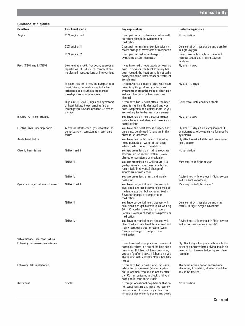
Guidance at a glance
Condition Functional status Lay explanation Restriction/guidance
Angina CCS angina IeII Chest pain on considerable exertion withno recent change in symptoms ormedication
No restriction
CCS angina III Chest pain on minimal exertion with norecent change of symptoms or medication
Consider airport assistance and possiblein-flight oxygen
CCS angina IV Chest pain at rest or a change insymptoms and/or medication
Defer travel until stable or travel withmedical escort and in-flight oxygenavailable
Post-STEMI and NSTEMI Low risk: age <65, first event, successfulreperfusion, EF >45%, no complications,no planned investigations or interventions
If you have had a heart attack but you areaged <65 years, the blocked artery hasbeen opened, the heart pump is not badlydamaged and no further tests or treatmentare planned
Fly after 3 days
Medium risk: EF >40%, no symptoms ofheart failure, no evidence of inducibleischaemia or arrhythmia, no plannedinvestigations or interventions
If you have had a heart attack, your heartpump is quite good and you have nosymptoms of breathlessness or chest painand no other tests or treatments areplanned
Fly after 10 days
High risk: EF <40%, signs and symptomsof heart failure, those pending furtherinvestigation, revascularisation or devicetherapy
If you have had a heart attack, the heartpump is significantly damaged and youhave symptoms of breathlessness or youare waiting for further tests or treatment
Defer travel until condition stable
Elective PCI uncomplicated You have had the heart arteries treatedwith a balloon and stent and there are nocomplications
Fly after 2 days
Elective CABG uncomplicated Allow for intrathoracic gas resorption. Ifcomplicated or symptomatic, see heartfailure
You have had heart bypass surgery andtime must be allowed for any air in thechest to be absorbed
Fly after 10 days if no complications. Ifsymptomatic, follow guidance for specificsymptoms
Acute heart failure You have been in hospital or treated athome because of ‘water in the lungs’which made you very breathless
Fly after 6 weeks if stabilised (see chronicheart failure)
Chronic heart failure NYHA I and II You get breathless on mild to moderateexercise but no recent (within 6 weeks)change of symptoms or medication
No restriction
NYHA III You get breathless on walking 20e100yards/metres at your own pace but norecent (within 6 weeks) change ofsymptoms or medication
May require in-flight oxygen
NYHA IV You are breathless at rest and mainlybedbound
Advised not to fly without in-flight oxygenand medical assistance
Cyanotic congenital heart disease NYHA I and II You have congenital heart disease withblue blood and get breathless on mild tomoderate exertion but no recent (within6 weeks) change of symptoms ormedication
May require in-flight oxygen*
NYHA III You have congenital heart disease withblue blood and get breathless on walking20e100 yards/metres but no recent(within 6 weeks) change of symptoms ormedication
Consider airport assistance and mayrequire in flight oxygen advisable*
NYHA IV You have congenital heart disease withblue blood and are breathless at rest andmainly bedbound but no recent (within6 weeks) change of symptoms ormedication
Advised not to fly without in-flight oxygenand airport assistance available*
Valve disease (see heart failure)
Following pacemaker inplantation If you have had a temporary or permanentpacemaker there is a risk of the lung beingpunctured. If it has not been punctured,you can fly after 2 days. If it has, then youshould wait until 2 weeks after it has fullyhealed
Fly after 2 days if no pneumothorax. In theevent of a pneumothorax, flying should bedeferred for 2 weeks following completeresolution
Following ICD implantation If you have had a defibrillator, the sameadvice for pacemakers (above) appliesbut, in addition, you should not fly afterthe ICD has delivered a shock until yourcondition is considered stable
The same advice as for pacemakersabove but, in addition, rhythm instabilityshould be treated
Arrhythmia Stable If you get occasional palpitations that donot cause fainting and have not recentlybecome more frequent or you have anirregular pulse which is treated and stable
No restriction
Continued
Fitness to fly
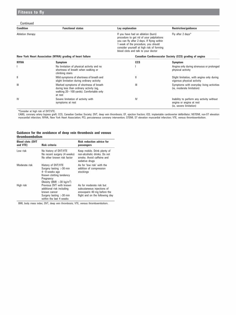
Continued
Condition Functional status Lay explanation Restriction/guidance
Ablation therapy If you have had an ablation (burn)procedure to get rid of your palpitationsyou can fly after 2 days. If flying within1 week of the procedure, you shouldconsider yourself at high risk of formingblood clots and talk to your doctor
Fly after 2 days*
New York Heart Association (NYHA) grading of heart failure Canadian Cardiovascular Society (CCS) grading of angina
NYHA Symptom CCS Symptom
I No limitation of physical activity and noshortness of breath when walking orclimbing stairs
I Angina only during strenuous or prolongedphysical activity
II Mild symptoms of shortness of breath andslight limitation during ordinary activity
II Slight limitation, with angina only duringvigorous physical activity
III Marked symptoms of shortness of breathduring less than ordinary activity (eg,walking 20e100 yards). Comfortable onlyat rest
III Symptoms with everyday living activities(ie, moderate limitation)
IV Severe limitation of activity withsymptoms at rest
IV Inability to perform any activity withoutangina or angina at rest(ie, severe limitation)
*Consider at high risk of DVT/VTE.CABG, coronary artery bypass graft; CCS, Canadian Cardiac Society; DVT, deep vein thrombosis; EF, ejection fraction; ICD, implantable cardioverter defibrillator; NSTEMI, non-ST elevationmyocardial infarction; NYHA, New York Heart Association; PCI, percutaneous coronary intervention; STEMI, ST elevation myocardial infarction; VTE, venous thromboembolism.
Guidance for the avoidance of deep vein thrombosis and venousthromboembolism
Blood clots (DVTand VTE) Risk criteria
Risk reduction advice forpassengers
Low risk No history of DVT/VTENo recent surgery (4 weeks)No other known risk factor
Keep mobile. Drink plenty ofnon-alcoholic drinks. Do notsmoke. Avoid caffeine andsedative drugs
Moderate risk History of DVT/VTESurgery lasting >30 min4e8 weeks agoKnown clotting tendencyPregnancyObesity (BMI >30 kg/m2)
As for ‘low risk’ with theaddition of compressionstockings
High risk Previous DVT with knownadditional risk includingknown cancerSurgery lasting >30 minwithin the last 4 weeks
As for moderate risk butsubcutaneous injections ofenoxoparin 40 mg before theflight and on the following day
BMI, body mass index; DVT, deep vein thrombosis; VTE, venous thromboembolism.
Fitness to fly

Fitness to fly for passengers with cardiovasculardisease
David Smith,1 William Toff,2 Michael Joy,3 Nigel Dowdall,4 Raymond Johnston,5
Liz Clark,6 Simon Gibbs,7 Nick Boon,8 David Hackett,9 Chris Aps,10 Mark Anderson,11
John Cleland12
SUMMARYFollowing this review of evidence and after due consideration, it is clear that there are few cardiovascularconditions that warrant the denial of fitness to fly as a passenger. Given the right aircraft, on-boardequipment and appropriately qualified and experienced escort personnel, aircraft can act as flying intensivecare units and carry extremely ill passengers.1
For those with cardiovascular disease who are not critically ill but who wish to fly on commercialaircraft, the aircraft environment does not pose a significant threat to their health. It is only when theirunderlying condition is associated with a significant risk of acute deterioration that reasonable restrictionsshould apply. For those at the more severe end of the spectrum of their specific cardiovascular condition,services exist to help make the journey more easily and safely. Most carriers and airport authorities provideassistance on the ground and in the air. Oxygen is available on most major carriers, although this issometimes subject to a charge and at least 7 days notice is normally required.2
Passengers are advised to plan their arrival at the airport in plenty of time to avoid having to rush and towarn the carrier and/or airport authority of any requirements for assistance, including requirement forin-flight oxygen, well in advance of the date of departure. They are strongly advised to ensure they have anappropriate supply of their medication, a clear list of the medications and doses they take and a letter ofexplanation from their doctor regarding their condition, drugs, allergies and devices (eg, pacemaker).Physicians are advised to consider the stability of a passenger ’s condition and apply the guidance
herein.The authors have contributed to this document in good faith and consider it to be an honest conclusion of
the reviewof evidence and assessment of the risks. It is guidance only and responsibility for declaring a patientfit to travel rests with the attending physician.The airline carrier should always be informed if a sick passenger is intending tofly. It has a right to refuse the
carriage of a passenger at its own discretion, even if they technically fulfil these guidelines recommendationsshown overleaf.
1. INTRODUCTION1.1 This Working Group of the British Cardiovas-cular Society was established to produce a report onPassenger Fitness to Fly in response to the House ofLords Science and Technology Committee report onAir Travel and Health3 in which it is suggested thatspecialist cardiology guidance would be of assis-tance to general practitioners, passengers andpassenger carrying organisations when determiningthe risks of passenger flight for those with cardio-vascular disorders.1.2 There are many existing guidelines on passengerfitness to fly,4e7 most of which include somereference to certain cardiovascular disorders, butthere is variation in the recommendations, partic-ularly in the time required to elapse between anevent or medical procedure and the flight.1.3 There is a lack of clarity over the purpose ofthe current guidelines. The suggestion by theHouse of Lords Committee on Air Travel andHealth that there be specialist cardiology guidanceis vague about the overall goal of the guidance.There are widespread concerns among the publicthat air travel has the potential to be harmful,many of which are expressed by the House of Lords
committee and are reflected in the following fewparagraphs.1.3.1 Although passenger flight is commonplace,the aircraft cabin provides what might be consid-ered a relatively alien, restrictive and hostile envi-ronment. Passengers are strapped for long periods inan upright sitting position and subjected tocontinuous noise, low humidity, cosmic radiationand hypobaric hypoxia, any or all of which mayhave a deleterious effect on their health.1.3.2 Passengers may already have a medicalcondition and exposure to the flying environmentmay precipitate an acute deterioration or poten-tially catastrophic event.1.3.3 The aircraft is an isolated environment withlimited facilities for acute health care and out ofreach of assistance other than that which can begained from radio communication. It is not an idealplace in which to fall ill.1.3.4 There are now a very large number ofpassengers, and a high proportion of them areelderly. The number of passenger hours in the air isincreasing and with it the chance of spontaneousevents occurring in flight but not actually caused orprecipitated by the flight.
1Cardiac Department, RoyalDevon and Exeter NHSFoundation Trust, Exeter, UK2Department of CardiovascularSciences, University ofLeicester, Faculty Member ofthe NIHR LeicesterCardiovascular BiomedicalResearch Unit, Leicester, UK3Postgraduate Medical School,Surrey University, UK4British Airways, UK5UK Civil Aviation Authority, UK6Peninsula Heart and StrokeNetwork, Plymouth, UK7National Heart and LungInstitute, Imperial CollegeLondon and Department ofCardiology, HammersmithHospital, London, UK8Past President, BritishCardiovascular Society, London,UK9British Cardiovascular Society,West Hertfordshire HospitalsNHS Trust, Hemel HempsteadGeneral Hospital, UK10Guy’s and St Thomas’ NHSFoundation Trust, London, UK11The Cardiac Centre, MorristonHospital, Swansea, UK12Department of Cardiovascularand Respiratory Disease,University of Hull, Castle HillHospital, Hull, UK
Correspondence toDavid Smith, CardiacDepartment, Royal Devon andExeter NHS Foundation Trust,Barrack Road, Exeter EX2 5DW,UK; [email protected]
Accepted 19 May 2010
Heart 2010;96:ii1eii16. doi:10.1136/hrt.2010.203091 ii1
Fitness to fly
1.3.5 Events in flight may cause disruption for staff and otherpassengers and, at worst, may lead to the aircraft being divertedto where appropriate medical care can be provided. Diversionsare very costly and disruptive for the carrier and it is desirablethat such events are kept to a minimum.1.4 Guidelines may be directed at any of the above problems, butthe British Cardiovascular Society and this Working Group haveconsidered the House of Lords requirements and feel this guid-ance should be directed neither at limiting cost and disruption toairline carriers nor to avoiding potential dangers of the in-flightenvironment to healthy passengers, but solely to providingadvice about the risks of flying for passengers with recognisedcardiovascular disease.
2. TERMS OF REFERENCE2.1 To review the evidence for safety and risk of air travel forpeople with cardiovascular conditions.2.2 To agree appropriate advice and guidance for air travel whichcan be expected to be safe for patients with cardiovascularconditions.2.3 To agree appropriate advice and guidance for healthcarerequirements for people undertaking air travel with potentiallyunsafe cardiovascular conditions, or suspected cardiovascularconditions, including ‘medical repatriation’.2.4 To report and to make recommendations to the Board ofTrustees of the British Cardiovascular Society.
3. MEMBERSHIPDr L D R Smith (Chairman), Dr C Aps, Dr N Boon (ex officioBritish Cardiovascular Society), Mrs E Clark, Dr N Dowling,Dr S Gibbs, Dr D Hackett (ex officio British CardiovascularSociety), Professor M Joy, Dr M Anderson, Dr W Toff,Dr R Johnston (co-opted) and Professor J Cleland (co-opted).
4. WHAT THIS DOCUMENT DOES NOT COVER4.1 Cardiovascular disease is a termwhich may be used to includeany pathological condition of the heart and its components, thegreat vessels and the peripheral vasculature. It could thereforeinclude diabetes mellitus, the idiopathic arteritides, peripheralarteriosclerosis and abdominal aortic aneurysm. It could includestroke, multi-infarct dementia, carotid artery disease and cerebralarteriovenous malformations, phlebitis and varicose veins. TheWorking Group considered the scope of the report and agreedthat it would not be appropriate to include all these conditions.For the purposes of this document, the term ‘cardiovascular ’willrefer to conditions of the heart and the great vessels.4.2 This guidance does not refer to cardiovascular fitness ofindividuals to take control of an aircraft, nor does it deal withthe risks associated with flight in private aircraft, militaryaircraft, aircraft with non-pressurised cabins or other means offlight such as paragliders or balloons.
5. WHAT THIS DOCUMENT DOES COVER5.1 This document is solely about the fitness to fly of air travelpassengers who have cardiovascular conditions and covers allaspects of cardiovascular fitness to fly for commercial passen-gers. It deals with the common questions and problems thatpatients, their relatives and their doctors ask about the advis-ability of flying, and it reviews the existing evidence in theformulation of guidelines for passenger fitness to fly.5.2 The overriding consideration for the Working Group wasthat the aim of the guidelines should be, wherever possible, toallow people to fly and not to be unnecessarily restrictive.
5.3 This document considers the effects of passenger flight andprovides guidance accordingly for the conditions that comeunder the broad headings of ischaemic heart disease, heartfailure, cyanotic congenital heart disease, abnormalities ofcardiac rhythm and cardiac devices.5.4 It is now widely accepted that guidelines published byprofessional societies, national and joint international commit-tees follow a specific style that includes an indication of the levelof importance of a particular guideline and the level ofsupporting evidence that justifies it. There is such a paucity ofrandomised controlled trials, meta-analyses and registriesregarding the risks of commercial flight for passengers withcardiovascular disease that the Working Group felt unable toadopt this style. What evidence there is has been reviewed andused, but a significant element of the guidance within thedocument is based on professional judgement and an under-standing of the interaction between the flight environment andthe pathophysiology of the cardiovascular condition.5.5 There was much discussion about the inclusion of venousthromboembolism (VTE) since this condition has so clearly beenimplicated in long-haul air travel. Much has been written on thesubject of VTE already, but the group considered that a sectionon VTE should be included.
6. REVIEW OF EVIDENCEThere are very few direct clinical studies of the pathophysiologicaleffects of commercial passenger flight on patients with existingcardiovascular diseases. Concerns that there may be deleteriouseffects are based on extrapolation from an understanding of thephysics of altitude, cardiorespiratory physiology and studies thatattempt to simulate the conditions of flight, either by studyingpatients adapting to living on land at altitude or by studying theeffects of normobaric hypoxia in a laboratory setting.We have considered the physics and physiology and formed
a view on whether there is evidence to suggest that beinga passenger in a commercial aircraft has any adverse effects thatmay increase the risk of a cardiac event.Wehave also considered peoplewhomaybe considered high risk
by dint of a recent cardiac event or procedure and in whomrestrictions onflying are designed to limit the chance of an in-flightevent where facilities for managing medical emergencies are poor.
6.1 Physics of air travel6.1.1 Dalton’s law6.1.1.1 The atmosphere is made up of a mixture of a number ofgases, the main components being nitrogen, oxygen and watervapour. The atmosphere is not of uniform thickness, being greaterat the poles than at the equator. The pressure it exerts variesaccordingly and also with weather systems, but is considered tohave a mean value at sea level of 100 kPa (760 mm Hg) whichdecreases with increasing altitude above sea level (figure 1).6.1.1.2 Since Dalton’s law states that the pressure exerted bya mixture of non-reacting gases is equal to the sum of the partialpressures of the component gases, the partial pressure of oxygenalso falls with increasing altitude (figure 1). Oxygen forms 21%of dry air, nitrogen forming almost all the rest. At 3000 m(9842 ft) the partial pressure of inspired oxygen is 13.3 kPa andat 8900 m (29 200ft, the altitude of the top of Mount Everest)the partial pressure of oxygen falls to approximately 6.2 kPa.6.1.1.3 Conventional commercial aircraft fly at altitudes from6500 m (22 000 ft) to 13 500 m (44 000 ft), and without a pres-surised aircraft cabin would be untenable for humans.6.1.1.4 In the USA the cabins of commercial aircraft are requiredto be pressurised to a pressure equivalent to an altitude
ii2 Heart 2010;96:ii1eii16. doi:10.1136/hrt.2010.203091
Fitness to fly
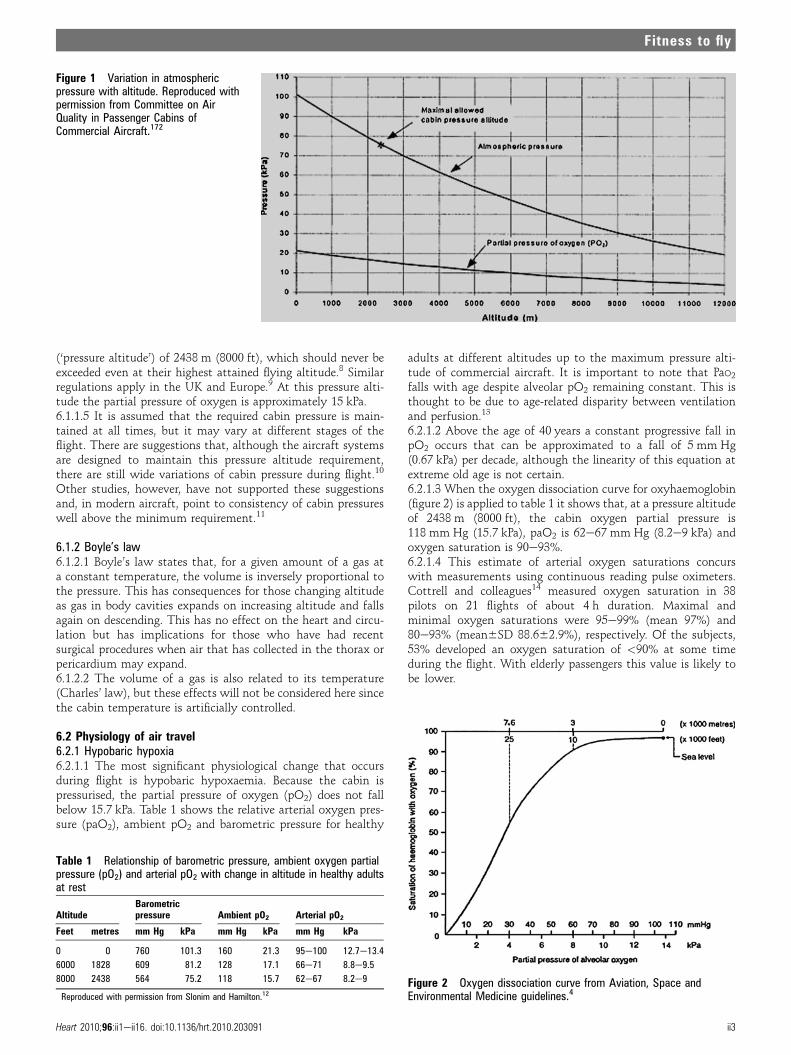
(‘pressure altitude’) of 2438 m (8000 ft), which should never beexceeded even at their highest attained flying altitude.8 Similarregulations apply in the UK and Europe.9 At this pressure alti-tude the partial pressure of oxygen is approximately 15 kPa.6.1.1.5 It is assumed that the required cabin pressure is main-tained at all times, but it may vary at different stages of theflight. There are suggestions that, although the aircraft systemsare designed to maintain this pressure altitude requirement,there are still wide variations of cabin pressure during flight.10
Other studies, however, have not supported these suggestionsand, in modern aircraft, point to consistency of cabin pressureswell above the minimum requirement.11
6.1.2 Boyle’s law6.1.2.1 Boyle’s law states that, for a given amount of a gas ata constant temperature, the volume is inversely proportional tothe pressure. This has consequences for those changing altitudeas gas in body cavities expands on increasing altitude and fallsagain on descending. This has no effect on the heart and circu-lation but has implications for those who have had recentsurgical procedures when air that has collected in the thorax orpericardium may expand.6.1.2.2 The volume of a gas is also related to its temperature(Charles’ law), but these effects will not be considered here sincethe cabin temperature is artificially controlled.
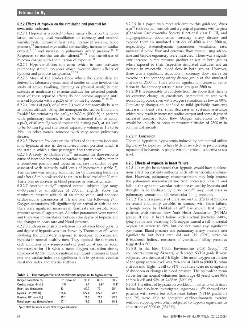
6.2 Physiology of air travel6.2.1 Hypobaric hypoxia6.2.1.1 The most significant physiological change that occursduring flight is hypobaric hypoxaemia. Because the cabin ispressurised, the partial pressure of oxygen (pO2) does not fallbelow 15.7 kPa. Table 1 shows the relative arterial oxygen pres-sure (paO2), ambient pO2 and barometric pressure for healthy
adults at different altitudes up to the maximum pressure alti-tude of commercial aircraft. It is important to note that PaO2
falls with age despite alveolar pO2 remaining constant. This isthought to be due to age-related disparity between ventilationand perfusion.13
6.2.1.2 Above the age of 40 years a constant progressive fall inpO2 occurs that can be approximated to a fall of 5 mm Hg(0.67 kPa) per decade, although the linearity of this equation atextreme old age is not certain.6.2.1.3 When the oxygen dissociation curve for oxyhaemoglobin(figure 2) is applied to table 1 it shows that, at a pressure altitudeof 2438 m (8000 ft), the cabin oxygen partial pressure is118 mm Hg (15.7 kPa), paO2 is 62e67 mm Hg (8.2e9 kPa) andoxygen saturation is 90e93%.6.2.1.4 This estimate of arterial oxygen saturations concurswith measurements using continuous reading pulse oximeters.Cottrell and colleagues14 measured oxygen saturation in 38pilots on 21 flights of about 4 h duration. Maximal andminimal oxygen saturations were 95e99% (mean 97%) and80e93% (mean6SD 88.662.9%), respectively. Of the subjects,53% developed an oxygen saturation of <90% at some timeduring the flight. With elderly passengers this value is likely tobe lower.
Figure 1 Variation in atmosphericpressure with altitude. Reproduced withpermission from Committee on AirQuality in Passenger Cabins ofCommercial Aircraft.172
Table 1 Relationship of barometric pressure, ambient oxygen partialpressure (pO2) and arterial pO2 with change in altitude in healthy adultsat rest
AltitudeBarometricpressure Ambient pO2 Arterial pO2
Feet metres mm Hg kPa mm Hg kPa mm Hg kPa
0 0 760 101.3 160 21.3 95e100 12.7e13.4
6000 1828 609 81.2 128 17.1 66e71 8.8e9.5
8000 2438 564 75.2 118 15.7 62e67 8.2e9
Reproduced with permission from Slonim and Hamilton.12Figure 2 Oxygen dissociation curve from Aviation, Space andEnvironmental Medicine guidelines.4
Heart 2010;96:ii1eii16. doi:10.1136/hrt.2010.203091 ii3
Fitness to fly
6.2.2 Effects of hypoxia on the circulation and potential formyocardial ischaemia6.2.2.1 Hypoxia is reported to have many effects on the circu-lation including local vasodilation of coronary and cerebralvascular beds, increase in heart rate, increase in systemic bloodpressure,15 increased myocardial contractility, increase in cardiacoutput16 17 and increase in pulmonary artery pressure.18 19
Responses to exercise are also altered,20 21 and the effects ofhypoxia change with the duration of exposure.21
6.2.2.2 Hyperventilation can occur which in turn activatespulmonary stretch receptors which override direct effects ofhypoxia and produce tachycardia.22 23
6.2.2.3 Most of the studies from which the above data arederived are laboratory-based animal studies or have involved thestudy of active (walking, climbing or physical work) humansubjects at moderate to extreme altitude for extended periods.Most of these reported effects do not become apparent untilmarked hypoxia with a paO2 of #40 mm Hg occurs.15 18 23
6.2.2.4 Levels of paO2 of 40 mm Hg would not normally be seenat modest altitude. Using the nomogram devised by Dillard andEwald24 for estimating the paO2 at 2438 m (8000 ft) in patientswith pulmonary disease, it can be estimated that to attaina paO2 of 40 mm Hg would require the resting paO2 at sea levelto be 60 mm Hg and the forced expiratory volume in 1 s to be29%din other words, someone with very severe pulmonarydisease.6.2.2.5 There are few data on the effects of short-term isocapnicmild hypoxia at rest in the semi-recumbent position which isthe state in which airline passengers find themselves.6.2.2.6 A study by Phillips et al25 measured the dose-responsecurve of isocapnic hypoxia and cardiac output in healthy men ina recumbent position and found an increase in cardiac outputassociated with relatively mild levels of hypoxaemia (table 2).The increase was entirely accounted for by increasing heart rateand after a 5 min peak tended to return to base level after 20 min.There was no increase in blood pressure or ventilation rate.6.2.2.7 Another study26 exposed normal subjects (age range6e83 years) to an altitude of 2900 m, slightly above themaximum pressure altitude of an airline cabin, and analysedcardiovascular parameters at 1 h and over the following 24 h.Oxygen saturations fell significantly on arrival at altitude andthere were significant increases in heart rate and systolic bloodpressure across all age groups. All other parameters were normaland there was no correlation between the degree of hypoxia andthe changes in heart rate and blood pressure.6.2.2.8 Such an inconsistent relationship between blood pressureand degree of hypoxia was also shown by Thomson et al27 whenstudying the circulatory response to isocapnic hyperoxia andhypoxia in normal healthy men. They exposed the subjects toeach condition in a semi-recumbent position at normal roomtemperature for 1 h with a mean oxygen saturation duringhypoxia of 82.6%. Hypoxia induced significant increases in heartrate and cardiac index and significant falls in systemic vascularresistance index and arterial stiffness.
6.2.2.9 In a paper even more relevant to this guidance, Wysset al28 took normal controls and a group of patients with angina(Canadian Cardiovascular Society functional class IIeIII) andangiographically documented coronary artery disease andexposed them to simulated altitudes of 4500 m and 2500 m,respectively. Haemodynamic parameters, ventilation rate,myocardial blood flow and coronary flow reserve using adeno-sine and bicycle ergometry were measured. There was a signifi-cant increase in rate pressure product at rest in both groupswhen exposed to their respective simulated altitudes and anincrease in myocardial blood flow in both groups. However,there was a significant reduction in coronary flow reserve onexercise in the coronary artery disease group at the simulatedaltitude of 2500 m. There was no significant increase in venti-lation in the coronary artery disease group at 2500 m.6.2.2.10 It is reasonable to conclude from the above that there isno extreme change in circulatory parameters at rest withisocapnic hypoxia, even with oxygen saturations as low as 80%.Circulatory changes are confined to mild (probably transient)increases in heart rate, small decreases in systemic resistancewhich may result in increased cardiac output and some degree ofincreased coronary blood flow. Oxygen saturations of 80%would be unlikely to occur at pressure altitudes of 2500 m incommercial aircraft.
6.2.2.11 ConclusionThe mild hypobaric hypoxaemia induced by commercial airlineflight may be expected to have little or no effect in precipitatingmyocardial ischaemia in people without critical ischaemia at sealevel.
6.2.3 Effects of hypoxia in heart failure6.2.3.1 It might be expected that hypoxia would have a delete-rious effect on patients suffering with left ventricular dysfunc-tion. However, pulmonary vasoconstriction may help protectthe pulmonary microvasculature from increased pressure andfalls in the systemic vascular resistance caused by hypoxia andthought to be mediated by nitric oxide29 may limit rises inpulmonary venous and left ventricular filling pressures.6.2.3.2 There is a paucity of literature on the effects of hypoxiaon central circulatory variables in humans with heart failure,although work by Hobkirk et al30 has shown that, in 21patients with treated New York Heart Association (NYHA)grades III and IV heart failure with ejection fractions <40%,lying supine and breathing 15% oxygen caused a fall in arterialoxygen saturation to 86% but did not cause any significantsymptoms. Blood pressure and pulmonary artery pressure rosesignificantly but heart rate did not (19 (90%) were onb blockers). Indirect measures of ventricular filling pressuressuggested a fall.6.2.3.3 In the Ideal Cabin Environment (ICE) Study,31 72volunteers (mean age 54 years) with stable NYHA grade II weresubjected to a simulated 7-h flight. The mean oxygen saturationof the group at ‘sea level’ was 94% and at 2438 m (8000 ft) cabinaltitude mid ‘flight’ it fell to 91%, but there were no symptomsof dyspnoea or changes in blood pressure. The equivalent meanvalues for the normal volunteers (mean age 43 years) were 96%at ‘sea level’ and 93% at 2438 m (8000 ft).6.2.3.4 The effect of hypoxia on workload in patients with heartfailure has also been investigated. Agostoni et al32 showed thatpatients with severe but stable heart failure (NYHA grades IIIand IV) were able to complete cardiopulmonary exercisewithout stopping even when subjected to hypoxia equivalent toan altitude of 3000 m (9842 ft).
Table 2 Haemodynamic and ventilatory response to hypoxaemia
Oxygen saturation (%) 97 (room air) 90.0 85.3 80.5
Cardiac output (l/min) 7.0 7.1 7.9* 8.4*
Heart rate (beats/min) 63 69.7 72 75*
Systolic BP (mm Hg) 125.6 127.6 127.1 128.2
Diastolic BP (mm Hg) 72.1 74.5 74.1 73.2
Respiratory rate (breaths/min) 17.1 17.3 16.6 16.8
*p<0.0005 for room air and 90% oxygen saturation.
ii4 Heart 2010;96:ii1eii16. doi:10.1136/hrt.2010.203091
Fitness to fly

6.2.3.5 ConclusionThe available evidence suggests that, for patients with stableheart failure including NYHA grades III and IV, short-term (upto 1 h) hypoxia at rest produces no significant deleterious effects.Periods of up to 7 h are tolerated by those with mild to moderatestable heart failure (NYHA grade II).
6.2.4 Effects of hypoxia on the ECG, arrhythmia and pacingthresholds6.2.4.1 It might be anticipated that hypoxia and the associatedincrease in a- and b-adrenergic stimulation would increase thesusceptibility to arrhythmia, particularly in the presence ofmyocardial ischaemia, but there are surprisingly few data on thesubject. In a study of healthy men aged 50e64 years, increasedatrial and ventricular ectopy was observed during acute ascent ina cable car to altitudes up to 2632 m (863 4ft) above sea level,the frequency of ectopy being proportional to the altitude.33
6.2.4.2 In another study healthy volunteers subjected toa simulated ascent of Mount Everest in a hypobaric chamber34
showed transient changes in mean frontal QRS axis and voltagesand increases in heart rate proportional to the degree of hypoxia.There were no ECG signs of ischaemia or arrhythmia even withan oxygen saturation of 49%.6.2.4.3 It is not known whether hypoxia during air travel resultsin an increased predisposition to sustained ventriculararrhythmia and implantable cardioverter defibrillator (ICD)activation in susceptible individuals.6.2.4.4 Profound hypoxaemia induced by inhalation of a gasmixture containing 10% oxygen has been shown to result ina significant and reversible increase in pacing threshold.35
However, acute exposure of 13 patients with implanted pace-makers to an altitude equivalent of 4000 m, simulated in a hypo-baric chamber, did not result in any alteration of stimulationthresholds after 30 min.36
6.2.4.5 ConclusionHypoxia is unlikely to produce an increase in susceptibility toarrhythmia or to have any adverse effect on pacing threshold atcabin altitudes likely to be encountered during air travel.
6.2.5 Effects of hypoxia on thrombogenicity (see also section onDVT/VTE)6.2.5.1 Passengers with existing atheromatous coronary disease,whether diagnosed or not, may be at risk of acute coronarysyndromes if being a passenger on a commercial flight hasa prothrombotic effect which might predispose them toformation of occlusive coronary thrombus.6.2.5.2 Hypoxia has been shown not to enhance exercise-inducedactivation of clotting factors in healthy athletes37 and, althoughshort-term and long-term periods at high altitude (>5000 m)have been shown to produce activation of clotting factors,38 39
well-controlled studies of both normobaric40 and hypobaric41
isocapnic hypoxia simulating an altitude of 3600 m for 8 hrevealed no significant effect.6.2.5.3 Toff et al42 simulated a long-haul flight by subjecting 73low-risk ‘passengers’ to 8 h of either sedentary normoxia orhypoxia in a crossover trial using a hypobaric chamber. A smallproportion of women taking oral contraceptives were includedbut subjects with factor V Leiden and prothrombin G20210mutations were not. Measures of endothelial function, plateletactivation and clotting showed no significant difference betweenthe hypoxic and normoxic conditions suggesting that, in low-risk passengers, hypoxia has no prothrombotic effect over andabove prolonged sitting.
6.2.5.4 In a study of predominantly high-risk ‘passengers’43 witha high proportion of participants taking oral contraceptives orbearing the factor V Leiden mutation, an 8-h flight wascompared with other 8-h periods of sitting in a cinema or lightambulatory activity. There were increases in clotting factors (D-dimer, thrombin/antithrombin complex and prothrombin frag-ments 1 and 2). This suggests that, in high-risk individuals, mildhypoxia alone may have a prothrombotic effect.
6.2.5.5 ConclusionCommercial airline passengers breathe air with a reduced oxygencontent which results in low blood oxygen saturations. Thelevels of blood oxygen saturations attained appear to have littleor no adverse circulatory effects which would make thepassenger more liable to myocardial ischaemia, myocardialinfarction, left ventricular failure or arrhythmia. Passengersalready at high risk may suffer additional risk from hypoxiaduring flight. This conclusion is supported by the availableevidence and applies to short or medium length flights. There isno adequate information to indicate whether extended flying(>12 h) might have adverse effects.
6.2.6 Effects of pre- and post-flight environmentTaking a flight with a commercial airline involves more than thetime spent in the aircraft during flight, and the potential threatsto the health of passengers with cardiac disease may occur beforeand after flight.6.2.6.1 Anxiety: A significant proportion of the population(10e40%) report a fear of flying,44 although this prevents fewfrom completely avoiding it.45 Fear and anxiety may beheightened at take-off and landing or if turbulence is experi-enced. The threat of terrorism adds to the fear of flying and cana have a major impact on passenger willingness to fly. Ito andLee assessed the September 11 attacks in New York as havinga major effect on reducing passenger numbers, an effect whichcontinued at least until 2003.46
6.2.6.2 The security measures which are now in place meanpassengers have to arrive well ahead of their flight time. Concernabout missing the flight, uncertainties about where to check inand identifying the departure gate all add to the stress and meanpassengers may be hurrying and anxious.6.2.6.3 Frustration about delays has been shown to result inanger,47 and anger is one of the more potent of the many mentalstresses that may provoke myocardial ischaemia.48
6.2.6.4 Exercise: After arrival at the airport a considerable amountof physical activity is involved, much of it carrying, pushing orpulling bags, which may be well in excess of the passenger ’snormal exercise limits. Similar stresses apply at the destinationairport andmay also be exacerbated by the altitude or temperatureof the destination. Many major destination cities are above5000 ft, especially in North and South America, Africa and theEast, and many have extremes of temperature. The potential forthe airport stresses to induce a deterioration of a chronic conditionis well recognised and most airports provide excellent services toassist the disabled passenger. These services can be found onarrival, but it is better to call ahead and make the arrangementsbeforehand.
6.2.7 Sleep deprivation and circadian rhythm disruption6.2.7.1 Departure times in the early hours of the morning oftenmean that, with travel time to the airport and the delay beforeboarding, passengers may be awake much of the night beforetravel. This may result in sleep deprivation and disruption of
Heart 2010;96:ii1eii16. doi:10.1136/hrt.2010.203091 ii5
Fitness to fly
circadian rhythm that can also occur with flights that crossmany time zones.6.2.7.2 The circadian rhythm of the cardiovascular systemreflects the changing sympathetic-vagal balance and is recog-nised to result in periods of cardiac vulnerability.49 Studies ofshift workers suggest that sleep deprivation and disruption ofcircadian rhythm may increase the risk of coronary events.50
Heart rate variability as a marker of circadian variation may bealtered in patients after infarction and in those with congestiveheart failure, hypertension and diabetes51 and can be modulatedby b blockers.6.2.7.3 Despite this, one study has shown that, in patients withheart failure, there is no relationship of circadian rhythm withsudden cardiac death52 and there is no direct evidence ofincreased cardiac vulnerability resulting from occasional short-term disruptions such as those experienced by airline passengers.6.2.7.4 Probably the most important effect of disturbed diurnalpatterns for the passenger with cardiovascular disease is the easewith which the regularity of medications is disrupted. It is verydifficult to adhere to a pattern of tablet-taking when the dailyroutine is altered, especially when confusion about the righttime compounds the problem. For passengers with stable heartfailure, angina or arrhythmia, it is very important that theymaintain the regularity of their medication.
7. PASSENGER FLIGHT WITH CHRONIC CONDITIONS ANDAFTER CARDIAC EVENTS OR PROCEDURESRegardless of any direct deleterious effects that the aircraft cabinenvironment may have, it does not provide an ideal environmentin which to manage medical problems and an acute in-flightevent may result in diversion of the flight, poor medical outcomesor both. Cardiac events or procedures, diagnostic or therapeutic,may leave people at short-term high risk of a further event orcomplication. This acute risk normally reduces in the short tomedium term until a stable situation is reached. If a cardiac eventor procedure carries a significant risk of events or complicationsoccurring, then consideration must be given to the time intervalbefore the risk becomes acceptable and the cabin environment nolonger a concern. It is generally the instability of the conditionthat predicts risk rather than the severity alone.
Although some considerations are relatively easy to define,such as the time it takes for post-thoracotomy intrathoracic air tobe reabsorbed, most of the situations are complex. Patients havea multitude of factors that contribute to the risk of an event andconsequently any guidance is necessarily a generalisation basedon reasonable judgement and should not be considered absolute.
In the following section acute and chronic conditions anddifferent cardiac procedures are considered and the appropriatetime that should pass between the event and the flightsuggested. The conditions and procedures considered are listed intable 3.
7.1 Acute coronary syndromes (ACS) including ST elevationmyocardial infarction (STEMI) and non-ST elevation myocardialinfarction (NSTEMI) and troponin-negative acute coronarysyndromes7.1.1 The observations of the limited effects of the cabin envi-ronment on cardiac physiology (see above) should be equallyapplicable to patients following an acute coronary syndrome,there being little or no deleterious effect.However, following an acute coronary syndrome, patients
may be at risk of events as a direct result of their condition. Theearly risk following an acute coronary syndrome is multifactorialand includes age, heart rate, ventricular function/Killip class,severity of the coronary disease and the degree ofrevascularisation.53e58 In existing guidelines on fitness to fly,only acute myocardial infarction (AMI) is considered withoutdistinction between different clinical presentations although it issometimes separated into complicated and uncomplicated.7.1.2 There is conflicting advice regarding the delay to flyingfollowing AMI, with varying guidance from professionalbodies4e7 and varying acceptance by passenger carriers. Data onwhich to base guidance are relatively sparse.7.1.3 Retrospective analyses of patients following myocardialinfarction have suggested that flyingdboth short and longhauldis safe at 2e4 weeks,59 60 but in these studies the elapsedtime between myocardial infarction and the flight was not anadjustable variable.7.1.4 Another retrospective study61 analysed 213 aeromedicalrepatriation patients after myocardial infarction and concludedthat travel <2 weeks after AMI can be safely undertaken andthat there is a low incidence of transfer-related complications.The patients were clinically heterogeneous, some having STelevation myocardial infarction (STEMI) and some non-STelevation myocardial infarction (NSTEMI), with varyingnumbers having undergone revascularisation. All the patients inthis study were accompanied by an appropriately trainedmedical escort, and it is not clear whether this activelycontributed to the low complication rate.7.1.5 A small prospective trial62 analysed the effects of flight2 weeks after AMI in patients who were able to climb a flight ofstairs and who were randomised to receive oxygen (2 l/min) orno oxygen during the flight. Holter monitoring was performedfor ST shift and arrhythmia and oxygen saturation wasmeasured throughout the flight. The authors noted someasymptomatic hypoxia (oxygen saturation <90%), which wasalso noted in a few patients in the paper by Thomas et al,61 butonly one episode of ST shift on the Holter tracing which wasasymptomatic. There was no difference in outcome betweenpatients with supplemental oxygen and those without. Theauthors concluded that, in this group of patients, commercialflight was safe and did not induce myocardial ischaemia, thatsupplemental oxygen as routine was not required and thatmedical attendants were unnecessary.
Table 3 Conditions, procedures and events considered in this section
Acute conditions Chronic conditions Diagnostic procedures Therapeutic procedures
Acute coronarysyndromesAcute left ventricularfailure
Stable anginaStable heart failureParoxysmal SVTPersistent/permanent AFCyanotic congenital heartdiseaseAnaemia
Cardiac catheterisationElectrophysiologicalstudies
Emergency PCIElective PCIInsertion of pacemaker/AICDCABGSurgical valve replacementPercutaneous valvotomyAblation flutter, right-sided pathways,left-sided pathways, AF
AF, atrial fibrillation; AICD, implantable cardioverter defibrillator; CABG, coronary artery bypass grafting; PCI, percutaneous coronaryintervention; SVT, supraventricular tachycardia.
ii6 Heart 2010;96:ii1eii16. doi:10.1136/hrt.2010.203091
Fitness to fly
7.1.6 Longer distance medical transport is also reported ina retrospective study by Essebag et al63 in which 51 patientswere transported within 7 days of AMI, half of them high riskwith Killip class IIeIV. This study suggested that transfer wassafe 3e7 days after admission.7.1.7 These studies do not distinguish between high-risk andlow-risk post-infarction patients. There is a spectrum of riskwhich will depend on the success of reperfusion, extent ofmyocardial dysfunction, predisposition to arrhythmia andpresence of major complications.
7.1.8 STEMI7.1.8.1 Figures from the Myocardial Infarction National AuditProject (MINAP) suggest overall mortality from STEMI ishighest in the acute phase, falling to 2% after 10 days,64 butthese figures include all patients with STEMI and do notdistinguish between high- and low-risk groups.7.1.8.2 Successful revascularisation during primary angioplastyfor STEMI confers a prognostic benefit in the short and mediumterm, especially in low-risk STEMI,65 and in a cohort of 1791patients treated by primary angioplasty de Luca et al deviseda scoring system to identify the risk of 30-day mortality.66 Theauthors’ score card (see table 4) was able to identify patients atvery low risk of early mortality or cardiac arrest (0.1% at 2 daysand 0.2% at 2e10 days). They were able to demonstrate thestability of the score over the following year. Although patientswith higher scores had higher early and late mortality in allgroups, actuarial survival curves showed that relative stabilityof risk had been reached by 10 days. They concluded that a largeproportion of patients following primary percutaneous coro-nary intervention (PCI) for STEMI may be identified as verylow risk (score #3) with a stable risk from 2 days onwards.There was a strong correlation between risk score and ejectionfraction (EF), patients with scores of 0e3 having mean6SD EFof 45610% and those with scores of 4e6 having a mean6SD EFof 40612%. This suggests that patients with low risk scores
and EF >40% have a very low chance of events from day 3onwards.7.1.8.3 The treatment of STEMI with primary angioplasty isincreasingly widespread but still not the norm. In 2007, MINAPreported 4228 primary angioplasties in England and Wales out ofa total of 27 199 STEMI (John Birkhead, MINAP, personalcommunication, April 2009). The British Cardiovascular Inter-vention Society (BCIS) audit returns67 reported a slightly higherfigure at 4858 primary angioplasties. Either way, it only repre-sents about 15% of those with STEMI but it is rapidlyincreasing. The remainder received fibrinolytic agents or noreperfusion therapy. In these patients, successful reperfusion,extent of coronary disease and degree of left ventriculardysfunction are not known so quickly after hospitalisation andit is recommended that patients undergo further investigationand risk stratification using echocardiography within 48 h andcoronary angiography during the same admission.68 If thereremain concerns about possible inducible ischaemia, stresstesting may be undertaken in the following 4e6 weeks andappropriate revascularisation planned. Revascularisation withPCI or coronary artery bypass graft (CABG) almost halves therisk of subsequent unstable angina or death.69
7.1.8.4 Left ventricular EF is a marker of increased risk ofventricular arrhythmia and sudden cardiac death (SCD). Patientswith an EF $40% and no ventricular tachycardia after day 2 areat low risk of SCD. Those with EF #30% and prolonged QRSduration may be considered for an implantable defibrillator(AICD).70
7.1.8.5 Bradyarrhythmia complications may require temporaryor permanent insertion of a pacemaker. In either case thereis a risk of pneumothorax (see section on pacemaker insertion).
7.1.9 NSTEMI and troponin-negative acute coronary syndromesPatients with NSTEMI are at increased risk of death or furthermyocardial infarction and are recommended to undergo coro-nary angiography and appropriate revascularisation beforedischarge.71
7.1.10 HaemorrhageCurrent management of all acute coronary syndromes involvesthe use of antithrombotic agents and anticoagulants as well asinvasive strategies for revascularisation. There is a thereforea risk of haemorrhage related to the arterial access site as well asa risk of spontaneous bleeding from the gastrointestinal tract or,rarely, retroperitoneal haemorrhage.7.1.10.1 Haemorrhagic complications are more likely withfemoral artery than radial artery access,72 but are unlikely tooccur after 1 week. Subacute gastrointestinal haemorrhage isreported after both primary angioplasty73 and fibrinolytictherapy74 and may complicate the in-hospital course. Heparinand the small molecule IIbIIIa receptor antagonists have a shorthalf life, but abciximab may still have effects at 7e10 days.Gastrointestinal haemorrhage from the use of the antiplateletagents may also occur after discharge, but it is relatively rare at0.07 per patient-year.75
7.1.10.2 If there are haemorrhagic complications during the in-hospital course after an acute coronary syndrome, it may alterthe advisability of flying early after discharge.
7.1.11 ConclusionPassengers who have suffered an acute coronary syndrome canbe reasonably divided into those at very low risk who may safelyfly as early as 3 days after the event, those at medium risk who
Table 4 Zwolle risk score for ST elevation myocardial infarction(STEMI)
Killip class Points Risk scoreRR (95% CI) ofdeath by 30 days
1 0 0e1 0.03 (0.008 to 0.13)
2 4
3e4 9 2 0.09 (0.02 to 0.37)
TIMI flow post PCI
3 0 3 1.04 (0.04 to 2.45)
2 1
0e1 2 4 1.40 (0.5 to 3.98)
Age
<60 0 5 2.48 (0.96 to 6.42)
$60 2
3 vessel disease 6 2.52 (0.75 to 8.46)
No 0
Yes 1 7 5.99 (1.98 to 18.1)
Anterior infarct
No 0 $8 32.1 (18.6 to 55.8)
Yes 1
Ischaemic time <4 h
No 0
Yes 1
Total available score 16
Reproduced with permission from de Luca et al.66
PCI, percutaneous coronary intervention; TIMI, thrombolysis in myocardial infarction.
Heart 2010;96:ii1eii16. doi:10.1136/hrt.2010.203091 ii7
Fitness to fly
may fly from 10 days onwards and those at high risk or awaitingfurther investigation/treatment in whom flying should bedeferred until a more stable situation is achieved.
Very low risk: age <65 years, first event, successful reperfu-sion, EF >45%, no complications and no planned investigationsor interventions.
Low risk: EF >40%, no symptoms of heart failure, no evidenceof inducible ischaemia or arrhythmia and no further investigationsor interventions planned.
High risk EF: <40% with signs and symptoms of heart failure,those pending further investigation with a view torevascularisation or device therapy.
7.2 Heart failure7.2.1 Acute left ventricular failureThe risk of hospitalisation for heart failure following AMI ishighest in the few weeks after the infarction but stabilises after45 days.76 Episodes of acute left ventricular failure may becaused by an acute coronary syndrome (see above) or provokedby anaemia or infection on the background of chronic leftventricular dysfunction. Once any precipitant is identified andtreated, most cases should be stabilised within 6 weeks andshould be safe to fly.
7.2.2 Chronic heart failurePassengers with stable chronic heart failure without recentchanges in symptoms or medication are likely to be able totolerate the mild hypoxia of the aircraft cabin environment evenif they have severe heart failure.30e32 It is advisable that theytake the precautions of avoiding physical exertion at the airportand making sure that they take their regular medication. It isprobably prudent for passengers who are severely limitedwith NYHA class IV symptoms not to fly without specialconsideration and the availability of in-flight oxygen.
7.2.3 ConclusionFollowing an episode of acute heart failure, most patients shouldbe stable within 6 weeks and be able to fly. With chronic heartfailure there should be no restriction, although those withNYHA III and IV should consider airport assistance and requestthe availability of in-flight oxygen.
7.3 Stable anginaThe available evidence suggests that it is unlikely that flyingwould precipitate acute myocardial ischaemia and no restrictionshould be applied. Passengers should be advised to allow plentyof time at either end of their journey to limit anxiety and haste.Passengers should be advised to seek assistance with personaltransport at the airport if needed. They should also be advised toremember to take their medication in the usual way and at theusual times.
7.4 Arrhythmia7.4.1 Paroxysmal supraventricular arrhythmiaFlight itself does not appear to induce paroxysmal supraven-tricular tachycardia, atrial fibrillation or atrial flutter, andproviding the passengers are symptomatically stable with a lowfrequency of events, there should be no restriction to flying.7.4.2 Passengers with permanent or persistent atrial fibrillationshould be stable with appropriate rate control and anti-coagulation.7.4.3 Passengers with a history of ventricular arrhythmiasshould have these controlled before flying. Ventricular arrhyth-
mias may be treated by implantable defibrillators (see section onDVT and VTE).7.4.4 Patients with uncontrolled haemodynamically significantventricular arrhythmias should not travel on commercialaircraft.
7.5 Cyanotic congenital heart disease7.5.1 Patients with congenital cyanotic heart disease withEisenmenger syndrome77 78 are hypoxaemic with a consequenterythrocytosis and haemoglobin levels of 15e24 g/l. They mightbe thought to be at risk when flying owing to the effects ofhypoxia and hyperviscosity.7.5.2 The circulatory pathology, however, does not impairalveolar gas transfer and the arterial hypoxia does not resultfrom low alveolar oxygen tension but from the central right-to-left shunting. As a result, small changes in the alveolar oxygenconcentration have little effect on hypoxaemia. In addition,a chronic adaptation of a shift in the oxygen dissociation curveto the right has been suggested by Harinck et al79 and subse-quently confirmed in a study by Broberg et al.80
7.5.3 In the study by Broberg et al, 53 patients with Eisenmengersyndrome were compared with 48 acyanotic patients anda 10-year record of 1157 flights was studied. One or two of thepatients reported symptoms of breathlessness during flight andwere treated with in-flight oxygen therapy but the authorsreported no serious adverse effects on account of the relativeadditional hypoxia experienced in-flight and suggested there wasno justification for limiting air travel in these patients.7.5.4 The erythrocytosis that occurs may increase the alreadyexisting risk of venous thromboembolic disease but with theadditional possibility of a ‘paradoxical’ embolus. One patient inthe study by Broberg et al was thought to have had a transientcerebral event.
7.5.5 ConclusionPassengers with the Eisenmenger syndrome should not berestricted from flying although they are advised to request in-flight oxygen. They are also advised to maintain good levels ofhydration, avoid alcohol and follow the guidance for VTEprophylaxis for high-risk groups (see section on DVTand VTE).
7.6 Anaemia7.6.1 Reduced circulating haemoglobin restricts the oxygen-carrying capacity of a unit volume of blood and inducescompensatory mechanisms in the circulation that result inincreased cardiac output and ventricular stroke work.81 82
7.6.2 In the normal healthy heart, remarkably low haemoglobinlevels can be tolerated83 although chronic anaemia may precip-itate angina in those with normal coronary arteries.84 However,in an already compromised circulation, anaemia may riskpulmonary oedema or critical myocardial ischaemia.85
7.6.3 It is well recognised that anaemia commonly complicateschronic heart failure86 and that treatment significantly improvesthe functional class.87 The same applies to those with chronicangina88 and following either PCI89 or CABG.90
7.6.4 The Canadian Cardiac Society Guidelines on Flying6
recommend that a haemoglobin level of 9 g/l is a threshold valuebelow which travel is inadvisable in passengers who haveundergone CABG.
7.6.5 ConclusionWhen considering passengers with cardiovascular disease wishingto fly, physicians should consider the additional potential effect ofanaemia on the underlying cardiovascular pathology.
ii8 Heart 2010;96:ii1eii16. doi:10.1136/hrt.2010.203091
Fitness to fly

7.7 Cardiac catheterisationUneventful cardiac catheterisation should not restrict flying andpatients may fly the next day. Bleeding from the access site maybe a reason to defer a flight, as may other complications.
7.8 Elective PCIElective PCI varies in its complexity from simple single vessel PCIto complex three vessel treatments and the use of adjunctivedevices and pharmacology. With aggressive anticoagulant andantiplatelet regimens, bleeding is a more likely event than withelective cardiac catheterisation and bigger contrast loads are morelikely to cause renal impairment. In an uncomplicated case there isno reason to restrict flying for more than 2 days, but there shouldbe discretion in the advice given by the physician and accounttaken of the degree of ventricular and renal dysfunction.
7.9 Pacemaker insertion including temporary systems,implantable cardioverter defibrillators (ICD) and biventriculardevices7.9.1 The insertion of any type of pacemaker including tempo-rary wires, permanent pacemakers, biventricular pacemaker andimplantable cardio-defibrillators (ICD) may involve subclavianvein puncture. Pneumothorax is a recognised complication.Before discharge an anteroposterior chest x-ray is routinelyobtained to check the lead positions and to exclude pneumo-thorax, although it is possible that very small pneumothoracesmay not be evident on the x-ray. In the absence of complica-tions, patients are typically discharged from hospital within 24 hof implantation of the device.7.9.2 Pneumothorax requiring intervention (aspiration or chestdrain insertion) occurs in 0.6e0.8% of cases with less significantpneumothoraces (occupying <10% of the lung field) in a further1%.91 92
7.9.3 Passengers should not fly with a pneumothorax because ofthe risk of gaseous expansion at altitude which may compromiserespiratory function and the possible development of tensionpneumothorax. If implantation has been complicated by pneu-mothorax, travel should be deferred for 2 weeks after fullradiographic resolution.93
7.9.4 In the context of aeromedical repatriation with a medicalescort, a stable patient with a persistent bronchopleural fistulamay be able to fly safely with a chest drain attached toa one-way Heimlich valve.7.9.5 In the absence of pneumothorax or other complicationssuch as bleeding or electrode problems, there is no absolutecontraindication to air travel within a 1e2 days of implantation.However, travel with a fresh surgical wound might be associatedwith discomfort and an increased risk of mechanical or infectivecomplications. A small pneumothorax might not be evident ordetected on the initial postoperative x-ray and a delay of a fewdays might allow an undetected pneumothorax to resolve. Inthe early weeks after pacemaker implantation, patients aregenerally advised to restrict arm movements on the ipsilateralside, to avoid raising the elbow above the shoulder and to avoidheavy lifting in order to minimise the risk of lead displacement.This may limit their ability to carry luggage and use theoverhead lockers in the aircraft cabin.
7.10 Ablation7.10.1 Electrophysiological studies with or without ablationtherapy involve the often prolonged insertion of a number ofcatheters into the systemic veins. The clotting cascade is acti-vated by the presence of the catheters and increases with theduration of the procedure.94 Femoral vein thrombosis is a well-
recognised complication and, although relatively rare as clinicalcomplication, can be found subclinically much more frequently.In a study of 52 cases (68 femoral veins), non-occlusivethrombus was found in 17% but had disappeared after 1 week.95
7.10.2 Clinically evident thromboembolic complicationsincluding femoral vein thrombosis and pulmonary embolus havebeen reported in 0.14%,96 0.8%97 and 1.04%98 of right-sidedprocedures. This rate is sufficiently low and the benefit of aspirinas prophylaxis for systemic venous thrombosis insufficientlyhigh99 that use of routine aspirin after right-sided ablation is nota recommendation in the consensus document of the EuropeanHeart Rhythm Association.100
7.10.3 It is recommended that patients undergoing left heartelectrophysiological studies and ablation involving transeptalpuncture for atrial fibrillation or ventricular tachycardia shouldbe given warfarin for at least 3 months and 1 month, respec-tively.100
7.10.4 Given the small additional risk of thromboembolismduring or following flight, it is advisable for any passengerwishing to fly within 1 week after left- or right-sided ablationtherapy for arrhythmia to be considered to be in a high-riskgroup and follow the advice for high-risk passengers with DVT/VTE (see below).
7.11 Open chest heart surgery including coronary artery bypassgrafting and valve replacementAfter the chest has been opened to perform cardiac surgery it isinevitable that some air will remain once the chest wound isclosed. It takes 3e10 days for air to be reabsorbed. If anysignificant amounts of air remain in the pericardial space or inthe thoracic cavity it may expand by up to 60% (see section onphysics of flight) and this may be painful or dangerous.Fast-track postoperative care and early extubation are leading
to earlier discharge for patients following surgery101 but withoutan impact on readmission to hospital.102 Patients undergoingcardiac surgery which is without complication are mobile and athome in less than 10 days. Cardiac function should be improvedfollowing the surgery, but there may be complications ofarrhythmia, pleural effusion, wound infection, anaemia, rhythmdisturbances and left ventricular dysfunction. In patientswithout any of these complications, flying should be safe at10 days. If complications do occur or there are symptoms ofangina, heart failure or arrhythmia, patients should be advisedabout flying according to the guidance for those specific condi-tions (see appropriate section).Patients may be in discomfort after cardiac surgery and should
be advised not to carry heavy bags or hurry. If travelling between10 days and 6 weeks after surgery, ground assistance may berequired.
8. PACEMAKERS AND ICDS8.1 Background8.1.1 In the UK over 40 000 new patients receive an implantedpacemaker or ICD each year.103 The majority of these devices areconventional pacemakers, but there are an increasing number ofcardiac resynchronisation therapy (CRT) pacemakers and ICDs,with or without CRT capability.8.1.2 It is estimated that there are currently 380 000 patientswith pacemakers, 6000 patients with CRT pacemakers and33 000 patients with ICDs (including 7000 with CRTcapability)in the UK (D Cunningham, personal communication, 2009).The majority of pacemaker recipients are elderly (mean age atimplant 75.5 years), but patients may receive a pacemaker at anyage from infancy to adult life.
Heart 2010;96:ii1eii16. doi:10.1136/hrt.2010.203091 ii9
Fitness to fly
8.1.3 The main indications for pacing are atrioventricular blockor sinus node disease, the predominant aetiologies being age-related fibrosis or ischaemic heart disease. Patients with pace-makers have a comparable survival to those without, and mostreturn to a full and active life with little limitation of work andleisure activities in relation to the device.8.1.4 Patients receiving CRTand ICDs tend to be younger (meanage at implant 69.9 and 62.5 years, respectively) and more oftenhave significant underlying cardiac disease such as previousmyocardial infarction, cardiomyopathy, impaired left ventricularfunction and heart failure. When assessing the patient’s fitnessto fly, the nature and stability of these underlying conditionsrequire careful consideration and may be of greater significancethan the presence of the device.8.1.5 Although the majority of patients with an implanteddevice may travel safely by air, there are a few specific issues thatshould be considered before travel and a number of concerns forwhich the patient may require guidance or reassurance.
8.2 Electromagnetic interference (EMI)8.2.1 Pacemakers and ICDs are inherently susceptible to elec-tromagnetic interference (EMI) in the presence of strong electricor magnetic fields. Modern devices are well shielded againstelectromagnetic radiation and interference signals predomi-nantly enter the devices via the lead which effectively acts asa loop aerial. Unipolar systems are more susceptible than bipolarsystems owing to the larger electrode separation and greatereffective area for inductive coupling.104
8.2.2 Most pacing systems are now implanted using bipolarleads which markedly reduce the risk of EMI. The devicesincorporate specific design features to detect and reject EMI, andalso a noise reversion mode (typically fixed-rate pacing) to whichpacemakers will switch in the presence of EMI that is recognisedas such by the device.8.2.3 The possible effects of undetected EMI on pacemakerfunction include inappropriate inhibition of output (missedpulses or complete cessation of pacing) due to the input signalbeing falsely interpreted to be of cardiac origin and triggering,according to the operating mode. Alteration of the programmedsettings may occasionally occur. The clinical consequences ofEMI will be influenced by its duration and by the extent towhich the patient is pacemaker-dependent. Fewer than one-fifthof patients have symptoms or fail to develop an adequate escaperhythm in response to abrupt inhibition.105
8.2.4 In an ICD, EMI may be falsely interpreted as tachyar-rhythmia and trigger inappropriate therapies (antitachycardiapacing or shocks). Interference protection has improvedsubstantially since the introduction in recent years of feed-through capacitors which are now incorporated into themajority of modern pacemakers and ICDs. These filters effec-tively attenuate most EMI in the frequency range from 30 MHzto 10 GHz.106
8.3 Airport security8.3.1 Before boarding the aircraft passengers are normallyrequired to pass through a metal detector gate. These gatestypically incorporate one or more coils through which alternatingcurrent is passed to induce a primary magnetic flux. Movementof metallic objects within the coils results in a secondarymagnetic flux which causes voltage changes in the coils that areused for detection. Studies of typical installations showedmagnetic field frequencies between 0.1 kHz and 3.5 kHz withpredominantly saw-toothed or pulsed waveforms and relativelyhigh field strengths of up to 299 A/m (3741 mG) peak to peak.107
8.3.2 Nonetheless, clinical experience and experimental datasuggest that clinically significant EMI is highly unlikely ifpatients walk briskly through the gate. In an early experimentalstudy reported 20 years ago, 103 patients with implanted pace-makers were monitored as they walked through a typical airportmetal detector gate.108 No EMI or adverse effect on pacemakerfunction was detected. In a more recent study, 348 patients (200with a pacemaker and 148 with an ICD) were monitored duringpassage through a security gate with no evident interference todevice function.109 Patients should be advised to walk throughthe gate at a normal pace if requested to do so and not to lingerin the vicinity. They should, however, alert the security staff tothe presence of the device as the metal casing of the device mayactivate the alarm.8.3.3 In some instances, passengers may also be asked to submitto scanning using a hand-held metal detector. These typicallyoperate at a higher frequency than the walk-through gates(89e133 kHz) with sinusoidal waveforms and substantiallylower field strengths of up to 6 A/m (76 mG) peak to peak.107
There has been at least one report of a spurious ICD shocktriggered by a hand-held metal detector.110 Consequently, somereviewers have suggested that patients with ICDs requesta hand search as an alternative to use of the hand-helddetector.111 If a hand-held detector is used in a passenger with animplanted device, it should not be held close to the implant forlonger than absolutely necessary and repeated sweeps to and froshould be avoided.8.3.4 Notwithstanding the remoteness of the risk of clinicallysignificant EMI, the US Transportation Security Administrationand some other airport authorities permit or may even recom-mend that patients with an implanted device who have been soadvised by their physician request a pat-down search rather thanpassing through the metal detector gate or being hand-wanded.112 113 Bearing in mind the possibility that unadvertisedchanges in screening technology might be introduced, a reason-able approach might be to reassure patients that the risk of EMIfrom metal detectors is remote and suggest that they inform thesecurity staff of the presence of an implanted device and beguided by them.
8.4 EMI during flight8.4.1 The risk of EMI affecting pacemaker function incommercial aircraft has been comprehensively assessed in thecontext of pilots with pacemakers seeking recertification tofly.114e117
8.4.2 The aircraft environment has many sources of broadbandradiation which may give rise to transients of relatively highfield strength associated with activation of aircraft equipment.While these are a potential source of EMI, their brief durationmakes it unlikely that they would cause clinically significanteffects. Aircraft systems also include a wide variety of radio-frequency emitters in the HF, VHF and microwave bands whichpresent a greater theoretical hazard. Several investigators haveempirically assessed the effect of the environment in differenttypes of aeroplane on the function of explanted pacemakers ina variety of test rigs. These either showed no effects or, at worst,one or two missed pacing pulses.115 117e120
8.4.3 Experimental studies in the 1980s showed that explantedunipolar pacemakers were susceptible to EMI at field strengthsand frequencies that might be encountered in commercialaircraft.114 115 117 Susceptibility was greatest in the HF and VHFbands and increased by modulation and pulsing of the radiatedsignal. However, exposure of similar implanted devices showedthem to be unaffected at field strengths at which the explants
ii10 Heart 2010;96:ii1eii16. doi:10.1136/hrt.2010.203091
Fitness to fly
showed interference and failure. This provided support for thehypothesis that shielding by body tissues offers at least someprotection against the effects of EMI. Explanted bipolar deviceswere also shown to be less susceptible to EMI.8.4.4 Since these studies were conducted, there have beensignificant improvements in EMI protection of pacemakers andICDs, notably the widespread use of feed-through capacitorswhich are most effective in the VHF and microwave bands.8.4.5 The potential for portable electronic devices to interferewith aircraft systems is well recognised and the potential risksassociated with the use of implanted devices in commercialaircraft have been considered by the aviation authorities. Therisk of significant interference arising from implanted devices islikely to be negligible and both the US Federal AviationAdministration and the UK Civil Aviation Authority suggestthat they should be exempted from the general restriction on theuse of portable electronic devices, although policy is ultimatelydetermined by the individual airlines.121 122
8.4.6 ConclusionThe accumulated data suggest that the risk of clinically signifi-cant EMI affecting pacemakers or ICDs in the aeroplane envi-ronment is minimal.
8.5 Cosmic radiation8.5.1 It has been recognised for many years that cosmic radiationcan disrupt the function of electronic devices, even at sealevel.123 The interaction of high-energy neutrons with the siliconnuclei in the static random access memory integrated circuit ofan ICD can result in a ‘single-event upset’ that may affect thecritical software control of the device.124 The ICD architectureincorporates internal checks that routinely screen for suchevents and either correct them or, when a manufacturer-definedthreshold is reached, trigger an automated software reset. Theseresets typically result in reversion from the programmed settingsto the manufacturer ’s nominal settings for the device and theydo not usually result in any significant hazard.8.5.2 Events of this type are distinct from the very rare but moreserious disruption of ICD function due to ‘latch-up’. This causesbattery depletion, transient loss of output and hardware resetwhich suspend tachycardia detection and therapy and has beenreported in relation to the effects of cosmic radiation ona particular chip used in some specific older devices.125
8.5.3 The estimated overall frequency of ICD software resetsinduced by cosmic radiation is approximately 1 in 100 over5e6 years (V Ivans, personal communication, 2009).8.5.4 Experimental studies have shown that single-event upsetsdue to cosmic radiation are more frequent at higher altitude andit is likely that the risk will be increased several fold during airtravel.123 The radiation exposure at typical cruising altitude isabout 100 times that on the ground.126
8.5.5 A single centre conducting ICD follow-up recently reportedthree cases of single-event upsets resulting in device electricalresets in three patients with an ICD during air travel.127 128 Ineach case, time and date stamping of the event confirmed itsoccurrence during a flight.8.5.6 In the absence of other reports, this is likely to be a rela-tively rare phenomenon but the observation of three events ina single follow-up clinic in as many years suggests that it maysimply be unrecognised or under-reported. Software resets oftentrigger an audible patient alert, but this was overlooked by thepatient in one of the three reported cases and the reset onlycame to light at a routine follow-up visit.8.5.7 It may be prudent to alert patients with an ICD to thepossible effects of cosmic radiation, particularly when
a prolonged flight is planned. They may, however, be reassuredthat such interactions are unlikely to have a significant effect onthe ability of the device to detect and treat life-threateningarrhythmia.128
8.6 Vibration8.6.1 Activity-sensing rate-adaptive pacemakers and ICDscommonly incorporate a piezoelectric crystal or accelerometer todetect vibration from heel strike during walking or body motion,which is used to match the pacing rate to the user ’s perceivedlevel of activity. These sensors may be sensitive to the effects ofexternal vibration during vehicular transport of any sort. Theextent and impact on pacemaker function of vibration during airtravel have been extensively investigated in devices incorpo-rating a piezoelectric crystal sensor.117 129e131
8.6.2 A more recent study has also assessed the function of twoaccelerometer-based rate-adaptive pacemakers in single-enginefixed-wing aircraft.120 In fixed-wing aircraft, vibration levels aregenerally low and unlikely to cause significant rate rises exceptduring take off, landing and turbulence when modest increasesin pacing rate may occur.8.6.3 In helicopters, however, vibration levels are highthroughout the flight and sustained rises in pacing rate are seenwhich might be problematic for some patients.117 129e131 Theproblem may be avoided by reprogramming the device toattenuate or disable the rate-response function prior to theflight. In the absence of a programmer, application of a magnetwill usually disable the rate-adaptive function of a pacemakerand cause it to switch to asynchronous pacing at an increasedrate.8.6.4 Magnet application over an ICD will cause temporarysuspension of tachyarrhythmia detection and therapy but doesnot usually affect bradycardia pacing functions.
9. DEEP VEIN THROMBOSIS (DVT) AND VENOUSTHROMBOEMBOLISM (VTE)9.1 World scheduled airlines carried some 2.1 billion passengersin 2006 on flights with an average duration of 2 h. This meanvalue has doubled over the last 30 years due to increasinglylengthy international sectors now spanning 12e14 h or more.Prolonged immobility, discomfort and dehydration maycontribute to an increased risk of deep venous thrombosis (DVT)and its significant complication venous thromboembolism(VTE). These pathologies have a substantial morbidity andmortality. The Olmsted County Study132 indicated an age- andsex-corrected annual incidence of 117 per 100 000 populationwhile the Clinical Outcomes in the International CooperativePulmonary Embolism Registry133 recorded a 3-month adjustedmortality of 15.3%.9.2 In 1940 Simpson134 recorded an increased incidence ofpulmonary embolism in subjects confined in air raid shelters, butit was Homans135 who first recorded a relationship between airtravel and VTE. Symington and Stack later labelled this the‘economy class’ syndrome.136
9.3 One and half centuries ago Virchow suggested that thetriadevenous stasis, vessel wall trauma and altered coagu-lationdwere implicated in venous thrombosis. Inheritedincreased susceptibility to venous thrombosiseethe thrombo-philic syndromesdinclude antithrombin III deficiency, protein Sdeficiency, protein C mutation, hyperhomocysteinaemia, factorV Leiden mutation and antiphospholipid antibodies. ‘Idiopathic’venous thrombosis is associated with a 2e3-fold increased risk ofneoplasm over the succeeding 12e24 months, affecting 13.0% of854 patients followed for 8.1 years.137
Heart 2010;96:ii1eii16. doi:10.1136/hrt.2010.203091 ii11
Fitness to fly
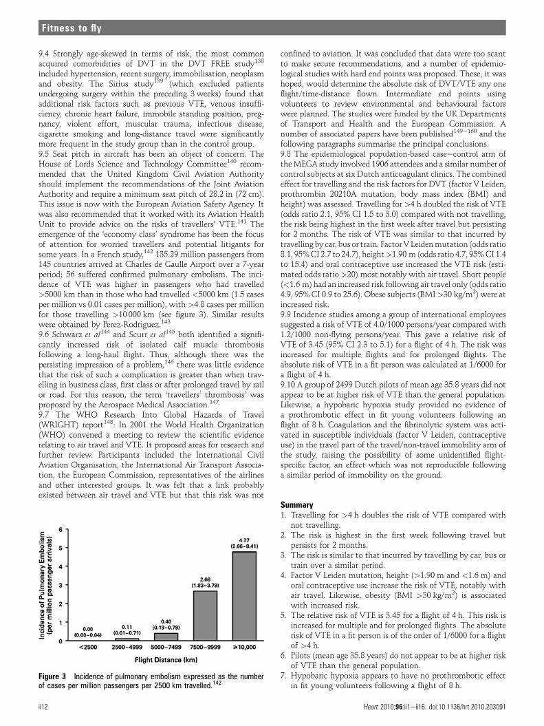
9.4 Strongly age-skewed in terms of risk, the most commonacquired comorbidities of DVT in the DVT FREE study138
included hypertension, recent surgery, immobilisation, neoplasmand obesity. The Sirius study139 (which excluded patientsundergoing surgery within the preceding 3 weeks) found thatadditional risk factors such as previous VTE, venous insuffi-ciency, chronic heart failure, immobile standing position, preg-nancy, violent effort, muscular trauma, infectious disease,cigarette smoking and long-distance travel were significantlymore frequent in the study group than in the control group.9.5 Seat pitch in aircraft has been an object of concern. TheHouse of Lords Science and Technology Committee140 recom-mended that the United Kingdom Civil Aviation Authorityshould implement the recommendations of the Joint AviationAuthority and require a minimum seat pitch of 28.2 in (72 cm).This issue is now with the European Aviation Safety Agency. Itwas also recommended that it worked with its Aviation HealthUnit to provide advice on the risks of travellers’ VTE.141 Theemergence of the ‘economy class’ syndrome has been the focusof attention for worried travellers and potential litigants forsome years. In a French study,142 135.29 million passengers from145 countries arrived at Charles de Gaulle Airport over a 7-yearperiod; 56 suffered confirmed pulmonary embolism. The inci-dence of VTE was higher in passengers who had travelled>5000 km than in those who had travelled <5000 km (1.5 casesper million vs 0.01 cases per million), with >4.8 cases per millionfor those travelling >10 000 km (see figure 3). Similar resultswere obtained by Perez-Rodriguez.143
9.6 Schwarz et al144 and Scurr et al145 both identified a signifi-cantly increased risk of isolated calf muscle thrombosisfollowing a long-haul flight. Thus, although there was thepersisting impression of a problem,146 there was little evidencethat the risk of such a complication is greater than when trav-elling in business class, first class or after prolonged travel by railor road. For this reason, the term ‘travellers’ thrombosis’ wasproposed by the Aerospace Medical Association.147
9.7 The WHO Research Into Global Hazards of Travel(WRIGHT) report148: In 2001 the World Health Organization(WHO) convened a meeting to review the scientific evidencerelating to air travel and VTE. It proposed areas for research andfurther review. Participants included the International CivilAviation Organisation, the International Air Transport Associa-tion, the European Commission, representatives of the airlinesand other interested groups. It was felt that a link probablyexisted between air travel and VTE but that this risk was not
confined to aviation. It was concluded that data were too scantto make secure recommendations, and a number of epidemio-logical studies with hard end points was proposed. These, it washoped, would determine the absolute risk of DVT/VTE any oneflight/time-distance flown. Intermediate end points usingvolunteers to review environmental and behavioural factorswere planned. The studies were funded by the UK Departmentsof Transport and Health and the European Commission. Anumber of associated papers have been published149e160 and thefollowing paragraphs summarise the principal conclusions.9.8 The epidemiological population-based caseecontrol arm oftheMEGA study involved 1906 attendees and a similar number ofcontrol subjects at six Dutch anticoagulant clinics. The combinedeffect for travelling and the risk factors for DVT (factor V Leiden,prothrombin 20210A mutation, body mass index (BMI) andheight) was assessed. Travelling for >4 h doubled the risk of VTE(odds ratio 2.1, 95% CI 1.5 to 3.0) compared with not travelling,the risk being highest in the first week after travel but persistingfor 2 months. The risk of VTE was similar to that incurred bytravelling by car, bus or train. FactorVLeidenmutation (odds ratio8.1, 95%CI 2.7 to 24.7), height>1.90 m (odds ratio 4.7, 95%CI 1.4to 15.4) and oral contraceptive use increased the VTE risk (esti-mated odds ratio>20) most notably with air travel. Short people(<1.6 m) had an increased risk following air travel only (odds ratio4.9, 95% CI 0.9 to 25.6). Obese subjects (BMI>30 kg/m2) were atincreased risk.9.9 Incidence studies among a group of international employeessuggested a risk of VTE of 4.0/1000 persons/year compared with1.2/1000 non-flying persons/year. This gave a relative risk ofVTE of 3.45 (95% CI 2.3 to 5.1) for a flight of 4 h. The risk wasincreased for multiple flights and for prolonged flights. Theabsolute risk of VTE in a fit person was calculated at 1/6000 fora flight of 4 h.9.10 A group of 2499 Dutch pilots of mean age 35.8 years did notappear to be at higher risk of VTE than the general population.Likewise, a hypobaric hypoxia study provided no evidence ofa prothrombotic effect in fit young volunteers following anflight of 8 h. Coagulation and the fibrinolytic system was acti-vated in susceptible individuals (factor V Leiden, contraceptiveuse) in the travel part of the travel/non-travel immobility arm ofthe study, raising the possibility of some unidentified flight-specific factor, an effect which was not reproducible followinga similar period of immobility on the ground.
Summary1. Travelling for >4 h doubles the risk of VTE compared with
not travelling.2. The risk is highest in the first week following travel but
persists for 2 months.3. The risk is similar to that incurred by travelling by car, bus or
train over a similar period.4. Factor V Leiden mutation, height (>1.90 m and <1.6 m) and
oral contraceptive use increase the risk of VTE, notably withair travel. Likewise, obesity (BMI >30 kg/m2) is associatedwith increased risk.
5. The relative risk of VTE is 3.45 for a flight of 4 h. This risk isincreased for multiple and for prolonged flights. The absoluterisk of VTE in a fit person is of the order of 1/6000 for a flightof >4 h.
6. Pilots (mean age 35.8 years) do not appear to be at higher riskof VTE than the general population.
7. Hypobaric hypoxia appears to have no prothrombotic effectin fit young volunteers following a flight of 8 h.
Figure 3 Incidence of pulmonary embolism expressed as the numberof cases per million passengers per 2500 km travelled.142
ii12 Heart 2010;96:ii1eii16. doi:10.1136/hrt.2010.203091
Fitness to fly
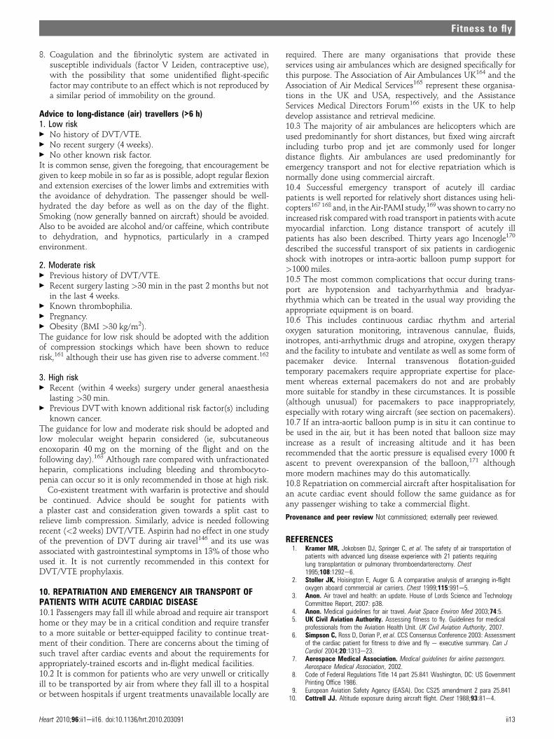
8. Coagulation and the fibrinolytic system are activated insusceptible individuals (factor V Leiden, contraceptive use),with the possibility that some unidentified flight-specificfactor may contribute to an effect which is not reproduced bya similar period of immobility on the ground.
Advice to long-distance (air) travellers (>6 h)1. Low risk< No history of DVT/VTE.< No recent surgery (4 weeks).< No other known risk factor.It is common sense, given the foregoing, that encouragement begiven to keep mobile in so far as is possible, adopt regular flexionand extension exercises of the lower limbs and extremities withthe avoidance of dehydration. The passenger should be well-hydrated the day before as well as on the day of the flight.Smoking (now generally banned on aircraft) should be avoided.Also to be avoided are alcohol and/or caffeine, which contributeto dehydration, and hypnotics, particularly in a crampedenvironment.
2. Moderate risk< Previous history of DVT/VTE.< Recent surgery lasting >30 min in the past 2 months but not
in the last 4 weeks.< Known thrombophilia.< Pregnancy.< Obesity (BMI >30 kg/m2).The guidance for low risk should be adopted with the additionof compression stockings which have been shown to reducerisk,161 although their use has given rise to adverse comment.162
3. High risk< Recent (within 4 weeks) surgery under general anaesthesia
lasting >30 min.< Previous DVTwith known additional risk factor(s) including
known cancer.The guidance for low and moderate risk should be adopted andlow molecular weight heparin considered (ie, subcutaneousenoxoparin 40 mg on the morning of the flight and on thefollowing day).163 Although rare compared with unfractionatedheparin, complications including bleeding and thrombocyto-penia can occur so it is only recommended in those at high risk.
Co-existent treatment with warfarin is protective and shouldbe continued. Advice should be sought for patients witha plaster cast and consideration given towards a split cast torelieve limb compression. Similarly, advice is needed followingrecent (<2 weeks) DVT/VTE. Aspirin had no effect in one studyof the prevention of DVT during air travel146 and its use wasassociated with gastrointestinal symptoms in 13% of those whoused it. It is not currently recommended in this context forDVT/VTE prophylaxis.
10. REPATRIATION AND EMERGENCY AIR TRANSPORT OFPATIENTS WITH ACUTE CARDIAC DISEASE10.1 Passengers may fall ill while abroad and require air transporthome or they may be in a critical condition and require transferto a more suitable or better-equipped facility to continue treat-ment of their condition. There are concerns about the timing ofsuch travel after cardiac events and about the requirements forappropriately-trained escorts and in-flight medical facilities.10.2 It is common for patients who are very unwell or criticallyill to be transported by air from where they fall ill to a hospitalor between hospitals if urgent treatments unavailable locally are
required. There are many organisations that provide theseservices using air ambulances which are designed specifically forthis purpose. The Association of Air Ambulances UK164 and theAssociation of Air Medical Services165 represent these organisa-tions in the UK and USA, respectively, and the AssistanceServices Medical Directors Forum166 exists in the UK to helpdevelop assistance and retrieval medicine.10.3 The majority of air ambulances are helicopters which areused predominantly for short distances, but fixed wing aircraftincluding turbo prop and jet are commonly used for longerdistance flights. Air ambulances are used predominantly foremergency transport and not for elective repatriation which isnormally done using commercial aircraft.10.4 Successful emergency transport of acutely ill cardiacpatients is well reported for relatively short distances using heli-copters167 168 and, in theAir-PAMI study,169was shown to carry noincreased risk comparedwith road transport in patientswith acutemyocardial infarction. Long distance transport of acutely illpatients has also been described. Thirty years ago Incenogle170
described the successful transport of six patients in cardiogenicshock with inotropes or intra-aortic balloon pump support for>1000 miles.10.5 The most common complications that occur during trans-port are hypotension and tachyarrhythmia and bradyar-rhythmia which can be treated in the usual way providing theappropriate equipment is on board.10.6 This includes continuous cardiac rhythm and arterialoxygen saturation monitoring, intravenous cannulae, fluids,inotropes, anti-arrhythmic drugs and atropine, oxygen therapyand the facility to intubate and ventilate as well as some form ofpacemaker device. Internal transvenous flotation-guidedtemporary pacemakers require appropriate expertise for place-ment whereas external pacemakers do not and are probablymore suitable for standby in these circumstances. It is possible(although unusual) for pacemakers to pace inappropriately,especially with rotary wing aircraft (see section on pacemakers).10.7 If an intra-aortic balloon pump is in situ it can continue tobe used in the air, but it has been noted that balloon size mayincrease as a result of increasing altitude and it has beenrecommended that the aortic pressure is equalised every 1000 ftascent to prevent overexpansion of the balloon,171 althoughmore modern machines may do this automatically.10.8 Repatriation on commercial aircraft after hospitalisation foran acute cardiac event should follow the same guidance as forany passenger wishing to take a commercial flight.
Provenance and peer review Not commissioned; externally peer reviewed.
REFERENCES1. Kramer MR, Jokobsen DJ, Springer C, et al. The safety of air transportation of
patients with advanced lung disease experience with 21 patients requiringlung transplantation or pulmonary thromboendarterectomy. Chest1995;108:1292e6.
2. Stoller JK, Hoisington E, Auger G. A comparative analysis of arranging in-flightoxygen aboard commercial air carriers. Chest 1999;115:991e5.
3. Anon. Air travel and health: an update. House of Lords Science and TechnologyCommittee Report, 2007: p38.
4. Anon. Medical guidelines for air travel. Aviat Space Environ Med 2003;74:5.5. UK Civil Aviation Authority. Assessing fitness to fly. Guidelines for medical
professionals from the Aviation Health Unit. UK Civil Aviation Authority, 2007.6. Simpson C, Ross D, Dorian P, et al. CCS Consensus Conference 2003: Assessment
of the cardiac patient for fitness to drive and fly e executive summary. Can JCardiol 2004;20:1313e23.
7. Aerospace Medical Association. Medical guidelines for airline passengers.Aerospace Medical Association, 2002.
8. Code of Federal Regulations Title 14 part 25.841 Washington, DC: US GovernmentPrinting Office 1986.
9. European Aviation Safety Agency (EASA). Doc CS25 amendment 2 para 25.84110. Cottrell JJ. Altitude exposure during aircraft flight. Chest 1988;93:81e4.
Heart 2010;96:ii1eii16. doi:10.1136/hrt.2010.203091 ii13
Fitness to fly

11. Kelly PT, Seccombe LM, Rogers PG, et al. Directly measured cabin pressureconditions during Boeing 747-400 commercial aircraft flights. Respirology2007;12:511e15.
12. Slonim NB, Hamilton LH. Respiratory physiology. 2nd edn. St Louis, Missouri, USA:C V Mosby, 1971.
13. Sorbini CA, Grassi V, Solinas E, et al. Arterial oxygen tension in relation to age inhealthy adults. Respiration 1968;25:3e13.
14. Cottrell JJ, Lebovitz B, Fennell RG, et al. In flight arterial saturation: continuousmonitoring by pulse oximetry. Aviat Space Environ Med 1995;66:126e30.
15. Heistad DD, Abboud FM. Circulatory adjustments to hypoxia. Circulation1980;61:463e70.
16. Vatner SF, Rutherfor JD. Control of the myocardial contractile state by carotidchemo-and baroreceptor and pulmonary inflation reflexes in conscious dogs. J ClinInvest 1978;61:1593e601.
17. Lugliani F, Whipp BJ, Wasserman K. A role for the carotid body in cardiovascularcontrol in man. Chest 1973;63:744e50.
18. Pelletier CL. Circulatory responses to graded stimulation of the carotidchemoreceptors in dogs. Circ Res 1972;31:431.
19. Von Euler US, Liljestrand G. Observations on the pulmonary arterial blood pressurein the cat. Acta Physiol Scand 1946;12:301e20.
20. Hultgren HN. Effects of altitude upon cardiovascular diseases. J Wilderness Med1992;3:301e8.
21. Bartsch P, Gibbs SR. Effect of altitude on the heart and lung. Circulation2007;116:2191e202.
22. De Burgh Daly M, Scott MJ. An analysis of the primary cardiovascular reflexeffects of stimulation of the carotid body chemoreceptors in the dog. J Physiol1962;162:555e73.
23. Cutaia M, Rounds S. Hypoxic pulmonary vasoconstriction: physiologic significance,mechanism and clinical relevance. Chest 1990;97:706e18.
24. Dillard Ewald F. The use of pulmonary function testing in piloting, air travel,mountain climbing, and diving. Clin Chest Med 2001;22:803.
25. Phillips BA, McConnell JW, Smith MD. The effects of hypoxaemia on cardiacoutput. A dose response curve. Chest 1988;93:471e5.
26. Veglio M, Maule S, Cametti G. The effects of exposure to moderate altitudeon cardiovascular autonomic function in normal adults. Clin Auton Res1999;9:123e7.
27. Thomson AJ, Drummond GB, Waring WS, et al. Effects of short-term isocapnichyperoxia and hypoxia on cardiovascular function. J Appl Physiol 2006;101:809e16.
28. Wyss CA, Koepfli P, Fretz G, et al. Influence of altitude exposure on coronary flowreserve. Circulation 2003;108:1202.
29. Pohl U, Busse R. Hypoxia stimulates release of endothelium derived relaxant factor.Am J Physiol 1989;256:H1595e600.
30. Hobkirk J, Damy T, Bennett A, et al. Effects of reduced inspired oxygenconcentration in patients with chronic heart failure (abstract). Proceedings of ESC2009.
31. Ideal Cabin Environment study.32. Agostini P, Cattadori G, Guazzi M, et al. Effects of simulated altitude-induced
hypoxia on exercise in patients in chronic heart failure. Am J Med2000;109:450e5.
33. Kujanik S, Snincak M, Galajdova K, et al. Cardiovascular changes during suddenascent in a cable cabin to moderate altitude. Physiol Res 2000;49:729e31.
34. Malconian M, Rock P, Hultgren H, et al. The electrocardiogram at rest andexercise during a simulated ascent of Mt Everest (Operation Everest II). Am JCardiol 1990;65:1475e80.
35. Westerholm CJ. Threshold studies in transvenous cardiac pacemaker treatment.Direct measurement with special reference to short and long term stimulation andinfluence of certain metabolic, respiratory and pharmacological factors. Scand JThorac Cardiovasc Surg Suppl 1971;8:1e35.
36. Weilenmann D, Duru F, Schonbeck M, et al. Influence of acute exposure to highaltitude and hypoxemia on ventricular stimulation thresholds in pacemaker patients.PACE 2000;23:512e15.
37. Siedler K, Kreutzberger R, Menold E, et al. Acute normobaric hypoxia (FIO2¼0.12)does not enhance exercise-induced thrombin and fibrin formation. Int J Sports Med2002;23:S120.
38. Le Roux G, Larmignat P, Marchal M, et al. Haemostasis at high altitude. Int JSports Med 1992;13:S49e51.
39. Mannucci PM, Gringeri A, Peyvandi F, et al. Short-term exposure to high altitudecauses coagulation activation and inhibits fibrinolysis. Thromb Haemost2002;87:342e3.
40. Crosby A, Talbot NP, Harrison P, et al. Relation between acute hypoxia andactivation of coagulation in human beings. Lancet 2003;361:2207e8.
41. Hodkinson PD, Hunt BJ, Parmar K, et al. Is mild normobaric hypoxia a risk factorfor venous thromboembolism? J Thromb Haemost 2003;1:2131e3.
42. Toff WD, Jones CI, Ford I, et al. Effect of hypobaric hypoxia, simulating conditionsduring long-haul air travel, on coagulation, fibrinolysis, platelet function, andendothelial activation. JAMA 2006;295:2251e61.
43. Schreijer AJM, Cannegieter SC, Meijers JCM, et al. Activation of coagulationsystem during air travel: a cross over study. Lancet 2006;367:832e8.
44. Dowdall N. Customer health: a new role for occupational physicians. Occup Med2003;53:19e23.
45. Van Gerwen LJ, Diekstra RF. Fear of flying treatment programs for passengers: aninternational review. Aviat Space Environ Med 2000;71:430e7.
46. Ito H, Lee D. Assessing the effect of September 11th terrorist attacks on US airlinedemand. J Econ Business 2005;57:75e95.
47. Folkes VS, Kaletsky S, Graham JL. A field study of causal inferences and consumerreaction: the view from the airport. J Consumer Res 1987;13:534e9.
48. Strike PC, Steptoe A. A systematic review of mental stress-induced myocardialischaemia. Eur Heart J 2003;24:690e703.
49. Hu K, Ivanov PCh, Hilton MF, et al. Endogenous circadian rhythm in an index ofcardiac vulnerability independent of changes in behavior. Proc Natl Acad Sci USA2004;101:18223e7.
50. Knutson A. Health disorders of shift workers. Occup Med 2003;53:103e8.51. Kerut EK, Winterton J, Roffidal L, et al. Congestive heart failure: influence of
hypertension with or without diabetes mellitus on circadian variation in autonomicbalance. Am J Hypertens 1995;8:174A.
52. Carson PM, O’Connor CM, Miller AB, et al. Circadian rhythm and sudden death inheart failure: results from prospective randomised amplodipine survival trial. J AmColl Cardiol 2000;36:541e6.
53. Boersma E, Pieper KS, Steyerberg EW, et al. Predictors of outcome in patientswith acute coronary syndromes without persistent ST-segment elevation. Resultsfrom an international trial of 9461 patients. The PURSUIT Investigators. Circulation2000;101:2557e67.
54. Califf RM, Pieper KS, Lee KL, et al. Prediction of 1-year survival after thrombolysisfor acute myocardial infarction in the global utilization of streptokinase and TPA foroccluded coronary arteries trial. Circulation 2000;101:2231e8.
55. Granger CB, Goldberg RJ, Dabbous O, et al. Predictors of hospital mortalityin the global registry of acute coronary events. Arch Intern Med2003;163:2345e53.
56. Morrow DA, Antman EM, Giugliano RP, et al. A simple risk index for rapid initialtriage of patients with ST-elevation myocardial infarction: an InTIME II substudy.Lancet 2001;358:1571e5.
57. Dorsch MF, Lawrance RA, Sapsford RJ, et al. A simple benchmark for evaluatingquality of care of patients following acutemyocardial infarction.Heart 2001;86:150e4.
58. Kurz DJ, Bernstein A, Hunt K, et al. Simple point-of-care risk stratification in acutecoronary syndromes: the AMIS model. Heart 2009;95:662e8.
59. Zahger D, Leibowitz D, Tabb IK, et al. Long-distance air travel soon after an acutecoronary syndrome: a prospective evaluation of a triage protocol. Am Heart J2000;140:241e2.
60. Cox GR, Peterson T, Bouchel L, et al. Safety of commercial air travel followingmyocardial infarction. Aviat Space Environ Med 1996;67:976e82.
61. Thomas MD, Hinds R, Walker C, et al. Safety of aeronautical repatriation aftermyocardial infarction: a retrospective study. Heart 2006;92:1864e5.
62. Roby H, Lee A, Hopkins A. Safety of air travel following acute myocardial infarctionAviat Space Environ Med 2002;73:91e6.
63. Essebag V, Lutchmedial S, Churchill-Smith M. Safety of long distance aeromedicaltransport of the cardiac patient: a retrospective study. Aviat Space Environ Med2001;72:182e7.
64. Myocardial Infarction National Audit Project (MINAP). 2004.65. Heggunje PS, Harjal KJ, Stone GW, et al. Procedural success versus clinical risk
status in determining discharge of patients after primary angioplasty for acutemyocardial infarction. J Am Coll Cardiol 2004;44:1400e7.
66. deLuca G, Suryapranata H, van’t Hof AWJ, et al. Prognostic assessment ofpatients with acute myocardial infarction treated with primary angioplasty.Implications for early discharge. Circulation 2004;109:2737e43.
67. Ludman PF. British Cardiovascular Intervention Society (BCIS) audit returns for2007. http://www.bcis.org.uk/resources/documents/BCIS%20Audit%20web.pdf.
68. Van de Verf F, Bax J, Betriu A, et al. for the Task Force of the European Society ofCardiology. Management of acute myocardial infarction for patients presenting withpersistent ST segment elevation. Eur Heart J 2008;29:2909e45.
69. Jan K, Madsen JK, Peer G, Saunamaki K, et al. Danish multicenter randomizedstudy of invasive versus conservative treatment in patients with inducible ischemiaafter thrombolysis in acute myocardial infarction (DANAMI). Circulation1997;96:748e55.
70. National Institute for Health and Clinical Excellence (NICE). Guidance onimplantable defibrillator and arrhythmia. London: NICE, 2006.
71. Task Force of the European Society of Cardiology. Guideline for the diagnosisand treatment of non ST elevation acute coronary syndromes. Eur Heart J2007;28:1598e660.
72. Ludman PF. British Cardiovascular Intervention Society audit returns 2007 and2008. http://www.bcis.org.uk/resources/audit.
73. Abbas AE, Brodie B, Dixon S, et al. Incidence and prognostic impact ofgastrointestinal bleeding after percutaneous coronary intervention for acutemyocardial infarction. Am J Cardiol 2005;96:173e6.
74. Berkowitz SD, Granger CB, Pieper KS, et al. for the Global Utilization of Streptokinaseand Tissue Plasminogen Activator for Occluded Coronary Arteries (GUSTO) IInvestigators. Incidence and predictors of bleeding after contemporary thrombolytictherapy for myocardial infarction. Circulation 1997;95:2508e16.
75. Buresly K, Eisenberg MJ, Zhang X, et al. Bleeding complications associated withcombinations of aspirin, thienopyridine derivatives, and warfarin in elderly patientsfollowing acute myocardial infarction. Arch Intern Med 2005;165:784e9.
76. Lewis EF, Velazquez EJ, Solomon SD, et al. Predictors of the first heart failurehospitalization in patients who are stable survivors of myocardial infarctioncomplicated by pulmonary congestion or left ventricular dysfunction: a VALIANTstudy. Eur Heart J 2008;29:748e56.
ii14 Heart 2010;96:ii1eii16. doi:10.1136/hrt.2010.203091
Fitness to fly

77. Eisenmenger V. Die angeborenen Defekte der Kammerscheidewande desHerzens. Zeitschr. Klin Med 1897;32:1e28.
78. Partin C. The evolution of Eisenmenger’s eponymic enshrinement. Am J Cardiol2003;92:1187e91.
79. Harinck E, Hutter PA, Hoorntje TM, et al. Air travel and adults with cyanoticcongenital heart disease. Circulation 1996;93:272e6.
80. Broberg CS, Uebing A, Cuomo L, et al. Adult patients with Eisenmenger syndromereport flying safely on commercial aircraft. Heart 2007;93:1599e603.
81. Duke M, Abelmann WH. The hemodynamic response to chronic anemia. Circulation1969;39:503e13.
82. Roy SB, Bhatia ML, Mathur VS, et al. Hemodynamic effects of chronic severeanemia. Circulation 1963;28:346e56.
83. Weiskopf RB, Viele MK, Feiner J, et al. Human cardiovascular and metabolicresponse to acute, severe isovolemic anemia. JAMA 1998;279:217e21.
84. Roig E, Betriu A, Castaner A, et al. Disabling angina pectoris with normal coronaryarteries in patients undergoing long-term hemodialysis. Am J Med 1981;71:431e4.
85. Case RB, Berglund E, Sarnoff SJ. Ventricular function. Changes in coronaryresistance and ventricular function resulting from acutely induced anemia and theeffect thereon of coronary stenosis. Am J Med 1955;18:397e405.
86. Cromie N, Lee C, Struthers AD. Anaemia in chronic heart failure: what is itsfrequency in the UK and its underlying causes? Heart 2002;87:377e8.
87. Silverberg DS, Wexler D, Sheps D, et al. The effect of correction of mild anemia insevere, resistant congestive heart failure using subcutaneous erythropoietin andintravenous iron: a randomized controlled study. J Am Coll Cardiol2001;37:1775e80.
88. Levy PS, Kim SJ, Eckel PK, et al. Limit to cardiac compensation during acuteisovolemic hemodilution: Influence of coronary stenosis. Am J Physiol1993;265:H340e9.
89. Lee PC, Kini AS, Chowdhury A, et al. Anemia is an independent predictor ofmortality after percutaneous coronary intervention. J Am Coll Cardiol2004;44:541e6.
90. Koch CG, Weng Y, Zhou SX, et al. Prevalence of risk factors, and not gender perse, determines short- and long-term survival after coronary artery bypass surgery.J Cardiothorac Vasc Anesth 2003;17:585e93.
91. Chauhan A, Grace AA, Newell SA, et al. Early complications after dual chamberversus single chamber pacemaker implantation. PACE 1994;17:2012e15.
92. Aggarwal RK, Connelly DT, Ray SG, et al. Early complications of permanentpacemaker implantation: no difference between dual and single chamber systems.Heart 1995;73:571e5.
93. British Thoracic Society Standards of Care Committee. Managing passengerswith respiratory disease planning air travel. British Thoracic Societyrecommendations. http://www.brit-thoracic.org.uk/Portals/0/Clinical%20Information/Air%20Travel/Guidelines/FlightRevision04.pdf (accessed2 Jan 2009).
94. Dorbala S, Cohen AJ, Hutchinson LA, et al. Does radiofrequency ablation inducea prethrombotic state? Analysis of coagulation system activation and comparison toelectrophysiologic study. J Cardiovasc Electrophysiol 1998;9:1152e60.
95. Chen JY, Chang KC, Lin YC, et al. Safety and outcomes of short-term multiplefemoral venous sheath placement in cardiac electrophysiological study andradiofrequency catheter ablation. Jpn Heart J 2004;45:257e64.
96. Scheinman MM, Huang S. The 1998 NASPE prospective catheter ablationregistry. PACE 2000;23:1020e8.
97. Greene T, Huang S, Wagshal A, et al. Cardiovascular complications after RFcatheter ablation of supraventricualr tachyarrhythmias. Am J Cardiol1994;74:615e17.
98. Hindricks G. The Multicentre European RF Survey (MERFS): complications of RFcatheter ablation of arrhythmias. The Multicentre European RF Survey (MERFS)investigators of the Working Group on Arrhythmias of the European Society ofCardiology. Eur Heart J 1993;14:1644e53.
99. Antiplatelet Trialists’ Collaboration. Collaborative overview of randomised trialsof antiplatelet therapy III. Reduction in venous thrombosis and pulmonary embolismby antiplatelet prophylaxis among surgical and medical patients. BMJ1994;308:235e46.
100. Blanc JJ, Almendral J, Brignole M, et al. Scientific Initiatives Committee of theEuropean Heart Rhythm Association. Consensus document on antithrombotictherapy in the setting of electrophysiological procedures. Europace2008;10:513e27.
101. Harold L, Lazar HL, Fitzgerald CA, et al. Early discharge after coronary artery bypassgraft surgery: are patients really going home earlier? J Thorac Cardiovasc Surg2001;121:943e50.
102. Beggs VL, Birkemeyer NJ, Nugent WC, et al. Factors related to rehospitalizationwithin thirty days of discharge after coronary artery bypass grafting. Best PractBenchmarking Healthc 1996;1:180e6.
103. Network Devices Survey Group. Pacemakers and implantable defibrillators:UK National Survey 2006 (version 2.1). London: Department of Health, 2008.http://www.devicesurvey.com (accessed 2 Jan 2009).
104. Irnich W. Electronic security systems and active implantable medical devices.Pacing Clin Electrophysiol 2002;25:1235e58.
105. Rokas S, Crick JCP, Sowton E. Does pacemaker dependence exist? Br Heart J1981;45:340.
106. Stevenson RA. Design and application of broadband ceramic feedthroughcapacitor EMI filters to cardiac pacemakers and implantable defibrillators.
Proceedings 19th International Conference, IEEE Engineering in Medicine andBiology Society 1997;6:2558e62.
107. Boivin W, Coletta J, Kerr L. Characterization of the magnetic fields around walk-through and hand-held metal detectors. Health Phys 2003;84:582e93.
108. Copperman Y, Zarfati D, Laniado S. The effect of metal detector gates onimplanted permanent pacemakers. Pacing Clin Electrophysiol 1988;11:1386e7.
109. Kolb C, Schmieder S, Lehmann G, et al. Do airport metal detectors interfere withimplantable pacemakers or cardioverter-defibrillators? J Am Coll Cardiol2003;41:2054e9.
110. Pinski SL, Trohman RG. Interference in implanted cardiac devices, Part I. PacingClin Electrophysiol 2002;25:1367e81.
111. Possick SE, Barry M. Evaluation and management of the cardiovascular patientembarking on air travel. Ann Intern Med 2004;141:148e54.
112. Transportation Security Administration (TSA). Travelers with Disabilities andMedical Conditions: Pacemakers, Defibrillators, Other Implanted Medical Devices,and Metal Implants. http://www.tsa.gov/travelers/airtravel/specialneeds/editorial_1374.shtm (accessed 2 Jan 2009).
113. BAA. http://www.baa.com/portal/controller/dispatcher.jsp?ChPath¼Global%5EMisc%5ESearch&searchChannel¼Global&queryString¼pacemaker (accessed 2Jan 2009).
114. Toff WD, Deller AGMcC, Hobbs RA, et al. Radiofrequency interference withpacemaker function in the aircraft environment. New Trends in Arrhythmias1988;III/IV:777e80.
115. Toff WD, Camm AJ. Implanted devices and aviation. Eur Heart J1988;9(Suppl G):133e8.
116. Deller AGMcC, Toff WD, Hobbs RA, et al. The development of a system for theevaluation of electromagnetic interference with pacemaker function: hazards in theaircraft environment. J Med Eng Technol 1989;13:161e5.
117. Toff WD. Cardiac pacemaker function in the aviation environment. MD thesis.London: University of London, 1994.
118. Carleton RA, Sessions RW, Graettinger JS. Environmental influence on implantablecardiac pacemakers. JAMA 1964;190:938e40.
119. Brown JH, Carter ET, Silverstein MN, et al. Would you fly with these pilots?Aerospace Med 1973;44:786e93.
120. De Rotte AA, Van Der Kemp P. Electromagnetic interference in pacemakers insingle-engine fixed-wing aircraft: a European perspective. Aviat Space Environ Med2002;73:179e83.
121. Federal Aviation Administration. Use of portable electronic devices aboardaircraft. Advisory Circular AC 91e21.1B. 8/25/06. http://rgl.faa.gov/Regulatory_and_Guidance_Library/rgAdvisoryCircular.nsf/list/AC%2091.21-1B/$FILE/AC%2091.21-1B.pdf (accessed 2 Jan 2009).
122. Civil Aviation Authority. Portable electronic devices. 2009. http://www.caa.co.uk/default.aspx?catid¼1770&pagetype¼90&pageid¼9853 (accessed 2 Jan 2009).
123. Ziegler JF, Curtis HW, Muhlfeld HP, et al. IBM experiments in soft fails in computerelectronics 1978e1994. IBM J Res Develop 1996;40:3e18.
124. Bradley PD, Normand E. Single event upsets in implantable cardioverterdefibrillators. IEEE Trans Nucl Sci 1998;45:2929e40.
125. St Jude Medical Inc. Physician communication: important physician advisory. 2005.http://www.sjm.com/companyinformation/physicianletter.html (accessed 2 Jan 2009).
126. World Health Organization. Information sheet Nov. 2005: Cosmic radiation andair travel. http://who.int/ionizing_radiation/env/cosmic/en/print.html (accessed 2Jan 2009).
127. Ferrick AM, Bernstein N, Aizer A, et al. Cosmic radiation induced softwareelectrical resets in ICDs during air travel. Heart Rhythm 2008;5:1201e3.
128. Bhakta D, Foreman LD. Cosmic radiation: not science fiction but clinical reality.Heart Rhythm 2008;5:1204e5.
129. French RS, Tillman JG. Pacemaker function during helicopter transport. Ann EmergMed 1989;18:305e7.
130. Gordon RS, O’Dell KB. Activity-sensing permanent internal pacemaker dysfunctionduring helicopter aeromedical transport. Ann Emerg Med 1990;19:1260e3.
131. Toff WD, Leeks C, Joy M, et al. The effect of aircraft vibration on the function of anactivity-sensing pacemaker. Br Heart J 1987;57:573e4.
132. Silverstein MD, Heit JA, Mohr DN, et al. Trends in the incidence of deep veinthrombosis and pulmonary embolism.A 25-year population-based study. Arch InternMed 1998;158:585e93.
133. Goldhaber SZ, Visani L, De Rosa M. Pulmonary embolism: clinical outcomes in theInternational Cooperative Pulmonary Embolism Registry (ICOPER). Lancet1999;353:1386e9.
134. Simpson K. Shelter deaths from pulmonary embolism. Lancet 1940;2:744.135. Homans J. Thrombosis of the deep leg veins due to prolonged sitting. N Engl J
Med 1954;250:148e9.136. Symington IS, Stack BHR. Pulmonary thromboembolism after travel. Br J Dis Chest
1977;71:138e40.137. Schulman S, Lindmarker P. Incidence of cancer after prophylaxis with warfarin
against recurrent venous thromboembolism for the Duration of Anticoagulation Trial.N Engl J Med 2000;342;1953e8.
138. Goldhaber SZ, Tapson VF. DVT FREE Steering Committee. A prospective registry of5,451 patients with ultrasound-confirmed deep vein thrombosis. Am J Cardiol2004;93:259e62.
139. Meyer-Michel S. for the Sirius Study Group. Epidemiologic study of risk factors fordeep vein thrombosis in medical outpatients. The Sirius Study. Arch Intern Med2000;160:3415e20.
Heart 2010;96:ii1eii16. doi:10.1136/hrt.2010.203091 ii15
Fitness to fly
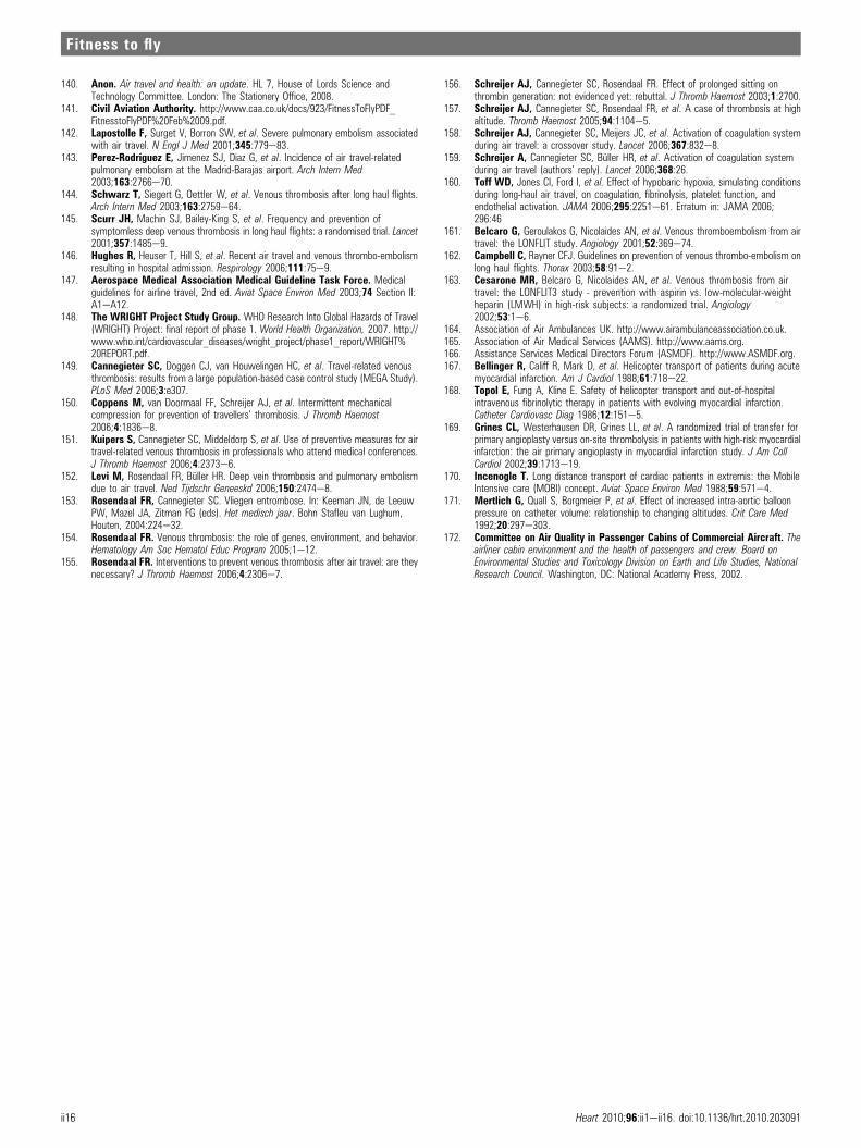
140. Anon. Air travel and health: an update. HL 7, House of Lords Science andTechnology Committee. London: The Stationery Office, 2008.
141. Civil Aviation Authority. http://www.caa.co.uk/docs/923/FitnessToFlyPDF_FitnesstoFlyPDF%20Feb%2009.pdf.
142. Lapostolle F, Surget V, Borron SW, et al. Severe pulmonary embolism associatedwith air travel. N Engl J Med 2001;345:779e83.
143. Perez-Rodriguez E, Jimenez SJ, Diaz G, et al. Incidence of air travel-relatedpulmonary embolism at the Madrid-Barajas airport. Arch Intern Med2003;163:2766e70.
144. Schwarz T, Siegert G, Oettler W, et al. Venous thrombosis after long haul flights.Arch Intern Med 2003;163:2759e64.
145. Scurr JH, Machin SJ, Bailey-King S, et al. Frequency and prevention ofsymptomless deep venous thrombosis in long haul flights: a randomised trial. Lancet2001;357:1485e9.
146. Hughes R, Heuser T, Hill S, et al. Recent air travel and venous thrombo-embolismresulting in hospital admission. Respirology 2006;111:75e9.
147. Aerospace Medical Association Medical Guideline Task Force. Medicalguidelines for airline travel, 2nd ed. Aviat Space Environ Med 2003;74 Section II:A1eA12.
148. The WRIGHT Project Study Group. WHO Research Into Global Hazards of Travel(WRIGHT) Project: final report of phase 1. World Health Organization, 2007. http://www.who.int/cardiovascular_diseases/wright_project/phase1_report/WRIGHT%20REPORT.pdf.
149. Cannegieter SC, Doggen CJ, van Houwelingen HC, et al. Travel-related venousthrombosis: results from a large population-based case control study (MEGA Study).PLoS Med 2006;3:e307.
150. Coppens M, van Doormaal FF, Schreijer AJ, et al. Intermittent mechanicalcompression for prevention of travellers’ thrombosis. J Thromb Haemost2006;4:1836e8.
151. Kuipers S, Cannegieter SC, Middeldorp S, et al. Use of preventive measures for airtravel-related venous thrombosis in professionals who attend medical conferences.J Thromb Haemost 2006;4:2373e6.
152. Levi M, Rosendaal FR, Buller HR. Deep vein thrombosis and pulmonary embolismdue to air travel. Ned Tijdschr Geneeskd 2006;150:2474e8.
153. Rosendaal FR, Cannegieter SC. Vliegen entrombose. In: Keeman JN, de LeeuwPW, Mazel JA, Zitman FG (eds). Het medisch jaar. Bohn Stafleu van Lughum,Houten, 2004:224e32.
154. Rosendaal FR. Venous thrombosis: the role of genes, environment, and behavior.Hematology Am Soc Hematol Educ Program 2005;1e12.
155. Rosendaal FR. Interventions to prevent venous thrombosis after air travel: are theynecessary? J Thromb Haemost 2006;4:2306e7.
156. Schreijer AJ, Cannegieter SC, Rosendaal FR. Effect of prolonged sitting onthrombin generation: not evidenced yet: rebuttal. J Thromb Haemost 2003;1:2700.
157. Schreijer AJ, Cannegieter SC, Rosendaal FR, et al. A case of thrombosis at highaltitude. Thromb Haemost 2005;94:1104e5.
158. Schreijer AJ, Cannegieter SC, Meijers JC, et al. Activation of coagulation systemduring air travel: a crossover study. Lancet 2006;367:832e8.
159. Schreijer A, Cannegieter SC, Buller HR, et al. Activation of coagulation systemduring air travel (authors’ reply). Lancet 2006;368:26.
160. Toff WD, Jones CI, Ford I, et al. Effect of hypobaric hypoxia, simulating conditionsduring long-haul air travel, on coagulation, fibrinolysis, platelet function, andendothelial activation. JAMA 2006;295:2251e61. Erratum in: JAMA 2006;296:46
161. Belcaro G, Geroulakos G, Nicolaides AN, et al. Venous thromboembolism from airtravel: the LONFLIT study. Angiology 2001;52:369e74.
162. Campbell C, Rayner CFJ. Guidelines on prevention of venous thrombo-embolism onlong haul flights. Thorax 2003;58:91e2.
163. Cesarone MR, Belcaro G, Nicolaides AN, et al. Venous thrombosis from airtravel: the LONFLIT3 study - prevention with aspirin vs. low-molecular-weightheparin (LMWH) in high-risk subjects: a randomized trial. Angiology2002;53:1e6.
164. Association of Air Ambulances UK. http://www.airambulanceassociation.co.uk.165. Association of Air Medical Services (AAMS). http://www.aams.org.166. Assistance Services Medical Directors Forum (ASMDF). http://www.ASMDF.org.167. Bellinger R, Califf R, Mark D, et al. Helicopter transport of patients during acute
myocardial infarction. Am J Cardiol 1988;61:718e22.168. Topol E, Fung A, Kline E. Safety of helicopter transport and out-of-hospital
intravenous fibrinolytic therapy in patients with evolving myocardial infarction.Catheter Cardiovasc Diag 1986;12:151e5.
169. Grines CL, Westerhausen DR, Grines LL, et al. A randomized trial of transfer forprimary angioplasty versus on-site thrombolysis in patients with high-risk myocardialinfarction: the air primary angioplasty in myocardial infarction study. J Am CollCardiol 2002;39:1713e19.
170. Incenogle T. Long distance transport of cardiac patients in extremis: the MobileIntensive care (MOBI) concept. Aviat Space Environ Med 1988;59:571e4.
171. Mertlich G, Quall S, Borgmeier P, et al. Effect of increased intra-aortic balloonpressure on catheter volume: relationship to changing altitudes. Crit Care Med1992;20:297e303.
172. Committee on Air Quality in Passenger Cabins of Commercial Aircraft. Theairliner cabin environment and the health of passengers and crew. Board onEnvironmental Studies and Toxicology Division on Earth and Life Studies, NationalResearch Council. Washington, DC: National Academy Press, 2002.
ii16 Heart 2010;96:ii1eii16. doi:10.1136/hrt.2010.203091
Fitness to fly