fistulinella cinereoalba sp. nov. and new distribution
TRANSCRIPT

Fistulinella cinereoalba sp. nov. and new distributionrecords for Austroboletus from Guyana
Tara D. FulgenziDepartment of Biological Sciences, Humboldt StateUniversity, Arcata, California 95521
Roy E. HallingInstitute of Systematic Botany, New York BotanicalGarden, Bronx, New York 10458, and Department ofBiological Sciences, Humboldt State University, Arcata,California 95521
Terry W. Henkel1
Department of Biological Sciences, Humboldt StateUniversity, Arcata, California 95521
Abstract: Fistulinella cinereoalba sp. nov., Austrobole-tus rostrupii, previously known from southeasternAsia, and Austroboletus festivus from Brazilian Amazo-nia are described for the first time from the GuianaShield. These boletes were collected from tropicalforests dominated by ectomycorrhizal Dicymbe spp.(Fabaceae subfam. Caesalpinioideae) in the PakaraimaMountains of western Guyana.
Key words: Boletaceae, Dicymbe, ectomycorrhizalfungi, Guiana Shield, Neotropics
INTRODUCTION
In monodominant forests of Dicymbe spp. (Fabaceaesubfam. Caesalpinioideae tribe Amherstieae) in Guyanataxa of Boletaceae sensu lato (Boletineae, Boletales,Agaricomycetes, Basidiomycota) figure prominently inthe ectomycorrhizal (EM) fungal assemblage (Cowanand Lindeman 1989; Henkel 1999; Henkel et al 2002;Fulgenzi et al 2007, 2008; Mayor et al 2008). Thispaper describes three species within FistulinellaHenn. or Austroboletus (Corner) Wolfe that areinfrequently encountered in Dicymbe forests.
Fistulinella encompasses , 27 species with primar-ily tropical distributions (Pegler and Young 1981;Pegler 1983; Singer et al 1983, 1991; Singer 1986;Watling and Gregory 1989; Ortiz-Santana et al 2007;Watling 2008). Fistulinella has been variably definedand recognized according to author (Wolfe 1979,1982; Pegler and Young 1981; Pegler 1983; Singer1986; Watling and Gregory 1989; Lewis and Cibula2000; Redeuilh and Soop 2006; Watling 2008). Herewe use the diagnostic features of Fistulinella defined
by Watling (1989, 2008) as bolete species withbasidiospores that are smooth, elongate-fusoid, andpink-brown, cinnamon-brown to purple brown indeposit, with some or all showing a weak to strongpseudoamyloid reaction, a strongly gelatinized lateralstratum in the tube trama, a pileipellis that is atrichodermium, ixotrichodermium or ixocutis, abasidioma that is viscid to glutinous, and a whitehymenophore that is pink to pink-gray at maturation.
Austroboletus contains , 40 species with primarilytropical distributions with some species occurring inthe temperate northern and southern hemispheres(McNabb 1967; Wolfe 1979; Horak 1980; Singer et al1983, 1991; Singer 1986; Watling and Gregory 1986;Watling and de Meijer 1987; Watling and Lee 1999;Halling et al 2006; Ortiz-Santana et al 2007). We usethe morphological concept of Austroboletus as boleteswith basidiospores that are lightly to heavily orna-mented with pits, warts or reticulations and are flesh-pink, vinaceous-brown to rust-brown in deposit(Wolfe 1979, Pegler and Young 1981, Singer 1986).
Here we describe Fistulinella cinereoalba sp. nov.and new distribution records for Austroboletus rostru-pii (Syd. & P. Syd.) E. Horak, previously known fromsoutheastern Asia, and Austroboletus festivus (Singer)Wolfe, previously known from eastern Brazil. Thesefungi were collected from tropical forests dominatedby EM Dicymbe spp. in the Pakaraima Mountains ofGuyana.
MATERIALS AND METHODS
Collecting expeditions were conducted in the May–Jul 1999rainy season in the Upper Ireng River Basin along Guyana’swestern border with Brazil in the west-central PakaraimaMountains and annually in May–Jul 2001–2004 in theUpper Potaro River Basin (5u18904.80N, 59u54940.40W) ,30 km north of the Ireng site. At each site fungi werecollected within a 5 km radius of a previously establishedfield camp in forests dominated by Dicymbe corymbosaSpruce ex Benth (Henkel 2003).
Basidiomata were examined in the field for their freshcharacteristics. Color was subjectively described and record-ed according to Kornerup and Wanscher (1978) with colorplates noted in parentheses (e.g. 3C4). Macrochemical spottests were performed following Singer (1986). Basidiomatawere field dried with silica gel (Miller et al 2002).
Microscopic anatomical details were determined on freshspecimens at the field camps with an EPOI microscope andon dried specimens in the laboratory with an OlympusBX51 microscope with bright field and phase contrast
Submitted 4 Apr 2009; accepted for publication 22 May 2009.1 Corresponding author. E-mail: [email protected]
Mycologia, 102(1), 2010, pp. 224–232. DOI: 10.3852/09-059# 2010 by The Mycological Society of America, Lawrence, KS 66044-8897
224

optics. Fungal tissue of dried specimens was rehydrated andmounted in either H20, 3% KOH, or Melzer’s reagent. Foreach taxon a minimum of 20 basidiospores, basidia, cystidiaand other structures were measured. Line drawings weremade with a drawing tube and scanned. Scanning electronmicrographs of basidiospores were obtained with a TopconABT32 scanning electron microscope at 15 kV or 30 kV.Specimens were deposited in these herbaria as indicated:BRG, HSU and NY (Holmgren et al 1990). A DNA sequencefor the 28S rDNA region was obtained from the holotype ofFistulinella cinereoalba following the methods of Smith et al(2007).
TAXONOMY
Fistulinella cinereoalba Fulgenzi and T.W. Henkelsp. nov. FIGS 1a, b; 2; 6bPileus griseus, fibrillosus, aequus, glutinosus, 19–43(55)
mm latus. Stipes albus, brunneus ubi contusum, gelatinosus,squamis ubique tenuibus, erectis, 55–80 3 3–6(13) mm.Hymenophorum brunneum ubi contusum. Basidiosporaecinnamomeo-brunneae, subfusiformae, leves, omnino pseu-doamyloideae, 12.4–19.8(24.8) 3 3.7–4.9 mm. Basidia 4-sterigmatibus. Pleurocystidia cylindricea ad aciculate.
HOLOTYPE: Henkel 8471 (BRG; ISOTYPE: HSU,NY)
Pileus 19–43 (55) mm broad, convex, dark gray(6D2–6E2) throughout initially, lighter gray (4C2–4C4) with age, finely rugulose, matted-fibrillose overwhite ground (under hand lens), overlain by agelatinous pellicle throughout, glutinous, viscid indry weather; margin lacking fibrils, entire, slightly in-rolled when young; trama 0.5–1.5 mm thick at margin,3–5 mm over tubes, 12–14 mm over stipe, white, solid,unchanging. Odor mildly fungoid; flavor mild. Tubes 2–4 mm long at margin, 6–8 mm centrally, 10–12 mm atstipe, narrowly and strongly depressed around stipe, ofirregular lengths, concolorous with pores, slightlybrowning on exposure; pores white when young,maturing to light pink-gray (7B3–7C3), slightly brown-ing with pressure, 1.5–3 per mm, isodiametric tosubovate. Stipe 55–80 3 3–6 (13) mm, subequal,gradually and tapering slightly inward toward base,entirely white, browning slightly with handling, withfine erect scales imbedded in a dense gelatinouspellicle throughout; extreme base with fine whitehyphal cords and occasional subtending concolorousectomycorrhizas; trama white, solid, unchanging.
Basidiospores cinnamon-brown (7D8–7E8) in heavydeposit, 12.4–19.8(24.8) 3 3.7–4.9 mm, Q mean(range) 5 3(2.5–4[7]), subfusiform, light yellow-pinkin H2O, lighter in KOH, most exhibiting a yellow,light red, or burgundy dextrinoid reaction in Mel-zer’s, uni- to multiguttulate, smooth; hilar appendage0.2–0.5 mm long. Basidia 27.2–34.6 3 9.8–12.4 mm,narrowly clavate, hyaline in H2O and KOH, often with
granular contents, 4-sterigmate; sterigmata 1.5–3.5 mm long. Pleurocystidia frequent, 37.1–61 3 3.7–6.2 mm, projecting 18.5–24.7(44.5) mm above hyme-nial palisade, cylindrical to aciculate, occasionally withgranular contents, thin-walled, hyaline in H2O andKOH. Cheilocystidia frequent, cylindrical, with 2–3septa, projecting 44.5–84 mm, slightly interwoven inlower two-thirds. Hymenophoral trama boletoid,strongly divergent; mediostratum (19.8)24.7–61.7 mm wide, of many thin, highly interwovenhyphae, these hyaline in H2O and KOH, gelatinousin KOH; lateral stratum hyphae 2.5–5 mm wide,hyaline in H2O and KOH, gelatinous, more so inKOH. Pileipellis a trichodermium superimposed on atrichodermial palisade of the ground, all overlain bythe gelatinous pellicle; terminal cells of trichoder-mium cylindrical, with 1–3 septa, orange-gray in H2O,lighter in KOH, projecting 49.4–86.5 mm from pileustrama, of irregular lengths; terminal cells of tricho-dermial palisade clavate, hyaline in H2O and KOH,projecting 12.4–32.1 mm from pileus trama; pellicle(14.8)24.7–123(217.5) mm thick, hyphae interwoven,periclinal, gelatinized, 2.4–6.1 mm wide, in massorange-gray in H2O, lighter and more gelatinous inKOH, with external burgundy acerose crystals. Pileustrama highly interwoven; individual hyphae 4.9–7.4 mm wide, hyaline in H2O and KOH, gelatinousin KOH. Stipitipellis with concentrated tufts of slightlyinterwoven, inflated cylindrical elements immersed ina dense pellicle of thin-walled, gelatinous hyphae;cylindrical elements with 1–3 septa, projecting 69.2–84 mm from stipe trama, concentrated on stipe scales;pellicle 74.1–173 mm thick; pellicle hyphae 2.5–3.7 mm wide, in mass light yellow in H2O, paler andgelatinous in KOH. Stipe trama of densely interwovenhyphae, these 2.5–4.9 mm wide, hyaline in H2O andKOH. Clamp connections absent. Macrochemical reac-tions: NH4OH nil on pileus.
Habit, habitat, and distribution. Infrequent, solitaryor in groups of 2–3 on root mat in forests dominatedby Dicymbe corymbosa, known only from the typelocality in the Upper Potaro Basin of Guyana.
Etymology. Cinereo 5 gray, alba 5 white (Latin),referring to the distinctive gray pileus and white stipe.
Specimens examined. BRAZIL. AMAZONAS: Fistulinellacampinaranae var. scrobiculata, Estrada Manaus-Caracarai, 12Jan 1979, Singer 12089 (HOLOTYPE of variety, INPA).GUYANA. REGION 8 POTARO-SIPARUNI: PakaraimaMountains, Upper Potaro River Basin, elevation 710–750 m; vicinity of Potaro base camp, 4 June 2000, Henkel7441 (BRG; HSU); Dicymbe plot 3, 5 May 2001, Henkel 8030(BRG; HSU); Dicymbe plot 3, 12 May 2001, Henkel 8108(BRG; HSU); vicinity of Potaro base camp, 17 June 2002,Henkel 8471 (HOLOTYPE, BRG; ISOTYPE: HSU, NY; 28SGenBank accession number GQ477439; MycoBank numberMB 513404).
FULGENZI ET AL: FISTULINELLA CINEREOALBA SP. NOV. 225

FIG. 1. Basidiomata of Fistulinella cinereoalba. a. HOLOTYPE (Henkel 8471). b. Field habit, Upper Potaro Basin, Guyana(photograph by Michael Pilkington). Basidiomata of Austroboletus rostrupii. c. Henkel 8189. d. Field habit, Upper Ireng Basin,Guyana. Bar 5 10 mm.
226 MYCOLOGIA
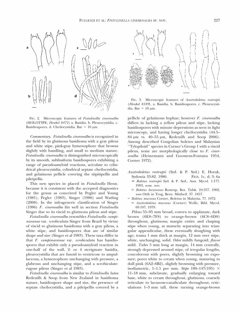
Commentary. Fistulinella cinereoalba is recognized inthe field by its glutinous basidioma with a gray pileusand white stipe, pink-gray hymenophore that brownsslightly with handling, and small to medium stature.Fistulinella cinereoalba is distinguished microscopicallyby its smooth, subfusiform basidiospores exhibiting arange of pseudoamyloid reactions, aciculate to cylin-drical pleurocystidia, cylindrical septate cheilocystidia,and gelatinous pellicle covering the stipitipellis andpileipellis.
This new species in placed in Fistulinella Henn.because it is consistent with the accepted diagnosticsfor the genus as conceived by Pegler and Young(1981), Pegler (1983), Singer (1986) and Watling(2008). In the infrageneric classification of Singer(1986) F. cinereoalba fits well in section FistulinellaSinger due to its viscid to glutinous pileus and stipe.
Fistulinella cinereoalba resembles Fistulinella campi-naranae var. scrobiculata Singer from Brazil by virtueof viscid to glutinous basidioma with a gray pileus, awhite stipe, and basidiospores that are of similarshape and size (Singer et al 1983). These taxa differ inthat F. campinaranae var. scrobiculata has basidio-spores that exhibit only a pseudoamyloid reaction inone-half of the wall, 2- or 4 sterigmate basidia,pleurocystidia that are fusoid to ventricose to ampul-laceous, a hymenophore unchanging with pressure, aglabrous and unchanging stipe, and a scrobiculate-rugose pileus (Singer et al 1983).
Fistulinella cinereoalba is similar to Fistulinella luteaRedeuilh & Soop from New Zealand in basidiomastature, basidiospore shape and size, the presence ofseptate cheilocystidia, and a pileipellis covered by a
pellicle of gelatinous hyphae; however F. cinereoalbadiffers in lacking a yellow pileus and stipe, lackingbasidiospores with minute depressions as seen in lightmicroscopy, and having longer cheilocystidia (44.5–84 mm vs. 40–55 mm, Redeuilh and Soop 2006).Among described Congolian boletes and Malaysian‘‘Tylopiloid’’ species in Corner’s Group 1 with a viscidpileus, none are morphologically close to F. ciner-eoalba (Heinemann and Goossens-Fontana 1954,Corner 1972).
Austroboletus rostrupii (Syd. & P. Syd.) E. Horak,Sydowia 33:82. 1980. FIGS. 1c, d; 3; 6a; Boletus rostrupii Syd. & P. Syd., Ann. Mycol. 1:177.
1903, nom. nov.; Boletus lacunosus Rostrup, Bot. Tidsk. 24:357. 1902,
non Otth in Trog, Bern. Mittheil. 37. 1857.5 Boletus mucosus Corner, Boletus in Malaysia, 77. 1972.
; Austroboletus mucosus (Corner) Wolfe, Bibl. Mycol.69:107. 1979.
Pileus 55–95 mm broad, convex to applanate, darkbrown (6E8–7F8) to orange-brown (6C8–6D8)throughout, glutinous; margin entire and claspingstipe when young, at maturity separating into trian-gular appendiculae, these eventually sloughing withage; trama 1 mm thick at margin, 12 mm over stipe,white, unchanging, solid. Odor mildly fungoid; flavormild. Tubes 5 mm long at margin, 14 mm centrally,strongly depressed around stipe, of irregular lengths,concolorous with pores, slightly browning on expo-sure; pores white to cream when young, maturing todull pink (6A2–6B2), slightly browning with pressure,isodiametric, 1–1.5 per mm. Stipe 100–147(195) 3
11–18 mm, subclavate, gradually enlarging towardbase, white to cream throughout, glutinous, coarselyreticulate to lacunose-canaliculate throughout, retic-ulations 1–3 mm tall, these turning orange-brown
FIG. 2. Microscopic features of Fistulinella cinereoalba(HOLOTYPE; Henkel 8471) a. Basidia. b. Pleurocystidia. c.Basidiospores. d. Cheilocystidia. Bar 5 10 mm.
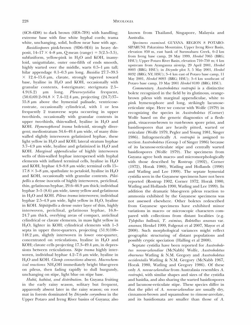
FIG. 3. Microscopic features of Austroboletus rostrupii(Henkel 8189). a. Basidia. b. Basidiospores. c. Pleurocysti-dia. Bar 5 10 mm.
FULGENZI ET AL: FISTULINELLA CINEREOALBA SP. NOV. 227
(6C8–6D8) to dark brown (6E8–7F8) with handling;extreme base with fine white hyphal cords; tramawhite, unchanging, solid, hollow near base with age.
Basidiospores pink-brown (9D6–9E6) in heavy de-posit, 14–17 3 4–8 mm, Q mean (range) 5 3(2.5–3.5),subfusiform, yellow-pink in H2O and KOH, inamy-loid, uniguttulate, outer one-fifth of ends smooth,highly warted over the middle, warts 0.5–1 mm tall;hilar appendage 0.1–0.3 mm long. Basidia 27.7–39.53 12.4–15.6 mm, clavate, strongly tapered towardbase, hyaline in H2O and KOH, occasionally withgranular contents, 4-sterigmate; sterigmata 2.5–4.9(6.2) mm long. Pleurocystidia frequent,(50.6)69.2–94.8 3 7.4–12.4 mm, projecting (24.7)37–55.8 mm above the hymenial palisade, ventricose-rostrate, occasionally cylindrical, with 1 or lessfrequently 2 transverse septa occurring in uppertwo-thirds, occasionally with granular contents inupper two-thirds, thin-walled, hyaline in H2O andKOH. Hymenophoral trama boletoid, strongly diver-gent; mediostratum 34.6–49.4 mm wide, of many thin-walled slightly interwoven gelatinized hyphae, theselight yellow in H2O and KOH; lateral stratum hyphae3.7–4.9 mm wide, hyaline and gelatinized in H2O andKOH. Marginal appendiculae of highly interwovenwefts of thin-walled hyphae interspersed with hyphalelements with inflated terminal cells, hyaline in H2Oand KOH, hyphae 4.9–9.8 mm wide; terminal cells 15–17.8 3 5–8 mm, spathulate to petaloid, hyaline in H2Oand KOH, occasionally with granular contents. Pilei-pellis a dense ixocutis of highly interwoven, periclinal,thin, gelatinous hyphae, 29.6–46.9 mm thick; individualhyphae 3–5 (8.6) mm wide, tawny yellow and gelatinousin H2O and KOH. Pileus trama interwoven; individualhyphae 2.5–4.9 mm wide, light yellow in H2O, hyalinein KOH. Stipitipellis a dense outer layer of thin, highlyinterwoven, periclinal, gelatinous hyphae, 16.8–24.7 mm thick, overlying areas of compact, anticlinalcylindrical or clavate elements, in mass light yellow inH2O, lighter in KOH; cylindrical elements with 1–3septa in upper three-quarters, projecting (51.9)100–148.2 mm, slightly interwoven in lower one-quarter,concentrated on reticulations, hyaline in H2O andKOH; clavate cells projecting 17.3–49.4 mm, in depres-sions between reticulations. Stipe trama highly inter-woven, individual hyphae 4.2–7.6 mm wide, hyaline inH2O and KOH. Clamp connections absent. Macrochem-ical reactions: NH4OH immediately bright blue-greenon pileus, then fading rapidly to dull burgundy,unchanging on stipe, light blue on stipe base.
Habit, habitat, and distribution. In Guyana fruitingin the early rainy season, solitary but frequent,apparently absent later in the rainy season; on rootmat in forests dominated by Dicymbe corymbosa in theUpper Potaro and Ireng River basins of Guyana; also
known from Thailand, Singapore, Malaysia andAustralia.
Specimens examined. GUYANA. REGION 8 POTARO-SIPARUNI: Pakaraima Mountains, Upper Ireng River Basin,elevation 850 m, east bank of Suruwabaru Creek, 0–2 kmfrom Ireng base camp, 28 May 1999, Henkel 7065 (BRG;HSU); Upper Potaro River Basin, elevation 710–750 m; 4 kmupstream from Ayanganna airstrip, 29 April 2001, Henkel8003 (BRG; HSU); in Dicymbe plot 3, 5 May 2001, Henkel8032 (BRG; NY; HSU); 3–4 km east of Potaro base camp, 11May 2001, Henkel 8093 (BRG; HSU); 3–4 km southeast ofPotaro base camp, 19 May 2001 Henkel 8189 (BRG; HSU).
Commentary. Austroboletus rostrupii is a distinctivebolete recognized in the field by its glutinous, orange-brown pileus with marginal appendiculae, white topink hymenophore and long, strikingly lacunose-reticulate stipe. Here we concur with Wolfe (1979) inrecognizing the species in Austroboletus (Corner)Wolfe based on the generic diagnostics of a flesh-pink, vinaceous-brown to rust-brown spore print, andbasidiospores that are heavily pitted, warted orreticulate (Wolfe 1979, Pegler and Young 1981, Singer1986). Infragenerically A. rostrupii is assigned tosection Austroboletus (Group 1 of Singer 1986) becauseof its lacunose-reticulate stipe and centrally wartedbasidiospores (Wolfe 1979). The specimens fromGuyana agree both macro- and micromorphologicallywith those described by Rostrup (1902), Corner(1972), Horak 1980), Watling and Hollands (1990)and Watling and Lee 1999). The septate hymenialcystidia seen in the Guyanese specimens have not beenreported (Rostrup 1902, Corner 1972, Horak 1980,Watling and Hollands 1990, Watling and Lee 1999). Inaddition the dramatic blue-green pileus reaction toammonia exhibited by the Guyanese specimens wasnot assessed elsewhere. Other boletes redescribedfrom Guyanese specimens have exhibited minorvariations in macro- or microscopic characters com-pared with collections from distant localities (e.g.Tylopilus ballouii, T. eximius, Boletellus ananas var.ananas; Henkel 1999, Fulgenzi et al 2007, Mayor et al2008). Such morphological variances might reflectgeographic structuring of distant populations andpossibly cryptic speciation (Halling et al 2008).
Septate cystidia have been reported for Austrobole-tus novae-zelandiae (McNabb) Wolfe, Austroboletuseburneus Watling & N.M. Gregory and Austroboletusoccidentalis Watling & N.M. Gregory (McNabb 1967,Horak 1980, Watling and Gregory 1986). Of theseonly A. novae-zelandiae from Australasia resembles A.rostrupii, with similar shapes and sizes of the cystidiaand basidia, and also sharing the warted basidiosporesand lacunose-reticulate stipe. These species differ inthat the pilei of A. novae-zelandiae are usually dry,cinnamon-brown and squamulose to rimose-areolate,and its basidiomata are smaller than those of A.
228 MYCOLOGIA

rostrupii, pileus 25–60 mm, stipe 50–100 mm vs.pileus 55–95 mm, stipe 100–147(195) mm. In addi-tion the basidiospores of A. novae-zelandieae areconsistently wider, 8–9.5(10.5) mm vs. 4–8 mm, andits gray-rose pileus reaction with NH4OH and redauto-oxidation in the stipe context are lacking in A.rostrupii (McNabb 1967, Horak 1980).
The occurrence of A. rostrupii in Guyana greatlyincreases the known geographic and host range ofthis putatively ectomycorrhizal species, previouslyunknown from the Neotropics. The species has beencollected in dipterocarp forests of Thailand (Rostrup1902) and subsequently Singapore and Malaysia(Corner 1972, Horak 1980, Watling and Hollands1990, Watling and Lee 1999), in sclerophyllous forestsof Australia (Watling and Li 1999) and now withcaesalpinioid hosts in Guyana. All collections of A.rostrupii in Guyana coincide with the onset of themain rainy season, which is consistent with the earlymonsoon fruiting phenology of A. rostrupii in Asia(Horak 1980, Watling and Lee 1999).
Austroboletus festivus (Singer) Wolfe Bibl. Mycol.69:92. 1979. FIGS 4, 5, 6c; Porphyrellus festivus Singer, Univ. Recife Inst. Mic.
Publ. 304:18. 1961.
Pileus 18–54 mm broad, convex to planoconvex,dark red-brown (8D8–8E8) throughout, densely
matted-fibrillose near center becoming minutelysquamulose and areolate near margin, on whiteground, dry; margin entire, in-rolled; trama 1–2 mmthick at margin, 6–8 mm over stipe, white, unchang-ing, solid. Odor and flavor mildly fungoid. Tubes 1–3 mm long at margin, 10–12 mm centrally, 3–5 mm at
FIG. 4. Basidiomata of Austroboletus festivus. a. Henkel 8164. b. Mature specimen, Upper Potaro Basin, Guyana(photograph by Michael Pilkington). Bar 5 10 mm.
FIG. 5. Microscopic features of Austroboletus festivus(Henkel 8164). a. Basidia. b. Pleurocystidia. c. Basidio-spores. d. Cheilocystidia. Bar 5 10 mm.
FULGENZI ET AL: FISTULINELLA CINEREOALBA SP. NOV. 229

stipe, depressed around stipe, with slight decurrenttooth, concolorous with pores, unchanging; poreswhite to pale orange-cream (5A2–5A3) at maturity,unchanging with pressure, isodiametric, 2–3 per mm.Stipe 42–110 3 3–10 mm, equal, gradually and slightlyenlarging toward base, with tan-gray to pink-gray(6B2–7B3) fibrils, these more concentrated towardbase; extreme apex smooth, white; basal tomentumrose-burgundy (11B6–11C8) to burgundy (11D6–11E7); trama white to gray-green (28E5) in base,unchanging, solid.
Basidiospores dull flesh (5B3–5B4) in light depositto orange-brown (5E7) in heavy deposit, 12.6–17.5 3
4.7–7.4 mm, Q mean (range) 5 2.6(2–3.3), subfusi-form, light yellow-pink in H2O and KOH, inamyloid,uni- to multiguttulate, finely reticulate throughout;hilar appendage 0.2–1 mm long. Basidia 28.4–40.3 3
11.1–13.6 mm, clavate, strongly tapered toward base,hyaline in H2O and KOH, occasionally with granularcontents, 4-sterigmate; sterigmata 1.7–4.9 mm long.Pleurocystidia frequent, 37–44.5 3 7.9–13.3 mm, pro-jecting 15.3–17.3 mm above the hymenial palisade,ventricose to narrowly obclavate, thin-walled; hyalinein H2O and KOH. Cheilocystidia of cylindricalelements in concentrated tufts projecting 36.6–66.7 mm, elements with rounded tips and occasionallywith 1 or rarely 2 septa in upper two-thirds.Hymenophoral trama boletoid, strongly divergent;mediostratum 14.8–29.6 mm wide, of thin-walled,interwoven, gelatinized, regularly septate hyphae,these 2–5 mm wide, light yellow in H2O and KOH;lateral stratum hyphae 3.5–5 mm wide, thin-walled,hyaline and highly gelatinized in H2O and KOH.Pileipellis a trichodermium with tufts of inflated,
erect, slightly interwoven cylindrical elements; tufts111.2–345.8 mm wide; cylindrical elements of irregu-lar lengths, 24.7–197.6 3 12–19.8 mm, with 2–3 septain upper two-thirds, basal or penultimate cell occa-sionally inflated to 12–19.8 mm wide, light yellow-brown in H2O and KOH. Pileus trama interwoven;individual hyphae 4.4–5.4 mm wide, light yellow inH2O, nearly hyaline in KOH. Stipitipellis with tufts oferect, inflated cylindrical elements more concentrat-ed and robust toward base; tufts 25–345.8 mm wide;cylindrical elements of unequal lengths, 49.4–140.8 3
4.4–5.4 mm, with 2–3 septa in upper two-thirds,interwoven in lower one-third. Stipe trama interwoven,individual hyphae 4.2–7.6 mm wide, regularly septate,light yellow in H2O and KOH, with scattered, externalorange-red acerose crystals. Clamp connections absent.Macrochemical reactions: NH4OH nil on pileus andpileus trama, yellow to orange-red on stipe and stipetrama; KOH nil on pileus, yellow on pileus trama andtube trama, orange (6A5) on stipe and stipe trama;FeSO4 olivaceous-yellow (4D7) on pileus, blue-green(25E6) on pileus trama and tube trama, nil on stipeand stipe trama.
Habit, habitat and distribution. In Guyana infre-quently encountered during the May-Jul rainy season,solitary or in groups of 2–3 on root mat in forestsdominated by Dicymbe corymbosa. Also reported fromeastern and southeastern coastal Brazil (Singer 1961,1986; Singer et al 1983; Watling et al 1997).
Specimens examined. GUYANA. REGION 8 POTARO-SIPARUNI: Pakaraima Mountains, Upper Potaro River Basin,elevation 710–750 m; vicinity of Potaro base camp, 17 May2001, Henkel 8164 (BRG; HSU); vicinity of Potaro base camp,4 July 2004, Henkel 8732 (BRG; HSU).
Commentary. Austroboletus festivus is recognized inthe field by its red-brown, squamulose-areolate pileus,pale orange-cream hymenophore, and a stipe that isincreasingly covered by tan-gray to pink-gray fibrilsfrom the apex to the base, terminating in a rose-burgundy basal tomentum. We concur with Wolfe(1979) in placing this species in Austroboletus (Corner)Wolfe because it fits the generic diagnostics in having aspore print that is flesh-pink, vinaceous-brown to rust-brown, and basidiospores that are pitted, warted orreticulate (Wolfe 1979, Pegler and Young 1981, Singer1986). Infragenerically A. festivus fits into Group 3 ofSinger (1986) but bridges the boundaries of sectionsGraciles and Austroboletus erected around temperatetaxa by Wolfe (1979); its stipe ornamentation is that ofsect. Graciles, but basidiospore ornamentation that ofsect. Austroboletus.
Austroboletus festivus specimens from Guyana areconsistent macro- and micromorphologically withthose previously described from Brazil. The basidio-spores of Guyanese specimens are more strongly
FIG. 6. Scanning electron micrographs of basidiospores.a. Austroboletus rostrupii (Henkel 8189). b. Fistulinellacinereoalba (HOLOTYPE; Henkel 8471). c. Austroboletusfestivus (Henkel 8164). Bar 5 1 mm.
230 MYCOLOGIA

reticulate with more pronounced anastamosing ridg-es under light microscopy and SEM than the Brazilianmaterial (Singer 1961, Wolfe 1979, Singer et al 1983,Watling and de Meijer 1997). Such variation mightreflect divergence in geographically distant popula-tions (. 3000 km) or greater variability in basidio-spore ornamentation in A. festivus than previouslyrecorded (Henkel 1999, Halling et al 2008, Fulgenziet al 2007, Mayor et al 2008). The association of A.festivus with D. corymbosa in Guyana is the first recordfor this putative EM fungus with a confirmed EM hosttree species because host associations for the Braziliancollections were unknown.
ACKNOWLEDGMENTS
This research was supported by grants to TWH and REHfrom the National Geographic Society’s Committee forResearch and Exploration. The Smithsonian Institution’sBiological Diversity of the Guiana Shield Program, theLinnaean Society of London, the Duke University Depart-ment of Botany, the Humboldt State University Foundation,and NSF DEB-0918591 provided support for TWH. REHacknowledges support from the National Science Founda-tion grant DEB-041466. We gratefully acknowledge MattSmith for assistance with molecular work, Michael Pilk-ington for providing original photographs for FIGS. 1b and5b and Adrianna Wenzel for the SEM images. Fieldassistance in Guyana was provided by Mimi Chin, CathieAime, Christopher Andrew, Leonard Williams, ValentinoJoseph, Francino Edmond and Luciano Edmond. Researchpermits were granted by the Guyana EnvironmentalProtection Agency and Ministry for Amerindian Affairs.This paper is No. 157 in the Smithsonian Institution’sBiological Diversity of the Guiana Shield Program publica-tion series.
LITERATURE CITED
Corner EJH. 1972. Boletus in Malaysia. Singapore: BotanicGardens. 263 p.
Cowan RS, Lindeman JC. 1989. Caesalpiniaceae. Fascicle 7.Flora of the Guianas, Series A. Phanerogams. Koenig-stein: Koeltz Scientific Books. 167 p.
Fulgenzi TD, Henkel TW, Halling RE. 2007. Tylopilusorsonianus sp. nov. and Tylopilus eximius from Guyana.Mycologia 99:622–627.
———, Mayor JR, Henkel TW, Halling RE. 2008. New speciesof Boletellus from Guyana. Mycologia 100:490–495.
Halling RE, Osmundson TW, Neves MA. 2006. Austroboletusmutabilis sp. nov. from Northern Queensland. Muel-leria 24:31–36.
———, ———, ———. 2008. Pacific boletes: implicationsfor biogregraphic relationships. Mycol Res 112:437–447.
Heinemann P, Goossens-Fontana M. 1954. Flore iconogra-phique des champignons du Congo. In: Fascicle 3.Boletineae. Brussels: Le jardin Botanique de l’Etat.p 51–78.
Henkel TW. 1999. New taxa and distribution records ofTylopilus from Dicymbe forests of Guyana. Mycologia 91:655–665.
———. 2003. Monodominance in the ectomycorrhizalDicymbe corymbosa (Caesalpiniaceae) from Guyana. JTropic Ecol 19:417–437.
———, Terborgh J, Vilgalys RJ. 2002. Ectomycorrhizal fungiand their leguminous hosts in the Pakaraima Moun-tains of Guyana. Mycol Res 106:515–531.
Holmgren PK, Holmgren NH, Barnett LC. 1990. Indexherbariorum I: the herbaria of the world. RegnumVegetabile 120:1–693.
Horak E. 1980. Supplementary remarks to Austroboletus(Corner) Wolfe (Boletaceae). Sydowia 33:71–87.
Kornerup A, Wanscher JH. 1978. Methuen handbook ofcolour. 3rd ed. London: Eyre Methuen Ltd. 252 p.
Lewis DP, Cibula WG. 2000. Studies on Gulf Coast Agarics(Basidiomycota: Agaricaceae): notes on some interest-ing and rare species. Texas J Sci 52:65–78.
Mayor JR, Fulgenzi TD, Henkel TW, Halling RE. 2008.Boletellus piakaii sp. nov. and a new distribution recordfor Boletellus ananas var. ananas from Guyana.Mycotaxon 105:387–398.
McNabb RFR. 1967. The Strobilomycetaceae of New Zealand.NZ J Bot 5:532–547.
Miller SL, Aime MC, Henkel TW. 2002. The Russulaceae ofthe Pakaraima Mountains of Guyana I. New species ofpleurotoid Lactarius. Mycologia 94:545–553.
Ortiz-Santana B, Lodge DJ, Baroni TJ, Booth EE. 2007.Boletes from Belize and the Dominican Republic.Fungal Diversity 27:247–416.
Pegler DN. 1983. Agaric flora of the Lesser Antilles. KewBull Additional Ser 9:1–668.
———, Young TWK. 1981. A natural arrangement of theBoletales with reference to spore morphology. TransBrit Mycol Soc 76:103–146.
Redeuilh G, Soop K. 2006. Nomenclature et tazinomie desgeneres affines a Fistulinella (Boletaceae) et Fistulinellalutea sp. nov. de Nouvelle-Zelande. Bull Soc MycolFrance 122:291–304.
Rostrup FGE. 1902. Boletus lacunosus sp. nov. Bot Tidskrift24:357.
Singer R. 1961. Porphyrellus festivus sp. nov. Univ Racife InstMic Publ 304:18–22.
———. 1986. The Agaricales in modern taxonomy. 4th ed.Koenigstein, Germany: Koeltz Scientific Books. 980 p.
———, Araujo I, Ivory MH. 1983. The ectotrophicallymycorrhizal fungi of the Neotropical lowlands, especiallyCentral America. Beihefte Nova Hedwigia 77:1–352.
———, Garcia J, Gomez LD. 1991. The Boletineae ofMexico and Central America III. Beihefte NovaHedwigia. 102:1–99.
Smith ME, Douhan GW, Rizzo DM. 2007. Ectomycorrhizalcommunity structure in a xeric Quercus woodland asinferred from rDNA sequence analysis of bulkedectomycorrhizal roots and sporocarps. New Phytologist174:847–863.
Watling R. 2008. A manual and source book on the boletesand their allies. Synopsis Fungorum 24:51–100.
———, de Meijer AR. 1997. Macromycetes from the state of
FULGENZI ET AL: FISTULINELLA CINEREOALBA SP. NOV. 231
Parana, Brazil 5. Poroid and lamellate boletes. Edin-burg J Bot 54:231–251.
———, Gregory NM. 1986. Observations on the boletes ofthe Cooloola Sandmass, Queensland and notes ontheir distribution in Australia. Proc Royal Soc Queens-land 97:97–128.
———, ———. 1989. Observations on the boletes of theCooloola Sandmass, Queensland, and notes on theirdistribution in Australia 2C: smooth-spored taxa—Strobilomycetaceae. Proc Royal Soc Queensland 100:13–30.
———, Hollands R. 1990. Boletes from Sarawak. NotesRoyal Botanic Garden Edinburgh 46:405–422.
———, Lee SS. 1999. Some larger fungi of SemangkokForest Reserve, Selangor. Malayan Nature J 53:315–322.
———, Li TH. 1999. Australian boletes. A preliminarysurvey. Edinburgh: Royal Botanic Garden. 71 p.
Wolfe CB. 1979. Austroboletus and Tylopilus subg. Porphyr-ellus. Bibliotheca Mycologica 69:1–148.
———. 1982. A taxonomic evaluation of the generic statusof Ixechinus and Mucilopilus (Ixechineae, Boletaceae).Mycologia 74:36–43.
232 MYCOLOGIA