fingolimod (fty720): discovery and development of an oral ... · ~85% of patients have...
TRANSCRIPT
Multiple sclerosis (MS) is a chronic autoimmune disorder of the central nervous system (CNS) that is characterized by inflammation leading to astrogliosis, demyelination, and loss of oligodendrocytes and neurons1. MS is the leading cause of neurological disability in young and middle-aged adults, affecting an estimated 2.5 million individuals worldwide2. The prevalence is greatest in Caucasians, with high prevalence rates reported in Europe, Canada, USA, Australia, New Zealand and northern Asia3,4.
Most patients are diagnosed between the ages of 20 and 40 years (in a 2:1 female to male ratio)1. At diagnosis, ~85% of patients have relapsing–remitting MS (RRMS), which is characterized by recurrent acute exacerbations (relapses) of neurological dysfunction, followed by recovery. A substantial proportion (42–57%) of relapses may result in incomplete recovery of function and lead to permanent disability and impairment5. Within 6–10 years of disease onset, 30–40% of patients with RRMS have progressed to secondary progressive MS6, in which a less inflammatory and more neurodegenerative course of disease seems to take precedence. Secondary progressive MS presents with steady progression in disability, with or without superimposed relapses. A summary of the pathophysiology of MS is given in FIG. 1.
Treatment strategies for MS usually involve the man-agement of symptoms and the use of disease-modifying drugs to reduce the frequency of relapses and to slow the progression of disability. Established first-line therapies — interferon-β (IFN-β) products and glati-ramer acetate (Copaxone; Teva) — provide ~30–35% reduction in the relapse rate compared with placebo over 2 years7–10 (TABLE 1). IFN-β1a has also been shown to reduce the progression of disability in patients with RRMS11. These agents are administered by injections (with dosing schedules ranging from daily subcutane-ous injections to weekly intramuscular injections), and may affect the immune system on several levels. More frequent side effects include influenza-like symptoms and injection-site reactions, which can affect tolerabil-ity and compliance12. Less commonly reported adverse events for IFN-β therapies include liver dysfunction and cytopaenias13.
A more recently approved therapy, natalizumab (Tysabri; Elan/Biogen-Idec), is a humanized monoclonal antibody specific for the α4 subunit of the integrin α4βl (also known as very late antigen 4) on lymphocytes14,15. It is administered through intravenous infusions every 4 weeks and seems to offer enhanced efficacy compared with other approved products15. However, natalizumab has been associated with hypersensitivity reactions and
*Department of Autoimmunity, Transplantation and Inflammation, Novartis Institutes for BioMedical Research, Novartis Campus Forum 1, WSJ-386, CH-4056 Basel, Switzerland Correspondence to V.B. e-mail: [email protected]:10.1038/nrd3248
Fingolimod (FTY720): discovery and development of an oral drug to treat multiple sclerosisVolker Brinkmann*, Andreas Billich*, Thomas Baumruker*, Peter Heining‡, Robert Schmouder§, Gordon Francis||, Shreeram Aradhye¶ and Pascale Burtin#
Abstract | The discovery of fingolimod (FTY720/Gilenya; Novartis), an orally active immunomodulatory drug, has opened up new approaches to the treatment of multiple sclerosis, the most common inflammatory disorder of the central nervous system. Elucidation of the effects of fingolimod — mediated by the modulation of sphingosine 1‑phosphate (S1P) receptors — has indicated that its therapeutic activity could be due to regulation of the migration of selected lymphocyte subsets into the central nervous system and direct effects on neural cells, particularly astrocytes. An improved understanding of the biology of S1P receptors has also been gained. This article describes the discovery and development of fingolimod, which was approved by the US Food and Drug Administration in September 2010 as a first‑line treatment for relapsing forms of multiple sclerosis, thereby becoming the first oral disease‑modifying therapy to be approved for multiple sclerosis in the United States.
C A S E H I S TO RY
DemyelinationDamage of the myelin sheath of axons. A demyelinating disease is any disease of the nervous system in which the myelin sheath is damaged. This impairs the conduction of signals in the affected nerves.
REVIEWS
NATURE REvIEWS | Drug Discovery voLUME 9 | NovEMBER 2010 | 883
© 20 Macmillan Publishers Limited. All rights reserved10

Oligodendrocytes Brain cells protecting the axons (the long projection of nerve cells) in the central nervous system (the brain and spinal cord) of higher vertebrates. A single oligodendrocyte can extend its processes to more than 50 axons, wrapping around ~1 mm of myelin sheath around each axon.
Calcineurin inhibitorsInhibitors (including cyclosporine and tacrolimus) that bind to the cytosolic protein cyclophilin of immunocompetent lymphocytes, especially T cells, to inhibit the phosphatase calcineurin and, thus, interleukin‑2 (IL‑2) production and IL‑2‑dependent proliferation.
Lymph nodesSmall, spherical organs of the immune system that are distributed widely throughout the body and linked by lymphatic vessels. Lymph nodes are garrisons of T, B and other immune cells and act as filters for foreign particles and antigens. These foreign antigens are then presented to T cells by professional antigen‑presenting cells (called dendritic cells), and the activated T cells recirculate to blood and initiate protective immunity against infection.
with progressive multifocal leukoencephalopathy, a rare but seriously disabling or fatal infectious demyelinating disease of the brain14. Another product, the cytostatic agent mitoxantrone (for which the cellular target has not been identified), is approved for use in severe forms of relapsing MS. However, cumulative dose-related car-diac toxicity and a risk of secondary leukaemia limit the total amount that can be administered16. Because of their safety profiles, natalizumab and mitoxantrone are cur-rently used only as second- and third-line treatments.
Drugs under development for MS include the mono-clonal antibodies rituximab, ocrelizumab and ofatumu-mab, which target CD20 to deplete B cells, as well as alemtuzumab (Campath-1H), which targets CD52 to deplete T and B cells and some monocyte-derived den-dritic cells17. Also in development are small molecules, including the oral agents cladribine (a cytotoxic adeno-sine deaminase-resistant purine nucleoside), fumarate (an activator of the nuclear factor E2-related factor 2 transcriptional pathway), laquinimod (the cellular target of which has not been identified), and teriflu-nomide (a cytostatic inhibitor of dihydroorotate dehy-drogenase, which catalyses the rate-limiting step in the de novo synthesis of pyrimidines). All these agents target lymphocytes as well as other cells with the aim of inhibiting the immune-system-mediated attack on the CNS18.
Given the limitations of currently available therapies, the development of oral MS treatments that might offer more effective and more convenient treatment has been the focus of considerable drug discovery and devel-opment efforts in recent years. Here, we describe the research that led to the approval of the novel oral sphin-gosine 1-phosphate (S1P) receptor modulator fingolimod (FTY720/Gilenya; Novartis) by the US Food and Drug Administration as the first oral, first-line treatment for relapsing MS, discussing the mechanistic aspects of S1P receptors in MS and highlighting key results from the Phase III studies.
Discovery and characterization of fingolimodInitial investigations. The structure of fingolimod was first described in 1995–1996, following a chemical deri-vatization programme based on the fungal metabolite myriocin (also known as ISP-1)19,20 (FIG. 2). Early studies in animals showed that fingolimod synergized with calcineurin inhibitors, which inhibit the proliferation of T cells and prolong organ graft survival21–24. Compared
with myriocin, fingolimod had no activity against serine palmitoyltransferase25,26 and did not inhibit the activa-tion and proliferation of T and B cells or the production of cytokines and antibodies27,28. In a clinically relevant model of bone marrow transplantation, fingolimod prevented the development of graft-versus-host disease, whereas in the same animals, the drug had no effect on the donor T cell-dependent graft-versus-leukaemia reac-tion29. Together, these data indicated that fingolimod might be acting differently to classical immunosuppres-sants (such as cyclosporine, tacrolimus, corticosteroids, methotrexate, mitoxantrone or lymphocyte-depleting antibodies) — an intriguing observation that prompted extensive follow up.
In animal models and in humans, fingolimod reduced peripheral blood lymphocyte counts, affecting CD4+ T cells, CD8+ T cells and B cells22,30,31, and it was speculated that the drug might accelerate the homing of lymphocytes into lymph nodes22,24,32. In vitro stud-ies initially suggested that fingolimod may promote T cell apoptosis33 or inhibit cytosolic phospholipase A2 (REF. 34) and S1P lyase35; however, these effects occurred at drug concentrations more than 100-fold in excess of the low nanomolar exposures required for in vivo activ-ity23,36. These hypotheses had to be discarded when it was shown that fingolimod’s mode of action is linked to G protein-coupled receptors (GPCRs) for S1P36,37 and altered lymphocyte trafficking38,39.
Fingolimod targets S1P receptors. Early studies had raised the possibility that fingolimod might interfere directly with the trafficking of T cells rather than with their activation28,40. Furthermore, GPCRs were involved, as pretreatment of lymphocytes with Pertussis toxin (PTX) to block GPCR-Gαi signalling prevented the retention of T cells in lymph nodes by fingolimod28,41. The close struc-tural homology of fingolimod with sphingosine (FIG. 2), a metabolite of the cell-membrane constituent sphingo-myelin, prompted investigation of whether fingolimod affects intracellular sphingolipid metabolism. This led to the discovery that, like sphingosine, fingolimod is a substrate of sphingosine kinases (SPHKs) and that the fingolimod phosphate (fingolimod-P) thus generated (FIG. 2) acted at a new class of GPCRs, which were termed S1P receptors36,37.
Bioassays for receptor binding and function revealed that fingolimod-P (but not parent fingolimod) functions as an agonist at S1P1, S1P4 and S1P5 receptors (EC50 values of ~0.3–0.6 nM) and at 10-fold higher concentrations at S1P3 receptors (EC50 values of ~3 nM) in vitro, but it had no activity at S1P2 receptors36,37. Analysis of chiral ana-logues of fingolimod showed that fingolimod-P was the biologically active form in vivo36 and that fingolimod was primarily phosphorylated by SPHK2 (REF. 42) to produce (S)-fingolimod-P, whereas the (R)-enantiomer was not found in vivo43 (FIG. 2).
The key structural features of fingolimod are first, the aminodiol polar head group, which is phosphory-lated by SPHK2; second, a 1,4 disubstituted phenyl ring, which acts as a rigid linker group; and, third, the lipophilic tail, which is important for interacting with
Author addresses‡ Department of Translational Sciences, Novartis Institutes for BioMedical Research, WKL-135, CH-4057 Basel, Switzerland.
§ Department of Translational Sciences, Novartis Institutes for BioMedical Research, 602-330J, 220 Massachusetts Avenue, Cambridge, Massachusetts 02139, USA.
|| Global Development Neuroscience and Ophthalmics, Novartis Pharmaceuticals Corporation, One Health Plaza, East Hanover, 07936-1080 New Jersey, USA.
¶ Department of Biopharma, Sandoz International GmbH, Industriestrasse 25, D-83607 Holzkirchen, Germany.
# Global Development Neuroscience and Ophthalmics, Novartis Pharma AG, Kirschgarten Site, Kirschgartenstrasse 14, WSJ-790, CH-4051 Basel, Switzerland.
R E V I E W S
884 | NovEMBER 2010 | voLUME 9 www.nature.com/reviews/drugdisc
© 20 Macmillan Publishers Limited. All rights reserved10

Blood–brainbarrier
Centralnervoussystem
Nature Reviews | Drug Discovery
Antigen processing anduptake onto MHC class II
Immature dendritic cell
Maturedendritic cell
TGFβ
IFN-γ
IFN-γ
IFN-γIL-17IFN-γTNF
IL-17TNF
TNF
IL-4IL-10
IL-4IL-10
TGFβIL-6 IL-1βIL-12
TReg cell TH2 cell
B cell
B cell
Plasma cell
TH17 cell
IL-17
TH1 cell
Naive T cell Naive T cell
CTL/CD8+ T cell
TH1 cell
CTL/CD8+ T cell
↑Glutamate
OGCOGC
Neuron
NOMMPsNO/O2•
Innate
Adaptive
Innate
Adaptive
Autoantibodies
Astrocyte
Microglia macrophage
Macrophage
Reactivation
Activation
Activation
TH17 cell
Figure 1 | A simplified view of the pathophysiology of multiple sclerosis and therapeutic targets. Immature dendritic cells are central players in innate immune responses and are involved in the maintenance of peripheral tolerance, presumably by promoting regulatory T cell responses. Abnormally activated (mature) antigen‑presenting dendritic cells can be found in patients with multiple sclerosis (MS), and effective regulation and immune tolerance is partially lost1–6,151. This favours the clonal expansion of self‑cross‑reactive T cells in lymph nodes. The nature of the antigen, the co‑stimulatory signals and the cytokine environment in the lymph nodes directs the differentiation of cells towards CD4+ T helper 1 (T
H1), T
H2, T
H17 or regulatory T (T
Reg) cells and CD8+ cytotoxic T lymphocytes (CTLs). Primed autoreactive
cells recirculate to blood and, after reactivation by phagocytes at leptomeningeal vessels76, penetrate the blood–brain barrier to invade the central nervous system (CNS), where they are reactivated, clonally expanded and terminally differentiated by self antigen presented on dendritic cells74,78. The presence of autoreactive T
H1 and T
H17 cells, CTLs and
plasma B cells in the CNS, together with abnormally activated astrocytes and microglia, lead to an increased production of inflammatory cytokines, reactive oxygen species, excitotoxicity, autoantibody production and direct cytotoxicity, which are all involved in demyelination and axonal, neuronal and blood–brain barrier damage that is present in patients with MS. Fingolimod targets sphingosine 1‑phosphate (S1P) receptors, of which the mRNA is widely expressed in immune, vascular and neural cells. The expression levels of the five S1P receptor subtypes in cell membranes versus cytoplasmic compartments may vary considerably depending on the activation and differentiation stage of the cells and the tissue levels of the ligand S1P48. Relevant targets of fingolimod may include cell membrane‑expressed S1P
1 on T and B cells38,55,
S1P1 and S1P
3 on astrocytes114,115 and, perhaps, S1P
1 on neurons102, S1P
1 and S1P
5 on oligodendrocytes (OGCs)101,109, and
S1P1 on endothelial cells of the blood–brain barrier48. S1P receptors modulate the actin cytoskeleton of cells, thereby
affecting pseudopodia formation and migration of lymphocytes, process outgrowth and retraction in neural cells, junctional communication between those cells, endothelial cell migration, and angiogenesis and endothelial permeability barriers48. IFN, interferon; IL, interleukin; MHC class II, major histocompatibility complex class II; MMPs, matrix metalloproteinases; NO, nitric oxide; TGFβ, transforming growth factor‑β; TNF, tumour necrosis factor. Figure adapted from Nature Reviews Drug Discovery REF. 151 © (2008) Macmillan Publishers Ltd. All rights reserved.
R E V I E W S
NATURE REvIEWS | Drug Discovery voLUME 9 | NovEMBER 2010 | 885
© 20 Macmillan Publishers Limited. All rights reserved10
Experimental autoimmune encephalomyelitis(EAE). An animal model of inflammatory demyelinating diseases of the central nervous system, including multiple sclerosis and acute disseminated encephalomyelitis. The most commonly used antigens in rodents are spinal cord homogenate, purified myelin, myelin protein such as myelin basic protein, proteolipid protein and myelin oligodendrocyte glycoprotein, or peptides of these proteins, all resulting in distinct models with different disease characteristics regarding both immunology and pathology.
the hydrophobic binding pocket of the S1P receptors44,45. The phosphorylation is a stereospecific process with only the (pro-S) hydroxymethyl group being efficiently phosphorylated36,43. Derivatives that lack a (pro-S) hydroxymethyl group are not phosphorylated and do not show immunomodulatory activity in vivo. Limited sub-stitution of the central phenyl ring is permitted without loss of activity.
Daily doses of fingolimod at 0.1–0.3 mg per kg were highly effective at inhibiting the development of experi‑mental autoimmune encephalomyelitis (EAE), an animal model of human MS36,46, whereas 10–100-fold higher doses or combination with classical immunosuppres-sants were required to prolong organ graft survival in animals19,23,24. Phase II and Phase III clinical trials for renal transplantation ultimately showed that fingo limod, even at doses 10-fold above those currently used in MS, did not provide sufficient immunosuppressive poten-tial to allow reduction of concomitant cyclosporine treatment or to provide a greater benefit than standard care47. Collectively, these data suggested that fingolimod may provide more benefit in the treatment of autoim-mune and neurodegenerative diseases, thus turning the focus of clinical development from organ transplantation to MS.
Biology of S1P and S1P receptors. Many studies have examined the immunomodulatory actions of S1P, and REFS 48–52 provide comprehensive reviews of S1P biol-ogy. S1P concentrations are high in plasma but low in tissues, and excessive production of the pleiotropic mediator at inflammatory sites may participate in vari-ous pathological conditions. A detailed description of S1P receptor function is beyond the scope of this review and the reader is referred to REF. 48 for details; a brief summary is given below.
The S1P1 receptor is predominantly expressed by immune cells, neural cells, endothelial cells and smooth muscle cells, and it is found with a gradient of brain > lung = spleen > heart and vasculature > kidney. Genetic deletion of the S1P1 receptor in mice suggests that it has a key role in angiogenesis and neurogenesis, as well as in the regulation of immune cell trafficking, endothelial barrier function and vascular tone. The S1P2 receptor also shows widespread expression, and its loss leads to a large increase in the excitability of neocortical pyramidal neurons, demonstrating a role in the development and/or mediation of neuronal excit-ability. Furthermore, the S1P2 receptor is essential for the proper functioning of the auditory and vestibular systems, and S1P2 deficiency results in deafness. The S1P3 receptor is expressed in the heart, lung, spleen, kidney, intestine, diaphragm and at certain cartilagi-nous regions, but genetic deletion of the S1P3 receptor does not result in an obvious phenotype. However, the receptor may fine-tune some cardiovascular functions, including the regulation of heart rate, but the role of the S1P1 receptor relative to the S1P3 receptor in heart rate regulation in humans is still debated. The S1P4 receptor has a more restricted expression pattern and is detect-able predominantly within immune compartments and on leukocytes, but it is also found on human airway smooth muscle cells. S1P4-deficient mice have not been described, and its functional role remains elusive. The S1P5 receptor is identical to the rat nerve growth factor-regulated GPCR neuregulin 1, and is predominantly expressed in oligodendrocytes in the white matter tracts of the CNS. However, no deficits in myelina-tion are observed in S1P5-deficient mice and, thus, its precise role in oligodendrocyte function remains to be determined. Below, we outline how fingolimod acts on S1P1 receptors to exert its effects relevant to MS.
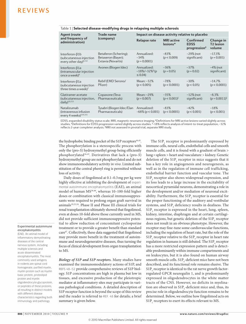
Table 1 | Selected disease-modifying drugs in relapsing multiple sclerosis
Agent (route and frequency of administration)
Trade name (company)
impact on disease activity relative to placebo
relapse rate Mri active lesions*
confirmed eDss progression‡
change in T2 lesion volume
Interferon‑β1b (subcutaneous injection every other day)8,152
Betaferon (Schering); Betaseron (Bayer); Extavia (Novartis)
Annualized: −34% (p = 0.0001)
–83% (p < 0.009)
−29% (not significant)
–17.3% (p = 0.001)
Interferon‑β1a (intramuscular injection once a week)9
Avonex (Biogen Idec) Annualized: –18%/–32%§ (p ≤ 0.04)
–36% (p = 0.05)
–37% (p = 0.02)
–4% (not significant)
Interferon‑β1a (subcutaneous injection three times a week)7
Rebif (EMD Serono/Pfizer)
Mean: –32% (p < 0.005)
−78% (p < 0.0001)
–30% (p < 0.05)
–14.7% (p < 0.0001)
Glatiramer acetate (subcutaneous injection, daily)10,153
Copaxone (Teva Pharmaceuticals)
Mean: –29% (p = 0.007)
−35% (p < 0.001)||
−12% (not significant)
−8.3% (p = 0.0011)||
Natalizumab (intravenous infusion every 4 weeks)154,155
Tysabri (Biogen Idec/Elan Pharmaceuticals)
Annualized: −68% (p < 0.001)
−83% (p < 0.0001)
−42% (p < 0.001)
−18% (p < 0.001)
EDSS, expanded disability status scale; MRI, magnetic resonance imaging.*Definitions for MRI active lesions varied slightly across studies. ‡Definitions for EDSS progression varied slightly across studies. §–18% reflects analysis of intent-to-treat population, –32% reflects 2‑year completer analysis. ||MRI not assessed in pivotal trial, separate MRI study.
R E V I E W S
886 | NovEMBER 2010 | voLUME 9 www.nature.com/reviews/drugdisc
© 20 Macmillan Publishers Limited. All rights reserved10
Nature Reviews | Drug Discovery
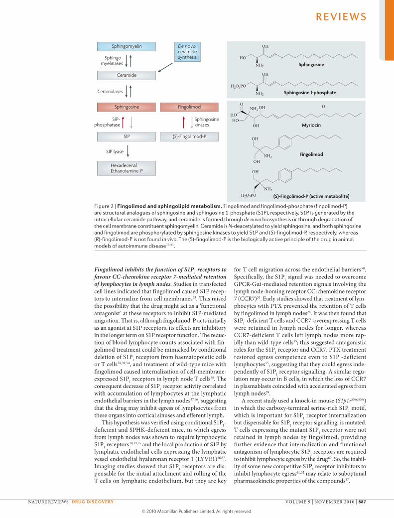
HO
NH2
OH
H2O3PO
NH2
OH
OH
OHNH2
OH
H2O3PONH2
Sphingosine
Sphingosine 1-phosphate
Myriocin
Fingolimod
(S)-Fingolimod-P (active metabolite)
Sphingomyelin De novo ceramidesynthesis
Ceramide
Sphingosine
Sphingosine kinases
S1P-phosphatase
Ceramidases
Sphingo-myelinases
S1P lyase
S1P
Fingolimod
(S)-Fingolimod-P
HexadecenalEthanolamine-P
OOH
OH
NH2
HO
O
HO
Fingolimod inhibits the function of S1P1 receptors to favour CC-chemokine receptor 7-mediated retention of lymphocytes in lymph nodes. Studies in transfected cell lines indicated that fingolimod caused S1P recep-tors to internalize from cell membranes53. This raised the possibility that the drug might act as a ‘functional antagonist’ at these receptors to inhibit S1P-mediated migration. That is, although fingolimod-P acts initially as an agonist at S1P receptors, its effects are inhibitory in the longer term on S1P receptor function. The reduc-tion of blood lymphocyte counts associated with fin-golimod treatment could be mimicked by conditional deletion of S1P1 receptors from haematopoietic cells or T cells38,39,54, and treatment of wild-type mice with fingolimod caused internalization of cell-membrane-expressed S1P1 receptors in lymph node T cells55. The consequent decrease of S1P1 receptor activity correlated with accumulation of lymphocytes at the lymphatic endothelial barriers in the lymph nodes37,56, suggesting that the drug may inhibit egress of lymphocytes from these organs into cortical sinuses and efferent lymph.
This hypothesis was verified using conditional S1P1-deficient and SPHK-deficient mice, in which egress from lymph nodes was shown to require lymphocytic S1P1 receptors38,39,55 and the local production of S1P by lymphatic endothelial cells expressing the lymphatic vessel endothelial hyaluronan receptor 1 (LYvE1)56,57. Imaging studies showed that S1P1 receptors are dis-pensable for the initial attachment and rolling of the T cells on lymphatic endothelium, but they are key
for T cell migration across the endothelial barriers58. Specifically, the S1P1 signal was needed to overcome GPCR-Gαi-mediated retention signals involving the lymph node-homing receptor CC-chemokine receptor 7 (CCR7)55. Early studies showed that treatment of lym-phocytes with PTX prevented the retention of T cells by fingolimod in lymph nodes28. It was then found that S1P1-deficient T cells and CCR7-overexpressing T cells were retained in lymph nodes for longer, whereas CCR7-deficient T cells left lymph nodes more rap-idly than wild-type cells55; this suggested antagonistic roles for the S1P1 receptor and CCR7. PTX treatment restored egress competence even to S1P1-deficient lymphocytes55, suggesting that they could egress inde-pendently of S1P1 receptor signalling. A similar regu-lation may occur in B cells, in which the loss of CCR7 in plasmablasts coincided with accelerated egress from lymph nodes59.
A recent study used a knock-in mouse (S1p1rS5A/S5A) in which the carboxy-terminal serine-rich S1P1 motif, which is important for S1P1 receptor internalization but dispensable for S1P1 receptor signalling, is mutated. T cells expressing the mutant S1P1 receptor were not retained in lymph nodes by fingolimod, providing further evidence that internalization and functional antagonism of lymphocytic S1P1 receptors are required to inhibit lymphocyte egress by the drug60. So, the inabil-ity of some new competitive S1P1 receptor inhibitors to inhibit lymphocyte egress61,62 may relate to suboptimal pharmacokinetic properties of the compounds57.
Figure 2 | Fingolimod and sphingolipid metabolism. Fingolimod and fingolimod‑phosphate (fingolimod‑P) are structural analogues of sphingosine and sphingosine 1‑phosphate (S1P), respectively. S1P is generated by the intracellular ceramide pathway, and ceramide is formed through de novo biosynthesis or through degradation of the cell membrane constituent sphingomyelin. Ceramide is N‑deacetylated to yield sphingosine, and both sphingosine and fingolimod are phosphorylated by sphingosine kinases to yield S1P and (S)‑fingolimod‑P, respectively, whereas (R)‑fingolimod‑P is not found in vivo. The (S)‑fingolimod‑P is the biologically active principle of the drug in animal models of autoimmune disease36,43.
R E V I E W S
NATURE REvIEWS | Drug Discovery voLUME 9 | NovEMBER 2010 | 887
© 20 Macmillan Publishers Limited. All rights reserved10
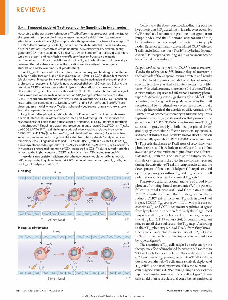
Nature Reviews | Drug Discovery
Tn TCM TEM
–
+
CCR7
S1P1
S1P
–
+Loss ofCCR7
HEVBlood
Efferent lymph
Lymphnode
LEC
a No drug
Tn TCM TEM
–CCR7
S1P
–
Loss ofCCR7
HEVBlood
Efferent lymph
Lymphnode
LEC
b Fingolimod treatment
Collectively, the above described findings support the hypothesis that S1P1 signalling in lymphocytes overrides CCR7-mediated retention to promote their egress from lymph nodes, and that functional antagonism of S1P1 by fingolimod favours lymphocyte retention in lymph nodes. Egress of terminally differentiated CCR7– effector T cells and effector memory T cells63 may be less depend-ent on S1P1 receptor signalling and, as a consequence, be less affected by fingolimod.
Fingolimod selectively retains CCR7+ central memory T cells: the relevance to MS. Immunological memory is the hallmark of the adaptive immune system and results from the clonal expansion and differentiation of antigen-specific lymphocytes that ultimately persist for a life-time63,64. In adult humans, more than 60% of blood T cells express antigen-experienced effector and memory pheno-types63,64. According to the ‘signal-strength model’ of T cell activation, the strength of the signals delivered by the T cell receptor and by co-stimulatory receptors drives T cells through hierarchical thresholds of differentiation63,64. Formation of protective memory in humans requires a high-intensity antigenic stimulation that promotes the generation of CCR7–CD45RA– effector memory T (TEM) cells that migrate readily to inflamed peripheral tissues and display immediate effector function. By contrast, antigenic stimuli of low intensity and/or short duration preferentially generate CCR7+CD45RA– central memory T (TCM) cells that home to T cell areas of secondary lym-phoid organs, and have little or no effector function but await antigenic restimulation to proliferate and differen-tiate into TEM cells63–71. The nature of the antigen, the co-stimulatory signals and the cytokine environment present during the activation of T cells in lymph nodes directs the development of functional T helper (TH), regulatory and cytolytic phenotypes within TCM and TEM cells, with full polarization achieved at the terminal TEM state63.
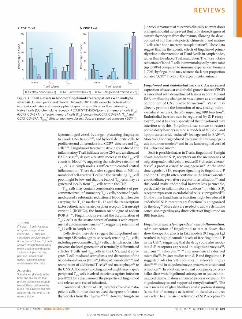
Phenotypic and functional analysis of blood lym-phocytes from fingolimod-treated mice72, from patients following renal transplant39 and from patients with MS31,73 provided evidence that the drug preferentially reduced CCR7+ naive T cells and TCM cells in blood, but it spared CCR7– TEM cells (BOX 1; FIG. 3), which is consist-ent with S1P1- and CCR7-dependent regulation of egress from lymph nodes. It is therefore likely that fingolimod may retain all TCM cell subsets in lymph nodes, irrespec-tive of TH1, TH2, TH17 cell or cytolytic commitment, but may spare all these subsets at the TEM stage. According to their TEM phenotype, blood T cells from fingolimod-treated patients secreted less interleukin-2 (IL-2) but more IFN-γ on a per cell basis following ex vivo restimulation by superantigens31.
The retention of TCM cells might be sufficient for the therapeutic effect of fingolimod, because in MS more than 90% of T cells that accumulate in the cerebrospinal fluid (CSF) express a TCM phenotype, and the T cell infiltrate does not contain naive T cells and is relatively depleted of TEM cells74. The clonal expansion of disease-relevant TCM cells may occur first in CNS-draining lymph nodes follow-ing low-intensity cross-reaction on self antigen75. These cells could then recirculate and could be restimulated at
Box 1 | Proposed model of T cell retention by fingolimod in lymph nodes
According to the signal strength model of T cell differentiation (see part a of the figure), the generation of protective immune responses requires high intensity antigenic stimulation of naive T cells (T
n) in lymph nodes; this generates CC-chemokine receptor 7
(CCR7)– effector memory T cells (TEM
), which recirculate to infected tissues and display effector function63. By contrast, antigenic stimuli of weaker intensity predominantly generate CCR7+ central memory T cells (T
CM), which home to T cell areas of secondary
lymphoid organs, and have little or no effector function but await antigenic restimulation to proliferate and differentiate into T
EM cells (the thickness of the wedges
between the cell subsets indicates the duration and intensity of the antigenic stimulation and the resulting T cell proliferation).
Tn and T
CM cells recirculate between blood and secondary lymphoid organs and home
to lymph nodes through high endothelial venules (HEVs) in a CCR7-dependent manner (black arrows). To egress from lymph nodes, they require activation of the sphingosine 1-phosphate receptor 1 (S1P
1) by lymphatic endothelial cell (LEC)-derived S1P, and this
overrides CCR7-mediated retention in lymph nodes55 (light grey arrows). Fully differentiated T
EM cells have irreversibly lost CCR7 (REF. 63) and related retention signals
and, as a consequence, are less dependent on S1P1 for egress55 (red arrows, see also
BOX 2). Accordingly, treatment with Pertussis toxin, which blocks CCR7–Gαi-signalling, restored egress competence to lymphocytes28,55 and to S1P
1-deficient T cells55. These
data suggest a model whereby T cells that have divided several times switch to a state favouring egress over retention31,55,73.
Fingolimod, after phosphorylation, binds to S1P1 receptors36,37 on T cells and causes
aberrant internalization of the receptor55 (see part b of the figure). This reduces the responsiveness of T cells to the egress signal S1P and favours CCR7-mediated retention in lymph nodes55. Fingolimod was shown to predominantly retain CD62LhiCD44low T
n cells
and CD62LhiCD44hi TCM
cells in lymph nodes of mice, causing a relative increase in CD62LlowCD44hiIFN-γ (interferon-γ)hi T
EM cells in blood72 (not shown). A similar subset
selectivity was observed in fingolimod-treated transplant patients39 and patients with multiple sclerosis: fingolimod retained CCR7+CD45RA+ T
n cells and CCR7+CD45RA– T
CM
cells in lymph nodes, but spared CCR7–CD45RA– and CCR7–CD45RA+ TEM
cell subsets31,73. In humans, a preferential retention of CD4+ compared to CD8+ T cells occurred31, and this related to the higher content of CCR7+ naive cells in the CD4+ compartment31,63.
These data are consistent with a model whereby down-modulation of lymphocytic S1P
1 receptors by fingolimod favours CCR7-mediated retention of T
n and T
CM cells, but
not TEM
cells, in lymph nodes31,55.
R E V I E W S
888 | NovEMBER 2010 | voLUME 9 www.nature.com/reviews/drugdisc
© 20 Macmillan Publishers Limited. All rights reserved10
Nature Reviews | Drug Discovery
Perc
enta
ge in
blo
od
100
80
60
40
20
0Naive
T cell subsetTCM TEM
a CD4+ T cell
Perc
enta
ge in
blo
od
100
80
60
40
20
0Naive
T cell subsetTCM TEM
b CD8+ T cell
Healthy donors (n = 7) MS – untreated (n = 3) MS – fingolimod treated (n = 5)
TH17 cell(T helper 17 cell). A subset of TH cells that produce interleukin‑17. They are considered developmentally distinct from TH1 and TH2 cells and are thought to have a key role in autoimmune diseases such as multiple sclerosis, psoriasis, autoimmune uveitis, juvenile diabetes and rheumatoid arthritis.
AstrocytesStar‑shaped glial cells in the brain and spinal cord that provide biochemical support to endothelial cells from the blood–brain barrier and that also provide nutrients to the nervous tissue.
leptomeningeal vessels by antigen-presenting phagocytes, to invade CNS tissues76,77, and by local dendritic cells, to proliferate and differentiate into CCR7– effectors and TEM cells74,78. Fingolimod treatment strikingly reduced the inflammatory T cell infiltrate in the CNS and ameliorated EAE disease79, despite a relative increase in the TEM cell counts in blood31,72, suggesting that selective retention of TCM cells in lymph nodes is sufficient to control central inflammation. These data also suggest that, in MS, the number of self-reactive T cells in the circulating TEM cell pool might be low and that the bulk of TEM cells may be generated locally from TCM cells within the CNS.
TCM cells may contain considerable numbers of pre-committed pro-inflammatory TH17 cells, because fingoli-mod caused a substantial reduction of blood lymphocytes carrying the TH17 marker IL-17 and the transcription factor retinoic acid-related orphan receptor C transcript variant 2 (RoRC2), the human orthologue of rodent RoRγt73,80. Fingolimod prevented the accumulation of TH17 cells in the sciatic nerves of animals with experi-mental autoimmune neuritis81,82, suggesting retention of TH17 cells in lymph nodes.
Collectively, these data suggest that fingolimod may interrupt MS pathology by selectively retaining TCM cells, including pre-committed TH17 cells, in lymph nodes. This prevents the local generation of terminally differentiated effector T cells and TEM cells in the CNS, and it abro-gates T cell-mediated astrogliosis and disruption of the blood–brain barrier (BBB)83, killing of neural cells84,85 and recruitment of additional T cells84 and macrophages86 to the CNS. At the same time, fingolimod might largely spare peripheral TEM cells involved in defence against infection (see BOX 2 for a discussion of the properties of fingolimod and relevance to risk of infection).
Conditional deletion of S1P1 receptors from haemato-poietic cells in mice also reduced the egress of mature thymocytes from the thymus38,54,87. However, long-term
(16 week) treatment of mice with clinically relevant doses of fingolimod did not prevent (but only slowed) egress of mature thymocytes from the thymus, allowing the devel-opment of full haematopoietic chimerism and mature T cells after bone marrow transplantation72. These data suggest that the therapeutic effects of fingolimod prima-rily relate to the retention of T and B cells in lymph nodes, rather than to reduced T cell maturation. The more notable reduction of blood T cells in immunologically naive mice (up to 98%) compared to immune-experienced humans (~70%) by fingolimod may relate to the larger proportion of naive CCR7+ T cells in the experimental animals.
Fingolimod and endothelial barriers. An increased expression of vascular endothelial growth factor (vEGF) is associated with demyelinated lesions in both MS and EAE, implicating changes in vasculature as a potential component of CNS plaque formation77. vEGF may directly promote the formation of new (leaky) micro-vascular structures, thereby impairing BBB function88. Endothelial barriers can be regulated by S1P recep-tors89,90, and it has been speculated that fingolimod may interfere with this. Fingolimod was shown to restore permeability barriers in mouse models of vEGF-91 and lipopolysaccharide-induced92 leakage and in EAE93,94. Moreover, the drug reduced excessive de novo angiogen-esis in tumour models95 and in the lumbar spinal cord of EAE-diseased mice96.
So, it is possible that, as in T cells, fingolimod-P might down-modulate S1P1 receptors on the membranes of migrating endothelial cells to reduce S1P-directed chemo-taxis97, a process crucial to angiogenesis98. At the same time, agonistic S1P1 receptor signalling by fingolimod-P and/or S1P might often continue in the intact vascular endothelium, even after receptor internalization99, and this could make endothelial barriers less permeable, particularly in inflammatory situations91 in which S1P1 receptor expression in endothelial cells is upregulated100. on the other hand, barrier function might be reduced if endothelial S1P1 receptors are functionally antagonized by the drug90. More data are needed to allow a definitive conclusion regarding any direct effects of fingolimod on BBB function.
Fingolimod and S1P-dependent neuroinflammation. Administration of fingolimod to rats at doses that show therapeutic effects in EAE models (0.3 mg per kg) resulted in high picomolar levels of free fingolimod-P in the CSF46, suggesting that the drug could also modu-late S1P receptors expressed in oligodendrocytes101, neurons102, astrocytes103,104 and non-neurally derived microglia102. In vitro studies with S1P and fingolimod-P suggested roles for S1P receptors in astrocyte migra-tion105–107 and in oligodendrocyte process extension and retraction108. In addition, treatment of organotypic cere-bellar slices with fingolimod subsequent to lysolecithin-induced demyelination enhanced process extension by oligodendrocytes and supported remyelination109. The early increase of glial fibrillary acidic protein staining (a marker of astrocyte activation) in the slice cultures may relate to a transient activation of S1P receptors by
Figure 3 | T cell subsets in blood of fingolimod-treated patients with multiple sclerosis. Human peripheral blood CD4+ and CD8+ T cells were characterized for expression of naive and memory phenotypes using multicolour flow cytometry. Naive T cells (CC‑chemokine receptor 7 (CCR7)+CD45RA+); central memory T cells (T
CM)
(CCR7+CD45RA–); effector memory T cells (TEM
) (containing CCR7–CD45RA– ‘TEM
’ and CCR7–CD45RA+ ‘T
EMRA’ effector memory subsets). Data are presented as means ± SD31,73.
R E V I E W S
NATURE REvIEWS | Drug Discovery voLUME 9 | NovEMBER 2010 | 889
© 20 Macmillan Publishers Limited. All rights reserved10

Nature Reviews | Drug Discovery
Rela
tive
expr
essio
n
High
LowNaive
CCR7+
CD45RA+
Retained by fingolimod Spared by fingolimodCCR7+
CD45RA–CCR7–
CD45RA–CCR7–
CD45RA+
TCM TEM
IFN-γPerforinCD45RACD62L
IL-2CD28CCR7
HCVInfluenza
EBVHIV
CMV
Distribution of virus-specific T cells
the drug, before receptor internalization and functional antagonism and suppression of astrogliosis (see below). Together, these data suggest the possibility that fingolimod may directly affect neural cells.
Several in vivo studies support a key role of S1P and S1P1 receptors in the development of the nervous system. S1P1 receptors are involved in the migration of neural stem cells and progenitor cells102, and genetic deletion of either the S1P1 receptor or SPHKs (to eliminate S1P) caused severe disturbance of neurogenesis and neural tube closure, resulting in increased apoptosis in the devel-oping brain110, which suggest cell-protective, pro-survival effects of S1P. In disease, however, excessive S1P produc-tion and cellular survival may sustain inflammation: var-ious inflammatory cytokines including IL-1 and tumour necrosis factor activate SPHKs111, and activation of the SPHK1–S1P pathway in microglia increased expression of pro-inflammatory cytokines and production of nitric oxide112. In patients with MS, increasing disability cor-related with higher S1P levels in the CSF113. Likewise, S1P levels were increased in the spinal cord of mice after injury102 and during EAE-related inflammation114; injec-tion of S1P into the striata caused astrogliosis and central inflammation104, perturbing gap junctional communica-tion of astrocytes with other neural cells115.
It is, therefore, plausible to assume that these patho-genic mechanisms are influenced beneficially by down-modulation of S1P responses. Recent data showed that IL-17 directly activates astrocytes116 and that reactive astrocytes in active and chronic inactive MS lesions show dramatically increased expression of S1P1 and S1P3 receptors117. The S1P1 receptor55 has been shown to be internalized by fingolimod, and mice with conditional deletion of S1P1 in neural cells, particularly astrocytes, showed reduced EAE disease severity and attenuated astrogliosis, demyelination and axonal damage com-pared with wild-type counterparts114. Furthermore, genetic deletion of either SPHK1 (to reduce S1P levels) or the S1P3 receptor in mice reduced astrogliosis and improved motor function during the terminal stages of neuronopathic Sandhoff disease103. These data provide evidence to suggest that functional antagonism of S1P receptors in the CNS might reduce central inflammation in neurodegenerative disorders, including MS (BOX 3). The therapeutic effect might, at least in part, relate to a restoration of effective gap-junctional communication of astrocytes with other neural cells and endothelial cells in the BBB, as gap junction channels connect the cytoplasm of contacting cells and coordinate electric and metabolic activity115. Together, these processes could support repair mechanisms and remyelination and explain the thera-peutic effects of fingolimod in animal models of MS.
Fingolimod in animal models of MS. Protective effects of fingolimod in animal models of MS were first described in 2002; in a Wistar rat model, administration of the drug at 0.3 mg per kg per day prevented the onset of EAE36. Protection required the conversion of fingolimod to its phosphate metabolite36, which acted at S1P receptors on lymphocytes36,38. The protective effect correlated with reduced blood lymphocyte counts36, which is consistent with the retention of T cells in lymph nodes38,56,115.
Follow-up studies confirmed prophylactic and thera-peutic activities of fingolimod in other rat and mouse EAE models79,118,119. Protection was associated with a reduction
Box 2 | Sparing of CCR7– effector memory T cells by fingolimod
Current data suggest that, in multiple sclerosis (MS), peripheral central memory T cells (T
CM) may contain the bulk of the autoreactive T cells, which invade the central nervous
system31,74,80. By contrast, the frequency of MS-pathogenic T cells within the peripheral effector memory T cell (T
EM) pool may be low31,74, but T
EM cells are pivotal in the defence
against infection63. The figure shows phenotypic and functional associations within virus-specific CD8+ T
CM cells and T
EM cells in humans, indicating the expression and fate
of the co-stimulatory receptor CD28, the lymph node homing receptors CC-chemokine receptor 7 (CCR7) and CD62L (also known as L-selectin), the effector molecules perforin and interferon-γ (IFN-γ), and the growth factor interleukin-2 (IL-2) in a resting state of the T cell subsets146. The relative induction of virus-specific T
CM versus T
EM subsets is
indicated after clearance of the influenza virus or during latent infection with hepatitis C virus (HCV), Epstein–Barr virus (EBV), human immunodeficiency virus (HIV), and cytomegalovirus (CMV).
Studies in models of lymphocytic choriomeningitis virus and vesicular stomatitis virus infection did not indicate suppressive effects of fingolimod on antigen presentation or on T cell and B cell activation, proliferation, differentiation and effector function in vivo40. Current data from humans and animals suggest that CCR7– T
EM cells generated
in the lymph nodes in response to infection or vaccination would recirculate largely independently of sphingosine 1-phosphate receptor 1 (S1P
1) (BOX 1) and, thus, may not
be affected by fingolimod. Indeed, a recently completed open-label, observational, prospective clinical study showed that fingolimod-treated individuals mount an influenza virus vaccine-specific humoral and cellular immune response that is comparable to drug-untreated controls147.
In mice, incomplete retention of antigen-activated T cells by fingolimod has also been observed after primary infection with Listeria monocytogenes 148 and after adoptive transfer of ex vivo primed T cells149. In L. monocytogenes‑148 and Mycobacterium bovis150-immune mice, cellular and humoral recall immunity was not impaired by the drug, allowing clearance of the infection.
Collectively, these data might provide a rationale for why the overall incidence of infection observed in clinical trials was similar between the fingolimod-treated patients and control groups125,127, despite a reduced blood lymphocyte count (see main text). However, long-term follow up of fingolimod-treated patients is necessary to substantiate this possibility further.
Figure adapted, with permission, from REF. 146 © (2008) Wiley.
R E V I E W S
890 | NovEMBER 2010 | voLUME 9 www.nature.com/reviews/drugdisc
© 20 Macmillan Publishers Limited. All rights reserved10
TE
Nature Reviews | Drug Discovery
–
+
CCR7
S1P1
S1P1
S1P1 S1P1S1P3 S1P5
S1P
TCM TCM
Down-modulation of S1P receptors by fingolimod
Dendritic cell
Oligo-dendrocyte
Astrocyte
TEM
SPHK↑
S1P↑
Lymph node
Blood
Brain
Inflammatory cytokines
IL-17receptor
IL-17
in the numbers of T cells and macrophages in spinal cords, and this correlated with a reduction of mRNA encoding the inflammatory markers IFN-γ, IL-2, IL-6 and gran-ulocyte–macrophage colony-stimulating factor79,118,119. Treatment with fingolimod started at the peak of the first relapse was more efficacious than IFN-β therapy119, an observation that was also seen later in a clinical trial (see below).
In a dark agouti rat model, late-stage rescue treatment started 4 weeks after disease onset reversed paralysis in established EAE and normalized the electrophysiological responses, including visual and somatosensory evoked potentials, and this correlated with decreased demyelina-tion in the brain and spinal cord93,94. Furthermore, BBB breakdown was reversed, as measured by immuno-globulin precipitation93, and this was associated with a reduction in the levels of mRNA encoding the matrix metalloproteinase 9 gene (Mmp9) and an increase in the mRNA levels of its counter-regulator, tissue inhibitor of metalloproteinase 1 (Timp1), resulting in a proteolytic
balance that favours the preservation of BBB integrity93. Collectively, these data suggest that fingolimod might also have therapeutic value beyond relapsing MS in the more chronic forms of the disease, and analysis in the respective models is ongoing.
Maximal efficacy of the drug was achieved with a dose of 0.3 mg per kg, resulting in fingolimod levels of 5.4 ng per ml in plasma and 0.07 ng per ml in CSF, and fingolimod-P levels of 7.4 ng per ml in plasma and 0.23 ng per ml in the CSF46. These data provided guid-ance towards the dose selection in humans, in which a similar blood exposure was achieved with a dosing of 1.25–0.5 mg fingolimod per patient per day.
Clinical studies of fingolimodClinical pharmacology. The clinical pharmacology of fin-golimod has been investigated in more than 1,000 subjects in 30 studies. Fingolimod has a slow absorption period, with the time of maximal concentration at 12–24 hours post dose120–122, when comparable concentrations of
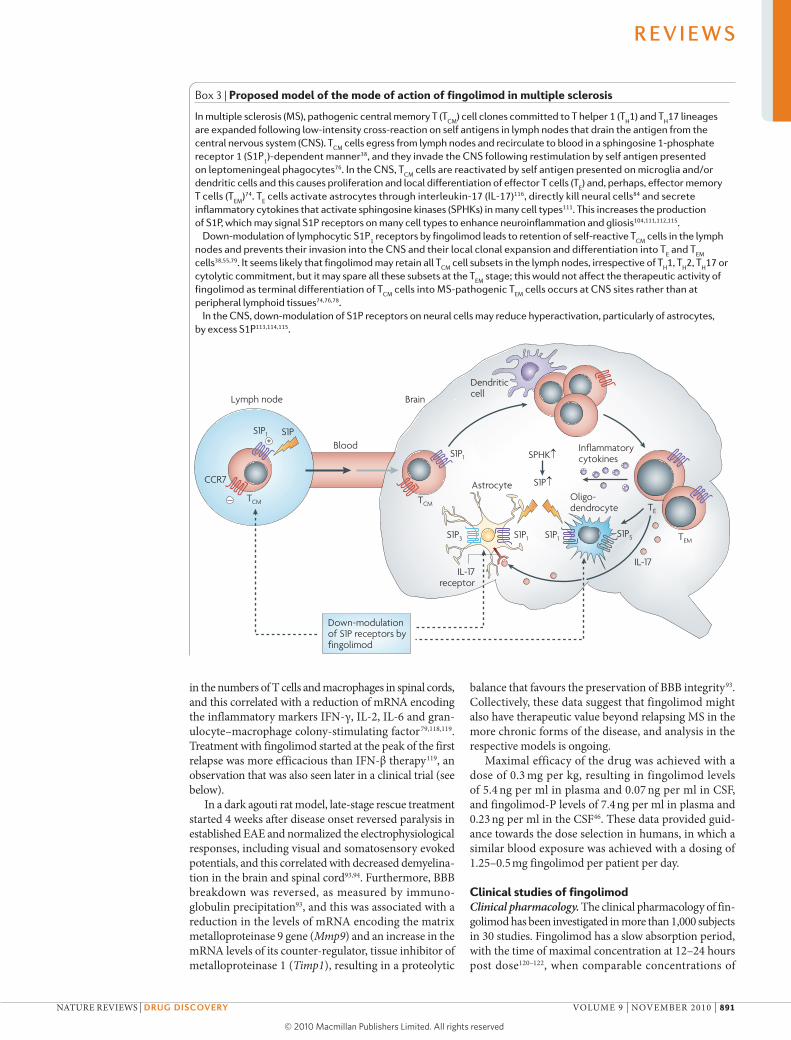
Box 3 | Proposed model of the mode of action of fingolimod in multiple sclerosis
In multiple sclerosis (MS), pathogenic central memory T (TCM
) cell clones committed to T helper 1 (TH1) and T
H17 lineages
are expanded following low-intensity cross-reaction on self antigens in lymph nodes that drain the antigen from the central nervous system (CNS). T
CM cells egress from lymph nodes and recirculate to blood in a sphingosine 1-phosphate
receptor 1 (S1P1)-dependent manner38, and they invade the CNS following restimulation by self antigen presented
on leptomeningeal phagocytes76. In the CNS, TCM
cells are reactivated by self antigen presented on microglia and/or dendritic cells and this causes proliferation and local differentiation of effector T cells (T
E) and, perhaps, effector memory
T cells (TEM
)74. TE cells activate astrocytes through interleukin-17 (IL-17)116, directly kill neural cells84 and secrete
inflammatory cytokines that activate sphingosine kinases (SPHKs) in many cell types111. This increases the production of S1P, which may signal S1P receptors on many cell types to enhance neuroinflammation and gliosis104,111,112,115.
Down-modulation of lymphocytic S1P1 receptors by fingolimod leads to retention of self-reactive T
CM cells in the lymph
nodes and prevents their invasion into the CNS and their local clonal expansion and differentiation into TE and T
EM
cells38,55,79. It seems likely that fingolimod may retain all TCM
cell subsets in the lymph nodes, irrespective of TH1, T
H2, T
H17 or
cytolytic commitment, but it may spare all these subsets at the TEM
stage; this would not affect the therapeutic activity of fingolimod as terminal differentiation of T
CM cells into MS-pathogenic T
EM cells occurs at CNS sites rather than at
peripheral lymphoid tissues74,76,78.In the CNS, down-modulation of S1P receptors on neural cells may reduce hyperactivation, particularly of astrocytes,
by excess S1P113,114,115.
R E V I E W S
NATURE REvIEWS | Drug Discovery voLUME 9 | NovEMBER 2010 | 891
© 20 Macmillan Publishers Limited. All rights reserved10
Gadolinium-enhanced lesionsGadolinium is used as a contrast agent in magnetic resonance imaging (MRI). In multiple sclerosis, gadolinium causes areas of inflammation to be more pronounced than other areas of the brain. This can be seen on the MRI results and indicates where the disease is active.
fingolimod and fingolimod-P are detectable in the blood120. owing to a high volume of distribution, fingoli-mod has a half-life of ~9–10 days123. With daily dosing of fingolimod, pharmacokinetic steady state is achieved after 1–2 months. The drug is cleared by a metabolic pathway that predominantly utilizes cytochrome P450 4F124. Given that this oxidative enzyme system is not known to contribute to the metabolism of any other drugs, there seems to be a low potential of drug–drug interactions.
Pharmacodynamic studies show that the drug has a rapid onset. Within hours of the first dose of fingolimod, a dose-dependent decrease in the peripheral lymphocyte count is apparent. With continued daily dosing, both a stable blood concentration and a stable reduction in the number of circulating blood lymphocytes are observed124, with an average reduction of 77% with the 1.25 mg dose and 73% with the 0.5 mg dose; cell counts remained stable for the entire treatment period125–127. An increase of the peripheral blood lymphocyte count was evident within days of stopping fingolimod treatment, and it typically returned to normal range within 6 weeks128.
Therapeutic effects in clinical studies. The first clinical evidence that fingolimod has therapeutic benefits in MS was provided in a 6-month, placebo-controlled Phase II trial involving 281 patients with relapsing MS129. Patients
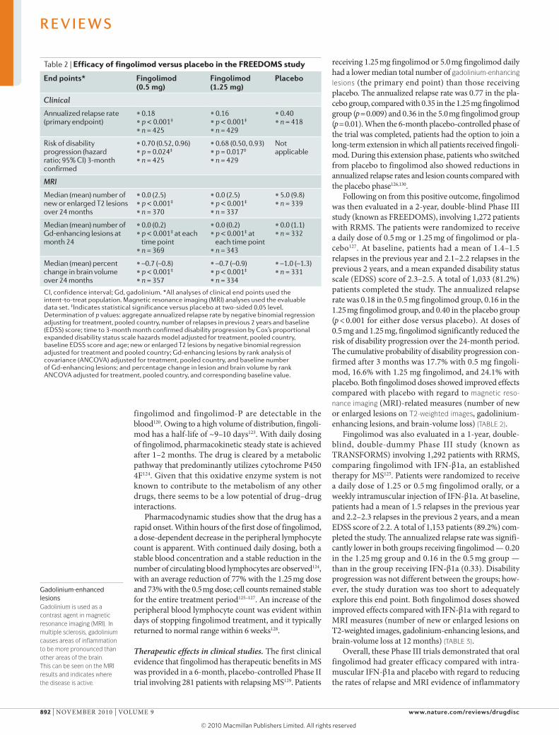
receiving 1.25 mg fingolimod or 5.0 mg fingolimod daily had a lower median total number of gadolinium‑enhancing lesions (the primary end point) than those receiving placebo. The annualized relapse rate was 0.77 in the pla-cebo group, compared with 0.35 in the 1.25 mg fingolimod group (p = 0.009) and 0.36 in the 5.0 mg fingolimod group (p = 0.01). When the 6-month placebo-controlled phase of the trial was completed, patients had the option to join a long-term extension in which all patients received fingoli-mod. During this extension phase, patients who switched from placebo to fingolimod also showed reductions in annualized relapse rates and lesion counts compared with the placebo phase126,130.
Following on from this positive outcome, fingolimod was then evaluated in a 2-year, double-blind Phase III study (known as FREEDoMS), involving 1,272 patients with RRMS. The patients were randomized to receive a daily dose of 0.5 mg or 1.25 mg of fingolimod or pla-cebo127. At baseline, patients had a mean of 1.4–1.5 relapses in the previous year and 2.1–2.2 relapses in the previous 2 years, and a mean expanded disability status scale (EDSS) score of 2.3–2.5. A total of 1,033 (81.2%) patients completed the study. The annualized relapse rate was 0.18 in the 0.5 mg fingolimod group, 0.16 in the 1.25 mg fingolimod group, and 0.40 in the placebo group (p < 0.001 for either dose versus placebo). At doses of 0.5 mg and 1.25 mg, fingolimod significantly reduced the risk of disability progression over the 24-month period. The cumulative probability of disability progression con-firmed after 3 months was 17.7% with 0.5 mg fingoli-mod, 16.6% with 1.25 mg fingolimod, and 24.1% with placebo. Both fingolimod doses showed improved effects compared with placebo with regard to magnetic reso‑nance imaging (MRI)-related measures (number of new or enlarged lesions on T2‑weighted images, gadolinium-enhancing lesions, and brain-volume loss) (TABLE 2).
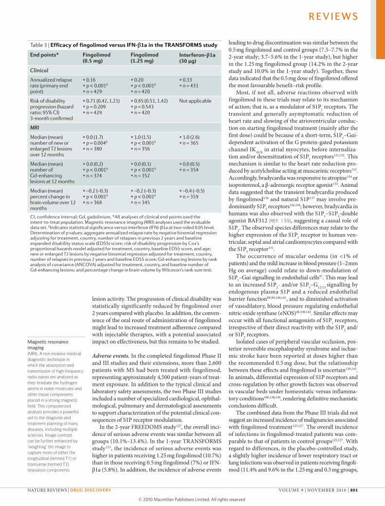
Fingolimod was also evaluated in a 1-year, double-blind, double-dummy Phase III study (known as TRANSFoRMS) involving 1,292 patients with RRMS, comparing fingolimod with IFN-β1a, an established therapy for MS125. Patients were randomized to receive a daily dose of 1.25 or 0.5 mg fingolimod orally, or a weekly intramuscular injection of IFN-β1a. At baseline, patients had a mean of 1.5 relapses in the previous year and 2.2–2.3 relapses in the previous 2 years, and a mean EDSS score of 2.2. A total of 1,153 patients (89.2%) com-pleted the study. The annualized relapse rate was signifi-cantly lower in both groups receiving fingolimod — 0.20 in the 1.25 mg group and 0.16 in the 0.5 mg group — than in the group receiving IFN-β1a (0.33). Disability progression was not different between the groups; how-ever, the study duration was too short to adequately explore this end point. Both fingolimod doses showed improved effects compared with IFN-β1a with regard to MRI measures (number of new or enlarged lesions on T2-weighted images, gadolinium-enhancing lesions, and brain-volume loss at 12 months) (TABLE 3).
overall, these Phase III trials demonstrated that oral fingolimod had greater efficacy compared with intra-muscular IFN-β1a and placebo with regard to reducing the rates of relapse and MRI evidence of inflammatory
Table 2 | Efficacy of fingolimod versus placebo in the FREEDOMS study
end points* Fingolimod (0.5 mg)
Fingolimod (1.25 mg)
Placebo
Clinical
Annualized relapse rate (primary endpoint)
• 0.18 • p < 0.001‡
• n = 425
• 0.16 • p < 0.001‡
• n = 429
• 0.40 • n = 418
Risk of disability progression (hazard ratio; 95% CI) 3-month confirmed
• 0.70 (0.52, 0.96) • p = 0.024‡ • n = 425
• 0.68 (0.50, 0.93) • p = 0.017‡
• n = 429
Not applicable
MRI
Median (mean) number of new or enlarged T2 lesions over 24 months
• 0.0 (2.5) • p < 0.001‡
• n = 370
• 0.0 (2.5) • p < 0.001‡
• n = 337
• 5.0 (9.8) • n = 339
Median (mean) number of Gd‑enhancing lesions at month 24
• 0.0 (0.2) • p < 0.001‡ at each
time point • n = 369
• 0.0 (0.2) • p < 0.001‡ at
each time point • n = 343
• 0.0 (1.1) • n = 332
Median (mean) percent change in brain volume over 24 months
• –0.7 (–0.8) • p < 0.001‡
• n = 357
• –0.7 (–0.9) • p < 0.001‡
• n = 334
• –1.0 (–1.3) • n = 331
CI, confidence interval; Gd, gadolinium. *All analyses of clinical end points used the intent‑to‑treat population. Magnetic resonance imaging (MRI) analyses used the evaluable data set. ‡Indicates statistical significance versus placebo at two‑sided 0.05 level. Determination of p values: aggregate annualized relapse rate by negative binomial regression adjusting for treatment, pooled country, number of relapses in previous 2 years and baseline (EDSS) score; time to 3‑month month confirmed disability progression by Cox’s proportional expanded disability status scale hazards model adjusted for treatment, pooled country, baseline EDSS score and age; new or enlarged T2 lesions by negative binomial regression adjusted for treatment and pooled country; Gd‑enhancing lesions by rank analysis of covariance (ANCOVA) adjusted for treatment, pooled country, and baseline number of Gd‑enhancing lesions; and percentage change in lesion and brain volume by rank ANCOVA adjusted for treatment, pooled country, and corresponding baseline value.
R E V I E W S
892 | NovEMBER 2010 | voLUME 9 www.nature.com/reviews/drugdisc
© 20 Macmillan Publishers Limited. All rights reserved10
Magnetic resonance imaging(MRI). A non‑invasive medical diagnostic technique in which the absorption and transmission of high‑frequency radio waves are analysed as they irradiate the hydrogen atoms in water molecules and other tissue components placed in a strong magnetic field. This computerized analysis provides a powerful aid to the diagnosis and treatment planning of many diseases, including multiple sclerosis. Image contrast can be further enhanced by ‘weighting’ the image to capture more of either the longitudinal (termed T1) or transverse (termed T2) relaxation components.
lesion activity. The progression of clinical disability was statistically significantly reduced by fingolimod over 2 years compared with placebo. In addition, the conven-ience of the oral route of administration of fingolimod might lead to increased treatment adherence compared with injectable therapies, with a potential associated impact on effectiveness, but this remains to be studied.
Adverse events. In the completed fingolimod Phase II and III studies and their extensions, more than 2,600 patients with MS had been treated with fingolimod, representing approximately 4,500 patient-years of treat-ment exposure. In addition to the typical clinical and laboratory safety assessments, the two Phase III studies included a number of specialized cardiological, ophthal-mological, pulmonary and dermatological assessments to support characterization of the potential clinical con-sequences of S1P receptor modulation.
In the 2-year FREEDoMS study127, the overall inci-dence of serious adverse events was similar between all groups (10.1%–13.4%). In the 1-year TRANSFoRMS study125, the incidence of serious adverse events was higher in patients receiving 1.25 mg fingolimod (10.7%) than in those receiving 0.5 mg fingolimod (7%) or IFN-β1a (5.8%). In addition, the incidence of adverse events
leading to drug discontinuation was similar between the 0.5 mg fingolimod and control groups (7.5–7.7% in the 2-year study; 3.7–5.6% in the 1-year study), but higher in the 1.25 mg fingolimod group (14.2% in the 2-year study and 10.0% in the 1-year study). Together, these data indicated that the 0.5 mg dose of fingolimod offered the most favourable benefit–risk profile.
Most, if not all, adverse reactions observed with fingolimod in these trials may relate to its mechanism of action; that is, as a modulator of S1P1 receptors. The transient and generally asymptomatic reduction of heart rate and slowing of the atrioventricular conduc-tion on starting fingolimod treatment (mainly after the first dose) could be because of a short-term, S1P1–Gαi-dependent activation of the G protein-gated potassium channel IKACh in atrial myocytes, before internaliza-tion and/or desensitization of S1P1 receptors131,132. This mechanism is similar to the heart rate reduction pro-duced by acetylcholine acting at muscarinic receptors133. Accordingly, bradycardia was responsive to atropine134 or isoproterenol, a β-adrenergic receptor agonist135. Animal data suggested that the transient bradycardia produced by fingolimod136 and natural S1P137 may involve pre-dominantly S1P3 receptors136,138; however, bradycardia in humans was also observed with the S1P1–S1P5-double agonist BAF312 (REF. 139), suggesting a causal role of S1P1. The observed species differences may relate to the higher expression of the S1P1 receptor in human ven-tricular, septal and atrial cardiomyocytes compared with the S1P3 receptor131.
The occurrence of macular oedema (in <1% of patients) and the mild increase in blood pressure (1–2 mm Hg on average) could relate to down-modulation of S1P1–Gαi signalling in endothelial cells97. This may lead to an increased S1P2- and/or S1P3–G12/13 signalling by endogenous plasma S1P and a reduced endothelial barrier function89,90,140,141, and to diminished activation of vasodilatory, blood pressure regulating endothelial nitric oxide synthase (eNoS)48,100,142. Similar effects may occur with all functional antagonists of S1P1 receptors, irrespective of their direct reactivity with the S1P2 and/or S1P3 receptors.
Isolated cases of peripheral vascular occlusion, pos-terior reversible encephalopathy syndrome and ischae-mic stroke have been reported at doses higher than the recommended 0.5 mg dose, but the relationship between these effects and fingolimod is uncertain129,143. In animals, differential expression of S1P receptors and cross-regulation by other growth factors was observed in vascular beds under homeostatic versus inflamma-tory conditions100,138,140, rendering definitive mechanistic conclusions difficult.
The combined data from the Phase III trials did not suggest an increased incidence of malignancies associated with fingolimod treatment125,127. The overall incidence of infections in fingolimod-treated patients was com-parable to that of patients in control groups125,127. With regard to differences, in the placebo-controlled study, a slightly higher incidence of lower respiratory tract or lung infections was observed in patients receiving fingoli-mod (11.4% and 9.6% in the 1.25 mg and 0.5 mg groups,
Table 3 | Efficacy of fingolimod versus IFN-β1a in the TRANSFORMS study
end points* Fingolimod (0.5 mg)
Fingolimod (1.25 mg)
interferon-β1a (30 μg)
Clinical
Annualized relapse rate (primary end point)
• 0.16 • p < 0.001‡
• n = 429
• 0.20 • p < 0.001‡
• n = 420
• 0.33 • n = 431
Risk of disability progression (hazard ratio; 95% CI) 3‑month confirmed
• 0.71 (0.42, 1.21) • p = 0.209 • n = 429
• 0.85 (0.51, 1.42) • p = 0.543 • n = 420
Not applicable
MRI
Median (mean) number of new or enlarged T2 lesions over 12 months
• 0.0 (1.7) • p = 0.004‡
• n = 380
• 1.0 (1.5) • p < 0.001‡
• n = 356
• 1.0 (2.6) • n = 365
Median (mean) number of Gd‑enhancing lesions at 12 months
• 0.0 (0.2) • p < 0.001‡
• n = 374
• 0.0 (0.1) • p < 0.001‡ • n = 352
• 0.0 (0.5) • n = 354
Median (mean) percent change in brain volume over 12 months
• –0.2 (–0.3) • p < 0.001‡
• n = 368
• –0.2 (–0.3) • p < 0.001‡
• n = 345
• –0.4 (–0.5) • n = 359
CI, confidence interval; Gd, gadolinium. *All analyses of clinical end points used the intent‑to‑treat population. Magnetic resonance imaging (MRI) analyses used the evaluable data set. ‡Indicates statistical significance versus interferon (IFN)‑β1a at two‑sided 0.05 level. Determination of p values: aggregate annualized relapse rate by negative binomial regression adjusting for treatment, country, number of relapses in previous 2 years and baseline expanded disability status scale (EDSS) score; risk of disability progression by Cox’s proportional hazards model adjusted for treatment, country, baseline EDSS score, and age; new or enlarged T2 lesions by negative binomial regression adjusted for treatment, country, number of relapses in previous 2 years and baseline EDSS score; Gd‑enhancing lesions by rank analysis of covariance (ANCOVA) adjusted for treatment, country, and baseline number of Gd‑enhancing lesions; and percentage change in brain volume by Wilcoxon’s rank sum test.
R E V I E W S
NATURE REvIEWS | Drug Discovery voLUME 9 | NovEMBER 2010 | 893
© 20 Macmillan Publishers Limited. All rights reserved10
T2-weighted imagesThe areas of abnormality on transverse (T2)‑weighted magnetic resonance imaging (MRI) scans in patients with multiple sclerosis are pathologically nonspecific as they may represent areas of oedema, inflammation, demyelination, gliosis or tissue destruction. Such lesions are usually permanent, although they may decrease in size as acute lesions recover. Counting the number of new, or enlarging, T2 lesions over a period of time is an integral measure of MRI‑detected disease activity over that time period.
respectively) compared with placebo (6.0%), whereas the incidence of urinary tract infections in patients receiving fingolimod (4.9% and 8.0% in the 1.25 mg and 0.5 mg groups, respectively) was lower than in those receiving placebo (11.2%)127. In the trial comparing fingolimod to IFN-β1a, two lethal herpes infections (one primary varicella zoster infection and one case of herpes simplex encephalitis) occurred in patients receiving 1.25 mg fingolimod125. These might have involved confounding factors related to the use of high-dose steroids to treat relapses in these patients. The primary varicella zoster infection occurred in a woman who was previously her-pes zoster antibody negative, and who was exposed to a child with chickenpox during a course of steroids admin-istered for a relapse, and the herpes simplex encephalitis occurred in a patient who received a course of steroids to treat a suspected relapse (see REF. 125 for details).
The mechanism behind the observed increase in liver enzymes125,127 remains elusive; so far, there are no data from animal experiments indicating a direct effect on hepatocytes, vascular or biliary structures using doses that are several times higher than doses given to patients with MS144. Furthermore, fingolimod reportedly amelio-rated the microcirculatory, biochemical and histological manifestations of hepatic ischaemia–reperfusion injury in rodents145.
To date, the benefit–risk profile of fingolimod in MS has been characterized within the clinical programme for up to 2 years in controlled studies and up to 5 years in a long-term extension study. Fingolimod was shown to be effective and well tolerated, with a more favourable safety profile for the 0.5 mg dose than the 1.25 mg dose; identified drug-related adverse effects were generally reversible after drug discontinuation. Thorough obser-vation and long-term follow up will continue to generate a more informed assessment of the benefits and risks of this new treatment option for relapsing MS.
Future development of fingolimodIn established EAE, fingolimod was effective as a res-cue therapy, reversing paralysis, electrophysiological responses and BBB function93,94. These data suggest that the drug may find application beyond RRMS in primary
progressive MS (PPMS) and more chronic forms of the disease. A Phase III study in patients with PPMS has now been initiated, to evaluate the efficacy and safety of the drug for up to 3–5 years, and the primary outcome is time to disability progression.
IL-17 secreted by TH17 cells has been shown to be a key cytokine involved in the pathogenesis of autoim-mune disease. In human MS and rodent experimental autoimmune neuritis, fingolimod reduced the numbers of circulating TH17 cells80–82, suggesting that the drug may find use in other autoimmune diseases involving the TH17–IL-17 axis, including systemic lupus erythema-tosus, psoriasis, arthritis and diabetes (see REFS 23,48 for more details).
Apart from its immunomodulatory properties, fin-golimod may act directly on neural cells, particularly astrocytes, reducing astrogliosis in models of MS114. Astrocytes are fundamental for brain homeostasis and are at the fulcrum of neurological diseases — includ-ing Alzheimer’s disease, Huntington’s disease, and Parkinson’s disease — suggesting that such patients may also benefit from treatment with S1P receptor modulators.
ConclusionThe discovery and exploration of the new mode of action of fingolimod required extensive research over more than a decade and a complex clinical develop-ment programme that was challenging even for a large pharmaceutical organization. Fingolimod has shown improved efficacy compared with placebo and with a current first-line therapy in patients with RRMS. Additionally, fingolimod has shown an effect on dis-ability and has demonstrated improved efficacy com-pared with one of the first-line IFN-β products in terms of relapses and MRI measurements over 12 months. Finally, fingolimod has prospectively shown a benefit in reducing brain atrophy compared with placebo and IFN-β on intent-to-treat analysis over the full duration of the studies125–127. Given the efficacy and tolerability seen in the clinical trials, and the convenience of once-daily oral administration, fingolimod might offer substantial benefits over current first-line therapies.
1. Compston, A. & Coles, A. Multiple sclerosis. Lancet 359, 1221–1231 (2002).
2. Multiple Sclerosis International Federation. Atlas of MS Database. Multiple Sclerosis International Federation website [online], http://www.atlasofms.org/index.aspx (2008).
3. Rosati, G. The prevalence of multiple sclerosis in the world: an update. Neurol. Sci. 22, 117–139 (2001).
4. Noseworthy, J. H., Lucchinetti, C., Rodriguez, M. & Weinshenker, B. G. Multiple sclerosis. N. Engl. J. Med. 343, 938–952 (2000).
5. Lublin, F. D., Baier, M. & Cutter, G. Effect of relapses on development of residual deficit in multiple sclerosis. Neurology 61, 1528–1532 (2003).
6. Weinshenker, B. G. et al. The natural history of multiple sclerosis: a geographically based study. I. Clinical course and disability. Brain 112, 133–146 (1989).
7. PRISMS Study Group. Randomised double-blind placebo-controlled study of interferon β-1a in relapsing/remitting multiple sclerosis. Lancet 352, 1498–1504 (1998).
8. The IFNB Multiple Sclerosis Study Group. Interferon β-1b is effective in relapsing-remitting multiple sclerosis. I. Clinical results of a multicenter, randomized, double-blind, placebo-controlled trial. Neurology 43, 655–661 (1993).
9. Jacobs, L. D. et al. Intramuscular interferon β-1a for disease progression in relapsing multiple sclerosis. The Multiple Sclerosis Collaborative Research Group (MSCRG). Ann. Neurol. 39, 285–294 (1996).
10. Johnson, K. P. et al. Copolymer 1 reduces relapse rate and improves disability in relapsing-remitting multiple sclerosis: results of a phase III multicenter, double-blind placebo-controlled trial. The Copolymer 1 Multiple Sclerosis Study Group. Neurology 45, 1268–1276 (1995).
11. Goodin, D. S. et al. Disease modifying therapies in multiple sclerosis: report of the Therapeutics and Technology Assessment Subcommittee of the American Academy of Neurology and the MS Council for Clinical Practice Guidelines. Neurology 58, 169–178 (2002).
12. Patti, F. Optimizing the benefit of multiple sclerosis therapy: the importance of treatment adherence. Patient Prefer. Adherence 4, 1–9 (2010).
13. Rice, G. P. et al. Interferon in relapsing-remitting multiple sclerosis. Cochrane Database Syst. Rev. CD002002 (2001).
14. Steinman, L. Blocking adhesion molecules as therapy for multiple sclerosis: natalizumab. Nature Rev. Drug Discov. 4, 510–518 (2005).
15. Putzki, N. et al. Natalizumab reduces clinical and MRI activity in multiple sclerosis patients with high disease activity: results from a multicenter study in Switzerland. Eur. Neurol. 63, 101–106 (2010).
16. Kingwell, E. et al. Cardiotoxicity and other adverse events associated with mitoxantrone treatment for MS. Neurology 74, 1822–1826 (2010).
17. Buttmann, M. Treating multiple sclerosis with monoclonal antibodies: a 2010 update. Expert Rev. Neurother. 10, 791–809 (2010).
R E V I E W S
894 | NovEMBER 2010 | voLUME 9 www.nature.com/reviews/drugdisc
© 20 Macmillan Publishers Limited. All rights reserved10
18. Niino, M. & Sasaki, H. Update on the treatment options for multiple sclerosis. Expert Rev. Clin. Immunol. 6, 77–88 (2010).
19. Suzuki, S., Li, X. K., Enosawa, S. & Shinomiya, T. A new immunosuppressant, FTY720, induces bcl-2-associated apoptotic cell death in human lymphocytes. Immunology 89, 518–523 (1996).
20. Adachi, K. et al. Design, synthesis, and structure-activity relationships of 2-substituted-2-amino-1, 3-propanediols: discovery of a novel immunosuppressant, FTY720. Bioorg. Med. Chem. Lett. 5, 853–856 (1995).The first description of fingolimod.
21. Chiba, K. et al. FTY720, a novel immunosuppressant possessing unique mechanisms. I. Prolongation of skin allograft survival and synergistic effect in combination with cyclosporine in rats. Transplant. Proc. 28, 1056–1059 (1996).
22. Chiba, K. et al. FTY720, a novel immunosuppressant, induces sequestration of circulating mature lymphocytes by acceleration of lymphocyte homing in rats. I. FTY720 selectively decreases the number of circulating mature lymphocytes by acceleration of lymphocyte homing. J. Immunol. 160, 5037–5044 (1998).
23. Brinkmann, V. & Lynch, K. R. FTY720: targeting G-protein-coupled receptors for sphingosine 1-phosphate in transplantation and autoimmunity. Curr. Opin. Immunol. 14, 569–575 (2002).
24. Yanagawa, Y. et al. FTY720, a novel immunosuppressant, induces sequestration of circulating mature lymphocytes by acceleration of lymphocyte homing in rats. II. FTY720 prolongs skin allograft survival by decreasing T-cell infiltration into grafts but not cytokine production in vivo. J. Immunol. 160, 5493–5499 (1998).
25. Miyake, Y., Kozutsumi, Y., Nakamura, S., Fujita, T. & Kawasaki, T. Serine palmitoyltransferase is the primary target of a sphingosine-like immunosuppressant, ISP-1/myriocin. Biochem. Biophys. Res. Commun. 211, 396–403 (1995).
26. Chen, J. K., Lane, W. S. & Schreiber, S. L. The identification of myriocin-binding proteins. Chem. Biol. 6, 221–235 (1999).
27. Brinkmann, V. et al. FTY720 alters lymphocyte homing and protects allografts without inducing general immunosuppression. Transplant. Proc. 33, 530–531 (2001).
28. Brinkmann, V., Pinschewer, D., Chiba, K. & Feng, L. FTY720: a novel transplantation drug that modulates lymphocyte traffic rather than activation. Trends Pharmacol. Sci. 21, 49–52 (2000).
29. Kim, Y. M., Sachs, T., Asavaroengchai, W., Bronson, R. & Sykes, M. Graft-versus-host disease can be separated from graft-versus-lymphoma effects by control of lymphocyte trafficking with FTY720. J. Clin. Invest. 111, 659–669 (2003).
30. Morris, M. A. et al. Transient T-cell accumulation in lymph nodes and sustained lymphopenia in mice treated with FTY720. Eur. J. Immunol. 35, 3570–3580 (2005).
31. Mehling, M. et al. FTY720 therapy exerts differential effects on T-cell subsets in multiple sclerosis. Neurology 71, 1261–1267 (2008).This study shows that fingolimod reduces the number of naive T cells and TCM cells, but spares TEM cells, in human blood.
32. Henning, G. et al. CC chemokine receptor 7-dependent and -independent pathways for lymphocyte homing: modulation by FTY720. J. Exp. Med. 194, 1875–1881 (2001).
33. Enosawa, S., Suzuki, S., Kakefuda, T., Li, X. K. & Amemiya, H. Induction of selective cell death targeting on mature T-lymphocytes in rats by a novel immunosuppressant, FTY720. Immunopharmacology 34, 171–179 (1996).
34. Payne, S. G. et al. The immunosuppressant drug FTY720 inhibits cytosolic phospholipase A2 independently of sphingosine-1-phosphate receptors. Blood 109, 1077–1085 (2007).
35. Bandhuvula, P., Tam, Y. Y., Oskouian, B. & Saba, J. D. The immune modulator FTY720 inhibits sphingosine-1-phosphate lyase activity. J. Biol. Chem. 280, 33697–33700 (2005).
36. Brinkmann, V. et al. The immune modulator FTY720 targets sphingosine 1-phosphate receptors. J. Biol. Chem. 277, 21453–21457 (2002).This study identified S1P receptors as the targets of fingolimod and showed that the drug is phosphorylated by SPHKs. The study also provided the first evidence that the phosphate metabolite is the active principle and that fingolimod‑P is effective in treating EAE.
37. Mandala, S. et al. Alteration of lymphocyte trafficking by sphingosine-1-phosphate receptor agonists. Science 296, 346–349 (2002).This study identified S1P receptors as the targets of fingolimod and showed that the drug causes the accumulation of lymphocytes at lymphatic endothelial barriers.
38. Matloubian, M. et al. Lymphocyte egress from thymus and peripheral lymphoid organs is dependent on S1P receptor 1. Nature 427, 355–360 (2004).This study showed that egress of lymphocytes from lymph nodes requires lymphocytic S1P1 receptors.
39. Brinkmann, V., Cyster, J. G. & Hla, T. FTY720: sphingosine 1-phosphate receptor-1 in the control of lymphocyte egress and endothelial barrier function. Am. J. Transplant. 4, 1019–1025 (2004).
40. Pinschewer, D. D. et al. FTY720 immunosuppression impairs effector T-cell peripheral homing without affecting induction, expansion, and memory. J. Immunol. 164, 5761–5770 (2000).This paper reports that fingolimod does not impair the induction, expansion or memory of T cells.
41. Brinkmann, V. et al. FTY720: dissection of membrane receptor-operated, stereospecific effects on cell migration from receptor-independent antiproliferative and apoptotic effects. Transplant. Proc. 33, 3078–3080 (2001).
42. Zemann, B. et al. Sphingosine kinase type 2 is essential for lymphopenia induced by the immunomodulatory drug FTY720. Blood 107, 1454–1458 (2006).
43. Albert, R. et al. Novel immunomodulator FTY720 is phosphorylated in rats and humans to form a single stereoisomer. Identification, chemical proof, and biological characterization of the biologically active species and its enantiomer. J. Med. Chem. 48, 5373–5377 (2005).
44. Parrill, A. L. et al. Identification of Edg1 receptor residues that recognize sphingosine 1-phosphate. J. Biol. Chem. 275, 39379–39384 (2000).
45. Deng, Q. et al. Identification of Leu276 of the S1P1 receptor and Phe263 of the S1P3 receptor in interaction with receptor specific agonists by molecular modeling, site-directed mutagenesis, and affinity studies. Mol. Pharmacol. 71, 724–735 (2007).
46. Foster, C. A. et al. Brain penetration of the oral immunomodulatory drug FTY720 and its phosphorylation in the central nervous system during experimental autoimmune encephalomyelitis: consequences for mode of action in multiple sclerosis. J. Pharmacol. Exp. Ther. 323, 469–475 (2007).
47. Mansoor, M. & Melendez, A. J. Recent trials for FTY720 (fingolimod): a new generation of immunomodulators structurally similar to sphingosine. Rev. Recent Clin. Trials 3, 62–69 (2008).
48. Brinkmann, V. Sphingosine 1-phosphate receptors in health and disease: mechanistic insights from gene deletion studies and reverse pharmacology. Pharmacol. Ther. 115, 84–105 (2007).A comprehensive summary of S1P biology and its relevance to fingolimod’s mode of action.
49. Igarashi, J. & Michel, T. Sphingosine-1-phosphate and modulation of vascular tone. Cardiovasc. Res. 82, 212–220 (2009).
50. Mutoh, T. & Chun, J. Lysophospholipid activation of G protein-coupled receptors. Subcell. Biochem. 49, 269–297 (2008).
51. Schwab, S. R. & Cyster, J. G. Finding a way out: lymphocyte egress from lymphoid organs. Nature Immunol. 8, 1295–1301 (2007).
52. Skoura, A. & Hla, T. Lysophospholipid receptors in vertebrate development, physiology, and pathology. J. Lipid Res. 50, S293–S298 (2009).
53. Graler, M. H. & Goetzl, E. J. The immunosuppressant FTY720 down-regulates sphingosine 1-phosphate G-protein-coupled receptors. FASEB J. 18, 551–553 (2004).
54. Allende, M. L., Dreier, J. L., Mandala, S. & Proia, R. L. Expression of the sphingosine 1-phosphate receptor, S1P1, on T-cells controls thymic emigration. J. Biol. Chem. 279, 15396–15401 (2004).
55. Pham, T. H., Okada, T., Matloubian, M., Lo, C. G. & Cyster, J. G. S1P1 receptor signaling overrides retention mediated by Gαi-coupled receptors to promote T-cell egress. Immunity 28, 122–133 (2008).This paper provides a description of the antagonistic roles of S1P1 and CCR7 in the egress of lymphocytes from lymph nodes.
56. Pham, T. H. et al. Lymphatic endothelial cell sphingosine kinase activity is required for lymphocyte egress and lymphatic patterning. J. Exp. Med. 207, 17–27 (2010).This study identified lymphatic endothelial cells as the source of S1P required for egress of T cells from lymph nodes.
57. Pappu, R. et al. Promotion of lymphocyte egress into blood and lymph by distinct sources of sphingosine-1-phosphate. Science 316, 295–298 (2007).This study demonstrates the key role of S1P in the egress of T cells from lymph nodes.
58. Grigorova, I. L. et al. Cortical sinus probing, S1P1-dependent entry and flow-based capture of egressing T-cells. Nature Immunol. 10, 58–65 (2009).
59. Wehrli, N. et al. Changing responsiveness to chemokines allows medullary plasmablasts to leave lymph nodes. Eur. J. Immunol. 31, 609–616 (2001).A demonstration that loss of CCR7 in B cells is associated with accelerated egress of these cells from lymph nodes.
60. Thangada, S. et al. Cell-surface residence of sphingosine 1-phosphate receptor 1 on lymphocytes determines lymphocyte egress kinetics. J. Exp. Med. 207, 1475–1483 (2010).
61. Wei, S. H. et al. Sphingosine 1-phosphate type 1 receptor agonism inhibits transendothelial migration of medullary T-cells to lymphatic sinuses. Nature Immunol. 6, 1228–1235 (2005).
62. Sanna, M. G. et al. Enhancement of capillary leakage and restoration of lymphocyte egress by a chiral S1P1 antagonist in vivo. Nature Chem. Biol. 2, 434–441 (2006).
63. Sallusto, F., Geginat, J. & Lanzavecchia, A. Central memory and effector memory T-cell subsets: function, generation, and maintenance. Annu. Rev. Immunol. 22, 745–763 (2004).A comprehensive review of TCM and TEM cell functions and their role in immunological memory.
64. Iezzi, G., Scheidegger, D. & Lanzavecchia, A. Migration and function of antigen-primed nonpolarized T lymphocytes in vivo. J. Exp. Med. 193, 987–993 (2001).
65. Iezzi, G., Karjalainen, K. & Lanzavecchia, A. The duration of antigenic stimulation determines the fate of naive and effector T-cells. Immunity 8, 89–95 (1998).
66. Lanzavecchia, A. & Sallusto, F. Dynamics of T lymphocyte responses: intermediates, effectors, and memory cells. Science 290, 92–97 (2000).
67. Reinhardt, R. L., Khoruts, A., Merica, R., Zell, T. & Jenkins, M. K. Visualizing the generation of memory CD4 T-cells in the whole body. Nature 410, 101–105 (2001).
68. Sallusto, F., Lenig, D., Forster, R., Lipp, M. & Lanzavecchia, A. Two subsets of memory T lymphocytes with distinct homing potentials and effector functions. Nature 401, 708–712 (1999).This study identified the presence of TCM and TEM cells in humans.
69. Wang, X. & Mosmann, T. In vivo priming of CD4 T-cells that produce interleukin (IL)-2 but not IL-4 or interferon (IFN)-γ, and can subsequently differentiate into IL-4- or IFN-γ-secreting cells. J. Exp. Med. 194, 1069–1080 (2001).
70. Lauvau, G. et al. Priming of memory but not effector CD8 T-cells by a killed bacterial vaccine. Science 294, 1735–1739 (2001).
71. Masopust, D., Vezys, V., Marzo, A. L. & Lefrancois, L. Preferential localization of effector memory cells in nonlymphoid tissue. Science 291, 2413–2417 (2001).
72. Metzler, B. et al. Modulation of T-cell homeostasis and alloreactivity under continuous FTY720 exposure. Int. Immunol. 20, 633–644 (2008).This study shows that fingolimod reduces the numbers of naive T cells and TCM cells, but not TEM cells, in the blood.
73. Brinkmann, V. FTY720 (fingolimod) in multiple sclerosis: therapeutic effects in the immune and the central nervous system. Br. J. Pharmacol. 158, 1173–1182 (2009).
74. Kivisakk, P. et al. Expression of CCR7 in multiple sclerosis: implications for CNS immunity. Ann. Neurol. 55, 627–638 (2004).This study demonstrated that most T cells found in the CNS of patients with MS express TCM and not TEM phenotypes.
75. Fabriek, B. O. et al. In vivo detection of myelin proteins in cervical lymph nodes of MS patients using ultrasound-guided fine-needle aspiration cytology. J. Neuroimmunol. 161, 190–194 (2005).
R E V I E W S
NATURE REvIEWS | Drug Discovery voLUME 9 | NovEMBER 2010 | 895
© 20 Macmillan Publishers Limited. All rights reserved10

76. Bartholomäus, I. et al. Effector T-cell interactions with meningeal vascular structures in nascent autoimmune CNS lesions. Nature 462, 94–98 (2009).
77. Roscoe, W. A., Welsh, M. E., Carter, D. E. & Karlik, S. J. VEGF and angiogenesis in acute and chronic MOG(35–55) peptide induced EAE. J. Neuroimmunol. 209, 6–15 (2009).
78. Chang, T. T. et al. Recovery from EAE is associated with decreased survival of encephalitogenic T-cells in the CNS of B7–1/B7-2-deficient mice. Eur. J. Immunol. 33, 2022–2032 (2003).
79. Fujino, M. et al. Amelioration of experimental autoimmune encephalomyelitis in Lewis rats by FTY720 treatment. J. Pharmacol. Exp. Ther. 305, 70–77 (2003).The first demonstration that fingolimod inhibits recirculation of T cells to the CNS in EAE.
80. Mehling, M. et al. TH17 central memory T cells are reduced by FTY720 in patients with multiple sclerosis. Neurology 75, 403–410 (2010).
81. Zhang, Z., Zhang, Z. Y., Fauser, U. & Schluesener, H. J. FTY720 ameliorates experimental autoimmune neuritis by inhibition of lymphocyte and monocyte infiltration into peripheral nerves. Exp. Neurol. 210, 681–690 (2008).
82. Zhang, Z. Y., Zhang, Z. & Schluesener, H. J. FTY720 attenuates lesional interleukin-17+ cell accumulation in rat experimental autoimmune neuritis. Neuropathol. Appl. Neurobiol. 35, 487–495 (2009).
83. Huppert, J. et al. Cellular mechanisms of IL-17-induced blood–brain barrier disruption. FASEB J. 24, 1023–1034 (2010).
84. Kebir, H. et al. Human TH17 lymphocytes promote blood–brain barrier disruption and central nervous system inflammation. Nature Med. 13, 1173–1175 (2007).
85. Aktas, O. et al. Neuronal damage in autoimmune neuroinflammation mediated by the death ligand TRAIL. Neuron 46, 421–432 (2005).
86. Rausch, M. et al. Predictability of FTY720 efficacy in experimental autoimmune encephalomyelitis by in vivo macrophage tracking: clinical implications for ultrasmall superparamagnetic iron oxide-enhanced magnetic resonance imaging. J. Magn. Reson. Imaging 20, 16–24 (2004).
87. Yagi, H. et al. Immunosuppressant FTY720 inhibits thymocyte emigration. Eur. J. Immunol. 30, 1435–1444 (2000).
88. Kirk, S. L. & Karlik, S. J. VEGF and vascular changes in chronic neuroinflammation. J. Autoimmun. 21, 353–363 (2003).
89. McVerry, B. J. & Garcia, J. G. In vitro and in vivo modulation of vascular barrier integrity by sphingosine 1-phosphate: mechanistic insights. Cell Signal. 17, 131–139 (2005).
90. Rosen, H., Sanna, M. G., Cahalan, S. M. & Gonzalez-Cabrera, P. J. Tipping the gatekeeper: S1P regulation of endothelial barrier function. Trends Immunol. 28, 102–107 (2007).
91. Sanchez, T. et al. Phosphorylation and action of the immunomodulator FTY720 inhibits vascular endothelial cell growth factor-induced vascular permeability. J. Biol. Chem. 278, 47281–47290 (2003).
92. Peng, X. et al. Protective effects of sphingosine 1-phosphate in murine endotoxin-induced inflammatory lung injury. Am. J. Respir. Crit. Care Med. 169, 1245–1251 (2004).
93. Foster, C. A. et al. FTY720 rescue therapy in the dark agouti rat model of experimental autoimmune encephalomyelitis: expression of central nervous system genes and reversal of blood–brain-barrier damage. Brain Pathol. 19, 254–266 (2009).
94. Balatoni, B. et al. FTY720 sustains and restores neuronal function in the DA rat model of MOG-induced experimental autoimmune encephalomyelitis. Brain Res. Bull. 74, 307–316 (2007).This study shows that fingolimod restores neuronal function in EAE.
95. LaMontagne, K. et al. Antagonism of sphingosine-1-phosphate receptors by FTY720 inhibits angiogenesis and tumor vascularization. Cancer Res. 66, 221–231 (2006).
96. Hiestand, P. & Schnell, C. Oral therapy with FTY720 inhibits angiogenesis in the spinal cord of lewis rats in the relapse phase of experimental autoimmune encephalomyelitis. Clin. Immunol. 123, S143 (2007).
97. Oo, M. L. et al. Immunosuppressive and anti-angiogenic sphingosine 1-phosphate receptor-1 agonists induce ubiquitinylation and proteasomal degradation of the receptor. J. Biol. Chem. 282, 9082–9089 (2007).
98. Allende, M. L., Yamashita, T. & Proia, R. L. G-protein-coupled receptor S1P1 acts within endothelial cells to regulate vascular maturation. Blood 102, 3665–3667 (2003).
99. Mullershausen, F. et al. Persistent signaling induced by FTY720-phosphate is mediated by internalized S1P1 receptors. Nature Chem. Biol. 5, 428–434 (2009).
100. Igarashi, J., Erwin, P. A., Dantas, A. P., Chen, H. & Michel, T. VEGF induces S1P1 receptors in endothelial cells: implications for cross-talk between sphingolipid and growth factor receptors. Proc. Natl Acad. Sci. USA 100, 10664–10669 (2003).
101. Jaillard, C. et al. Edg8/S1P5: an oligodendroglial receptor with dual function on process retraction and cell survival. J. Neurosci. 25, 1459–1469 (2005).
102. Kimura, A. et al. Essential roles of sphingosine 1-phosphate/S1P1 receptor axis in the migration of neural stem cells toward a site of spinal cord injury. Stem Cells 25, 115–124 (2007).
103. Wu, Y. P., Mizugishi, K., Bektas, M., Sandhoff, R. & Proia, R. L. Sphingosine kinase 1/S1P receptor signaling axis controls glial proliferation in mice with Sandhoff disease. Hum. Mol. Genet. 17, 2257–2264 (2008).
104. Sorensen, S. D. et al. Common signaling pathways link activation of murine PAR-1, LPA, and S1P receptors to proliferation of astrocytes. Mol. Pharmacol. 64, 1199–1209 (2003).
105. Yamagata, K. et al. Sphingosine 1-phosphate induces the production of glial cell line-derived neurotrophic factor and cellular proliferation in astrocytes. Glia 41, 199–206 (2003).
106. Rao, T. S. et al. Pharmacological characterization of lysophospholipid receptor signal transduction pathways in rat cerebrocortical astrocytes. Brain Res. 990, 182–194 (2003).
107. Mullershausen, F. et al. Phosphorylated FTY720 promotes astrocyte migration through sphingosine-1-phosphate receptors. J. Neurochem. 102, 1151–1161 (2007).
108. Miron, V. E. et al. FTY720 modulates human oligodendrocyte progenitor process extension and survival. Ann. Neurol. 63, 61–71 (2008).
109. Miron, V. E. et al. Fingolimod (FTY720) enhances remyelination following demyelination of organotypic cerebellar slices. Am. J. Pathol. 176, 2682–2694 (2010).
110. Mizugishi, K. et al. Essential role for sphingosine kinases in neural and vascular development. Mol. Cell Biol. 25, 11113–11121 (2005).
111. Chalfant, C. E. & Spiegel, S. Sphingosine 1-phosphate and ceramide 1-phosphate: expanding roles in cell signaling. J. Cell Sci. 118, 4605–4612 (2005).A comprehensive review of the role of S1P in inflammation.
112. Nayak, D. et al. Sphingosine kinase 1 regulates the expression of proinflammatory cytokines and nitric oxide in activated microglia. Neuroscience 166, 132–144 (2010).
113. Kulakowska, A. et al. Intrathecal increase of sphingosine 1-phosphate at early stage multiple sclerosis. Neurosci. Lett. 477, 149–152 (2010).
114. Choi, J. W., Herr, D., Kennedy, G. & Chun, J. Astrocytic sphingosine 1-phosphate (S1P) receptor subtype 1 signalling influences levels of S1P and cytokines during experimental autoimmune encephalimyelitis and fingolimod (FTY720) intervention. Mult. Scler. 15, S58 (2009).
115. Rouach, N. et al. S1P inhibits gap junctions in astrocytes: involvement of G and Rho GTPase/ROCK. Eur. J. Neurosci. 23, 1453–1464 (2006).
116. Kang, Z. et al. Astrocyte-restricted ablation of interleukin-17-induced Act1-mediated signaling ameliorates autoimmune encephalomyelitis. Immunity 32, 414–425 (2010).
117. Van Doorn, R. et al. Sphingosine 1-phosphate receptor 1 and 3 are upregulated in multiple sclerosis lesions. Glia 58, 1465–1476 (2010).
118. Webb, M. et al. Sphingosine 1-phosphate receptor agonists attenuate relapsing-remitting experimental autoimmune encephalitis in SJL mice. J. Neuroimmunol. 153, 108–121 (2004).
119. Kataoka, H. et al. FTY720, sphingosine 1-phosphate receptor modulator, ameliorates experimental autoimmune encephalomyelitis by inhibition of T-cell infiltration. Cell. Mol. Immunol. 2, 439–448 (2005).
120. Kovarik, J. M. et al. Oral-intravenous crossover study of fingolimod pharmacokinetics, lymphocyte responses and cardiac effects. Biopharm. Drug Dispos. 28, 97–104 (2007).
121. Kovarik, J. M., Schmouder, R. L. & Slade, A. J. Overview of FTY720 clinical pharmacokinetics and pharmacology. Ther. Drug Monit. 26, 585–587 (2004).
122. Budde, K. et al. First human trial of FTY720, a novel immunomodulator, in stable renal transplant patients. J. Am. Soc. Nephrol. 13, 1073–1083 (2002).
123. Kahan, B. D. et al. Pharmacodynamics, pharmacokinetics, and safety of multiple doses of FTY720 in stable renal transplant patients: a multicenter, randomized, placebo-controlled, phase I study. Transplantation 76, 1079–1084 (2003).
124. Kovarik, J. M. et al. Screening for a drug interaction of FTY720 on cyclosporine in renal transplant patients. Am. J. Trans. 3, 483 (2003).
125. Cohen, J. A. et al. Oral fingolimod or intramuscular interferon for relapsing multiple sclerosis. N. Engl. J. Med. 362, 402–415 (2010).The first Phase III clinical study comparing fingolimod with IFN‑β1a in relapsing MS.
126. Comi, G. et al. Phase II study of oral fingolimod (FTY720) in multiple sclerosis: 3-year results. Mult. Scler. 16, 197–207 (2010).A Phase II extension study of fingolimod in relapsing MS.
127. Kappos, L. et al. A placebo-controlled trial of oral fingolimod in relapsing multiple sclerosis. N. Engl. J. Med. 362, 387–401 (2010).A 2‑year Phase III clinical study comparing fingolimod with placebo in relapsing MS.
128. Tedesco-Silva, H. et al. FTY720, a novel immunomodulator: efficacy and safety results from the first phase 2A study in de novo renal transplantation. Transplantation 77, 1826–1833 (2004).
129. Kappos, L. et al. Oral fingolimod (FTY720) for relapsing multiple sclerosis. N. Engl. J. Med. 355, 1124–1140 (2006).The first Phase II study of fingolimod in relapsing MS.
130. O’Connor, P. et al. Oral fingolimod (FTY720) in multiple sclerosis: two-year results of a phase II extension study. Neurology 72, 73–79 (2009).A 2‑year Phase II extension study of fingolimod in relapsing MS.
131. Mazurais, D. et al. Cell type-specific localization of human cardiac S1P receptors. J. Histochem. Cytochem. 50, 661–670 (2002).
132. Koyrakh, L., Roman, M. I., Brinkmann, V. & Wickman, K. The heart rate decrease caused by acute FTY720 administration is mediated by the G protein-gated potassium channel, I. Am. J. Transplant. 5, 529–536 (2005).A demonstration that bradycardia induced by fingolimod is due to activation of the cardiac inwardly rectifying potassium (IKACh) channels.
133. Kurachi, Y. G protein regulation of cardiac muscarinic potassium channel. Am. J. Physiol. 269, C821–C830 (1995).
134. Kovarik, J. M. et al. The ability of atropine to prevent and reverse the negative chronotropic effect of fingolimod in healthy subjects. Br. J. Clin. Pharmacol. 66, 199–206 (2008).
135. Kovarik, J. M. et al. A mechanistic study to assess whether isoproterenol can reverse the negative chronotropic effect of fingolimod. J. Clin. Pharmacol. 48, 303–310 (2008).
136. Forrest, M. et al. Immune cell regulation and cardiovascular effects of sphingosine 1-phosphate receptor agonists in rodents are mediated via distinct receptor subtypes. J. Pharmacol. Exp. Ther. 309, 758–768 (2004).
137. Bunemann, M. et al. Activation of muscarinic K+ current in guinea-pig atrial myocytes by sphingosine-1-phosphate. J. Physiol. 489, 701–707 (1995).
138. Sanna, M. G. et al. Sphingosine 1-phosphate (S1P) receptor subtypes S1P1 and S1P3, respectively, regulate lymphocyte recirculation and heart rate. J. Biol. Chem. 279, 13839–13848 (2004).
139. Gergely, P. et al. Phase I study with the selective S1P1/S1P5 receptor modulator BAF312 indicates that S1P1 rather than S1P3 mediates transient heart rate reduction in humans. Mult. Scler. 15, S125 (2009).The first demonstration that an S1P1 and S1P5‑selective agonist causes bradycardia in humans independent of S1P3 receptors.
140. Rosen, H. & Liao, J. Sphingosine 1-phosphate pathway therapeutics: a lipid ligand-receptor paradigm. Curr. Opin. Chem. Biol. 7, 461–468 (2003).
R E V I E W S
896 | NovEMBER 2010 | voLUME 9 www.nature.com/reviews/drugdisc
© 20 Macmillan Publishers Limited. All rights reserved10

141. Lee, J. F. et al. Balance of S1P1 and S1P2 signaling regulates peripheral microvascular permeability in rat cremaster muscle vasculature. Am. J. Physiol. Heart Circ. Physiol. 296, H33–H42 (2009).
142. Huang, P. L. et al. Hypertension in mice lacking the gene for endothelial nitric oxide synthase. Nature 377, 239–242 (1995).
143. Schwarz, A., Korporal, M., Hosch, W., Max, R. & Wildemann, B. Critical vasospasm during fingolimod (FTY720) treatment in a patient with multiple sclerosis. Neurology 74, 2022–2024 (2010).
144. Tawadrous, M. N., Mabuchi, A., Zimmermann, A. & Wheatley, A. M. Effects of immunosuppressant FTY720 on renal and hepatic hemodynamics in the rat. Transplantation 74, 602–610 (2002).
145. Martin, M. et al. Protective effects of early CD4+ T-cell reduction in hepatic ischemia/ reperfusion injury. J. Gastrointest. Surg. 14, 511–519 (2010).
146. Appay, V., van Lier, R. A., Sallusto, F. & Roederer, M. Phenotype and function of human T lymphocyte subsets: consensus and issues. Cytometry A 73, 975–983 (2008).
147. Mehling, M. et al. Cellular and humoral influenza vaccine-specific immune responses in patients with multiple sclerosis treated with FTY720 or interferon-β. Neurology 74, A372 (2010).
148. Kursar, M. et al. Requirement of secondary lymphoid tissues for the induction of primary and secondary T-cell responses against Listeria monocytogenes. Eur. J. Immunol. 38, 127–138 (2008).
149. Bartholomäus, I., Schläger, C., Brinkmann, V., Wekerle, H. & Flügel, A. Intravital 2-photon imaging of encephalitogenic effector cells during fingolimod (FTY720) treatment of experimental autoimmune encephalomyelitis. Mult. Scler. 14, S30 (2008).
150. Connor, L. M. et al. Lung-resident memory lymphocytes are sufficient for protection against a mycobacterial lung infectious challenge. Eur. J. Immunol. (in the press).
151. Lopez-Diego, R. S. & Weiner, H. L. Novel therapeutic strategies for multiple sclerosis — a multifaceted adversary. Nature Rev. Drug Discov. 7, 909–925 (2008).
152. Paty, D. W. & Li, D. K. Interferon beta-1b is effective in relapsing-remitting multiple sclerosis. II. MRI analysis results of a multicenter, randomized, double-blind, placebo-controlled trial. UBC MS/MRI Study Group and the IFNB Multiple Sclerosis Study Group. Neurology 43, 662–667 (1993).
153. Comi, G., Filippi, M. & Wolinsky, J. S. European/Canadian multicenter, double-blind, randomized, placebo-controlled study of the effects of glatiramer acetate on magnetic resonance imaging-measured disease activity and burden in patients with relapsing
multiple sclerosis. European/Canadian Glatiramer Acetate Study Group. Ann. Neurol. 49, 290–297 (2001).
154. Polman, C. H. et al. A randomized, placebo-controlled trial of natalizumab for relapsing multiple sclerosis. N. Engl. J. Med. 354, 899–910 (2006).
155. Miller, D. H. et al. MRI outcomes in a placebo-controlled trial of natalizumab in relapsing MS. Neurology 68, 1390–1401 (2007).
AcknowledgementsWe acknowledge collaborations with the research groups of K. Lynch, University of Virginia, Charlottesville, Virginia, USA; J. Cyster, University of California San Francisco, San Francisco, California, USA; T. Hla, Cornell University, New York, New York, USA; J. Chun, The Scripps Research Institute, La Jolla, California, USA; L. Pott, University of Bochum, Bochum, Germany; S. Kaufmann, Max Planck Institute for Infection Biology, Berlin, Germany; M. Van der Giet, Charite-University Medical Center, Berlin, Germany; D. Pinschewer, University of Geneva, Switzerland; L. Kappos, University Hospital Basel, Switzerland; and G. LeGros, Malaghan Research Institute, Wellington, New Zealand. We also thank D. Pinschewer for critical comments on the manuscript and Oxford PharmaGenesis Ltd for expert editorial support.
Competing interests statement The authors declare competing financial interests: see web version for details.
R E V I E W S
NATURE REvIEWS | Drug Discovery voLUME 9 | NovEMBER 2010 | 897
© 20 Macmillan Publishers Limited. All rights reserved10