fine structural changes in the ileum of mice fed on [dgr
TRANSCRIPT

Fine Structural Changes in the Ileum ofMice Fed on d Endotoxin-Treated Potatoes
and Transgenic Potatoes
Nagui H. Fares*1 and Adel K. El-Sayed2
1Department of Zoology, Faculty of Science, Ain Shams University, Cairo, Egypt2Department of Entomology, Faculty of Science, Ain Shams University, Cairo, Egypt
ABSTRACT The present work has been designed to study the effect of feeding on transgenic potatoes,which carry the CryI gene of Bacillus thuringiensis var. kurstaki strain HD1, on the light and electronmicroscopic structure of the mice ileum, in comparison with feeding on potatoes treated with the `�-endotoxin' isolated from the same bacterial strain. The microscopic architecture of the enterocytes of theileum of both groups of mice revealed certain common features such as the appearance of mitochondriawith signs of degeneration and disrupted short microvilli at the luminal surface. However, in the group ofmice fed on the `�-endotoxin', several villi appeared with an abnormally large number of enterocytes(151.8 in control group versus 197 and 155.8 in endotoxin and transgenic-treated groups, respectively).Fifty percent of these cells were hypertrophied and multinucleated. The mean area of enterocyte wassigni®cantly increased (105.3 mm2 in control group versus 165.4 mm2 and 116.5 mm2 in endotoxin andtransgenic-treated groups, respectively). Several forms of secondary lysosomes or auotophagic vacuoleswere recognized in these cells. These changes were con®rmed with the scanning electron microscopewhich revealed a remarkable increase in the topographic contour of enterocytes (23 mm in control groupversus 44 mm and 28 mm in endotoxin and transgenic-treated groups, respectively) at the divulged surfaceof the villi. The basal lamina along the base of the enterocytes was damaged at several foci. Severaldisrupted microvilli appeared in association with variable-shaped cytoplasmic fragments. Some of thesefragments contained endoplasmic reticulum, as well as ring-shaped annulate lamellae. In addition, thePaneth cells were highly activated and contained a large number of secretory granules. These changes maysuggest that �-endotoxin-treated potatoes resulted in the development of hyperplastic cells in the miceileum. Although mild changes are reported in the structural con®guration of the ileum of mice fed ontransgenic potatoes, nevertheless, thorough tests of these new types of genetically engineered crops mustbe made to avoid the risks before marketing. Copyright 1998 John Wiley & Sons, Ltd.
Key words: scanning; ultrastructure; ileum; Bacillus thuringiensis var. kurstaki; transgenic potatoes; �-endotoxin
INTRODUCTION
This study respects the efforts of several investigatorsagainst the dangerous use of chemical insecticides forpest control; these chemicals are still widely marketed(Fares, 1996). In the mid 1970s, the World HealthOrganization (WHO) and other international institutionsinitiated studies on the development of existing and newbiological control agents (de Barjac, 1989). The mostpopular of these agents are strains of the‘Bacillusthuringiensis’. Among theseBacillus thuringiensisvar.kurstaki, was proven to produce an effective toxin againstlepidopteran insects (Tyrellet al., 1981, de Barjac, 1989;Singsitet al., 1997). These spore-forming entomopatho-
genic bacteria are gram-positive and have a unique abilityto produce parasporal-proteinaceous crystalline inclu-sions during sporulation (Caramoriet al., 1991; Sanchiset al., 1996). The insecticidal properties of this proteincrystal (�-endotoxin) have stimulated studies leading toits commercial production for use as a biological controlagent (Sanchiset al., 1996). Scientists at AGERI
Natural ToxinsNat. Toxins 6: 219±233 (1998)
*Correspondence to: Dr N. H. Fares, Department of Zoology,Faculty of Science, Ain Shams University, Cairo, Egypt.E-mail: [email protected]: This work is partially supported by a Fullbright Post-doctoral Scholarship to Nagui Hassan Fares.
CCC 1056±9014/98/060219±15 $17.50Copyright 1998 John Wiley & Sons, Ltd.
Received 6 October 1998; Accepted 13 January 1999.
RESEARCH ARTICLE
(Agriculture Genetic Engineering Research Institute,Guiza, Egypt) were able to produce transgenic potatoesin which the CryI gene ofBacillus thuringiensisvar.kurstakiwas transmitted into the plant cells via a shuttleplasmid vector after cloning inE. coli. The presentinvestigation has been designed to evaluate feeding ofexperimental animals on ‘transgenic potatoes’ (as yet notmeasured) on the ileum of mice at the microscopic level,compared with feeding on potatoes treated with thebacterial toxin ‘�-endotoxin’.
MATERIALS AND METHODSPreparation of Bacterial Endotoxin
Bacterial isolates of the strain HD14 ofBacillusthuringiensis var. kurstaki were allowed to grow insterilized T3 medium (5.0 g peptone, 1.5 g yeast extract,0.005 g Mn Cl2 and 0.5 M sodium phosphate buffer at pH6.8) according to Traverset al. (1987). Sporulation wasexamined at intervals using a light microscope. Bacterialspores and crystals were collected using a Backman J-2MC centrifuge equipped with a JA-14 titanium rotator at1200 rev minÿ1 for 20 min at 4°C. Sedimented spores
and crystals (�-endotoxin) were washed in distilled waterand dried for 6 h (under vacuum) in ‘Labconco, FreezeDry/Shell Freeze’ system (model ilyph, lock 6) according
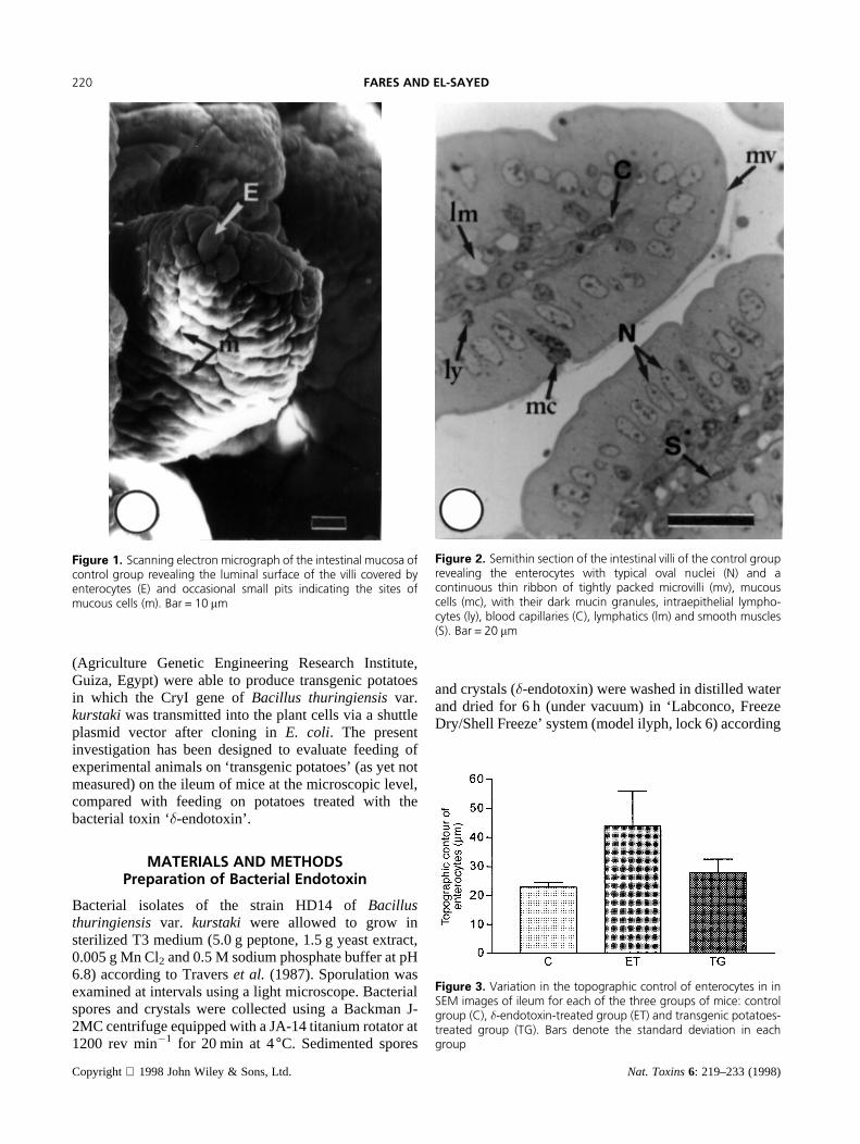
Figure 1. Scanning electron micrograph of the intestinal mucosa ofcontrol group revealing the luminal surface of the villi covered byenterocytes (E) and occasional small pits indicating the sites ofmucous cells (m). Bar = 10 mm
Figure 2. Semithin section of the intestinal villi of the control grouprevealing the enterocytes with typical oval nuclei (N) and acontinuous thin ribbon of tightly packed microvilli (mv), mucouscells (mc), with their dark mucin granules, intraepithelial lympho-cytes (ly), blood capillaries (C), lymphatics (lm) and smooth muscles(S). Bar = 20 mm
Figure 3. Variation in the topographic control of enterocytes in inSEM images of ileum for each of the three groups of mice: controlgroup (C), �-endotoxin-treated group (ET) and transgenic potatoes-treated group (TG). Bars denote the standard deviation in eachgroup
Copyright 1998 John Wiley & Sons, Ltd. Nat. Toxins6: 219–233 (1998)
220 FARES AND EL-SAYED

to Redway and Lapage (1974). The dried�-endotoxinwas stored at 20°C. Fresh potatoes were cut into smallpieces and immersed in a suspension of the�-endotoxin,of Bacillus thuringiensisvar. kurstaki, in distilled water(1 g lÿ1) for 30 min.
Feeding of Mice
A group of 5 1-month-old male mice (Mus musculus),was fed daily for 2 weeks on a diet consisting of the�-endotoxin-treated potatoes. Another group of 5 mice wasfed on a diet consisting of transgenic potatoes, carryingthe CryI gene ofBacillus thuringiensisvar.kurstaki, for 2weeks. These transgenic plants were provided by AGERI(Guiza, Egypt). A control group of 5 mice was fed onfresh potatoes for the same 2-week period.
Table 1. Statistical analysis of the mean perimeter (topographic contour) of enterocyte in scanning electron microscopic images of ileum for eachof the three different groups of mice: control group (C),�-endotoxin-treated group (ET), and transgenic potatoes-treated group (TG)
Control (C) Endotoxin (ET) Transgenic (TG)
Number of measured cells 50 50 50Mean perimeter of cell in 5 mice 23.00 44.00 28.00Minimum perimeter of cell 21.00 30.00 22.00Median perimeter of cell 24.00 44.00 30.00Maximum perimeter of cell 24.00 58.00 32.00Standard deviation 1.477 11.94 4.513Standard error 0.4264 3.447 1.303p value (two-tailed) p< 0.0001 p< 0.0001 p< 0.0001Significant (alpha = 0.05)? Yes Yes YesUnpairedt-test: Two-tailedp value:1: C vs ET p value<0.0001 (means are significantly different,p< 0.05)2: C vs TG p value = 0.0014 (means are significantly different,p< 0.05)
Figure 4. Variation in the area of enserocytes in semithin sectionsof ileum of each of the three different groups of mice: control group(C), �-endotoxin-treated group (ET) and transgenic potatoes-treatedgroup (TG). Bars denote the standard deviation in each group
Table 2. Statistical analysis of the mean area of enterocyte in semithin sections of ileum of each of the three different groups of mice. controlgroup (C),�-endotoxin-treated group (ET), and transgenic potatoes-treated group (TG)
Control (C) Endotoxin (ET) Transgenic (TG)
Number of measured cells 750 750 750Mean area of cell in 5 mice 105.3 165.4 116.5Minimum area of cell 99.00 125.0 111.0Median area of cell 103.5 172.0 115.0Maximum area of cell 115.0 192.5 125.0Standard deviation 7.762 28.70 5.972Standard error 3.881 14.35 2.986p value (two tailed) p< 0.0001 p< 0.0014 p< 0.0001Significant (alpha = 0.05)? Yes Yes YesUnpairedt-test: Two-tailedp value:1: C vs ET p value = 0.0068 (means are significantly different,p< 0.05)2: C vs TG p value = 0.06 (means are not significantly different,p< 0.05)
Copyright 1998 John Wiley & Sons, Ltd. Nat. Toxins6: 219–233 (1998)
CHANGES IN MICE FED ON �-ENDOTOXIN-TREATED AND TRANSGENIC POTATOES 221

Preparation of Microscopic Samples
Animals from the three different groups were killed bysevering the spinal cord and the ileum of each animal wasdissected out, cut into small pieces and fixed in 2.5 %glutaraldehyde in 0.l M phosphate buffer (Sigma, StLouis, USA) at pH 7.2 for 90 min, for light, scanning andelectron microscopic studies. Tissues were postfixed for2 h in 1 % OsO4 in the same phosphate buffer, dehydratedthrough ascending grades of acetone and embedded inSpurr’s medium. Semithin sections (0.5mm) wereprepared on an MT600-XL RMC ultratome (Tokyo,Japan), stained with toluidine blue and used for lightmicroscopic studies. Thin sections (80–90 nm) were cutwith a Diatom diamond knife (Washington, USA) on anMT600-XL RMC ultratome (Tokyo, Japan). Sectionswere collected on 200-mesh nickel grids, stained in 5%
uranyl acetate in distilled water for 10 min, washed indistilled water and stained in lead citrate for 6 min(Venable and Coggeshall, 1965) and examined with a bi-functional Joel JTM-1200 EX II electron microscope(Tokyo, Japan). For scanning electron microscopicstudies, small pieces of the OsO4-postfixed tissues wereexposed to the critical point dry and spotter coatingprocesses and examined by the same electron micro-scope.
Morphometric Analysis
Semithin sections, as well as scanning and electronmicroscopic photographs, of the ileum of each of thethree different groups of mice were used in themorphometric studies. Images of the ileum from thesepreparations were transferred into an IBM computer
Figure 5. Variation in number of enterocytes pervillus insemithin sections of ileum of each of the three differentgroups of mice: control group (C), �-endotoxin-treated group(ET) and transgenic potatoes-treated group (TG), Bars denotethe standard deviation in each group
Table 3. Statistical analysis of the mean number of enterocytes in semithin sections of ileum of each of the three different groups of mice: controlgroup (C),�-endotoxin-treated group (ET), and transgenic potatoes-treated group (TG)
Control (C) Endotoxin (ET) Transgenic (TG)
Number of villi selected in 5 mice 625 625 625Mean number of cells per villus (in 5 mice) 151.8 197.0 155.8Minimum number of cells 148.0 190.0 140.0Median number of cells 149.5 196.5 160.5Maximum number of cells 160.0 205.0 162.0Standard deviation 5.560 6.164 10.53Standard error 2.780 3.082 5.266p value (two tailed) p< 0.0001 p< 0.0001 p< 0.0001Significant (alpha = 0.05)? Yes Yes YesUnpairedt-test: Two-tailedp value:1: C vs ET p value< 0.0001 (means are significantly different,p< 0.05)2: C vs TG p value = 0.5268 (means are not significantly different,p< 0.05)
Copyright 1998 John Wiley & Sons, Ltd. Nat. Toxins6: 219–233 (1998)
222 FARES AND EL-SAYED

attached to an Olympus2 light microscope (Japan) via aSony2 video-camera (Japan). The captured images werethen digitized on the computer using an ‘Alpha-Viewer’image analysis program, version 1.0, for measuring thetopographic contour and the area of enterocytes. Thenumber of enterocytes, multinucleated enterocytes, andhypertrophied nuclei were also counted. The mean valueof each parameter was calculated per 5 animals, in eachof the three different groups, and the data werestatistically analysed using Paired Student’st-test of the‘GraphPad Prism2’ program, version 2.01, from Graph-Pad Software Inc., USA.
OBSERVATIONMicroscopic Observations
In relation to the digestive and absorptive functions of thesmall intestine of mammals, the mucosa of the ileum isthe most important absorptive layer (Fawcett, 1997).Accordingly, the present investigation was designed tofocus mainly on the microscopic structure of this layer in
mice of the three different groups: the control group, thegroup fed on the�-endotoxin-treated potatoes and thegroup fed on transgenic potatoes.
Control GroupAs revealed by the scanning electron microscopicexamination, the intestinal mucosa was thrown up intoseveral finger-like, as well as leaf-like forms of villiextending into the intestinal lumen (Figure 1). Thesurface of these villi was almost entirely covered by theenterocytes, which were the principle absorptive cells ofthe intestinal epithelium. The topographic contour (meanperimeter) of the enterocyte was 23mm, p< 0.0001(Table 1, Figure 3). Scattered among the enterocyteswere occasional small pits indicating the sites of mucouscells. The light microscopic examination of semithinsections of these villi revealed the enterocyte as a tallcolumnar cell with typical oval nuclei in the lower thirdof the cell (Figure 2). The mean area of the enterocytewas 105.3mm2, p< 0.0001 (Table 2, Figure 4), while themean number per one villus was 151.8,p< 0.0001
Figure 6. Electron micrograph of the intestinal epithelium of control group revealing a mucous cell with apical mucin globules(g), basal ¯attened nucleus (n), rough endoplasmic reticulum (RER) and Golgi complex (G). The enterocytes display luminalmicrovilli (mv), large oval euchromatic nuclei (N), tight junction (arrows), rough endoplasmic reticulum (arrow heads) andmitochondria (M). Bar = 2.0 mm
Copyright 1998 John Wiley & Sons, Ltd. Nat. Toxins6: 219–233 (1998)
CHANGES IN MICE FED ON �-ENDOTOXIN-TREATED AND TRANSGENIC POTATOES 223

(Table 3, Figure 5). The luminal surface of these cellswas covered by a continuous thin ribbon, which was ahighly specialized region of this epithelium, consisting oftightly packed microvilli. Mucous cells were locatedamong the enterocytes and were distinguished by theirmucin granules which occupy the upper portion of thecells. Their nuclei were small in size and oval in shapeand were located at the basal side of the cells.Intraepithelial lymphocytes were located in a basalposition between the lateral intercellular spaces. Theypossessed relatively small dark nuclei. Underneath thebasal lamina of the intestinal epithelium, the laminapropria penetrated the core of the villi, taking along bloodcapillaries, lymphatics and smooth muscles.
At the ultrastructural level, the mucous cells wererecognized by their mucin globules (Figure 6). Thesedroplets occupied the apical region of the cell andconsisted of a homogeneous matrix, which varied inintensity from highly electron dense to more lightlyelectron dense, enveloped by a delicate membrane. Thebase of the cell was relatively free of secretory material
and formed a slender stem or stalk. The nucleus tended tobe flattened and was surrounded by a thin layer ofcytoplasm. This cytoplasmic area contained severalprofiles of longitudinally oriented rough endoplasmicreticulum running parallel to the lateral edges of the cell.A highly developed Golgi complex was situated betweenthe nucleus and the mucin droplets.
The enterocytes displayed large oval euchromaticnuclei with a few patches of heterochromatin (Figure6). The lateral walls of these cells formed a well-developed tight junction, specially at the uppermostregion. The upper cytoplasmic region was rich in roughendoplasmic reticulum and mitochondria.
The mitochondria were relatively large and had aninternal structure with several large cristae traversingacross the inner mitochondrial space (Figure 7). Thestriated or brush border of the enterocytes was made up oflarge numbers of closely packed parallel microvilli(Figures 6 and 7). Each microvillus was a cylindricalprotrusion of the apical cytoplasm and consisted of a cellmembrane enclosing a filamentous core. In the interior of
Figure 7. Electron micrograph of the intestinal epithelium of control group revealing a part of an enterocyte with relativelytall mitochondria (M), several pro®les of endoplasmic reticulum (ER) and a large numbers of closely packed parallel microvilli(mv). Each microvillus has a bundle of thin striated ®laments (F) connected to terminal web (arrow heads) in a clear zone ofunderlying cytoplasm. Bar = 0.2 mm
Copyright 1998 John Wiley & Sons, Ltd. Nat. Toxins6: 219–233 (1998)
224 FARES AND EL-SAYED

each microvillus was a bundle of thin striated filaments ofrunning longitudinally in an otherwise homogeneousfine-textured cytoplasmic matrix (Figure 7). Underneaththe microvilli was a clear zone usually devoid oforganelles, except for a few profiles of endoplasmicreticulum, but occupied by filamentous striations, orterminal web, parallel to the apical surface of the cell(Figure 7).
Several well-developed Golgi apparatuses occupied asupranuclear position and consisted of parallel cisternaeand large vesicles (Figure 8). A few primary lysosomeswere located in the area of Golgi apparatus (Figure 8).The subnuclear cytoplasmic area was occupied by a largenumber of mitochondria and a few profiles of endoplas-mic reticulum (Figure 9). The base of the enterocytes wasbased on a thick basal lamina (Figure 9). A small numberof Paneth cells were recognized in the lower third of thecrypts of Lieberku¨hn by their characteristic basal nucleiand secretory granules in their luminal surface (inset,Figure 9).
�-Endotoxin-Treated Group
In the group of mice fed on the�-endotoxin-treated
potatoes, the scanning electron microscopic examinationrevealed a remarkable increase in the topographiccontour of the enterocytes at the divulged surface of thevilli (Figure 10). The mean perimeter of the enterocytewas 44mm, p< 0.0001 (Table 1, Figure 2). In addition,several variable-shaped structures, ranging from round toelongate, were recognized adhering to these villi. Insemithin sections, the villi appeared with an abnormallylarge number of enterocytes and consequently wereextremely large (Figure 11). The mean number ofenterocytes per villus was 197,p< 0.0001 (Table 3,Figure 5), while the mean area of enterocytes was165.4mm2, p< 0.0014 (Table 2, Figure 4). A largenumber (50 %) of the enterocytes in these villi weremultinucleated. The great majority of these nuclei werehypertrophied and acquired a round shape, rather than theoval appearance revealed in the enterocytes of the controlgroup. At the ultrastructural level, the nuclei of theenterocytes displayed a typical rounded configuration
Figure 8. Electron micrograph of the intestinal epithelium ofcontrol group revealing a part of an enterocyte with well-developedGolgi apparatus (G) and a primary lysosome (Ls). Bar = 0.2 mm
Figure 9. Electron micrograph of a basal region of intestinalepithelium of a control mouse revealing a large number ofsubnuclear mitochondria (M), a few pro®les of endoplasmicreticulum (ER) and a thick basal lamina (asterisk). Bar = 2.0 mm.The inset reveals a semithin section of a part of the crypt ofLieberkuÈ hn containing a few Paneth cells with dark secretorygranules (arrowhead). Bar = 20 mm
Copyright 1998 John Wiley & Sons, Ltd. Nat. Toxins6: 219–233 (1998)
CHANGES IN MICE FED ON �-ENDOTOXIN-TREATED AND TRANSGENIC POTATOES 225

(Figures 11 and 12). In addition, the basal lamina alongthe base of the enterocytes was severely destructive atseveral foci. A number of enterocytes lost their luminalmicrovilli and appeared in association with variable-shaped cytoplasmic fragments (Figure 12). The roundedforms of these fragments contained several unrecogniz-able membranous structures, while the elongated formscontained several profiles of endoplasmic reticulum, aswell as ring-shaped annulate lamellae (Figures 12 and13). At one side, these cytoplasmic fragments possessedclear zones which extended laterally into vermiformprocesses (Figure 13). Most of these cytoplasmicfragments were in association with much smaller roundedstructures which were remarkable for their highlyelectron dense contour and lightly dense core. The lateralplasma membranes of the enterocytes were detached in anumber of foci (Figure 14). Their supranuclear cytoplas-mic area contained several profiles of endoplasmicreticulum, a few mitochondria and several forms ofsecondary lysosomes, or auotophagic vacuoles (Figures14 and 15). Several degenerated mitochondria, as well asendoplasmic reticulum, were located within the autopha-gic vacuoles (Figure 15). The luminal surface of the
enterocytes were covered by short microvilli. Themucous cells in these villi contained several coagulatedmucin granules (Figure 16 and inset). In the crypts ofLieberkuhn, the Paneth cells were highly activated andcontained large number of secretory granules (Figure17).
Transgenic Potatoes-treated GroupIn the group of mice fed on transgenic potatoes, bothscanning and light microscopic architecture of theintestinal villi and their cellular structures, includingenterocytes, Paneth cells, and mucous cells were almostas normal as the control group (Figures 18–20). The meanperimeter of enterocyte was 28mm (p< 0.0001, Table 1,Figure 2), with a mean area of 116.5 (p< 0.0001, Table2, Figure 4) and a mean number of 155.8 enterocytes pervillus (p< 0.0001, Table 3, Figure 5). However, at theultrastructural level the enterocytes possessed severaldilated mitochondria with short cristae (Figure 21). Inaddition, the luminal surface of certain foci possesseddisrupted short microvilli. Nevertheless, in the greatmajority of the enterocytes the microvilli displayed
Figure 10. Scanning electron micrograph of the intestinal mucosaof �-endotoxin-treated group revealing remarkably increasedtopographic contour of enterocytes (asterisks) and associatedvariable-shaped structures (arrows). Bar = 10mm
Figure 11. Semithin section of the intestinal mucosa of �-endotox-in-treated group revealing a villus with an abnormally large numberof multinucleated and hypertrophied enterocytes (E). A numbervariable-shaped cytoplasmic fragments (arrowheads) are in associa-tion with this villus. Bar = 20 mm
Copyright 1998 John Wiley & Sons, Ltd. Nat. Toxins6: 219–233 (1998)
226 FARES AND EL-SAYED

regular striated appearance (Figure 22). The basal laminawas relatively intact (Figure 18). Mucous cells possesseda homogeneously electron dense mucin granules (Figure20).
DISCUSSION
In the present investigation, the enterocytes of theintestinal epithelium in the group of mice fed on�-endotoxin-treated potatoes were remarkably enlarged asa result of multiplication and hypertrophy of their nuclei,degeneration of mitochondria and endoplasmic reticu-lum, and the ensuing appearance of autophagic vacuoles.These features were reflected on the scanning topo-graphic architecture of these cells, which showed aremarkably large contour. In addition, these changeswere accompanied by the detachment of the lateralplasma membranes in several foci and the discontinua-tion of the basal lamina of these cells. Several investiga-tions revealed that solubilized�-endotoxin ofBacillusthuringiensis kurstakiis cytolytic to a wide range of
vertebrate and invertebrate cells (Wu and Chang, 1985;Ibarra and Federici, 1986b; Chilcott and Ellar, 1988).Additionally, Thomas and Ellar (1983a) showed thatsolubilized endotoxin preparations are lethal wheninjected into suckling mice. It has been suggested thatthe high toxicity of this endotoxin is due not to a singleprotein, but rather to a set of nergistic interactions of the25-kDa protein with one or more of the higher molecularweight proteins (Chilcott and Ellar, 1988). Although theprecise mode of action of the�-endotoxin ofBacillusthuringiensisvar. kurstakiis not fully understood, Lu¨thyand Ebersold, (1981) suggested that intoxication ininsects may result from an osmotic imbalance acrossthe midgut epithelial membranes which leads quickly tohypertrophy and lysis of midgut cells. Lysis is followedby disruption of the basement membrane, leakage ofdigestive juices into the hemocoel, and larval death.Thomas and Eliar (1983b) provided good evidence thatBacillus thuringiensis kurstakiendotoxin’s cytolyticactivity was due to a detergent-like action in which thetoxin disrupted membranes by binding to specific lipids.
Figure 12. Electron micrograph of the intestinal mucosa of �-endotoxin-treated group revealing enterocytes with roundednuclei (N), intracellular vacuoles (V) and discontinuous basal lamina (B) interrupted by a lymphocyte (ly) and congested bloodcapillary (BC). A cytoplasmic fragment (asterisk) enclosing membranous structures is in association with fragmented microvilli(mv). Bar = 2.0 mm
Copyright 1998 John Wiley & Sons, Ltd. Nat. Toxins6: 219–233 (1998)
CHANGES IN MICE FED ON �-ENDOTOXIN-TREATED AND TRANSGENIC POTATOES 227

Figure 13. Electron micrograph of the intestinal mucosa of �-endotoxin-treated group showing an elongatedform of cytoplasmic fragments containing several pro®les of endoplasmic reticulum (RER), ring-shapedannulate lamellae (AL), clear zones of laterally extended vermiform processes (P), and in association with smallrounded structures (arrows) with highly electron dense contour and lightly dense core. Bar = 0.5 mm. Inset: asemithin section of the same area. Bar = 20 mm
Figure 14. Electron micrograph of the intestinal mucosa of �-endotoxin-treated group showing detachedlateral plasma membranes (arrows), several pro®les of endoplasmic reticulum (ER), auotophagic vacuoles(Av), a few mitochondria (M) and a cytoplasmic vacuole. Bar = 2.0 mm
Copyright 1998 John Wiley & Sons, Ltd. Nat. Toxins6: 219–233 (1998)
228 FARES AND EL-SAYED
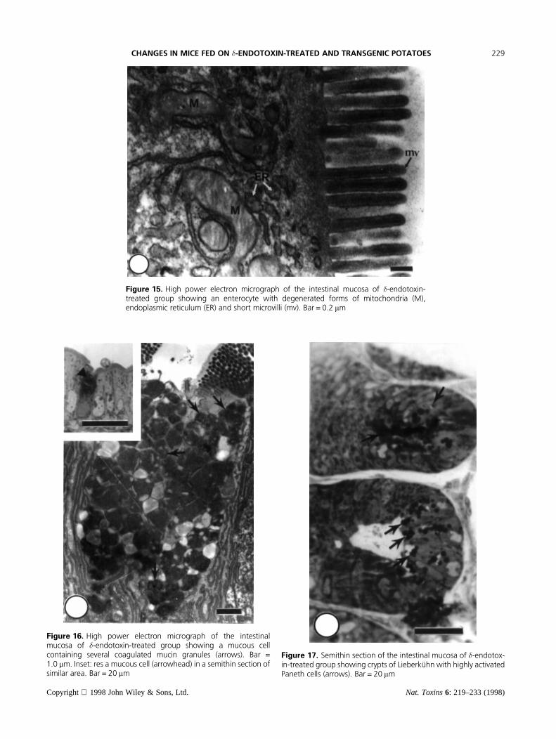
Figure 15. High power electron micrograph of the intestinal mucosa of �-endotoxin-treated group showing an enterocyte with degenerated forms of mitochondria (M),endoplasmic reticulum (ER) and short microvilli (mv). Bar = 0.2 mm
Figure 16. High power electron micrograph of the intestinalmucosa of �-endotoxin-treated group showing a mucous cellcontaining several coagulated mucin granules (arrows). Bar =1.0 mm. Inset: res a mucous cell (arrowhead) in a semithin section ofsimilar area. Bar = 20 mm
Figure 17. Semithin section of the intestinal mucosa of �-endotox-in-treated group showing crypts of LieberkuÈ hn with highly activatedPaneth cells (arrows). Bar = 20 mm
Copyright 1998 John Wiley & Sons, Ltd. Nat. Toxins6: 219–233 (1998)
CHANGES IN MICE FED ON �-ENDOTOXIN-TREATED AND TRANSGENIC POTATOES 229
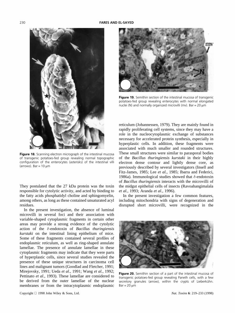
They postulated that the 27 kDa protein was the toxinresponsible for cytolytic activity, and acted by binding tothe fatty acids phosphatidyl choline and sphingomyelin,among others, as long as these contained unsaturated acylresidues.
In the present investigation, the absence of luminalmicrovilli in several foci and their association withvariable-shaped cytoplasmic fragments in certain otherareas may provide a strong evidence of the cytolyticaction of the �-endotoxin of Bacillus thuringiensiskurstaki on the intestinal lining epithelium of mice.Some of these fragments contained several profiles ofendoplasmic reticulum, as well as ring-shaped annulatelamellae. The presence of annulate lamellae in thesecytoplasmic fragments may indicate that they were partsof hyperplastic cells, since several studies revealed thepresence of these unique structures in carcinoma celllines and malignant tumors (Goodlad and Fletcher, 1991;Mirejovsky, 1991; Uedaet al., 1991; Wanget al., 1992;Pettinatoet al., 1993). These lamellae are considered tobe derived from the outer lamellae of the nuclearmembranes or from the intracytoplasmic endoplasmic
reticulum (Johannessen, 1979). They are mainly found inrapidly proliferating cell systems, since they may have arole in the nucleocytoplasmic exchange of substancesnecessary for accelerated protein synthesis, especially inhyperplastic cells. In addition, these fragments wereassociated with much smaller and rounded structures.These small structures were similar to parasporal bodiesof the Bacillus thuringiensis kurstakiin their highlyelectron dense contour and lightly dense core, aspreviously described by several investigators (Insell andFitz-James, 1985; Leeet al., 1985; Ibarra and Federici,1986a). Immunological studies showed that�-endotoxinof Bacillus thuringiensisinteracts with the microvilli ofthe midgut epithelial cells of insects (Ravoahangimalalaet al., 1993; Arandaet al., 1996).
In the present investigation a few common features,including mitochondria with signs of degeneration anddisrupted short microvilli, were recognized in the
Figure 18. Scanning electron micrograph of the intestinal mucosaof transgenic potatoes-fed group revealing normal topographiccon®guration of the enterocytes (asterisks) of the intestinal villi(arrows). Bar = 10 mm
Figure 19. Semithin section of the intestinal mucosa of transgenicpotatoes-fed group revealing enterocytes with normal elongatednuclei (N) and normally organized microvilli (mv). Bar = 20 mm
Figure 20. Semithin section of a part of the intestinal mucosa oftransgenic potatoes-fed group revealing Paneth cells, with a fewsecretory granules (arrow), within the crypts of LieberkuÈ hn.Bar = 20 mm
Copyright 1998 John Wiley & Sons, Ltd. Nat. Toxins6: 219–233 (1998)
230 FARES AND EL-SAYED

Figure 21. Electron micrograph of the intestinal mucosa of transgenic potatoes-fed group revealingenterocytes with elongated nuclei (N), a relatively intact basal lamina (B) and several dilated mitochondriawith short cristae (M). The luminal surface of certain foci possessed disrupted short microvilli (mv). Bar = 2.0 mm
Figure 22. High power electron micrograph of the intestinal mucosa of transgenic potatoes-fed grouprevealing normally organized microvilli (mv). Bar = 0.2 mm
Copyright 1998 John Wiley & Sons, Ltd. Nat. Toxins6: 219–233 (1998)
CHANGES IN MICE FED ON �-ENDOTOXIN-TREATED AND TRANSGENIC POTATOES 231

ultrastructure of the intestinal epithelium in both groupsof mice fed on�-endotoxin-treated potatoes and trans-genic potatoes. However, in the group of mice fed on the�-endotoxin-treated potatoes, the Paneth cells of thecrypts of Lieberku¨hn were highly activated and containeda large number of secretory granules. These cells arebelieved to have an important role in the activation ofphagocytes and controlling the bacterial flora of the gut(Ariza et al., 1996; Fawcett, 1997). They contain elevatedlevels of lysozyme in their large eosinophilic secretorygranules, an enzyme capable of digesting bacterial cellswalls, and antibacterial peptides called cryptdins (Jun-queiraet al., 1998). Ouellette (1997) revealed that Panethcell secretory products seem to contribute both to innateimmunity of the crypt lumen and to defining the apicalenvironment of neighboring cells. Wadaet al. (1993)revealed that the incidence of Paneth cells increases inadenomas and adenocarcinoma, as well as in severalother diseased digestive tracts. The antimicrobial poly-peptides of the Paneth cell secretory products kill a widerange of organisms, including bacteria, fungi, viruses andtumor cells (Aleyet al., 1995).
In conclusion, the present investigation revealed mildchanges in the microscopic structure of the differentcellular compartments of the ileum of a group of mice fedon transgenic potatoes as compared with another group ofmice fed on the�-endotoxin-treated potatoes, despite thepresence of the same type of toxin ofBacillusthuringiensisvar. kurstaki in the transgenic potatoes asa result of gene expression. The appearance of several
multinucleated and hypertrophied enterocytes, as well asseveral associated cytoplasmic fragments with highlyrecognized annulate lamellae may suggest the possibleparticipation of feeding on the�-endotoxin-treatedpotatoes in the hyperplastic development in the miceileum. Although transgenic crop plants used in food andfeed production carry different beneficial transgenes,mostly for resistance to pests, herbicides and diseases(Ondrej and Drobnik, 1997), before releasing for market-ing thorough tests and all possible consequences of thesenew types of heredity and new genetic structures must beevaluated to avoid any potential risks,
ACKNOWLEDGEMENTS
The authors wish to express their appreciation to Dr John C. Herr,Professor of Cell Biology and Director of the Center for RecombinantGamete Contraceptive Vaccinogens, at the University of VirginiaScience Health Center, for providing computer facilities for thefinalizing of the present manuscript. Thanks are also due to Dr.Mohamed Salama, Associate Professor of Molecular Biology, at theDepartment of Entomology, Faculty of Science, Ain Shams Uni-versity, for providing the transgenic potatoes.
REFERENCES
Aley S, Zimmerman M, Hetsko M, Selsted M, Gillin F (1995). Killingof Giardia lamblia by cryptdins and cationic neutrophil peptides.Infection and Immunity62:5397–5403.
Aranda E, Sanchez J, Peferoen M, Guereca L, Bravo (1996). AInteractions of Bacillus thuringiensiscrystal proteins with the
Figure 23. High power electron micrograph of the intestinal mucosa oftransgenic potatoes-fed group revealing a mucous gland with homogeneousmucin granules (g). Bar = 0.5 mm
Copyright 1998 John Wiley & Sons, Ltd. Nat. Toxins6: 219–233 (1998)
232 FARES AND EL-SAYED

midgut epithelial cells ofSpodoptera frugiperda(Lepidoptera:Noctuidae).J Invertebr Pathol68:203–212.
Ariza A, Lopez D, Castella E, Munoz C, Zujar M, Mate L (1996).Expression of CD15 in normal and metaplastic Paneth cells of thedigestive tract.J Clin Pathol (London)49:474–477.
Caramori T, Albertini AM, Galizzi A (1991).In vivo generation ofhybrids between twoBacillus thuringiensisinsect-toxin-encodinggenes.Gene98:37–44.
Chilcott C, Ellar D (1988). Comparative toxicity ofBacillusthuringiensisvar. israelensiscrystal proteinsin vivo and in vitro. JGen Microbiol134:2551–2558.
de Barjac H (1989). New facts and trends in bacteriological control ofmosquitoes.Memorias do Instituto Oswaldo Cruz84(suppl 3):101–105.
Fares NH (1996). Histological and fine structural changes in the kidneycortex of mice treated by the carbamate insecticide sevin.J EgyptGer Soc Zool21(C):53–85.
Fawcett DW (1997). Bloom and Fawcett:Concise Histology. R. P.Jensh, Contributing Editor. Chapman and Hall, Thomson Interna-tional Publishing, New York, NY, USA.
Goodlad J, Fletcher C (1991). Malignant peripheral nerve sheath tumorwith annulate lamellae mimicking pleomorphic malignant fibroushistiocytomaJ Pathol164:23–30.
Ibarra J, Federici B (1986a). Isolation of a relatively nontoxic 65-kilodalton protein inclusion from the parasporal body ofBacillusthuringiensissubsp.Israelensis. J Bacteriol165:527–533.
Ibarra J, Federici B (1986b). Parasporal bodies ofBacillus thuringien-sis subsp.Morrisoni (PG-14) andBacillus thuringiensissubsp.israelensisare similar in protein composition and toxicity.FEMSMicrobiol Lett 34:79–84.
Insell J, Fitz-James P (1985). Composition and toxicity of the inclusionof Bacillus thuringiensissubsp.Israelensis. Appl Environ Microbiol50:56–62.
Johannessen J, ed. (1979).Electron microscopy in Human medicine,vol. 9, Urogenital System and Breast. New York: McGraw-HillInternational Book Company.
Junqueira L, Carneiro R, Kelley R (1998). Basic Histology, 8th edn.Appleton and Lange, USA.
Lee S, Eckblad W, Bulla L (1985). Diversity of protein inclusionbodies and identification of mosquitocidal protein inBacillusthuringiensis subsp. Israelensi. Biochem Biophys Res Commun126:953–960.
Luthy P, Ebersold H (1981).Bacillus thuringiensisdelta endotoxin:histopathology and molecular mode of action. In: Davidson W, ed.Parthenogenesis of invertebrate microbial diseases. New Jersey:235–267.
Mirejovsky P (1991). The ultrastructure of respiratory tract tumors.Acta Universitatis Carolinae Medica Monographia139:1–25.
Ondrej M, Drobnik M (1997). The safety and usefulness of transgenicplants.Cas Lek Cesk136:331–336.
Ouellette A (1997). Paneth cells and innate immunity in the cryptmicroenvironment.Gastroenterology113:1779–1784.
Pettinato G, Manivel J, Ravetto C T, Gould E, Tuoro A, Jaszcz W,Albores-Saavedra J (1993). Papillary cystic tumor of the pancreas: alinicopathologic study of 20 cases with cytologic, immunohisto-chemical, ultrastructural, and flow cytometric observations, and areview of the literature.Am J Clin Pathol98:478–488.
Redway K, Lapage S (1974). Effect of carbohydrates and relatedcompounds on the long-term reservation of freeze-dried bacteria.Cryobiology11:73–79.
Ravoahangimalala O, Charles J, Schoeller-Raccaud J (1993). Im-munological localization ofBacillus thuringiensis serovar israe-lensis toxins in midgut cells of intoxicatedAnopheles gambiaelarvae (Diptera: Culicidae).Res Microbiol144:271.
Sanchis V, Agaisse H, Chaufaux J, Lereclus D (1996). Construction ofnew insecticidalBacillus thuringiensisrecombinant strains by usingthe sporulation non-dependent expression system of cryIIIA and asite specific recombination vector.J Biotechnol48:81–96.
Singsit C, Adang M, Lynch R, Anderson W, Wang A, Cardineau G,Ozias-Akins P (1997). Expression of aBacillus thuringiensiscryIA(c) gene in transgenic peanut plants and its efficacy againstlesser cornstalk borer.Transgenic Res6:169–176.
Thomas W, Ellar D (1983a).Bacillus thuringiensisvar. Israelensiscrystal�-endotoxin: effects on insects and mammalian cellsin vitroandvivo. J Cell Sci60:181–197.
Thomas W, Ellar D (1983b). Mechanism of action ofBacillusthuringiensisvar. Israelensisinsecticidal �-endotoxin.FEBS Lett154:362–368.
Travers R, Martin P, Reichelderfer C (1987). Selective process forefficient isolation of soilBacillus spp. Appl Environ Microbiol53:1263–1266.
Tyrell D, Bulla L, Andrews R, Kramer K, Davidson L, Nordin P(1981). Comparative biochemistry of entomocidal parasporalcrystals of selectedBacillus thuringiensisstrains. J Bacteriol145:1052–1062.
Ueda N, Nagakawa T, Nakamura A, Ueno K, Miyazaki I, Kurachi M,Konishi I, Hirono T (1991). Clinicopathological studies on solid andcystic tumors of the pancreas.Gastroenterologia, Joponica26:497–502.
Venable J, Coggeshall R (1965). A simplified lead citrate stain for usein electron microscopy.J Cell Biol 25:407–408.
Wada R, Miwa H, Abe H, Santo R, Kitamura S, Kuwabara N, Suda K,Kondo K, Yamada S (1993). Incidence of Paneth cells in minutetubular adenomas and adenocarcinomas of the large bowel.ActaPathol Jap42:579–584.
Wang N, Liu C, Emond J, Tsao M (1992). Annulate lamellae in a largecell lung carcinoma cell line with high expression of tyrosine kinasereceptor and proto-oncogenes.Ultrastructural Pathol16:439–449.
Wu D, Chang F (1985). Synergism in mosquitocidal activity of 26 and65 kDa proteins ofBacillus thuringiensissubsp.Israelensiscrystal.FEBS Lett190:232–236.
Copyright 1998 John Wiley & Sons, Ltd. Nat. Toxins6: 219–233 (1998)
CHANGES IN MICE FED ON �-ENDOTOXIN-TREATED AND TRANSGENIC POTATOES 233