fine-needle aspiration biopsy of metastatic malignant melanoma resembling a malignant peripheral...
TRANSCRIPT

DIAGNOSTIC DILEMMASSection Editor: Claire W. Michael, M.D.
Fine-Needle Aspiration Biopsy ofMetastatic Malignant MelanomaResembling a MalignantPeripheral Nerve Sheath TumorSvetoslav Bardarov, M.D., Claire W. Michael, M.D.,*David Lucas, M.D., Yijun Pang, M.D., Ph.D.,and Robert T. Pu, M.D., Ph.D.
We report a case of metastatic malignant melanoma resembling amalignant peripheral sheath tumor, which posed a significantdiagnostic challenge. The patient is a 76-year-old male, who pre-sented in the emergency room with bilateral chest pain exacer-bated by inspiration. The pain was present for 3 week and wasnot exacerbated by physical exercise. The diagnostic workuprevealed bilateral parenchymal pulmonary infiltrates. The CT-scanguided fine-needle aspiration and the core biopsies of the largestpulmonary lesion revealed high-grade spindle cell neoplasm withindividual cell apoptosis and necrosis. The immunohistochemicalprofile on the cell block showed that the cells are positive forVimentin. The S-100 stain showed only focal positivity. The immu-nohistochemical stains for HMB45, Melan A, pancytokeratin, andsmooth muscle actin were negative. Five years ago the patientwas diagnosed with melanoma on the back with Clark level of IV.The melanoma was excised with clear margins and sentinel lymphnodes were negative. Careful examination of patient’s previousslides revealed an area of spindle cell melanoma adjacent to anodular type melanoma. Based on the patient’s previous history,current clinico-pathologic presentation and immunohistochemicalprofile, the diagnosis of metastatic malignant melanoma resem-bling peripheral nerve sheath tumor was favored over the diagno-sis of metastatic malignant spindle cell neoplasm of unknown pri-mary site, which by itself is very rare clinical scenario. Diagn.Cytopathol. 2008;36:754–757. ' 2008 Wiley-Liss, Inc.
Key Words: melanoma; MPNST; immunohistochemistry
Melanoma is an increasingly common, potentially fatal
form of skin cancer arising from the malignant transfor-
mation of melanocytes. Currently, melanoma is the sev-
enth most common type of cancer and the most common
malignancy in women ages 25–29.1 Melanoma has the
ability to spread and give metastasis to different sites
sometimes years after the initial diagnosis is made. Pri-
mary as well as metastatic melanomas can have different
morphologic features on light microscopy, which make
the diagnosis difficult. It can resemble small round cell
tumor, poorly differentiated carcinomas as well as low
and high-grade soft tissue sarcoma. Because of the mor-
phologic heterogeneity, malignant melanoma is always in
the differential diagnosis of metastatic tumors of unknown
primary site. In this report, we present a case of meta-
static melanoma which morphologically resembles malig-
nant peripheral nerve sheath tumor, which posed a signifi-
cant diagnostic challenge.
Case Reports
A 76-year-old man presented to the emergency room
complaining of sharp bilateral chest pain associated with
shortness of breath. The pain worsened with coughing
and inspiration. The pain had been present for the prior 3
weeks, was stable and was not exacerbated with walking
or physical activity. The patient’s past medical history
included: hypertension, atrial fibrillation, and melanoma
on the back excised about 5 years ago. The patient’s
social and family history was noncontributory. Chest X-
ray revealed large pleural effusion on the left. Computer
tomography scan revealed multiple bilateral opacities, the
Department of Pathology, University of Michigan, Ann Arbor, Michigan*Correspondence to: Claire W. Michael, M.D., Department of Pathol-
ogy, 1500 East Medical Drive, Ann Arbor, MI 48109.E-mail: [email protected]
Received 26 March 2008; Accepted 16 May 2008DOI 10.1002/dc.20893Published online in Wiley InterScience (www.interscience.wiley.com).
754 Diagnostic Cytopathology, Vol 36, No 10 ' 2008 WILEY-LISS, INC.

largest one measuring 3.5 cm on the left lower lobe. The
patient was referred for CT-scan guided fine-needle aspi-
ration and core biopsy of the largest pulmonary lesion.
Cytological Finding
The examination of the cell block revealed a paucicellular
specimen composed of scattered fragments of spindled
cells, scattered leukocytes, and individually apoptotic malig-
nant cells (Fig. 1A). Occasional larger, epithelioid cells con-
taining a moderate amount of cytoplasm, slightly enlarged
hyperchromatic nuclei containing small ‘‘cherry red’’ nucle-
oli were also noted (Fig. 1B). One important morphologic
sign one should keep in mind is that the nucleoli of the
metastatic melanomas are usually oxyphilic, whereas the
macro nuclei of the malignant peripheral nerve sheath tu-
mor are usually basophilic. The tissue core biopsies
obtained during the same procedure showed similar appear-
ing cells with large areas of necrosis, cellular pleomor-
phism, and cellular apoptosis. The tumor cells were mainly
discohesive, however, clumps of malignant looking cells
with necrosis and acute inflammation were also noted.
Past Medical History and Pathology
Diagnostic slides from the patient’s previously diagnosed
melanoma (2004) were obtained, which showed both
invasive and in-situ Malignant Melanoma with Clark level
IV (Breslow depth of 1.2 mm) (Fig. 2). The margins of
excision were free of tumor. A total of nine sentinel
lymph nodes were obtained and showed no evidence of
metastatic melanoma. The original case showed nodular
type of melanoma composed of pigment containing plas-
macytoid cells with abundant cytoplasm and single
Fig. 1. Fine-needle aspiration biopsy of patients current disease. (A) Cell block (H&E, 3200) and (B) Core biopsy obtained during the same procedure(H&E, 3200). [Color figure can be viewed in the online issue, which is available at www.interscience.wiley.com.]
Fig. 2. Examination of the patients previously diagnosed melanoma revealed: (A) Usual type nodular melanoma and (B) Areas adjacent to the nodularmelanoma showing spindle cell melanoma but no necrosis (H&E, 3200). [Color figure can be viewed in the online issue, which is available at www.interscience.wiley.com.]
MALIGNANT MELANOMA RESEMBLING MPNST
Diagnostic Cytopathology, Vol 36, No 10 755
Diagnostic Cytopathology DOI 10.1002/dc
‘‘cherry red’’ nucleoli. Adjacent to the usual type nodular
melanoma, there was an area with spindle cells with nu-
clear atypia and scant melanin pigment (Fig. 2B). No ne-
crosis or mitoses were seen on the original slides.
Immunohistochemistry
The immunohistochemistry were performed on paraffin sec-
tions of the core biopsies as well as on the cell block, with
the use of avidin-biotin peroxidase complex technique. The
tumor cells were negative for Melan A, HMB45, Calretinin,
CK7, CK20, CD15, TTF-1, and Pancytokeratin. Additional
stains for smooth muscle actin were negative as well. The
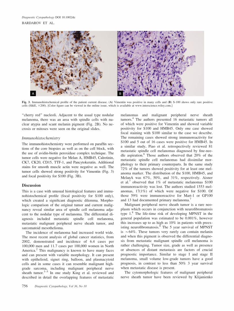
tumor cells showed strong positivity for Vimentin (Fig. 3)
and focal positivity for S100 (Fig. 3B).
Discussion
This is a case with unusual histological features and immu-
nohistochemical profile (focal positivity for S100 only),
which created a significant diagnostic dilemma. Morpho-
logic comparison of the original tumor and current malig-
nancy reveal similar area of spindle cell melanoma adja-
cent to the nodular type of melanoma. The differential di-
agnosis included metastatic spindle cell melanoma,
metastatic malignant peripheral nerve sheath tumor, and
sarcomatoid mesothelioma.
The incidence of melanoma had increased world wide.The most recent analysis of global cancer statistics, from
2002, demonstrated and incidence of 6.4 cases per
100,000 men and 11.7 cases per 100,000 women in NorthAmerica.2 This malignancy is known to have many faces
and can present with variable morphology. It can presentwith epithelioid, signet ring, balloon, and plasmacytoid
cells and in some cases it can resemble malignant high-
grade sarcoma, including malignant peripheral nervesheath tumor.3–5 In one study King et al. reviewed and
described in detail the overlapping features of metastatic
melanomas and malignant peripheral nerve sheath
tumors.4 The authors presented 16 metastatic tumors all
of which were positive for Vimentin and showed variablepositivity for S100 and HMB45. Only one case showed
focal staining with S100 similar to the case we describe.
The remaining cases showed strong immunoreactivity forS100 and 5 out of 16 cases were positive for HMB45. In
a similar study, Piao et al. retrospectively reviewed 81metastatic spindle cell melanomas diagnosed by fine-nee-
dle aspiration.6 Those authors observed that 20% of the
metastatic spindle cell melanomas had dissimilar mor-phology to their primary counterparts. In the same study
72% of the tumors showed positivity for at least one mel-anoma marker. The distribution of the S100, HMB45, and
MelanA was 67%, 50%, and 31%, respectively. Aisner
et al.7 observed that 1% of metastatic melanomas S100immunoreactivity was lost. The authors studied 1553 mel-
anomas, 17(1%) of which were negative for S100. Ofthose 59% were immunoreactive for Mart-1 or GP100
and 13 had documented primary melanoma.7
Malignant peripheral nerve sheath tumor is a rare neo-
plasm which occurs in conjunction with neurofibromatosis
type 1.8 The life-time risk of developing MPNST in the
general population was estimated to be 0.001%, however
this increases up to as high as 10% in patients with preex-
isting neurofibromatosis.8 The 5 year survival of MPNST
is *64%. These tumors very rarely can contain melanin
and when this pigment is observed the differential diagno-
sis from metastatic malignant spindle cell melanoma is
rather challenging. Tumor size, grade as well as presence
or absences of distant metastasis are factors of crucial
prognostic importance. Similar to stage I and stage II
melanoma, small volume low-grade tumors have a good
prognosis, in contrast to less than 50% 3 year survival
when metastatic disease is present.
The cytomorphologic features of malignant peripheral
nerve sheath tumor have been reviewed by Klijanienko
Fig. 3. Immunohistochemical profile of the patient current disease. (A) Vimentin was positive in many cells and (B) S-100 shows only rare positivecells (H&E, 3200). [Color figure can be viewed in the online issue, which is available at www.interscience.wiley.com.]
BARDAROV ET AL.
756 Diagnostic Cytopathology, Vol 36, No 10
Diagnostic Cytopathology DOI 10.1002/dc
et al.9 and described in great detail by Gupta et al.10 In
summary MPNST can present with variable cellularity on
FNA, usually with clusters of cells arranged in fascicular
or parallel pattern. The nuclear pleomorphism is variable
with variable mitotic activity. Nucleoli are usually present
and basophilic and the cytoplasm may contain vacuoles.
Nuclear inclusions usually are not present and individual
cell apoptosis is rarely observed.
King et al.4 advanced a list of clinicopathological fea-
tures that may serve as guidelines for the diagnosis of MM
mimicking MPNST. Briefly, the diagnosis of metastatic
MM should be entertained when one or more of the follow-
ing are seen in a spindle cell neoplasm: (1) tumor localized
within or close to a lymphnode; (2) absence of continuity
with a major nerve or a neurofibroma, or absence of neuro-
fibromatosis; (3) presence, or history of past primary MM;
(4) strong and diffuse S-100 protein immunoreactivity (S-
100 protein is usually focal and/or weak in MPNSTs); and
(5) immunoreactivity for other melanoma markers. It is pos-
sible that in the clinical practice one encounter a patient
with history of melanoma and MPNST and in these cases
the differentiation of between both neoplasms may be
impossible. The cytomorphologic features which may assist
in this difficult cases are: presence of fibrilary or myxoid
background, fascicular arrangement, basophilic nucleoli,
cytoplasmic vacuoles, and no nuclear inclusions all of
which will favor MPNST over metastatic spindle cell ma-
lignant melanoma (Table I).
In this article, we presented a case which posed a signifi-
cant diagnostic challenge. The patient presented with a met-
astatic high-grade spindle cell neoplasm, with prominent ne-
crosis. He had a previous history of melanoma with Clark
lever IV, however, with negative sentinel nodes. The patient
had no history of neurofibromatosis and no other known
primary malignancies were detected. The lesion was nega-
tive for melanoma markers and showed only focal positivity
for S-100 which favored a MPNST. Based on the clinical
history of malignant melanoma as well as identification of
similar appearing spindle cells in the original slides, we
favor the diagnosis of metastatic malignant melanoma over
metastatic high-grade spindle cell neoplasm of unknown
primary site. Even though the presence of high-grade bulky
metastatic disease confers a poor prognosis for this patient
it is important to discriminate between both neoplasms in
order to guide future treatments such as experimental treat-
ment plans or protocols.
References1. Rigel DS, Friedman RJ, Kopf AW. The incidence of malignant mel-
anoma in the United States: Issues as we approach the 21st century.J Am Acad Dermatol 1996;34:839–847.
2. Parkin DM, Bray F, Ferlay J, Pisani P. Global cancer statistics,2002. CA Cancer J Clin 2005;55:74–108.
3. Lodding P, Kindblom LG, Angervall L. Metastases of malignantmelanoma simulating soft tissue sarcoma. A clinico-pathological,light- and electron microscopic and immunohistochemical study of21 cases. Virchows Arch A Pathol Anat Histopathol 1990;417:377–388.
4. King R, Busam K, Rosai J. Metastatic malignant melanoma resem-bling malignant peripheral nerve sheath tumor: Report of 16 cases.Am J Surg Pathol 1999;23:1499–1505.
5. Banerjee SS, Harris M. Morphological and immunophenotypicvariations in malignant melanoma. Histopathology 2000;36:387–402.
6. Piao Y, Guo M, Gong Y. Diagnostic challenges of metastatic spindlecell melanoma on fine-needle aspiration specimens. Cancer 2008;114(2):94–101.
7. Aisner DL, Maker A, Rosenberg SA, Berman DM. Loss of S100 anti-genicity in metastatic melanoma. Hum Pathol 2005;36:1016–1019.
8. Ferner RE, Gutmann DH. International consensus statement on ma-lignant peripheral nerve sheath tumors in neurofibromatosis. CancerRes 2002;62:1573–1577.
9. Klijanienko J, Caillaud JM, Lagace R, Vielh P. Cytohistologic cor-
relations of 24 malignant peripheral nerve sheath tumor (MPNST)in 17 patients: The Institut Curie experience. Diagn Cytopathol2002;27:103–108.
10. Gupta K, Dey P, Vashisht R. Fine-needle aspiration cytology ofmalignant peripheral nerve sheath tumors. Diagn Cytopathol 2004;31:1–4.
Table I. Cytomorphologic Features of Metastatic Spindle Cell Melanoma Resembling MPNST vs. MPNST
Features Spindle cell melanoma resembling MPNST MPNST
Clinical Presentations Prerequisites History of melanoma Neurofibromatosis type 1History of MPNST
Presentation Can represent with metastaticdisease of unknown primary site
Metastatic disease as initialpresentation is very rare
Low-power features Cellularity Variable VariableBackground Can have large areas of necrosis Fibrilary or MyxoidCellular arrangement Mostly discohesive Clusters; Fascicular and
parallel arrangementHigh-power features Cellular Morphology Spindle cells with sharp ends;
occasional epithelioidcells present
Fusiform nucleiwith round ends
N:C ratio Increased VariablePleomorphism Variable VariableCytoplasmic vacuoles None VariableNucleoli Oxyphillic BasophilicMitotic figures Many VariableApoptosis Present NoneIntranuclear inclusions Present Rare
MALIGNANT MELANOMA RESEMBLING MPNST
Diagnostic Cytopathology, Vol 36, No 10 757
Diagnostic Cytopathology DOI 10.1002/dc