fibroma of tendon sheath as a cause of carpal tunnel syndrome · fibroma of tendon sheath as a...
TRANSCRIPT
Fibroma of tendon sheath as a cause of carpaltunnel syndrome
Jaggi Rao BSc, Achilleas Thoma MD FRCSC FACS, Sam Salama MB FRCPC
Department of Surgery, Division of Plastic and Reconstructive Surgery, and Department of Pathology,
St Joseph’s Hospital, McMaster University, Hamilton, Ontario
In 1949, Geschickter and Copeland (1) first described fi-
broma of tendon sheath (FTS) as a distinct entity, but it
was not described further until 1979, when Chung and
Enzinger (2) presented the clinical and pathological findings
of 138 cases selected from the files of the Armed Forces In-
stitute of Pathology. In all cases of FTS reported in literature,
only six have been implicated as a cause of carpal tunnel syn-
drome (CTS). The fibroma described in this report is an ex-
ample of this rare tumour; it led to the progression of CTS
and demonstrated characteristics unique to FTS. Carpal tun-
nel release and local excision of the tumour promptly re-
lieved the patient’s symptoms.
CASE PRESENTATIONA 28-year-old Caucasian male presented with a complaint of
cramping in the left hand, as well as numbness to the left mid-
dle and ring fingers. With minimal activity of the hand, there
was swelling of the midpalm, and he became more sympto-
matic.
Seven months earlier, the patient underwent surgery to re-
move the tumour in a peripheral hospital. The tumour was
adhered to the median nerve. The original surgeon, not feel-
ing confident about the nature of the tumour, prudently aban-
doned the procedure. A biopsy was performed instead,
which, although nondiagnostic, suggested a benign fibrovas-
cular tumour.
The patient continued to suffer numbness in the median
nerve distribution, particularly on the contiguous surfaces of
the middle and ring fingers. Three months following the ini-
tial attempt at resection, he was forced to take leave from his
occupation as a butcher because of midpalmar pain.
At the present examination, the patient had a palpable,
soft tissue mass in the left palm, located at the distal portion
of the carpal tunnel. The mass was tender to pressure and the
provocative tests for CTS, including Tinel’s and Phalen’s
tests, were positive. Accompanying this was paresthesia on
both sides of the middle and ring fingers.
Consent was obtained for exploration and resection of the
tumour with the strong possibility of sural nerve grafting. At
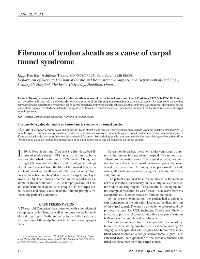
surgery, an encapsulated whitish-grey firm tumour was iden-
tified which resembled a benign schwannoma (Figure 1). It
extended from the hypothenar to the thenar eminence and
filled the distal portion of the carpal tunnel.
176 Can J Plast Surg Vol 5 No 3 Autumn 1997
CASE REPORT
Correspondence and reprints: Dr Achilleas Thoma, 200 James Street
South (Suite 208), Hamilton, Ontario L8P 3A9. Telephone 905-523-0019,
fax 905-523-0229, e-mail [email protected]
J Rao, A Thoma, S Salama. Fibroma of tendon sheath as a cause of carpal tunnel syndrome. Can J Plast Surg 1997;5(3):176-178. This re-port describes a 28-year-old male with a fibrovascular tumour of the left midpalm, extending into the carpal tunnel. It compressed the mediannerve, producing carpal tunnel syndrome. Upon carpal tunnel decompression and local excision, the symptoms were relieved. Histopathologicalstudy of the tumour revealed characteristics suggestive of fibroma of tendon sheath, an uncommon tumour of the hand and rare cause of carpaltunnel syndrome.
Key Words: Carpal tunnel syndrome, Fibroma of tendon sheath
Fibrome de la gaine du tendon en cause dans le syndrome du tunnel carpien
RÉSUMÉ : Ce rapport décrit le cas d’un homme de 28 ans atteint d’une tumeur fibrovasculaire au milieu de la paume gauche s’étendant vers letunnel carpien. La tumeur comprimait le nerf médian entraînant un syndrome du tunnel carpien. Lors de la décompression du tunnel carpien etd’une excision locale, les symptômes ont été soulagés. L’examen histopathologique de la tumeur a révélé des caractéristiques évocatrices d’unfibrome de la gaine du tendon, une tumeur rare de la main et une cause rare du syndrome du tunnel carpien.

Removal of the tumour required complete decompression
of the carpal tunnel through a linear incision extending to the
proximal wrist crease, and exposure of Guyon’s canal to
identify the ulnar artery and nerve.
The communicating branch of the ulnar and median
nerves stretched over the capsule, and was identified and pre-
served. The common digital artery to the middle and ring fin-
gers was encased by the tumour, and was therefore ligated
proximally and distally, and sacrificed. All the digital nerves
were preserved.
The motor branches to the lumbricals of the middle and
ring fingers were involved in the tumour and were sacrificed
(during surgery, these nerves were thought to be the origin of
this presumptive schwannoma). The recurrent branch of the
median nerve was teased off the capsule and preserved. The
tenosynovial tissue was stripped off the flexer tendons in the
carpal tunnel and excised en bloc with the tumour.
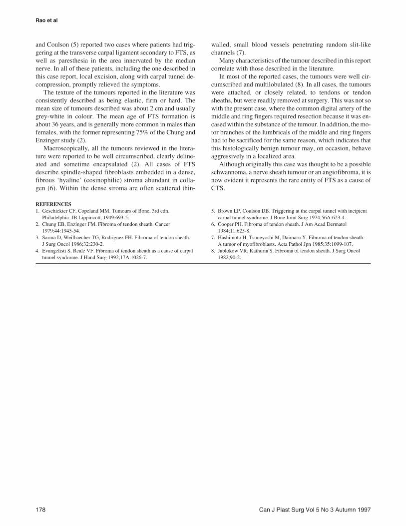
The excised tumour measured 3.6x1.6x0.9 cm and was
firm, nodular, well circumscribed and grey-tan coloured,
with a thin layer of adipose tissue (Figure 2). Microscopic ex-
amination revealed an encapsulated benign fibrovascular le-
sion consisting of spindle-shaped fibroblasts embedded
within dense fibrocoliagenous stroma (Figure 3). The stroma
also exhibited vascular spaces, but no giant cells were noted
(Figure 4). Few lymphocytes were seen scattered along the
periphery of the tumour. No neural structures were seen
around the tumour.
Postoperatively, the patient’s symptoms disappeared, and
he was able to return to work four weeks later.
DISCUSSIONCompression from benign tumours has been included in the
differential diagnosis of CTS, and although documentation is
rare, FTS is no exception. In the Chung and Enzinger study
(2), two patients who had FTS in the hand showed symptoms
of CTS, while another patient showed paresthesia. Sarma et
al (3) reported an encapsulated FTS found during carpal tun-
nel release. This tumour was attached to the flexer digitorum
profundus tendon of the middle finger and was compressing
the median nerve enough to cause a complete loss of sensa-
tion along its distribution, as well as partial block of flexion
of the third digit. One patient suffering from FTS in the mid-
palm demonstrated most of the classic symptoms of CTS, in-
cluding diminished sensitivity and thenar atrophy (4). Brown
Can J Plast Surg Vol 5 No 3 Autumn 1997 177
Fibroma of tendon sheath as a cause of CTS
Figure 3) The tumour appears encapsulated and shows fibrocollage-
nous stroma. Notice the prominent vasculature with small blood vessels
(hematoxylin and eosin x100)
Figure 4) Section of the tumour stained with the factor VIII antibody,
showing prominent vascularized stroma (peroxidase antiperoxidasex100)Figure 2) Gross appearance of removed tumour
Figure 1) Intraoperative appearance of fibroma of tendon sheath. Infe-
rior arrow points to distal extent of the tumour. A branch of the median
nerve is stretched by the underlying tumour (superior arrow)
and Coulson (5) reported two cases where patients had trig-
gering at the transverse carpal ligament secondary to FTS, as
well as paresthesia in the area innervated by the median
nerve. In all of these patients, including the one described in
this case report, local excision, along with carpal tunnel de-
compression, promptly relieved the symptoms.
The texture of the tumours reported in the literature was
consistently described as being elastic, firm or hard. The
mean size of tumours described was about 2 cm and usually
grey-white in colour. The mean age of FTS formation is
about 36 years, and is generally more common in males than
females, with the former representing 75% of the Chung and
Enzinger study (2).
Macroscopically, all the tumours reviewed in the litera-
ture were reported to be well circumscribed, clearly deline-
ated and sometime encapsulated (2). All cases of FTS
describe spindle-shaped fibroblasts embedded in a dense,
fibrous ‘hyaline’ (eosinophilic) stroma abundant in colla-
gen (6). Within the dense stroma are often scattered thin-
walled, small blood vessels penetrating random slit-like
channels (7).
Many characteristics of the tumour described in this report
correlate with those described in the literature.
In most of the reported cases, the tumours were well cir-
cumscribed and multilobulated (8). In all cases, the tumours
were attached, or closely related, to tendons or tendon
sheaths, but were readily removed at surgery. This was not so
with the present case, where the common digital artery of the
middle and ring fingers required resection because it was en-
cased within the substance of the tumour. In addition, the mo-
tor branches of the lumbricals of the middle and ring fingers
had to be sacrificed for the same reason, which indicates that
this histologically benign tumour may, on occasion, behave
aggressively in a localized area.
Although originally this case was thought to be a possible
schwannoma, a nerve sheath tumour or an angiofibroma, it is
now evident it represents the rare entity of FTS as a cause of
CTS.
REFERENCES1. Geschickter CF, Copeland MM. Tumours of Bone, 3rd edn.
Philadelphia: JB Lippincott, 1949:693-5.
2. Chung EB, Enzinger FM. Fibroma of tendon sheath. Cancer
1979;44:1945-54.
3. Sarma D, Weilbaecher TG, Rodriguez FH. Fibroma of tendon sheath.
J Surg Oncol 1986;32:230-2.
4. Evangelisti S, Reale VF. Fibroma of tendon sheath as a cause of carpal
tunnel syndrome. J Hand Surg 1992;17A:1026-7.
5. Brown LP, Coulson DB. Triggering at the carpal tunnel with incipient
carpal tunnel syndrome. J Bone Joint Surg 1974;56A:623-4.
6. Cooper PH. Fibroma of tendon sheath. J Am Acad Dermatol
1984;11:625-8.
7. Hashimoto H, Tsuneyoshi M, Daimaru Y. Fibroma of tendon sheath:
A tumor of myofibroblasts. Acta Pathol Jpn 1985;35:1099-107.
8. Jablokow VR, Kathuria S. Fibroma of tendon sheath. J Surg Oncol
1982;90-2.
178 Can J Plast Surg Vol 5 No 3 Autumn 1997
Rao et al