fibrinolytic activities of a medicinal mushroom: …studentsrepo.um.edu.my/4918/1/thesis.pdf · dan...
TRANSCRIPT
FIBRINOLYTIC ACTIVITIES OF A MEDICINAL MUSHROOM: LIGNOSUS RHINOCEROTIS (COOKE) RYVARDEN
KHO TIENG TIENG
FACULTY OF SCIENCE UNIVERSITY OF MALAYA
KUALA LUMPUR
2014
FIBRINOLYTIC ACTIVITIES OF A MEDICINAL MUSHROOM: LIGNOSUS RHINOCEROTIS (COOKE) RYVARDEN
KHO TIENG TIENG
DISSERTATION SUBMITTED IN FULFILLMENT OF THE REQUIREMENTS FOR THE DEGREE OF
MASTER OF BIOTECHNOLOGY
INSTITUTE OF BIOLOGICAL SCIENCES FACULTY OF SCIENCE
UNIVERSITY OF MALAYA KUALA LUMPUR
2014
UNIVERSITI MALAYA
ORIGINAL LITERARY WORK DECLARATION Name of Candidate: KHO TIENG TIENG I/C/Passport No: 871203-52-5872
Regisration/Matric No.: SGF110014
Name of Degree: MASTER OF BIOTECHNOLOGY Title of Project Paper/Research Report/Dissertation/Thesis (“this Work”):
“FIBRINOLYTIC ACTIVITIES OF A MEDICINAL MUSHROOM: LIGNOSUS RHINOCEROTIS (COOKE) RYVARDEN”
Field of Study: MUSHROOM BIOTECHNOLOGY I do solemnly and sincerely declare that: (1) I am the sole author/writer of this Work, (2) This Work is original, (3) Any use of any work in which copyright exists was done by way of fair dealing and for
permitted purposes and any excerpt or extract from, or reference to or reproduction of any copyright work has been disclosed expressly and sufficiently and the title of the Work and its authorship have been acknowledged in this Work,
(4) I do not have any actual knowledge nor do I ought reasonably to know that the making of this work constitutes an infringement of any copyright work,
(5) I hereby assign all and every rights in the copyright to this Work to the University of Malaya (“UM”), who henceforth shall be owner of the copyright in this Work and that any reproduction or use in any form or by any means whatsoever is prohibited without the written consent of UM having been first had and obtained,
(6) I am fully aware that if in the course of making this Work I have infringed any copyright whether intentionally or otherwise, I may be subject to legal action or any other action as may be determined by UM.
(Candidate Signature) Date: Subscribed and solemnly declared before, Witness’s Signature Date:
Name PROFESSOR DR VIKINESWARY SABARATNAM
Designation
Witness’s Signature Date:
Name PROFESSOR DR SEKARAN A/L MUNIANDY
Designation
ii
ABSTRACT
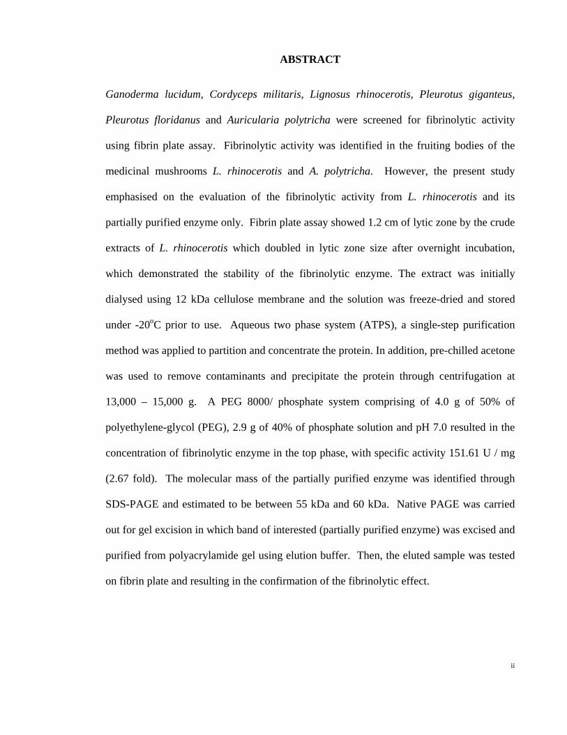
Ganoderma lucidum, Cordyceps militaris, Lignosus rhinocerotis, Pleurotus giganteus,
Pleurotus floridanus and Auricularia polytricha were screened for fibrinolytic activity
using fibrin plate assay. Fibrinolytic activity was identified in the fruiting bodies of the
medicinal mushrooms L. rhinocerotis and A. polytricha. However, the present study
emphasised on the evaluation of the fibrinolytic activity from L. rhinocerotis and its
partially purified enzyme only. Fibrin plate assay showed 1.2 cm of lytic zone by the crude
extracts of L. rhinocerotis which doubled in lytic zone size after overnight incubation,
which demonstrated the stability of the fibrinolytic enzyme. The extract was initially
dialysed using 12 kDa cellulose membrane and the solution was freeze-dried and stored
under -20oC prior to use. Aqueous two phase system (ATPS), a single-step purification
method was applied to partition and concentrate the protein. In addition, pre-chilled acetone
was used to remove contaminants and precipitate the protein through centrifugation at
13,000 – 15,000 g. A PEG 8000/ phosphate system comprising of 4.0 g of 50% of
polyethylene-glycol (PEG), 2.9 g of 40% of phosphate solution and pH 7.0 resulted in the
concentration of fibrinolytic enzyme in the top phase, with specific activity 151.61 U / mg
(2.67 fold). The molecular mass of the partially purified enzyme was identified through
SDS-PAGE and estimated to be between 55 kDa and 60 kDa. Native PAGE was carried
out for gel excision in which band of interested (partially purified enzyme) was excised and
purified from polyacrylamide gel using elution buffer. Then, the eluted sample was tested
on fibrin plate and resulting in the confirmation of the fibrinolytic effect.

iii
ABSTRAK
Ganoderma lucidum, Cordyceps militaris, Lignosus rhinocerotis, Pleurotus giganteus,
Pleurotus floridanus dan Auricularia polytricha ditapis melalui asai fibrinolitik. Aktiviti
fibrinolitik telah dikenalpasti dalam janabuah cendawan perubatan L. rhinocerotis dan A.
polytricha. Walau bagaimanapun, kajian ini akan menekankan penilaian aktiviti
fibrinolitik daripada L. rhinocerotis dan enzim yang separa tulen sahaja. Asai fibrinolitik
daripada ekstrak mentah L. rhinocerotis menunjukkan zon penguraian sebesar 1.2 cm dan
membesar dua kali ganda selepas eraman semalaman, di mana ia menunjukkan kestabilan
enzim fibrinolitik tersebut. Ekstrak tersebut telah didialisis dengan menggunakan 12 kDa
membran selulosa sebelum larutan tersebut dikeringkan secara sejuk-beku. Selepas proses
pengeringan, ekstrak tersebut disimpan di dalam -20oC sebelum mengguna. Sistem dua
fasa cair yang merupakan cara satu langkah penulenan telah digunakan untuk memisah dan
menumpukan protein. Secara tambahan, aseton sejuk digunakan untuk mengeluarkan
bendasing dan memendakkan protein melalui pengemparan pada 13,000–15,000 g. Sistem
yang menggunakan PEG 8000/ phosphate mengandungi 4 g daripada 50% polietilena
glikol (PEG), 2.9 g daripada 40% fosfat larutan dan pH 7.0, telah memberi keputusan di
mana enzim fibrinolitik telah ditumpukan ke fasa atas dengan aktiviti spesifik 151.61 U /
mg (2.67 ganda). Jisim molekul enzim yang ditulenkan telah dianggarkan oleh SDS-PAGE
dan anggaran jisim molekul adalah 55 kDa dan 60 kDa. Native PAGE telah digunakan
untuk pemotongan gel di mana enzim yang separa tulen dipotong keluar dan gel
dibersihkan dengan menggunakan penampan. Aktiviti fibrinolitik telah dikenalpasti
daripada enzim-enzim yang dibersihkan.

iv
ACKNOWLEGEMENT
With completion of this thesis, there are many who deserve a word of appreciation. First, I
would like to express my sincere appreciation and deepest gratitude to my supervisor, Prof.
Dr. Vikineswary Sabaratnam, Faculty of Science, University of Malaya, whom had
provided me the opportunity to perform and complete my project research in Mycology and
Plant Pathology Laboratory. Thanks a million for her countless advice, patience and
guidance, definitely deserves more recognition than a word of merit. Also, I would like to
thank to my co-supervisor, Prof. Dr. Sekaran A/L Muniandy, from the Faculty of Medicine,
University of Malaya, for his advice and guidance. Next, I would like to thank all my
friends who had been with me with their constant support throughout the progression of the
thesis since day one. Thank to University of Malaya’s grant and scholarships. Last but not
least, I would like to express my gratitude to my lovely family members who always
supported me, accommodating and understanding in every way. Thank you to all who had
made this thesis a success.
v
TABLE OF CONTENTS
ABSTRACT ii
ACKNOWLEDGEMENT iv
TABLE OF CONTENTS v
LIST OF TABLES viii
LIST OF FIGURES ix
LIST OF ABBREVIATIONS x
INTRODUCTION 1
CHAPTER I
LITERATURE REVIEW
CHAPTER II
2.1 Cardiovascular disease 8
2.1.1 Fibrinolytic mechanism 9
2.1.2 Atherosclerosis 12
2.2 Aqueous two phase system (ATPS) 14
MATERIAL AND METHODS
CHAPTER III
3.1 Preparation of crude extract 16
3.2 Preliminary fibrinolytic test (fibrin plate method) 16
3.3 Aqueous two phase system (ATPS) 17

vi
3.4 Acetone precipitation of proteins 17
3.5 Fibrinolytic assay (Folin-spectrophotometric method) 18
3.5.1 Preparation of L-tyrosine calibration plot 20
3.5.2 Preparation of protein calibration plot 21
3.6 Sodium dodecyl sulfate – polyacrylamide gel electrophoresis (SDS-PAGE) 22
3.7 Protein purification of polyacrylamide gel
3.7.1 Identification and excision of band of interest 24
3.7.2 Protein elution from the gel matrix 24
3.8 Statistical analysis 25
RESULTS
CHAPTER IV
4.1 Preliminary fibrinolytic assay (fibrin plate assay) 26
4.2 Fibrin plate assay for crude extract and partially purified enzyme 27
4.3 Fibrinolytic assay (Folin-spectrophotometric method) 28
4.4 Sodium dodecyl sulfate-polyacrylamide gel electrophoresis 30
4.5 Excision of the band of interest 31
DISCUSSION AND CONCLUSIONS
Chapter V
5.1 Preparation of crude extracts 33
5.2 Preliminary fibrinolytic assay (fibrin plate method) 35
5.3 Purification of fibrinolytic enzyme 36
5.4 Fibrin plate assay for crude extract and partially purified enzyme 38
vii
5.5 Fibrinolytic assay (Folin-spectrophotometric method) 39
5.6 Molecular mass estimation of partially purified enzyme 41
5.7 Native PAGE and excision of band of interest 42
5.8 Recommendations 42
5.9 Conclusions 43
REFERENCES 45
Appendix A: MEDIA AND METHODS 55
Appendix B: STATISTICAL ANALYSIS 60
Appendix C: RAW DATA 62

viii
LIST OF TABLES
Table Description Page 3.1 Summary of experimental procedures for preparation of
calibration plot with L-tyrosine solution (µM) and the changes of absorbance reading at 660 nm
21
3.2 Changes of absorbance at 595 nm 22 3.3 Preparation of 12% SDS-PAGE gel 23 4.1 Diameter of lytic zone (cm) observed after one hour of incubation 26 4.2 Fibrin plate assay for crude extract and partially purified enzyme 27 4.3 Specific activity and purification fold of crude extract and
partially purified enzyme from Lignosus rhinocerotis 29
B.1 Normality test - Fibrinolytic activities of crude extract and partially purified enzymes from Lignosus rhinocerotis
62
B.2 Test of homogeneity of variances - Fibrinolytic activities of crude extract and partially purified enzymes from Lignosus rhinocerotis
62
B.3 ANOVA - Fibrinolytic activities of crude extract and partially purified enzymes from Lignosus rhinocerotis
62
B.4 Duncan post-hoc test - Fibrinolytic activities of crude extract and partially purified enzymes from Lignosus rhinocerotis
63
C.1 Diameter of lytic zone (cm) observed in 18 plates after one hour of incubation at 37±2oC
64
C.2 Fibrin plate assay for crude extract and partially purified enzyme 65 C.3 Fibrinolytic activities of crude extract and partially purified
enzymes from L. rhinocerotis 66
ix
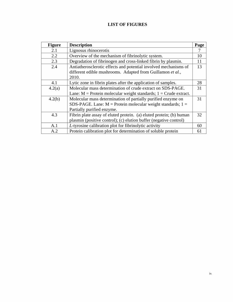
LIST OF FIGURES
Figure Description Page2.1 Lignosus rhinocerotis 7 2.2 Overview of the mechanism of fibrinolytic system. 10 2.3 Degradation of fibrinogen and cross-linked fibrin by plasmin. 11 2.4 Antiatherosclerotic effects and potential involved mechanisms of
different edible mushrooms. Adapted from Guillamon et al., 2010.
13
4.1 Lytic zone in fibrin plates after the application of samples. 28 4.2(a) Molecular mass determination of crude extract on SDS-PAGE.
Lane: M = Protein molecular weight standards; 1 = Crude extract. 31
4.2(b) Molecular mass determination of partially purified enzyme on SDS-PAGE. Lane: M = Protein molecular weight standards; 1 = Partially purified enzyme.
31
4.3 Fibrin plate assay of eluted protein. (a) eluted protein; (b) human plasmin (positive control); (c) elution buffer (negative control)
32
A.1 L-tyrosine calibration plot for fibrinolytic activity 60 A.2 Protein calibration plot for determination of soluble protein 61

x
LIST OF SYMBOLS AND ABBREVIATIONS
µg Microgram µg/mL Microgram per microlitre µl Microlitre µM Micromolar µmole Micromole ANOVA Analysis of Variance APS Ammonium persulfate ATPS Aqueous two phase system BSA Bovine serum albumin cm Centimeter g Gravity g Gram HCl Hydrochloric acid kDa Kilo Dalton LSD Least significant difference M Molar mg Milligram mg/ml Milligram per millilitre ml Millilitre mM Millimolar N Normality NaCl Sodium chloride nm nanometer OD Optical density PEG Polyethylene glycol rpm Rotation per minute SDS Sodium dodecyl sulfate-polyacrylamide SDS-PAGE Sodium dodecyl sulfate-polyacrylamide gel electrophoresis Tris-HCl Tris hydrochloride U Unit U/mg Unit per milligram v/v Volume per volume w/v Weight per volume

1
CHAPTER I
INTRODUCTION
Mushrooms are commonly used as food, food flavouring substances and traditional
medicines in eastern countries. They are reported to be high in protein, with a
significant content of essential amino acid, but low in fat (Guillamon et al., 2010).
Besides, mushrooms supply a large amount of carbohydrates, fiber, vitamins (B1, B2,
B12, C and D) and mineral ions (Ca, K, Mg, Na, P, Cu, Fe, Mn and Se).
The amino acid composition of mushroom proteins is comparable to animal
protein, and it can counterbalance the high consumption of animal proteins especially in
developed countries. Besides, edible mushroom is a unique food which contains many
different bioactive compounds such as eritadenine (Mattila et al., 1999) and phenolic
compounds (Barros et al., 2007 & 2008) that may be beneficial in cardiovascular
disease treatment. Cardiovascular disease is major cause of death and therefore, there is
worldwide interest in discovering the potential therapeutic drugs, supplements, and food
for better health and lifestyle.
In recent years, variety of mushrooms have been studied for their
pharmacological effect and reported to have great benefits for human health. These
mushrooms or their extracts which can be used as therapeutic agents are generally
known as medicinal mushrooms. Typically, medicinal mushrooms are edible.
Nevertheless, edible mushrooms may not necessarily possess therapeutic properties and
are usually used only as food.

2
According to the world health statistics 2012 by World Health Organization
(WHO), the annual number of deaths due to cardiovascular disease will increase from
17 million in 2008 to 25 million in 2030 (World Health Organization, 2012). In
Malaysia, the mortality rate from cardiovascular disease in 2008 is 263 deaths per
100,000 population, and the majority caused by ischaemic heart disease (OECD/WHO,
2012). Cardiovascular disease has long been the leading cause of death in developed
countries. It becomes increasingly prevalent in Asian countries, comprising about one
third of all deaths in Asians. Cardiovascular disease includes a range of diseases that
are related to circulatory system. The most prevalent diseases are ischaemic heart
disease (or heart attack) and cerebrovascular disease (or stroke). These two diseases
have contributed to three quarters of all cardiovascular deaths in Asian countries
(OECD/WHO, 2012).
Atherosclerosis is a condition in which fatty material collects along the walls of
arteries. The fatty material thickens and hardens with calcium deposition, and
eventually blocks the arteries. Once the blood vessels are injured, platelets tend to
aggregate and react with fibrin to form thrombus. Thrombus is abnormal blood clot
formed within the blood vessel that obstructs the flow of blood and nutrients to vital
tissue. When there is phenomenon of reduced blood supply to heart muscle, the
syndrome is known as Ischaemic heart disease.
In biological system, thrombosis can be prevented through fibrinolysis. The
insoluble fibrin fiber is hydrolyzed into fibrin degradation products by plasmin, which
is generated from plasminogen by plasminogen activators such as the tissue

3
plasminogen activator, vascular plasminogen activator, blood plasminogen activator,
urokinase, Hageman factor and streptokinase-plasminogen complex (Shen et al., 2007).
Thrombolytic agents have been extensively used in therapeutic treatment of thrombosis
nowadays. The commonly used thrombolytic agents include streptokinase, urokinase,
alteplase, reteplase and tenecteplase. Thrombolytic drugs breakdown dangerous blood
clots in blood vessels, improve blood flow, and prevent damage to tissues and organs.
However, these thrombolytic agents are of relatively high cost and exhibit low
specificity to fibrin. Besides, all of these thrombolytic agents could cause undesirable
side effect such as bleeding complications, which is the most common consequences of
anticoagulant therapy. Streptokinase is capable of eliciting antigenic response in human
since the protein is obtained from streptococci cultures. The usage of streptokinase is
previously prohibited or contraindicated because of the risk of anaphylaxis.
Streptokinase is also relatively non- specific thrombolytic agent which can lead to
systemic fibrinolysis and lysis of normal hemostatic plugs. Hence, the search for safe
thrombolytic agents from other sources is necessary. Fortunately, fibrinolytic enzymes
were discovered in insects (Hahn et al., 1999), earthworm (Cho et al., 2004), snake
venom (Sun et al., 2006) and food-grade microorganism (Wang et al., 2006).
For mushrooms, some studies reported that mushrooms do have therapeutic
benefit for cardiovascular disease. The hypocholesterolemic action of edible
mushrooms has been reported in the early work of Kaneda and Tokuda (1966).
Lentinus edodes (Berk.) Singer, Auricularia polytricha (Mont.) Sacc., Flammulina
velutipes (Curtis) Singer and Agaricus bisporus (J.E.Lange) Imbach have been reported
to have cholesterol lowering properties. On the other hand, some mushrooms have
hypotensive effect when blood pressure is already high (Kabir et al., 1987 & 1988;
4
Miyazawa et al., 2008). Besides, the properties of mushrooms such as antioxidant
(Cheung et al., 2003; Wong & Chye, 2009) and anti-inflammatory (Jose & Janardhanan,
2004; Khohno et al., 2008) might be helpful in the management of heart and blood
circulation.
Today, several mushrooms had been studied for their fibrinolytic activities and
the corresponding fibrinolytic enzymes were also identified: Schizophyllum commune
(Fr) (Lu et al., 2010), Armillariella mellea (Vahl) P. Karst (Kim and Kim, 1999),
Grifola frondosa (Dicks.) Gray (Nonaka et al., 1997), Pleurotus ostreatus (Jacq.) P.
Kumm. (Shen et al., 2007) and Fomitella fraxinea (Bull.) Imazeki (Lee et al., 2006).
Differing from plasminogen activators such as streptokinase and urokinase, these
fibrinolytic enzymes are plasmin-like proteins which directly perform fibrinolytic action.
An ideal thrombolytic drug should be effective in breaking down fresh and older
thrombi, be rapid in its action with complete dissolution of the thrombus, be able to be
given as a bolus and be safe without hypotensive, allergic or immunogenic reactions
(Thomson, 1999).
According to current dietary recommendations for health, mushrooms are
considered as appropriate choice and its consumption can affect some known
cardiovascular risk biomarkers. Besides the known hypocholestrolemic effect, the
presence of antioxidant and anti-inflammatory compound in mushrooms may
synergistically contribute to reduce the atherosclerosis risk. Compared to other sources,
mushrooms are considered as a good choice to treat or prevent cardiovascular disease as
it can be included in our daily diet and suitable for vegetarians. Although some

5
mushrooms had been consumed for therapeutic purpose in past, they have yet to
scientifically verified and reported. On the other hand, therapeutic effects of
mushrooms obtained from different geographical region might act differently.
Therefore, in this study, medicinal mushrooms investigated include Ganoderma
lucidum (Curtis) P. Karst, Cordyceps militaris (L.) Fr., and Lignosus rhinocerotis
(Cooke) Ryvarden. For edible medicinal mushrooms, Pleurotus giganteus (Berk.)
Karun. & K.D. Hyde, Pleurotus floridanus (Singer) and A. polytricha were studied.
OBJECTIVES:
The objectives were to:
a. screen the selected edible and medicinal mushrooms for their fibrinolytic
activities.
b. isolate and purify the fibrinolytic enzymes from the mushroom.
c. characterize the fibrinolytic enzyme by estimation of the molecular mass.

6
CHAPTER II
LITERATURE REVIEW
Mushrooms are universally consumed as food, food favouring substances and
traditional medicines in eastern countries due to its tastiness, high nutritional values,
and pharmacological properties (Chang & Miles, 1989; Lindequist et al., 2005). It is
well established that mushroom extracts contain a wide variety of compounds such as
protein, polysaccharides, fibre, lectins, and polyphenols, and each of the compounds
consists its own pharmacological effects (Smiderle et al., 2010).
Lignosus rhinocerotis (Figure 2.1), also known as “tiger milk mushroom”,
belongs to the Polyporaceae family and is one of the most important medicinal
mushrooms used by the indigenous people of Southeast Asia and China. In Malaysia,
the mushroom is locally named as “cendawan susu rimau” which means tiger’s milk
mushroom. It is traditionally used by the natives in peninsular Malaysia as traditional
medicine to cure cough, fever, asthma, food poisoning and as a general tonic (Lee et al.,
2011). In China, L. rhinocerus sclerotium is an expensive folk medicine used by
traditional Chinese physicians to treat liver cancer, chronic hepatitis, and gastric ulcer
(Wong & Cheung, 2008). It has more than 15 medicinal uses, however, there are
limited usage of this mushroom because of its low availability. The mushroom is
difficult to cultivate and previously, was only available from forest. Successful
cultivation of the mushroom was carried out in agar, solid, and spawn medium with
good yield, therefore contributing to large quantity production of the mushroom for
investigation and therapeutic purpose (Lee et al., 2011). In recent years, variety of
mushrooms have been tested for their pharmacological effect and reported to have great
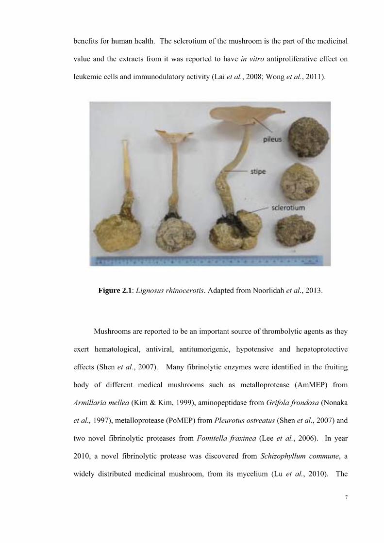
benefits for human health. The sclerotium of the mushroom is the part of the medicinal
value and the extracts from it was reported to have in vitro antiproliferative effect on
leukemic cells and immunodulatory activity (Lai et al., 2008; Wong et al., 2011).
Figure 2.1: Lignosus rhinocerotis. Adapted from Noorlidah et al., 2013.
Mushrooms are reported to be an important source of thrombolytic agents as they
exert hematological, antiviral, antitumorigenic, hypotensive and hepatoprotective
effects (Shen et al., 2007). Many fibrinolytic enzymes were identified in the fruiting
body of different medical mushrooms such as metalloprotease (AmMEP) from
Armillaria mellea (Kim & Kim, 1999), aminopeptidase from Grifola frondosa (Nonaka
et al., 1997), metalloprotease (PoMEP) from Pleurotus ostreatus (Shen et al., 2007) and
two novel fibrinolytic proteases from Fomitella fraxinea (Lee et al., 2006). In year
2010, a novel fibrinolytic protease was discovered from Schizophyllum commune, a
widely distributed medicinal mushroom, from its mycelium (Lu et al., 2010). The
7

8
mushroom was cultured with fermentation technology, and protein purification was
carried out by cross-flow filtration and fast performance liquid chromatography (FPLC)
system.
Fibrinolytic activity was determined by the fibrin plate assay in which fibrin clot
was made in petri dish at room temperature by 1.5% agarose, 0.2% human fibrinogen
and 10 U of human thrombin, the assay was modified from the method described by
Astrup and Mullertz, (1952). For the determination of protease activity, azocasein
assay was used, where azocasein hydrolysis was used as an measure of fibrinolytic
activity. Activity was determined by measuring the release of acid-soluble material
after protease digestion of azocasein. The protease isolated was fractionated through
Superdex 75 10/300 GL column and SDS-PAGE. This enzyme showed 21.32 kDa in
molecular mass and monomeric form in protein structure. The effect of temperature
and pH for optimal protease activity was being estimated as well, the optimal protease
activity displayed at the condition of pH 5.0 and 45oC. The activity was enhanced by
magnesium and inhibited by EDTA. Despite of low production ratio and recovery rate,
the fibrinolytic protease revealed 9.29 fold in specific activity after purification, and
showed greater activity than human plasmin.
2.1 Cardiovascular disease
Cardiovascular diseases are a group of heart and blood vessels disorders, including
rheumatic heart disease, hypertensive heart disease, ischemic heart disease,
cerebrovascular disease, inflammatory heart disease, congenital heart disease and heart
9
failure. Diseases like acute myocardial, valvular heart disease and stroke is the leading
cause of death in developed countries (Lu et al., 2010).
2.1.1 Fibrinolysis mechanism
Fibrinolysis is the aseptic dissolution of fibrin brought about by the direct action of a
mechanism existing in normal blood (Macfarlane & Biggs, 1948). Generally,
dissolutions take days to week to complete, but the action may be accelerated as a result
of natural changes occurring in the living subject or of experimental procedures as to
occur within a few hours or minutes (Macfarlane & Biggs, 1948). This acceleration
phenomenon had raised the interest of scientists, though a particular set of factors have
been studied as being apparently those mainly responsible, it is recognized that others
may be involved.
Under physiological conditions, fibrinolysis is highly regulated mechanism that
integrates with the coagulation system through several direct molecular links. Both
coagulation and fibrinolysis are precisely regulated by measured participation of
substrates, activators, inhibitors, cofactors and receptors. Besides preventing blood loss,
these co-ordinated molecular events insure blood fluidity (Cesarman-Maus & Hajjar,
2005). Plasminogen (PLG) is a circulating plasma zymogen which can be converted to
plasmin by both tissue plasminogen activator (tPA) and by urokinase (uPA). Through
positive feedback mechanism, plasmin cleaves both tPA and uPA from single chain to
more active two-chain polypeptides. Fibrin, which is the substrate to plasmin, bind with
both plasminogen and tPA on its surface to regulate its own degradation and therefore,
localizing and enhancing plasmin generation. Plasminogen activator (tPA) is a weak

activator in the absence of fibrin, its catalytic efficiency for PLG activation is enhanced
by at least two orders of magnitude in the presence of fibrin. In other words, the
affinity between tPA and plasminogen is low in the absence of fibrin, and vice versa.
The mechanism of fibrinolytic system is illustrated in Figure 2.2 below.
Figure 2.2: Overview of the mechanism of fibrinolytic system. The zymogen plasminogen is converted to the active serine protease, plasmin, through the action primarily of two-chain tissue plasminogen activator (tc-tPA) or two-chain urokinase (tc-uPA). These activators are secreted as single-chain (sc-tPA and scuPA) forms from endothelial cells, and from renal epithelium, monocyte/macrophages, or endothelial cells respectively. Both tPA and uPA can be inhibited by plasminogen activator inhibitor-1 (PAI), while plasmin is inhibited by its major inhibitor, a2-plasmin inhibitor (a2-PI), and to a lesser extent by a2-macroglobulin (a2-MG). Once plasmin is generated, it converts single chain tPA and uPA to double chain forms. It is then rapidly inhibited unless it remains bound to fibrin or to its cell surface receptors. Adapted from Cesarman-Maus and Hajjar (2005).
10
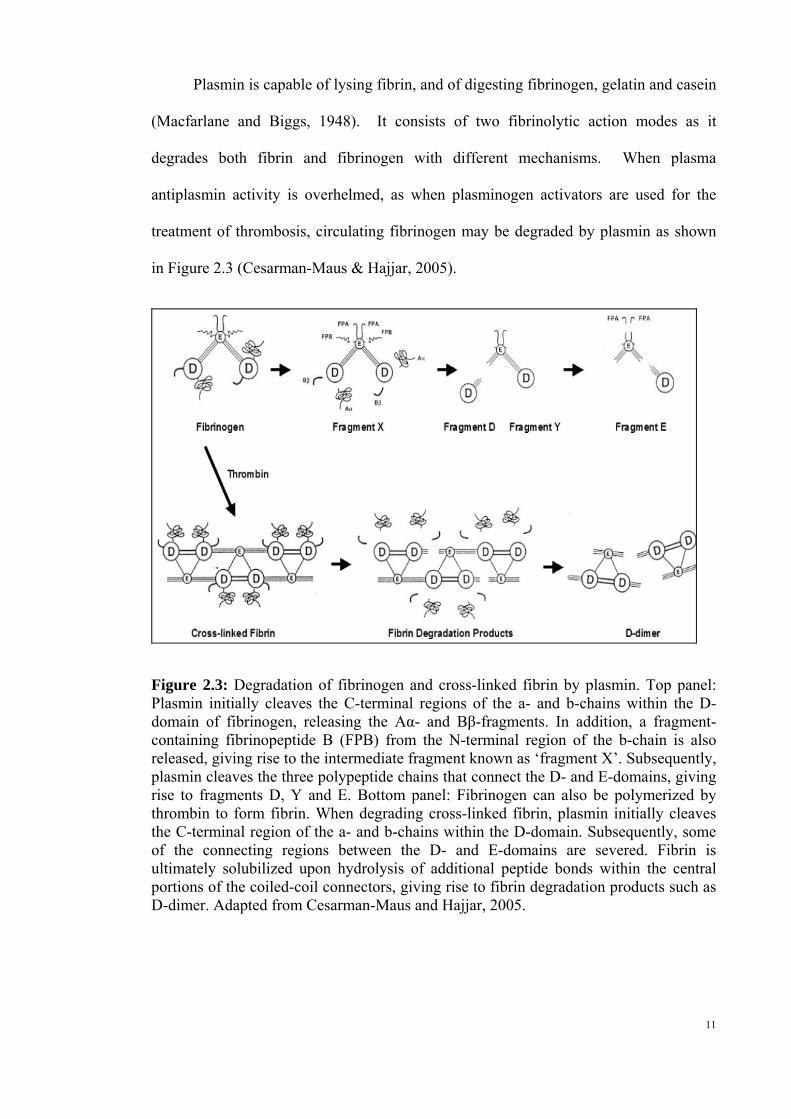
Plasmin is capable of lysing fibrin, and of digesting fibrinogen, gelatin and casein
(Macfarlane and Biggs, 1948). It consists of two fibrinolytic action modes as it
degrades both fibrin and fibrinogen with different mechanisms. When plasma
antiplasmin activity is overhelmed, as when plasminogen activators are used for the
treatment of thrombosis, circulating fibrinogen may be degraded by plasmin as shown
in Figure 2.3 (Cesarman-Maus & Hajjar, 2005).
Figure 2.3: Degradation of fibrinogen and cross-linked fibrin by plasmin. Top panel: Plasmin initially cleaves the C-terminal regions of the a- and b-chains within the D-domain of fibrinogen, releasing the Aα- and Bβ-fragments. In addition, a fragment-containing fibrinopeptide B (FPB) from the N-terminal region of the b-chain is also released, giving rise to the intermediate fragment known as ‘fragment X’. Subsequently, plasmin cleaves the three polypeptide chains that connect the D- and E-domains, giving rise to fragments D, Y and E. Bottom panel: Fibrinogen can also be polymerized by thrombin to form fibrin. When degrading cross-linked fibrin, plasmin initially cleaves the C-terminal region of the a- and b-chains within the D-domain. Subsequently, some of the connecting regions between the D- and E-domains are severed. Fibrin is ultimately solubilized upon hydrolysis of additional peptide bonds within the central portions of the coiled-coil connectors, giving rise to fibrin degradation products such as D-dimer. Adapted from Cesarman-Maus and Hajjar, 2005.
11
12
Plasmin act on the proteolytic cleavage sites on fibrinogen, and subsequently give
rise to fragments [Aα, Bβ and fragment fibrinopeptide B (FPB)] for the C- and N-
termini of fibrinogen’s three polypeptide chains. The resulting molecule is called
fragment X which represents a clottable form of fibrinogen. Additional cleavage results
in releasing other peptides, and in a series of subsequent reactions, plasmin may further
cleave the three polypeptide chains that convert the D- and E- domains. Some of these
fragments inhibit the spontaneous polymerization of fibrinogen (Cesarman-Maus &
Hajjar, 2005). On the other hand, when fibrin cross-linked by factor XIII, is degraded
by plasmin and subsequently fragments known as D-dimers are released (Cesarman-
Maus & Hajjar, 2005). In clinical therapy, assays for cross-linked D-dimer fragments
are employed to identify disseminated intravascular coagulation-like states associated
with excessive plasmin-mediated fibrinolysis (Cesarman-Maus & Hajjar, 2005).
2.1.2 Atherosclerosis
Atherosclerosis is a progressive disease characterized by the accumulation of lipids and
fibrous elements in the large arteries, constitutes the single most important contributor
to the growing burden of cardiovascular diseases (Libby, 2002). Several mechanisms
involved in the antiatherosclerotic effect have been reported (Figure 2.4) (Guillamon et
al., 2010). Research in the last two decades has discovered that inflammatory and
oxidative processes are common features in several cardiovascular conditions, such as
atherosclerosis (Guillamon et al., 2010). Abnormal blood clot called thrombus within
the vascular system is formed by the aggregation of fibrin, and the formation of fibrin is
triggered from a precursor fibrinogen through the proteolytic action of thrombin.
Consequently, it obstructs the flow of blood and nutrients to vital tissue.

In biological system, thromboses can be prohibited through fibrinolysis. The
insoluble fibrin fiber is hydrolyzed into fibrin degradation products by plasmin, which
is generated from plasminogen by plasminogen activators such as the tissue
plasminogen activator, vascular plasminogen activator, blood plasminogen activator,
urokinase, Hageman factor and streptokinase-plasminogen complex (Shen et al., 2007).
Figure 2.4: Antiatherosclerotic effects and potential involved mechanisms of different edible mushrooms. Adapted from Guillamon et al., 2010.
Today, thrombolytic agents have been extensively used in therapeutic treatment
of thrombosis. Thrombolytic agents are classified into: plasminogen activator and
plasmin-like protein according to their fibrinolysis mechanisms. Plasminogen
activators such as tissue-type plasminogen activator (tPA), urokinase-type plasminogen
activator and bacterial plasminogen activator streptokinase, activate the zymogen
plasminogen to generate plasmin for fibrinolysis system. In clinical therapy,
plasminogen activators are the most widely used among the thrombolytic agents
13

14
available. Despite the extensive use, all of these plasminogen activators have
undesirable side effects such as resistance to reperfusion, the occurrence of acute
coronary reocclusion, and bleeding complications (Kim & Kim, 1999). Besides the
relatively high cost, the thrombolytic agents also exhibit low specificity for fibrin (Shen
et al., 2007). Plasmin-like proteins which perform fibrinolytic actions directly were
discovered from snake venom, earthworm, microorganisms and fermented foods like
Japanese natto, Korean chungkook-jang and Chinese douchi (Shen et al., 2007).
2.2 Aqueous two phase system (ATPS)
Aqueous two phase system (ATPS) is an ideal technology combining or integrating
clarification, concentration, and partial purification into one single step, and therefore it
is prevalent as it is able to shorten and reduce the purification process of protein.
Comparing to the conventional liquid-liquid extraction, ATPS has the advantage of
preserving the targeted biomolecule with high water content of both phases (70-85%
w/w), high biocompatibility and low interfacial tension, low degradation of
biomolecules, good resolution, high separation yield, relatively high capacity, ease of
scale-up, low material costs and the possibility of polymer and salt recycling
(Ramyadevi et al., 2012). Hence, ATPS have been studied extensively on a laboratory
scale for the partitioning of whey milk protein, lysozyme (Su & Chiang, 2006), amino
acids and peptides (Ramyadevi et al., 2012).
Recently, the potential application of aqueous two phase systems (ATPS) has
been demonstrated for the recovery of recombinant protein expressed in plants such as
tobacco and seeds (Gu & Glatz, 2007; Platis & Labrou, 2006). Ibarra-Herrera et al.

15
(2011) reported that potential application of selected ATPS as primary step in the
recovery process of recombinant human proteins expressed in plants green-tissue,
alfalfa. The partition using PEG 8000/phosphate systems comprising of 16.1% (w/w)
of polyethylene glycol (PEG), 10% phosphate, tie-line length of 35.7% (w/w), volume
ratio equal to one and pH of 7.0 resulted in the potential recovery of 88% of the rhG-
CSF (granulocyte-colony stimulating factor) in the top phase. Meanwhile, there was
concentration of 93% of alfalfa contaminant proteins at the interface and bottom phase.
Biopharmaceutical products have been discovered in plant species including
tobacco, potato, rice, soybean alfalfa, tomato and lettuce (Ma et al., 2003; Stoger et al.,
2002). Bioproducts were found to have therapeutic value or diagnostics proteins,
industrial proteins, nutritional supplements such as minerals, vitamins, carbohydrates
and biopolymers (Ibarra-Herrera et al., 2011). Several reports have revealed that
therapeutic proteins produced by the bioreactors obtained from plants consist of several
advantages. These advantages include the lack of animal pathogenic contaminants, low
cost, larger production level, the presence of natural storage organs such as seeds and
tubers, and existing technology for harvesting and processing of plant material (Ibarra-
Herrera et al., 2011).

16
CHAPTER III
MATERIALS AND METHODS
3.1 Preparation of crude extract
Lyophilised powder of L. rhinocerotis (Tiger milk mushroom), C. militaris, G. lucidum,
A. polytricha, P. floridanus and P. giganteus were available in the lab and used in this
study. For each sample, 10g of the freeze- dried powder was initially suspended in 200
ml of 20mM Tris-hydrochloride buffer (pH 8.0) [Tris-HCl buffer (pH 8.0)] and then
physically mixed in an ice bath for 30 minutes. The suspension was then centrifuged at
10,000 g for 30 minutes at 4 ± 2 oC to remove cell debris. Then, the supernatant was
dialysed two to three times using a cellulose membrane with molecular weight 12,000
kDa cut off range, against distilled water. The dialysed crude extract was stored at -20
± 2 oC prior to use.
3.2 Preliminary fibrinolytic assay (fibrin plate method)
The fibrin plate method used was a modified method from Kim and Kim (1999). Raw
fibrin (bovine blood) was freeze-dried and 0.6 % (w/v) freeze-dried fibrin powder was
dissolved in distilled water followed by homogenation at 11,000 g for 30 seconds. The
homogenate was then centrifuged at 2,800 g for 6 minutes to spin down the un-
dissolved fibrin. The supernatant was collected as fibrin solution and warmed up to 45
± 2 oC. Agarose was prepared at 2 % (w/v) with distilled water and maintained at 55 ±
2 oC. Fibrin solution was mixed with 2 % agarose in 1:1 ratio and then poured into petri
dishes. The clot was allowed to stand for one hour at room temperature. After the
medium solidified, a well was made in each of the plates with borer (5mm). Different
amount of freeze-dried crude extract was dissolved in 20 mM Tris-HCl buffer to obtain
17
different concentration of the crude extract. Twenty microliters (20 µl) of crude
extract/s was dropped into the well and then incubated at 37±2 oC for one hour. The
lytic zone (the diameter of clear transparent zone) was measured. The steps were
repeated by using 3mg/ml plasmin as positive control and Tris-HCl as negative control.
3.3 Aqueous two phase systems (ATPS)
The optimised system used was referred from the study of Ibarra-Herrera et al. (2011).
Four grams of polyethylene glycol 50 % (PEG 50 %) was added with 2.9 g of
phosphate 40 % and 1 g of sample. The system was topped up to 10 g by adding 2.1 g
of distilled water, and then mixed the solution by inverting the tube. Then, the tube was
centrifuged at 1,500 g for 10 minutes. The top and bottom phase was separated into two
different tubes. A blank was prepared by replacing the 1 g of sample with 1 g of Tris-
HCl.
3.4 Acetone precipitation of proteins
Partially purified enzyme from ATPS was precipitated by using acetone precipitation of
protein method (Wessel & Flugge, 1984). A required volume of acetone was cooled to
-20 oC. Protein sample was placed in acetone-compatible tube. Four times of the
sample volume of cold (-20oC) acetone was added. The tube was mixed using vortex
and incubated for 60 minutes at -20 oC. Then, the tube was centrifuged 10 minutes at
13,000 - 15,000 g. The supernatant was decanted and disposed properly, being careful
to not dislodge the protein pellet. The acetone was allowed to be evaporated from the
uncapped tube at room temperature for 30 minutes. Pellet was prevented from being
over-dried as it may not dissolve properly. Appropriate amount of distilled water was

18
added for the downstream process and mixed thoroughly through vortex to dissolve
protein pellet.
3.5 Fibrinolytic activity assay (Folin-spectrophotometry method)
The crude extract and partially purified enzyme were further evaluated by Folin-
spectrophotometer method (Yun et al., 2003) for their fibrinolytic capacity. Fibrin was
dissolved in 0.1 M Mcllvain buffer (pH 7.0) at concentration of 0.6 % (w/v). The
solution was then homogenised at 11,000 g for 30 seconds followed by centrifugation at
2,800 g for 10 minutes at room temperature. Supernatant was collected as fibrin
solution.
After that, 0.3 ml crude extract or partially purified enzyme was added to 0.6 ml
fibrin solution and incubated at 40oC for 10 minutes. The reaction was terminated by
adding 0.6 ml of 0.55 M trichloro-acetic acid solution for 10 minutes. Mixture was then
centrifuged at 2,800 g for 10 minutes to get the supernatant. One milliliter of
supernatant was mixed with 1 ml of 0.4 M sodium carbonate and subsequently 0.2 ml (1
N) of Folin-Ciocalteau reagent. The mixture was maintained at room temperature for
30 minutes and absorbance reading was then taken at 660 nm. A blank was prepared in
the same way except that 0.3 ml of enzyme sample was replaced with an equivalent
amount of Mcllvain buffer (pH 7.0) – for crude extract fibrinolytic assay. Another
blank was prepared in the same way except that 0.3 ml of enzyme sample was replaced
with an equivalent amount of ATPS blank – for partially purified enzyme fibrinolytic
assay. The fibrinolytic activity was calculated by comparison with a standard curve
generated using tyrosine. Crude extract or partially purified enzyme (0.2 ml) was

mixed thoroughly with 5 ml of Coomassie Brilliant Blue reagent. Blank was only 0.2
ml of distilled water with 5 ml of Coomassie Brilliant Blue reagent. Absorbance was
taken at 595 nm and the amount of the soluble protein in the sample was calculated by
using the protein calibration plot.
The protease activity was determined in terms of Units, which was the amount
in micromoles of tyrosine released from casein per minute. The calculation for
protease activity (U/ml) was done by using the formula below:
Protease activity (U/ml) = incubation of minutes 10 extractml3.0
g) ( released tyrosine
L
While, L-tyrosine released (µg) = 025.0
y
The specific activity was determined by dividing the protease activity with the soluble
protein obtained. For the calculation of soluble protein (mg/ml), the formula used was,
Soluble protein (mg/ml) = μg 1000
mg 1
ml 0.2
1
0.007
y
Based on the standard plots, equation for L-tyrosine standard plot was y = 0.025x
(Figure A.1) whereas equation for bovine serum albumin standard plot was y = 0.007
(Figure A.2).
19

3.5.1 Preparation of L-tyrosine calibration plot
The calibration plot (Folin & Marenzi, 1929) was prepared in order to estimate the
amount of L-tyrosine released at 660 nm on spectrophotometer. L-tyrosine at
concentrations from 25 to 200 µM was prepared to generate a calibration plot. One
milliliter of different concentrations of L-tyrosine solution was mixed with 1 ml of 0.4
M sodium carbonate and subsequently 0.2 ml of 1 N Folin-Ciocalteu reagent. After 30
minutes, absorbance readings for the mixtures were taken at 660 nm. A blank was
prepared in the same way except that L-tyrosine was replaced with 1 ml of 0.1 M
Mcllvain buffer (pH 7.0). The absorbance was determined at 660 nm with
spectrophotometer.
Preparation of L-tyrosine solution for calibration plot was summarised in the
Table 3.1. Conversion of µM of L-tyrosine to µg tyrosine was needed as the unit of
fibrinolytic activity was defined in µg tyrosine released. Therefore, the L-tyrosine
calibration plot was plotted with changes of absorbance against concentrations of L-
tyrosine (µg). The calculation method for the conversion was described as below:
Molecular weight of L-tyrosine = 181.19 g / mol
Mole = weightmolecular
x
1 mole = 181.19g
Mole = ml 1000
MV
M = mol x 1000 ml / V
1 M = 181.19 g
20

Hence 25, 50, 75, 100, 150 and 200 µM of L-tyrosine were converted to become
4.53, 9.06, 13.59, 18.12, 27.18 and 36.24 µg respectively.
The changes in absorbance of the crude extracts and partially purified enzyme were
translated into L-tyrosine released using the L-tyrosine calibration plot with the
following formula:
L- tyrosine released (µg) = 025.0
y
Table 3.1: Summary of experimental procedures for preparation of calibration plot with L-tyrosine solution (µM) and the changes of absorbance reading at 660 nm
Concentration of L-tyrosine
(µM)
L-tyrosine stock solution
(ml)
Weight of L-tyrosine
(µg)
0.1M Mcllvain buffer
(ml)
Absorbance reading at 660nm
0 0.000 0.000 1.000 0.000 25 0.050 4.530 0.950 0.116 50 0.100 9.060 0.900 0.226 75 0.150 13.590 0.850 0.342 100 0.200 18.120 0.800 0.460 150 0.300 27.180 0.700 0.688 200 0.400 36.240 0.600 0.893
3.5.2 Preparation of protein calibration plot
According to the standard method by Bradford (1976), bovine serum albumin solution
(BSA) with 10 to 100 µg/ml of protein was prepared and then the final volume was
made up to 1 ml with distilled water in each test tube. Blank contained only 1 ml
distilled water without BSA. Five milliliters of Coomassie Brilliant Blue reagent was
21

22
added to each test tube and mixed thoroughly. After 30 minutes, absorbance readings
for the mixtures were taken at 595 nm and the weight of protein was plotted against the
change of absorbance to generate a standard curve. The absorbance readings for
different concentrations of bovine serum albumin are summarised in the Table 3.2.
Table 3.2: Standard calibration graph of determination of soluble protein
Concentration of bovine serum albumin (µg/ml)
Absorbance reading at 595 nm
0 0.075 10 0.120 20 0.218 30 0.305 40 0.356 50 0.472 60 0.537 70 0.606 80 0.688 90 0.795 100 0.075
3.6 Sodium dodecyl sulfate-polyacrylamide gel electrophoresis (SDS-PAGE)
analysis
Protein samples were analyzed with SDS polyacrylamide gel electrophoresis. Protein
samples were concentrated and precipitated using pre-chilled acetone solution before
the SDS-PAGE analysis. Glass plate and spacer were cleaned and assembled
“sandwich” in a casting frame. The lower edges of glass plates and spacers were well
aligned to make sure there is no leaking of gel. Then, the whole frame was assembled
in a casting stand. Fresh ammoniumpersulfate (APS) solution was prepared in 10% and
before use only. Acrylamide gel made up of 12% was used in this study. Both
resolving and stacking gel mixture were prepared as shown in Table 3.3.
23
Resolving gel was poured until 2 cm free from the top of the short plate,
followed by a quick but steady pipette of distilled water onto the top to create a smooth
top surface. After 30 minutes, stacking gel solution was prepared when the resolving
gel had polymerized. Then, distilled water was poured off from the top of the resolving
gel completely. APS and TEMED were then added into the stacking gel solution and
mixed well by pipetting up and down gently to avoid bubbles. Then, stacking gel
solution was poured to the top of the resolving gel and comb was inserted. The stacking
gel was left for 30 minutes for it to polymerize. The gel sandwich was attached to the
electrode assembly. When one gel was used, the gasket was turned from the other side
around and acryl block was put on that side. Then, running buffer was poured inside
the gel assembly until the buffer covered and be in contact with the gel. After make
sure there is no leakage, buffer was poured into the tank to cover the bottom of the gel.
Samples were prepared by mixing 15µl of sample and 5µl of sample buffer. Then,
electrophoresis was run with 110V for 1 hour 30 minutes. After electrophoresis,
polyacrylamide gel was stained with coomassie brilliant blue R-250 for 16-18 hours.
Table 3.3: Preparation of 12% SDS-PAGE gel
Solutions Resolving gel Stacking gel
Distilled water 4.4. ml 5.9 ml Buffer Tris-HCl (1.5M, pH 8.8) 2.5 ml - Buffer Tris-HCl (0.5M, pH 6.8) - 2.5 ml 10% SDS 0.1 ml 0.1 ml 40% bis-acrylamide 3.0 ml 1.5 ml 10% APS 50.0 µl 50.0 µl TEMED 10.0 µl 15.0 µl

24
3.7 Protein elution from polyacrylamide gel
3.7.1 Identification and excision of band of interest
Instead of SDS-PAGE, the band of interest was obtained from native PAGE. Native
PAGE was carried out same method as SDS-PAGE without the presence of SDS. After
gel electrophoresis, clean scalpel was used to cut off a strip on the right or left of the gel.
Then, the strip was placed in a tray for staining while the rest of the gel in a container
which had filled with distilled water. The cut strip of gel was stained using coomassie
brilliant blue R-250 and this strip was used as the ‘reference’ gel strip. After 30
minutes, the stained strip of gel was aligned with the unstained gel portion and the band
of gel that aligns with the stained protein of interest in the reference strip was cut out.
The entire remaining gel after excision of bands was stained to determine the accuracy
of excision.
3.7.2 Protein elution from the gel matrix
Excised gel pieces were placed in clean screw-up culture or microcentrifuge tubes.
Then, 1ml of elution buffer was added so that the gel pieces were completely immersed.
Gel pieces were crushed using a clean pestle and incubated in a rotary shaker at 30oC
overnight. After that, the tube was centrifuged at 5,000-10,000 g for 10 minutes and the
supernatant was carefully pipetted into a new microcentrifuge tube. The eluted sample
used for fibrin plate assay was prepared from the excised band of interest from native
page. A total of 25µl of eluted sample was loaded on each fibrin plate and incubated at
37oC for one hour.

25
3.8 Statistical Analysis
Mean values of triplicate data for all the parameters tested were obtained and subjected
to one-way analysis of variance (ANOVA). The significant was tested by using the
multiple range tests at 95% least significant difference (LSD). The least significant
difference was conducted to detect any significant difference in fibrinolytic activity of
L.rhinocerotis enzymes (Appendix B, Table B1 to Table B4).

26
CHAPTER IV
RESULTS
4.1 Preliminary fibrinolytic assay (fibrin plate method)
Among the six mushroom samples, both L. rhinocerotis and A. polytricha showed
fibrinolytic activity (lytic zone observed) in fibrin plates. As the fibrinolytic activity of
A. polytricha is well documented, this study focused on the potential of L. rhinocerotis
as a fibrinolytic agent. Several concentrations of the sample were tested in the
preliminary screening, and the observations are summarized in Table 4.1.
According to Table 4.1, there was no lytic zone observed in the fibrin plate that
contained 0.2 mg/ml of crude extract. For the 0.3 mg/ml crude extract, one centimeter
of lytic zone was observed which indicated the lowest concentration for fibrinolytic
activity in this study. The observation showed that crude extracts from 0.4 mg/ml
increasing to 0.7 did not increase the lytic zone, which was 1.2 cm in diameter. The
diameter of lytic zone for positive control was one centimetre, while no lytic zone was
found in negative control (Tris-HCl buffer).
Table 4.1: Diameter of lytic zone (cm) observed in 18 plates after one hour of incubation at 37±2oC
Concentration of crude extract (mg/ml) Average of diameter of lytic zone (cm)
0.2 - 0.3 1.0 ± 0.05 0.4 1.2 ± 0.05 0.5 1.2 ± 0.02 0.6 1.2 ± 0.03 0.7 1.2 ± 0.02

27
4.2 Fibrin plate assay for crude extract and partially purified enzyme
The sample was partially purified through aqueous two phase system (ATPS) by using
the optimized system (Ibarra-Herrera et al., 2010). In order to determine which phase
would concentrate the fibrinolytic enzyme, fibrin plate assay was carried out for both
top and bottom phase of aqueous two phase system. From the result summarised in
Table 4.2 and Figure 4.1, the lytic zone observed in crude extract and top phase of
ATPS was 1.2 cm and 1.0 cm respectively.
For positive control, one centimetre of lytic zone was observed. While, there
was no lytic zone for sample bottom phase from ATPS, top and bottom phase of ATPS
blank, and negative control (Tris-HCl buffer). The present research found that the
incubation’s duration of the assay influenced the diameter of lytic zone, the overnight
plate of crude extract showed a double- increased of diameter (2.6cm) [Figure 4.1(e)].
Table 4.2: Fibrin plate assay for crude extract and partially purified enzyme
Sample Diameter of lytic zone (cm) Crude extract 1.2 ± 0.01 Human plasmin (positive control) 1.0 ± 0.01 Top phase of ATPS 1.0 ± 0.01 Bottom phase of ATPS - Top phase of blank - Bottom phase of blank - Tri-HCl buffer (Negative control) -

(a) (b) (c)
(d) (e)
Figure 4.1: Lytic zone in fibrin plates after the application of samples. (a) 3 mg/ml of human plasmin (positive control); (b) Tris-HCl buffer (negative control); (c) ATPS top phase; (d) 3 mg/ml of crude extract; (e) 3 mg/ml of crude extract incubated for 16 hours. Fibrin plate was prepared from raw fibrin solution and 2% (w/v) agarose solution that mixed in 1:1 ratio. Samples (20 µl) were loaded in the well made by borer and incubated at 37 ± 2oC for one hour.
4.3 Fibrinolytic assay (Folin-spectrophotometric method)
Crude extract and partially purified enzyme were evaluated for their fibrinolytic activity
by Folin-spectrophotometric method. Prior to the evaluation of fibrinolytic activity,
standard curve for L-tyrosine (Appendix A, Figure A.1) and bovine serum albumin was
plotted (Appendix A, Figure A.2). Based on the standard plots, equation for L-tyrosine
standard plot was y = 0.025x (Appendix A, Figure A.1) whereas equation for bovine
serum albumin standard plot was y=0.007 (Appendix A, Figure A.2). Specific activity
28

29
and purification fold of crude extract and partially purified enzyme are summarized in
Table 4.3.
According to Table 4.3, the protease activity of crude extract was 2.817 U/ml
which was higher than ATPS top phase and bottom phase, 1.520 U/ml and 0.007 U/ml
respectively. However, the protease activity of crude extract was lower than the
protease activity of acetone precipitated ATPS top phase, which was 4.907 U/ml.
Table 4.3: Specific activity and purification fold of crude extract and partially purified enzyme from L. rhinocerotis
Sample Protease activity (U/ml)
Protein (mg/ml)
Specific activity (U/mg)
Purification fold
Crude extract
2.817 (± 0.002)
0.050 (± 0.003)
56.850 1.000
ATPS top phase
1.520 (± 0.240)
0.015 (± 0.004)
93.850 1.650
ATPS bottom phase
0.007 (± 0.010)
0.005 (± 0.007)
1.390 0.020
Acetone precipitated
protein (ATPS top phase)
4.907 (± 0.350)
0.032 (± 0.004)
151.610 2.670
The concentration of protein was measured using Bradford protein assay. Crude
extract contained the highest amount of protein, 0.05 mg/ml, whereas ATPS bottom
phase contained the lowest amount of protein, 0.005 mg/ml. ATPS top phase contained
0.015 mg/ml of protein which was higher than ATPS bottom phase but lower than crude
extract and acetone precipitated ATPS top phase (0.0324 mg/ml).

30
For specific activity, acetone precipitated ATPS top phase showed the highest
activity which was 151.61 U/mg, followed by 93.85 U/mg from ATPS top phase, 56.85
U/mg from crude extract and the lowest specific activity was 1.39 U/mg from ATPS
bottom phase. The partially purification method resulted in a 2.67 fold purification of
the enzyme.
Based on Table B.1 in Appendix B, the sample was said to be normally
distributed since all the p-values were greater than the significant level of 0.05 in the
normality test. Levene Test was not significant as the p-value was greater than 0.05, we
could assume that the data showed homogeneity of variances (Appendix B, Table B.2).
For ANOVA test, the p-value was less than 0.05, which showed that there were
significant differences between the groups (Appendix B, Table B.3). Using Duncan
post-hoc test, the degree of specific activity was determined where acetone precipitated
protein showed the highest activity while ATPS bottom as the lowest (Appendix B,
Table B.4).
4.4 Sodium dodecyl sulfate-polyacrylamide gel electrophoresis (SDS-PAGE)
analysis
The molecular mass of the partially purified enzyme was estimated by SDS-PAGE.
SDS-PAGE analysis of crude extract and partially purified enzyme and were shown in
Figure 4.2(a) and Figure 4.2(b) respectively. The SDS-PAGE image shown in Figure
4.2(a) illustrates the proteins present in crude extract of L. rhinocerotis. Based on the
SDS-PAGE image shown in Figure 4.2(b), the partially purified enzyme showed double
bands on SDS-PAGE, with the molecular weight of 55 kDa and 60 kDa approximately.

~60k ~55k
31
Figure 4.2(a): Molecular mass determination of crude extract on SDS-PAGE. Lane: M = Protein molecular weight standards; 1=Crude extract.
Figure 4.2(b): Molecular mass determination of partially purified enzyme on SDS-PAGE. Lane: M = Protein molecular weight standards; 1= Partially purified enzyme.
4.5 Elution of protein from band of interest
The partially purified enzyme was the fibrinolytic enzyme that present in L.
rhinocerotis. In order to reconfirm the presence of the fibrinolytic effect, biological test
was carried out. In this study, fibrin plate assay was used as the biological test. The
band strips of interest obtained from native page gel were crushed gently and eluted by
using buffer (50 mM Tris-HCl, 150 mM NaCl; pH7.5). The supernatant of the eluted
protein were tested on fibrin plates, and as a result, the eluted protein showed visible
digestive zone, with measurement of diameter 1.3 cm and 1.2 cm respectively [Figure
4.3 (a) and (b)].

(a) (b)
(c) (d)
Figure 4.3: Fibrin plate assay of eluted protein. (a) eluted protein of ~60kDa band; (b) eluted protein of ~55kDa band; (c) elution buffer (negative control).
32
33
CHAPTER V
DISCUSSION AND CONCLUSIONS
5.1 Preparation of crude extracts
The supernatant of crude extract was dialyzed in order to remove protein of smaller
molecular weight. Cellulose membrane with 12 kDa cut off range was used in the
dialysis process since the molecular mass of fibrinolytic enzyme obtained from a related
source, honey mushroom (A. mellea) fruiting bodies was 18 kDa (Kim and Kim, 1999).
Besides, fibrinolytic protease purified from S. commune, one of the medical mushrooms
was reported to have 21.32 kDa (Lu et al., 2010). In addition, the molecular mass of
fibrinolytic metalloprotease discovered from P. ostreatus was estimated to be 32 kDa
(Shen et al., 2007).
According to Dixon et al. (1979a), drying in high vacuum at low temperature
from the frozen state was useful in producing an active and soluble powder which can
be stored at room temperature. Hence, freeze-dried sample was preferred in this study
since fibrinolytic enzyme can be preserved better during the freeze-drying process.
Besides, the crude extract of freeze-dried fruit bodies showed the highest fibrinolytic
activity using the fibrin plate assay compared to the fresh and oven-dried fruit bodies
obtained in a previous study by Kho (2008). Hence, the dialyzed crude extract used in
this study was freeze-dried.
One of the factors that affect the protein stability during the preparation of crude
extracts was temperature, especially high temperature. Due to thermal stability, many

34
proteins could be slowly denatured above 25oC. Hence, the preparation of crude
extracts in this study was performed on an ice bath (4oC).
Besides temperature, constant pH of buffer was essential to maintain protein
stability (Hames and Hooper, 2000). In this study, Tris-HCl buffer with constant pH of
8.0 was used. Tris buffer was preferred since it is comparatively inexpensive, very
freely soluble in water, inert in the enzymatic system and had a high buffer capacity. In
addition, the sample powder was physically mixed instead of blending and
homogenizing so as to minimize frothing and at the same time, maintaining the stability
of the protein.
Many proteins could be denatured by contact with the air-water interface, and
significant fraction of protein may be lost through adsorption to surfaces. Therefore,
the sample powder and solvent buffer were mixed in 1:20 ratio to make sure that
enzyme was relatively concentrated and fully dissolved (Mohammad Ali et al., 2014).
5.2 Preliminary fibrinolytic assay (fibrin plate method)
In this study, increased concentration of the L. rhinocerotis sample resulted in the
increment of the size of the lytic zone, which indicating increase of the fibrinolytic
activity. At the concentration of 0.4 mg/ml to 0.7, the lytic zones observed were
constant which was 1.2 cm in diameter. In fact, the slight increase of the fibrinolytic
activity could not be accurately determined through the fibrin plate assay. Hence,
quantitative assay was carried out in this study. Fibrinolytic effect of traditional
35
medicinal mushrooms, G. lucidum and C. militaris, has been previously reported
(Kumaran et al., 2011 and Cui et al., 2008). However, there was no fibrinolytic activity
observed with this mushroom and this might be due to the geographical factors that
could affect the properties of the mushrooms.
The fibrin plate assay used in this study was modified from the method of Kim
and Kim (1999) in which raw bovine blood collected from local abattoir was utilized as
raw blood fibrin. In place of the purified protein that available in market, raw bovine
blood was preferred to be used due to several reasons. Besides low material cost, raw
bovine blood could form blood clot naturally which was considered as a more suitable
model for fibrin clot formation in this study. The utilization of whole bovine blood
may reduce the loss of any co-factor and co-enzyme which involved in the fibrin clot
formation (Kho, 2008).
In the fibrin plate assay, clear transparent regions indicated hydrolysis of fibrin,
and its lytic zone diameter was directly proportional to the potency of the fibrinolytic
activity. A borer was used to make well in fibrin plate so that sample was loaded in the
center of fibrin plate without spreading, as an even circular shape of lytic zone was
necessary for diameter estimation. The incubation temperature was set at 37oC to give
optimal enzymatic activity.
During the preparation of fibrin plates, the fibrin agarose mixture must be
poured evenly in every plate in terms of the thickness of the fibrin agar. The ideal
thickness of the fibrin agar was the thickness in which the level was parallel to the
36
height of 20µl of sample loaded in the well. This is to ensure the accuracy and
reproducibility of the estimation of fibrinolytic activity.
5.3 Purification of fibrinolytic enzyme
Crude extracts are complex mixture which contains substances that will interfere with
the downstream applications. Hence, purification is necessary to extract certain specific
enzyme from the complex mixture. The conventional methods of purification of
biomolecules involve several steps of operations and therefore are usually expensive.
Moreover, some quantity of target molecule is lost in each step of purification and
resulting in a big overall loss. The aqueous two phase system (ATPS) is an alternative
method for the separation of biomolecules which reduces number of steps and also the
cost of operation. The technique is used in recovery and purification of many biological
products including proteins, genetic material, bionanoparticles, cells and organelles
(Andrews and Asenjo, 2010; Rito-Palomares, 2004).
Complete phase separation was achieved when two phases were formed. The top
phase was polymer phase and the bottom phase was the salt phase. The fibrinolytic
enzyme should be concentrated in one of the phases and the contaminants in the other.
The phase preferences were attributed to the hydrophilic nature of the proteins present
in the extracts and the effect of system parameters on the partition behaviour of the
proteins (Ibarra-Herrera et al., 2011). Parameters were manipulated to partition the
enzyme in polymer (PEG with molecular weight of 8000 g mol-1) because the presence
of salt may interfere the downstream applications. Based on Ibarra-Herrera et al.
(2011), ATPS using PEG with molecular weight of 8000 g mol-1 can be easily

37
implemented for the recovery of hydrophobic proteins that exhibit top phase preference.
The fibrinolytic protease from Streptomyces sp. DPUA1576 was partitioned
preferentially to the PEG rich phase under these conditions (Medeiros e Silva et al.,
2013). In addition, protease extraction through ATPS was reported from A. bisporus, in
which the protease was also partitioned to the PEG rich phase (Deloisa et al., 2009).
Due to cost considerations, the aqueous two phase polymer/salt system was
preferred over the other systems. Polymer/salt systems have advantages including
larger differences in density, greater selectivity, lower viscosity, lower cost and larger
relative size of the drops (Chunha and Aznar, 2009). Polyethylene glycol (PEG) was
used as the polymer in ATPS because it is available at low cost and able to form a two-
phase system with salts. In addition, PEG can significantly enhance the refolding of
proteins to recover the activity (Cleland et al., 1992).
There are several factors that influence the recovery of biomolecules from a
mixture by ATPS such as phase components and their concentration, tie-line length
(TLL), pH, temperature, and sample concentration. In order to optimize the ATPS,
many number of experiments have to be carried out. This is laborious, time consuming
and increases the overall cost. Different systems selected for ATPS could allow
differential partitioning between target protein and contaminants. Therefore, the
system selected for the evaluation of the partition behaviour of the proteins from L.
rhinocerotis was based on the previous experiences by Aguilar and Rito-Palomares
(2008). The strategy behind the selection of the experimental systems was well
explained in another paper of Rito-Palomares (2004). According to Aguilar et al.

38
(2006), the system tie-line length (TLL) that represents the length of the line which
connects the compositions of the top and bottom phases in a phase diagram for a
defined system, was calculated and described. In addition, the application of the
selected system on other mushroom samples was investigated in the laboratory recently
and showed ideal results.
Using ATPS, it is necessary to precipitate and concentrate the proteins after the
partitioning step. In this study, acetone precipitation of proteins was employed to
remove undesirable substances which may interfere with downstream applications and
analyses. After centrifugation to pellet the precipitated proteins, the supernatant which
contained the interfering substances was discarded. The acetone precipitation of protein
step was performed once only instead of repeating the purification step to prevent
sample loss in each cycle of precipitation.
5.4 Fibrin plate assay for crude extract and partially purified enzyme
Fibrinolytic activity was observed in both the crude extract and partially purified
enzyme of L. rhinocerotis when assayed by using fibrin plate after one hour of
incubation. In comparison, fibrinolytic activity of the culture filtrate from C. militaris
was observed after 5 hours when it was incubated in fibrin plate (Cui et al., 2008).
Therefore, the result explains that the fibrinolytic enzyme found in L. rhinocerotis is
higher than in C. militaris a property might be helpful in the thrombolytic therapy in the
future. On the other hand, the present study found that the fibrinolytic activity of L.
rhinocerotis was stable overnight. A 2x increase of the lytic zone was observed after 16
hours which demonstrate the stability of the enzyme derived from L. rhinocerotis. The
39
partially purified enzyme obtained in the top phase of ATPS had a slightly smaller
diameter lytic zone on the fibrin plate than the crude extract of freeze dried L.
rhinocerotis. The result may due to the amount of the fibrinolytic enzyme recovered
was reduced throughout each of the purification process.
5.5 Fibrinolytic assay (Folin-spectrophotometric method) for crude extracts and
partially purified enzyme preparations
The fibrinolytic protease from L. rhinocerotis in top phase of ATPS showed the highest
specific activity after partitioning and precipitation by cold acetone (151.61 U/mg). It is
because protein was concentrated through the precipitation action on the partially
purified enzyme produced by ATPS, while a number of contaminants in the sample had
been eliminated after partitioning by ATPS. This assay showed that the purification
fold in the top phase of ATPS (1.65 fold) was higher than in the bottom phase (0.02
fold). It was demonstrated that fibrinolytic enzyme was well partitioned into top phase.
The top phase of ATPS is usually the polymer (PEG) which was hydrophobicity, and
hence, the hydrophobic protein preferably partitioned in the top phase of ATPS
(polymer phase). There was a small detection of activity in the bottom phase of ATPS
and some of them had no activity at all. It may be caused by technical error during
pipetting out the bottom phase solution. The bottom phase solution might have mixed
with some amount of the ATPS top phase solution causing a small detection of
fibrinolytic activity. As compared to the recent study by Mohamed Ali et al. (2014), the
fibrinolytic enzyme from a mushroom was also partitioned to the top phase of the ATPS
and with good recovery outcome.

40
From the result shown in Table 4.3, the specific activity of ATPS top phase
protein after acetone precipitation was 2.67 fold. In the report by Kim and Kim (1999),
the specific activity of the fibrinolytic enzyme from the sample A. mellea was 2.41 fold
after ammonium sulphate precipitation. As a comparison, the purification fold found in
this study is slightly higher than the ammonium sulphate purification method. Hence,
ATPS may be considered as a better option since the recovery of the fibrinolytic
enzyme in mushroom using ammonium sulphate extraction method was low (Mohamed
Ali et al., 2014). The utilization of ATPS for primary recovery of phytase produced by
S. commune was reported in a recent study by Salmon et al., (2014) with considerably
high purification fold (5.43) of protein, which indicated the efficiency of the method.
The Enzyme Commission recommended that specific activity should be given as
unit per mg protein. Therefore, Folin-Ciocalteau reagent was used in the present study
to estimate the quantity of fibrinolytic enzyme more accurately. This reagent reacted on
chromogenic nature of the amino acid (tyrosine) side chain was sensitive, enabling it to
detect 0.01 to 0.1 mg of protein in the assay (Dixon and Webb, 1979b).
Folin-spectrophotometric method was used in the assay of fibrinolytic activity
because of its simplicity, sensitivity and ease. According to Dixon and Webb (1979b),
enzyme unit is defined in term of the change of absorbance in a certain time of
incubation with other reaction conditions being constructed. Ten minutes of incubation
time at 40oC was used in this study. These conditions were sufficient to assay as a
fibrinolytic activity because of short reaction time.
41
In this assay, casein was the substrate and when protease broke peptide bonds of
casein, the amino acid tyrosine was released along with other amino acids and peptide
fragments. Folin’s reagent reacted primarily with free tyrosine to produce a blue
colored chromophore, which was quantifiable and measurable as an absorbance value at
660 nm in a UV spectrophotometer. The absorbance values generated by the activity of
protease were compared to a standard curve. The amount of tyrosine released was
proportional to the protease activity. In other words, the more the chromophore were
generated, the greater the protease activity.
5.6 Molecular mass estimation for partially purified enzyme
The molecular weights of the partially purified enzyme were confirmed as 55kDa and
60kDa after the fibrin plate assay and gel elution. The highest molecular weight of
fibrinolytic enzyme was reported in G. lucidum (100kDa), whereas the lowest in
Pleurotus eryngii (14kDa) (Choi and Sa, 2000; Lu and Chen, 2012). The molecular
weights found in this study were closest to the molecular weight reported in A.
polytricha, which is 66kDa (Mohamed Ali et al., 2014). The molecular weight of the
fibrinolytic enzyme found in this study did not match to the reported ones. In fact, the
molecular weight of the fibrinolytic protease might differ from diverse species (Choi et
al., 2011).
The purpose of the SDS (sodium dodecyl sulfate) detergent was to denature the
proteins and dissociate them from each other. Besides, it solubilizes all proteins and
then covered the proteins with negative charges. During SDS-PAGE, all proteins
migrate downwards to the positively charged electrode (anode). Good quality of SDS
42
was used to ensure that sharp protein bands were observed. Poor quality or old SDS
would cause protein with stained background along the individual gel tract with
indistinct protein band. The concentration of SDS should not be too high as high
concentration of SDS might interfere with Coomassie Blue staining.
5.7 Native PAGE and excision of band of interest
Both of the bands on native PAGE were eluted in order to detect the enzyme activity.
The partially purified enzyme from L. rhinocerotis exhibited fibrinolytic activity. In
order to reconfirm the presence of the fibrinolytic effect, its biological activity was
assayed. In this study, fibrin plate assay was used as the biological assay. The bands of
interest (55kDa and 60kDa) were excised from native page gel, crushed gently and the
protein eluted by using buffer (50 mM Tris-HCl, 150 mM NaCl; pH7.5). Native PAGE
was employed instead of SDS-PAGE for gel excision because active and non-
denaturing conditions were necessary for biological assay. In the present study, the
eluted protein showed fibrin digestion and confirmed the fibrinolytic activity of L.
rhinocerotis.
5.8 Recommendations for future studies
Future study may focus on the large scale of the protein purification through aqueous
two phase system (ATPS) in the considerations of commercial use of the product. In
addition, blood parameters such as C-reactive protein, fibrinogen, leukocyte count,
monocyte count, total cholesterol, high density lipoprotein-cholesterol, LDL-cholesterol
and triglycerides in animal models should be evaluated in order to characterize the in

43
vivo potential of this enzyme to reduce the risk of the cardiovascular disease (Boudjeltia
et al., 2006).
5.9 Conclusions
Lignosus rhinocerotis and A. polytricha were found to exhibit fibrinolytic activities,
while P. giganteus and P. floridanus did not show any fibrinolytic activity. However, G.
lucidum and C. militaris which were recognized traditional medicinal mushrooms were
found to have no fibrinolytic activity.
This was the first report of fibrinolytic activity of L. rhinocerotis. The fibrin
plate assay result had shown that L. rhinocerotis had the potential to lyse the fibrin clot.
Besides, the fibrinolytic effect remained after overnight (16 hours), which demonstrated
the stability of the fibrinolytic activity. The specific activity of the crude extract
evaluated through Folin-spectrophotometric method was 56.85 U/mg.
The fibrinolytic enzyme in crude extract from L. rhinocerotis was partially
purified through aqueous two phase system (ATPS) and subsequently concentrated by
using cold acetone. Fibrin plate assay was employed to examine fibrinolytic activity of
the ATPS partitions (top and bottom phases). The assay demonstrated that the
fibrinolytic enzyme showed more affinity to top phase compared to the bottom phase.
After evaluation by using the Folin-spectrophotometric method, the specific activity of
the partially purified enzyme was 151.61 U / mg, which represented a 2.67 of
purification fold.

44
The molecular mass of the partially purified enzyme estimated through SDS-
PAGE and resulted was 55 kDa and 60 kDa approximately. Native PAGE and gel
purification step was carried out and confirmed the fibrinolytic activity of the partially
purified enzyme.
45
CHAPTER VI
REFERENCES
Aguilar O. and Rito-Palomares M. (2008). Processing of soybean (Glycine max)
extracts in aqueous two-phase systems as a first step for the potential recovery of
recombinant proteins. Journal of Chemical Technology and Biotechnology. 83:
386–293.
Aguilar O., Albiter V., Serrano-Carreon L. and Rito-Palomares M. (2006). Direct
comparison between ion-exchange chromatography and aqueous two-phase
processes for the partial purification of penicillin acylase produced by E. coli.
Journal of Chromatography B: Analytical Technologies in the Biomedical and Life
Sciences. 835: 77-83.
Andrews B.A. and Asenjo J.A. (2010). Theoretical and experimental evaluation of
hydrophobicity of proteins to predict their partitioning behaviour in aqueous two-
phase systems. Separation Science and Technology. 45: 2165-2170.
Astrup T. and Mullertz S. (1952). The fibrin plate method for estimating fibrinolytic
activity. Archieves of Biochemistry and Biophysics. 40:346-351.
Barros L., Cruz T., Baptista P., Estevinho L.M., Ferreira I.C.F.R. (2008). Wild and
commercial mushrooms as source of nutrients and neutraceuticals. Food and
Chemical Toxicology. 46: 2742-2747.
Barros L., Ferreira M.J., Queirós B., Ferreira I.C.F.R. and Baptista P . (2007). Total
phenols, ascorbic acid, β-carotene and lycopene in Portuguese wild edible
mushrooms and their antioxidant activities. Food Chemistry. 103: 413-419.
46
Boudjeltia K.Z., Guillaume M., Henuzet C. Delree Cauchie P., Remacle C., Ducobu J.,
Vanhaeverbeek M. and Brohee D. (2006). Fibrinolysis and cardiovascular risk
factors: Association with fibrinogen, lipids and monocyte count. European Journal
of Internal Medicine. 17: 102-108.
Bradford M.M. (1976). A rapid and sensitive for the quantitation of microgram
quantities of protein utilizing the principle of protein-dye binding. Analytical
Biochemistry. 72: 248-254.
Cui L., Dong M.S., Chen X.H. Jiang M., Lv X. and Yan G.J. (2008). A novel
fibrinolytic enzyme from Cordyceps militaris, a chinese traditional medicinal
mushroom. World Journal of Microbiology and Biotechnology. 24: 483-489.
Cesarman-Maus G. and Hajjar K.A. (2005). Molecular mechanisms of fibrinolysis.
British Journal of Haematology. 129: 307–321.
Chang S. T. and Miles P. G. (1989). Edible mushrooms and their cultivation. Boca
Raton, USA: CRC Press.
Cheung L.M., Cheung P.C.K. and Ooi V.E.C. (2003). Antioxidant activity and total
phenolics of edible mushroom extracts. Food Chemistry. 81: 249-255.
Cho H.S., Choi E.S., Lim H.G. and Lee H.H. (2004). Purification and characterization
of six fibrinolytic serine-proteases from earthworm Lumbricus rubellus. Journal of
Biochemistry and Molecular Biology. 37: 199-205.
Choi D., Cha W., Park N., Kim H., Lee J.H., Park J.S. and Park S. (2011). Purification
and characterization of a novel fibrinolytic enzyme from sporocarps of Korean
Cordyceps militaris. Bioresource Technology. 3: 3279-3285.

47
Choi H.S. and Sa Y.S. Fibrinolytic and antithrombotic protease from Ganoderma
lucidum. Mycologia. 92:545-552.
Chunha E. V. C. and Aznar M. (2009). Liquid-liquid equilibrium in aqueous two-phase
(water + PEG 8000 + salt): Experimental determination and thermodynamic
modelilng. Journal of Chemical and Engineering Data. 54: 3242-3246.
Cleland J.L., Hedgepeth C. and Wang D.I.C. (1992). Polyethylene glycol enhanced
refolding of bovine carbonic anhydrase B. Journal of Biological Chemistry. 267:
13327-13334.
Deloisa K. M., Hernandez M.R.T. and Palomares M. R. (2009).Recovery of laccase
from the residual compost of Agaricus bisporus in aqueous two-phase systems.
Process Biochemistry. 44: 435-439.
Dixon M. and Webb E.C. (1979a). Enzyme techniques. London: Longman Group
Limited.
Dixon M. and Webb E.C. (1979b). Enzyme isolation. London: Longman Group Limited.
Folin O. and Marenzi A.D. (1929). Tyrosine and tryptophane determinations in one-
tenth gram of protein. Journal of Biological Chemistry. 83: 89-102.
Gu Z.R. and Glatz C.E. (2007). Aqueous two-phase extraction for protein recovery
from corn extracts. Journal of Chromatography B. 845: 38-50.
Guillamon E., Garcia-Lafuente A., Lozano M., D’Arrigo M., Rostagno M.A., Villares
A. and Martinez J.A. (2010). Edible mushrooms: Role in the prevention of
cardiovascular diseases. Fitoterapia. 81: 715-723.

48
Hahn B.S., Cho S.Y., Wu S.J., Chang I.M., Baek K.H., Kim Y.C. and Kim Y.S. (1999).
Purification and characterization of a serine protease with fibrinolytic activity from
Tenodera sinensis (praying mantis). Biochimica et Biophysica Acta, 1430: 376-386.
Hames B.D. and Hooper N.M. (2000). Section B – Amino acids and proteins. Guildford,
United Kingdom: BIOS Scientific Publishers Limited.
Ibarra-Herrera C.C., Aguilar O. and Rito-Palomares M. (2011). Application of an
aqueous two phase systems strategy for the potential recovery of a recombinant
protein from alfalfa (Medicago sativa). Separation and Purification Technology.
77:94-98.
Imazeki R. and Hongo T. (1989). Colored illustrations of mushrooms of Japan. Osaka:
Hoikusha Publishing.
Jose N., Ajith T.A. and Janardhanan K.K. (2004). Methanol extract of the oyster
mushroom, Pleurotus floridanus, inhibits inflammation and platelet aggregation.
Phytotherapy Research. 18: 43-46.
Kabir Y., Kimura S. and Tamura T. (1988). Dietary effect of Ganoderma lucidum
mushroom on blood pressure and lipid levels in spontaneously hypertensive rats
(SHR). Journal of Nutritional Science Vitaminology. 34: 433-438.
Kabir Y., Yamaguchi M. and Kimura S. (1987). Effect of shiitake (Lentinus edodes)
and maitake (Grifola frondosa) mushrooms on blood pressure and plasma lipids of
spontaneously hypertensive rats. Journal of Nutritional Science Vitaminology. 33:
341-346.
Kaneda T. and Tokuda S. (1966). Effect of various mushroom preparations on
cholesterol levels in rats. Journal of Nutrition. 90: 371-376.

49
Kho Y.S. (2008). Antimicrobial, antioxidant and fibrinolytic activities of Auricularia
auricular-judae (FR.) Quél. Dissertation of master degree of science, Faculty of
Science, University of Malaya.
Khohno K., Miyake M., Sano O., Tanaka-Kataoka M., Yamamoto S., Koya-Miyata S.,
Arai N., Fujii M., Watanabe H. and Ushio S. (2008). Anti-inflammatory and
immunomodulatory properties of 2-amino-3H-phenoxazin-3-one. Biological and
Pharmaceutical Bulletin. 31: 1938-1945.
Kim J.H and Kim Y.S. (1999). A fibrinolytic metalloprotease from the fruiting bodies
of an edible mushroom, Armillariella mellea. Bioscience, Biotechnology, and
Biochemistry. 63:2130-2136.
Kumaran S., Palani P., Nishanthi R., Srimathi S. and kaviyarasan V. (2011).
Purification of an intracellular fibrinolytic protease from Ganoderman lucidum
Vk12 and its susceptibility to different enzyme inhibitors. Tropical Journal of
Pharmaceutical Research. 10: 413-420.
Lai C.K.M., Wong K.H. and Cheung P.C.K. (2008). Antiproliferative effects of
sclerotial polysaccharides from Polyporus rhinocerus cooke
(Aphyllophoromycetideae) on different kinds of leukemic cells. International
Journal of Medicinal Mushrooms. 10: 255-264.
Lai W.H.,Siti Murni M.J., Fauzi D., Abas Mazni O. and Saleh N.M. (2011). Optimal
culture conditions for mycelia growth of Lignosus rhinocerotis. Mycobiology. 39:
92-95.

50
Lee J.S., Baik H.S. and Park S.S. (2006). Purification and characterization of two novel
fibrinolytic proteases from mushroom, Fomitella fraxinea. Journal of Microbiology
and Biotechnology. 16: 264-271.
Lee L.M., Tan N.H., Fung S.Y., Tan C.S. and Ng S.T. (2011). The antiproliferative
activity of sclerotia of Lignosus rhinocerotis (Tiger Milk mushroom). Evidence-
Based Complementary and Alternative Medicine. 10.1155/2012/697603.
Lee S.Y., Kim J.S., Kim J.E., Sapkota K., Shen M.H. , Kim S., Chun H.S., Yoo J.C.,
Choi H.S., Kim M.K. and Kim S.J. (2005). Purification and characterization of
fibrinolytic enzyme from cultured mycelia of Armillaria mellea. Protein Expression
and Purification. 43: 10-17.
Libby P. (2002). Inflammation in atherosclerosis. Nature. 420: 868-74.
Lindequist U., Niedermeyer T. H. J. and Julich W. D. (2005). The pharmacological
potential of mushrooms. Evidence-Based Complementary and Alternatice Medicine.
2: 285-299.
Lu C. and Chen S. (2012). Fibrinolytic enzymes from medicinal mushrooms. Protein
Structure. 15: 338-362.
Lu C.L., Chen S.W. and Chen S.N. (2010). Purification and characterization of a novel
fibrinolytic protease from Schizophyllum commune. Journal of Food and Drug
Analysis. 18: 69-76.
Ma J.K.C., Drake P.M.W. and Christou P. (2003). The production of recombinant
pharmaceutical proteins in plants. Nature Reviews Genetics. 4: 794-805.
51
Macfarlane R. G. and Biggs R. (1948). Fibrinolysis. Its mechanism and significance.
Blood. 3: 1167-1187.
Mattila P., Konko K., Eurola M., Philava J.M., Astola J., Vahteristo L., Hietaniemi V.,
Kumpulainen J., Valtonen M. and Piironen V. (2001). Contents of vitamins, mineral
elements, and some phenolic compounds in cultivated mushrooms. Journal of
Agriculture and Food Chemistry. 49: 2343 – 2348.
Medeiros e Silva G.M., Araujo Viana Marques D., Porto T.S., Filho J.L.L., Teixeira
J.A.C, Junior A.P. and Porto A.L.F. (2013). Extraction of fibrinolytic protease from
Streptomyces sp. DPUA1576 using PEG-phosphate aqueous two-phase systems.
Fluid Phase Equilibria. 339: 52-57.
Miyazawa N., Okazaki M. and Ohga S. (2008). Antihypertensive effect of Pleurotus
nebrodensis in spontaneously hypertensive rats. Journal of Oleo Science. 57: 675-
681.
Mohamed Ali S., Ling T.C., Sekaran M., Tan Y.S., Jegadeesh R. and Vikineswary S.
(2014). Recovery and partial purification of fibrinolytic enzymes of Auricularia
polytricha (Mont.) Sacc by an aqueous two-phase system. Separation and
Purification Technology. 122: 359-366.
Nonaka T., Naoshi D., Yohichi H., and Koji T. (1997). Amino acid sequences of
metalloendopeptidases specific for acyl-lysine bonds from Grifola frondosa and
Pleurotus ostreatus fruiting bodies. Journal of Biological Chemistry. 272: 30032-
30039.
52
Noorlidah A., Muhammad Z.D.H., Lau B.F. and Suffian M.A. (2013). Domestication of
a wild medicinal sclerotial mushroom, Lignosus rhinocerotis (Cooke) Ryvarden.
Industrial Crops and Products. 47: 256-261.
OECD/WHO. (2012). Mortality from cardiovascular disease. Health at a Glance:
Asia/Pacific 2012. OECD Publishing. Retrieved from:
http://dx.doi.org./10.1787/9789264183902-8-en.
Platis D. and Labrou N.E. (2006). Development of an aqueous two phase partitioning
system for fractionating therapeutic proteins from tobacco extract. Journal of
Chromatography A. 1128: 114-124.
Ramyaderi D., Subathira A. & Saravanan S. (2012). Central composite design
application for optimization of aqueous two phase extraction of protein from shrimp
waste. Journal of Chemical and Pharmaceutical Research. 4: 2087-2095.
Rito-Palomares M. (2004). Practical application of aqueous two-phase partition to
process development for the recovery of biological products. Journal of
Chromatography B: Analytical Technologies in the Biomedical and Life Sciences.
807: 3 -11.
Salmon D.N.X., Walter A., Porto T.S., Moreira K.A., Souza Vandenberghe L.P., Soccol
C.R., Porto A.L.F. and Spier M.R. (2014). Aqueous two-phase extraction for partial
purification of Schizophyllum commune phytase produced under solid-state
fermentation. Biocatalyst and Biotransformation. 32: 45-52.
Shen M.H., Kim J.S., Kumar S., Park S.E., Choi B.S., Kim S., Lee H.H., Kim C.S.,
Chun H.S., Ryoo C.I. and Kim S.J. (2007). Purification, characterization and

53
cloning of fibrinolytic metalloprotease from Pleurotus ostreatus mycelia. Journal of
Microbiology and Biotechnology. 17: 1271-1283.
Smiderle F. R., Sassaki G. L., Arkel J. van, Iacomini M., Wichers H. J. and Griensven L.
J. van. (2010). High molecular weight glucan of the culinary medicinal mushroom
Agaricus bisporus is an α-Glucan that forms complexes with low molecular weight
galactan. Molecules. 15: 5818–5830.
Stoger E., Sack M., Fischer R. and Christou P. (2002). Plantibodies: applications,
advantages, and bottlenecks. Current Opinion in Biotechnology. 13: 161-166.
Su C.K. and Chiang B.H. (2006). Partitioning and purification of lysozyme from
chicken egg white using aqueous two-phase system. Process Biochemistry. 41: 257–
263.
Sun M.Z., Liu S.Q. and Greenaway F.T. (2006). Characterization of a fibrinolytic
enzyme (ussurenase) from Agkistrodon blomhoffii ussurensis snake venom: Insights
into the effects of Ca2+ on function and structure. Biochimica et Biophysica Acta.
1764: 1340-1348.
Tan C.S. (2009). Lignosus rhinocerotis (Cooke) Ryvarden 1972. Encyclopedia of Life.
Retrieved from: http://www.eol.org/pages/192772.
Thomson A. (1999). New thrombolytic drugs. Australian and New Zealand Journal of
Medicine. 29: 433-435.
Wang C.T., Ji B.P., Li B., Nout R., Li P.L., Ji H. and Chen L.F. (2006). Purification and
characterization of fibrinolytic enzyme of Bacillus subtilis DC33, isolated from
Chinese traditional Douchi. Journal of Industrial Microbiology and Biotechnology.
33: 750-758.
54
Wessel D. and Flugge U.I. (1984). A method for the quantitative recovery of protein in
dilute solution in the presence of detergent and lipids. Analytical Biochemistry. 138:
141-143.
WHO. (2012). World health statistics. World Health Organization. Retrieved from:
www.who.int.
Wong J.Y. and Chye F.Y. (2009). Antioxidant properties of selected tropical wild
edible mushrooms. Journal of Food Composition and Analysis. 22: 269-277.
Wong K. H. and Cheung P. C. K.. (2008). Sclerotia: emerging functional food derived
from Mushrooms. Hoboken, NJ, USA: John Wiley & Sons.
Wong K.H., Lai C.L.M. and Cheung P.C.K. (2011). Immunomodulatory activities
of mushroom sclerotial polysaccharides. Food Hydrocolloids. 25: 150–158.
Yun G.H., Lee E.T. and Kim S.D. (2003). Purification and characterization of a
fibrinolytic enzyme produced from Bacillus amyloliquefaciens K42 isolated from
Korean soy sauce. Korean Journal of Microbiology and Biotechnology. 31: 284-291.
Appendix A: MEDIA AND METHODS
A.1 Reagents
20mM Tris-HCl buffer (pH 8.0), 0.1 M citric acid, 0.2 M disodium hydrogen phosphate, 0.1 M Mcllvain buffer (pH 7.0), 0.55 M trichloro-acetic acid solution, 0.4 M sodium carbonate, 1 N Folin-Ciocalteus reagent, and L-tyrosine, 95% ethanol, Coomassie Brilliant Blue solution and bovine serum albumin stock solution. 40% phosphate stock solution and 50% PEG stock solution
A.2 Preparation of reagents
A.2.1 20 mM Tris-HCl buffer (pH 8.0)
Molecular weight of trizma = 121.14 g/mol To prepare 1000 ml,
Mole = ml 1000
ml)1000( M1020 3
= 0.02 mol
0.02 mole = mol / g 14.121
x
x = 2.4228 g To prepare 20 mM Tris-HCl buffer (pH 8.0), 2.4228 g of trizma was dissolved
in 900 ml of distilled water and pH was adjusted to pH 8.0 with HCl before the final volume was brought to 1000 ml with distilled water
A.2.2 0.1 M citric acid solution
Molecular weight of citric acid = 210.14 g / mol To prepare 100 ml,
Mole = ml 1000
ml 100 M 1.0
= 0.01 mol
0.01 mole = mol / g 210.14
x
x = 2.1014 g To prepare 0.1 M citric acid solution, 2.1014 g of citric acid was dissolved in
100 ml of distilled water.
A.2.3 0.2 M disodium hydrogen phosphate solution
Molecular weight of disodium hydrogen phosphate = 141.96 g / mol To prepare 1000 ml,
Mole = ml 1000
ml 1000 M 0.2
55

= 0.2 mol
0.2 mole = mol / g 141.96
x
X = 28.392 g To prepare 0.2 M disodium hydrogen phosphate solution, 28.392 g of disodium
hydrogen phosphate was dissolved in 1000 ml of distilled water.
A.2.4 0.1 M Mcllvain buffer (pH7.0)
To prepare 100 ml of 0.1 M of Mcllvain buffer (pH7.0), 17.65 ml of 0.1 M citric acid was mixed thoroughly with 82.35 ml of 0.2 M disodium hydrogen phosphate solution.
A.2.5 0.55 M trichloro-acetic acid solution
Molecular weight of trichloro-acetic acid = 163.39 g / mol To prepare 100 ml,
Mole = ml 1000
ml 100 M 55.0
= 0.055 mol
0.055 mole = /molg 163.39
x
X = 8.9865 g To prepare 0.55 M trichloro-acetic acid solution, 8.9865 g of trichloro-acetic
acid was dissolved in 100 ml of distilled water.
A.2.6 0.4 M sodium carbonate
Molecular weight of sodium carbonate = 105.99 g / mol To prepare 100 ml,
Mole = ml 1000
ml 100 M 4.0
=0.04 mol
0.04 mole = mol / g 105.99
x
x = 4.2396 g To prepare 0.4 M sodium carbonate, 4.2396 g of sodium carbonate was
dissolved in 100ml of distilled water.
A.2.7 1 N Folin-Ciocalteu reagent
To prepare 1 N Folin-Ciocalteu reagent, Folin-Ciocalteu reagent stock (2 N) was mixed evenly with distilled water in 1:1 ratio and kept in dark prior to use.
56

A.2.8 L-tyrosine stock solution
Molecular weight of L-tyrosine = 181.2 g / mol To prepare 1000 ml,
Mole = ml1000
ml) (1000 M 10 500 -6
= 0.0005 mol
0.0005 mole = mol / g 181.2
x
X = 0.0906 g To prepare 500 µM L-tyrosine, 0.0906 g of L-tyrosine was dissolved in 1000 ml
of 0.1 M Mcllvain buffer (pH 7.0)
A.2.9 95% ethanol
95 ml of pure ethanol (100%) was mixed thoroughly with five milliliters of distilled water.
A.2.10 Coomassie Brilliant Blue solution
To prepare 1 L of Coomassie Brilliant Blue solution, approximately 100 mg of Coomassie Brilliant Blue G-250 was dissolved in 50 ml of 95% ethanol. After that, 100ml of 85 % (w / v) phosphoric acid was added into this mixture and brought up to final volume of 1 L.
A.2.11 Bovine serum albumin stock solution
0.01 % (w / v) of bovine serum albumin stock solution was prepared by dissolving 0.01 g of bovine serum albumin in 100 ml of distilled water.
A.2.12 40% phosphate stock solution
To prepare 100g, 21.7g of K2HPO4 mixed with 18.3 g of KH2PO4 and 60 g of distilled water.
A.2.13 50% PEG stock solution
To prepare 80 g, 40 g of PEG 8000 was mixed with 40 g of distilled water.
57

A.3 L-tyrosine calibration plot (Folin & Marenzi, 1929)
Figure A.1: L-tyrosine calibration plot for fibrinolytic activity
58
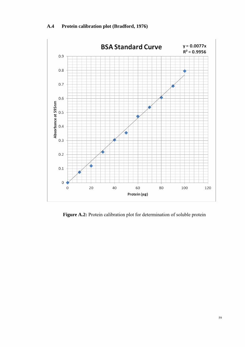
A.4 Protein calibration plot (Bradford, 1976)
Figure A.2: Protein calibration plot for determination of soluble protein
59

60
Appendix B: STATISTICAL ANALYSIS
B.1 Normality test and Homogeneity of Variances
Table B.1: Normality test - Fibrinolytic activities of crude extract and partially purified enzymes from L. rhinocerotis Kolmogorov-Smirnova Shapiro-Wilk
Samples Statistic df Sig. Statistic df Sig.
Crude extract .369 3 . .789 3 .088ATPS Top .312 3 . .896 3 .374ATPS Bottom .185 3 . .998 3 .923Specific_activity
Acetone precipitated protein
.233 3 . .979 3 .722
a. Lilliefors Significance Correction
B.1.1 Test of homogeneity of variances
Table B.2: Test of homogeneity of variances - Fibrinolytic activities of crude extract and partially purified enzymes from L. rhinocerotis
Levene Statistic d.f. 1 d.f. 2 Sig. 3.312 3 8 .078
B.2 One-way ANOVA
Table B.3: ANOVA - Fibrinolytic activities of crude extract and partially purified enzymes from L. rhinocerotis
Sum of Squares
d.f. Mean of square
F-ratio Sig. level #
Between Groups 35906.922 3 11968.974 1348.695 .000
Within Groups 70.996 8 8.874 Total 35977.917 11
# p = 0.0000 (denotes a statistically significant difference)

61
B.3 Post-Hoc for ANOVA
Table B.4: Duncan post-hoc test - Fibrinolytic activities of crude extract and partially purified enzymes from L. rhinocerotis
Subset for alpha = 0.05
Samples N 1 2 3 4
ATPS Bottom 3 1.3900 Crude extract 3 56.8467 ATPS Top 3 93.8500 Acetone precipitated protein
3
151.6100
Duncana
Sig. 1.000 1.000 1.000 1.000Means for groups in homogeneous subsets were displayed. a. Uses Harmonic Mean Sample Size = 3.000.

Appendix C: RAW DATA
Table C.1: Diameter of lytic zone (cm) observed in 18 plates after one hour of incubation at 37±2oC
Concentration of crude extract (mg/ml) Replicate Diameter of lytic zone (cm) 0.2 1 -
2 - 3 -
0.3 1 1.00 2 1.00 3 1.09
0.4 1 1.20 2 1.10 3 1.20
0.5 1 1.23 2 1.21 3 1.19
0.6 1 1.23 2 1.21 3 1.18
0.7 1 1.19 2 1.21 3 1.23
62

Table C.2: Fibrin plate assay for crude extract and partially purified enzyme
Sample Replicate Diameter of lytic zone (cm) Crude extract 1 1.21
2 1.23 3 1.22
Human plasmin (positive control) 1 1.40 2 1.20 3 1.30
Top phase of ATPS 1 1.04 2 1.02 3 1.03
Bottom phase of ATPS 1 - 2 3
Top phase of blank 1 - 2 3
Bottom phase of blank 1 - 2 3
Tri-HCl buffer (Negative control) 1 - 2 3
63

64
Table C.3: Fibrinolytic activities of crude extract and partially purified enzymes from L. rhinocerotis
Sample Replicate Protease activity (U/ml) Protein (mg/ml) Specific activity (U/mg) purification fold
1 2.790 0.047 59.360 2 2.840 0.051 55.686
Crude extract
3 2.830 0.051 55.490
1.000
1 1.510 0.016 92.073 2 1.060 0.011 92.982
ATPS top phase
3 1.930 0.020 96.500
1.650
1 0.007 0.005 1.400 2 0.007 0.006 1.167
ATPS bottom phase
3 0.008 0.005 1.600
0.020
1 5.000 0.034 147.059 2 4.820 0.031 157.003
Acetone precipitated protein
3 4.900 0.033 150.769
2.670