fetal /perinatal insults scott m. kulich department of pathology division of neuropathology...
TRANSCRIPT
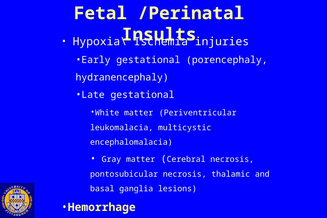
Fetal /Perinatal Insults
Scott M. KulichDepartment of Pathology
Division of NeuropathologyUniversity of Pittsburgh School of Medicine
Fetal /Perinatal Insults: Overview
• Hypoxia\ Ischemia injuries
•Early gestational (porencephaly, hydranencephaly)
•Late gestational
•White matter (Periventricular leukomalacia, multicystic
encephalomalacia)
• Gray matter (Cerebral necrosis, pontosubicular necrosis,
thalamic and basal ganglia lesions)
•Hemorrhage
•Germinal matrix hemorrhage
•Kernicterus
Fetal /Perinatal Insults
• Hypoxia\ Ischemia injuries
•Early gestational (porencephaly, hydranencephaly)
•Late gestational
•White matter (Periventricular leukomalacia, multicystic
encephalomalacia)
• Gray matter (Cerebral necrosis, pontosubicular necrosis,
thalamic and basal ganglia lesions)
•Hemorrhage
•Germinal matrix hemorrhage
•Kernicterus
Hypoxia\Ischemia: Overview
•Very common injury•1.8-47 per 1000 live births
•Sequela variable but include•Cerebral palsy•Mental retardation•Seizures
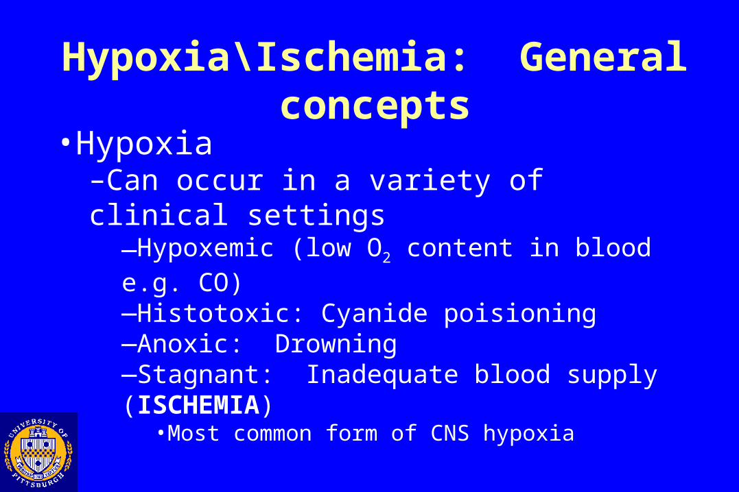
Hypoxia\Ischemia: General concepts
•Hypoxia–Can occur in a variety of clinical settings
—Hypoxemic (low O2 content in blood e.g. CO)—Histotoxic: Cyanide poisioning—Anoxic: Drowning—Stagnant: Inadequate blood supply (ISCHEMIA)
•Most common form of CNS hypoxia
Hypoxia\Ischemia: General concepts
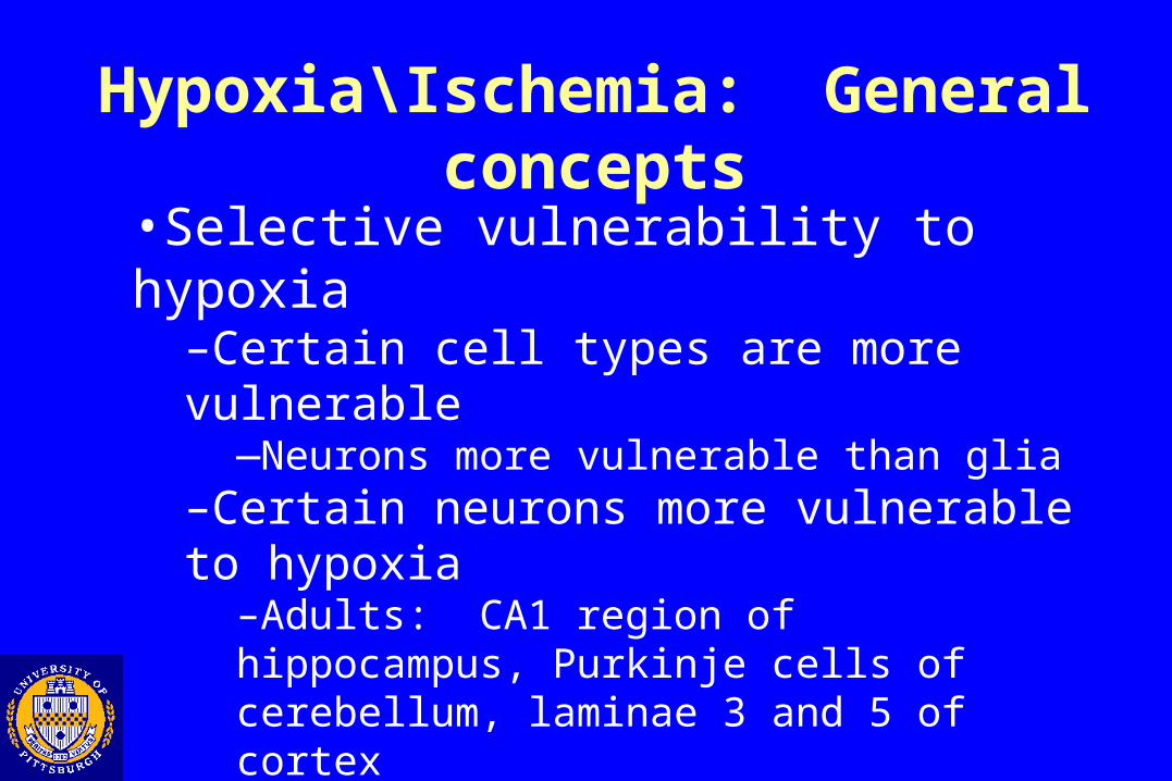
•Selective vulnerability to hypoxia–Certain cell types are more vulnerable
—Neurons more vulnerable than glia–Certain neurons more vulnerable to hypoxia
–Adults: CA1 region of hippocampus, Purkinje cells of cerebellum, laminae 3 and 5 of cortex–Infants: Pons, subiculum, thalamus\basal ganglia
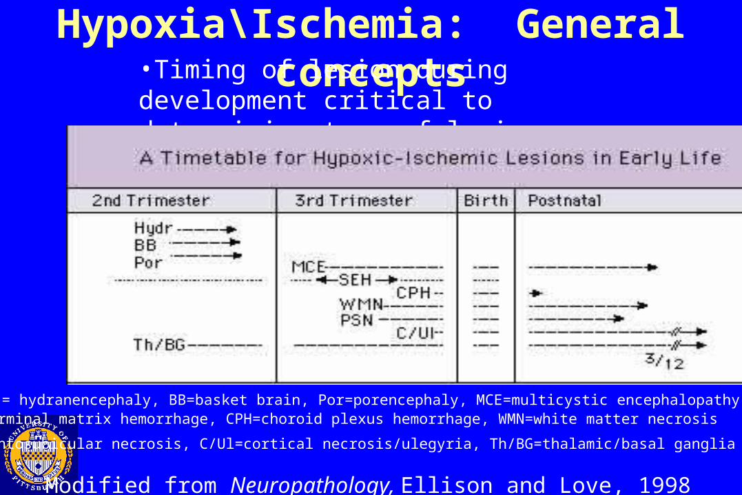
Hypoxia\Ischemia: General concepts•Timing of lesion during development critical to determining type of lesion produced
Modified from Neuropathology, Ellison and Love, 1998
(Hydr = hydranencephaly, BB=basket brain, Por=porencephaly, MCE=multicystic encephalopathySHE=germinal matrix hemorrhage, CPH=choroid plexus hemorrhage, WMN=white matter necrosis
PSN=pontosubicular necrosis, C/Ul=cortical necrosis/ulegyria, Th/BG=thalamic/basal ganglia lesions)

Hypoxia\Ischemia: General concepts
•Timing of lesion during development critical to determining type of lesion produced
•Lack of astrocytes during early development•Smooth-walled cystic lesions of hydran\porencephaly
•Metabolic demands of different regions of the brain differ at various points of development
•White matter necrosis in 3rd trimester injuries•Hypoxic change in neurons differ depending upon time of injury
•Karyorrhexis versus eosinophilia
Hypoxia\Ischemia: Early developmental lesions•Hydranencephaly•Porencephaly (Basket brain, Schizencephaly)
Hydranencephaly
Due to hypoxic-ischemic injury during second trimester
Usually affects the territories of middle and anterior cerebral arteries– Sparing of posterior fossa
May live up to several years depending upon extent of central gray matter involvement
Hydranencephaly: Gross
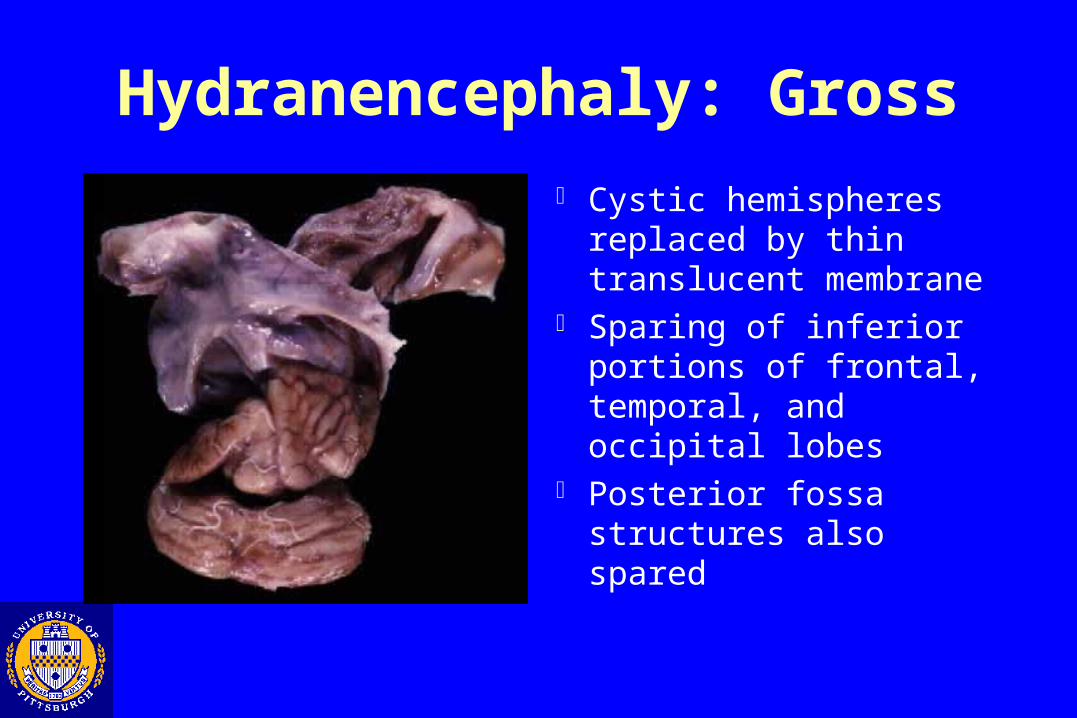
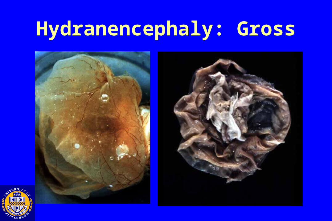
Cystic hemispheres replaced by thin translucent membrane
Sparing of inferior portions of frontal, temporal, and occipital lobes
Posterior fossa structures also spared
Hydranencephaly: Gross
Hydranencephaly: Micro
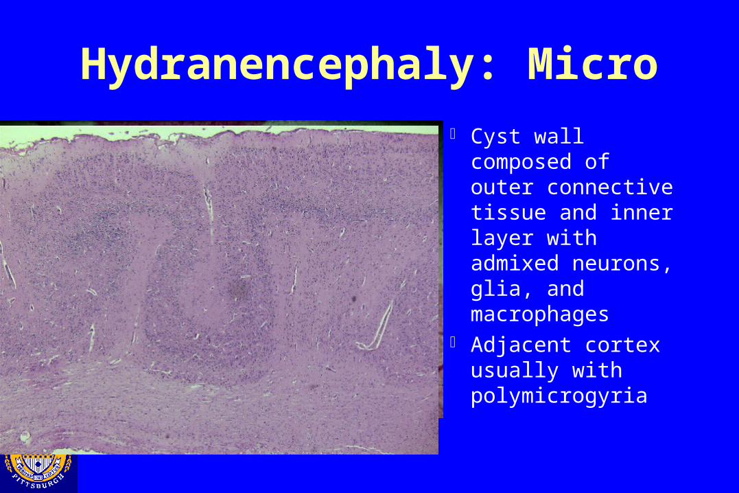
Cyst wall composed of outer connective tissue and inner layer with admixed neurons, glia, and macrophages
Adjacent cortex usually with polymicrogyria
Porencephaly Circumscribed hemispheric defect Also due to hypoxic-ischemic injury during second trimester Usually bilateral, symmetrical, and involves the Sylvian
fissure or central sulcus Severe bilateral cases may also be called by other terms
(schizencephaly, basket brain) Variable clinical manifestations
– Severe cases: MR, epilepsy, blindness, tetrapelegia– Mild cases may survive into adulthood
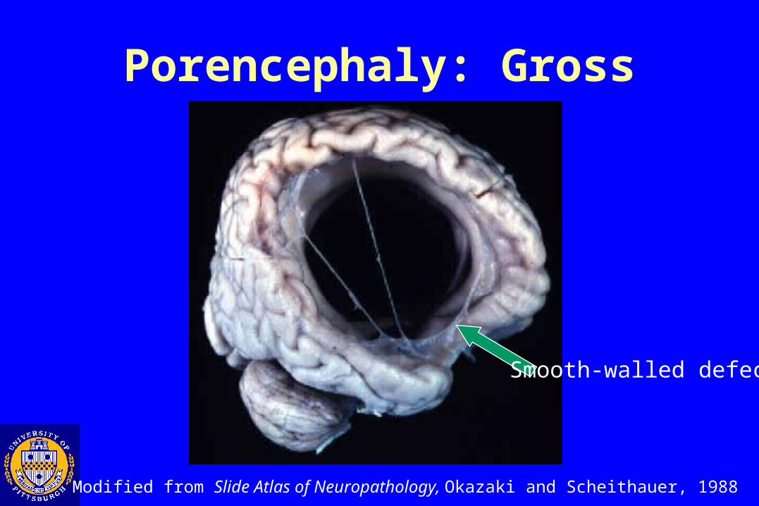
Porencephaly: Gross
Modified from Slide Atlas of Neuropathology, Okazaki and Scheithauer, 1988
Smooth-walled defect
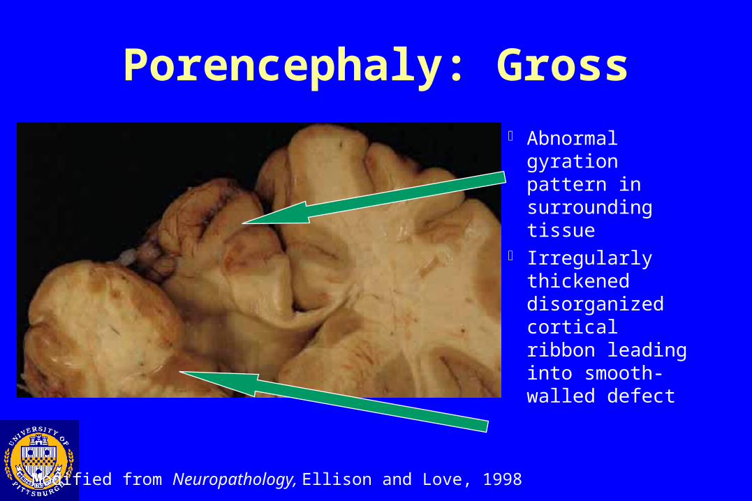
Porencephaly: Gross
Abnormal gyration pattern in surrounding tissue
Irregularly thickened disorganized cortical ribbon leading into smooth-walled defect
Modified from Neuropathology, Ellison and Love, 1998
Hypoxia\Ischemia: Late developmental lesions•White matter lesions
•Periventricular leukomalacia•Multicystic encephalomalacia
•Gray matter lesions•Cerebral necrosis•Pontosubicular necrosis•Status marmoratus•Ulegyria

Periventricular Leukomalacia
• AKA: PVL, white matter necrosis, white matter ischemia, and periventricular leukoencephalopathy• 5 % of all hospital births and up to 35 % of low birth weight newborns• Pathogenesis: Late 3rd trimester (28-32 weeks gestational age) hypoxic/ischemic damage
•Watershed area•Area of high metabolic demand
• Cystic lesions after resolution• Most infants develop spastic motor dysfunction (cerebral palsy)
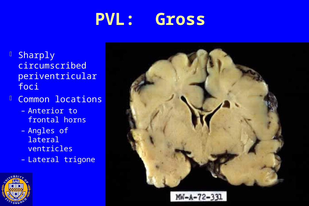
PVL: Gross
Sharply circumscribed periventricular foci
Common locations– Anterior to frontal
horns
– Angles of lateral ventricles
– Lateral trigone
Modified from Neuropathology, Ellison and Love, 1998
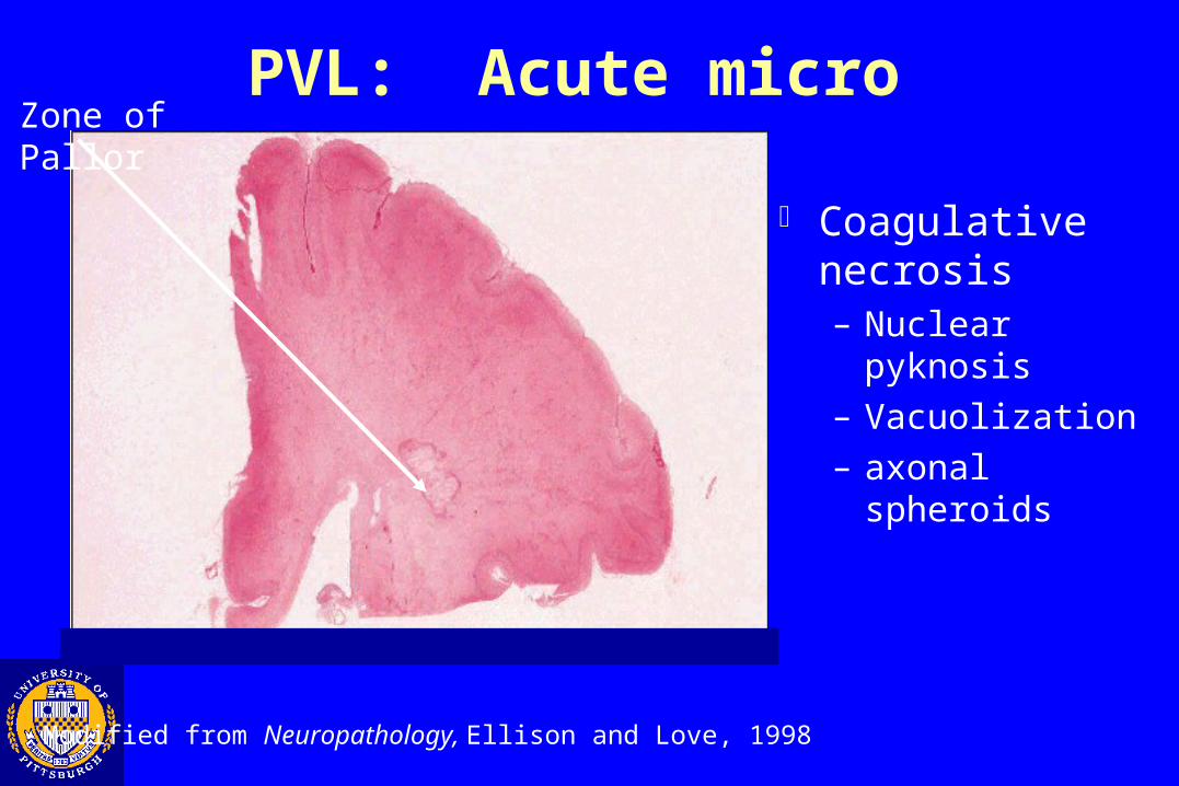
PVL: Acute microZone of Pallor
Coagulative necrosis– Nuclear pyknosis
– Vacuolization
– axonal spheroids
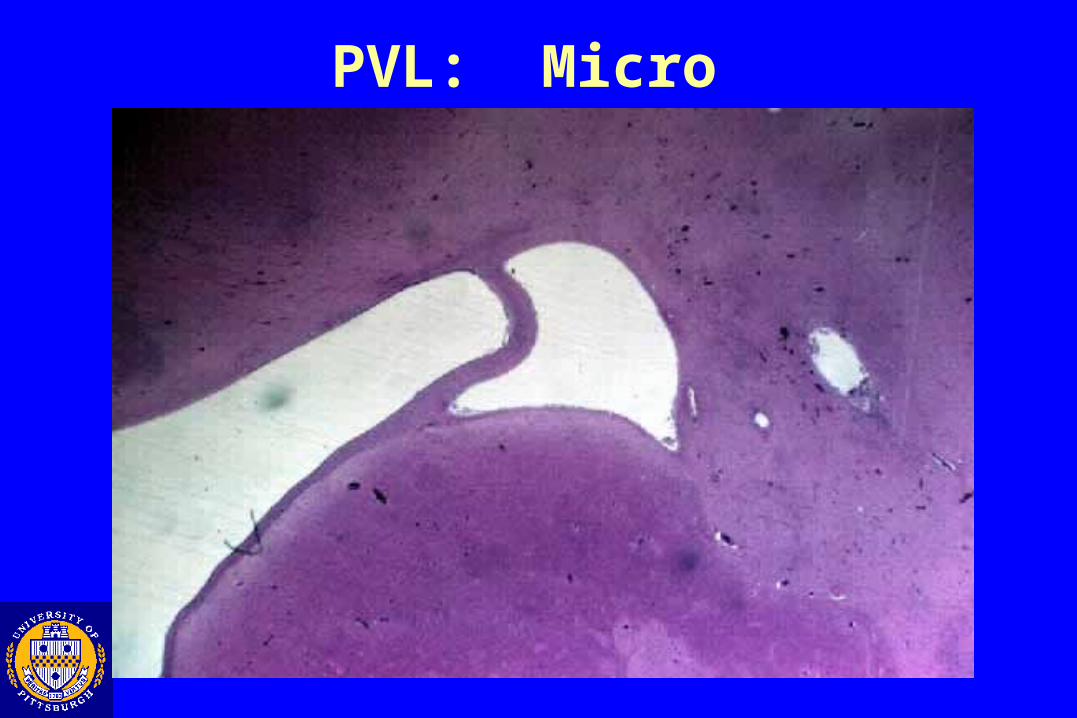
PVL: Micro
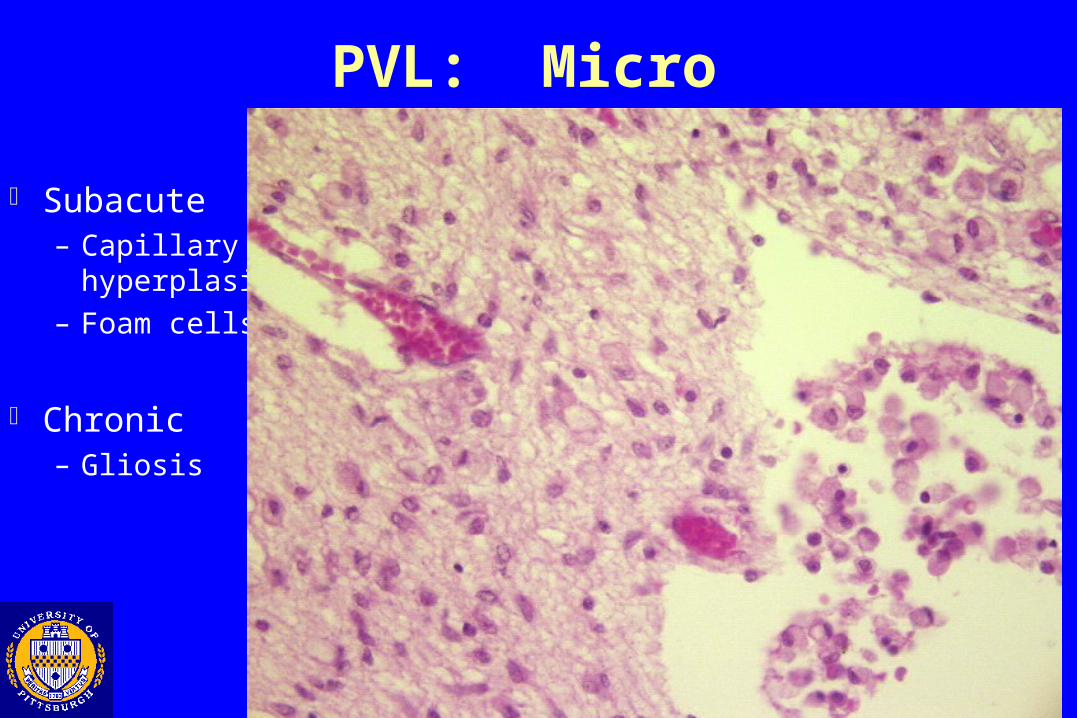
PVL: Micro
Subacute– Capillary
hyperplasia
– Foam cells
Chronic– Gliosis

Multicystic Encephalomalacia
• Believed to result from hypoxic\ischemic insults near term or in the early post-natal period•Can be seen with other conditions (e.g. Herpes)•Usually results in death within weeks to months after insult.
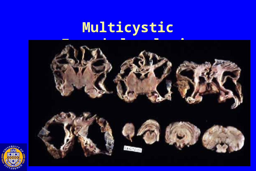
Multicystic Encephalomalacia
Hypoxia\Ischemia: Late developmental lesions•White matter lesions
•Periventricular leukomalacia•Multicystic encephalomalacia
•Gray matter lesions•Cerebral necrosis•Pontosubicular necrosis•Basal ganglia/thalamic lesions•Ulegyria
Cerebral Necrosis
• Observed in term infants associated with•Intrapartum vascular complication (e.g. placental abruption)• Perinatal vascular problems
• Congenital heart defects, hypotension• Lesion common between anterior and middle cerebral artery distributions• Neurological consequences
• Hypotonia,abnormal eye movement, seizures, coma
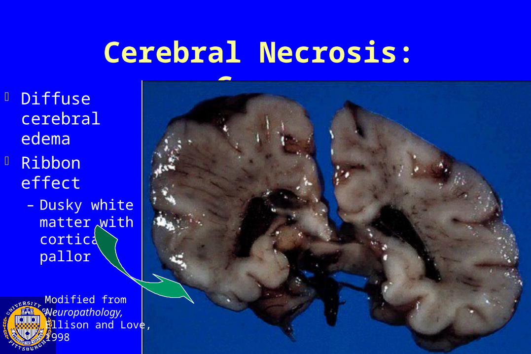
Cerebral Necrosis: Gross Diffuse
cerebral edema Ribbon effect
– Dusky white matter with cortical pallor
Modified from Neuropathology, Ellison and Love, 1998
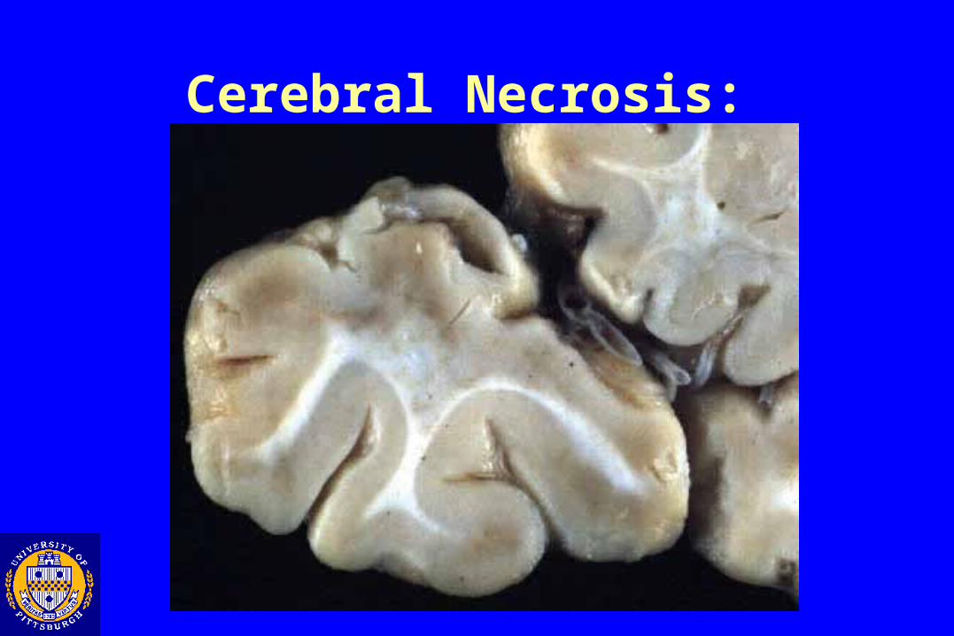
Cerebral Necrosis: Gross
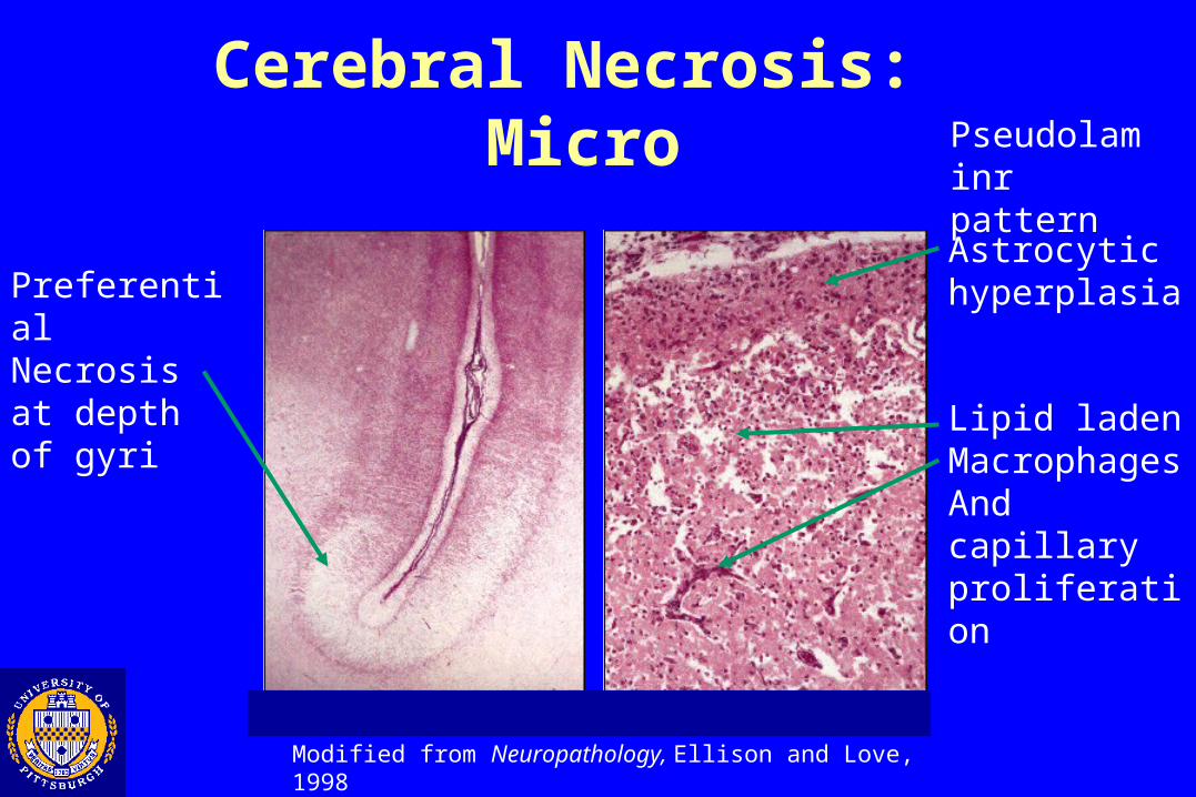
Cerebral Necrosis: Micro
Preferential Necrosis at depth of gyri
Pseudolaminr pattern
Astrocytic hyperplasia
Lipid laden MacrophagesAnd capillary proliferation
Modified from Neuropathology, Ellison and Love, 1998
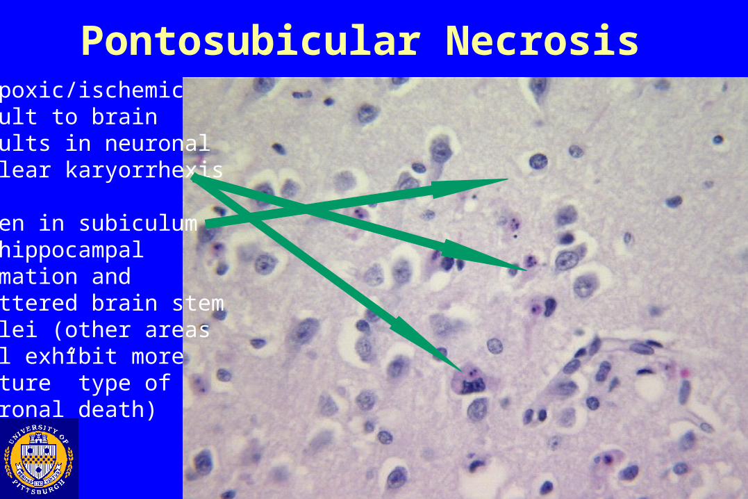
Pontosubicular Necrosis-Hypoxic/ischemic insult to brain results in neuronal nuclear karyorrhexis
-Seen in subiculumof hippocampalformation and scattered brain stemnuclei (other areaswill exhibit more “mature” type of neuronal death)
Ulegyria
• “Scarred gyrus”•Chronic healed hypoxic ischemic insult to the cortex• Preferential involvement of
•Depths of sulci (mushroom morphology)•Anterior-middle cerebral artery territories
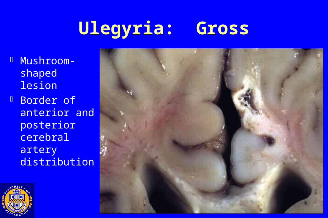
Ulegyria: Gross
Mushroom-shaped lesion
Border of anterior and posterior cerebral artery distribution
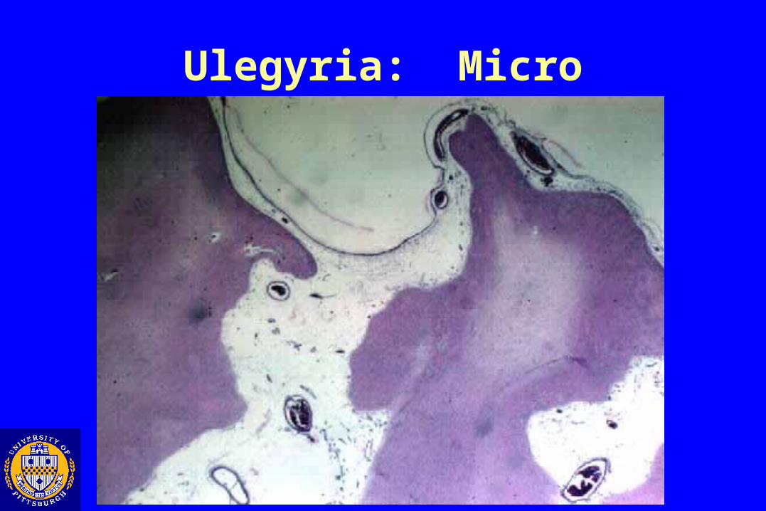
Ulegyria: Micro

Thalamic and Basal Ganglia Lesions
• Microinfarcts of thalamus and basal ganglia
• Abnormal myelination (Status Marmoratus)
• Clinical manifestations
• choreoathetosis
• mental retardation
• spastic paraplegia
• epilepsy
• hyperkinetic if caudate is involved
• Average age of death 12 years old
Thalamic and basal ganglia lesions:Pathogenesis
• Complicated parturition in 70 % of cases• cyanosis
• resuscitation
• convulsions
• neurological signs
• 1/3 have umbilical cord complications
• Male predilection 2:1
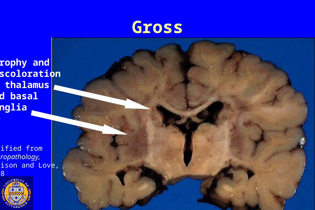
Gross
Atrophy anddiscoloration of thalamus and basalganglia
Modified from Neuropathology, Ellison and Love, 1998
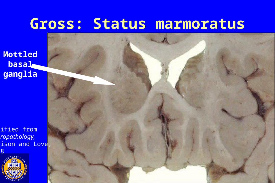
Gross: Status marmoratus
Mottled basalganglia
Modified from Neuropathology, Ellison and Love, 1998
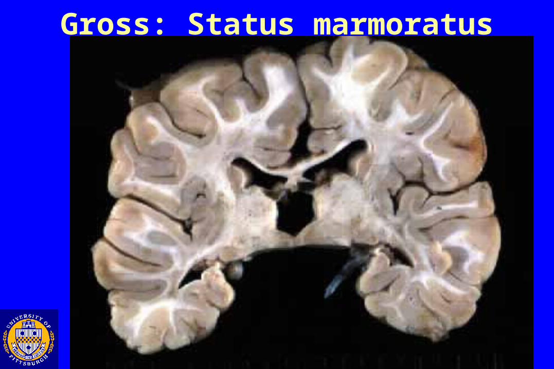
Gross: Status marmoratus
Fetal /Perinatal Insults
• Hypoxia\ Ischemia injuries
•Early gestational (porencephaly, hydranencephaly)
•Late gestational
•White matter (Periventricular leukomalacia, multicystic
encephalomalacia)
• Gray matter (Cerebral necrosis, pontosubicular necrosis,
thalamic and basal ganglia lesions)
•Hemorrhage
•Germinal matrix hemorrhage
•Kernicterus
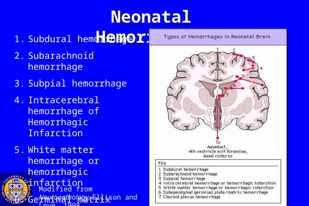
Neonatal Hemorrhages1. Subdural hemorrhage
2. Subarachnoid hemorrhage
3. Subpial hemorrhage
4. Intracerebral hemorrhage of Hemorrhagic Infarction
5. White matter hemorrhage or hemorrhagic infarction
6. Germinal matrix hemorrhage
7. Choroid plexus hemorrhage
Modified from Neuropathology, Ellison and Love, 1998
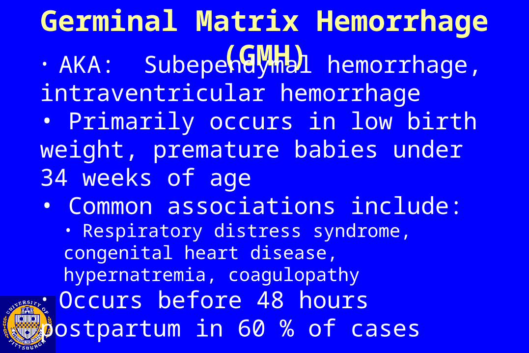
Germinal Matrix Hemorrhage (GMH)• AKA: Subependymal hemorrhage, intraventricular hemorrhage• Primarily occurs in low birth weight, premature babies under 34 weeks of age• Common associations include:
• Respiratory distress syndrome, congenital heart disease, hypernatremia, coagulopathy
• Occurs before 48 hours postpartum in 60 % of cases

Pathogenesis of GMH
• Fragile microcirculation at germinal matrix lacking support• Hypoxia -> Autoregulation failure -> Overperfusion• Focal endothelial cell necrosis• High levels of tissue plasminogen activator
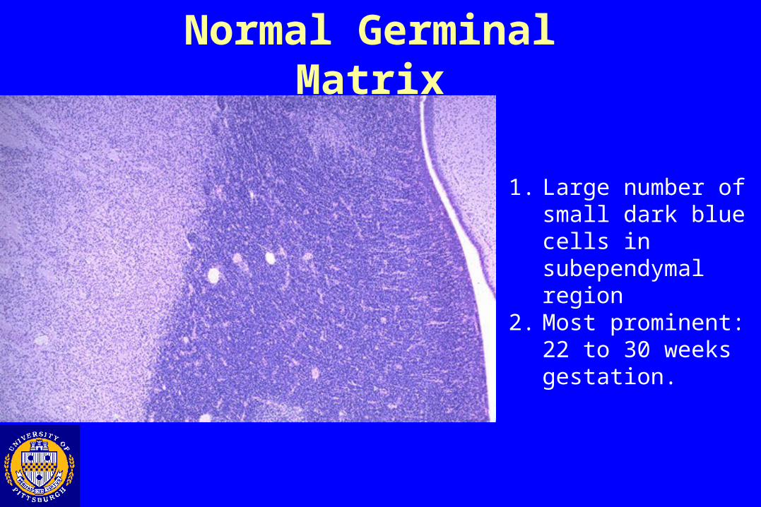
Normal Germinal Matrix
1. Large number of small dark blue cells in subependymal region
2. Most prominent: 22 to 30 weeks gestation.
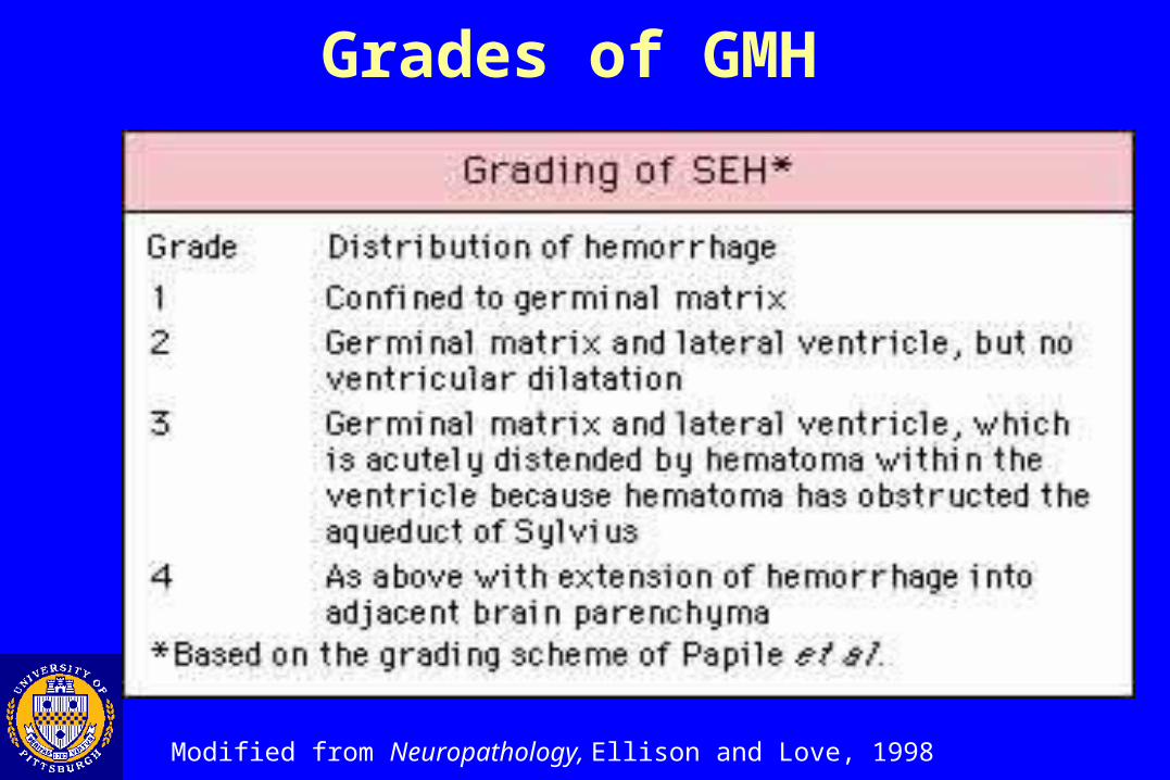
Grades of GMH
Modified from Neuropathology, Ellison and Love, 1998
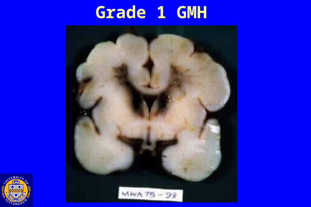
Grade 1 GMH

Grade 1 GMH
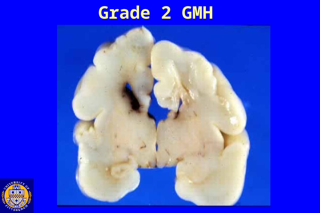
Grade 2 GMH
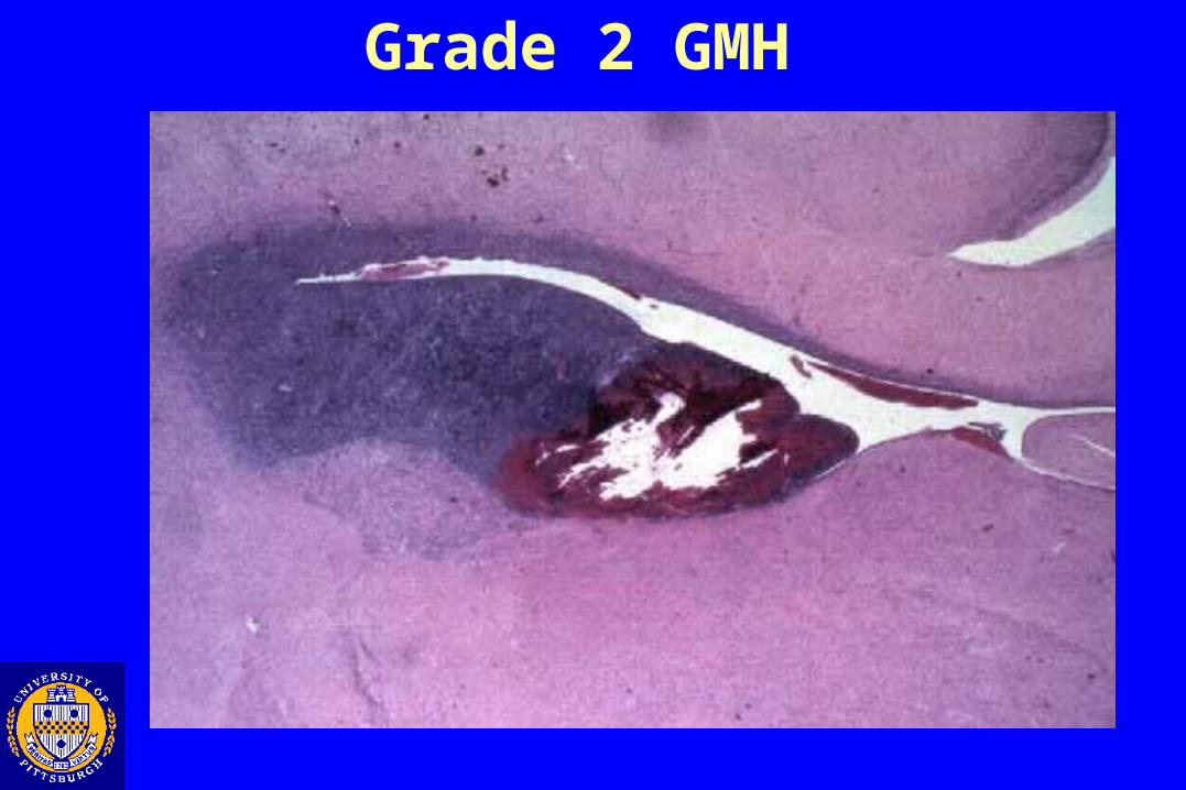
Grade 2 GMH
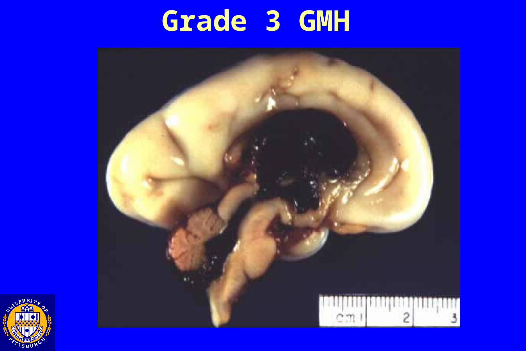
Grade 3 GMH
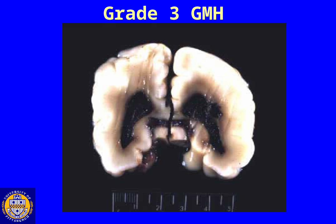
Grade 3 GMH

Grade 4 GMH
Extension into parenchyma
Modified from Neuropathology, Ellison and Love, 1998
Fetal /Perinatal Insults
• Hypoxia\ Ischemia injuries
•Early gestational (porencephaly, hydranencephaly)
•Late gestational
•White matter (Periventricular leukomalacia, multicystic
encephalomalacia)
• Gray matter (Cerebral necrosis, pontosubicular necrosis,
thalamic and basal ganglia lesions)
•Hemorrhage
•Germinal matrix hemorrhage
•Kernicterus
Fetal /Perinatal Insults
• Hypoxia\ Ischemia injuries
•White matter
•Periventricular leukomalacia
• Gray Matter Ischemia
• Cerebral Necrosis
• Pontosubicular Necrosis
• Thalamic and Basal Ganglia Lesions
•Hemorrhage
•Germinal matrix hemorrhage
•Kernicterus
Kernicterus “Jaundiced Nuclei” Selective yellow staining of the deep gray matter and
brain stem due to deposition of unconjugated bilirubin Associated with neuronal necrosis and resulting
neurologic sequelae Poor prognosis but condition is now rare in areas
where hyperbilirubinemia can be predicted, monitored, and treated appropriately
Pathogenesis of Kernicterus• Excessive production of unconjugated bilirubin (80 % from RBC’s) or insufficient conjugation/excretion by the liver• Unconjugated circulating form is neurotoxic• May occur in small or preterm infants at 10 mg/ml• Contributing factors
• Blood-brain barrier damage• Reduced albumin or albumin binding
•Dose dependence• 6-7 % of newborns > 12.9 mg/dl• 3 % of newborns > 15 mg/dl
• Hyperbilirubinemia cause suggested by age of onset•< 1 day: Hemolysis\hematoma, infection•2-3 days: Infection, Criglar-Najjar, physiologic jaundice•1 week: Breast milk jaundice (pregnane-3beta, 20alpha-diol), congenital or drug-induced hemolytic anemias, hypothyroidism, biliary atresia, infections
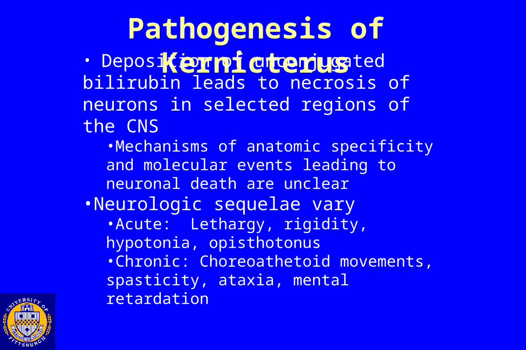
Pathogenesis of Kernicterus• Deposition of unconjugated bilirubin leads to necrosis of neurons in selected regions of the CNS
•Mechanisms of anatomic specificity and molecular events leading to neuronal death are unclear
•Neurologic sequelae vary•Acute: Lethargy, rigidity, hypotonia, opisthotonus•Chronic: Choreoathetoid movements, spasticity, ataxia, mental retardation
Modified from Neuropathology, Ellison and Love, 1998
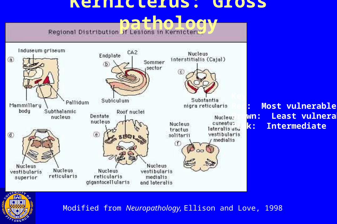
Kernicterus: Gross pathology
KeyRed: Most vulnerableBrown: Least vulnerablePink: Intermediate
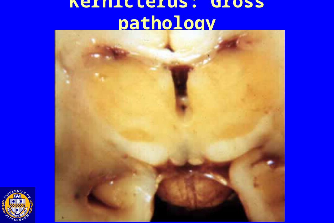
Kernicterus: Gross pathology
Modified fromNeuropathology, Ellison and Love, 1998
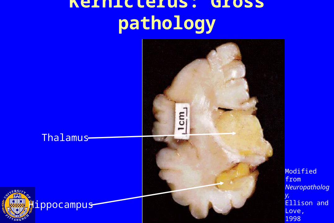
Kernicterus: Gross pathology
Hippocampus
Thalamus
Modified fromNeuropathology, Ellison and Love, 1998
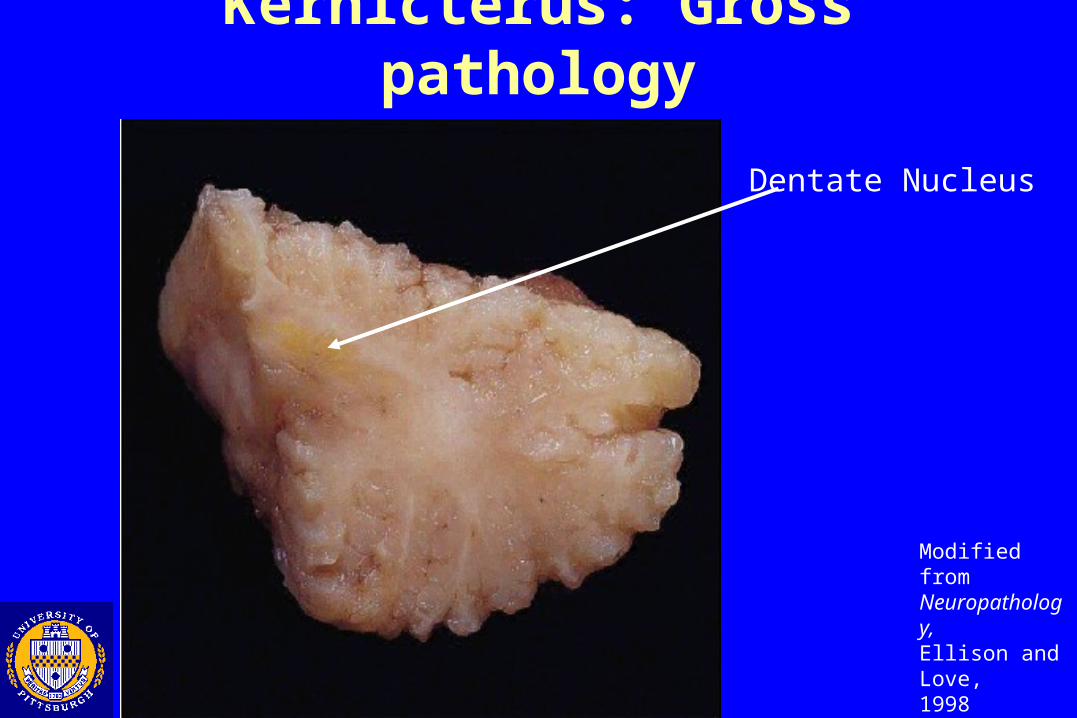
Kernicterus: Gross pathology
Dentate Nucleus
Modified fromNeuropathology, Ellison and Love, 1998
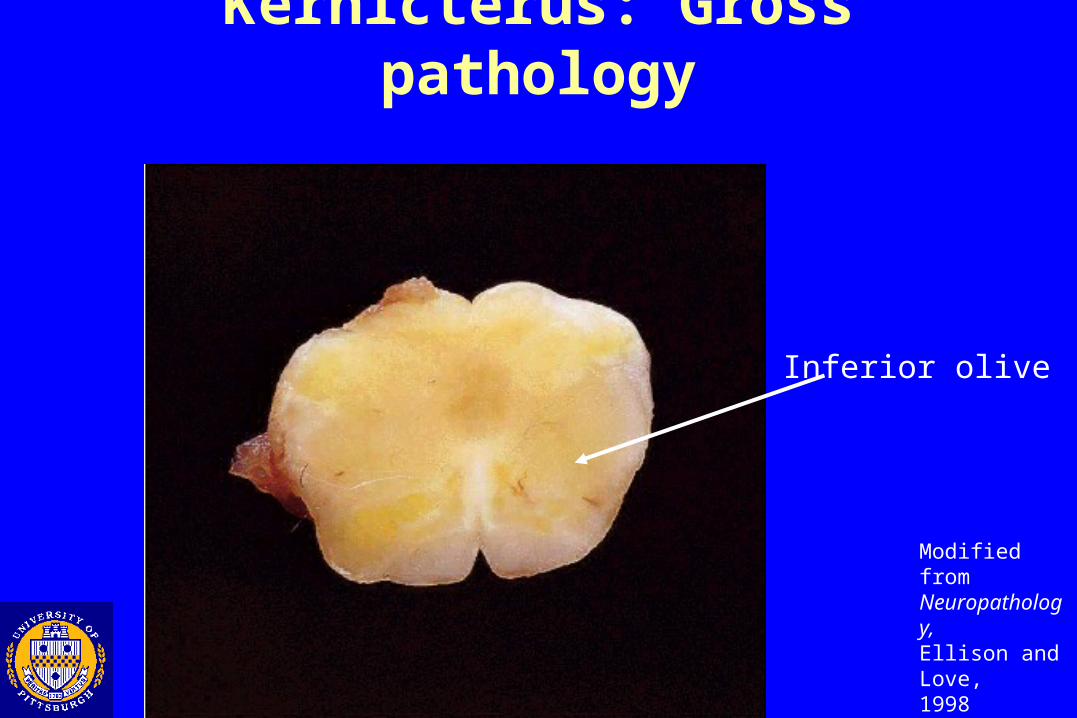
Kernicterus: Gross pathology
Inferior olive
Kernicterus: Microscopic pathology
• Changes do not necessarily correspond to intensity of staining
•Acute
•Cytoplasmic vacuolization, eosinophilia, chromatolysis, and spongy neuropil
•Subacute
•Astrogliosis and neuronal drop-out
Acknowledgments and references Drs. Julio Martinez, Gutti Rao, and David Van
Sickle Neuropathology, Ellison and Love, 1998 Greenfield’s Neuropathology, Graham and
Lantos, 1997 Slide Atlas of Neuropathology, Okazaki and
Scheithauer, 1988