feeding and activation of corpora allata in the cockroach blattella germanica (l.) (dictyoptera,...
TRANSCRIPT
J. Insect Physiol. Vol. 44, No. 1, pp. 31–38, 1998 1997 Elsevier Science LtdPergamon All rights reserved. Printed in Great Britain0022-1910/97 $19.00 + 0.00PII: S0022-1910(97)00102-9
Feeding and Activation of Corpora Allata in theCockroach Blattella germanica (L.)(Dictyoptera, Blattellidae)SERGIO OSORIO,* MARIA-DOLORS PIULACHS,* XAVIER BELLES*†
Received 6 April 1997; revised 10 July 1997
Adult females of the cockroach Blattella germanica have clearly-defined feeding cycles relatedto oogenesis. In the first cycle, food ingestion precedes volumetric increase in the corporaallata, which in turn precedes juvenile hormone production, whereas starved females do notdevelop the corpora allata and produce very low amounts of juvenile hormone. When thesecond gonadotropic cycle is provoked by removing the ootheca, the first event observed isan increase in food consumption, followed by an increase in corpora allata volume and activity.However, this increase in corpora allata volume (and activity) does not occur if females arestarved, thus indicating that the ootheca in the genital chamber inhibits primarily feeding,and indirectly corpora allata development and activity. Corpora allata volume in isolatedheads from starved and decapitated females was able to increase to levels similar to fed con-trols, but this increase was abolished by allatostatin treatment. We suggest that a factor pro-duced in the thoracico–abdominal compartment, which reaches the head mainly through anervous pathway, is released during starvation and inhibits corpora allata development. Thisfactor may stimulate allatostatin production or release, or may well be allatostatin itself. 1997 Elsevier Science Ltd. All rights reserved
Feeding Corpora allata Juvenile hormone Blattella germanica Allatostatin
INTRODUCTION
In most insect species availability of food is critical foroogenesis (see Wheeler, 1996 for review). This isespecially evident in cockroaches, where pioneering stud-ies had shown that starvation suppresses oocyte growthin Blattella germanica and Leucophaea maderae (Rothand Stay, 1962; Engelmann and Rau, 1965, respectively).In B. germanica further work confirmed the criticalimportance of food for reproduction (Kunkel, 1966;Cochran, 1983; Durbin and Cochran, 1985; Piulachs,1988), and the influence of dietary protein contents uponfood consumption and oogenesis (Hamilton and Schal,1988; Cooper and Schal, 1992).
The invention of a radiochemical system to measurerates of juvenile hormone synthesis by corpora allataincubated in vitro (Pratt and Tobe, 1974) shed light onthe relationships between feeding and oocyte growth. In
*Insect Physiology Unit, Department of Agrobiology (CID, CSIC),Jordi Girona 18, 0834 Barcelona Spain.
†To whom all correspondence should be addressed. Tel: 34-3-400 6124; Fax: 34-3-204 59 04. E-mail: [email protected].
31
Periplaneta americana (Weaver and Pratt, 1981;Weaver, 1984), Diploptera punctata (Woodhead andStay, 1989), L. maderae (Acle et al., 1990), and B. germ-anica (Schal et al., 1993) the suppression of oogenesisobserved in protein-deprived females correlated withreduced rates of juvenile hormone production. Themethod for measuring juvenile hormone production alsorevealed that nerve transection increased, to some extent,corpora allata activity in starved specimens of P. amer-icana (Weaver, 1984), D. punctata (Woodhead and Stay,1989) and B. germanica (Schal et al., 1993). However,juvenile hormone production in females of B. germanicawith denervated corpora allata that were fed protein-deficient diets was much lower than that observed infemales that were fed normal diets (Schal et al., 1993).The whole data suggest that the relationships betweennutrition and corpora allata function may be complex andinvolve nervous as well as humoral factors.
Here we used the species B. germanica as the modelto study the effects of feeding upon corpora allata devel-opment and function. Observations of the dynamics offeeding, corpora allata volume and juvenile hormonesynthesis led to ligature and decapitation experiments.
32 SERGIO OSORIOet al.
These, in turn, led us to postulate the involvement ofYXFGL-NH2 allatostatins, neuropeptides well known fortheir inhibitory properties upon the corpora allata, whichhave been isolated from various species of insects (Stayet al., 1994), including B. germanica (Belles et al., 1994).
MATERIALS AND METHODS
Insect rearing and study of the gonadotropic cycles
We used virgin or mated females of B. germanicaobtained from a colony fed on Panlab dog chow andwater, and reared in the dark at 30 ± 1°C and 60–70% r.h.Both, mated and virgin females, undergo similar corporaallata activity and feeding cycles (Gadot et al., 1989; Leeand Wu, 1994). For studies carried out within the firstgonadotropic cycle, freshly moulted females were iso-lated and used at appropriate ages, and were assessedwherever possible by measuring the basal oocyte length(Belles et al., 1987).
The second gonadotropic cycle was experimentallyprovoked by removing the ootheca from the genitalatrium or by severing the ventral nerve cord (see Rothand Stay, 1959) on mated females that had formed theootheca 24–48 h earlier. Intact females with the oothecaattached and sham-operated females (see below) wereused as respective controls. To assess the influence offood consumption, starved females with the oothecaremoved or with the nerve cord severed were also stud-ied. Food consumption, corpora allata volume, basaloocyte growth and juvenile hormone biosynthesis werethe parameters measured. The second gonadotropiccycle, either spontaneous or provoked, lasts between 10and 12 days in our rearing conditions. However, sinceour interest focused on the activation of the corporaallata, which occurs between days 6 and 8 of the secondcycle, the experimental specimens were monitored untilday 8.
Food consumption, and crop and midgut weight
To study the rhythm of feeding during the first andsecond gonadotropic cycles, food consumption wasdetermined with the method of Cochran (1983) withminor variations. Food was weighed on a Sartorius 2004MP balance (0–166 g, d = 0.01 mg). Individual speci-mens were provided with water ad libitum and a weighedfood portion (fresh weight of initial food, FW). After24 h the food remaining was transferred to an oven at60°C, left there for 24 h and then weighed again (driedweight of final food, DW). In parallel, a similar amountof food was placed in a control box containing only thewater vial, and a total of 4–6 of these observations werecarried out at every experimental session. The water lostto evaporation (evaporation factor, EF) estimated fromthese control experiments was used as a correction factor.With these parameters, the dry weight of food consumed(food consumption, FC) was then calculated from the for-mula: FC = FW − [DW + (FW × EF)].
For feeding rhythm studies the criterion of the basaloocyte length cannot be used to assess the physiologicalage, and food consumption during the gonadotropic cycleshowed considerable day-to-day variation and asynch-rony, mostly due to differences in the onset of feeding,which in turn influenced the length of the cycle. How-ever, in all cases food consumption was low after adultemergence, peaked around the middle of the gonado-tropic cycle, and declined thereafter. For data presen-tation, the date of adult emergence and that of the forma-tion of the first ootheca were used as references, and theday-to-day measurements of food consumption wererealigned by using the mean intervals between these tworeference points (following Cochran, 1983) and the datawere referred to a scale of 8 days, which is the mostcommon duration of the first gonadotropic cycle underour rearing conditions (Belles et al., 1987).
Corpora allata volume and oocyte length
All micrometrical measurements were carried out withan ocular micrometer adapted to the dissecting stereomic-roscope. Corpus allatum volume (VCA) was estimated bythe formula VCA = 4/3px1x2x3, where x1, x2 and x3 arethe radii of the three principal axes. For each specimen,both corpora allata were measured and averaged. Basaloocyte length was measured on six ovarioles chosen atrandom in the ovary pair, and the values were averaged.
Quantification of juvenile hormone synthesis
Individual pairs of corpora allata were incubated in100 ml of TC 199 medium (Flow, Ayrshire, Scotland,UK), containing L-methionine (0.1 mmol), Hank’s salts,Hepes buffer (20 mmol) plus Ficoll (20 mg ml−1), towhich L-[3H-methyl] methionine (Amersham, Bucks,UK) had been added to achieve a final specific activityof 7.4 GBq mmol−1. Details of the method for juvenilehormone III determination in vitro in B. germanica aregiven in Piulachs and Couillaud (1992). Routine quanti-fication of juvenile hormone III produced in vitro wascarried out in standard 2 h incubation periods. At the endof the incubations, juvenile hormone III was determinedin the medium plus homogenized glands.
Head ligature, decapitation and ventral nerve cord sev-erance
All the operations were carried out on specimens anae-sthetized with carbon dioxide. Head ligated specimenswere prepared by putting a ligature round the neck, whichwas then gently tightened and knotted, taking care thatthe neck cuticle was not severed. Decapitation was car-ried out following the same procedure, but in this casethe ligature was firmly tightened and knotted, and theneck was then sectioned posterior to the ligature. In allcases, the corpora allata remain isolated within the headcapsule. In head ligated specimens the haemocoelic iso-lation of the head with respect to the thoracico–abdomi-nal compartment was assessed at the end of the experi-ment by injecting methylene blue (0.2% in aqueous
33FEEDING IN BLATTELLA GERMANICA
solution) in the abdomen and observing the diffusion ofthe dye 5 min later. Those specimens showing dye withinthe head were discarded. In the experiments to provokethe second gonadotropic cycle (see above), the nervecord was severed with fine forceps introduced through alateral slit between the fifth and the sixth abdominal ster-nites. Sham-operations were performed according thesame procedure, but without severing the nerve cord.
Injection of allatostatin into isolated heads
We used one of the allatostatins of B. germanica(BLAST 2: Asp-Arg-Leu-Tyr-Ser-Phe-Gly-Leu-NH2),which was synthesized and characterized as described inBelles et al. (1994). It was injected with a Hamiltonmicrosyringe at a volume of 0.3 ml in Ringer saline con-taining 3% acetonitrile. Specimens were anaesthetizedwith carbon dioxide, and prepared with a lax ligatureplaced round the neck. The needle of the syringe chargedwith the allatostatin was introduced to the head throughthe occipital foramen, and the ligature was tightenedaround the needle. Then the allatostatin was gentlyinjected, after which the needle was removed and theligature was immediately knotted. Finally, the neck wassectioned posterior to the ligature.
RESULTS
Rhythm of feeding in the first gonadotropic cycle
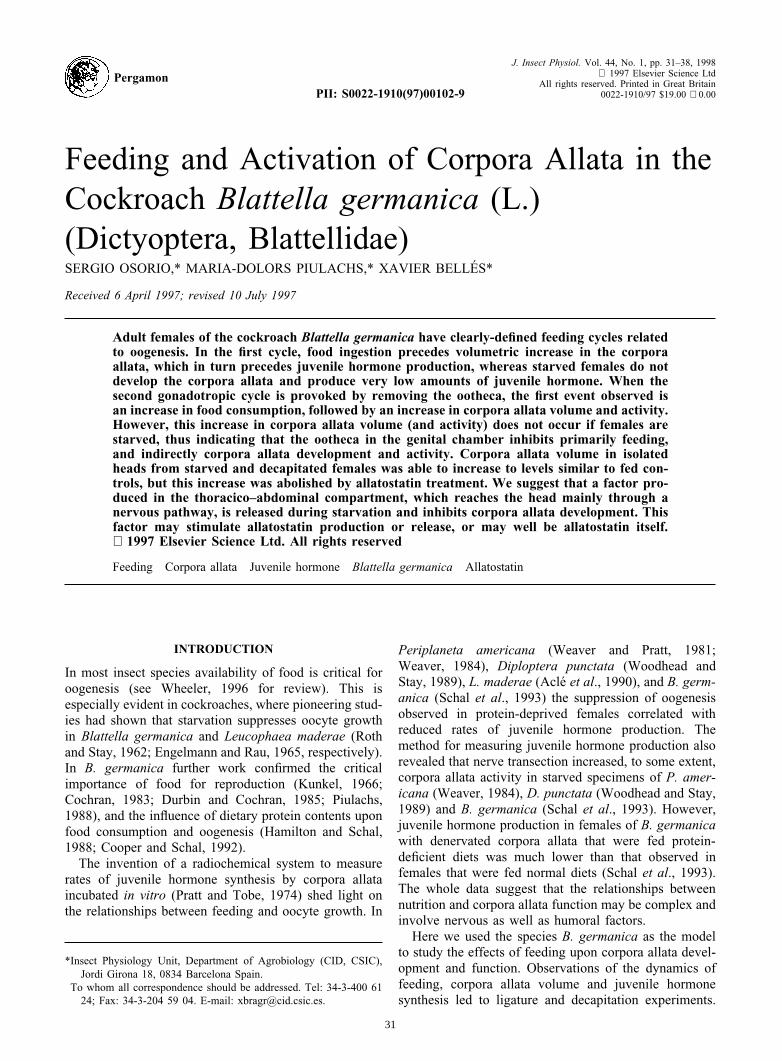
Food consumption by virgin females during the firstgonadotropic cycle [Fig. 1(A)] was low after imaginalecdysis (day 0, 0.50 ± 0.29 mg/day, n = 12), increasedprogressively to a maximum between days 3 and 4 (11.00± 1.00 mg/day, n = 9), and then rapidly decreased untilthe formation of the ootheca (day 7), when the valueswere similar to those measured in freshly emerged speci-mens. Food consumption in females transporting theootheca [day 8: Fig. 1(A), and also 2 to 3 days after theformation of the ootheca, not shown] was uniformly low,between 1 and 2 mg per day.
In addition, crop and midgut were weighed on thesame days, in order to obtain a food transit reflection ofthe feeding rhythm studied above. Results on crop weight[Fig. 1(B)] show increasing values from day 0 (0.72 ±0.13 mg, n = 10) to day 3 (3.15 ± 0.65 mg, n = 9), anda drop from day 4 onwards, to reach values similar tothose observed on day 0. Midgut weight [Fig. 1(C)] alsoshows a cyclic profile, although in this case the moreapparent change was observed between day 0 and 1, thenthe weight slowly increased until day 4 (1.60 ± 0.09 mg,n = 11), and decreased on days 5, 6 and 7 (0.66 ±0.07 mg, n = 8).
Feeding, corpora allata activity and oocyte growth
In another set of experiments we studied the effects offeeding upon corpora allata volume, juvenile hormonesynthesis and oocyte growth. For this purpose, normallyfed females were compared to starved females.
FIGURE 1. Food consumption (A), crop weight (B) and midgutweight (C) during the first gonadotropic cycle of virgin females of B.germanica. Results are expressed as x ± SEM (n = 9–12). The whitespace close to the base of (B) and (C) indicates the interval of variationof dry weight of empty crop and midgut, respectively (n = 25 in both
cases). 8 (O): 8-day-old females transporting an ootheca.
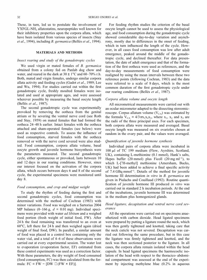
Normally fed females matured the first batch of basaloocytes and produced the first ootheca around the day 8of adult life. Conversely, starved females died aroundday 10 of adult life without showing any ovarian devel-opment (Fig. 2, inset).
Concerning corpora allata volume, fed females showeda cycle that can be divided into three stages (Fig. 2): (i)days 0–2, when there were no significant changes, (ii)days 2–6, when a steady increase was observed to amaximum of 1.88 ± 0.05 nl (n = 7) on day 6, and (iii)days 6–8, when a sharp decrease occurred, to values aslow as 0.70 ± 0.08 nl (n = 7) on day 8, which are similarto those of freshly ecdysed adults. In contrast, corporaallata of starved females (Fig. 2), decreased slightly(from 0.70 ± 0.08 nl, n = 8, on day 0, to 0.56 ± 0.03 nl,n = 7, on day 8).
Determinations of juvenile hormone synthesis on days2, 4, 6 and 8 (Fig. 2) in both fed and starved femalesgave results approximately parallel to those of corporaallata volume. Fed females showed the expected cyclicpattern, whereas starved females gave minimal values,
34 SERGIO OSORIOet al.
FIGURE 2. Corpora allata volume in fed (P) and starved (s) virgin females of B. germanica during the first 8 days of adultlife. For each specimen both corpora allata were measured and averaged. The values in the squares besides days 2, 4, 6 and8 correspond to juvenile hormone biosynthesis (in pmol h−1 × pair of corpora allata). The inset shows the basal oocyte lengthin both groups of experimental females. In all cases results are expressed as x ± SEM (n = 7–11). 8 (O): 8-day-old specimens
transporting an ootheca (in the case of fed females).
close to the limit of detection of the method, irrespectiveof the day of measurement (Fig. 2).
Feeding in the second gonadotropic cycle
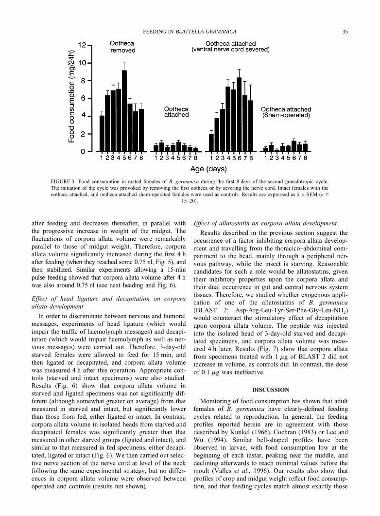
The second gonadotropic cycle was provoked byremoving the ootheca from the genital atrium or bysevering the nerve cord. These operations were carriedout on mated females 24–48 h after the formation of thefirst ootheca, and food consumption was measured duringthe next 8 days. Intact females carrying the first oothecanormally, and ootheca-carrying sham-operated femaleswere used as respective controls.
Results (Fig. 3) show that females either with theootheca removed or with the nerve cord severed beganto feed on the day of the operation, and steadily increasedfood consumption during the following 5–6 days. Con-versely, both control groups showed constantly low feed-ing rates throughout the period studied.
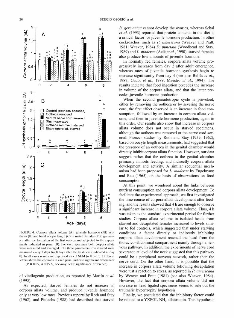
Corpora allata activity and oocyte growth in the secondgonadotropic cycle
Concerning the volume of the corpora allata [Fig.4(A)], the females with the ootheca removed showed par-allel behaviour to those with the nerve cord severed. Inboth cases, the volume of the corpora allata significantlyincreased with respect to controls and starved specimens.The differences between the females with the ootheca
removed or with the nerve cord severed with respect tothe other experimental groups began to be clearly sig-nificant 4 days after the beginning of the experiment.
Conversely, juvenile hormone biosynthetic rates [Fig.4(B)] remained low in all groups until day 8 after thebeginning of the experiment, when the rates measured onfemales with the ootheca removed or with the nerve cordsevered were clearly higher than in the other experi-mental groups (controls and starved specimens).
Data on basal oocyte length [Fig. 4(C)] are consistentwith those of the two former parameters. On day 6 afterthe beginning of the experiment the values measured onfemales with the ootheca removed or with the nerve cordsevered began to be significantly higher with respect tocontrols and starved specimens, and on day 8 the differ-ences were more apparent.
Time-course of corpora allata development after feeding
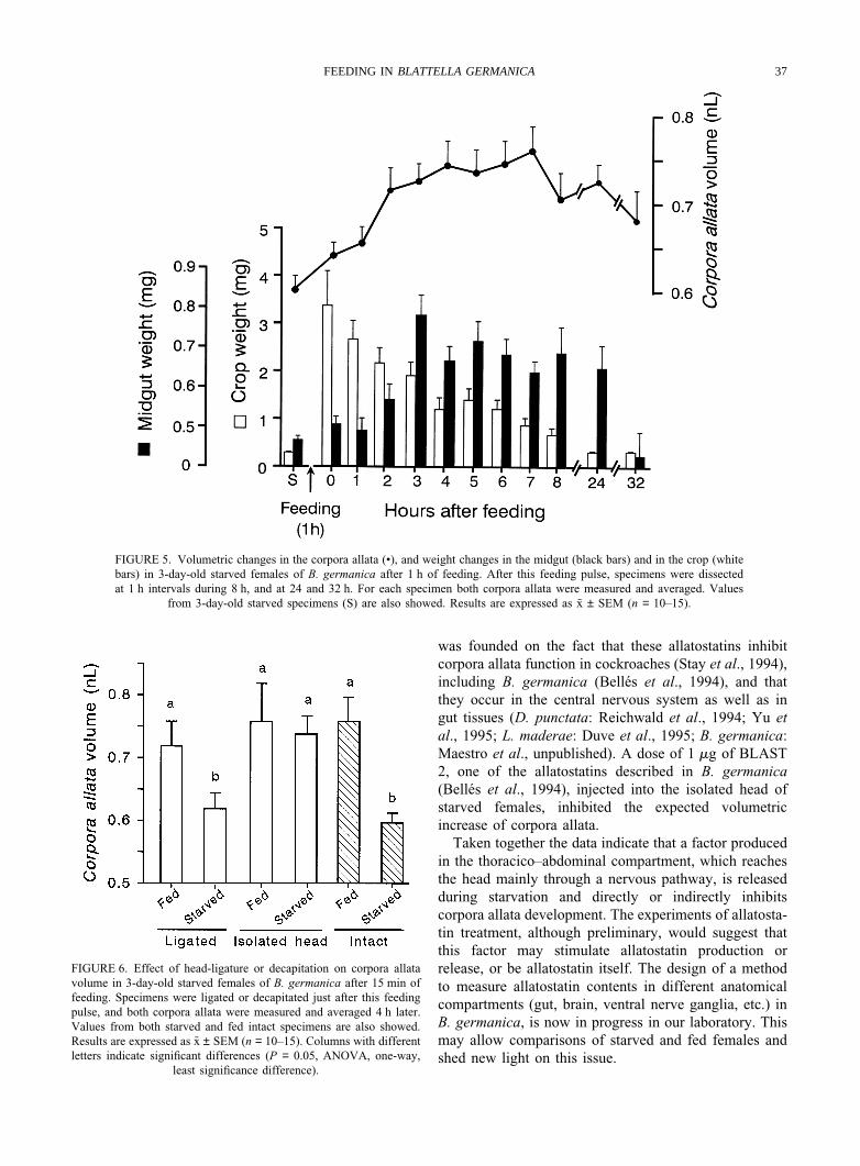
The previous results led us to study the links betweenfood consumption and corpora allata development andactivity, and we were first interested in minimizing thetime involved in the experiments. Therefore, specimensthat had been starved during the first three days of adultlife were allowed to feed for 1 h. Then, groups of themwere dissected at 1 h intervals and crop and midgutweight and corpora allata volume were measured. Results(Fig. 5) indicate that crop weight increases abruptly just
35FEEDING IN BLATTELLA GERMANICA
FIGURE 3. Food consumption in mated females of B. germanica during the first 8 days of the second gonadotropic cycle.The initiation of the cycle was provoked by removing the first ootheca or by severing the nerve cord. Intact females with theootheca attached, and ootheca attached sham-operated females were used as controls. Results are expressed as x ± SEM (n =
15–20).
after feeding and decreases thereafter, in parallel withthe progressive increase in weight of the midgut. Thefluctuations of corpora allata volume were remarkablyparallel to those of midgut weight. Therefore, corporaallata volume significantly increased during the first 4 hafter feeding (when they reached some 0.75 nl, Fig. 5), andthen stabilized. Similar experiments allowing a 15-minpulse feeding showed that corpora allata volume after 4 hwas also around 0.75 nl (see next heading and Fig. 6).
Effect of head ligature and decapitation on corporaallata development
In order to discriminate between nervous and humoralmessages, experiments of head ligature (which wouldimpair the traffic of haemolymph messages) and decapi-tation (which would impair haemolymph as well as ner-vous messages) were carried out. Therefore, 3-day-oldstarved females were allowed to feed for 15 min, andthen ligated or decapitated, and corpora allata volumewas measured 4 h after this operation. Appropriate con-trols (starved and intact specimens) were also studied.Results (Fig. 6) show that corpora allata volume instarved and ligated specimens was not significantly dif-ferent (although somewhat greater on average) from thatmeasured in starved and intact, but significantly lowerthan those from fed, either ligated or intact. In contrast,corpora allata volume in isolated heads from starved anddecapitated females was significantly greater than thatmeasured in other starved groups (ligated and intact), andsimilar to that measured in fed specimens, either decapi-tated, ligated or intact (Fig. 6). We then carried out selec-tive nerve section of the nerve cord at level of the neckfollowing the same experimental strategy, but no differ-ences in corpora allata volume were observed betweenoperated and controls (results not shown).
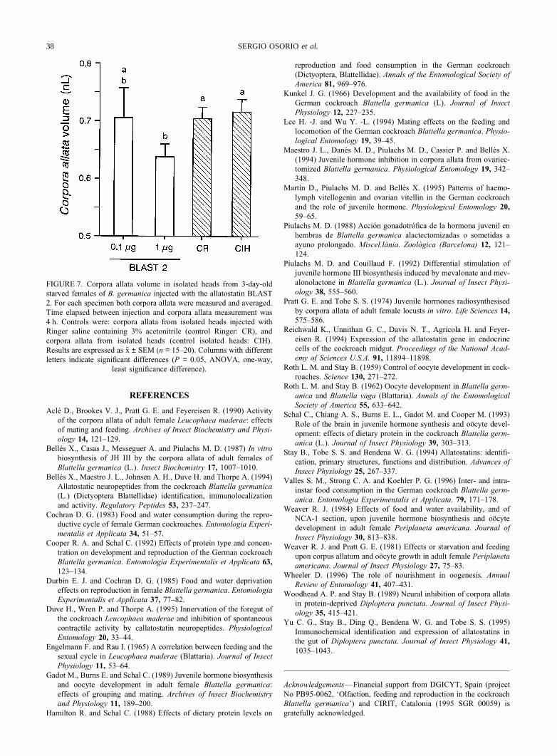
Effect of allatostatin on corpora allata development
Results described in the previous section suggest theoccurrence of a factor inhibiting corpora allata develop-ment and travelling from the thoracico–abdominal com-partment to the head, mainly through a peripheral ner-vous pathway, while the insect is starving. Reasonablecandidates for such a role would be allatostatins, giventheir inhibitory properties upon the corpora allata andtheir dual occurrence in gut and central nervous systemtissues. Therefore, we studied whether exogenous appli-cation of one of the allatostatins of B. germanica(BLAST 2: Asp-Arg-Leu-Tyr-Ser-Phe-Gly-Leu-NH2)would counteract the stimulatory effect of decapitationupon corpora allata volume. The peptide was injectedinto the isolated head of 3-day-old starved and decapi-tated specimens, and corpora allata volume was meas-ured 4 h later. Results (Fig. 7) show that corpora allatafrom specimens treated with 1 mg of BLAST 2 did notincrease in volume, as controls did. In contrast, the doseof 0.1 mg was ineffective.
DISCUSSION
Monitoring of food consumption has shown that adultfemales of B. germanica have clearly-defined feedingcycles related to reproduction. In general, the feedingprofiles reported herein are in agreement with thosedescribed by Kunkel (1966), Cochran (1983) or Lee andWu (1994). Similar bell-shaped profiles have beenobserved in larvae, with food consumption low at thebeginning of each instar, peaking near the middle, anddeclining afterwards to reach minimal values before themoult (Valles et al., 1996). Our results also show thatprofiles of crop and midgut weight reflect food consump-tion, and that feeding cycles match almost exactly those
36 SERGIO OSORIOet al.
FIGURE 4. Corpora allata volume (A), juvenile hormone (JH) syn-thesis (B) and basal oocyte length (C) in mated females of B. german-ica after the formation of the first ooheca and subjected to the experi-ments indicated in panel (B). For each specimen both corpora allatawere measured and averaged. The three parameters investigated weremeasured every 2 days for 8 days after the treatment (indicated as day0). In all cases results are expressed as x ± SEM (n = 6–15). Differentletters above the columns in each panel indicate significant differences
(P = 0.05, ANOVA, one-way, least significance difference).
of vitellogenin production, as reported by Martın et al.(1995).
As expected, starved females do not increase incorpora allata volume, and produce juvenile hormoneonly at very low rates. Previous reports by Roth and Stay(1962), and Piulachs (1988) had described that starved
B. germanica cannot develop the ovaries, whereas Schalet al. (1993) reported that protein contents in the diet isa critical factor for juvenile hormone production. In othercockroaches, such as P. americana (Weaver and Pratt,1981; Weaver, 1984) D. punctata (Woodhead and Stay,1989) and L. maderae (Acle et al., 1990), starved femalesalso produce low amounts of juvenile hormone.
In normally fed females, corpora allata volume pro-gressively increases from day 2 after adult emergence,whereas rates of juvenile hormone synthesis begin toincrease significantly from day 4 (see also Belles et al.,1987; Gadot et al., 1989; Maestro et al., 1994). Theresults indicate that food ingestion precedes the increasein volume of the corpora allata, and that the latter pre-cedes juvenile hormone production.
When the second gonadotropic cycle is provoked,either by removing the ootheca or by severing the nervecord, the first effect observed is an increase in food con-sumption, followed by an increase in corpora allata vol-ume, and then in juvenile hormone production, again inthis order. Our results also show that increase in corporaallata volume does not occur in starved specimens,although the ootheca was removed or the nerve cord sev-ered. Pioneer studies by Roth and Stay (1959, 1962),based on oocyte length measurements, had suggested thatthe presence of an ootheca in the genital chamber woulddirectly inhibit corpora allata function. However, our datasuggest rather that the ootheca in the genital chamberprimarily inhibits feeding, and indirectly corpora allatadevelopment and activity. A similar sequential mech-anism had been proposed for L. maderae by Engelmannand Rau (1965), on the basis of observations on foodconsumption.
At this point, we wondered about the links betweennutrient consumption and corpora allata development. Tofacilitate the experimental approach, we first investigatedthe time-course of corpora allata development after feed-ing, and the results showed that 4 h are enough to observea significant increase in corpora allata volume. Thus, 4 hwas taken as the standard experimental period for furtherstudies. Corpora allata volume in isolated heads fromstarved and decapitated females increased to levels simi-lar to fed controls, which suggested that under starvingconditions a factor directly or indirectly inhibitingcorpora allata development reached the head from thethoracico–abdominal compartment mainly through a ner-vous pathway. In addition, the experiments of nerve cordseverance at level of the neck suggested that this pathwaycould be a peripheral nervous network, rather than thenerve cord. On the other hand, it is possible that theincrease in corpora allata volume following decapitationwere just a reaction to stress, as reported in P. americanaby Weaver and Pratt (1981) (see also Weaver, 1984).However, the fact that corpora allata volume did notincrease in head ligated specimens seems to rule out thetraumatic hypertrophy hypothesis.
Finally, we postulated that the inhibitory factor couldbe related to a YXFGL-NH2 allatostatin. This hypothesis
37FEEDING IN BLATTELLA GERMANICA
FIGURE 5. Volumetric changes in the corpora allata (•), and weight changes in the midgut (black bars) and in the crop (whitebars) in 3-day-old starved females of B. germanica after 1 h of feeding. After this feeding pulse, specimens were dissectedat 1 h intervals during 8 h, and at 24 and 32 h. For each specimen both corpora allata were measured and averaged. Values
from 3-day-old starved specimens (S) are also showed. Results are expressed as x ± SEM (n = 10–15).
FIGURE 6. Effect of head-ligature or decapitation on corpora allatavolume in 3-day-old starved females of B. germanica after 15 min offeeding. Specimens were ligated or decapitated just after this feedingpulse, and both corpora allata were measured and averaged 4 h later.Values from both starved and fed intact specimens are also showed.Results are expressed as x ± SEM (n = 10–15). Columns with differentletters indicate significant differences (P = 0.05, ANOVA, one-way,
least significance difference).
was founded on the fact that these allatostatins inhibitcorpora allata function in cockroaches (Stay et al., 1994),including B. germanica (Belles et al., 1994), and thatthey occur in the central nervous system as well as ingut tissues (D. punctata: Reichwald et al., 1994; Yu etal., 1995; L. maderae: Duve et al., 1995; B. germanica:Maestro et al., unpublished). A dose of 1 mg of BLAST2, one of the allatostatins described in B. germanica(Belles et al., 1994), injected into the isolated head ofstarved females, inhibited the expected volumetricincrease of corpora allata.
Taken together the data indicate that a factor producedin the thoracico–abdominal compartment, which reachesthe head mainly through a nervous pathway, is releasedduring starvation and directly or indirectly inhibitscorpora allata development. The experiments of allatosta-tin treatment, although preliminary, would suggest thatthis factor may stimulate allatostatin production orrelease, or be allatostatin itself. The design of a methodto measure allatostatin contents in different anatomicalcompartments (gut, brain, ventral nerve ganglia, etc.) inB. germanica, is now in progress in our laboratory. Thismay allow comparisons of starved and fed females andshed new light on this issue.
38 SERGIO OSORIOet al.
FIGURE 7. Corpora allata volume in isolated heads from 3-day-oldstarved females of B. germanica injected with the allatostatin BLAST2. For each specimen both corpora allata were measured and averaged.Time elapsed between injection and corpora allata measurement was4 h. Controls were: corpora allata from isolated heads injected withRinger saline containing 3% acetonitrile (control Ringer: CR), andcorpora allata from isolated heads (control isolated heads: CIH).Results are expressed as x ± SEM (n = 15–20). Columns with differentletters indicate significant differences (P = 0.05, ANOVA, one-way,
least significance difference).
REFERENCES
Acle D., Brookes V. J., Pratt G. E. and Feyereisen R. (1990) Activityof the corpora allata of adult female Leucophaea maderae: effectsof mating and feeding. Archives of Insect Biochemistry and Physi-ology 14, 121–129.
Belles X., Casas J., Messeguer A. and Piulachs M. D. (1987) In vitrobiosynthesis of JH III by the corpora allata of adult females ofBlattella germanica (L.). Insect Biochemistry 17, 1007–1010.
Belles X., Maestro J. L., Johnsen A. H., Duve H. and Thorpe A. (1994)Allatostatic neuropeptides from the cockroach Blattella germanica(L.) (Dictyoptera Blattellidae) identification, immunolocalizationand activity. Regulatory Peptides 53, 237–247.
Cochran D. G. (1983) Food and water consumption during the repro-ductive cycle of female German cockroaches. Entomologia Experi-mentalis et Applicata 34, 51–57.
Cooper R. A. and Schal C. (1992) Effects of protein type and concen-tration on development and reproduction of the German cockroachBlattella germanica. Entomologia Experimentalis et Applicata 63,123–134.
Durbin E. J. and Cochran D. G. (1985) Food and water deprivationeffects on reproduction in female Blattella germanica. EntomologiaExperimentalis et Applicata 37, 77–82.
Duve H., Wren P. and Thorpe A. (1995) Innervation of the foregut ofthe cockroach Leucophaea maderae and inhibition of spontaneouscontractile activity by callatostatin neuropeptides. PhysiologicalEntomology 20, 33–44.
Engelmann F. and Rau I. (1965) A correlation between feeding and thesexual cycle in Leucophaea maderae (Blattaria). Journal of InsectPhysiology 11, 53–64.
Gadot M., Burns E. and Schal C. (1989) Juvenile hormone biosynthesisand oocyte development in adult female Blattella germanica:effects of grouping and mating. Archives of Insect Biochemistryand Physiology 11, 189–200.
Hamilton R. and Schal C. (1988) Effects of dietary protein levels on
reproduction and food consumption in the German cockroach(Dictyoptera, Blattellidae). Annals of the Entomological Society ofAmerica 81, 969–976.
Kunkel J. G. (1966) Development and the availability of food in theGerman cockroach Blattella germanica (L). Journal of InsectPhysiology 12, 227–235.
Lee H. -J. and Wu Y. -L. (1994) Mating effects on the feeding andlocomotion of the German cockroach Blattella germanica. Physio-logical Entomology 19, 39–45.
Maestro J. L., Danes M. D., Piulachs M. D., Cassier P. and Belles X.(1994) Juvenile hormone inhibition in corpora allata from ovariec-tomized Blattella germanica. Physiological Entomology 19, 342–348.
Martın D., Piulachs M. D. and Belles X. (1995) Patterns of haemo-lymph vitellogenin and ovarian vitellin in the German cockroachand the role of juvenile hormone. Physiological Entomology 20,59–65.
Piulachs M. D. (1988) Accion gonadotrofica de la hormona juvenil enhembras de Blattella germanica alactectomizadas o sometidas aayuno prolongado. Miscel.lania. Zoologica (Barcelona) 12, 121–124.
Piulachs M. D. and Couillaud F. (1992) Differential stimulation ofjuvenile hormone III biosynthesis induced by mevalonate and mev-alonolactone in Blattella germanica (L.). Journal of Insect Physi-ology 38, 555–560.
Pratt G. E. and Tobe S. S. (1974) Juvenile hormones radiosynthesisedby corpora allata of adult female locusts in vitro. Life Sciences 14,575–586.
Reichwald K., Unnithan G. C., Davis N. T., Agricola H. and Feyer-eisen R. (1994) Expression of the allatostatin gene in endocrinecells of the cockroach midgut. Proceedings of the National Acad-emy of Sciences U.S.A. 91, 11894–11898.
Roth L. M. and Stay B. (1959) Control of oocyte development in cock-roaches. Science 130, 271–272.
Roth L. M. and Stay B. (1962) Oocyte development in Blattella germ-anica and Blattella vaga (Blattaria). Annals of the EntomologicalSociety of America 55, 633–642.
Schal C., Chiang A. S., Burns E. L., Gadot M. and Cooper M. (1993)Role of the brain in juvenile hormone synthesis and oocyte devel-opment: effects of dietary protein in the cockroach Blattella germ-anica (L.). Journal of Insect Physiology 39, 303–313.
Stay B., Tobe S. S. and Bendena W. G. (1994) Allatostatins: identifi-cation, primary structures, functions and distribution. Advances ofInsect Physiology 25, 267–337.
Valles S. M., Strong C. A. and Koehler P. G. (1996) Inter- and intra-instar food consumption in the German cockroach Blattella germ-anica. Entomologia Experimentalis et Applicata. 79, 171–178.
Weaver R. J. (1984) Effects of food and water availability, and ofNCA-1 section, upon juvenile hormone biosynthesis and oocytedevelopment in adult female Periplaneta americana. Journal ofInsect Physiology 30, 813–838.
Weaver R. J. and Pratt G. E. (1981) Effects or starvation and feedingupon corpus allatum and oocyte growth in adult female Periplanetaamericana. Journal of Insect Physiology 27, 75–83.
Wheeler D. (1996) The role of nourishment in oogenesis. AnnualReview of Entomology 41, 407–431.
Woodhead A. P. and Stay B. (1989) Neural inhibition of corpora allatain protein-deprived Diploptera punctata. Journal of Insect Physi-ology 35, 415–421.
Yu C. G., Stay B., Ding Q., Bendena W. G. and Tobe S. S. (1995)Immunochemical identification and expression of allatostatins inthe gut of Diploptera punctata. Journal of Insect Physiology 41,1035–1043.
Acknowledgements—Financial support from DGICYT, Spain (projectNo PB95-0062, ‘Olfaction, feeding and reproduction in the cockroachBlattella germanica’) and CIRIT, Catalonia (1995 SGR 00059) isgratefully acknowledged.