feederlayerand nutritionalrequirementsfor the...
TRANSCRIPT

[CANCER RESEARCH 39, 1748-i 759, May i 97910008-5472/79/0039-0000$02.00
Feeder Layer and NutritionalRequirementsfor the EstablishmentandCloningof HumanMalignantLymphomaCell Lines1
Alan L. Epstein2 and Henry S. Kaplan3
Cancer Biology A4search Laboratory, Department of Radiology, Stanford University School of Medicine, Stanford, California 94305
itt's lymphoma cultures, of which there are more than 100established cell lines (for review, see Ref. 43), 11 histiocytic(i 4, 52), 4 lymphocytic lymphoma (5, 46), and several acutelymphoblastic leukemia cell lines (i 7, 25, 29, 32, 33, 38—40,42, 50, Si ) have been established. A few permanent cell lineshave also been derived from tissues involved by Hodgkin'sdisease (34, 56). More commonly, attempts to establish humanmalignant lymphoma cells in culture have been frustrated bythe outgrowth of diploid, polyclonal, EBV-infected lymphoblastoid cell lines which originate from EBV-carrying precursor B-lymphocytes present within the tumor biopsy explant of seropositive patients (18, 45).
Utilizing a multifactorial approach to screen a number of cellculture conditions, neoplastic lymphoid cell lines were firstestablished in our laboratory from the malignant effusions of 3patients with a biopsy diagnosis of diffuse histiocytic lymphoma(1 2). Subsequently, cell lines have been established from 7
additional patients with diffuse histiocytic lymphoma, 2 patientswith North American Burkitt's lymphoma, and one patient withacute lymphoblastic leukemia. These cell lines have been characterized with respect to their morphological and functionalfeatures (10, 12, i 4), their neoplastic properties, both byheterotransplantation techniques (i i ) and cytogenetic analysis(27, 36), and their relationship to EBV infection (i 0, i 4).
To investigate further the nutritive growth requirements ofthe human lymphomas, the tumor cells were tested for theircapacity for clonal growth on agar in the presence of normaland malignant human feeder layers and various chemical andbiological supplements. This paper describes the techniquesused in establishing these human malignant lymphoma celllines in liquid suspension culture and in agar clonal culture.
MATERIALS AND METHODS
Clinical Data. The patients involvedin this study were referred by physicians from the Clinical Cancer Research Centerand the Division of Medical Oncology, Stanford UniversityMedical Center, and the Children's Hospital at Stanford, Stanford, Calif. Pertinent clinical data are presented in Table 2.
Preparation and Storage of Malignant Tissues. Malignantpleural and peritoneal effusions obtained from lymphoma patients were collected in sterile Vacutainer bottles containing i 0units of sodium heparin per ml (The Upjohn Co., Kalamazoo,Mich.). The effusion fluids were stored at 4°until cytologicalexamination confirmed their malignancy. The effusions werethen gently centrifuged, and the RBC were removed by theFicoll-Hypaque method (55). The cells were then spun at i ,200rpm for i 0 mm and resuspended in complete RPMI 1640medium (Grand Island Biological Co.) containing 20% heatinactivated fetal calf serum (Microbiological Associates, Bethesda, Md.), 1% minimal Eagle's medium vitamin solution, i %sodium pyruvate solution, 1% L-glutamine (200 mM), and 1%penicillin-streptomycin solution (Grand Island Biological Co.).
1748 CANCER RESEARCH VOL. 39
ABSTRACT
Cell lines were successfully established in continuous suspension culture from i 0 patients with a histopathological diagnosis of diffuse histiocytic lymphoma (SU-DHL-i to SU-DHL10), two with North American Burkitt's lymphoma (SU-AmB-iand SU-AmB-2), and one with acute lymphoblastic leukemia(SU-ALL-i ). By screening a variety of parameters, includingmedia, sera, effusion fluids, feeder layers, and chemical supplements, the nutritive growth requirements of lymphoma cellsobtained from malignant effusions and lymph node biopsieswere determined for each tumor. Most of these cell lines initiallyrequired human skin fibroblast or epithelial cell feeder layersfrom which they could be weaned after one to six weeks inculture and maintained in Aoswell Park Memorial InstituteTissue Culture Medium 1640 containing 20% fetal calf serumand 10% pooled human serum. Several of these cell lines weresuccessfully cloned on 0.5% Noble agar substrates. In thepresence of human serum and selected feeder monolayers,cloning efficiencies increased significantly from <1 % to 15 to25%. In addition, the cloning efficiencies of certain cell linesshowed a concentration-dependent increase with specificchemical supplements including L-Cysteine and dithiothreitol.Placental colony-stimulating factor, nerve growth factor, epithelial growth factor, and fibroblastic growth factor were ineffective in augmenting the cloning efficiencies of the humanlymphoma cell lines. After a single passage on agar, cellssubpassaged from visible colonies showed markedly increasedcloning efficiencies to levels as high as 50%. Such cloningefficiencies, coupled with the use of replica plating, make thistechnique applicable to genetic and quantitative radiobiological, immunological, and chemotherapeutic studies. Althoughthese methods have thus far been used only with lymphoreticular tumors, they may also be applicable to the cell culture ofother human neoplasms and normal tissues.
INTRODUCTION
Since the successful culture of EBV4-infected African Burkitt's lymphoma cells by Epstein and Barr in i 964 (i 5), littleprogress has been made in establishing human malignantlymphoma cells in continuous culture. Other than African Burk
1 Supported by Contract NOi -CP-43228 from the National Cancer Institute,
NIH, Departmentof Health,Education,and Welfare,Bethesda,Md., and by giftsto the Joseph Edward Luetje Memorial Fund for Lymphoma Research. Presentedin part at the American Association of Clinical Oncology meetings, Denver, Cob.,May i 977(13).
2 Medical and graduate student in the Medical Scientist Training Program,
Stanford University School of Medicine. Supported by NIH Grant GM-O1922.Present address: Eleanor Roosevelt Institute for Cancer Research, University ofColorado Medical Center, 4200 East Ninth Ave. B-i 29, Denver, Cob. 80262.
3 To whom requests for reprints should be addressed.
4 The abbreviations used are: EBV, Epstein-Barr virus; RPMI, Roswell Park
Memorial Institute.Received August 29, 1978; accepted February 8, i 979.
on March 18, 2019. © 1979 American Association for Cancer Research.cancerres.aacrjournals.org Downloaded from

EffusionfluidsPatientInitialsDiagnosisTypeCytologyDate1
2
3
45
6
789
101112M.
C.V. S.
K. P.
K. H.B. B.
M. S.
B. D.T. 0.I. V.M. C.R. R.P. F.Ovarian
carcinomaHodgkin's disease, nodular scle
rosingNorthAmericanBurkitt's lym
phomaDiffuse histiocytic lymphomaHodgkin's disease, lymphocyte
depletionHodgkin's disease, nodular scie
rosingAdenocarcinomaUndifferentiatedlymphomaDiffuse histiocytic lymphomaOvarianadenocarcinomaUndifferentiated lymphomaAdenocarcinomaPeritoneal
Pleural
Peritoneal
PleuralPleural
Pleural
PleuralPleuralPeritonealPeritonealPleuralPleuralPositive
Negative
Positive
PositivePositive
Negative
NegativePositivePositivePositiveNegativeNegative5/73
5/73
7/73
7/739/73
11/73
12/7312/73
1/743/745/758/75
Estab!ishment and C!oning of Lymphoma Ce!! Lines
Grossly involved spleen and lymph node tissues from untreated patients with non-Hodgkin's lymphomas were obtainedat staging laparotomy. Involved areas were placed sterilely intobottles containing precooled Hanks' balanced salt solution(Grand Island Biological Co.) and 1% antibiotic-antimycoticmixture (Grand Island Biological Co.) and transported to thelaboratory, where tumor nodules were selectively excised andplaced into small beakers containing cooled RPMI i 640 medium, sodium heparin (10 units/mI), 20% heat-inactivated fetalcalf serum, and 1% antibiotic-antimycotic mixture. The noduleswere then fragmented with scissors and repeatedly pipetedwith a wide-bore Pasteur pipet to dissociate the clumps ofcells. Finally, the disaggregated cells were poured through asterile cotton gauze column to remove any residual debris andclumps, and the cells were spun on a Ficoll-Hypaque layer toremove the ABC. Acute lymphoblastic leukemia cells frompatient D. A. were obtained by leukophoresis at a time whenthe patient was not on chemotherapy.
After processing, excess cells from all sources of malignanttissue were resuspended in complete RPMI 1640 mediumcontaining 10% dimethyl sulfoxide (certified spectra-analyzed;Fisher Scientific Co., Fair Lawn, N. J.) at a concentration of 5to 10 x 106 cells /ml and frozen in a Linde biological freezer(Union Carbide Co., New York, N. Y.) at —1°/min.Ampuls (2ml Pro-vial; Cooke Laboratories, Division of Dynatech Laboratories, Inc., Alexandria, Va.) of each cell preparation were thenstored in liquid nitrogen for future use.
Feeder Layers and Supplements. Normal fibroblast andepithelial cell cultures were established from pediatric andadult human skin punch biopsies and from splenic capsulesobtained at staging laparotomy. The tissue specimens weredecontaminated in medium containing 2% antibiotic-antimycotic solution for 3 hr at 37°.They were then washed twicewith Hanks' balanced salt solution, fragmented with scissors,and repeatedly pipeted with a wide-bore Pasteur pipet. After 3more washes, the small fragments were placed into tissueculture flasks containing McCoy's medium (Grand Island Biological Co.), I 0% fetal calf serum, and 1% penicillin-streptomycin solution. After 3 weeks, the explant cultures were removed by trypsinization with a solution of 0.05% trypsin (1:300hog pancreas; ICN Pharmaceuticals, Life Sciences Group,Cleveland, Ohio) and 0.05% disodium EDTA (J. T. BakerChemical Co., Phillipsburg, N. Y.) in phosphate-buffered saline
(8 g NaCl:0.2 g KCI:2.0 g Na2HPO4:0.4 g KH2PO4 in 1 liter
distilled water), washed, and transferred to new flasks forcontinued cultivation.
In addition to these cultures, adult mesothelial cells obtainedfrom 2 nonmalignant pleural effusion fluids were grown inMcCoy's mediumas above. Wl-38 embryolung fibroblastcultures, obtained from the laboratory of Dr. Leonard Hayflick,Department of Medical Microbiology, Stanford University, weregrown in Eagle's basal medium (Grand Island Biological Co.)10% fetal calf serum, and aureomycin (50 pg/mI; LederleLaboratories Division, American Cyanamid Co., Pearl River,N. Y.). All cultures were maintained in a humidified 5% CO2
incubator at 37°and were subcultured once each week bytrypsinization. A complete list of these and other cultures testedfor their feeder effect is presented in Table 5.
A numberof supplementswere also tested for their capacityto support the establishment and cloning of lymphoma cells invitro. Biological supplements included pleural and peritonealeffusion fluids, pooled human serum, fetal calf serum, bovine,and horse serum (Grand Island Biological Co.), tryptose phosphate broth (Difco Laboratories, Detroit, Mich.), chick and beefembryo extracts (Grand Island Biological Co.), insulin (U-40re9ular; Eli Lilly & Co., Indianapolis, Ind.), normal human serumalbumin (Miles Laboratories, Inc., Elkhart, Ind.), Bacto-Ascitesfluid (Difco Laboratories), nonessential amino acids of minimalEagle's medium (Grand Island Biological Co.) placenta colonystimulating factor (diluted 1:20; kindly supplied by Dr. DonaldMetcalf, Cancer Research Unit, Walter and Eliza Hall Institute,Royal Melbourne Hospital, Australia), fibroblastic and epithelialgrowth factors at concentrations of 125 and 25 ng/mI, respectively (kindly supplied by Dr. Denis Gospodarowicz, CancerResearch Unit, University of California, San Francisco MedicalCenter, San Francisco, Calif.), and nerve growth factor at aconcentration of 50 ng/ml (kindly supplied by Dr. Eric Shooter,Department of Neurobiology, Stanford University Medical Center, Stanford, Calif.). The biological activity of these growthfactors was rigorously tested by the various investigators before being sent to our laboratory. Before use, the effusion fluidswere centrifuged twice at 10,000 rpm for 15 mm, heat-mactivated at 56°for 45 mm, and stored frozen in i 0- and 30-mIaliquots at _200 within 2 days of collection. A list of theeffusion fluids is presented in Table 1 along with pertinentinformation concerning their sources. The human serum, which
Table1
MAY 1979 1749
on March 18, 2019. © 1979 American Association for Cancer Research.cancerres.aacrjournals.org Downloaded from
000000 0000 00000000 000000000000000000000000000000000000000000000000000000000000000000000000000000
NONE A B C D E
A. L. Epstein and H. S. Kaplan
was collected and pooled from healthy donor volunteers, wasprepared in the same manner. Finally, commercial sera werealso heat-inactivated to destroy endogenous complement.
Chemical supplements included L-cysteine, dithiothreitol,and a-thioglycerol (Sigma Chemical Co., St. Louis, Mo.), lipopolysaccharide (Difco Laboratories), pokeweed mitogen(Grand Island Biological Co.), concanavalin A (Miles Laboratories), and 2-mercaptoethanol (Sigma Chemical Co.).
Establishment in Vitro
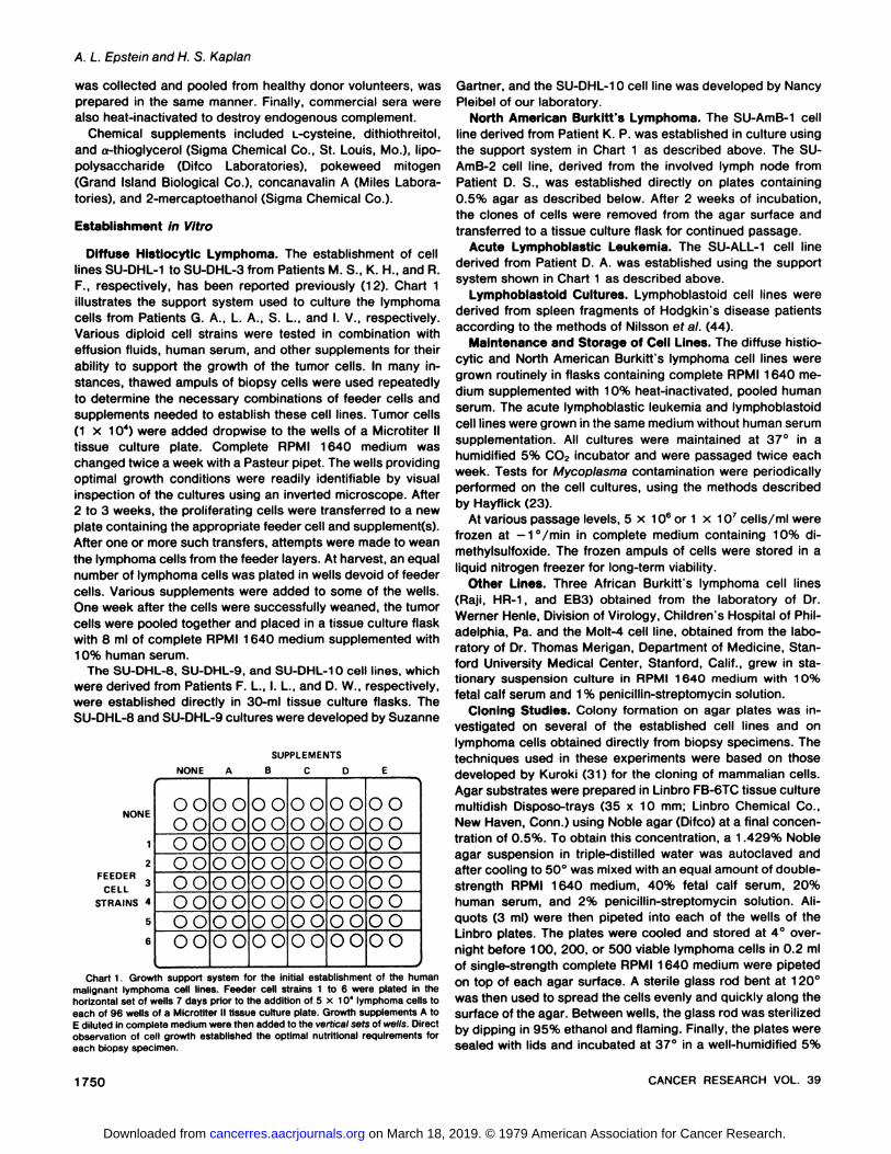
Diffuse Hlstiocytlc Lymphoma. The establishmentof celllines SU-DHL-i to SU-DHL-3 from Patients M. S., K. H., and A.F., respectively, has been reported previously(i 2). Chart 1illustrates the support system used to culture the lymphomacells from Patients G. A., L. A., S. L., and I. V., respectively.Various diploid cell strains were tested in combination witheffusion fluids, human serum, and other supplements for theirability to support the growth of the tumor cells. In many instances, thawed ampuls of biopsy cells were used repeatedlyto determine the necessary combinations of feeder cells andsupplements needed to establish these cell lines. Tumor cells(1 x 10@)were added dropwise to the wells of a Microtiter IItissue culture plate. Complete RPMI 1640 medium waschanged twice a week with a Pasteur pipet. The wells providingoptimal growth conditions were readily identifiable by visualinspection of the cultures using an inverted microscope. After2 to 3 weeks, the proliferatingcells were transferredto a newplate containing the appropriate feeder cell and supplement(s).After one or more such transfers, attempts were made to weanthe lymphoma cells from the feeder layers. At harvest, an equalnumber of lymphoma cells was plated in wells devoid of feedercells. Various supplements were added to some of the wells.One week after the cells were successfully weaned, the tumorcells were pooled together and placed in a tissue culture flaskwith 8 ml of complete RPMI 1640 medium supplemented with10% human serum.
The SU-DHL-8, SU-DHL-9, and SU-DHL-i 0 cell lines, whichwere derived from Patients F. L., I. L., and D. W., respectively,were established directly in 30-mI tissue culture flasks. TheSU-DHL-8 and SU-DHL-9 cultures were developed by Suzanne
Gartner, and the SU-DHL-i 0 cell line was developed by NancyPleibel of our laboratory.
North American Burkltt's Lymphoma. The SU-AmB-i cellline derived from Patient K. P. was established in culture usingthe support system in Chart i as described above. The SUAmB-2 cell line, derived from the involved lymph node fromPatient D. S., was established directly on plates containing0.5% agar as described below. After 2 weeks of incubation,the clones of cells were removed from the agar surface andtransferred to a tissue culture flask for continued passage.
Acute Lymphoblastic Leukemia. The SU-ALL-i cell linederived from Patient D. A. was established using the supportsystem shown in Chart i as described above.
Lymphoblastoid Cultures. Lymphoblastoidcell lines werederived from spleen fragments of Hodgkin's disease patientsaccording to the methods of Nilsson et a!. (44).
Maintenance and Storage of Cell Lines. The diffusehistiocytic and North American Burkitt's lymphoma cell lines weregrown routinely in flasks containing complete RPMI i 640 medium supplemented with 10% heat-inactivated, pooled humanserum. The acute lymphoblastic leukemia and lymphoblastoidcell lines were grown in the same medium without human serumsupplementation. All cultures were maintained at 37° in ahumidified 5% CO2 incubator and were passaged twice eachweek. Tests for Mycop!asma contamination were periodicallyperformed on the cell cultures, using the methods describedby Hayflick (23).
At various passage levels, 5 x 10⁶ or i x 1O@cells/mI werefrozen at —1°/min in complete medium containing 10% dimethylsulfoxide. The frozen ampuls of cells were stored in aliquid nitrogen freezer for long-term viability.
Other Lines. Three African Burkitt's lymphoma cell lines(Raji, HA-i , and EB3) obtained from the laboratory of Dr.Werner Henle, Division of Virology, Children's Hospital of Philadelphia, Pa. and the MoIt-4 cell line, obtained from the laboratory of Dr. Thomas Merigan, Department of Medicine, Stanford University Medical Center, Stanford, Calif., grew in stationary suspension culture in RPMI 1640 medium with 10%fetal calf serum and i % penicillin-streptomycmnsolution.
Cloning Studies. Colony formationon agar plates was investigated on several of the established cell lines and onlymphoma cells obtained directly from biopsy specimens. Thetechniques used in these experiments were based on thosedeveloped by Kuroki (3i ) for the cloning of mammalian cells.Agar substrates were prepared in Linbro FB-6TC tissue culturemultidish Disposo-trays (35 x 10 mm; Linbro Chemical Co.,New Haven, Conn.) usingNobleagar (Difco) at a finalconcentration of 0.5%. To obtain this concentration, a 1.429% Nobleagar suspension in triple-distilled water was autoclaved andafter cooling to SO@was mixed with an equal amount of doublestrength RPMI 1640 medium, 40% fetal calf serum, 20%human serum, and 2% penicillin-streptomycin solution. Aliquots (3 ml) were then pipeted into each of the wells of theLinbro plates. The plates were cooled and stored at 4°overnight before 100, 200, or 500 viable lymphoma cells in 0.2 mlof single-strength complete RPMI 1640 medium were pipetedon top of each agar surface. A sterile glass rod bent at 120°was then used to spread the cells evenly and quickly along thesurface of the agar. Between wells, the glass rod was sterilizedby dipping in 95% ethanol and flaming. Finally, the plates weresealed with lids and incubated at 37°in a well-humidified 5%
SUPPLEMENTS
NONE
2FEEDER
CELLSTRAINS 4
5
6
Chart1. Growthsupportsystemfor the initial establishmentof the humanmalignant lymphoma cell lines. Feeder cell strains 1 to 6 were plated in thehorizontal set of wells 7 days prior to the addition of 5 x 1O@lymphoma cells toeach of 96 wells of a Microtiter II tissue culture plate. Growth supplements A toE dilutedIncompletemediumwere thenaddedto the verticalsetsof wells.Directobservationof cell growthestablishedthe optimalnutritionalrequirementsforeach biopsy specimen.
CANCERRESEARCHVOL. 391750
on March 18, 2019. © 1979 American Association for Cancer Research.cancerres.aacrjournals.org Downloaded from

Estab!ishment and C!oning of Lymphoma Ce!! Lines
CO2 incubator for 2 weeks with no additional feeding. Foroptimal results, it was necessary to leave the plates undisturbedfor at least the first week of incubation to allow the cells tobecome firmly attached to the agar. In addition, it was importantto restrict the amount of fluid which accumulated on top of theplates, since a watery surface tended to dislodge the colonies.This was easily achieved by seeding the plates with smallinocula and cooling them at 4°for 1 hr prior to incubation tocondense the excess water. Visible colonies were then scoredwithout fixing or staining, and cloning efficiencies were calculated by the following equation:
No.of visiblecoloniesClonIngefficiency= x 100
No.of cellsseeded
In several experiments, this procedure was modified to determine the effects of feeder layers and supplements on thecloning efficiencies of the cell lines. As shown in Chart 2,feeder layers were prepared in the Linbro plates by seeding 5x 1o@trypsinized cells suspended in 3 ml of complete McCoy'smedium into each well (35 x 10 mm). After 7 days of incubation,the medium was decanted, and 0.5% agar overlay was added.In certain cases, an autoclaved dialysis membrane (UnionCarbide Corp., Chicago, III.) shaped to cover one-half of themonolayer was inserted before the agar was pipeted into thewells. In order to test the effects of varying concentrations ofsupplements on the cloning efficiencies of the cell lines, supplements were first mixed with the agar solution in disposabletubes at 45°.Aliquots (3 ml) were then pipeted into each of thewells of the Linbro plates. Because of the added volume of thesupplement, the agar concentration had to be adjusted in eachtube to obtain a final concentration of 0.5%. Stock solutions ofthe supplements were sterilized by filtration through a 0.45-@tmMillex disposable filter unit (Millipore, Bedford, Mass.), aliquoted, and stored frozen at —20°until use.
To subclone the lymphoma cell lines, individual colonieswere removed with a Pasteur pipet and placed into single wellsof a Microtiter II tissue culture plate containing complete medium. After a few days of incubation, the cells could be replatedin multiple wells and, finally, after 3 weeks they were transferred to tissue culture flasks for continued cultivation. Subcloned lines were again grown on agar plates to determinetheir new cloning efficiencies.
RESULTS
Establishment and Growth in Cell Culture
Ce!! Lines
Table 2 lists the lymphoma cell lines established in vitroalong with pertinent clinical information.
Diffuse Histlocytlc Lymphoma. SU-DHL-1 throughSU-DHL3. The establishment of these cell lines from Patients M. S., K.
H., and A. F., respectively, has been reported previously (12).SU-DHL-4 through SU-DHL- 10. Lymphoma cells from Pa
@@@@@@@ LYMPHOMACELL COLONIES
AGAR
__________________________ FEEDER CELL MONOLAYER
Chart 2. Schematic diagram of the agar cloning assay. Lymphoma cells werecloned on agar overlays in plates containing feeder cell monolayers.
tients G. A., L. A., and S. L. were found to proliferate only inthose wells containing monolayers of pediatric skin fibroblastsfrom Patient M. S. Further supplementation with human serumor effusion from various patients retarded or, in some instances,prevented the growth of the lymphoma cells during this initialphase of cultivation. Tumor cells from Patient I. V. were foundto grow in wells containing monolayers of adult or pediatricskin fibroblasts from Patients G. A. and M. S., respectively.However, cell growth was dependent upon the presence of10% human serum in combination with these feeder layers.
Neoplastic cells from Patients F.L. and I. L. were established inflasks containing autologous feeder cells present in the effusionfluids of these patients. Malignant cells from Patient D. W.,unlike the other diffuse histiocytic lymphoma cell lines, did notrequire the presence of a feeder layer for initial growth in vitro.
All 7 cell lines were established in complete RPMI 1640medium. Under the phase-contrast microscope, the lymphomacells were seen resting on top of the feeder layers as roundedcells, oftentimes with clear cytoplasmic vacuoles. After incubation for i week, negative wells showed a disappearance ofthese cells while positive wells showed areas of increased celldensity (Fig. 1A). After 2 weeks, positive wells showed a furtherincrease in tumor cell number (Fig. i B) and, finally, after 3weeks, a lawn of lymphoma cells was observed covering thesurface of the underlying monolayer of feeder cells (Fig. 1C).The SU-DHL-4, SU-DHL-5, SU-DHL-8, and SU-DHL-9 cell lineswere then successfully weaned from the feeder layers in complete RPMI 1640 medium containing 10% human serum. TheSU-DHL-6 and DU-DHL-7 cell lines were also successfullyweaned from the feeder layers in complete RPMI 1640 mediumcontaining 10% human serum but had to be subcultured several times on the feeder layers before they could be establishedin stationary suspension culture.
North American Burkltt's Lymphoma. SU-AmB- 1. Lymphoma cells from the malignant peritoneal effusion from PatientK. P. were foundtogrowonlyinthosewellscontainingpediatricskin fibroblasts from Patient M. S. After one transfer, thesecells were weaned from the feeder cells and established insingle-cell suspension culture in medium containing i 0% human serum.
Su-AmB-2. After incubation for 2 weeks, clones of cellsdeveloped on the surface of the agar plates. These cells weredirectly established in suspension culture in medium supplemented with 10% human serum.
Acute Lymphoblastlc Leukemia. SU-ALL-1. Leukemiacellsfrom Patient 0. A. were only able to grow in the presence offeeder cells from the skin punch biopsy of Patient G. A. Afterseveral passages on the feeder layers, the leukemia cells werefinally weaned from the underlying monolayer and establishedin single-cell suspension culture at a relatively high cell density(i x i 06 cells/mI). Contrary to the other lymphoma cell lines,these cells did not require a human serum supplement beforeor after weaning.
Lymphoblastold. Culturesof explantsprepared from Hodgkin's disease tissues spontaneously underwent lymphoblastoidtransformation 4 to 6 weeks after initiation in vitro. Clusters oflymphoblasts were observed growing on top of the fibroblastsfrom multiple areas of the flasks (Fig. 2). The clumps of cellseventually became detached from the underlying fibroblastsand grew in suspension. By removing the supernatant culturefluid, it was possible to isolate the lymphoblasts and establish
MAY 1979 1751
on March 18, 2019. © 1979 American Association for Cancer Research.cancerres.aacrjournals.org Downloaded from

Cell line Patient Age Sex Diagnosis Source of malignant cells
A. L. Epstein and H. S. Kaplan
Table 2
Human malignant lymphoma cell lines
Diffuse jiistiocytic lymphomaSUa@DHL@1SU-DHL-i (Cl 1)
MS. 10 M DHL Pleuraleffusion
SU-DHL-2SU-DHL-2(2)SU-DHL-2(2 Cl 1)
K. H. 73 F DHL Pleuraleffusion
SU-DHL-3SU-DHL-3(Cl 1)SU-DHL-3(Cl 2)
R. F. 35 M DHL Peritonealeffusion
SU-DHL-4SU-DHL-4 (Cl 1)SU-DHL-4 (Cl 2)SU-DHL-4 (Cl 3)SU-DHL-4 (Cl 4)SU-DHL-4 (M)SU-DHL-4 (M CI 1)SU-DHL-4 (M Cl 2)
GA. 38 M DHL Peritonealeffusion
SU-DHL-5SU-DHL-5(Cl 1)SU-DHL-5(Cl 2)
L. A. 17 F DHL Lymph node
SU-DHL-6SU-DHL-6(M)
I. V. 43 M DMHL Peritoneal effusion
SU-DHL-7S. L.47FDHLPleuraleffusionSU-DHL-8F.
L.59MDHLPleuraleffusionSU-DHL-9I.
L.64FDHLPleuraleffusionSU-DHL-i
0D. W.26MDHLPeritoneal effusion
North American Burkitt's lymphomaSU-AmB-i K. P.
SU-AmB-i (M)@U-AmB-2
SU-AmB-2(Cl 1)SU-AMB-2 (Cl 2)
ii M DU(Burkitt's)
16 M DU(Burkitt's)
Peritonealeffusion
Lymph node0. S.
Acute lymphoblastic leukemiaSU-ALL-1 D.A. 14 F ALL
a The abbreviations used are: SU, Stanford University; DHL, diffuse histiocytic lymphoma; AmB, North
AmericanBurkitt's Iymphoma;ALL,acutelymphoblasticleukemia;DMHL,diffusemixedhistiocyticandlymphocyticlymphoma;DU, diffuse undifferentiated;Cl, clone; M, derived from heterotransplantedtumor.
Blood
permanent cell lines. Four such cell lines, designated SU-LB-2through SU-LB-5, were established (Table 3). All of the celllines grew typically as free-floating clumps of cells in completeRPMI1640medium.
C!oning Studies
As illustrated in Fig. 3, the human malignant lymphoma celllines were successfully cloned on agar substrates. After 2weeks of incubation, visible discrete colonies of cells werefound to grow on the surface of the agar plates, facilitatingquantitation and subcloning procedures. Individual colonieswere composed of rounded cells which grew radially from the
efficiencies of the human malignant lymphoma cell lines alsovaried, as shown in Table 4. Interestingly, the EBV-positive SUAmB-2 cell line, which originally was established from cellsgrown on agar, had the highest cloning efficiency (3i .0%). Ofthe diffuse histiocytic lymphoma cell lines, the SU-DHL-8 andSU-DHL-9 cell lines had distinctively higher cloning efficiencies. Several clones of the diffuse histiocytic and North American Burkitt's lymphoma cell lines were isolated and successfully established in suspension culture. A complete list of thesesublines is presented in Table 2. As shown in Table 4, all of thesublines tested had increased cloning efficiencies in the rangeof45 to50%.
center of each clone. Colonies from different cell lines showed In order to quantitatively screen a variety of normal andmorphological variations in size, color, and density. The cloning malignant cell cultures for their ‘‘feeder'â€effect, lymphoma
1752 CANCER RESEARCH VOL. 39
on March 18, 2019. © 1979 American Association for Cancer Research.cancerres.aacrjournals.org Downloaded from

Lymphoblastoid cell linesSourceofCell
linePatientDiseasetissueEBVSUa@LB@2C.
B.NSHDSpleen+SU-LB-3C.S.NSHDSpleen+SU-LB-4W.M.NSHDSpleen+SU-LB-5N.
M.PDLSpleen+
Feederlayerscreeningdata%cloningSua@SU
No.Source of tissueType oftissueMolt-4AmB-1DHL-21Patient
D. S.Adultskin0002PatientG. A.Adultskin0003PatientD. M.Adultskin0004CM
1522°Adultskin00.805CM1568bAdultskin0.600.26Patient
M. S.Pediatric skin2.41.01.07GL2722°Pediatric skin1 .01.00.48GL2723°Pediatricskin2.00.41.09GL31 @Pediatricforeskin0.80.20.41
0GL3280―Fetus00011Patient J. S.Adultspleen00012Patient A. C.Adultspleen00013Patient B. P.Adultspleen00014Patient W. S.Adultspleen000151 021 B―Adultovary00016Patient M. M.Adultmesothelial00017PatIent F. L.Adultmesothelial00018NP 1016BbTeratoma00019AU302CFibrosarcoma00020AO
676COsteogenicsarcoma000211021 C°Ovarianteratoma00022A2O4CRhabdomyosarcoma000
Cloningefficienciesof thehumanmalignantlymphomacelllinesThediffusehistiocyticand NorthAmericanBurkitt's lymphomacelllines
wereclonedon 0.5%NobleagarcontainingRPMIMedium1640,20%fetal calf serum, 10% human serum, and 1% penicillin-strepto
mycin solution. The SU-ALL-i cell line was cloned with thesamenutrientsas the otherculturesexceptwithouthumanserum.Cloning
efficiency(%)Cell
line Established lineSubcboneDHLSU-DHL-i
o.3aSU-DHL-25.0SU-DHL-3
4.048.0SU-DHL-410.046.0SU-DHL-4
(M) 15.044.0SU-DHL-52.1SU-DHL-6<0.1SU-DHL-7
<0.1SU-OHL-815.0SU-DHL-922.5SU-DHL-1O
NDAMBSU-AmB-i
0.7SU-AmB-231.0ALLSU-ALL-i
0
%cloningsul
sU@su su suFeederlayerDHL-1DHL-2DHL-4DHL-5AmB-1Without
humanserum20<10006010<1080<10009000001000000With
humanserum201415556013iiii258000009000001000000a
The abbreviations used are the sameas in Table5.
Estab!ishment and C!oning of Lymphoma Ce!! Lines
Table3 cell lines. In Charts 4 and 5, respectively, the effects of Lcysteine and dithiothreitol on the cloning efficiencies of severalof the diffuse histiocytic lymphoma cell lines are presented.These results clearly show that, at very narrow concentrationranges, both chemicals produce marked increases (from 16 to28%) in the cloning efficiencies of some but not all the diffuse
histiocytic lymphoma cell lines tested. As shown in Table 7,increases in cloning efficiencies of certain lymphoma cell lineswere also observed with concanavalin A for the SU-DHL-4 cellline; with Iipopolysaccharide for the SU-DHL-4, SU-DHL-5, and
Table5
aThe abbreviationsusedare: SU, StanfordUniversity;LB, lymphoblastoid; NSHD,nodular sclerosingHodgkin'sdisease;PDL, poorlydifferentiatedlymphocyticlymphoma.
Table4
a The abbreviations used are: SU, Stanford University; AmB, North
AmericanBurkitt's lymphoma;DHL,diffusehistiocyticlymphoma.6 Obtained from the laboratory of Dr. Barbara McCaw, Department
of MedicalGenetics,Universityof OregonSchoolof Medicine,Portland,Oreg.
C Obtained from the Cell Culture Laboratory, University of California
Schoolof PublicHealth,NavalBiomedicalResearchLaboratory,NavalSupplyCenter,Oakland,Calif.
Table6Effects of feeder layers and human serum on the cloning efficiency of
humanmalignantlymphomacell lines
a Each percentage represents the average of several experiments.
b The abbreviations used are: SU, Stanford University; DHL, diffuse
histiocytic lymphoma; M, derived from heterotransplanted tumor; AmB,NorthAmericanBurkitt's lymphoma;ND,notdone.
cells were plated on agar overlays in plates containing adherentmonolayers. As shown in Table 5, only a few of these culturesimproved the cloning efficiencies of the test lymphoma andleukemia cell lines. Some of these cultures, which were derivedfrom pediatric and adult skin punch biopsies, were the sameones used to establish many of the lymphoma cell lines. Asshown in Table 6, when the lymphoma cells were cloned onplates containing both human serum and feeder layers, thecloning efficiencies of all but SU-DHL-i increased dramaticallyfrom <i % to as high as 25%. If, however, a dialysis membranewas placed between the feeder layer and the lymphoma cells,colony formation was inhibited on that half of the plate containing the dialysis membrane (Chart 3). These results stronglysuggest that a diffusible factor(s) may be responsible for thefeeder effect of these cultures. Unfortunately, medium conditioned by these feeder layers does not have colony-stimulatingactivity. Thus, this factor(s) appears to be extremely labileunder the present experimental conditions.
In addition to feeder layers and human serum, certain chemical supplements were found to produce concentration-dependent increases in the cloning efficiencies of the lymphoma
MAY 1979 1753
on March 18, 2019. © 1979 American Association for Cancer Research.cancerres.aacrjournals.org Downloaded from

@A I
A. L. Epstein and H. S. Kaplan
The use of the Microtiter II tissue culture plate as the supportsystem for the initial establishment of the lymphoma cell linesoffered several advantages which contributed significantly tothe success of this investigation. It was possible to start withvery small initial tumor cell populations such as are often readilyavailable in lymph node biopsy specimens or body cavity fluids.In addition, the design of the Microtiter II tissue culture platepermitted the rapid multifactorial screening of nutrient growthrequirements. Without this multifactorial approach, it would nothave been possible to identify the various combinations ofmedia, supplements, and feeder layers that were optimal foreach tumor line. Thereafter, daily observation of individual cellsin each well enabled the ascertainment of optimal nutrientrequirements and the early weaning of evolving tumor cellpopulations from the feeder layers. Since cell suspensions ofmany of the original tumor specimens were frozen shortly aftercollection, multiple experimental approaches were possibleover an extended period of time. These factors have contributed to a very high success rate in the cultivation of the diffusehistiocytic and North American Burkitt's lymphomas. Thesemethods should be applicable to a number of other lymphomasand leukemias for which cell lines have not yet been established.
In order to quantitate the growth-supporting effects of variousfeeder layers, chemicals, and biological supplements on thehuman lymphomas, an agar cloning assay developed for mammalian cells by Kuroki (31) was utilized in a number of experiments. Using standard methods, most of the lymphoma andleukemia cell lines were found to have only relatively lowcloning efficiencies on agar. However, in the presence ofhuman serum and specific feeder layers, the cloning efficiencies of many of the cell lines increased dramatically. Althougha diffusible factor(s) synthesized by the feeder layer cells wasimplicated in these experiments, further studies regarding itschemical nature were not possible because of its apparentlability.
During the critical initial phase of tumor cell establishment in
-@ CELLCOLONIESLw@-w@—J AGARDIALYSIS MEMBRANE
FEEDERCELL MONOLAYER
Chart 3. Schematic drawing illustrating the position ofthe dialysis membranebarrier and its inhibition of colony formation in the agar cloning assay. The resultsstrongly suggest that a diffusable factor(s) synthesized by the feeder layer cellsmay be responsible for the observed increase in colony formation of the humanmalignant lymphoma cells.
>.0zuJ
0U.U.UJ
Qzz0-I0I-zLU0
LU0.
CONCENTRATIONL-CYSTEINE (mol)
Chart 4. Effect of L-cysteine on the cloning efficiencies of 2 diffuse histiocyticlymphoma cell lines.
SU-AmB-i cell lines; with a-thioglycerol for the SU-DHL-4, SUDHL-8, and SU-DHL-9 cell lines; and with 2-mercaptoethanolfor the SU-DHL-2, SU-DHL-4, and SU-DHL-9 cell lines.
Finally, among the biological supplements tested for theircolony-stimulating activity, select malignant pleural effusionfluids were capable of increasing the cloning efficiency of thelymphoma cell lines. However, none of the 4 growth factorstested had a significant effect on the cloning efficiencies ofselectedcelllines.
DISCUSSION
Since the inauguration in 1973 of a research program directed at studying the biology of the human malignant lymphomas, 13 tumor cell lines have been established in our laboratory. Evidence concerning the neoplastic nature and the functional and surface marker characteristics of these cell lines hasbeen presented in a series of earlier papers (10—i2, 14, 27,36). The original choice of malignant effusion fluid may havefacilitated our initial studies since, as has recently been suggested (6), tumor cell selection within the patient may alreadyhave occurred, yielding a population of highly malignant cellsgrowing in suspension under conditions not dissimilar fromtheir subsequent in vitro environment. However, our later experience with biopsy specimens (e.g. , SU-DHL-S and SU-AmB2) and peripheral blood (e.g. , SU-ALL-i ) has suggested thatthese methods are also applicable to the establishment oflymphoid tumor cells from sources other than malignant effusions.
Q 202z0.J0
2LU0
LU0.
CONCENTRATIONDITHIOTHREITOL(x10@mol)
Chart 5. Effect of dithiothreitoi on the cloning efficiencies of 3 diffuse histiocytic lymphoma cell lines.
1754 CANCER RESEARCH VOL. 39
on March 18, 2019. © 1979 American Association for Cancer Research.cancerres.aacrjournals.org Downloaded from

Effect of chemical supplements on the cloning efficiencies of the humanThe lymphomacell lines were cloned on 0.5% Nobleagar containin
serum,10% humanserum,and 1% penicillin-streptomycinsolution.malignang RPMIt
lymphoma1640, 20%cell
linesfetalcalf%
cloningSUa@
SU- SU- SU- SU SU SU SUSupplement DHL-i DHL-2 DHL-4 DHL-5DHL-7DHL-8DHL-9AmB-iNone
0.5 0.5 0.5 0022.515.00Concanavalin
A1.0 @g/ml 0 0 1.5 0045.05.002.5
@tg/ml 0 0 5.0 0 027.515.005.0pg/mI 0 0 2.0 0057.520.007.5@g/ml
0 0 1.0 0047.510.00Pokeweed
mitogen0.5%0 0 0 0031.5b01.0%0 0 0 0032.511.001
.5% 0 0 0 0 030.510.00Lipopolysaccharide1:100
0 0 0 0.5030.522.501:2000 0 7.5 00bb1.21
:400 0 0 7.5 3.00b22.53.01:800 0 0 7.5 0.5024.522.54.51:1,600
0 0 22.5 0031.520.05.2a-Thioglycerol1
x i03 M 0 0 3.5 0052.5b01x iO4 M 0 0 5.0 0036.043.501x 1o5 M 0 0 1.0 0032.022.001x 1O-°M 0 0 1.0 0 019.08.002-Mercaptoethanol5
x iO4 M 0 0 0 0024.07.501x 1o@ M 0 1.0 35.0 0049.030.005x 1O@ M 0 15.0 10.0 0 034.517.501x iO@ M 0 0 17.5 0044.5b05x i06 M 0 0 2.0 0024.09.001x iO@ M 0 0 0 0 028.014.50
Establishment and C!oning of Lymphoma Ce!! Lines
Table 7
a The abbreviations used are the same as in Table 5.
b In these plates, the number of colonies was not able to be counted accurately, since excess moisture
collecting on the surface of the agar caused the colonies to coalesce.
vitro, autologous or heterologous contact-inhibited feeder layers were essential for cell proliferation. Historically, the use offeeder layers in cell culture was first reported by Puck andMarcus (48), who described the effects of X-irradiated monolayers on the cloning of HeLa cells in vitro. Along with theinvestigations of Earle et a!. (9) which demonstrated the necessity of ‘‘conditioning'â€media by living cells, their pioneeringwork showed the importance of diffusible factors in the in vitrogrowth of single mammalian cells (48, 49). Despite the substantial improvements in synthetic tissue culture media in thelast few years, feeder layers are being used with increasingfrequency for the growth and establishment of a wide variety ofnormal and malignant mammalian tissues. Thus, Sundstromand Nilsson (52) used human glial cells to grow the only otherhuman histiocytic lymphoma cell line established to date; Hamburger and Salmon (22) used adherent spleen cell cultures andconditioned media to clone malignant cells from various humantumors, including human myeloma stem cells (21); Taylor-Papadimitriou et a!. (54) have used nondividing feeder cells togrow human mammary epithelial cells; Aaronson et a!. (i ) wereable to grow colonies of human sarcoma cells directly onconfluent monolayers of normal diploid cells; and Armstrongand Aosenau (2) grew human primary breast carcinomas with
embryonic mesenchyme feeder layers. Although it is clear thatcertain human cancers can clone in culture without the additionof feeder cells or diffusible factors (24, 26, 35), it is becomingincreasingly clear that most human tumors require the presence of normal cellular products for sustained growth in vitro.
The agar cloning assay as modified by our laboratory fromthe methods of Kuroki (31) was successfully used to screen alarge number of normal and malignant tissues for their growthsupporting effects. The results of these experiments demonstrate that only specific feeder cells are capable of stimulatingthe clonal growth of the human malignant lymphoma cells.Therefore, it is likely that the microenvironment of varioustissues may play an important role in the growth of individualor small clusters of tumor cells metastasizing in the lymphaticand vascular channels of the body.
In addition to feeder layers and biological supplements,certain chemicals were noted to produce concentration-dependent increases in the cloning efficiencies of the lymphomacell lines. The use of thiols and disulfides in these experimentswas stimulated by the previous observations of Broome andJeng (3), who first showed that L-cysteine was a specificgrowth-promoting substance for mouse lymphoma Li 2i 0 cells.These same investigators later demonstrated that a number of
MAY 1979 1755
on March 18, 2019. © 1979 American Association for Cancer Research.cancerres.aacrjournals.org Downloaded from
A. L. Epstein and H. S. Kap!an
other thiols and disulfide derivatives were able to stimulate thegrowth of mouse lymphoma cells in culture (4). Of 23 mouseleukemic and neoplastic lymphoid cell lines tested, 13 werefound to be thiol-disulfide-dependent in vitro. In addition, themitogenic responses of normal mouse splenic lymphocyteswere often increased by thiols and disulfides. Although theexact mechanism of action of these compounds is unknown,precise structure-activity relationships do seem to exist forthese molecules. Based on the structure of thioethane, Broomeand Jeng (4) were able to formulate a number of rules concerning the structure of active molecules. More recently, Tanapat et a!. (53) have produced evidence to suggest that murinelymphoma cells secrete autoinhibitory substances, possiblythiols or disulfides, the actions of which may be reversed byhigh concentrations of 2-mercaptoethanol. Our studies extendthe growth-promoting activities of the thiols and disulfides toinclude the human lymphomas. Taken together, these findingsstrongly suggest that these compounds are acting directly onthe replicative processes of lymphocytes and that malignantlytransformed cells are able to respond to or are dependent upontheir action for clonogenic growth.
The cloning efficiencies of some of the human lymphoma celllines were also found to increase in the presence of mitogens.Consistent with these results, phytohemagglutinmn and/orpokeweed mitogen have been used to clone human lymphocytes (7, 16) and acute myelogenous leukemia cells (8) in vitro.Recently, conditioned medium from phytohemagglutinin-stimulated normal human lymphocytes was found selectively tostimulate the growth of normal T-lymphocytes (41). Finally,other chemicals not yet tested on the human malignant lymphoma cell lines have been shown to improve the cloningactivities of hematopoietic and fibroblastic cell cultures. Theseinclude ascorbic acid for the formation of mouse plasmacytomacell colonies (47), steroid hormones on the plating and cloningefficiency of human fibroblasts (37), and selenite, transferrin,albumin, and lecithin on the growth of murine bone marrowcells (19). In a review of the unique requirements for clonalgrowth in culture, Ham (20) explores the advantages of clonalexperiments in the delineation of nutritive differences betweennormal and cancerous cells.
The successful adaptation of many of these human malignantlymphoma cell lines to clonal cultivation on agar substratescoupled with replica plating procedures for mammalian cells(31) opens the way to the selection and analysis of multipleclones of each cell line. Recently, Konrad et a!. (30) haverevealed previously unsuspected heterogeneity in an established line of cultured mammalian cells by studying clonalvariation in colony morphology and growth on agar. Theseexperiments suggest that the morphological appearance ofindividual clones may be used to study a new class of in vitrogrowth control phenomena. Such studies may be useful inassessing the relative importance of various specific cytogenetic abnormalities in the maintenance of the transformedphenotype. Clonal culture systems will also permit the quantitative analysis of the susceptibility of monospecific tumor celltypes to killing by ionizing radiation, hyperthermia, variouschemotherapeutic agents used singly or in combination, andexperimental immunotherapeutic methods. This procedure willenable investigators to determine the mutation frequency ofdrug and radiation resistance in malignant cells. In addition,cloning techniques will help determine the nutritional require
ments of individual lymphomas, which may lead to a greaterunderstanding of the metastatic patterns of tumors in vivo andhopefully to the development of more specific chemotherapeutic agents. Finally, the isolation of individual clones of cells mayenable investigators to segregate neoplastic properties suchas heterotransplantability and unstable chromosome complement from specific differentiation traits of malignant cells.
Currently, although modern radiotherapeutic and chemotherapeutic advances have dramatically improved the prognosis of patients with Hodgkin's disease (28), the prognosis ofmany of the non-Hodgkin's lymphomas, particularly the diffusehistiocytic and undifferentiated lymphomas, remains poor. It ishoped that new therapeutic approaches stemming from in vitrostudies with clonal cultures of the human malignant lymphomaswill contribute significantly to the treatment and prognosis ofthese neoplasms.
ACKNOWLEDGMENTS
The authors wish to thank Nancy Pleibel for her expert technical assistance.
REFERENCES
1. Aaronson, S. A., Todaro, G. J., and Freeman, A. E. Human sarcoma cells inculture: identification by colony-forming ability on monolayers of normalcells. Exp. Cell Res., 6 1: i —5,1970.
2. Armstrong,R. C., and Rosenau,W. Cocultivationof humanprimarybreastcarcinomas and embryonic mesenchyme resulting in growth and maintenance of tumor cells. Cancer Res., 38: 894—900,1978.
3. Broome, J. D., and Jeng, M. W. Growth stimulation of mouse leukemia cellsby thiols and disulfides in vitro. J. NatI. Cancer Inst., 49: 579—58i, 1972.
4. Broome, J. D., and Jeng, M. W. Promotion of replication in lymphoid cells byspecific thiols and disulfides in vitro. J. Exp. Med., 138: 574—592,1973.
5. Bruntsch, U., Gallmeier, W. M., Hossfeld, D. K., Boecker, W. R., Hertenstein,C., Gasch, J., and Schmidt, C. G. Biological characterization of two EBNAnegative human cell lines derived from non-Hodgkin's lymphomas, in press,1979.
6. Cailleau, R., Mackay, B., Young, R. k., and Reeves, W. J., Jr. Tissue culturestudies on pleural effusions from breast carcinoma patients. Cancer Res.,34: 801—809,1974.
7. Chol, k. W., and Bloom, A. D. Cloning human lymphocytes in vitro. Nature(Lond.), 227: 171—173,1970.
8. Dicke, K. A., 5pitzer, G., and Ahearn, M. J. Colony formation in vitro byleukemia cells in acute myebogenousleukemia with phytohaemagglutinin asstimulating factor. Nature (Lond.), 259: 129—130, 1976.
9. Earle, W. R., Bryant, J. C., and Schilling, E. L. Certain factors limiting thesize of the tissue culture and the development of massive cultures. Ann. N.Y. Acad. Sd., 58: 1000-101 1, 1954.
10. Epstein, A. L., Henle, W., Henle, G., Hewetson, J. F., and kaplan, H. S.SurfacemarkercharacteristicsandEpstein-Barrvirusstudiesof twoestablished North American Burkltt's lymphoma cell lines. Proc. NatI. Acad. Sci.U. S. A., 73: 228-232, i976.
i i . Epstein, A. L., Herman, M. M., kim, H., Dorfman, R. F., and Kaplan, H. S.Biology of the human malignant lymphomas. Ill. Intracranial heterotransplantation in the nude, athymic mouse. Cancer, 37: 2158—2176, 1976.
12. Epstein, A. L., and Kaplan, H. S. Biology of the human malignant lymphomas.I. Establishmentin continuouscell cultureandheterotransplantationofdiffuse histiocytic lymphomas. Cancer, 34: 1851—1872, 1974.
13. Epstein, A. L., and Kaplan, H. S. Feeder layer and nutritional requirementsfor the establishment and cloning of human malignant lymphoma cell lines.Proc. Am. Assoc. Cancer Res., 18: 293, 1977.
14. Epstein, A. L., Levy, R., Kim, H., Henle, W., Henle, G., and Kaplan, H. S.Biology of the human malignant lymphomas. Iv. Functional characterizationof ten diffuse histiocytic lymphoma cell lines. Cancer, 42: 2379—2391,1978.
i 5. Epstein, M. A., and Barr, Y. M. Cultivation in vitro of human lymphoblastsfrom Burkitts malignant lymphoma. Lancet, 1: 252—253,i 964.
16. Fibach, E., Gerassi, E., and Sacks, L. Induction of colony formation in vitroby human lymphocytes. Nature, (Lond.) 259: 127—i29, i 976.
i 7. Foley, G. E., Lazarus, H., Farber, S., Uzman, B. G., Boone, B. A., andMcCarthy, R. E. Continuous culture of human lymphoblasts from peripheralbloodof a childwithacuteleukemia.Cancer,18:522—529,1965.
18. Gerber, P. Is Epstein-Barr virus a human tumor virus? NatI. Cancer Inst.Monogr., 36: 65-71, 1973.
i 9. Guilbert, L. J., and Iscove, N. N. Partial replacement of serum by selenite,
i756 CANCER RESEARCH VOL. 39
on March 18, 2019. © 1979 American Association for Cancer Research.cancerres.aacrjournals.org Downloaded from

Estab!ishment and C!oning of Lymphoma Ce!! Lines
transferrin, albumin, and lecithin In haemopoietic cell cultures. Nature, 263:594—595.1976.
20. Ham, R. Unique requirements for cbonal growth. J. Nati. Cancer Inst. 53:i459-i463, 1974.
21. Hamburger,A. W., and Salmon,S. E. Primarybioassayof humanmyelomastem cells. J. Clin. Invest., 60: 846—854,1977.
22. Hamburger,A. W., and Salmon, S. E. Primary bioassayof humantumorstem cells. Science, 197: 46i —463,1977.
23. Hayfllck, L. Tissue culturesand mycoplasmas.Tex. Rep. Biol. Med., 23:285-303, i965.
24. Hinuma,V., and Grace, J., Jr. Cloningof immunogbobulin-producinghumanleukemic and lymphoma cells in long-term cultures. Proc. Soc. Exp. Biol.Med., 124: 107-110, i967.
25. Hirakl, S., Miyoshi,I., Masuji,H., kubonishi,I., Matsuda,Y., Nakayama,T.,Kishimoto, H., Chen, P., and kimura, I. Establishment of an Epstein-Barrvirus-determined nuclear antigen-negative human B-cell line from acutelymphoblastic leukemia. J. Nat). Cancer Inst. 59: 93-94, 1977.
26. Imamura,T., Huang, C., Minowada, J., Takahashi, M., and Moore, G. E.Cloning of human hematopoletic cell lines. J. NatI. Cancer Inst., 44: 845—854, 1970.
27. Kaiser-McCaw, B., Epstein, A. L., Kaplan, H. S., and Hecht, F. The cytogenetics of human lymphomas: chromosome 14 in Burkitt's, diffuse histiocytic,and related neoplasms. In: Chromosomes Today, A. de Ia Chapelle and M.Sorja (eds.), vol. 6, pp. 383—390.Amsterdam: Elsevier-North HollandBiomedical Press, 1977.
28. Kaplan, H. S. Hodgkin'sdisease and other humanmalignantlymphomas:advances and prospects—G. H. A. Cbowesmemorial lecture. Cancer Res.,36: 3863-3878, 1976.
29. Kaplan,J., Shope,T. C., andPeterson,W. D., Jr. Epstein-Barrvirus-negativehuman malignant T-celI lines. J. Exp. Med., 139: 1070—1076,1974.
30. Konrad, M. W., Stome, B., Glaser, D. A., and Thompson, L. H. CbonalvariationIn colonymorphologyandgrowthof CHOcellscultureson agar.Cell, 10: 305-312, 1977.
31. kuroki,T. Colonyformationof mammaliancellson agarplatesanditsapplication to Lederberg's replica plating. Exp. Cell Res., 80: 55—62,1973.
32. Lazarus,H., Barell,E. F., Krlshan,A., Uvingston,D. M., Hams, K., Schlossman, S. F., and Chess, L Characterization of a unique cell line (LAZ 221)from human acute lymphocytic (null cell―)leukemia. Cancer Res., 38:1362-1367, 1978.
33. Lazarus, H., Barell, E. F., Oppenheim, S., and krlshan, A. Divergent propertiesof two humanlymphoctyiccell linesisolatedfroma singlespecimenof peripheral blood. In vitro, 9: 303—310,i 974.
34. Long, J. C.. zamecnik, P. C., Aisenberg, A. C., and Atkins, L. Tissue culturestudies in Hodgkin's disease: morphologic, cytogenetic, cell surface, andenzymatic properties of cultures derived from splenic tumors. J. Exp. Med.,145: 1484—1500,1977.
35. McAllister, R. M., and Reed, G. Colonial growth in agar of cells derived fromneoplastic and non-neoplastic tissues of children. Pediatr. Res., 2: 356—360, 1968.
36. McCaw, B. K., Epstein, A. L., Kaplan, H. S., and Hecht, F. Chromosome 14transiocation in African and North American Burkitt's lymphoma. Int. J.Cancer,19:482—486,1977.
37. Mibo,G. E., Malarkey, W. B., Powell, J. E., Blakesless, J. R., and Yohn, D.S. Effectsof steroidhormonesin fetal bovineserumon platingand cloningof human cells in vitro. In Vitro, 12: 23—30,i 976.
38. Minowada, J., and Moore, G. E. T-lymphocyte cell lines derived from patients
with acute lymphoblastic leukemia. Bibi. Haematol., 40: 251—261, i 973.39. Minowada, J., Ohnuma, T., and Moore, G. E. Rosette-forming human lymph
oid cell lines. I. Establishment and evidence for origin of thymus-derivedlymphocytes. J. Nati. Cancer Inst., 49: 891 -894, 1973.
40. Miyoshl, I., Hiraki, S., Tsubota, T., Kubonishi, I., Matsuda, V., Nakayama,T., Kishimoto, H., Kimura, I., and Masuji, H. Human B cell, T cell, and nullcell leukemic cell lines derived from acute lymphoblastic leukemias. Nature(Lond.), 267: 843—844,1977.
41 . Morgan, D. A., Ruscetti, F. W., and Galbo, R. Selective in vitro growth of Tlymphocytes from normal human bone marrows. Science, 193: 1007-i 008,1976.
42. Morlkawa, S., Tatsumi,E., Babe, M., Harada, T., and Yasuhira,K. Two Erosette-forming lymphoid cell lines. Int. J. Cancer, 2 1: 166- i 70. 1978.
43. Nilsson, K. The nature of lymphoid cell lines and their relationship to EBvirus. In: M. A. Epstein and B. G. Achong (eds.), The Epstein-Barr Virus.Berlin: Springer Verlag, in press, 1978.
44. Nilsson, K., Klein, G., Henle, W., and Henle, G. The establishmentoflymphoblastold lines from adult and fetal human lymphoid tissue and itsdependence on EBV. mt. J. Cancer, 8: 443—450,1971.
45. Nilsson, K., and Pontén,J. Classification and biological nature of establishedhuman hematopoletic cell lines. Int. J. Cancer, 15: 321—341, 1975.
46. Nilsson, K., and Sundstrom, C. Establishment and characteristics of twounique cell lines from patients with lymphosarcoma. Int. J. Cancer, 13: 808—823.1974.
47. Park, C. H., Bergsagel, D. E., and McCulloch,E. A. Ascorbic acid: a culturerequirement for colony formation by mouse plasmacytoma cells. Science,174:720—722,1971.
48. Puck, T. T., and Marcus, P. I. A rapid method for viable cell titration andclone production with HeLa cells in tissue culture: the use of x-irradlatedcells to supply conditioning factor. Proc. NatI. Acad. Sci. U. S. A., 4 1: 432-437,1955.
49. Puck, T., Marcus, P., and Cieciura, S. Clonal growth of mammalian cells invitro: growth characteristics of colonies from single HeLa cells with andwithout a feeder layer. J. Exp. Med., 103: 273—284,1956.
50. Rosenfeld, C., Goutner, A., Vénaut,A. M., Choquet, C., Pico, J. L., Dore, J.F., Uabeuf, A., Durandy, A., Desgrange,C., and De-Thé,G. An effectivehuman leukemic cell line: REH. Eur. J. Cancer, 13: 377—379,i 977.
51. Schneider, U., Schwenk, H-U., and Bomkamm, J. Characterization of EBVgenome negative “null―and “T―cell lines derived from children with acutelymphoblastic leukemia and leukemic transformed non-Hodgkin's lymphoma. Int. J. Cancer, 19: 62i —626,1977.
52. Sundstrom, C., and Nllsson, K. Establishment and characterization of ahuman histiocytic lymphoma cell line (U -937). Int. J. Cancer, 17: 565—577,1976.
53. Tanapat, P., Gaetjens, E., and Broome, J. D. Production of autoinhibitoryfactors by mouse lymphoma cells in vitro and its relationship to thiol dependence. Proc. NatI. Acad. Sd. U. S. A., 75: 1849-1853, 1978.
54. Talyor-Papadimitrlou, J., Shearer, M., and Stoker, M. G. P. Growth requirements of human mammaryepithellal cells in culture. Int. J. Cancer, 20: 903—908, 1977.
55. Thorsby, E., and Bratlie, A. A new method for separation of lymphocytesfrom blood. In: P. Terasaki (ed), Histocompatibility Testing, Copenhagen:Munksgaard, pp. 655—656,1970.
56. Zamecnik,P. C., and Long,J. C. Growthof culturedcellsfrompatientswithHodgkin's disease and transplantation In nude mice. Proc. NatI. Acad. Sd.U. S. A., 74: 754—758,1977.
1757MAY 1979
on March 18, 2019. © 1979 American Association for Cancer Research.cancerres.aacrjournals.org Downloaded from

A. L. Epstein and H. S. Kap!an
- @!• d@
@ ( .-. ,-@—@ . .-, @A , ,@
- _ ,,@-- - ,-_‘@
@@@@ I I@ @iJ@@ @;@@ @; :@t@r v'@@@ @,
r@_ @@e' -‘.‘.@@‘ .‘@ ,‘@ .- ,@- -@
,.@4;., ;..@ pI,@ ,,.
—.‘, ,_.@ * /.. .
@ as-. . ‘@>.,_p@@ •1@@@@@ @-‘.-,
@ ., -@@ ,_‘ -‘p@
.,.-;-::‘@ . . ‘@‘@ ,‘@ /“@ .@ v,. ‘ ‘‘
@ . ,.—... ,. .P@,ç. @r- .. ,,@ -@
:@,-i;ç;@,@• (.@@ f. - @_: @:-. -WI;'@ p@ ‘@ lB
@ ,F:F -@ ,@ ,@ —@ A@‘@@mr‘@ .@:‘@;:@:-@;-:@@.@ r'@―@
s'—, @lr;.@ø:I)f:i@ @r_2@'@ ,;.@ â€â€”t. :@ -@ @:/ ‘.@ ‘@‘“ ‘ r2d')@ ‘
r' ‘@“@ @.““@ e@#II@ •,Vf - .. . —‘- .@ ,..r( r@Ø' , f_'—'
@ “ r ,@@ @@qt;.'c@@ •- •:f@@./. .- .- - ,(@@ - @.
, .r@ ... @â€@,-â€.- -@ .@â€- ,-. - I' ,r—@ . ‘@@ fl@ ,— ,@
., ::‘ “?@‘@ @‘@ ‘@:@. @ç: •@-@@ “@ @J
_-.;.f.,.-@ ‘:‘ ‘A@@@@@ r,@. ..@
@@ •@?‘-,@‘ @.c,'#'t@'.@@@
@ #@*_@ r@'@ p@@
. ‘@ -.@ -. , . ; @‘F@ a'@ ‘@ . -F@
@_f'_• c@. .@@ . S@ f'
. ( ,- ‘;-@ , :: :@1pi', ,@#Ib3e1fu@@,@,.#.‘ . It@@ A.,,@
@ , - ,@ -@ ..@ )‘L C
,, •@4.f@ ‘@ “T― . @iii;1'@.;@@ •@ -@@@ , ‘@:-.-‘-@--“@
-@ - -, .‘ .@ .\,‘ .‘ @: 1c@@@;―e:@.
:. ‘@ .,‘@‘-‘ -
@ ‘@ .@@ .@: @:
@@@ . -@ - .,-@‘-‘-., - . .“-@ -@@ \. , . @@_:@ ‘.-‘, @\@.•a:@r:.I
. %@ @,@ 4 * - -— I •,@ -@@ a'\:'I •@ . ‘•@ - .@V ,@\@@ ••@.@.â€@\.@.@IF,:@ -. •. ‘@‘‘@.- .,@ia.,@ ‘@
‘.‘ ‘@ @,‘@ ; ,‘.‘.‘@@
@ ,,@@@@ -
‘T@i.' ‘.@ . .@ .‘.@ ‘-@@@ @.
@ @b@@ .@ p@.@@ @_1h
..,@@ -@‘ .@ - @-. @,‘..
@@ % 4 •@‘L'@ ‘ ‘...@@9 @r@@ i,',@ ‘@‘@
@ . a@ @•‘@@
. “@‘r@@@@ 4@-,@
d'@@ ‘ __..i .4'..@ @“%@‘@L'@@ @‘@@
.&f@ .,@@@@@ *@‘ @-‘.‘@ ‘@“%. ‘1S@?
..‘.*...@ ‘@‘ @‘@@@ .e@4@b\..1,sa:4@; @-
@, ‘@@@@@@
i@@ @.I
Fig. 1. Microscopic appearance of SU-DHL-6 cells dividing on top of an epithelial cell feeder layer. A, 1 week in culture; B, 2 weeks in culture; C, 3 weeks inculture. Once the iymphoma cells grew as a confluent sheet of cells on top of the feeder layer, they were subpassaged to new wells for continued cultivation orweaning. Phase-contrast. x 250.
CANCER RESEARCH VOL. 39i758
on March 18, 2019. © 1979 American Association for Cancer Research.cancerres.aacrjournals.org Downloaded from

Estab!ishment and Cloning of Lymphoma Cell Lines
Fig. 2. Lymphoblastoid transformation of spleen cell cultures. After 4 to 6 weeks in vitro, numerous clones of lymphoblastoid cells were observed growing fromthe underlying explant cultures. Phase-contrast, x 250.
Fig. 3. Colony formation on agar. A, Appearance of SU-DHL-4 cell colonies after 2 weeks of incubation on 0.5% Noble agar. B and C. higher magnifications of thelymphoma cell clones.
1759MAY1979
on March 18, 2019. © 1979 American Association for Cancer Research.cancerres.aacrjournals.org Downloaded from

1979;39:1748-1759. Cancer Res Alan L. Epstein and Henry S. Kaplan Lines
CellEstablishment and Cloning of Human Malignant Lymphoma Feeder Layer and Nutritional Requirements for the
Updated version
http://cancerres.aacrjournals.org/content/39/5/1748
Access the most recent version of this article at:
E-mail alerts related to this article or journal.Sign up to receive free email-alerts
Subscriptions
Reprints and
To order reprints of this article or to subscribe to the journal, contact the AACR Publications
Permissions
Rightslink site. Click on "Request Permissions" which will take you to the Copyright Clearance Center's (CCC)
.http://cancerres.aacrjournals.org/content/39/5/1748To request permission to re-use all or part of this article, use this link
on March 18, 2019. © 1979 American Association for Cancer Research.cancerres.aacrjournals.org Downloaded from