fast and sensitive pretargeted labeling of cancer … · fast and sensitive pretargeted labeling of...
TRANSCRIPT
Live-Cell ImagingDOI: 10.1002/anie.200903233
Fast and Sensitive Pretargeted Labeling of Cancer Cells through aTetrazine/trans-Cyclooctene Cycloaddition**Neal K. Devaraj, Rabi Upadhyay, Jered B. Haun, Scott A. Hilderbrand,* and Ralph Weissleder*
There is considerable interest in the use of bioorthogonalcovalent chemistry, such as “click” reactions, to label smallmolecules located on live or fixed cells.[1] Such labeling hasbeen used for the visualization of glycans, activity-basedprotein profiling, the site-specific tagging of proteins, thedetection of DNA and RNA synthesis, investigation of thefate of small molecules in plants, and the detection ofposttranslational modification in proteins.[2–4] Most reportedapplications rely on either the copper-catalyzed azide–alkynecycloaddition, which is limited to in vitro application owing tothe cytotoxicity of copper, or the elegant strain-promotedazide–alkyne cycloaddition, which is suitable for live-cell andin vivo application but is hindered by relatively slow kineticsand the often difficult synthesis of cyclooctyne derivatives.[4,5]
New bioorthogonal reactions that do not require a catalystand show rapid kinetics are therefore of interest for differentmolecular-imaging applications at the cellular level. Hereinwe demonstrate the use of an inverse-electron-demand Diels–Alder cycloaddition between a serum-stable 1,2,4,5-tetrazineand a highly strained trans-cyclooctene to covalently label livecells. We applied this reaction to the pretargeted labeling ofepidermal growth factor receptor (EGFR) tagged withcetuximab (Erbitux) on A549 cancer cells. We found thatthe tetrazine cycloaddition to trans-cyclooctene-labeled cellsis fast and can be amplified by increasing the loading of thedienophile on the antibody. This highly sensitive targetingstrategy can be used to label proteins by treatment with asecondary agent at nanomolar concentrations for shortdurations of time.
Recently, we and others explored strain-promotedinverse-electron-demand Diels–Alder cycloaddition reac-tions of 1,2,4,5-tetrazines for bioconjugation.[6,7] We showedthat the cycloaddition of a tetrazine with a norbornene can beapplied to the pretargeted imaging of live breast cancer cells.However, the rate of cycloaddition of the tetrazine withnorbornene was 1.6m!1 s!1 in serum at 20 8C. This rate iscomparable to previously reported rates for optimized azide–
cyclooctyne cycloaddition reactions and requires micromolarconcentrations for sufficient labeling.[3,4] On the basis ofpreviously reported rate constants, we decided to investigatethe coupling of tetrazines with more-strained dienophiles.[8]
Higher rate constants would enable faster and more efficientlabeling. Thus, less of the labeling agent would be required,and the background signal would be decreased.
Fox and co-workers recently reported the use of a highlystrained trans-cyclooctene for bioconjugation.[6,9] Althoughthe rates reported were impressive, the tetrazine that yieldedthe fastest rate has limited stability to nucleophiles andaqueous media, with significant degradation observed afterseveral hours. In contrast, we reported the use of the novelasymmetric tetrazine 1, which is very stable in water as well asin whole serum: a prerequisite for in vivo applications.[7] Wehypothesized that tetrazine 1 would react with trans-cyclo-octene significantly faster than the previously reportednorbornene and this would greatly improve the sensitivityof cell labeling by tetrazine cycloaddition.
With this goal in mind, we synthesized the trans-cyclo-octene dienophile 2 in two steps from a commerciallyavailable cyclooctene epoxide. The trans-cyclooctene reactedreadily with tetrazine 1 to form isomeric dihydropyrazineconjugation products in greater than 95% yield (Figure 1a;see also the Supporting Information). The trans-cyclooctenol2 can be converted into the reactive succinimidyl carbonate,and the carbonate can be conjugated to amine-containingbiomolecules, such as monoclonal antibodies, through theformation of a carbamate linkage. To determine the second-order rate constant for the reaction of the tetrazine with thetrans-cyclooctene, we modified surface arrays of trans-cyclo-octene-functionalized antibodies with a fluorescent tetrazineprobe and monitored the fluorescence signal over time (seeFigure S3a in the Supporting Information). From these data,we determined a second-order rate constant of 6000"200m!1 s!1 at 37 8C (see Figure S3b in the Supporting Infor-mation). This rate constant is several orders of magnitudehigher than the previously reported value for the cyclo-addition of tetrazine 1with a norbornene derivative, as well asthe previously reported rate constants for bioorthogonal clickreactions used to label live cells covalently.[3,4,7]
To demonstrate the utility of the reaction of a tetrazinewith a trans-cyclooctene for live-cell imaging, we chose tolabel EGFR expressed on A549 lung cancer cells with theanti-EGFR monoclonal antibody cetuximab. The concept ofpretargeting is illustrated in Figure 1b. The multistep labelingof monoclonal antibodies is of interest as a result of the longblood half-life of antibodies. This property leads to poortarget-to-background ratios when the antibodies are labeleddirectly with imaging agents or cytotoxins.[10] A small-
[*] Dr. N. K. Devaraj, R. Upadhyay, Dr. J. B. Haun, Dr. S. A. Hilderbrand,Prof. R. WeisslederCenter for Systems Biology, Massachusetts General HospitalRichard B. Simches Research Center185 Cambridge Street, Suite 5.210, Boston, MA 02114 (USA)Fax: (+1)617-643-6133E-mail: [email protected]
[email protected]: http://csb.mgh.harvard.edu/
[**] We thank Dr. Ned Keliher for helpful advice. This research wassupported in part by NIH grants U01-HL080731 and T32-CA79443.
Supporting information for this article is available on the WWWunder http://dx.doi.org/10.1002/anie.200903233.
AngewandteChemie
1Angew. Chem. Int. Ed. 2009, 48, 1 – 5 ! 2009 Wiley-VCH Verlag GmbH & Co. KGaA, Weinheim
These are not the final page numbers! !!

molecule-based pretargeting strategy that relies on irrever-sible covalent chemistry may circumvent these problems andfind general application for the delivery of imaging agents andtherapeutics.[11]
For cell-labeling studies, we chose an anti-EGFR antibodyas our target, as EGFR is of central importance in cancer-cellsignaling[12] and therefore a key target for therapeuticinhibition,[13] and because prior studies with fluorophore-labeled antibodies would serve as a reference.[14] Commer-cially available cetuximab was labeled with trans-cyclooctenesuccinimidyl carbonate and used for pretargeting experi-ments. We chose to work with a green fluorescent protein(GFP) positive A549 lung cancer cell line that has been shownto have upregulated levels of EGFR.[15] To label cellspretargeted with trans-cyclooctene-bearing antibodies, thetetrazine amine 1 was conjugated to a commercially availablefar-red indocyanine fluorophore, Vivo-Tag 680 (VT680 pur-chased from VisEn Medical). The decision to use tetrazine–fluorophore probes was based on the commercial availabilityof numerous amine-reactive fluorophores. Furthermore, wehad used this compound previously and shown that thetetrazine moiety is serum-stable and reacts rapidly withstrained dienophiles.[7]
Iinitial pretargeting experiments used cetuximab modi-fied by both trans-cyclooctene and a single Alexa Fluor 555dye (AF555 purchased from Invitrogen). The AF555 dye wasconjugated to the antibody in addition to the trans-cyclo-octene to track the location of the antibody and determine thespecificity of sequential VT680–tetrazine labeling. A549cancer cells were first incubated with cetuximab–trans-cyclo-
octene/AF555 (100 nm) for45 minutes in serum. Thecells were then washed,incubated at 37 8C with tet-razine–VT680 (500 nm) for10 min in 100% fetal bovineserum (FBS), washed again,and imaged immediately byconfocal microscopy(Figure 2). Direct labelingof the antibodies withAF555 was monitored inthe red channel (Figure 2b,red). The antibody wasclearly visible both on thesurface of the cells andinside the cells as a resultof EGFR internaliza-tion.[16,17] Covalently boundtetrazine–VT680 could bevisualized clearly in thenear-infrared (NIR) channel(Figure 2c, green). Mergingof the red and NIR channelsrevealed excellent colocali-zation of the AF555 andVT680 signals with little
background fluorescence. This result indicates that thereaction of the tetrazine is extremely selective. As expected,the reaction occurred primarily on the surface of the cells,where EGFR concentrations are highest. A smaller amountof cell-internalized, vesicle-associated NIR fluorescence was
Figure 1. a) Reaction of benzylaminotetrazine 1 with trans-cyclooctenol 2 by an inverse-electron-demandDiels–Alder cycloaddition. Dinitrogen is released, and dihydropyrazine coupling products, such as 3, areformed. b) Live-cell pretargeting. Cancer cells (blue), which overexpress EGFR, are exposed to the cetuximab–trans-cyclooctene conjugate (red). In the next step, the pretargeted cells are labeled with a tetrazine bearing afluorophore such as VT680 (green).
Figure 2. Confocal microscopy of cetuximab-pretargeted GFP-positiveA549 lung cancer cells after tetrazine–fluorophore labeling. A) GFPchannel. Scale bar: 30 mm. B) Red channel: Cetuximab–trans-cyclo-octene antibodies were also labeled with AF555 directly and imaged inthe rhodamine channel. Some of the antibody has been internalized,as indicated by the signal inside the cells. C) Near-IR channel showingthe location of bound tetrazine–VT680 probe (500 nm, 10 min, 100%FBS, 37 8C). D) Merging of GFP, red, and near-IR channels. The redand near-IR channels show excellent colocalization, especially at thesurface of the cells. Intracellular cetuximab that has not reacted withtetrazine–VT680 is clearly visible and was probably internalized priorto the addition of the extracellular tetrazine–VT680 probe.
Communications
2 www.angewandte.org ! 2009 Wiley-VCH Verlag GmbH & Co. KGaA, Weinheim Angew. Chem. Int. Ed. 2009, 48, 1 – 5!!These are not the final page numbers!

also observed. This fluorescence is probably a result of EGFRinternalization after treatment with tetrazine–VT680.[16,17]
Control experiments with either unlabeled cetuximab andtetrazine–VT680 or trans-cyclooctene–cetuximab and unla-beled VT680 resulted in no NIR fluorescence (see Figure S4in the Supporting Information).
Next, we tested whether labeling could be observedwithout a washing step to remove the probe. The desire toavoid such a step is relevant to applications in which one isunable to perform stringent and multiple washing steps, suchas intracellular labeling, experiments in which cell handlinghas to be minimized (with rare cells or highly specializedcells), and in vivo labeling. The concentration of thetetrazine–VT680 label was lowered to 50 nm to enableobservation of the covalent modification in real time. Theimages in Figure 3 were taken during continuous imaging of
the cycloaddition of the tetrazine–VT680 to the pretargetedtrans-cyclooctene on live cancer cells in 100% FBS. Tetra-zine–VT680 first became visible as it reacted and concen-trated on the surface of cells; at later times, punctate spotswithin the cell were visible as tetrazine-labeled cetuximab wasinternalized.
In an attempt to improve the signal-to-background ratio,we increased the loading density of the reactive trans-cyclooctene on the targeted antibodies. A greater numberof reactive sites per antibody should lead to more fluorophoreper antibody after labeling and thus result in signal amplifi-cation. To vary the trans-cyclooctene loading, we exposedcetuximab to different molar excesses of the amine-reactive
trans-cyclooctene. The conjugates were modified with tetra-zine–VT680, and the resulting fluorochrome absorbance wasused to estimate the number of reactive trans-cycloocteneunits per antibody. In this fashion, cetuximab bearing one,three, five, and six tetrazine–VT680-reactive trans-cyclooc-tene moieties were prepared. Owing to the large size ofindocyanine dyes, for the higher loadings, the number ofreactive trans-cyclooctene moieties is probably lower than theactual number of trans-cyclooctene moieties on the antibody.These trans-cyclooctene–cetuximab conjugates bound toEGFR-expressing A549 cells with excellent stability (seeFigure S5 in the Supporting Information).
We used flow cytometry to gain a more quantitativeunderstanding of live-cell fluorescent labeling with thetetrazine. A549 cells were incubated with cetuximab (50 nm)modified with either zero, one, three, five, or six reactivetrans-cyclooctene units. The cycloaddition was carried outwith tetrazine–VT680 (500 nm) at 37 8C in 100% FBS. After30 min, the cells were washed, and the fluorescence intensitywas analyzed by flow cytometry. Figure 4a shows the relativeVT680 signal intensity after 30 min for all five loadings of thetrans-cyclooctene. To illustrate the practical effect of this
Figure 3. Real-time imaging of the tetrazine labeling of pretargetedA549 cells. Cells were exposed to cetuximab–trans-cyclooctene,washed, and imaged in 100% FBS by using the near-IR channel (topleft). The FBS was removed and immediately replaced with FBScontaining tetrazine–VT680 (50 nm ; top middle image). Images weretaken periodically over 40 min. The signal around the cell surfacescontinues to increase as a function of time. Scale bar (top left): 30 mm.
Figure 4. a) Analysis of live-cell labeling by flow cytometry. Fluores-cence intensity after treatment for 30 min with tetrazine–VT680(500 nm) versus the loading of reactive trans-cyclooctene on theantibody. b) Confocal microscopy of A549 cells pretargeted withcetuximab loaded with A) one, B) three, C) five, or D) six trans-cyclo-octene moieties and then labeled by treatment with tetrazine–VT680(100 nm) for 10 min. The increasing fluorescence signal correlates withthe increasing level of labeling of the antibody with the trans-cyclo-octene. (All cells were exposed to the antibody in the same concen-tration.) Scale bar (top left): 30 mm.
AngewandteChemie
3Angew. Chem. Int. Ed. 2009, 48, 1 – 5 ! 2009 Wiley-VCH Verlag GmbH & Co. KGaA, Weinheim www.angewandte.org
These are not the final page numbers! !!

amplification on the imaging of live cells, we pretargetedA549 cells with cetuximab (100 nm) conjugated to one, three,five, and six reactive trans-cyclooctene moieties, then exposedthe cells to tetrazine–VT680 (100 nm) for 10 min prior toimaging with confocal microscopy (Figure 4b). Cells pre-targeted with trans-cyclooctene-conjugated cetuximab con-structs with higher loadings of the trans-cyclooctene werevisualized readily, and the signal diminished as the amount ofthe dienophile on the antibody decreased. The ability toamplify signals by loading increased amounts of a smallmolecule on the antibody could provide a strategy forincreasing the signal-to-background ratio for in vivo pretar-geting schemes.
In conclusion, we have developed a highly sensitivetechnique for the covalent labeling of live cancer cells onthe basis of the cycloaddition of a tetrazine to a highlystrained trans-cyclooctene. With the appropriate choice of acell-permeable labeling agent, this method should be readilyextendable to intracellular labeling and could facilitate thetracking of tagged small-molecule drugs, signaling proteins, orother components of the cellular machinery within live cells.Furthermore, it may be possible to incorporate tetrazines andtrans-cyclooctene-substituted nonnatural amino acids intoproteins of interest in a site-specific manner and then toreveal them in live cells. Given its speed and sensitivity inwhole serum, this labeling reaction should be adaptable to invivo imaging applications.
Received: June 15, 2009Published online: && &&, 2009
.Keywords: bioorthogonal reactions · cancer · cycloaddition ·imaging agents · tetrazines
[1] J. M. Baskin, C. R. Bertozzi, QSAR Comb. Sci. 2007, 26, 1211 –1219; J. A. Prescher, C. R. Bertozzi, Nat. Chem. Biol. 2005, 1,13 – 21; A. S. Raghavan, H. C. Hang, Drug Discovery Today2009, 14, 178 – 184; A. M. Sadaghiani, S. H. L. Verhelst, M.Bogyo, Curr. Opin. Chem. Biol. 2007, 11, 20 – 28; H. C. Kolb,M. G. Finn, K. B. Sharpless, Angew. Chem. 2001, 113, 2056 –2075; Angew. Chem. Int. Ed. 2001, 40, 2004 – 2021; V. V.Rostovtsev, L. G. Green, V. V. Fokin, K. B. Sharpless, Angew.Chem. 2002, 114, 2708 – 2711; Angew. Chem. Int. Ed. 2002, 41,2596 – 2599.
[2] A. B. Neef, C. Schultz, Angew. Chem. 2009, 121, 1526 – 1529;Angew. Chem. Int. Ed. 2009, 48, 1498 – 1500; A. Salic, T. J.Mitchison, Proc. Natl. Acad. Sci. USA 2008, 105, 2415 – 2420;A. E. Speers, G. C. Adam, B. F. Cravatt, J. Am. Chem. Soc. 2003,125, 4686 – 4687; A. Deiters, T. A. Cropp, M. Mukherji, J. W.Chin, J. C. Anderson, P. G. Schultz, J. Am. Chem. Soc. 2003, 125,11782 – 11783; C. Y. Jao, A. Salic, Proc. Natl. Acad. Sci. USA
2008, 105, 15779 – 15784; F. Kaschani, S. H. L. Verhelst, P. F.van Swieten, M. Verdoes, C.-S. Wong, Z. Wang, M. Kaiser, H. S.Overkleeft, M. Bogyo, R. A. L. Van der Hoorn, Plant J. 2009, 57,373 – 385; B. R. Martin, B. F. Cravatt,Nat. Methods 2009, 6, 135 –138.
[3] X. H. Ning, J. Guo, M. A. Wolfert, G. J. Boons, Angew. Chem.2008, 120, 2285 – 2287; Angew. Chem. Int. Ed. 2008, 47, 2253 –2255.
[4] J. M. Baskin, J. A. Prescher, S. T. Laughlin, N. J. Agard, P. V.Chang, I. A. Miller, A. Lo, J. A. Codelli, C. R. Bertozzi, Proc.Natl. Acad. Sci. USA 2007, 104, 16793 – 16797.
[5] N. J. Agard, J. A. Prescher, C. R. Bertozzi, J. Am. Chem. Soc.2004, 126, 15046 – 15047.
[6] M. L. Blackman, M. Royzen, J. M. Fox, J. Am. Chem. Soc. 2008,130, 13518 – 13519.
[7] N. K. Devaraj, R. Weissleder, S. A. Hilderbrand, BioconjugateChem. 2008, 19, 2297 – 2299.
[8] J. Sauer, D. K. Heldmann, J. Hetzenegger, J. Krauthan, H.Sichert, J. Schuster, Eur. J. Org. Chem. 1998, 2885 – 2896.
[9] M. Royzen, G. P. Yap, J. M. Fox, J. Am. Chem. Soc. 2008, 130,3760 – 3761.
[10] A. M. Wu, P. D. Senter, Nat. Biotechnol. 2005, 23, 1137 – 1146.[11] D. A. Goodwin, C. F. Meares, Biotechnol. Adv. 2001, 19, 435 –
450; D. A. Goodwin, C. F. Meares, N.Watanabe, M.McTigue, W.Chaovapong, C. M. Ransone, O. Renn, D. P. Greiner, D. L.Kukis, S. I. Kronenberger, Cancer Res. 1994, 54, 5937 – 5946;R. M. Sharkey, H. Karacay, T. M. Cardillo, C. H. Chang, W. J.McBride, E. A. Rossi, I. D. Horak, D. M. Goldenberg, Clin.Cancer Res. 2005, 11, 7109s – 7121s.
[12] S. V. Sharma, D. W. Bell, J. Settleman, D. A. Haber, Nat. Rev.Cancer 2007, 7, 169 – 181.
[13] R. M. Bachoo, E. A. Maher, K. L. Ligon, N. E. Sharpless, S. S.Chan, M. J. You, Y. Tang, J. DeFrances, E. Stover, R. Weissleder,D. H. Rowitch, D. N. Louis, R. A. DePinho, Cancer Cell 2002, 1,269 – 277; T. J. Lynch, D. W. Bell, R. Sordella, S. Gurubhagava-tula, R. A. Okimoto, B. W. Brannigan, P. L. Harris, S. M.Haserlat, J. G. Supko, F. G. Haluska, D. N. Louis, D. C. Chris-tiani, J. Settleman, D. A. Haber, N. Engl. J. Med. 2004, 350,2129 – 2139.
[14] T. Barrett, Y. Koyama, Y. Hama, G. Ravizzini, I. S. Shin, B. S.Jang, C. H. Paik, Y. Urano, P. L. Choyke, H. Kobayashi, Clin.Cancer Res. 2007, 13, 6639 – 6648; A. J. Bogdanov, H. W. Kang,M. Querol, P. H. Pretorius, A. Yudina,Bioconjugate Chem. 2007,18, 1123 – 1130; Y. Koyama, T. Barrett, Y. Hama, G. Ravizzini,P. L. Choyke, H. Kobayashi,Neoplasia 2007, 9, 1021 – 1029; E. L.Rosenthal, B. D. Kulbersh, R. D. Duncan, W. Zhang, J. S.Magnuson, W. R. Carroll, K. Zinn, Laryngoscope 2006, 116,1636 – 1641; E. L. Rosenthal, B. D. Kulbersh, T. King, T. R.Chaudhuri, K. R. Zinn, Mol. Cancer Ther. 2007, 6, 1230 – 1238.
[15] W. J. Rachwal, P. F. Bongiorno, M. B. Orringer, R. I. Whyte, S. P.Ethier, D. G. Beer, Br. J. Cancer 1995, 72, 56 – 64; N. S. Wang, C.Liu, J. Emond, M. S. Tsao,Ultrastruct. Pathol. 1992, 16, 439 – 449.
[16] B. Vincenzi, G. Schiavon, M. Silletta, D. Santini, G. Tonini, Crit.Rev. Oncol. Hematol. 2008, 68, 93 – 106.
[17] D. Patel, A. Lahiji, S. Patel, M. Franklin, X. Jimenez, D. J.Hicklin, X. Kang, Anticancer Res. 2007, 27, 3355 – 3366.
Communications
4 www.angewandte.org ! 2009 Wiley-VCH Verlag GmbH & Co. KGaA, Weinheim Angew. Chem. Int. Ed. 2009, 48, 1 – 5!!These are not the final page numbers!

CommunicationsLive-Cell Imaging
N. K. Devaraj, R. Upadhyay, J. B. Haun,S. A. Hilderbrand,*R. Weissleder* &&&&—&&&&
Fast and Sensitive Pretargeted Labeling ofCancer Cells through a Tetrazine/trans-Cyclooctene Cycloaddition A label for six, please! A fluorescent
tetrazine derivative was used to imagetrans-cyclooctene-modified affinityligands on live cancer cells through abioorthogonal cycloaddition with a reac-tion rate of approximately
6000"200m!1 s!1 in serum at 37 8C (seescheme). To maximize the fluorescencesignal, up to six trans-cyclooctene moi-eties were attached to the antibody usedto pretarget cells for labeling.
AngewandteChemie
5Angew. Chem. Int. Ed. 2009, 48, 1 – 5 ! 2009 Wiley-VCH Verlag GmbH & Co. KGaA, Weinheim www.angewandte.org
These are not the final page numbers! !!

Supporting Information
© Wiley-VCH 2009
69451 Weinheim, Germany

Fast and Sensitive Pretargeted Labeling of Cancer Cells through a
Tetrazine/trans-Cyclooctene Cycloaddition
Neal K. Devaraj, Rabi Upadhyay, Jered B. Haun, Scott A. Hilderbrand,* Ralph Weissleder*
Supporting Information
Materials and Methods
General materials and methods. All chemicals were purchased from Sigma Aldrich
unless noted and were used as received. The amine reactive cyanine dye Vivo-Tag 680
(VT680) and cyanine dye Genhance 680 were purchased from VisEn Medical (Bedford, MA),
Alexa Fluor 555 (AF555) was purchased from Invitrogen (Carlsbad, CA), and Dylight 488 was
purchased from Thermo Fisher Scientific (Rockford, Il). All solvents were of reagent grade or
higher and were used without further purification. Analytical HPLC and LC/MS were performed
on a Waters 2695 HPLC equipped with a 2996 diode array detector, a Micromass ZQ4000 ESI-
MS module, and a Grace-Vydac RPC18 column (model 218TP5210) at a flow rate of 0.3
mL/min. For all HPLC runs, solvent A consists of water with 0.1% TFA and solvent B is
composed of acetonitrile with 10% water and 0.1% TFA. All UV/vis spectra and kinetics
experiments were recorded on a Thermoscientific Nanodrop 1000 spectrophotometer. All
kinetics data were calculated using Prism 4 for Mac.
Synthetic Methods
Tetrazine amine (1) and tetrazine VT680 were synthesized as previously reported.[1]
(Z)-9-Oxabicyclonon-4-ene was purchased from Aldrich and used as received. The synthesis of
(Z)-cyclooct-4-enol was performed as previously reported.[2]
(E)-cyclooct-4-enol (2) was synthesized from (Z)-cyclooct-4-enol using a modification of a
previously reported protocol.[3] Briefly, 1 gram of cyclooctenol (2) and 1.1 g methyl benzoate
sensitizer was added to 250 mL solvent (9:1 Ether:Hexanes) in a 500 mL quartz reaction vessel
(Southern New England Ultraviolet Company). No attempt to degas the solution was made.
The vessel was irradiated with 254 nm light in a Rayonet RPR-100 UV reactor (Southern New
England Ultraviolet Company) under constant stirring. At 30 minute intervals, the irradiation
was stopped and the entire solution was passed through a column packed with silver nitrate
(10%) impregnated silica (commercially available from Aldrich). The solution that passes
through was then transferred back into the quartz flask and irradiation was continued. After 6
hours the irradiation was stopped and the silica was added to a solution of ammonium
hydroxide and stirred for 5 minutes after which ether was added and stirring continued for 5
more minutes. After stirring the ether phase was decanted, washed with water, dried with
magnesium sulfate, and evaporated yielding trans-cyclooctenol (40%) as a mixture of isomers
as previously reported. The isomers were separated by column chromatography (1:1 Ethyl
Acetate Hexanes) and verified by proton NMR using the previously reported proton NMR
spectra.[3] The major isomer (more polar isomer) was used for the synthesis of (4)
(E)-cyclooct-4-enyl 2,5-dioxopyrrolidin-1-yl carbonate (4). 50 mg of (E)-cyclooct-4-enol (major
isomer) and 0.2 mL triethylamine were added to 3 mL anhydrous acetonitrile. To this solution
was slowly added 250 mg of N,N!-Disuccinimidyl carbonate. The reaction mixture was stirred at
room temperature until thin layer chromatography revealed that the reaction was complete
(approximately 48 hours). The acetonitrile was removed by rotary evaporation and the
remaining residue was suspended in ether, washed with 0.1M HCl followed by brine, and dried
with magnesium sulfate. The ether was evaporated and the resulting oil was purified by column
chromatography (1:1 Ethyl Acetate:Hexane) yielding 80 mg (75% yield) of the title compound. 1H
NMR (400 MHz CDCl3): ! 5.65-5.54 (m, 1H), 5.5-5.4 (m, 1H), 4.5-4.4 (m, 1H), 2.88-2.78 (s, 4H),
2.45-2.3 (m, 2H), 2.2-1.5 (m, 8H).
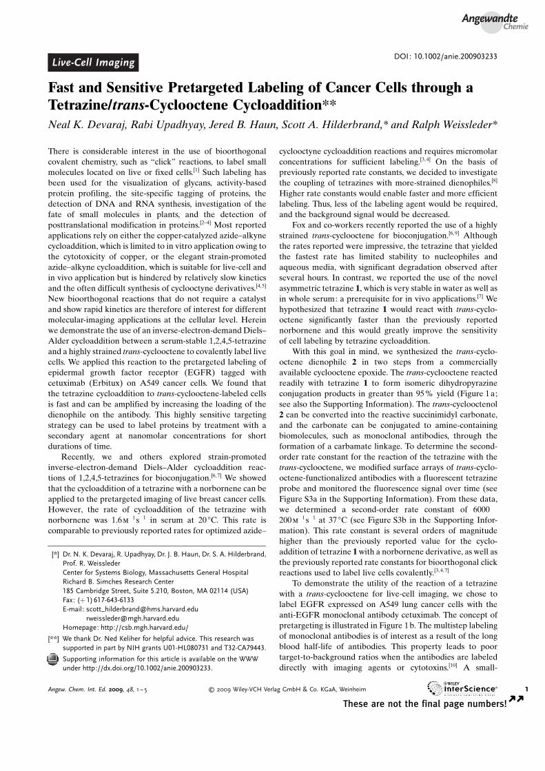
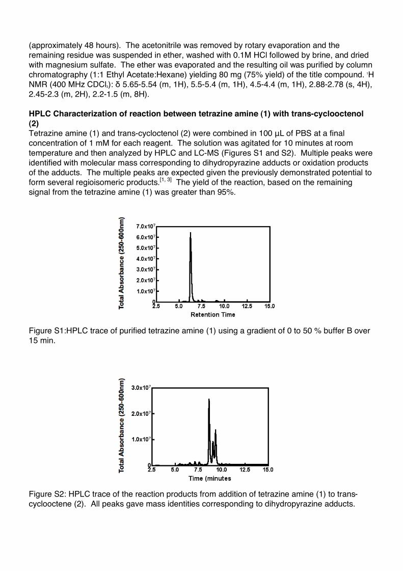
HPLC Characterization of reaction between tetrazine amine (1) with trans-cyclooctenol
(2)
Tetrazine amine (1) and trans-cycloctenol (2) were combined in 100 "L of PBS at a final
concentration of 1 mM for each reagent. The solution was agitated for 10 minutes at room
temperature and then analyzed by HPLC and LC-MS (Figures S1 and S2). Multiple peaks were
identified with molecular mass corresponding to dihydropyrazine adducts or oxidation products
of the adducts. The multiple peaks are expected given the previously demonstrated potential to
form several regioisomeric products.[1, 3] The yield of the reaction, based on the remaining
signal from the tetrazine amine (1) was greater than 95%.
Figure S1:HPLC trace of purified tetrazine amine (1) using a gradient of 0 to 50 % buffer B over
15 min.
Figure S2: HPLC trace of the reaction products from addition of tetrazine amine (1) to trans-
cyclooctene (2). All peaks gave mass identities corresponding to dihydropyrazine adducts.

Labeling Antibody with trans-cyclooctene
Cetuximab (ImClone 2 mg/mL) was purchased and the solvent exchanged for 0.1M NaHCO3
buffered at pH 8.5 with a final concentration of 7 mg/mL. To 200 !L of this stock solution was
added 10 !L of DMF. (E)-cyclooct-4-enyl 2,5-dioxopyrrolidin-1-yl carbonate was dissolved in
anhydrous DMF to make a 40 mM stock solution. For conjugation, the appropriate excess of
amine reactive trans-cyclooctene in DMF was aliquoted into the antibody solution, vortexed, and
reacted overnight at 4 oC. In the experiments reported, the final trans-cycloctene loadings of 1,
3, 5, and 6 correspond to using 2, 10, 30, and 100 equivalents of succinimidyl carbonate with
respect to antibody. After overnight reaction the antibodies were purified by centrifuge filtration
using 5% DMSO PBS, concentrated to 2 mg/mL and stored in PBS at 4 oC.
Antibody Labeling with Fluorescent Succinimidyl Esters
A solution of antibody (1 mg/mL) in 0.1M NaHCO3 (pH 8.5) was incubated with 2 equivalents of
fluorescent succinimidyl ester (VT680, AF555, or Dylight 488) for 2 hours. After incubation, the
antibody was purified by centrifuge filtration using 30000 dalton molecular weight cutoff filters
(Amicon) and stored in PBS. The number of fluorochromes per antibody was determined by
spectrophotometric analysis and determined to be approximately 1 per antibody for all dye
succinimidyl esters used.
Kinetic Measurements
trans-cyclooctene modified antibody was physically absorbed onto polystyrene by immersing
the surfaces in a 0.1 mg/mL solution of antibody in PBS for 3 hours. After numerous washes
with PBS, the surface was exposed to 750 nM tetrazine VT680 in PBS at 37 oC. After 5
minutes, the tetrazine solution was removed and the surface washed 3 times with PBS. The
fluorescence due to the VT680 dye was measured on a fluorescence plate reader (Tecan Safire
2) and corrected for background fluorescence. The surface was again exposed to the tetrazine
solution and the entire process repeated at 10, 15, 30, and 60 minutes. The fluorescence
measurements were plotted versus time, fitted to a first order exponential growth curve and the
pseudo first order rate constant determined (Figure S3 Left). The entire experiment was
repeated using two different concentrations of tetrazine (375 nM and 1000 nM) and the pseudo
first order rate constants from all three experiments were plotted versus concentration, fitted to a
straight line, and the slope taken as the second order rate constant.
Figure S3: Kinetics of tetrazine VT680 cycloaddition to trans-cyclooctene modified
antibody absorbed to a polystyrene plate. (A) Fluorescence monitoring of tetrazine
VT680 cycloaddition to a surface array of trans-cyclooctene antibody (1 reactive
trans-cyclooctene). The data were fit to a first order exponential (black line). From
the fit a pseudo first order rate constant (kobs) is determined. (B) A plot of kobs
versus the concentration of tetrazine VT680. The data were fit using linear

regression (black line). The slope of the line was 6000±200 M-1sec-1 and was
reported at the second order rate constant for the reaction between tetrazine VT680
and trans-cyclooctene bound to antibody.
Cell Culture
The human lung adenocarcinoma epithelial cell line A549 was selected for all experiments due
to its mid-level over-expression of EGFR. The cell line was maintained in a standard ATCC
formulated F-12K media supplemented with 10% fetal bovine serum and 5%
penicillin/streptomycin. In order to facilitate microscopy and visualize intracellular morphology,
EGFP labeling of the cell line was done using a third-generation lentiviral vector system. 293T
cells were transfected using lipofectamine 2000 in a subconfluent 10-cm dish with the vector
pCCLsin.PPT.hPGK (10 !g), into which EGFP had been cloned, as well as pMDLg/p packaging
(7 !g) and VSV-G envelope encoding pMD.G (5 !g) plasmids. These plasmids were obtained
from Rafaella Sordella at the MGH Center for Cancer Research and Luigi Naldini at the San
Raffaele Telethon Institute for Gene Therapy. Viral supernatant was collected after 48 hours,
filtered with a 0.45 micron syringe filter, and stored at -80°C. The A549 cell line was infected in
subconfluent wells of 24-well plates, using 300 !L of virus in 1 mL of F-12K culture media with
10% fetal calf serum. This protocol yielded an infection rate in excess of 80% (determined by
visual assessment using fluorescence microscopy). EGFP-negative cells were removed using a
modified 5-laser Becton-Dickinson FACSDiVa with standard techniques.
Confocal Microscopy
Cells were grown on break away glass chamber slides and washed six times after administering
either imaging agent. A multichannel upright laser-scanning confocal microscope (FV1000;
Olympus) was used to image live cells with a 60X water immersion objective lens. Image
collection and fluorophore excitations with lasers at 488nm (EGFP), 543nm (AF555), and
633nm (VT680) were done serially to avoid cross talk between channels. Data were acquired
with Fluoview software (version 4.3; Olympus) and image stacks were processed and analyzed
with ImageJ software (version 1.41, Bethesda MD).
Flow cytometry
Confluent A549 cells were suspended using 0.05% Tryspin/0.53 mM EDTA, washed by
centrifugation with PBS containing 2% FBS (PBS+), and 2.5x10⁵ cells were added to
microcentrifuge tubes. Cetuximab antibody with the following modifications was then added at
10 !g/ml concentration in 100 !L PBS+: none (control), 1 trans-cyclooctene per antibody, 3
trans-cyclooctene per antibody, 5 trans-cyclooctene per antibody, 6 trans-cyclooctene per
antibody. Following incubation for 15 minutes at room temperature, samples were washed with
PBS+. For stability studies, trans-cyclooctene antibodies were labeled with Dylight 488
fluorophore (Pierce, ~1 per antibody) and the cells were resuspended in 100 !L PBS+ and
incubated for 15, 30, or 60 minutes at 37ºC before addition of 1 ml PBS+ and 2 washes by
centrifugation. For clicking studies, labeled cells were resuspended in 100 !L FBS containing
500 nM tetrazine-VT680 and incubated for 30 minutes at 37ºC before addition of 1 ml PBS+ and
2 washes by centrifugation. VT680 and DyeLight-488 fluorescence was assessed using an
LSRII flow cytometer (Becton Dickinson) and analyzed using FlowJo software.

Confocal Microscopy of Control Experiments
Figure S4: Confocal microscopy of GFP-positive A549 lung cancer cells after control
labelings. Cells were exposed to unlabeled Cetuximab antibody, washed, exposed
to tetrazine-VT680 probe (500 nM 10 minutes 100% FBS 37oC), washed, and
imaged in the GFP channel (Panel A, White scale bar in top left panel denotes 30
microns) and the near-infrared channel (Panel B). In a separate control experiment,
cells were exposed to Cetuximab/trans-cyclooctene antibodies, washed, exposed to
unlabeled VT680 (Genhance 680, 500 nM 10 minutes 100% FBS 37oC, VisEn
Medical), washed, and imaged in the GFP channel (Panel C) and the near-infrared
channel (Panel D).
Stability data of trans-Cyclooctene modified Cetuximab
Figure S5: Stability study of Dylight 488 Cetuximab modified with either 1 (blue), 3
(green), 5 (yellow), or 6 (red) reactive trans-cyclooctene. A549 cells were exposed
to antibody, washed, and the fluorescence intensity monitored with time using flow
cytometry.
References
[1] Devaraj, N. K.; Weissleder, R.; Hilderbrand, S. A. Bioconjug Chem 2008, 19, 2297-2299.
[2] Hillmyer, M. A.; Laredo, W. R.; Grubbs, R. H. Macromolecules 1995, 28, 6311-6316.
[3] Royzen, M.; Yap, G. P.; Fox, J. M. J Am Chem Soc 2008, 130, 3760-3761.