fascin is a key regulator of breast cancer invasion that ... · fascin is a key regulator of breast...
TRANSCRIPT

Fascin Is a Key Regulator of Breast Cancer Invasion ThatActs via the Modification of Metastasis-AssociatedMoleculesMonther Al-Alwan1*, Safiah Olabi1, Hazem Ghebeh1, Eman Barhoush1, Asma Tulbah2, Taher
Al-Tweigeri3, Dahish Ajarim3, Chaker Adra1,4
1 Stem Cell Therapy Program, King Faisal Specialist Hospital and Research Centre, Riyadh, Saudi Arabia, 2 Department of Pathology and Laboratory Medicine, King Faisal
Specialist Hospital and Research Centre, Riyadh, Saudi Arabia, 3 Department of Oncology, King Faisal Specialist Hospital and Research Centre, Riyadh, Saudi Arabia,
4 Transplantation Research Centre (TRC), Brigham and Women’s Hospital and Children’s Hospital Boston, Harvard Medical School, Boston, Massachusetts, United States of
America
Abstract
The actin-bundling protein, fascin, is a member of the cytoskeletal protein family that has restricted expression inspecialized normal cells. However, many studies have reported the induction of this protein in various transformed cellsincluding breast cancer cells. While the role of fascin in the regulation of breast cancer cell migration has been previouslyshown, the underlying molecular mechanism remained poorly defined. We have used variety of immunological andfunctional assays to study whether fascin regulates breast cancer metastasis-associated molecules. In this report we found adirect relationship between fascin expression in breast cancer patients and; metastasis and shorter disease-free survival.Most importantly, in vitro interference with fascin expression by loss or gain of function demonstrates a central role for thisprotein in regulating the cell morphology, migration and invasion potential. Our results show that fascin regulation ofinvasion is mediated via modulating several metastasis-associated genes. We show for the first time that fascin down-regulates the expression and nuclear translocation of a key metastasis suppressor protein known as breast cancermetastasis suppressor-1 (BRMS1). In addition, fascin up-regulates NF-kappa B activity, which is essential for metastasis.Importantly, fascin up-regulates other proteins that are known to be critical for the execution of metastasis such asurokinase-type plasminogen activator (uPA) and the matrix metalloproteases (MMP)-2 and MMP-9. This study demonstratesthat fascin expression in breast cancer cells establishes a gene expression profile consistent with metastatic tumors andoffers a potential therapeutic intervention in metastatic breast cancer treatment through fascin targeting.
Citation: Al-Alwan M, Olabi S, Ghebeh H, Barhoush E, Tulbah A, et al. (2011) Fascin Is a Key Regulator of Breast Cancer Invasion That Acts via the Modification ofMetastasis-Associated Molecules. PLoS ONE 6(11): e27339. doi:10.1371/journal.pone.0027339
Editor: Wanjin Hong, Institute of Molecular and Cell Biology, Singapore
Received May 25, 2011; Accepted October 13, 2011; Published November 4, 2011
Copyright: � 2011 Al-Alwan et al. This is an open-access article distributed under the terms of the Creative Commons Attribution License, which permitsunrestricted use, distribution, and reproduction in any medium, provided the original author and source are credited.
Funding: This work was supported by the Research Advisory Council (proposal grant 2060 016), King Faisal Specialist Hospital and Research Centre. The fundershad no role in study design, data collection and analysis, decision to publish, or preparation of the manuscript.
Competing Interests: The authors have declared that no competing interests exist.
* E-mail: [email protected]
Introduction
Breast cancer is one of the leading causes of cancer mortality in
women worldwide. In spite of significant advances in cancer
treatment, mortality results from local invasion and/or distant
metastasis and not from tumor in the primary site [1,2]. Therefore,
there is a strong demand to understand the cellular and molecular
mechanisms that regulate tumor invasion and metastasis in order
to develop better treatment regimens. Metastasis is a multi-step
process involving neovascularization, stromal invasion by cancer
cells, and infiltration into vascular and lymphatic spaces,
extravasation and growth at a secondary site [1–6]. Many cellular
and molecular factors have been reported to regulate tumor
metastasis (Reviewed in [7]). However, a critical step of metastatic
tumor cells is the ability to cross extracellular matrix (ECM) tissue
boundaries [8,9], a process that can be accomplished by
expression of active metalloproteases (MMPs) by cancer cells,
which facilitate their migration. In addition, metastatic tumor cells
can down-regulates metastatic suppressors such as the Breast
Cancer Metastasis Suppressor 1 (BRMS1), which belongs to family
of metastasis suppressors that suppress metastasis without blocking
orthotopic tumor growth. BRMS1 has been reported to block lung
and regional lymph node metastasis in experimental breast models
[10] and decreased expression of this protein has been
demonstrated to correlate with reduced disease-free survival in
human breast cancer [11]. It was reported that BRMS1
suppression of tumor metastasis is mediated via inhibition of
NF-kB and subsequent suppression of the urokinase-type plasmin-
ogen activator (uPA) [12], a serine protease that is known to
activate the MMPs [13] leading to invasion and metastasis.
Manipulation of the actin cytoskeleton, leading to enhancement
of cell motility, is one of the dominant cellular mechanisms that
regulate metastasis [14]. Fascin is a member of the actin
cytoskeletal proteins that bundles actin filaments into tertiary
structures within dynamic cellular structures such as microspikes,
stress fibers and membrane ruffles [15], consistent with its
abundant expression at these sites. Interestingly, fascin expression
is highly restricted to certain tissues such as brain and spleen and
PLoS ONE | www.plosone.org 1 November 2011 | Volume 6 | Issue 11 | e27339

abundant fascin expression was reported in normal specialized
cells such as neurons, glia cells, endothelial cells and antigen
presenting dendritic cells [16–19].
It is widely believed that de-regulation of normal tissue
organization and homeostasis including cell adhesion, motility
and cytoskeleton can contribute to many human diseases including
cancers [19]. In fact, fascin over-expression induces membrane
protrusion and has been reported to enhance cell motility in
various systems [20]. In addition to its expression in specialized
normal cells, many studies have reported induction of fascin
expression in various transformed cells. In many human
carcinomas including breast cancer, fascin expression correlates
with clinically aggressive tumors and metastasis [21]. However, the
exact function of fascin and its regulation of other genes in breast
cancer are still poorly understood.
Development of membrane protrusions by non-immune cells
has been clearly demonstrated to enhance cell motility and to
facilitate interactions with other cell types. Therefore, we
proposed to investigate whether fascin expression in breast
cancer cells is directly involved in promoting cell migration and
invasion and to elucidate the molecular mechanism that regulate
this process. Our results clearly demonstrated a key role for fascin
in regulating both cell morphology and invasion. In addition, we
have shown that fascin facilitates this process via modulation of
metastasis-associated genes; specifically down-regulation of
the metastasis suppressor BRMS1 and up-regulation of NF-kB
activity as well as the induction of uPA, MMP-2, MMP-9
expression.
Materials and Methods
Cell linesThe human breast cancer cell line MDA-MB-231 (HTB-26)
and T-47D (HTB-20) were purchased from ATCC and seeded in
DMEM containing 10% FBS, 200 mM L-glutamine and antibi-
otic-antimycotic liquid (Invitrogen) at 37uC in a 5% CO2
humidified atmosphere.
Antibodies and reagentsThe mouse anti-human fascin mAb and APC-conjugated
secondary Ab were from Dako and Jackson Laboratories,
respectively. The mouse mAb to detect MMP-2 (2C1-1D12) and
MMP-9 (2C3) were from Invitrogen and Chemicon, respectively.
The mouse mAb (ab57082), rabbit polyclonal Ab (ab65244) to
detect BRMS1, mAb to detect uPA (PGM2005) and mAB to
detect TATA binding protein (TBP) were all from ABcam. Abs to
detect p65 (C22B4), phopho-p65 (93H1), IkBa (L35A5) and
phospho-IkBa (5A5), alpha-tubulin were all from Cell Signaling
Technology. The GAPDH, b-actin, PCNA and p50 (C-19) were
from Santa Cruz. The secondary anti-mouse HRP-conjugated and
anti-goat HRP-conjugated were from Southern Biotech and
Serotec, respectively. The Rhodamine-phalloidin to detect F-actin
was from Invitrogen. The remaining antibodies were from BD
Bioscience. Fascin specific-, negative control-SiRNA and GAP-
DH-SiRNA were purchased from Applied Biosystem. Fascin
specific-ShRNA and control were purchased from Thermo
Scientific. The MMP-2/MMP-9 inhibitor II was from Calbio-
chem. The cell proliferation reagent WST-1 was purchased from
Roche. The CSFE cell proliferation dye was purchased from
Guava Biotechnologies, Inc.
Flow cytometry and cell sortingCell permeablization and fascin staining were performed as
previously described [22]. Fluorescence was analyzed using a flow
cytometer (LSR II; BD, Mountain View, CA). Cell sorting was
performed on GFP-Fascin transfected cells using cell sorter (FACS
DIVA, BD, Mountain View, CA).
Fascin knockdownFor transient knockdown, MDA-MB-231 cells were seeded in
the presence of control SiRNA or Fascin (ID s13209) SiRNA as
per instructions of Applied Biosystem. Maximum inhibition of
fascin as assessed by western blot and FACS analysis was observed
72 hours post SiRNA treatment and thus this time was used for
the rest of the study.
To rule out any off target effect of the used SiRNA, stable
ShRNA clones were generated. For stable fascin knock-
down, lentiviral vectors expressing fascin shRNA (clone Id:
TRCN000012039) or control shRNA were obtained from
Thermo Scientific. Recombinant lentivirus was produced in
293T cells as previous described [23] and mixed with MDA-
MB-231 cells in the presence of polybrene (10 mg/ml) for 16 hour
at 37uC, medium was replaced and cultured for additional
72 hours. Transduced cells were selected with puromycin followed
by cloning and screening for the lack of fascin expressing using
FACS. ShRNA were designed and used especially for the
activation experiments where cells need to be seed in serum
containing media before stimulation in serum-free media. This
could not be done using the SiRNA approach as they were
generated in a serum-free media.
Generation of eGFP-fascin expressing cell stabletransfectants
Wild-type or mutant fascin-GFP fusion constructs were
previously described [24] and kindly provided to us by Dr.
Josephine C. Adam (Cleveland Clinic Foundation, Ohio, USA).
Briefly, mutation in fascin was generated by substituting the major
site that is phosphorylated by PKCa (Serine 39) with Aspartic
Acid. After transfection using the Nucleaofector reagent (Amaxa
BioSystems), stable clones were isolated post selection with G418
for 2–3 weeks followed by cloning. Fascin expression levels and
mean florescent intensity (MFI) were always higher in GFP
positive cells as assessed by FACS and western blots, respectively.
Chemotaxis and invasion assayThe migration and metastatic potential of tumor cells were
evaluated using the 24-well BD migration and BioCoat Matrigel
Invasion Chamber, respectively, as per the manufacturer guideline
(BD Bioscience). Cells (1–26105/well) were allowed to migrate for
18 hours and membranes were stained with a Diff Quick stain and
mounted on slides. Cells that had migrated to the underside of the
filter were counted using light microscope (Zeiss Axio Observer) in
5 randomly selected fields (magnification; 40x). Each assay was
performed in triplicate and repeated at least 5 times. Due to
variation in the number of migrated cells from different
experiment, the results are normalized to control cells and the
relative migration or invasion is expressed as mean 6 SD of
migrating cells relative to control cells.
Purification of total RNA and RT-PCRTotal RNA was extracted from cells using RNAeasy Mini kit
(QIAGEN). cDNA was synthesized using First Strand cDNA
synthesis kit (Invitrogen). The relative mRNA copy number of a
gene was determined by RT-PCR using molecular beacon method
and detected by ABI PRISM 7900HT (Applied Biosystems) and
expressed as previously described [25]. Sequences of forward,
reverse and probe used in this experiment are shown in Table S1.
Fascin Regulates Breast Cancer Metastasis Proteins
PLoS ONE | www.plosone.org 2 November 2011 | Volume 6 | Issue 11 | e27339

Western BlotCells were washed with cold PBS, total proteins were extracted
using RIPA buffer containing protease inhibitors, and concentra-
tions were determined using Bradford Assay (Bio-Rad). Proteins
(20 mg) were loaded onto an SDS-PAGE gel and transferred using
semidry electrobloting (Bio-Rad). Membranes were then incubated
in 5% (w/v) skimmed milk before incubation with primary
antibodies overnight at 4uC. After washing, membranes were
incubated with horseradish peroxidase-conjugated secondary anti-
body for 1 hour. Chemiluminescence Super Signal System (Thermo
Scientific) was used for subsequent detection of bound antibodies.
For activation, exponentially growing cells were stimulated with
10 ng/ml of TNF-a in serum-free media for the indicated time
points in the figures. Nuclear and cytoplasmic fractionation was
done as previously described [26]. Proteins (15 mg of cytoplasmic
and 10 mg of nuclear extract) were loaded and assayed as above.
Equal amounts of supernatants were loaded onto an SDS-PAGE
gel and protein expression was detected by western blot as
described above. Gelatin zymography was done as previously
described [27].
Dual-luciferase reporter assayNF-kB luciferase activity in cell lysates was measured using
dual-luciferase reporter assay system (Promega) previously de-
scribed [28]. pXPG-hIL-8p luciferase reporter plasmid was a
generous gift from Dr. Abbas Hawwari, King Faisal Specialist
Hospital and Research Centre, Riyadh, Saudi Arabia. Each
experiment group was done in triplicate and repeated at least 5
times. Firefly luciferase was divided by the Renilla luciferase activity
to normalize for transfection efficiency and the relative values are
presented as fold change over non-stimulated control.
ImmunohistochemistryFormalin-fixed, paraffin-embedded breast cancer sections of 71
patients deparaffinized and rehydrated. Antigen retrievals were
done by microwaving for 15 minutes in a special citrate solution
pH 6 (Dako). Endogenous peroxidase were blocked for 15 minutes
with 3% hydrogen peroxide (Sigma) in methanol. Sections were
then blocked with 10% goat serum (Sigma) for 60 minutes,
followed by addition of a primary mouse anti-human fascin (1/
200) or rabbit anti-BRMS1 (1/1000) antibodies for overnight
incubation at 4uC. After washing, sections were incubated with
labeled Polymer (EnVision+) HRP detection kit (Dako) for 30
minutes at room temperature. The HRP was detected using DAB
substrate (Novocastra) for 4 minutes and the sections were
counterstained for 1 minute with Instant hematoxylin (Shandon).
The intensity of staining and the percentages of fascin and BRMS1
positive cells were quantified at 5 to 10 increments by an
anatomical pathologist (AT) who had no prior knowledge of
patient details. Type of breast cancer was confirmed at the time of
reading. Histological grades of breast cancer sections were
evaluated according to Scarff-Bloom-Richardson (SBR) classifica-
tion [29].
For immunoflorescence staining of BRMS1, the above method
of fixation and primary staining was used followed by Alexa-555
anti-rabbit secondary antibody. F-actin staining was done as
previously described [30]. For cellular localization of BRMS1 or
F-actin detection, 100 cells were assessed using attovision software
on Pathway 855 (BD, Mountain View, CA).
Statistical AnalysisThe significance (0.05) of relationship between fascin expression
and patient’s clinicopathological parameters was assessed using
Fisher exact test. The software package SAS 9.1 (SAS Institute,
Cary, NC) was used for these analyses.
Results
Fascin expression in breast cancer is associated with poorprognosis, metastasis and reduced disease-free survival
Fascin expression in breast cancer correlates with poor
prognosis of the disease and shorter disease-free and overall
survival [21]. Here we have used immunohistochemistry to
reexamine the relationship between the expression of fascin and
metastasis in 71 breast cancer patients, which were diagnosed with
invasive ductal carcinoma. Fascin was negative in normal breast
luminal cells, but weak to moderate expression was seen in the
myoepithelial and endothelial cells (data not shown), consistent
with previous study [21]. In breast cancer samples however, fascin
was expressed in the tumor cells of 40.84% of breast cancer
patients. There were a strong correlation between fascin
expression and; basal-like phenotype (,0.001), hormone recep-
tor-negative (ER2, P,0.001), (PR2, P = 0.020), larger tumor size
(P = 0.034), high histological grade tumors (P = 0.091), known poor
prognostic markers (Table 1). Interestingly, fascin also significantly
correlated with increased expression level of B7-H1 (P = 0.008), a
T cell inhibitory molecule that is associated with bad prognostic
makers in breast cancer [31]. Importantly, our data showed
significant (P = 0.017) correlation between fascin expression and
local as well as systemic metastasis (Table 1). In addition, there was
a highly significant (P,0.001) association between fascin expres-
sion and decreased disease-free survival (Figure 1A), but the
association with the overall survival (Figure 1B) was borderline
significant (P = 0.058). Our in vivo findings demonstrate a strong
correlation between fascin induction in breast cancer cells and;
poor prognostic markers, increased tumor metastasis and reduced
disease-free survival.
Fascin regulates the morphology and migration of breastcancer cells
To address the relationship between fascin expression and
metastasis we measured parameters important for this process
using MDA-MB-231 breast cancer cell line as a model. This cell
line is known to be ER/PR-negative, a feature of breast cancer
cells with more invasive and motile phenotype [32–34]. FACS
analysis (Figure 2A) and western blot (Figure S1A) demonstrate
that these cells are fascin positive. To study fascin function in
MDA-MB-231 cells, we transiently down-regulated its expression
using fascin-specific siRNA (SiFascin). FACS histogram (Figure 1A)
shows that 60–70% of the SiFascin treated cells became fascin-
negative compared with the control-siRNA (SiCon) treated group.
The mean florescence intensity (MFI) of SiFascin-treated cells was
also reduced by greater than 60%. Similar to the FACS data,
western blot indicated 70% inhibition of fascin in the knockdown
cells (Figure S1A). Inhibition of the house keeping gene (GAPDH)
using specific SiRNA was used as positive control in all of the
assays (data not shown).
We examined whether fascin expression in human breast cancer
cells induces morphological changes, as we have previously
observed in mouse immune cells [22]. In culture, fascin-
knockdown cells lost their typical/normal spindle shape that is
seen in the control cells as shown by phase contrast inverted
microscope (Figure S1B). Furthermore, immunohistochemistry
staining demonstrated that while the control cells expressed fascin
and have a spindle shape with fine dendrites, fascin-knockdown
cells expressed lower fascin levels and exhibited more rounded
morphology (Figure 2B Top). Immunoflorescent staining of
Fascin Regulates Breast Cancer Metastasis Proteins
PLoS ONE | www.plosone.org 3 November 2011 | Volume 6 | Issue 11 | e27339

SiFascin cells demonstrated these alterations in cell morphology
and showed changes in the distribution of F-actin (Figure 2B
Bottom). Fascin expression had no effect on MDA-MB-231 cell
proliferation as assessed using the cell proliferation reagent WST1
and CSFE dye (data not shown).
We examined if fascin-mediated morphological changes affect
breast cancer cell motility. Migration of the fascin-knockdown cells
was significantly (P,0.001) inhibited by more than 50% compared
with control cells (Figure 2C). Furthermore, SiFascin cells were
also less migratory after 72 hours using the traditional wound
healing assay (Figure S2). Difference in wound closure was not a
reflection of cell division as similar number of cells was seeded and
became confluent at the same time before the wound was made.
To specifically link fascin expression to enhanced migration, we
have generated MDA-MB-231 cells stably over-expressing wild-
type (WT) or mutant fascin. Contrary to fascin-knockdown data,
over-expression of WT (Figure S3) and not mutant fascin
significantly (P = 0.01) enhanced MDA-MB-231 cell migration
(Figure 2D). Collectively, our data shows that fascin expression in
breast cancer cells regulates their morphology and migratory
potential.
Fascin regulates breast cancer cell invasionFascin involvement in mediating breast cancer metastasis was
then tested using a well-established invasion assay. Global actin
polymerization inhibitor (Cytochalasin D), at a dose that did not
effect cell viability (data not shown), altered MDA-MB-231
morphology and significantly (P,0.001) suppressed their invasion
by greater than 95% (Figure 3A), consistent with the role of actin
cytoskeleton in this process. Similarly, fascin-knockdown MDA-
MB-231 showed significantly (P,0.001) impaired invasion and
Table 1. #Correlation between fascin expression in tumorcells and the clinico-pathological parameters of 71 breastcancer patients.
Fascin
Negative Positive *P
Age
,40 years 16 §(76) 5 (24) 0.575
$40 years 33 (66) 17 (34)
Tumor Size
,4 CM 31 (79) 8 (21) 0.035
$4 CM 18 (56) 14 (44)
Histological Grade (SBR)
I 1 (50) 1 (50) 0.091
II 27 (82) 6 (18)
III 21 (58) 15 (42)
Her2/neu status
0 23 (68) 11 (32) 0.072
1 0 (0) 2 (100)
2 10 (91) 8 (33)
3 16 (67) 8 (33)
ER status
Negative 9 (39) 14 (61) ,0.001
Positive 70 (83) 5 (17)
PR status
Negative 20 (56) 16 (44) 0.020
Positive 29 (83) 6 (17)
B7H1
Negative 49 (72) 19 (28) 0.008
Positive 0 (0) 3 (100)
Basel
Negative 46 (77) 14 (23) ,0.001
Positive
¤Local and systematic metastasis
Negative 25 (89) 3 (11) 0.017
Local 4 (80) 1 (20)
Systemic 15 (56) 12 (27)
Neo-adjuvant Chemotherapy
Without 20 (67) 10 (33) 0.714
With 29 (69) 22 (31)
Abbreviations:#When interpreting data and correlating it with clinico-pathological parameters,
the 5% expression of cells was the cut-off point below which were consideredas negative and above as positive.
§Numbers between brackets are the percentages of patients,¤11 patients were reported as no show or dead,*P values in bold represent a significant data.doi:10.1371/journal.pone.0027339.t001
Figure 1. Fascin is associated with disease-free survival inhuman breast cancer samples. Survival with or without the diseasewas reported by oncologist and blotted in relation to fascin expression.Survival curves showing decreased disease-free (A) or overall (B) survivalin patients that have fascin positive tumor.doi:10.1371/journal.pone.0027339.g001
Fascin Regulates Breast Cancer Metastasis Proteins
PLoS ONE | www.plosone.org 4 November 2011 | Volume 6 | Issue 11 | e27339

altered morphology compared with control cells (Figure 3B). On
the contrary, over-expression of WT and not mutant fascin
in MDA-MB-231 cells significantly (P = 0.003) enhanced their
invasive capability (Figure 3C). Interference with fascin expression
in MDA-MB-231 had no effect on cell proliferation or survival
(data not shown). Collectively, these data demonstrate that fascin
expression in breast cancer cells is strongly associated with
enhanced cell motility and invasiveness.
Fascin inhibits nuclear expression of BRMS1 in breastcancer cells
BRMS1 is a known metastasis suppressor in many cancer types
including breast cancer [35]. We examined whether fascin-
mediated breast cancer cell invasion has an effect on the
expression levels and cellular localization of BRMS1. SiFascin
breast cancer cells showed significantly enhanced BRMS1 RNA
(Figure 4A) and protein (Figure 4B left) expression compared with
SiCon, consistent with the role of fascin in enhancing breast
cancer metastatic potential. Conversely, over-expression of WT
fascin in MDA-MB-231 dramatically suppresses BRMS1 expres-
sion (Figure 4B right), further confirming the inverse relationship
between these two proteins. BRMS1 expression was found to be
predominantly in the nucleus and fascin-knockdown clearly leaded
to enhanced expression of nuclear BRMS1 (Figure 4C). Examin-
ing the relationship between BRMS1 and fascin in our breast
cancer patient samples demonstrated variations in the intensity
and subcellular distribution of BRMS1 (Figure 4D). Most
importantly, 36 patients that were scored as having high levels
of nuclear BRMS1 ($50% of tumor cells express high levels
(intensity of +3) of nuclear BRMS1) showed reduced levels of
fascin (Figure 4E), demonstrating an inverse relationship between
fascin and nuclear BRMS1 that is statistically significant
(P,0.001). This data demonstrates that fascin can directly or
indirectly regulate the tumor suppressor BRMS1 nuclear expres-
sion.
Fascin enhances metastasis-associated genesWe tested whether fascin enhances metastasis via counteracting
BRMS1 effect on key downstream mediators that are involved in
this process such as uPA [12]. Consistent with enhanced invasion,
cells that over-expressed WT and not mutant fascin showed higher
levels of uPA expression (Figure 5A). To directly link invasion to
well established mediators of metastasis, we blocked activity of
MMP-2 and MMP-9, which are activated by uPA [9] and are
among the most well established proteases known to degrade
ECM and facilitate invasion and metastasis [36–38]. MMP-2 and
29 selective inhibitors significantly (P = 0.013) reduced the
invasion of control cells to comparable level with the fascin-
knockdown cells (Figure 5B), implicating a role for these proteases
Figure 2. Fascin knockdown alters cell morphology andreduces cell migration. MDA-MB-231 cells were treated with control(SiCon) or Fascin (SiFascin) SiRNA for 72 hours. A) Cells were thencollected and stained for fascin before detection by flow cytometry. B)Top: Immunohistochemistry showing cell morphology and fascin
expression after treatment with SiCon or SiFascin in 8-well chamber.Bottom: Immunoflorescent staining showing the altered morphologyand distribution of F-actin (red) in cells after treatment with SiFascin in8-well chamber. Blue color indicates nuclear stain (Dabi). C) Bar graphshowing reduced migration in fascin knockdown MDA-MB-231 cells.Data was normalized to SiCon cells and the relative migration isexpressed as mean 6 SD of triplicate experiments. D) Bar graphshowing enhanced cell migration in MDA-MB-231 cells that over-express wild type (WT) fascin compared with mutant (M) over-expressing cells. Data was normalized to parental MDA-MB-231 andthe relative migration is expressed as mean 6 SD of triplicateexperiments.doi:10.1371/journal.pone.0027339.g002
Fascin Regulates Breast Cancer Metastasis Proteins
PLoS ONE | www.plosone.org 5 November 2011 | Volume 6 | Issue 11 | e27339

in fascin-mediated invasion. Cytochalasin D, which significantly
diminished the cell invasion capability (Figure 3A), also inhibited
the expression of MMP-2 and MMP-9 (Figure S4).
Since TNF-a was shown to trigger uPA expression in MDA-
MB-231 cells [12], we activate our cells with TNF-a to further test
the link between fascin and uPA secretion in the supernatants.
Maximum induction of uPA RNA was seen at 6 hours post TNF-astimulation in fascin-positive breast cancer cells, while fascin-
knockdown cells failed to induce significant up-regulation
compared with the control cells (Figure 5C). uPA protein secretion
in control cells was detected in the supernatant 6 hours post TNF-
a stimulation and notable reduction was observed in fascin-
knockdown cells 12 hours post activation (Figure 5D). In parallel,
MMP9 was also detected in control cells 6 hours post TNF-astimulation and only detected at reduced levels 12 hours post
TNF-a stimulation of fascin-knock down cells. Furthermore,
reduced MMP-2 protein expression in fascin-knockdown cells
that was observed above is in line with decreased TNF-a-mediated
enzymatic activity in gelatin zymograms (Figure 5E).
Fascin enhanced NF-kB activitySince uPA promoter was reported to contain an NF-kB binding
site [39], we asked if fascin enhanced uPA secretion after TNF-aactivation is mediated via enhancement of NF-kB activity.
Inhibition of the NF-kB pathway significantly reduced cell
invasion in a dose-dependent manner (Figure 6A), implicating
the importance of NF-kB in this process. Importantly, fascin-
knockdown cells showed significantly (P = 0.040) impaired NF-kB
luciferase activity upon TNF-a activation when compared with
control cells (Figure 6B). In contrast, cells that over-expressed WT
fascin exhibited significantly (P = 0.002) enhanced NF-kB lucifer-
ase activity upon TNF-a activation (Figure 6C), indicating that
fascin positively regulates NF-kB transcriptional activity. No NF-
kB transcriptional activities was observed upon TNF-a activation,
when mutant NF-kB-luciferase reporter was used (data not
shown). Collectively, our data strongly demonstrated that fascin
suppressed BRMS1 and enhanced NF-kB activity.
Phosphorylation of the inhibitory subunit (IkBa) leads to
libration and translocation of NF-kB to the nucleus [40,41]. We
thus examine whether the observed fascin mediated NF-kB
transcriptional activity is related to enhanced NF-kB nuclear
translocation. The cytoplasmic levels of total IkBa were reduced in
control and fascin knockdown cells in response to activation
(Figure 7A). However, the magnitude of IkBa inhibition was more
pronounce in the control cells especially at earlier time points
(Figure 7B). There were a time dependent increase in IkBaphosphorylation in control cells after activation, which was
severely reduced in the fascin-knockdown cells (Figure 7 A and
C). The effect of fascin on IkBa phosphorylation and degradation
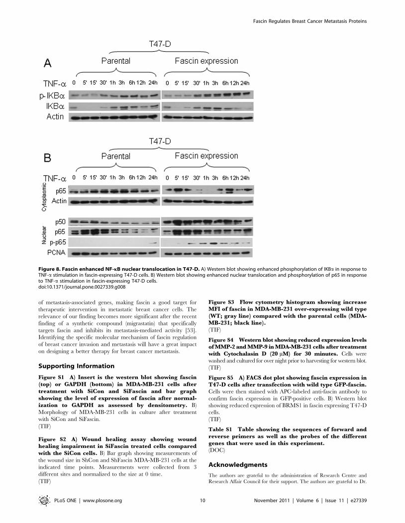
have also been observed in another breast cancer cell line (T47-D).
Expression of wild type fascin in T47-D cells, which are fascin
negative (Figure S5A), reduced BRMS1 expression (Figure S5B).
Interestingly, fascin expressing T47-D cells demonstrated reduced
total levels of IkBa and enhanced magnitude of IkBa phosphor-
ylation in response to activation especially at later time points
(Figure 8A).
To test whether reduced IkBa phosphorylation in fascin
knockdown cells would affect the phosphorylation of p65 and
translocation of p65/p50 to the nucleus, cytoplasmic and nuclear
fractions were extracted from cells after activation and the level of
these proteins and the status of phosphorylation were evaluated.
TNF-a activation induced time-dependent p65/p50 translocation
into the nucleus in control and fascin knockdown cells (Figure 7 D,
E and F). However, the levels of p65/p50 translocation into the
nucleus in fascin knockdown cells were notably lower especially at
the earlier time points, indicating less sustainable p65 nuclear
translocation in fascin knockdown cells. There were also time-
Figure 3. Fascin regulates breast cancer cell invasion. A) Bargraph showing inhibition of MDA-MB-231 cell invasion after pretreat-ment with 20 mM of Cytochalsin D (Cyt D) for 30 minutes prior to theassay. Data was normalized to untreated cells and the relative invasionis expressed as mean 6 SD of triplicate experiments. B) Bar graphshowing inhibition of MDA-MB-231 cell invasion after fascin knockdownwith SiRNA. Data was normalized to con SiRNA cells and the relativeinvasion is expressed as mean 6 SD of triplicate experiments. C) Bargraph showing enhanced cell invasion in MDA-MB-231 cells that over-express WT fascin. Data was normalized to parental MDA-MB-231 andthe relative invasion is expressed as mean 6 SD of triplicateexperiments.doi:10.1371/journal.pone.0027339.g003
Fascin Regulates Breast Cancer Metastasis Proteins
PLoS ONE | www.plosone.org 6 November 2011 | Volume 6 | Issue 11 | e27339

dependent increases of p65 phosphorylation in control and fascin
knockdown cells. However, the magnitude of induction of p65
phosphorylation in fascin knockdown cells was notably lowered
(Figure 7 D and G). In T47-D cells, TNF-a activation triggered
early p65 translocation into the nucleus which declined after 30
minutes (Figure 8B). Nuclear translocation of p65 in response to
TNF-a activation was more pronounced in the fascin expressing
T47-D cells. Most importantly, the nuclear translocation of p65
after 30 minutes was more sustainable in fascin expressing T47-D
cells. Furthermore, there were also time-dependent increases of
p65 phosphorylation in fascin expressing T47-D cells, which was
not seen in T47-D parental cells till 24 hours. Collectively, our
data demonstrated that fascin positively regulate NF-kB nuclear
translocation and transcriptional activity.
Discussion
Metastasis and not the primary tumors remained the main
causes of cancer mortalities [1,2], stressing the need to understand
the cellular and molecular mechanisms that regulate this process.
It is a complex process where cytoskeletal proteins were reported
to regulate multiple cellular processes including morphological
changes and motility, which are critical steps for metastasis
(Reviewed in [42]). In this study, we have shown an association
between the actin-bundling protein (fascin) and expression of bad
prognosis, metastasis and reduced disease-free survival. Our in
vitro data demonstrated fascin involvement in regulation of breast
cancer cell invasiveness and identifies some of the underlying
molecular mechanisms.
Breast cancers mainly arise from the luminal compartment of
the breast [43], but a small minority of tumors arise from multi-
potent progenitor cells, which can differentiate into luminal or
myoepithelial lineages [44,45]. The aggressive breast cancer with a
basal-like phenotype is triple negative (ER, PR and HER2) and
express both luminal and myoepithelial markers [46]. Our breast
cancer patients showed an association between fascin expression
and basal-like phenotype and high histological grade tumors, a
type of breast cancers that are associated with metastasis [46,47].
Furthermore, our patients showed an association between fascin
expression, increase metastasis and shorter disease-free survival
and reduced nuclear BRMS1, consistent with the in vitro data.
High fascin expression in our patients significantly correlated with
expression of other poor prognostic markers of breast cancer such
as tumor size and B7-H1. Whether fascin has a direct effect in
regulating the poor prognostic markers of breast cancer remains to
be elucidated.
NF-kB pathway is constitutively activated in many cancers,
which leads to enhancement of metastasis (reviewed in[48]) and
positively regulates the expression of uPA [39], which in turn can
activate the MMPs to facilitate invasion by degrading the ECM
[13]. BRMS1 negatively regulates uPA expression through
inhibition of the NF-kB activity in breast cancer and melanoma
cells [12]. Consistent with those findings, our data showed that
fascin expression in breast cancer cells, which enhances invasion,
Figure 4. Fascin inhibits nuclear expression of BRMS1 in breastcancer cells. A) Bar graph showing the relative RNA expression asassessed by real time PCR. Fascin RNA was inhibited, while BRMS1 RNAwas enhanced after fascin knockdown. B) Western blot showingenhanced BRMS1 protein expression when fascin was knockdown(Left) and BRMS1 inhibition when WT fascin was over-expressed (Right).C) Right; Representative photograph showing induction of BRMS1expression in the nucleus after fascin knockdown. Left; Bar graphshowing the mean intensity of BRMS1 expression in the nucleus afterfascin knockdown as assessed on more than 100 cells per group usingattovision software on Pathway 855 from BD. D) Paraffin-embedded
sections from breast cancer patients were stained for fascin and BRMS1and the expression profile and staining intensity were assessed bypathologist. Left: representative image showing strong BRMS1 nuclearstaining (Brown) and weakfascin cytoplasmic staining (red). Right:representative image showing weak BRMS1 nuclear staining (Brown)and strong fascin cytoplasmic staining (red). E) Bar graph showingreduced levels of fascin in patients that express high level ($50 positivewith +3 intensity) nuclear BRMS1.doi:10.1371/journal.pone.0027339.g004
Fascin Regulates Breast Cancer Metastasis Proteins
PLoS ONE | www.plosone.org 7 November 2011 | Volume 6 | Issue 11 | e27339

suppresses BRMS1 and counteract its effect on downstream
targets by increasing NF-kB activity, uPA secretion and MMP
enzymatic activity. Although we could not conclude whether fascin
suppression of BRMS1 is directly responsible for the inhibition of
BRMS1 downstream targets, we demonstrated that fascin
regulates MMPs expression and activation, key molecules that
facilitate invasion and metastasis. Furthermore, TNF-a stimulation
was shown to induce over-expression of fascin that in turn up-
regulate MMP-9 expression in cholangiocarcinoma [49].
Figure 6. Fascin enhanced NF-k -dependent transcriptionalactivity. A) Bar graph showing inhibition of MDA-MB-231 cell invasionthat were treated with NF-kB inhibitor. Data was normalized tountreated cells and the relative invasion is expressed as mean 6 SD oftriplicate experiments. B) ShCon or ShFascin MDA-MB-231 cells were co-transfected with the NF-kB luciferase promoter and Renilla promoter asin methods. Cells were stimulated with or without (20 ng/ml) TNF-a andluciferase activity was assessed as in methods. C) MDA-MB-231 parentalor cells that over-express WT or mutant fascin were co-transfected withthe NF-kB luciferase promoter and Renilla promoter as in methods. Cellswere stimulated with or without (20 ng/ml) TNF-a and luciferaseactivity was assessed as in methods.doi:10.1371/journal.pone.0027339.g006
Figure 5. Fascin enhanced uPA, MMP-2 and MMP-9 expression.A) Western blot showing increased expression of uPA in MDA-MB-231cells over-expressing WT fascin compared with the mutant over-expressing cells. B) Bar graph showing reduced invasion of SiCon-treated cells when MMP-2/MMP-9 inhibitor II (20 mM) was used. Datawas normalized to untreated SiCon cells and the relative invasion isexpressed as mean 6 SD of triplicate experiments. C) Bar graphshowing reduced RNA expression of uPA in fascin knockdown cells asassessed by real time PCR. D) Western blot showing reduced uPA (top)or MMP-9 (bottom) secretion in supernatants of fascin-knockdown cells.E) MMP-2 gelatinolytic activity showing reduced enzymatic activity ofsecreted MMP-2 in the supernatants of fascin-knockdown cells.doi:10.1371/journal.pone.0027339.g005
Fascin Regulates Breast Cancer Metastasis Proteins
PLoS ONE | www.plosone.org 8 November 2011 | Volume 6 | Issue 11 | e27339

While our data demonstrated a suppression of BRMS1 by
fascin, the exact mechanism by which this mediated has not been
elucidated. Interestingly, previous study reported down-regulation
of fascin when they expressed BRMS1 in a human ovarian
carcinoma, but the supporting data was not presented [50]. Both
Zhang et al and our findings support the existence of a direct or
indirect interaction between fascin and BRMS1. Fascin is mainly a
cytoplasmic protein with higher expression at the submemebrane
and around the nuclear membrane as shown in our work and
reviewed by Kureishy N et al [51]. Although BRMS1 is
predominantly nuclear, it is expressed in the cytoplasm as shown
by our data and by Frolova N et al [52]. It is therefore possible that
there is an active shuttling of this molecule between the cytoplasm
and the nucleus, where its translocation is controlled by direct or
indirect interaction with fascin molecules.
Our study showed that fascin acts at several cellular fronts to
facilitate cell invasion. It may act by negatively regulating the
expression of BRMS1, thereby enhancing NF-kB activity and
subsequent augmentation of uPA and MMPs expression. It is also
possible that fascin and BRMS1 have a feedback loop where
BRMS1 may also down-regulate fascin to inhibit cancer cells from
metastasis. This assumption is supported by a study where BRMS1
was demonstrated to down-regulate fascin expression in human
ovarian carcinoma cell line without showing data [50], and
concluded that this could be a potential mechanism underlying
BRMS1 suppression of metastasis. Alternatively, fascin may act via
yet unknown pathway to positively regulate NF-kB transcriptional
activity, which in turn enhances the expression of uPA and MMPs.
This study demonstrates a clear role for fascin in regulating
breast cancer invasion partially through modifying the expression
Figure 7. Fascin enhanced NF-kB nuclear translocation in MDA-MB-231. A) Western blot showing reduced phosphorylation of IKBa inresponse to TNF-a stimulation in fascin knockdown cells. B and C) Bar graph showing quantitation of total and phosphorylated IKBa in ShCon andShFascin cells after stimulation with TNF-a for the indicated time. Results showed the mean of triplicate experiments after normalization to actin andeach time point is normalized to 0 time. D) Western blot showing reduced nuclear translocation and phosphorylation of p65 in response to TNF-astimulation in Fascin knockdown cells. E-G) Bar graph showing quantitation of nuclear P50 and P65 and phosphorylated P65 in ShCon and ShFascincells after stimulation with TNF-a for the indicated time. Results showed the mean of triplicate experiments after normalization to PCNA and eachtime point is normalized to 0 time.doi:10.1371/journal.pone.0027339.g007
Fascin Regulates Breast Cancer Metastasis Proteins
PLoS ONE | www.plosone.org 9 November 2011 | Volume 6 | Issue 11 | e27339
of metastasis-associated genes, making fascin a good target for
therapeutic intervention in metastatic breast cancer cells. The
relevance of our finding becomes more significant after the recent
finding of a synthetic compound (migrastatin) that specifically
targets fascin and inhibits its metastasis-mediated activity [53].
Identifying the specific molecular mechanism of fascin regulation
of breast cancer invasion and metastasis will have a great impact
on designing a better therapy for breast cancer metastasis.
Supporting Information
Figure S1 A) Insert is the western blot showing fascin(top) or GAPDH (bottom) in MDA-MB-231 cells aftertreatment with SiCon and SiFascin and bar graphshowing the level of expression of fascin after normal-ization to GAPDH as assessed by densitometry. B)
Morphology of MDA-MB-231 cells in culture after treatment
with SiCon and SiFascin.
(TIF)
Figure S2 A) Wound healing assay showing woundhealing impairment in SiFascin treated cells comparedwith the SiCon cells. B) Bar graph showing measurements of
the wound size in ShCon and ShFascin MDA-MB-231 cells at the
indicated time points. Measurements were collected from 3
different sites and normalized to the size at 0 time.
(TIF)
Figure S3 Flow cytometry histogram showing increaseMFI of fascin in MDA-MB-231 over-expressing wild type(WT; gray line) compared with the parental cells (MDA-MB-231; black line).(TIF)
Figure S4 Western blot showing reduced expression levelsof MMP-2 and MMP-9 in MDA-MB-231 cells after treatmentwith Cytochalasin D (20 mM) for 30 minutes. Cells were
washed and cultured for over night prior to harvesting for western blot.
(TIF)
Figure S5 A) FACS dot plot showing fascin expression inT47-D cells after transfection with wild type GFP-fascin.Cells were then stained with APC-labeled anti-fascin antibody to
confirm fascin expression in GFP-positive cells. B) Western blot
showing reduced expression of BRMS1 in fascin expressing T47-D
cells.
(TIF)
Table S1 Table showing the sequences of forward andreverse primers as well as the probes of the differentgenes that were used in this experiment.(DOC)
Acknowledgments
The authors are grateful to the administration of Research Centre and
Research Affair Council for their support. The authors are grateful to Dr.
Figure 8. Fascin enhanced NF-kB nuclear translocation in T47-D. A) Western blot showing enhanced phosphorylation of IKBa in response toTNF-a stimulation in fascin-expressing T47-D cells. B) Western blot showing enhanced nuclear translocation and phosphorylation of p65 in responseto TNF-a stimulation in fascin-expressing T47-D cells.doi:10.1371/journal.pone.0027339.g008
Fascin Regulates Breast Cancer Metastasis Proteins
PLoS ONE | www.plosone.org 10 November 2011 | Volume 6 | Issue 11 | e27339

Josephine C. Adam for providing the wild-type and mutant of fascin-GFP
fusion constructs, Mr. Pulicat Manogaran for helping in the analysis of
FACS data. We are also thankful to Dr. Abbas Hawwari for providing the
pXPG-hIL-8p luciferase reporter plasmid.
Author Contributions
Conceived and designed the experiments: MAA. Performed the experi-
ments: SO HG EB. Analyzed the data: AT TAT DA. Wrote the paper:
MAA. Obtained fund for SO and provided administrative and materials
support: CA.
References
1. Bashyam MD (2002) Understanding cancer metastasis: an urgent need for using
differential gene expression analysis. Cancer 94: 1821–1829.
2. Chambers AF, Naumov GN, Varghese HJ, Nadkarni KV, MacDonald IC, et al.
(2001) Critical steps in hematogenous metastasis: an overview. Surg Oncol
Clin N Am 10: 243–255, vii.
3. Fidler IJ (2001) Seed and soil revisited: contribution of the organ
microenvironment to cancer metastasis. Surg Oncol Clin N Am 10:
257–269. vii-viiii.
4. Nicolson GL (1988) Organ specificity of tumor metastasis: role of preferential
adhesion, invasion and growth of malignant cells at specific secondary sites.
Cancer Metastasis Rev 7: 143–188.
5. Saaristo A, Karpanen T, Alitalo K (2000) Mechanisms of angiogenesis and
their use in the inhibition of tumor growth and metastasis. Oncogene 19:
6122–6129.
6. Woodhouse EC, Chuaqui RF, Liotta LA (1997) General mechanisms of
metastasis. Cancer 80: 1529–1537.
7. Smith SC, Theodorescu D (2009) Learning therapeutic lessons from metastasis
suppressor proteins. Nat Rev Cancer 9: 253–264.
8. Kim J, Yu W, Kovalski K, Ossowski L (1998) Requirement for specific proteases
in cancer cell intravasation as revealed by a novel semiquantitative PCR-based
assay. Cell 94: 353–362.
9. Mignatti P, Rifkin DB (1993) Biology and biochemistry of proteinases in tumor
invasion. Physiol Rev 73: 161–195.
10. Seraj MJ, Samant RS, Verderame MF, Welch DR (2000) Functional evidence
for a novel human breast carcinoma metastasis suppressor, BRMS1, encoded at
chromosome 11q13. Cancer Res 60: 2764–2769.
11. Hicks DG, Yoder BJ, Short S, Tarr S, Prescott N, et al. (2006) Loss of breast
cancer metastasis suppressor 1 protein expression predicts reduced disease-
free survival in subsets of breast cancer patients. Clin Cancer Res 12:
6702–6708.
12. Cicek M, Fukuyama R, Welch DR, Sizemore N, Casey G (2005) Breast cancer
metastasis suppressor 1 inhibits gene expression by targeting nuclear factor-
kappaB activity. Cancer Res 65: 3586–3595.
13. Dano K, Andreasen PA, Grondahl-Hansen J, Kristensen P, Nielsen LS, et al.
(1985) Plasminogen activators, tissue degradation, and cancer. Adv Cancer Res
44: 139–266.
14. Jiang P, Enomoto A, Takahashi M (2009) Cell biology of the movement of breast
cancer cells: Intracellular signalling and the actin cytoskeleton. Cancer Lett.
15. Edwards RA, Bryan J (1995) Fascins, a family of actin bundling proteins. Cell
Motil Cytoskeleton 32: 1–9.
16. Duh FM, Latif F, Weng Y, Geil L, Modi W, et al. (1994) cDNA cloning
and expression of the human homolog of the sea urchin fascin and Drosophila
singed genes which encodes an actin-bundling protein. DNA Cell Biol 13:
821–827.
17. Mosialos G, Birkenbach M, Ayehunie S, Matsumura F, Pinkus GS, et al. (1996)
Circulating human dendritic cells differentially express high levels of a 55-kd
actin-bundling protein. Am J Pathol 148: 593–600.
18. Mosialos G, Yamashiro S, Baughman RW, Matsudaira P, Vara L, et al. (1994)
Epstein-Barr virus infection induces expression in B lymphocytes of a novel gene
encoding an evolutionarily conserved 55-kilodalton actin-bundling protein.
J Virol 68: 7320–7328.
19. Pinkus GS, Pinkus JL, Langhoff E, Matsumura F, Yamashiro S, et al. (1997)
Fascin, a sensitive new marker for Reed-Sternberg cells of hodgkin’s disease.
Evidence for a dendritic or B cell derivation? Am J Pathol 150: 543–562.
20. Yamashiro S, Yamakita Y, Ono S, Matsumura F (1998) Fascin, an actin-
bundling protein, induces membrane protrusions and increases cell motility of
epithelial cells. Mol Biol Cell 9: 993–1006.
21. Yoder BJ, Tso E, Skacel M, Pettay J, Tarr S, et al. (2005) The expression of
fascin, an actin-bundling motility protein, correlates with hormone receptor-
negative breast cancer and a more aggressive clinical course. Clin Cancer Res
11: 186–192.
22. Al-Alwan MM, Rowden G, Lee TD, West KA (2001) Fascin is involved in the
antigen presentation activity of mature dendritic cells. J Immunol 166:
338–345.
23. Tran J, Kung SK (2007) Lentiviral vectors mediate stable and efficient gene
delivery into primary murine natural killer cells. Mol Ther 15: 1331–1339.
24. Adams JC, Schwartz MA (2000) Stimulation of fascin spikes by thrombospon-
din-1 is mediated by the GTPases Rac and Cdc42. J Cell Biol 150: 807–822.
25. Livak KJ, Schmittgen TD (2001) Analysis of relative gene expression data using
real-time quantitative PCR and the 2(-Delta Delta C(T)) Method. Methods 25:
402–408.
26. Wang W, Furneaux H, Cheng H, Caldwell MC, Hutter D, et al. (2000) HuR
regulates p21 mRNA stabilization by UV light. Mol Cell Biol 20: 760–769.
27. Xie JJ, Xu LY, Zhang HH, Cai WJ, Mai RQ, et al. (2005) Role of fascin in theproliferation and invasiveness of esophageal carcinoma cells. Biochem Biophys
Res Commun 337: 355–362.
28. He W, Liu Q, Wang L, Chen W, Li N, et al. (2007) TLR4 signaling promotes
immune escape of human lung cancer cells by inducing immunosuppressivecytokines and apoptosis resistance. Mol Immunol 44: 2850–2859.
29. Bloom HJ, Richardson WW (1957) Histological grading and prognosis in breast
cancer; a study of 1409 cases of which 359 have been followed for 15 years.
Br J Cancer 11: 359–377.
30. Al-Alwan MM, Rowden G, Lee TD, West KA (2001) The dendritic cellcytoskeleton is critical for the formation of the immunological synapse.
J Immunol 166: 1452–1456.
31. Ghebeh H, Mohammed S, Al-Omair A, Qattan A, Lehe C, et al. (2006) The B7-
H1 (PD-L1) T lymphocyte-inhibitory molecule is expressed in breast cancerpatients with infiltrating ductal carcinoma: correlation with important high-risk
prognostic factors. Neoplasia 8: 190–198.
32. Naffar-Abu-Amara S, Shay T, Galun M, Cohen N, Isakoff SJ, et al. (2008)
Identification of novel pro-migratory, cancer-associated genes using quantitative,microscopy-based screening. PLoS ONE 3: e1457.
33. Rochefort H, Platet N, Hayashido Y, Derocq D, Lucas A, et al. (1998) Estrogenreceptor mediated inhibition of cancer cell invasion and motility: an overview.
J Steroid Biochem Mol Biol 65: 163–168.
34. Tong D, Czerwenka K, Sedlak J, Schneeberger C, Schiebel I, et al. (1999)Association of in vitro invasiveness and gene expression of estrogen receptor,
progesterone receptor, pS2 and plasminogen activator inhibitor-1 in human
breast cancer cell lines. Breast Cancer Res Treat 56: 91–97.
35. Samant RS, Debies MT, Hurst DR, Moore BP, Shevde LA, et al. (2006)Suppression of murine mammary carcinoma metastasis by the murine
ortholog of breast cancer metastasis suppressor 1 (Brms1). Cancer Lett 235:
260–265.
36. Jezierska A, Motyl T (2009) Matrix metalloproteinase-2 involvement in breastcancer progression: a mini-review. Med Sci Monit 15: RA32–40.
37. Kamat AA, Fletcher M, Gruman LM, Mueller P, Lopez A, et al. (2006) Theclinical relevance of stromal matrix metalloproteinase expression in ovarian
cancer. Clin Cancer Res 12: 1707–1714.
38. Munshi HG, Wu YI, Ariztia EV, Stack MS (2002) Calcium regulation of matrix
metalloproteinase-mediated migration in oral squamous cell carcinoma cells.J Biol Chem 277: 41480–41488.
39. Sliva D, Rizzo MT, English D (2002) Phosphatidylinositol 3-kinase and NF-
kappaB regulate motility of invasive MDA-MB-231 human breast cancer cells bythe secretion of urokinase-type plasminogen activator. J Biol Chem 277:
3150–3157.
40. Madrid LV, Mayo MW, Reuther JY, Baldwin AS Jr. (2001) Akt stimulates the
transactivation potential of the RelA/p65 Subunit of NF-kappa B throughutilization of the Ikappa B kinase and activation of the mitogen-activated protein
kinase p38. J Biol Chem 276: 18934–18940.
41. Madrid LV, Wang CY, Guttridge DC, Schottelius AJ, Baldwin AS Jr., et al.
(2000) Akt suppresses apoptosis by stimulating the transactivation potential of theRelA/p65 subunit of NF-kappaB. Mol Cell Biol 20: 1626–1638.
42. Insall RH, Machesky LM (2009) Actin dynamics at the leading edge: fromsimple machinery to complex networks. Dev Cell 17: 310–322.
43. Moll R, Franke WW, Schiller DL, Geiger B, Krepler R (1982) The catalog of
human cytokeratins: patterns of expression in normal epithelia, tumors and
cultured cells. Cell 31: 11–24.
44. Bocker W, Moll R, Poremba C, Holland R, Van Diest PJ, et al. (2002) Commonadult stem cells in the human breast give rise to glandular and myoepithelial cell
lineages: a new cell biological concept. Lab Invest 82: 737–746.
45. Boecker W, Buerger H (2003) Evidence of progenitor cells of glandular and
myoepithelial cell lineages in the human adult female breast epithelium: a newprogenitor (adult stem) cell concept. Cell Prolif 36 Suppl 1: 73–84.
46. Rodriguez-Pinilla SM, Sarrio D, Honrado E, Hardisson D, Calero F, et al.
(2006) Prognostic significance of basal-like phenotype and fascin expression in
node-negative invasive breast carcinomas. Clin Cancer Res 12: 1533–1539.
47. Gordon LA, Mulligan KT, Maxwell-Jones H, Adams M, Walker RA, et al.(2003) Breast cell invasive potential relates to the myoepithelial phenotype.
Int J Cancer 106: 8–16.
48. Gilmore TD (2003) The Re1/NF-kappa B/I kappa B signal transduction
pathway and cancer. Cancer Treat Res 115: 241–265.
49. Onodera M, Zen Y, Harada K, Sato Y, Ikeda H, et al. (2009) Fascin is involved
in tumor necrosis factor-alpha-dependent production of MMP9 in cholangio-carcinoma. Lab Invest 89: 1261–1274.
50. Zhang S, Lin QD, Di W (2006) Suppression of human ovarian carcinoma
metastasis by the metastasis-suppressor gene, BRMS1. Int J Gynecol Cancer 16:522–531.
Fascin Regulates Breast Cancer Metastasis Proteins
PLoS ONE | www.plosone.org 11 November 2011 | Volume 6 | Issue 11 | e27339

51. Kureishy N, Sapountzi V, Prag S, Anilkumar N, Adams JC (2002) Fascins, and
their roles in cell structure and function. Bioessays 24: 350–361.52. Frolova N, Edmonds MD, Bodenstine TM, Seitz R, Johnson MR, et al. (2009) A
shift from nuclear to cytoplasmic breast cancer metastasis suppressor 1
expression is associated with highly proliferative estrogen receptor-negative
breast cancers. Tumour Biol 30: 148–159.53. Chen L, Yang S, Jakoncic J, Zhang JJ, Huang XY (2010) Migrastatin analogues
target fascin to block tumour metastasis. Nature 464: 1062–1066.
Fascin Regulates Breast Cancer Metastasis Proteins
PLoS ONE | www.plosone.org 12 November 2011 | Volume 6 | Issue 11 | e27339