faculty of science research · faculty of science research newsletter is a publication of o1 ......
TRANSCRIPT

Faculty of Science
NewsletterVolume 17 No 1 June 2012
RESEARCH
Turning off the “self-destruction” mechanism of a cell

Content
For more information on the publications, please contact Prof Loh Kian Ping(email: [email protected]) or Mr Soh Kok Hoe (email: [email protected])at the Dean's Offi ce, Faculty of Science, National University of Singapore.
Faculty of Science Research Newsletter is a publication of
O1Sequential Monte Carlo Methods in High DimensionsAssoc Prof Ajay Jasra, Department of Statistics and Applied Probability
O3Understanding Complex Energy Landscapes and Rare EventsAssoc Prof Ren Weiqing, Department of Mathematics
O6Molecular-Resolution Studies of Cell DivisionDr Lu Gan, Department of Biological Sciences
O8 Solving the Darwin-Wallace MysteryDr John van Wyhe, Department of Biological Sciences
1OTrapped Ion and Photon Quantum SystemDr Manas Mukherjee, Department of Physics
12Apoptosis as an Ultimate Weapon to Protect Us from Getting SickAssoc Prof Victor Chun Kong Yu, Department of Pharmacy
14Understanding Outer Membrane Assembly in Gram-Negative BacteriaDr Chng Shu Sin, Department of Chemistry

1
Facu
lty R
esea
rch
Ne
wsle
tte
r
Sequential Monte Carlo Methods in High Dimensions Assoc Prof Ajay JasraDepartment of Statistics and Applied Probability
Introduction
The amount of data in our world has
been increasing at a rapid rate in a
wide variety of applied disciplines such
as in fi nance (the advent of high-
frequency algorithmic trading) and
genetics (recent experimental advances
have led to vast amounts of new data-
sets). As a result, analyzing large data
sets is becoming a key basis of
competition, supporting new waves of
productivity growth and technological
innovations. The primary role of many
statisticians has evolved to develop
advanced and often highly complex
stochastic models to determine the
underlying physical phenomenon in
the datasets. In many of these statistical
models, it is important to estimate a
variety of parameters which can
accurately describe the data. For
example, in the fi nancial industry, fund
managers are concerned with the “price
volatility” of the fi nancial assets as it
helps them allocate their investments
to balance the risk level. In such
scenarios, this information is obtained
by evaluating an integral, or a
“theoretical average”, in very high-
dimensional space. If a fi nancial data set
consists of only a time-series of prices,
the integration task has to be carried
out in as much as over 1000 dimensions
to arrive at a correct estimate of the
price volatility In practice to calculate
this integral, the analytical value is not
known due to the high-dimensional
nature of the integral (i.e. one cannot
simply perform mathematical calculations
on a computer/by hand and obtain the
answer). As a result, this has lead to a
substantial literature on numerical
approximation of integrals, which can
be roughly divided into two main
groups - stochastic methods and
deterministic methods.
Methods to Handle Complex
Probability Distributions
It is widely believed that stochastic
numerical integration methods out-
perform deterministic methods for
dimensions greater than three. In
general, statisticians and probabilists
Academic Profile:A/P Jasra joined the Department of
Statistics and Applied Probability in
December 2011 as an Associate Professor.
He graduated from University of Exeter
(BSc, 2001), then from University of Oxford
(2002, MSc) and Imperial College London
(Ph.D, 2005) under the supervision of Prof.
Chris Holmes and Prof. Dave Stephens. He
has held a variety of post-doc positions
including University of Cambridge (2006)
with Prof. Arnaud Doucet, the Chapman
Fellowship in Mathematics at Imperial
College (2006-2008) and Institute of
Statistical Mathematics Japan (2008). He
joined National University of Singapore
from Imperial College London where
he was a tenured assistant professor in
Mathematics (2008-2011).
Research Interests: Monte Carlo Methods
Bayesian Statistics
Markov Chain Theory
Computational Finance
Contact Details:Department of Statistics and
Applied Probability
National University of Singapore
Block S16, Level 5, 6 Science Drive 2
Singapore, 117546
Telephone : (+65) 6601-1410
Email : [email protected]
Webpage : http://www2.ic.ac.uk/~aj2
focus upon stochastic ideas, in particular
Monte Carlo approaches. Monte Carlo
methods are based on the use of random
numbers and probability statistics to
investigate problems, and are believed
(wrongly) to always break the curse of
dimensionality and accurately estimate
integrals in very high-dimensions. This
is true in some scenarios, but there are
instances whereby the methodology
has very poor performance and yields
inaccurate outcomes. That is, if the
dimension of the problem is d, then it
is thought that the method can work
well if, for some “good” behavior the
cost is polynomial function of d, and
work poorly if the cost is an exponential
function of d. Roughly, the idea of
Monte Carlo is to re-write an integral
as a theoretical average of a function
with respect to a probability model
(this is automatic in most statistical
applications as the probability model is
often given by the problem of interest).
Then one seeks to simulate samples
from this probability distribution
and approximate the integral by an
average of the function, evaluated at
the simulated samples. It is a standard
result in probability that as the sample
size increases, the approximation will
converge in a probabilistic sense to the
integral. Although this method provides
a reasonably accurate result, a major
diffi culty lies in obtaining the samples.
This has lead to substantial eff orts from
physicists, statisticians and probabilists
to develop methods to sample from
complex probability distributions. This
includes the well-known “Markov Chain
Monte Carlo” (MCMC) method [1] and
the “Sequential Monte Carlo” (SMC)
method [2]. The cost and accuracy of
these methods in high-dimensions
is one of the major research areas in
the theoretical statistics and applied
probability community.
In short, the use of advanced
probabilistic models is often limited by
a lack of adequate computational tools
(or at least the understanding of these
tools) for extracting information from
existing data and performing inference
in complex models. For example, a
major bottleneck in the application
Assoc Prof Ajay Jasra

Facu
lty R
esea
rch
Ne
wsle
tte
r
2
of probabilistic models to biology is that their calibration is
thought to be computationally expensive. Thus, researchers
often prefer to use simple summary statistics to characterize
the underlying biological process; this approach is obviously
unsatisfactory and caution must be exercised when drawing
conclusions from it. Our research examines the computational
cost of applying advanced simulation techniques, which are
designed to deal with these latter complex models.
Computational Cost Considerations
As remarked above, it is important to evaluate the cost of
simulation methods for statisticians. In a landmark article,
Roberts et al. [3] established not only the cost, in d, of the
most used MCMC method, but also some optimality results
associated to it. The article showed that, using diff usion limits,
the computational cost was only a polynomial function of
d, although the major result of the article went far beyond
this point. It should be noted that this result is within the
confi nes of the Monte Carlo method: if the estimation of
an integral is of cost that is (in some sense) an exponential
function of d for Monte Carlo, it is still the case for MCMC. It is
only that the cost to obtain samples is a polynomial function
of d for MCMC. This is the context of all the results that we
discuss below.
In many empirical studies [4], it has been found that SMC
techniques out-perform MCMC methods and as yet, no study
of the computational cost has been undertaken. However a
distinctly negative result of Bickel et al. (2008) [5] showed that
importance sampling (the basis of SMC) has an exponential
cost in d, leading to many researchers avoiding SMC. It
should be noted that a study of SMC in high-dimensions is
important as it applies to many problems beyond MCMC
(the problems discussed in this article), such as “sequential
inference” (fi ltering). This refers to the updating of estimates
as new data becomes available and is used in many real-life
applications.
In his recent work, Beskos et al. [6, 7] has shown that a
particular SMC method has a cost in the dimension that is
only a polynomial function of d, albeit the same order as
some MCMC algorithms. These are the fi rst positive results
in this area and help to confi rm that SMC is a viable option
in high-dimensional problems. These mathematical results
also shed some light on the phenomena observed by
practitioners, although their impact is much less than Roberts
et al. (1997). Unfortunately, at present, they do not extend to
fi ltering applications, and this is perhaps the major research
challenge in the fi eld: to fi nd an SMC algorithm for fi ltering
applications, that has a cost determined by a polynomial
function of d for a wide class of problems.
References
1. Hastings W. K., “Monte Carlo sampling methods using
Markov chains and their applications”, Biometrika, Vol 57,
pp. 97-109 (1970).
2. Doucet A., De Freitas J.F.G. & Gordon N.J., “Sequential
Monte Carlo methods in practice”, Springer: New York
(2001).
3 Roberts G. O., Gelman A. & Gilks W. R., “Weak convergence
and optimal scaling of random walk Metropolis
algorithms”, Ann. Appl. Probab., Vol 7, pp. 110-120 (1997).
4 Del Moral P., Doucet A. & Jasra A., “Sequential Monte Carlo
samplers”, J. R. Statist. Soc. B, Vol 68, 411-436 (2006).
5. Bickel P., Li B. & Bengtsson T., “Sharp failure rates for the
bootstrap particle fi lter in high dimensions”, Pushing the
Limits of Contemporary Statistics, pp. 318-329, Institute of
Mathematical Statistics (2008).
6. Beskos A., Crisan D. & Jasra A., “On the stability of
Sequential Monte Carlo methods in high dimensions”,
Technical Report, Imperial College London (2011).
7 Beskos A., Crisan D, Jasra A. & Whiteley N., “Error bounds
and normalizing constants for sequential Monte Carlo
in high dimensions”, Technical Report, Imperial College
London (2011).

3
Facu
lty R
esea
rch
Ne
wsle
tte
r
Understanding Complex Energy Landscapes and Rare EventsAssoc Prof Ren WeiqingDepartment of Mathematics
Assoc Prof Ren Weiqing
Introduction
Many problems arising from applied
sciences can be abstractly formulated
as a system navigating over a complex
energy landscape. Well-known examples
include conformational changes of bio-
molecules, chemical reactions, nucleation
events during phase transitions, etc. The
dynamics proceeds by long waiting
periods around metastable states
followed by sudden jumps or transitions
from one state to another. These
transition events happen infrequently
compared with the relaxation time scale
of the system. However, when they
do happen, they usually happen
rather quickly and have important
consequences. Typically a small
amount of noise is present in the
system and it is this that drives these
rare events. For such an event to
happen, the system has to wait for a
long time in metastable states until the
diff erent components of the noise work
together to bring the system over some
energy barrier or go through a sequence
of correlated changes.
Figure 1: Alanine dipeptide
(upper) and the time history of the
(normalized) torsion angle (lower
panel).
For illustration purposes, let us consider
the dynamics of a small molecule, the
alanine dipeptide, at room temperature.
The molecule in vacuum has two main
meta-stable confi gurations, which can
be characterized by diff erent values of
the torsion angles along the backbone.
Figure 1 shows the time history of one
of the torsion angles obtained from
the Langevin dynamics. It is seen that
the system spends most of time in the
two metastable states, with infrequent
transitions (conformational changes)
from one to the other.
It should be noted that the rare events
that we are interested in are not really
unusual. For example, conformational
changes of biomolecules as in the above
example usually happen on the time
scale of microseconds or milliseconds.
These events are rare on the time scale
of molecular vibration (which is typically
on the order of femtoseconds, s), but
they are not rare on the time scale of
our daily lives, which is often measured
in minutes, hours or days. After all,
all biological processes are driven by
such events.
Our objective here is not to keep track
of the detailed dynamics of the system
but rather to capture statistically the
sequence of transitions between
diff erent metastable states. This means
that, eff ectively, the dynamics of the
system is modeled by a Markov chain:
the metastable states are the states
of the chain and the hopping rates
are transition rates between diff erent
metastable states. Therefore the
main objects we need to compute
are the transition pathways and the
transition rates. The computation of
these quantities represents one of the
major challenges in computational
science. The diffi culty is mainly due to
the disparity of time scales involved in
the system, which makes conventional
simulation techniques (e.g. the direct
simulation of the Langevin dynamics
or molecular dynamics, Monte
Carlo simulations, etc.) prohibitively
expensive. Indeed, one has to use a
very small time step and resolve the
relaxation time scale in Langevin
dynamics or molecular dynamics for
numerical stability, thus it takes a huge
Academic Profile:A/P Ren obtained his PhD from the Courant
Institute of Mathematical Sciences at New
York University in 2002. He was a member
of the Institute for Advanced Study at
Princeton (2002-2003) and an instructor
at Princeton University (2003-2005) before
joining the faculty of Courant Institute as
an assistant professor in 2005. In 2011, he
joined the National University of Singapore
as an Associate Professor of Mathematics
and the Institute of High Performance
Computing as a senior scientist.
Research Interests: Computational mathematics and
scientifi c computing
Analysis and algorithms for multiscale
problems
Complex energy landscapes and rare
events
Multi-phase fl ow and moving contact
lines
Contact Details:Department of Mathematics
National University of Singapore
Block S17, 10 Lower Kent Ridge Road
Singapore 119076
Telephone : (+65) 6516-8756
E-mail : [email protected]
Website : http://www.math.nus.edu/~matrw

Facu
lty R
esea
rch
Ne
wsle
tte
r
4
number of time steps on average to observe a transition
event in these simulations.
My work on modeling rare events (joint with Weinan E and
Eric Vanden-Eijnden) has centered on developing the string
method, which is now quite popular in computational
chemistry and materials science, as well as the minimum
action method for analyzing transition events in non-
gradient systems (i.e. systems without an underlying energy
landscape).
The minimum action method [1]
The Freidlin-Wentzel theory of large deviation is a rigorous
mathematical theory for analyzing rare events. It provides an
estimate for the probability of the transition events between
metastable states in terms of an action functional. In view of
this, fi nding the path with maximum probability becomes a
problem of fi nding the path with minimum action subject
to the constraint that the two end points of the path are
fi xed at two metastable states. Based on the large deviation
theory, we developed the minimum action method for
analyzing transition events in dynamical systems driven by
small noise. The method has been successfully applied to a
variety of problems, including the fi nite-time switching in
a Ginzburg–Landau system, the Lorenz system, the Kardar-
Parisi-Zhang equation for interface growth, and transitions in
the Kuramoto-Sivashinsky equation.
Smooth energy landscapes and the zero-temperature
string method [2,3]
For gradient systems with a smooth energy landscape in
which the metastable states are separated by a few isolated
barriers, the key objects are the transition states, which
are saddle points on the potential energy landscape that
separate the metastable states. The relevant notion for the
transition pathways is that of minimum energy paths (MEPs).
MEPs are the paths in confi guration space that connects the
metastable states along which the potential force is parallel
Figure 2: A smooth energy landscape and
the minimum energy path.
Figure 3: The critical points along a minimum energy path (path a) followed by the magnetization during the
switching of the element. The color code indicates the direction of the magnetization.
Figure 4: The critical points along a minimum energy path (path b) followed by the magnetization during the
switching of the element. The color code indicates the direction of the magnetization.
to the tangent vector (see Figure 2). MEP allows us to identify
the relevant saddle points which act as bottlenecks for a
particular transition. The zero-temperature string method is
designed to compute MEPs. It fi nds the MEP by evolving a
string using the steepest descent dynamics in the path space.
As an interesting application, we used the string method
to study the switching of micro-magnetic thin fi lms [4].
Submicro-sized magnetic elements have found a wide range
of applications in science and technology, particularly as
storage devices. As the elements get smaller, the eff ect of
thermal noise and the issue of data retention time become a
major concern. For this reason, thermally activated switching
has attracted considerable attention in the magnetics
community. From the viewpoint of fundamental sciences,
thermally activated switching of micro-sized magnetic
elements is an example of rare events that drive a relatively
complex system. Figures 3 and 4 show the critical points
along two MEPs that were obtained using the string method.
More details can be found in Reference 4.

5
Facu
lty R
esea
rch
Ne
wsle
tte
r
Figure 5: A rough energy landscape (upper) and
the transition tube (lower panel).
Rough energy landscapes and the fi nite-temperature
string method [5,6,7]
The situation is quite diff erent for systems with rough energy
landscapes, as is the case for typical chemical reactions of
solvated systems. An example of rough energy landscape
is shown in Figure 5. In this case, traditional notions of
transition states have to be reconsidered since there may
not exist specifi c microscopic confi gurations that identify
the bottleneck of the transition. Instead the potential energy
landscape typically contains numerous saddle points, most
of which are separated by barriers that are less than or
comparable to the noise, and therefore do not act as barriers.
There is not a unique most probable path for the transition.
Instead, a collection of paths is important.
In view of this, we developed the fi nite-temperature string
method for analyzing transitions in complex systems with
rough energy landscapes. The key objects in the fi nite-
temperature string method are the transition tube and
the transition state ensemble, which are defi ned with the
help of the so-called committor function – the solution of
the backward Kolmogorov equation in the confi guration
space with appropriate boundary conditions. Under the
assumption that the transition paths are localized, we
fi rst use a variational formulation to reduce the backward
Kolmogorov equation in the large dimensional confi guration
space to a large coupled system in one-dimensional space,
then use an iterative procedure to identify the transition
tube. An example of the transition tube computed using the
string method is shown in Figure 5.
The numerical tools we have developed have been
successfully applied to many problems arising from
various disciplines, including conformational changes of
biomolecules, switching of micro-magnetics thin fi lms,
phase transitions of complex fl uids, dislocation dynamics in
crystalline solids, etc. More information on these numerical
methods and their applications can be found on the website:
http://www.math.nus.edu/~matrw.
References
1. W. E, W. Ren & E. Vanden-Eijnden, “Minimal Action Method
for the Study of Rare Events”, Comm. Pure Appl. Math., Vol
57, pp. 637 (2004).
2. W. E, W. Ren & E. Vanden-Eijnden, “String Method for the
Study of Rare Events”, Phys. Rev. B, Vol 66, 052301 (2002).
3. W. E, W. Ren & E. Vanden-Eijnden, “Simplifi ed and Improved
String Method for Computing the Minimum Energy Paths
in Barrier-Crossing Events”, J. Chem. Phys., Vol 126, 164103
(2007).
4. W. E, W. Ren & E. Vanden-Eijnden, “Energy Landscape
and Thermally Activated Switching of Submicron-sized
Ferromagnetic Elements”, J. Appl. Phys., Vol 93, pp. 2275
(2003).
5. W. E, W. Ren & E. Vanden-Eijnden , “Finite-Temperature
String Method for the Study of Rare Events”, J. Phys. Chem.
B, Vol 109, pp. 6688 (2005).
6. W. Ren, E. Vanden-Eijnden, P. Maragakis & W. E, “Transition
Pathways in Complex Systems: Application of the Finite-
Temperature String Method to the Alanine Dipeptide”, J.
Chem. Phys., Vol 123, 134109 (2005).
7. W. E, W. Ren & E. Vanden-Eijnden, “Transition Pathways in
Complex Systems: Reaction Coordinates, Isocommittor
Surfaces, and Transition Tubes”, Chem. Phys. Lett., Vol 413,
pp. 242 (2005).

Facu
lty R
esea
rch
Ne
wsle
tte
r
6
Molecular-Resolution Studies of Cell Division
Dr Lu GanDepartment of Biological Sciences
Electron Cryotomography and its
Application in Understanding Cell
Biology
Cell division is one of the most
fascinating biological processes
known. Repeated rounds of cell
division allow a single fertilized egg
cell to transform into a human being.
Before a cell divides, however, it must
segregate its chromosomes so that
each of the two daughter cells inherits
a complete genome (see Figure 1).
Hundreds of millions of cells in the
human body divide every single day,
so chromosomes must be segregated
with exquisite precision. Unfortunately,
errors do occur, and chromosome mis-
segregation can lead to birth disorders
and diseases such as cancer [1].
Biologists have studied chromosome
segregation for more than a century
and have identifi ed most of the proteins
needed to segregate chromosomes and
the manner in which they function.
These proteins form huge molecular
machines that can detect and correct
segregation errors in the human
body. It is important to have a better
understanding of the interactions
between these protein-based molecular
machines in the cell at the nanometer
resolution [2].
Figure 1: Illustration showing
chromosome segregation.
Chromosomes (purple) are
segregated by straw-shaped protein
complex es called microtubules
(green). Microtubules work together
in a machine called the spindle.
Figure 2: Principle of electron
tomography. (A) A series of 2-D
images are recorded from a sample
(a protein-synthesis machine called
the ribosome in this cartoon) while
the sample is tilted around an axis.
(B) The 2-D images are combined
to create a 3-D image called a
tomogram.
Imaging cells at nanometer resolution in
3-D is challenging and requires the most
advanced microscopy techniques. One
of the best ways to generate molecular
images is by electron cryotomography:
a series of 2-D images is taken from
multiple viewpoints and then combined
to generate a detailed 3-D image called
a “tomogram” (See Figure 2). By using a
transmission electron cryomicroscope
(e.g. the Titan Krios at the NUS Centre
for BioImaging Sciences), exquisite
images of “frozen-hydrated” cells –
cells that are cooled to liquid nitrogen
temperatures so quickly that the water
inside does not have enough time to
reorganize into a damaging, crystalline
form (like the kind used in beverages)
– can be generated. Tomograms of
frozen-hydrated cells can reveal how
chromosome segregation machines are
organized in a life-like state, without the
artifacts that have clouded historical
electron microscopy studies for many
years [3]. By combining 3-D molecular
models with decades’ worth of
genetic, biochemical, and biophysical
knowledge, we may one day be able to
simulate how a cell divides, determine
the cause of chromosome segregation
failure, and design a possible remedy.
Dr Lu Gan
Academic Profile:Dr Lu Gan received his B.S. in 2001 from
the California Institute of Technology
and his Ph.D. in 2006 from The Scripps
Research Institute. After that, he did his
postdoctoral research at the California
Institute of Technology. He joined the
Department of Biological Sciences, at
the National University of Singapore in
August 2011.
Research Interests:• Chromosome segregation
• Picoplankton cell biology
• Eukaryotic ultrastructure
Contact Details:Department of Biological Sciences
National University of Singapore
S1A, level 2, 14 Science Drive 4
Singapore 117543
Telephone : (+65) 6516-8868
E-mail : [email protected]

7
Facu
lty R
esea
rch
Ne
wsle
tte
r
High-Resolution Analysis of Picoplankton Chromosome
Segregation
Picoplankton are among the smallest known cells, measuring
less than 2μm across (See Figure 3). These unicellular plants
can be found in all of the world’s oceans and are signifi cant
contributors to the global carbon sink, yet very little is
known about their life cycle. Picoplankton are nevertheless
excellent models for cell biology because they have a
simplifi ed ultrastructure: each cell typically has one nucleus,
one chloroplast, one mitochondrion, and one Golgi body.
Remarkably, picoplankton are able to pack 20 chromosomes
into their tiny nuclei [4]]!
Cells as diverse as humans, yeast, and plants use microtubules,
25-nm-wide straw-shaped protein assemblies, to grab
onto chromosomes. Microtubules work together in a huge
structure called the “spindle” to segregate chromosomes.
Contrary to expectations, our studies using electron
cryotomography showed that Ostreococcus uses a spindle
which has approximately 10 microtubules to segregate its 40
chromosomes during cell division (See Figure 4 and [5]). This
result was surprising because according to literature, there
should be more microtubules than chromosomes for cell
division to function correctly.
Figure 3: The picoplankton Ostreococcus is one of the
smallest known cells. (A) A fl uorescence image showing
dividing (magenta and green) and non-diving cells (blue
and green). The green bodies are chloroplasts (plant
solar cells); the magenta and blue bodies are the nuclei,
which contain the chromosomes. (B) Ostreococcus,
which is less than 2μm wide (scale bars), is even smaller
than (C) baker’s yeast.
Figure 4. (A) Virtual “tomographic” slice through a
frozen-hydrated dividing Ostreococcus cell. The outlines
of the chloroplasts and nucleus are outlined in green
and blue, respectively. The region enclosed by the
black box is enlarged in (B), showing a transverse view
of 4 microtubules (arrowheads). (C) Illustration of a
dividing Ostreococcus cells, showing the unresolved
chromosomes (blue) and spindle microtubules (green).
The team is researching on the mechanisms and processes
involved when the Ostreococcus segregates its 40
chromosomes using such a small spindle. This knowledge
gained can lead to a better understanding of the segregation
of cancer cells with their unusually large number of
chromosomes, which can be applied to develop anti-cancer
agents.
Fundamental Questions in Cell Biology
When you look at the inside of a eukaryotic cell for the fi rst
time (See Figure 5) [6], you cannot help but ask questions
that would not otherwise have come to mind: What are all
the organelles (specialized bodies within a cell that have
specifi c functions)? How do they divide? How does a cell
make sure that each descendent inherits the right number
of organelles (in this case, one)? What keeps the cell from
growing too big or too small? How does the cell adapt to
changes in environment?
We believe that electron cryotomography will be one of the
most powerful tools to address such questions through the
delineation of the molecular anatomy of cells in precisely
controlled conditions.
Figure 5: (Left) A tomographic slice of an Ostreococcus
cell. (B) A 3-D model of the organelles (colored bodies
that each have a unique function), generated from the
tomographic data in (A).
References
1. Holland AJ & Cleveland DW., “Boveri revisited:
chromosomal instability, aneuploidy and tumorigenesis”,
Nature Reviews Molecular Cell Biology, Vol 10, pp. 478 (2009).
2. McIntosh JR, Molodtsov MI & Ataullakhanov FI, “Biophysics
of mitosis”, Q Rev Biophys., Vol 10, pp. 1 (2012)
3. Robinson CV, Sali A & Baumeister W., “The molecular
sociology of the cell”, Nature, Vol 450, pp. 973-82 (2007).
4. Derelle E, Ferraz C, Rombauts S, Rouzé P, Worden AZ,
Robbens S, et al., “Genome analysis of the smallest free-
living eukaryote Ostreococcus tauri unveils many unique
features”, PNAS, Vol 103, pp. 11647 (2006).
5. Gan, L., Ladinsky, M.S. & Jensen, G.J.,“Organization of the
smallest eukaryotic spindle” , Curr. Biol., Vol 21, pp. 1578
(2011).
6. Henderson, G.P., Gan, L., & Jensen, G.J., “3-D ultrastructure
of O. tauri: electron cryotomography of an entire
eukaryotic cell”, PLoS One, Vol 2(8), e749 (2007).

Facu
lty R
esea
rch
Ne
wsle
tte
r
8
Solving the Darwin-Wallace Mystery Dr John van WyheDepartment of Biological Sciences
Introduction
Alfred Russel Wallace, the English
naturalist who spent eight years in
Singapore and South East Asia between
1854 and 1862, conceived of evolution
by natural selection independently of
Charles Darwin. Wallace had a dramatic
eureka moment while living on the
island of Ternate in the Moluccas (now
Indonesia). He wrote up an essay which
he sent, incredibly, to Charles Darwin
who had not yet published his own very
similar theory conceived many years
earlier. Wallace’s essay was published
together with an essay by Darwin in
1858. This was the fi rst publication of
the theory of evolution by natural
selection which over the ensuing
twenty years resulted in one of the
greatest scientifi c revolutions in history.
Figure 1: Alfred Russel Wallace
As is so common with history, Wallace’s
letter and essay no longer exist. His
essay was dated “February 1858” from
the island of Ternate. Darwin wrote a
letter to a colleague on 18 June 1858
mentioning he had just been surprised
to receive Wallace’s essay “today” – with
the amazing coincidence of the same
theory. In later years Wallace often
told the story of suddenly realizing the
theory and sending his essay to Darwin
“by the next post”. There was then only
a monthly mail ship service at Ternate.
So it was assumed that the essay must
have been sent to Darwin in March
1858.
Figure 2: Charles Darwin
How the Mystery Began
In 1972 a researcher named Lewis
McKinney found another letter from
Wallace to a friend named Frederick
Bates that was sent on that March 1858
steamer. The letter still bore postmarks
from Singapore and London which
showed that it arrived in London on 3
June 1858- two weeks before Darwin
said he received the essay from Wallace.
Thus began the mystery- how could
two letters from Wallace leave Ternate
on the same steamer and travel along
the same mail route back to London
but Darwin received his two weeks later
than Bates did? This mystery has led
to numerous conspiracy theories. For
example several writers have claimed
that Darwin stole ideas from Wallace’s
essay during the time he kept the
letter secret. But most other evidence
suggests that Darwin received the letter
when he said he did.
So Did Darwin Receive The Letter
When He Said He Did, or Not?
Here is what some writers have said
about this mystery over the years:
“This is the hard-est story in science,
and one day it is going to blow up”
John Langdon Brooks, 1984
“a problem that is…essentially
unresolvable” Barbara Beddall, 1988
Dr John van Wyhe
Academic Profile:Dr van Wyhe received his PhD in History
from the University of Cambridge in 2001.
He was a Senior Research Fellow at the
University Scholars Programme at NUS
2002 - 2003. From 2003 - 2004 he was
Research Assistant on the Correspondence
of Alfred Russel Wallace project at the
Open University. In 2004, he was visiting
Associate Professor at Aarhus University.
From 2005 – 2009, he was Director of the
award-winning Darwin Online project
at the University of Cambridge. Since
2009 he has continued Darwin Online at
NUS where he is Senior Lecturer in the
Departments of Biological Sciences and
History and a Fellow of Tembusu College.
Research Interests: History of science
History of evolutionary theories
Charles Darwin
Alfred Russel Wallace
Public understanding science
Contact Details:Department of Biological Sciences
National University of Singapore
Block S1, Level 4, 14 Science Drive 4
Singapore 117543
Telephone : (+65) 8448-4042
E-mail : [email protected]

9
Facu
lty R
esea
rch
Ne
wsle
tte
r
“Exactly what happened next is a small nutlike riddle that
science historians have never cracked. At its core is the
issue of who deserves credit for one of the greatest scientifi c
achievements in history” David Quammen, 1997
“one of the most persistent urban myths in evolutionary
biology” Sandy Knapp, 2012
I was asked to write a biographical chapter on Wallace for a
new Cambridge University Press encyclopaedia. In addition
I am writing a book on Wallace in South East Asia. I decided
to look into this mystery and see if anything could be found
out. I initially assumed that it was impossible to solve since
so many historians had examined it before. But it occurred
to me that we really have no contemporary evidence of
when Wallace sent the essay to Darwin, only his much later
recollection that he sent it by the next post after writing it in
February. That suggested that the essay was sent in March
1858. But this recollection from years later seemed to me
not very reliable as evidence of what really happened at
the time. The other evidence that Darwin received it on 18
June 1858 seemed more likely. All of his correspondence
changed dramatically after that date for example. Since that
side of the correspondence was all one really had to go on, it
occurred to me to trace the letter from Darwin’s end, rather
than Wallace’s.
Figure 3: Correspondence between Wallace and Darwin
[Reproduced by kind permission of the Trustees of the
Natural History Museum (London). With thanks to Judith
Magee.]
If Darwin really received it on 18 June- how could it get there?
It had come to his house in the countryside from London the
day before, the 17th. I then found that a steamer arrived in
England the day before, the 16th with mail from India and
South East Asia. Surely this was not a coincidence! Wallace’s
letter was probably on that ship. I then had to trace back the
remainder of the 9,240 miles of the journey from England,
through the Mediterranean, across Egypt, to Sri Lanka,
Penang, Singapore, Jakarta and so on. My colleague, the
Senior research fellow on the Darwin Online-Wallace Online
project, Dr Kees Rookmaaker, who speaks Dutch, was an
invaluable help as he was able to check the ship arrival and
departure times in the Dutch newspapers and sources for the
Dutch East Indies as I had through the English newspapers. It
was an exciting detective story, tracing the connections that
mail batch took from London to South East Asia. Eventually
our mail itinerary was completed all the way back to Ternate
and we were astonished to fi nd that there was an unbroken
series of mail connections to Ternate- not in March as all
other writers before had assumed, but in April 1858!
My further research has clarifi ed why Wallace mailed it later
than we assumed and many other parts of this famous, but
misunderstood chapter in the history of science. First of all,
we now know that Wallace was replying to an early letter
from Darwin- and that letter from Darwin arrived in Ternate
on the March steamer. We have assembled the fi rst complete
collection of all the surviving Wallace correspondence from
Ternate and nearby islands. These reveal that he never
replied to a letter on the same steamer which delivered it.
Apparently the turn over time was too short. Therefore this
is an additional reason to doubt that Wallace could have sent
the famous letter to Darwin in March as so long assumed [1].
My book, tentatively entitled “Apocalypse now: Wallace,
Darwin and the making of evolution” is almost fi nished and I
believe it will completely revise the story of Wallace in South
East Asia and how he really came to discover evolution here.
In addition to the continuing research and publication on
Darwin Online- the most widely consulted history of science
website in the world, we are preparing a new website on
Wallace- Wallace Online, a website which aims to be the
defi nitive and reliable source of Wallace’s work. It will contain
all of Wallace’s books and article, as well as a complete
collection of his specimens collected from South-east Asia,
and much more such as a revised itinerary of his whereabouts
during these years and his notebooks edited for the fi rst time
to modern scholarly standards. The website will be launched
in 2013, the centenary of the death of Wallace.
Reference
1. John van Wyhe & Kees Rookmaaker, "A new theory to
explain the receipt of Wallace’s Ternate Essay by Darwin in
1858", Biological Journal of the Linnean Society, Vol 105, pp.
249-252 (2012).

Facu
lty R
esea
rch
Ne
wsle
tte
r
10
Trapped Ion and Photon Quantum SystemDr Manas MukherjeeDepartment of Physics
Introduction
The laws of Physics as we know them
today are fundamentally diff erent
for massive objects (which we deal with
in every day life) as compared to tiny
objects like atoms and molecules. The
law governing the former is known as
the classical mechanics while the later is
known as quantum mechanics. Naively
speaking, the laws should have been
the same since any massive object is
ultimately formed of atoms. However
there is a boundary between these two
worlds and the search is on to identify
and manipulate this boundary. The
main motivation to explore this
boundary is to utilize the amazing non-
intuitive behavior of the quantum world
in some so-called classical objects.
Quantum Mechanics
Quantum mechanics permit two or
more states of an object to co-exist at a
given time. Translating it to the classical
world would mean co-existence of life
and death in a living object (a cat for
example), which in physics is known as
the Schrödinger’s cat state. This peculiar
behavior of a quantum object, apart
from many others has been utilized to
perform computation.
Theoretically speaking, a quantum
computer can perform tasks that are
known to be hard to compute using
a classical computer. This motivates
both physicists and engineers to come
up with possible quantum systems
which are capable of implementing
quantum computation. There has
been tremendous progress in the
implementation of the basic building
blocks for these computers and the
forerunner of them all is a system
consisting of a few charged atoms (ions)
which can interact with light beams of
diff erent colours (energy).
Quantum mechanics can be used
to describe a system consisting of a
single atom and a single light particle
(photon). This well-understood system
has been implemented to produce light
in particular quantum superposition
states similar to one described earlier as
CAT state [1]. Diff erent research groups
around the world has also shown that
the ions can be used very effi ciently to
perform quantum computation with
as large as 14 quantum bit (qubits) [2].
Naturally, the next logical step would
be to combine ions (matter qubit) with
photon (light qubit) [3]. Although, an
ion-photon system works well at a
preliminary level, the bulkiness of such
a system limits their scalability and is a
major drawback.
Research groups around the world
are searching for a quantum system
that can address the scalability issue
without compromising simplicity
and computational speed. Two
diff erent approaches have been under
consideration within our group to
address this issue.
One approach is to manipulate the state
of a large quantum object by slowly
varying the interaction of it (geometry)
in such a manner that it performs the
required quantum gate. The other
approach uses a quantum system
whereby an ion is interacting with a
superconducting wire. The long-term
aim of the research work is to enable
the transfer of quantum states between
an ion to a superconducting electronic
circuit (which is also a qubit) and
vice-versa.
Ion traps
At the heart of these experiments is a
device called an ion trap as shown in
Figure 1 that can confi ne a single or a
few ions in ultra high vacuum (factor
of 10^14 below the atmospheric
pressure). The ions can be brought to
almost rest by colliding them against
a stream of photons (laser cooling).
In this manner quantum system of
trapped ions are created. This quantum
system of trapped ion can exist in two
internal electronic states namely, the
ground state (lowest energy state) and
a long-lived excited state. An ion can
be transferred from its ground state
to the excited state by absorption of a
photon whose colour (energy) is equal
Dr Manas Mukherjee
Academic Profile:Dr Mukherjee obtained his Ph.D. in 2004
from the University of Heidelberg &
GSI, Darmstadt, Germany. He was a Lise
Meitner Fellow at University of Innsbruck,
Austria till 2007. He moved to the Indian
Association for the Cultivation of Science
in India as an Assistant Professor. In 2012,
he joined the Department of Physics, and
is a Principal Investigator in the Centre for
Quantum Technologies.
Research Interests: Precision measurements
Ion trap spectroscopy
Quantum Information
Contact Details:Centre for Quantum Technologies
National University of Singapore
Block S15, 3 Science Drive 2
Room #03-14C
Singapore 117543
Telephone : (+65) 6516-7518
E-mail : [email protected]

11
Facu
lty R
esea
rch
Ne
wsle
tte
r
to the energy gap between the ground and the excited
state. Depending on the time duration of the applied laser
pulse, its intensity etc. a quantum superposition of these two
states can also be created (similar to the CAT state mentioned
earlier). However, these states are fragile and they disperse
when the ions interact with any other system namely, other
atom or molecule, light particle etc.
Figure 1: Two diff erent variety of ion traps: (left) four rod
structure, (right) blade structure.
The system once prepared using the ion trap can be
considered in a simplistic manner as an ion subjected to a
force that is always directed towards the center of the trap.
The ion inside the trap oscillates like a ball attached to a
spring with typical frequency of about 1 million oscillations
per sec. The amplitude of the oscillations can be increased
or decreased by the application of certain laser sequences.
These states which are external to the ion’s internal structure
are called the motional states and it can be obtained by
certain quantum measurement techniques.
Quantum Phases and Quantum Phase Transitions
It has been shown that under certain trapping conditions
[5], this motion can exist in certain states that cannot be
explained by classical mechanics. These states and their
transitions are known as quantum phases and quantum
phase transitions, which is similar to the phase transitions
between water and ice observed in classical physics. They are
responsible for certain phenomena like superconductivity
and superfl uidity that has rocked the quantum technology
landscape for a long time. However, the basic understanding
of this phenomenon is still only partially clear.
6S1/2
6P1/2
6P3/2
5D3/2
5D5/2
493 nm
455 nm
650 nm
614 nm
2051 nm
1760 nm
Z
y
��
�z
x/y
�r
Z
(a) (b) (c)
U
Figure 2: The internal structure of an ion (Ba+ in this
case). (b and c) the motional energy levels in the three
dimensions of motions. The trapping potential is
considered to be harmonic.
Using the system of trapped ions, experiments can be
performed in a controlled manner to understand the
quantum phenomena. Complex quantum phase transitions
can be mimicked to extract the exact conditions under
which the phenomena will occur. This could include simple
signatures such as fl uctuations in the emitted light from the
ions [6].
The atom species whose ion is used for our experimentation
work is Barium. Its relevant internal structure is shown in
fi gure 2(a). The two energy levels that form the qubit are
known as the S1/2 and D3/2. The diff erent colours (in terms of
wavelengths) connecting the diff erent energy levels are also
shown. In addition fi gure 2(b) and 2(c) depicts the possible
motional states of the ion when confi ned by the ion trapping
potential.
Behaviour of Parity Violation
Trapped ion interacting with photon is a well controllable
system and therefore has fundamental interest in Physics.
This system under specifi c experimental confi guration
as mentioned in [7] can be used to address a peculiar
behavior of nature called the parity violation. The violation
of parity means that the law of nature selects one preferred
orientation among diff erent equally probable ones (a simple
example is the handedness of DNA helix). Fortunately, this
is not generally observed in the macroscopic world as only
one (known as the “weak force”) of the four fundamental
forces of nature violates the spatial symmetry. In a system of
trapped ions, this phenomenon which has a miniscule eff ect
on frequency shift can be observed. By understanding the
phenomena involved, we can throw light beyond the best
model for describing the nature as a whole, the Standard
Model of particle physics.
References
1. F. Dubin, D. Rotter, M. Mukherjee, S. Gerber & R. Blatt,
“Single-ion two-photon source”, Phys. Rev. Lett., Vol 99,
183001 (2007).
2. D. Leibfried, R. Blatt, C. Monroe & D. J. Wineland, “Quantum
dynamics of single trapped ions”, Rev. Mod. Phys., Vol 75,
pp. 281-324 (2003).
3. Thomas Monz et al., “14-Qubit Entanglement: Creation
and Coherence”, Phys. Rev. Lett., Vol 106, 130506 (2011).
4. D. L. Moehring, et al., “Entanglement of single-atom
quantum bits at a distance”, Nature, Vol 449, pp. 68 (2007).
5. D. Porras and J.I. Cirac, “Bose-Einstein Condensation and
Strong-Correlation Behavior of Phonons in Ion Traps”,
Phys. Rev. Lett., Vol 93, 263602 (2004).
6. T. dutta, M. Mukherjee & K. Sengupta, “Non-equilibrium
phonon dynamics in trapped ion systems”, accepted for
Physical Review A (2012).
7. P. Mandal and M. Mukherjee, “Quantum metrology to
probe atomic parity nonconservation”, Phys. Rev. A (Rapid
Comm.), Vol 82, 050101(R) (2010).

Facu
lty R
esea
rch
Ne
wsle
tte
r
12
Apoptosis as an Ultimate Weapon to Protect Us from Getting SickAssoc Prof Victor Chun Kong YuDepartment of Pharmacy
Introduction
Every day, hundreds of thousands of cells in our body die by committing
suicide. This is because on routine basis, many cells in our body will be infected by pathogens and others are undergoing mutations that may cause the cell to become cancerous. To contain the damage from spreading further to other cells and tissues and eventually causing diseases, these infected or mutated cells would spontaneously activate a suicide program, known as apoptosis, to permit the cells to be “self-destructed”.
Interestingly, many clinically used chemotherapeutic drugs work by taking advantage of the vulnerability of rapidly dividing cancer cells to DNA damages which can be effi ciently induced by these drugs. DNA damages are potent signals for apoptosis and in principle will bring death to the cancer cells [1]. Unfortunately, reduced effi ciency to sense or process apoptosis signals is also a common feature associated with cancer cells. Hence, it is diffi cult for cancer drugs to realize their full therapeutic potentials because of the intolerable toxic side eff ects associated with their use at doses needed to kill the cancer cells.
Mitochondria as Arbiter for the
Kill Order
Mitochondria are known to be the organelles (specialized bodies within a cell that have specifi c functions) that produce ATP which is the fuel for the cells. This means that mitochondria are needed to keep cells alive. Surprisingly, research in the past decade fi rmly established that mitochondria also play crucial role in executing the apoptosis program. It is now known that most apoptotic signals, regardless of origin, converge at mitochondria to prepare for the fi nal irreversible execution (point of no return) step of the apoptotic process. Detailed understanding of the components constituting the molecular circuitry and the information about the control of the apoptosis signaling mechanism operating at the level of
the mitochondria would therefore be vital to support the development of eff ective novel therapeutic strategies.
It is envisioned that small molecule compounds that target key proteins involved in regulating the execution of apoptosis signaling in mitochondria would have the potential to be used as drugs to widen the therapeutic windows of many of today’s cancer drugs [2].
Cracking the “Code” of the Apoptosis
Signaling Network at Mitochondria
Over the past decade, my lab has used a variety of approaches to help to identify a number of protein molecules that play an important role in regulating the apoptosis signaling network in mitochondria [3-7].
One recent approach used by my laboratory to investigate apoptosis signaling mechanism is based on a well established observation that bacterial pathogens are normally good at inhibiting (“unarming”) the apoptosis mechanism in the host cells they infected. Hence, this would make them capable of surviving in the host cells long enough to cause diseases. Little is known, however, on the means by which they manage to inhibit the execution of the cell suicide program of the host cells. One interesting question is about whether bacterial pathogens are able to inhibit apoptosis program of the host cells by directly hijacking the command center of the suicide program situated at mitochondria.
A post-doctoral fellow on my team, Dr Sunil Sukumaran whom is an expert on bacterial pathogens harboring in the human gut (e.g. Salmonella) worked on this issue. It is found that once the bacterial pathogen enters the cell, a protein known as “FimA” will be released by the bacteria which cause the host cell mitochondria to turn off its cell suicide program by binding to the VDAC-hexokinase protein complex (See Figure 1) [8].
VDAC-hexokinase protein complex has long been suspected by many cancer researchers that it plays an important role in switching off the suicide program in cancer cells. This fi nding not only have the potential to shed light on the early events
Assoc Prof Victor Chun Kong Yu
Academic Profile:Born in Hong Kong, A/P Victor Chun Kong
Yu obtained his B.Sc. Pharmacy (Hons)
in 1982 from University of Houston and
Ph.D. (Pharmaceutical Chemistry) in 1987
from University of California, San Francisco
where he spent fi ve years to investigate
molecular mechanism of action of the
oldest and most widely used narcotic
analgesic, morphine. In his postdoctoral
work, he studied nuclear hormone
receptors and had identifi ed the RXR
protein as the common co-regulator for
the retinoic acid (vitamin A), vitamin D and
thyroid hormone receptors. The discovery
had paved the way for making signifi cant
progress towards understanding of
the mechanism of actions of nuclear
hormones on gene regulation. He came
to Singapore in 1993 to join Institute
of Molecular and Cell Biology (IMCB) as
a Principal Investigator. In Singapore,
he has been focusing his research on
elucidating the molecular circuitry of the
apoptosis and other signaling networks
in mitochondria that are relevant for the
understanding of major human diseases
such as cancer. He joined the Department
of Pharmacy, NUS, in August, 2009 as
tenured Associate Professor.
Research Interests: Molecular Mechanisms of Cell Death
Mitochondria
Bcl-2 family of proteins
Diseases associated with de-
regulation of cell death mechanisms
in mitochondria
Contact Details:Department of Pharmacy
National University of Singapore
18 Science Drive 4
Singapore 117543
Telephone : (+65) 6516-8216
E-mail : [email protected]

13
Facu
lty R
esea
rch
Ne
wsle
tte
r
in the pathogenesis of certain infectious diseases caused by bacterial pathogens in the gut, it could also off er new insights on the role of these pathogens in promoting cancer of the gut such as stomach and colon cancers.
Figure 1: Bacteria pathogen releasing "FimA" to turn off
the suicide program of the host cells
The Almighty Mitochondria
Mounting evidence accumulated in the past few years revealed that that other than “apoptosis”, there are other forms of cell death that appears to be relevant in causing many other major human diseases [2].
Figure 2: Expanding roles of Mitochondria
It is increasingly clear that mitochondria are also playing critical roles in regulating “necrosis” and “autophagic death”, two other forms of cell death that are highly relevant for understanding certain diseases. Furthermore, example of cross-talks among cell death pathways has also begun to emerge [9]. More recently, mitochondria have been linked to other key physiological processes such as infl ammation [10]. From being known as the “powerhouse” of cells, mitochondria are rapidly being recognized to be the key link to understanding a wide variety of physiological processes that are intimately linked to health and diseases [11, 12]. Our team would certainly be well-positioned to investigate the broader roles of mitochondria in regulating cell death, infl ammation and ageing in future (See Figure 2).
References
1. N.N. Danial, and S. J. Korsmeyer, “Cell Death: Critical
Control Points”, Cell, Vol 116: pp. 205-19 (2004).
2. R.S. Hotchkiss, A. Strasser, J.E. McDunn and P.E. Swanson,
“Cell Death”, New England Journal of Medicine, Vol 361, pp.
1570-83 (2009).
3. K.O. Tan, K.M.L. Tan, S.L. Chan, K.S.Y. Yee, M. Bevort, K.C.
Ang and V.C. Yu, “MAP-1, a novel pro-apoptotic protein
containing a BH3-like motif that associates with Bax
through its Bcl-2 homology domains”, J. Biol. Chem., Vol
276, pp. 2802-2807 (2001).
4. K.O. Tan, N.Y. Fu, S.K. Sukumaran, S.L. Chan, K.L. Poon, K.J.
Hian, B.S. Chen and V.C. Yu, “MAP-1 is a mitochondrial
eff ector of Bax (Track II)”, Proc. Natl. Acad. Sci. USA, Vol 102,
pp. 14623-14688 (2005).
5. S. Baksh, S. Tommasi, S. Fenton, V.C. Yu, L.M. Martins, G.P.
Pfeifer, F. Latiff , J. Downward and B.G. Neel, “The tumor
suppressor RASSF1A and MAP-1 link death receptor
signaling to Bax conformational change and cell death”,
Molecular Cell, Vol 18: pp. 637-650 (2005).
6. N.Y. Fu, S.K. Sukumaran and V.C. Yu, “Inhibition of
ubiquitin-mediated degradation of MOAP-1 by apoptotic
stimuli promotes Bax function in mitochondria (Track II)”,
Proc. Natl. Acad. Sci. USA, Vol 104, pp. 10051-10056 (2007).
7. N.Y. Fu, S.K. Sukumaran and V.C. Yu, “ Baxβ: a constitutively
active human Bax isoform that is under tight regulatory
control by the proteasomal degradation mechanism”,
Molecular Cell, Vol 33, pp. 15-29 (2009).
8. S.K. Sukumaran, N.Y. Fu, B.T. Chua, K.F. Wan, S.S. Lee and
V.C. YU, “A soluble form of the pilus protein FimA targets
the VDAC-hexokinase complex at mitochondria to inhibit
host cell apoptosis”, Molecular Cell, Vol 37, pp. 768-783
(2010).
9. P.S. Welz, A. Wullaert, K. Vlantis, V. Kondylis, V. Fernández-
Majada, M. Ermolaeva, P. Kirsch, A. Sterner-Kock , G.
van Loo and M. Pasparakis, “FADD prevents RIP3-
mediated epithelial cell necrosis and chronic intestinal
infl ammation”, Nature, Vol 477, pp. 30-34 (2011).
10. R. Zhou, A.S. Yazdi, P. Menu and J. Tschopp, “A role for
mitochondria in NLRP3 infl ammasome activation”, Nature,
Vol 469, pp221-225 (2010).
11. D.R. Green, L. Galluzzi and G. Kroemer, “Mitochondria
and the Autophagy–Infl ammation–Cell Death Axis in
Organismal Aging”, Science, Vol 333, pp. 1109-1112 (2011).
12. J. Nunnari, A. Suomalainen, “Mitochondria: in Sickness
and in Health”, Cell, Vol 148, pp1145-1159 (2012).

Facu
lty R
esea
rch
Ne
wsle
tte
r
14
Understanding Outer Membrane Assembly in Gram-Negative BacteriaDr Chng Shu SinDepartment of Chemistry
The Gram-Negative Cell Envelope
and Antibiotic Resistance
The cell envelope of Gram-negative
bacteria consists of two lipid
bilayers: an inner membrane (IM) that
encloses the aqueous cytoplasm and
an outer membrane (OM) that faces the
extracellular milieu (See Figure 1).
Between these two membranes is a
second aqueous compartment known
as the periplasm, which contains the
peptidoglycan layer, or the cell wall,
that determines the shape of the
bacterial cell. This unique double-
membrane envelope renders Gram-
negative bacteria generally more
resistant to external insults than their
Gram-positive counterparts, which
lacks the OM. In particular, the OM
serves as a physical barrier to exclude
toxic compounds such as antibiotics,
detergents and dyes [1].
The OM is an asymmetric lipid bilayer in
which the inner and outer leafl ets are
composed of phospholipids (PLs) and
lipopolysaccharides (LPS), respectively
[1]. In the outer leafl et, LPS [2], an
anionic glycolipid that contains six
fatty acyl chains, pack together in the
presence of divalent cations to form an
impervious polyelectrolyte with gel-
like characteristics in the hydrophobic
interior. The resulting LPS leafl et
exhibits markedly decreased fl uidity
and makes the OM a very eff ective
permeability barrier, even against small
hydrophobic molecules.
Figure 1: Schematic diagram
of Gram-negative bacterial cell
envelope
Consequently, Gram-negative infections
are very diffi cult to treat; many classes
of antibiotics (macrolides, glycopeptides,
etc) are not eff ective because they
cannot penetrate the OM. Furthermore,
resistance to eff ective drug classes
(β-lactams, fl uoroquinolones, etc) is
already on the rise [3], underscoring the
need to invent new strategies to fi ght
Gram-negative pathogens, including
Pseudomonas aeruginosa. Since the
OM is essential for the survival of these
pathogens, and compromising the
integrity of the OM enables the use of
many antibiotics currently only eff ective
against Gram-positive pathogens, the
molecular machines that build the OM
have great potential as new targets for
antibiotic discovery. In this regard, a
mechanistic understanding of how the
OM is assembled would be extremely
valuable. In this article, our work towards
elucidating the pathway for LPS assembly
is described.
Figure 2: The Lpt proteins that mediate
LPS export forms a protein complex
that spans the bacterial cell envelope
LPS Export Across the Cell Envelope
LPS is synthesized at the inner leafl et of
the IM via a well-characterized pathway
and then translocated to the periplasmic
leafl et by an ABC transporter MsbA
[2]. Following that, seven essential Lpt
(lipopolysaccharide transport) proteins
mediate LPS transport across the
periplasm and assembly into the outer
leafl et of the OM (See Figure 2) [4]. We
Dr Chng Shu Sin
Academic Profile:Dr Chng Shu Sin obtained his B.Sc.(Hons)
in Chemistry from the National University
of Singapore in 2003 and completed his
Ph.D. in Chemistry at Harvard University
(MA, USA) in 2010. After a short post-
doctoral stint at the Harvard Medical
School (MA, USA), Dr Chng joined the
Department of Chemistry at NUS as an
Assistant Professor in August 2011.
Research Interests: Membrane assembly
Protein / lipid biochemistry
Anti-microbial discovery
Chemical biology
Contact Details:Department of Chemistry
National University of Singapore
3 Science Drive 3, S9-5-8,
Singapore 117543
Telephone : (+65) 6516-2682
E-mail : [email protected]
15
Facu
lty R
esea
rch
Ne
wsle
tte
r
demonstrated that the Lpt proteins form a trans-envelope
complex that connects the IM and the OM; all seven Lpt
proteins can be found in a cellular fraction that contains the
IM and OM, and they co-purify [5]. Based on this discovery,
a bridge model for LPS transport across the periplasm has
emerged where the periplasmic protein LptA interacts
with both IM protein LptC and OM β-barrel protein LptD to
establish a physical route for LPS movement. The proteins
LptBFG form an ABC transporter that uses ATP hydrolysis to
release LPS from the IM, while the lipoprotein LptE form a
tight complex with LptD at the OM to receive and assemble
LPS into the outer leafl et of the OM (see Figure 2).
Figure 3. Plug-barrel architecture of the LptD/E complex
(left) and a model for how LPS is inserted into the outer
leafl et of the OM (right)
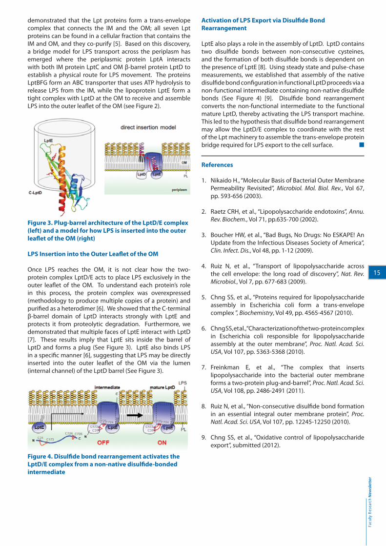
LPS Insertion into the Outer Leafl et of the OM
Once LPS reaches the OM, it is not clear how the two-
protein complex LptD/E acts to place LPS exclusively in the
outer leafl et of the OM. To understand each protein’s role
in this process, the protein complex was overexpressed
(methodology to produce multiple copies of a protein) and
purifi ed as a heterodimer [6]. We showed that the C-terminal
β-barrel domain of LptD interacts strongly with LptE and
protects it from proteolytic degradation. Furthermore, we
demonstrated that multiple faces of LptE interact with LptD
[7]. These results imply that LptE sits inside the barrel of
LptD and forms a plug (See Figure 3). LptE also binds LPS
in a specifi c manner [6], suggesting that LPS may be directly
inserted into the outer leafl et of the OM via the lumen
(internal channel) of the LptD barrel (See Figure 3).
Figure 4. Disulfi de bond rearrangement activates the
LptD/E complex from a non-native disulfi de-bonded
intermediate
Activation of LPS Export via Disulfi de Bond
Rearrangement
LptE also plays a role in the assembly of LptD. LptD contains
two disulfi de bonds between non-consecutive cysteines,
and the formation of both disulfi de bonds is dependent on
the presence of LptE [8]. Using steady state and pulse-chase
measurements, we established that assembly of the native
disulfi de bond confi guration in functional LptD proceeds via a
non-functional intermediate containing non-native disulfi de
bonds (See Figure 4) [9]. Disulfi de bond rearrangement
converts the non-functional intermediate to the functional
mature LptD, thereby activating the LPS transport machine.
This led to the hypothesis that disulfi de bond rearrangement
may allow the LptD/E complex to coordinate with the rest
of the Lpt machinery to assemble the trans-envelope protein
bridge required for LPS export to the cell surface.
References
1. Nikaido H., “Molecular Basis of Bacterial Outer Membrane
Permeability Revisited”, Microbiol. Mol. Biol. Rev., Vol 67,
pp. 593-656 (2003).
2. Raetz CRH, et al., “Lipopolysaccharide endotoxins”, Annu.
Rev. Biochem., Vol 71, pp.635-700 (2002).
3. Boucher HW, et al., “Bad Bugs, No Drugs: No ESKAPE! An
Update from the Infectious Diseases Society of America“,
Clin. Infect. Dis., Vol 48, pp. 1-12 (2009).
4. Ruiz N, et al., “Transport of lipopolysaccharide across
the cell envelope: the long road of discovery”, Nat. Rev.
Microbiol., Vol 7, pp. 677-683 (2009).
5. Chng SS, et al., “Proteins required for lipopolysaccharide
assembly in Escherichia coli form a trans-envelope
complex ”, Biochemistry, Vol 49, pp. 4565-4567 (2010).
6. Chng SS, et al., “Characterization of the two-protein complex
in Escherichia coli responsible for lipopolysaccharide
assembly at the outer membrane”, Proc. Natl. Acad. Sci.
USA, Vol 107, pp. 5363-5368 (2010).
7. Freinkman E, et al., “The complex that inserts
lipopolysaccharide into the bacterial outer membrane
forms a two-protein plug-and-barrel”, Proc. Natl. Acad. Sci.
USA, Vol 108, pp. 2486-2491 (2011).
8. Ruiz N, et al., “Non-consecutive disulfi de bond formation
in an essential integral outer membrane protein”, Proc.
Natl. Acad. Sci. USA, Vol 107, pp. 12245-12250 (2010).
9. Chng SS, et al., “Oxidative control of lipopolysaccharide
export”, submitted (2012).

Notes

Mailing List ...
If you wish to be on our mailing list, please complete the form below and
mail or fax (6873 1103) it back to us:
Dean's offi ce
Faculty of Science
National University of Singapore
S16, Level 8
Science Drive 2
Singapore 117546
Name:
Designation:
Address:
Tel: Fax: Email:
Tick on which best describes your job responsibility:
Chief Executive Offi cer/Chief Operating Offi cer
Business Development Manager
Consultant
Scientist/Teacher
Others ( please specify ):
Tick on which best describes your company's business:
Information Technology
Financial Institution
Manufacturing
Research & Development
Others ( please specify ):
Tick news items that interest you:
Conferences
Seminars
Workshops/Training sessions
Departmental/Faculty Open House
Others ( please specify ):
Faculty of Science
Newsletter
RESEARCH