faculty of pharmacy department of medical technology · human body. these microorganisms are...
TRANSCRIPT
Faculty of PharmacyDepartment of Medical Technology
36 QUERO, Marc Delvin 37 QUERUBIN, Ryan 38 RAMOS, Ma. Nicolei 39 RASPADO, Raphaelle 40 RAVELO, Aimee 41 ROBLE, Maria Lourdes
(Group 6, 1-G BSMT)
PROF. CARMELITA C. CARDONA Introduction to Medical Technology with STS
October 2009

WHAT IS MICROBIOLOGY?
Microbiology is the study of very small living organisms- organisms called microorganisms or microbes. Microorganisms are said to be ubiquitous, meaning they are virtually everywhere.
Microbiology is the branch of biological sciences involving study of all infectious diseases caused by bacteria, viruses and fungal infections
Microbiology includes virology, mycology, parasitology, bacteriology and other branches.
WHY STUDY MICROBIOLOGY?
Although they are very small, microorganisms play significant roles in our lives.
• Indigenous microflora or “normal flora”. Includes all the microbes that resides on and within the human body. These microorganisms are beneficial to us. They inhibit the growth of pathogens in those areas of the body where they live by occupying space, depleting the food supply, and secreting materials that may prevent or reduce the growth of pathogens.
NOTE: A fetus has no indigenous microflora.
Some of the organisms that colonize (inhibit) our bodies are known as opportunistic pathogens (or opportunists). These organisms do not cause any problems, they have the potential to cause infections if they gain access to a part of our anatomy where they do not belong. (e.g E. coli lives in our intestinal tracts. This organism does not cause us any harm as long as it stays in our intestinal tract but can cause disease if it gains access to our urinary bladder, bloodstream or a wound.
Some microbes produce oxygen by the process known as photosynthesis. Thus organisms that require O2 – humans for example owe a debt of gratitude to the algae and cyanobacteria that produce oxygen.
Many microbes are involved in the decomposition of dead organisms and the waste products of living organisms. They are known as decomposers or saprophytes. They aid in aid in fertilization by returning inorganic nutrients to the soil. They break down dead and dying organic materials (plants and animals) into nitrates, phosphates and other chemicals necessary for he growth of plants.
Many microbes are essential in various food and beverage industries, whereas others are used to produce certain enzymes and chemicals. The use of microorganisms in industry is called biotechnology.
Some bacteria and fungi produce antibiotics that are used to treat patients with infectious diseases. By definition, an antibiotic is a substance produced by a microorganism that is effective in killing or inhibiting the growth of other microorganisms.
Diseases. Microorganisms cause two categories of diseases: infectious and microbial intoxications. Anyone pursuing a career in a healthcare profession must be aware of infectious diseases, the pathogens that cause them, the sources of the pathogens, how these diseases are transmitted, and how to protect yourself and your patients from these diseases.

BACTERIA
Bacteria were one of the first microorganisms to be observed by human. In 1676, Antoine van Leeuwenhoek, using a single lens microscope first observed bacteria. He called bacteria as animalcules. The name bacterium was introduced much later by Christian Ehrenberg in 1838.
Bacteria are prokaryotic cells. Bacteria have no nucleus.
95% of the known bacteria are non-pathogens
Only 1% and about 4% of the all known bacteria cause human diseases and plant diseases respectively.
CELL MORPHOLOGY OF BACTERIA
There are three basic shapes of bacteria:
1. cocci (round or spherical bacteria)2. bacilli (rod-shaped or rectangular bacteria) and3. spirillum (curved and spiral-shaped bacteria)
Cocci may be seen singly or in pairs (diplococci), chains (streptococci), clusters (staphylococci), packets of four (tetrads) or packets of eight (octads).
Bacilli (often referred to as rods) may be or long, thick or thin and pointed or with curved or blunt ends.
Bacilli may be occur in singly, in pairs (diplobacilli), in chains (streptobacilli), in long filaments or branched. Some rods are quite short, resembling elongated cocci (coccobacilli).
FUNGI
Fungi are eukaryotic organisms that do not contain chlorophyll, but have cell walls, filamentous structures, and produce spores. These organisms grow as saprophytes and decompose dead organic matter. There are between 100,000 to 200,000 species depending on how they are classified. About 300 species are presently known to be pathogenic for man.
There are four types of mycotic diseases: 1. Hypersensitivity - an allergic reaction to molds and spores.2. Mycotoxicoses - poisoning of man & animals by feeds & food products contaminated by fungi which produce toxins from the grain substrate.3. Mycetismus- the ingestion of preformed toxin (mushroom poisoning).4. Infection

VIRUS
Viruses are acellular (containing no cell) microorganisms that are smaller than bacteria; obligate intracellular parasites; sometimes referred to as infectious agents.
Common diseases caused by viruses are small pox (variola virus), colds (rhinovirus), hepatitis (hepatitis types A&B), meningitis (coxsackievirus), influenza (orthomyxoviruses), mumps (myxovirus), AIDS (HIV).
Virus Type Viral Characteristics
Virus Disease
Poxviruses Large, brick shape with envelop, dsDNA
a) Variola
b) Vaccinia
a) Smallpox
b) Cowpox
Polyoma-papilloma
dsDNA, polyhedrala) Papillomavirus
b) Polyomavirus
a) Warts
b) Some tumors, some cancer
Herpesvirus Polyhedral with envelope, dsDNA
a) Herpes simplex I
b) Herpes simplex II
c) Herpes zoster
d) Varicella
a) Cold sores
b) Genital herpes
c) Shingles
d) Chickenpox
Adenovirus dsDNA, icosahedral with envelope
Respiratory infections, pneumonia, conjunctivitis, some tumors
Picornavirus(the name means small RNA viruses)
ssRNA, tiny, icosahedral, with envelope
a) Rhinovirus
b) Poliovirus
c) Hepatitis A & B
d) Coxsackievirus
a) Colds
b) Poliomyelitis
c) Hepatitis
d) Meningitis
Myxoviruses RNA, helical with envelope
a) Orthomyxoviruses types A & B
b) Myxovirus parotidis
c) Paramyxovirus
d) Rhabdovirus
a) Influenza
b) Mumps
c) Measles (rubeola)
d) Rabies

Arbovirus Arthropodborne RNA, cubic
a) Mosquitoborne B
b) Mosquitoborne A&B
c) Tickborne,Coronavirus
a) Yellow fever
b) Encephalitis
c) Colorado tick fever
Reovirus dsRNA, icohedral with envelope
a) Enterovirus a) Intestinal infections
Retrovirus dsDNA. Helical with envelope
a) RNA tumor virus
b) HTLV virus
c) HIV
a) Tumors
b) Leukemia
c) AIDS
GRAM-STAIN PROCEDURE
Gram-stain is used to differentiate bacteria into two large groups- gram positive and gram negative bacteria based on the physical and chemical characteristics of the cell wall.
Gram-stain procedure is the most important procedure in the bacteriology section of the laboratory. It was devised in 1844 by Hans Christian Gram, a Danish physician.
Gram-stain uses the following reagents:
Crystal Violet or Ammonium Oxilate (as Primary Stain) - Gram-positive bacteria will retain the crystal violet stain in their cell walls.
Iodine Crystal ( as Mordant) - Iodine fixes the crystal-violet dye molecules to the cell walls of gram-positive bacteria so the stain will not wash away when the slide is rinsed off with water.
Ethanol/ Acetone (as Decolorizer) Safranin (as Counter-stain)
Procedure:
I. Prepare the smear.
Transfer a drop amount of a suspended culture to be examined in a slide. If the culture is to be taken from a Petri dish, first add a drop of water on the slide and transfer a minute amount of a colony from the Petri dish.
If staining a clinical specimen, smear a very thin layer onto the slide, using a wooden stick. Do not use a cotton swab, if at all possible, as the cotton fibers may appear as artefacts. The smear should be thin enough to dry completely within a few seconds.
Spread the culture with an inoculation loop to an even thin film over a circle of 1.5 cm in diameter, approximately the size of a dime. Thus, a typical slide can simultaneously accommodate 3 to 4 small smears if more than one culture is to be examined.
Air-dry the culture and fix it or over a gentle flame, while moving the slide in a circular fashion to avoid localized overheating. The applied heat helps the cell adhesion on the glass slide to make possible the subsequent rinsing of the smear with water without a significant loss of the culture. Heat can also be applied to facilitate drying the the smear. However, ring patterns can form if heating is not uniform, e.g. taking the slide in and out of the flame.

II. Gram-stain
1. After drying the smear, apply crystal violet to the slide specimen. Wait for 60 seconds.2. The slide is washed with water for 10 seconds (or until it is completely rinse) after having stained
with crystal violet.3. Apply Gram’s Iodine and wait for 60 seconds.4. After the slide has been treated with Gram’s Iodine, rinse the slide with tap water. Now the gram’s
positive are stained.5. Apply ethanol or acetone to the slide until it turns blue then rinse. A mixture of ethanol and acetone
dissolves the lipid layer of the Gram-negative cell walls ensuring that none of the crystal violet remains. This process also helps fix the crystal violet to the Gram-positive cell walls.
6. Apply Safranin to the slide and wait for 45 seconds then rinse. The cell walls of most Gram-negative bacteria will absorb the Safranin dye. Sometimes another dye, Giemsa, is also applied to stain the remaining Gram-negative bacteria.
7. The slide is now ready to be observe in a microscope (with OIO as its objective)
Apply Crystal Violet Rinse smear w/ H2O Apply Gram’s Iodine Rinse smear w/ H2O
Rinse smear w/ H2O Apply Safranin Decolorized the smear with ethanol
THE SMEAR IS NOW READY TO BE VIEWED IN MICROSCOPE WITH OIO
FACTORS THAT AFFECT MICROBIAL GROWTH
Microbial is affected by many different environmental factors:
1. Availability of Nutrients
BACTERIUM DISEASES GRAM-STAIN REACTION
Bacillus anthracis Anthrax +Bordetella pertussis Whooping cough -Clostridium botulinum Botulism (food
poisoning)+
Clostridium perfringens Gas gangrene, wound infections
+
Clostridium tetani Tetanus (lockjaw) +Corynebacterium diphtheriae Diptheria +Escherichia coli Urinary tract
infection (UTI)-
Francisella tularensis Tularemia -Klebsiella pneumoniae Pneumonia -Mycobacterium tuberculosis Tuberculosis (TB) +/-Mycobacterium leprae Leprosy +/-Neisseria gonorrhoeae Gonorrhea -Salmonella typhi Typhoid fever -Salmonella species Gastroenteritis -Staphylococcus aureus Boils, septicaemia,
pneumonia+
Streptococcus pyogens Strep throat, rheumatic fever
+
Streptococcus pneumoniae Pneumonia +Treponema pallidum Syphilis -Vibrio cholerae Cholera -

To survive in a particular environment, appropriate nutrients must be available.
2. MoistureAll living organisms require water to carry out their metabolic processes. The organisms within the spores and cysts are in a dormant or in a resting state; if they are placed in a moist, nutrient-rich environment, they will grow and reproduce normally.
3. TemperatureEvery microorganism has an optimum growth temperature in which the organism grow best.
• Thermophiles – microorganisms that grow best at high temperature. The highest temperature at which a bacterium has been found living is around 113C—Pyrolobus fumarii.
• Mesophiles – microbes that grow best at moderate temperature (37C).• Psychrophiles – microbes that prefer cold temperature. Example of this is the
Escherichia coli that has survive in Arctic.
4. pH – refers to the acidity or alkalinity of a solution.
• Acidophilic microbes - prefer an environment with a pH of 2-5.• Alkaliphiles microbes - prefer an environment with a pH of 8.5. Vibrio cholerae,
the causative agent of cholera, is the only human pathogen that grows well above pH 8.
5. Osmotic Pressure and Salinity
Osmotic Pressure – pressure exerted on a cell membrane by solutions both inside and outside the cell.
Plasmolysis- in case of hypertonic solution, the cell membrane and cytoplasm shrink away from the cell wall.
Salts and sugar are added to certain foods as a way of preserving them. Bacteria that enter hypertonic environment will die due to desiccation.
Plasmoptysis – when bacterial are placed in a hypotonic solution, the fluid pressure within the cell increases greatly and this causes the escape of cytoplasm from the cell.
Sugar solutions for jellies and pickling brines for meats preserve foods by inhibiting the growth of most microorganisms.
6. Barometric Pressure
7. Gaseous Atmosphere
Types and concentrations of gases present in a particular environment determine which species of microbes are able to live there.
Encouraging the Growth of Microorganisms in VitroVitro – refers to the events outside the body
Reasons why the growth of microorganisms is encouraged in microbiology laboratory:
a.) Grow them on culture media so they can gather information that will enable identification of any pathogen;
b.) So that the scientists can learn more about them, harvest antibiotics and other microbial products, test new antimicrobial agents, and produce vaccines.
Culturing Bacteria in the Laboratory

Bacterial Growth – refers to an increase in the number of organisms rather than an increase in size.
When each bacterial cell reaches its optimum size, it divides by binary fission into two daughter cells.
Bacterial Colony – is a mound or pile of bacteria containing millions of cells.Generation Time – time it takes for one cell to become two cells by binary fission.
In the laboratory, a pure culture of a single species of bacteria can usually be maintained if the appropriate growth medium and environmental conditions are provided.
Fastidious Bacteria have complex nutritional requirements. Often, special mixtures of vitamins and amino acids must be added to the medium to culture these fastidious organisms. Viruses, rickettsias, and chlamydias are obligate intracellular pathogens therefore cannot grow on artificial culture.
Culture Media - Are used in microbiology laboratories to culture bacteria. - Referred to as artificial media because they are prepared in the laboratory.
Classifications of culture media
A. According to exact contents:a. Chemically defined media – was prepared by adding specific number of grams of each of the components.
b. Complex media – one in which the exact contents are not known.o Contain ground up or digested extracts from animal organs, yeasts and plants to
provide necessary vitamins and minerals.
Culture media can also be categorized as liquid or solid:
A. Liquid Media – are contained in tubes thus referred as tube media.B. Solid Media – prepared by adding agar to liquid media and then pouring the media into tubes or
Petri dishes where the media solidifies.
Agar – is a complex polysaccharide that is obtained from a red marine alga; it is used as a solidifying agent.
Enriched medium – solid medium containing a rich supply of special nutrients that promotes the growth of fastidious organisms. It is usually prepared by adding extra nutrients to a medium called nutrient agar.
o Examples of solid enriched media: blood agar (nutrient agar plus 5% sheep RBC) and chocolate agar (nutrient agar plus powdered hemoglobin).
Selective medium – has added inhibitors that discourage the growth of certain organisms without inhibiting the growth of the organism being sought.
o For example: MacConkey agar inhibits growth of Gram-positive bacteria and thus is selective for Gram-negative bacteria.
Differential medium – permits the differentiation of organisms that grow on the medium. o For example: MacConkey agar is frequently used to differentiate among various Gram-
negative, bacilli that are isolated form fecal specimens. Gram-negative bacteria able to ferment lactose—ingredient of Macconkey agar—produce pink colonies whereas those unable to ferment lactose produce colorless colonies. Thus, MacConkey agar differentiates between lactose-fermenting and nonlactose-fermenting Gram-negative bacteria.

USING ANTIMICROBIAL AGENTS TO CONTROL MICROBIAL GROWTH IN VIVO
*vivo refers to events inside the body
I. Compare and Contrast chemotherapeutic agents, antimicrobial agents, and antibiotics
a. Chemotheraphy – refers to the use of any chemical (drug) to treat any disease or condition
b. Chemotherapeutic agent – is any drug used to treat any condition or disease
c. Antimicrobial agent – is any chemical (drug) used to treat an infectious disease, either by inhibiting or killing pathogens in vivo.
d. Antibacterial agents – drugs used to treat bacterial diseases
e. Antifungal agents – drugs used to treat fungal diseases
f. Antiprotozoal agents – drugs used to treat protozoal diseases
g. Antiviral agents – drugs used to treat viral diseases
h. Antibiotic – is a substance produced by a microorganism that is effective in killing or inhibiting the growth of other microorganisms
NOTE: Although all antibiotics are antimicrobial agents, not all antimicrobial agents are antibiotics.Antibiotics are produced by certain molds and bacteria, usually those that live in soil. The antibiotics produced by soil organisms give them a selective advantage in the struggle for the available nutrients in the soil.Examples of antibiotics produced by molds
1. Penicillin
2. Cephalosporins
Examples of antibiotics produced by bacteria1. Bacitracin
2. Erythromycin
3. Chloramphenicol
Although originally produced by microorganisms, many antibiotics are now synthesized or manufactured in pharmaceutical laboratories. Also, many antibiotics have been chemically modified to kill a wider variety of pathogens or reduce side effects; these modified antibiotics are called semisynthetic antibiotics. Semisynthetic antibiotics include penicillins such as ampicillin and carbenicillin. Antibiotics are primarily antibacterial agents and are thus used to treat bacterial diseases.
II. Ideal qualities of an antimicrobial agent
a. Kill or inhibit the growth of pathogens (a specific agent causing disease)
b. Cause no damage to the host
c. Cause no allergic reaction to the host
d. Be stable when stored in solid or liquid form
e. Remain in specific tissues in the body long enough to be effective
f. Kill the pathogens before they mutate and become resistant to it.

* Unfortunately, most antimicrobial agents have some side effects, produce allergic reactions, or permit development of resistant mutant pathogens.
III. Five most common mechanisms of action of antimicrobial agentsa. Inhibition of cell wall synthesis
Example: mechanism of penicillin
b. Damage of cell membrane
c. Inhibition of enzyme activity
Example: mechanism of Sulfonamide drug
d. Inhibition of protein synthesis
e. Inhibition nucleic acid synthesis (as of DNA and RNA synthesis)
IV. Differentiate between bactericidal and bacteriostatic agentsa. Bacteriostatic – stop bacteria from growing and dividing
**Should only be used in patients whose host defense mechanisms are functioning properly, that is only in patients whose bodies are capable of killing the pathogen once its multiplication is stopped
b. Bactericidal – kills bacteria
**Should NOT be used in immunosuppressed or leukopenic patients, that is patients having an abnormally low number of white blood cells
V. Differentiate between narrow-spectrum and broad-spectrum antimicrobial agents
a. Narrow-spectrum antibiotics – destroys either Gram-positive bacteria or Gram-negative bacteria
Ex. Vancomycin – destroys only Gram-positive bacteria
Colistin and nalidixic acid – destroy only Gram-negative bacteria
b. Broad-spectrum antibiotics – destructive to both Gram-postive and Gram-negative bacteria
Ex. Ampicillin, chloramphenicol, tetracycline
*In some cases, a single antimicrobial agent is not sufficient to destroy all the pathogens that develop during the course of disease; thus two or more drugs may be used simultaneously to kill all the pathogens and to prevent resistant mutant pathogens from emerging. This way of treating a disease is called Multidrug Therapy.
Example: The multidrug-resistant strains of Mycobacterium tuberculosis. Four drugs namely isoniazid, rifampin, pyrazinamide, and either ethambutol or streptomycin are routinely prescribed, and as many as 12 drugs may be required especially resistant strains.
VI. Synergism Versus Antagonism

1. Synergism – use of 2 antimicrobial agents to treat an infectious disease that sometimes produces a degree of pathogen killing that is far greater than that achieved by either drug alone.
2. Antagonism – use of two drugs that actually work against each other. The extent of pathogen killing is less than that achieved by either drug alone.
VII. Four most common mechanisms by which bacteria become resistant to antimicrobial agents
Intrinsic resistance – situation wherein bacteria is naturally resistant to a particular antimicrobial agent
Acquired resistance – situation wherein bacteria that were once susceptible to a particular drug to become resistant to it.
MECHANISM EFFECTA chromosomal mutation that causes a change in the structure of a drug binding site
The drug cannot bind to the bacterial cell and thus the drug cannot enter the cell
a chromosomal mutation that causes a change in cell membrane permeability
The drug cannot pass through the cell and thus cannot enter the cell
Acquisition (by conjugation, transduction or transformation) of a gene that enables the bacterium to produce an enzyme that destroys or inactivates the drug
The drug is destroyed or inactivated by the enzyme
Acquisition (by conjugation, transduction, or transformation) of a gene that enables the bacterium to produce multidrug-resistance (MDR) pump
The drug is pumped out of the cell before it can damage or kill the cell
*conjugation - transfer of genetic material between bacteria through direct cell-to-cell contact, or through a bridge-like connection between the two cells
*transduction – whereby bacteriophages (viruses that infect bacteria) carry bacterial DNA from one bacterial cell to another
*transformation – the uptake of naked DNA from the environment
*plasmid – is an extra-chromosomal DNA molecule separate from the chromosomal DNA which is capable of replicating independently of the chromosomal DNA. In many cases, it is circular and double-stranded. Plasmids usually occur naturally in bacteria.
*Resistance factor (R-factor) - plasmid containing multiple genes for drug resistance
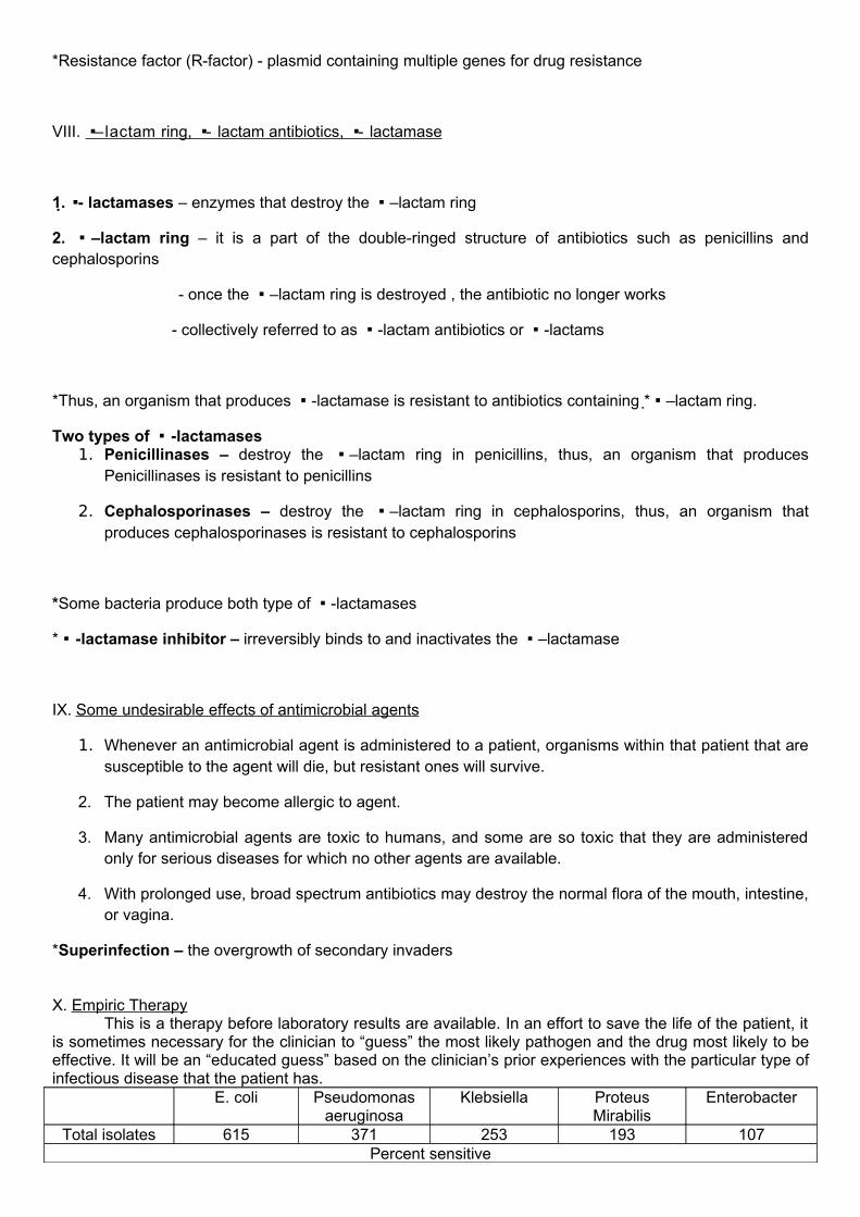
VIII. –lactam ring, - lactam antibiotics, - lactamase
1. - lactamases – enzymes that destroy the –lactam ring
2. –lactam ring – it is a part of the double-ringed structure of antibiotics such as penicillins and cephalosporins
- once the –lactam ring is destroyed , the antibiotic no longer works
- collectively referred to as -lactam antibiotics or -lactams
*Thus, an organism that produces -lactamase is resistant to antibiotics containing * –lactam ring.
Two types of -lactamases1. Penicillinases – destroy the –lactam ring in penicillins, thus, an organism that produces
Penicillinases is resistant to penicillins
2. Cephalosporinases – destroy the –lactam ring in cephalosporins, thus, an organism that produces cephalosporinases is resistant to cephalosporins
*Some bacteria produce both type of -lactamases
* -lactamase inhibitor – irreversibly binds to and inactivates the –lactamase
IX. Some undesirable effects of antimicrobial agents
1. Whenever an antimicrobial agent is administered to a patient, organisms within that patient that are susceptible to the agent will die, but resistant ones will survive.
2. The patient may become allergic to agent.
3. Many antimicrobial agents are toxic to humans, and some are so toxic that they are administered only for serious diseases for which no other agents are available.
4. With prolonged use, broad spectrum antibiotics may destroy the normal flora of the mouth, intestine, or vagina.
*Superinfection – the overgrowth of secondary invaders
X. Empiric TherapyThis is a therapy before laboratory results are available. In an effort to save the life of the patient, it
is sometimes necessary for the clinician to “guess” the most likely pathogen and the drug most likely to be effective. It will be an “educated guess” based on the clinician’s prior experiences with the particular type of infectious disease that the patient has.
E. coli Pseudomonas aeruginosa
Klebsiella Proteus Mirabilis
Enterobacter
Total isolates 615 371 253 193 107Percent sensitive

Cefotetan 100 1 100 100 77Ceftriaxone 100 80 100 100 90Ceftazidime 100 98 95 100 86
Amikacin 100 100 100 100 100Gentamicin 100 88 89 93 100
XI. Actions that clinicians or patients can take to help in war against drug resistance
1. Education of healthcare professional s and in turn, education of patients.
2. Patients must stop demanding antibiotics every time they are sick or have a sick child.
3. It is important that clinicians not allow themselves to be pressured by patients.
4. Clinicians should prescribe an inexpensive, narrow-spectrum drug whenever the laboratory results demonstrate that such a drug effectively kills the pathogen.
5. Patients must take their antibiotics in the exact manner in which they are prescribed.
6. Healthcare professionals must practice good infection prevention and control procedures.
7. It is critical that clinicians prescribe the appropriate amount of antibiotic necessary to cure the infection.
8. Patients should always destroy excess medications.
THE CLINICAL MICROBIOLOGY LABORATORY
Organization

Depending on the size of the hospital, the Clinical Microbiology Laboratory may be under the direction of a pathologist, microbiologist, or in smaller hospitals, a medical technologist who has had many years of experience working in microbiology. Most of the actual bench work that is performed in the CML is performed by MT’s and MLT’s.
Sections of the Laboratory
Primary Responsibility
Bacteriology To assist clinicians in the diagnosis of bacterial diseases-antimicrobial susceptibility testing is performed whenever it is appropriate to do so-bacterial pathogens are identified by gathering clue(phenotypic characteristics)-bacterial pathogens are isolated from the specimens -tests are performed to identify the bacterial pathogens
Mycobacteriology/TB LAB
To assist clinicians in the diagnosis of tuberculosis-sputum specimens are primarily processed here-acid-fast staining is performed -susceptibility testing is performed-Mycobacterium spp. Are identified using a combination of growth characteristics
Virology To assist clinicians in the diagnosis of viral diseases-many viral diseases are diagnosed using immunodiagnostic procedures-can be observed by using electron microscopy-nucleic acid probes and polymerase chain reaction assays-viruses are identified by the types of cell lines that they are able to infect and the physical changes they cause in the infected cellsz
Parasitology To assist clinicians in the diagnosis of parasitic diseasesspecifically, endoparasites-parasites are identified by their characteristic appearance of the various life cycle stages that are seen in clinical specimens
Mycology To assist clinicians in the diagnosis of fungal infections-most commonly submitted specimens: hair clippings, nail clippings, and skin scrapings-a potassium hydroxide preparation is performed on the hair clippings, nail clippings, and skin scrapings. It acts as a clearing agent by dissolving keratin in the specimens to enable the technologist to see into the specimens when they’re examined microscopically and to determine whether there are any fungal elements.-Sabouraud dextrose agar is used to inoculate specimens (a selective medium for fungi)-molds are identified by using a combination of growth and macroscopic and microscopic observations, not by biochemical tests.

Primary Responsibilities of the Lab.
1. Processing clinical specimens
2. Isolating pathogens from specimens
3. Identifying pathogens
4. Performing antimicrobial susceptibility testing
5. Assist physicians in the diagnosis of infectious diseases
6. All clinical specimens must be labeled properly

7. Laboratory slips must contain all the necessary information
8. When collecting blood from specimens for culture, the venipuncture site must be thoroughly cleansed and disinfected to prevent contamination of the specimen with the indigenous skin floras
GODBLESS 1-GMTHIT THE HIGH MARK!
-the microbiology group