factorviii-vonwillebrandfactorcomplexinhibits ... ... using biaeval 4.1 software (biacore). ... on...
TRANSCRIPT

Factor VIII-von Willebrand Factor Complex InhibitsOsteoclastogenesis and Controls Cell Survival*□S
Received for publication, June 5, 2009, and in revised form, August 18, 2009 Published, JBC Papers in Press, September 16, 2009, DOI 10.1074/jbc.M109.030312
Marc Baud’huin‡§1, Laurence Duplomb‡§¶, Stephane Teletchea‡§, Celine Charrier‡§, Mike Maillasson�,Marc Fouassier**, and Dominique Heymann‡§¶2
From ‡INSERM U957, Universite de Nantes, Nantes Atlantique Universites, Laboratoire de Physiopathologie de la ResorptionOsseuse et Therapie des Tumeurs Osseuses Primitives, §EA3822, Universite de Nantes, ¶CHU, Hotel-Dieu, �INSERM U892 andIFR26-Ouest Genopole, and the **Centre Regional de Traitement de l’Hemophilie, Laboratoire d’Hematologie, CHU, Hotel-Dieu,Nantes F-44035, France
Factor VIII-von Willebrand factor (FVIII�vWF) complex, amolecule involved in coagulation, can be physically associatedwithosteoprotegerin (OPG).OPG is an anti-osteoclastic proteinand a soluble receptor for the proapoptotic protein TRAIL(tumor necrosis factor-related apoptosis-inducing ligand), sug-gesting a potential role of FVIII�vWF complex in bone and can-cer biology.We, thus, assessed the effects of FVIII�vWFcomplexon osteoclastogenesis and cell survival. We first evidenced thatFVIII�vWF complex inhibited RANKL-induced osteoclastogen-esis and enhanced the inhibitory effect ofOPG. Interestingly,werevealed by surface plasmon resonance that FVIII�vWF complexbound toRANKL,whereas recombinant FVIII and vWFdid not.Bymodeling,we showed that theOPGbindingdomain to theA1domain of vWF was closely located and partially overlapped toits binding site to RANKL. Then, we demonstrated thatFVIII�vWF complex cancelled the inhibitory activity of OPG onTRAIL-induced apoptosis and characterized interactionsbetween these molecules. The present work evidenced a directactivity of FVIII�vWF complex on osteoclasts and on inducedcell apoptosis, pointing out its potential involvement in physio-logical bone remodeling or in bone damages associated withsevere hemophilia and cancer development.
Themolecular triadosteoprotegerin (OPG)3/RANK/RANKLisa crucial parameter of bone biology. Receptor activator ofnuclear factor �B ligand (RANKL), a member of the tumornecrosis factor family, is mainly expressed by osteoblasts in thebone microenvironment and acts as a pro-resorption factor (1,2); RANKL binds to its receptor RANK expressed at the cellsurface of osteoclast precursors and induces osteoclastic differ-
entiation and maturation, leading to bone resorption (3, 4).OPG, also mainly produced by osteoblasts, is a soluble decoyreceptor for RANKL, preventing the binding of RANKL toRANK and, thus, inhibiting osteoclastogenesis (5–7). Boneturnover is tightly controlled by theOPG/RANK/RANKL triad,and any change in the balance OPG/RANKL leads to patholog-ical conditions (7). OPG is also a receptor for tumor necrosisfactor-related apoptosis-inducing ligand (TRAIL) (8, 9), a cyto-kine that is able to induce a rapid cancer cell death by apoptosis(9–11). Interestingly, the binding of OPG to TRAIL completelyinhibits TRAIL-induced cytotoxicity (8). OPG possesses anti-apoptotic properties and, therefore, could be considered as apro-tumoral agent.Factor VIII is a plasma glycoprotein mainly synthesized by
hepatocytes but also by kidney, sinusoidal endothelial cells, andin small amounts by lymphatic tissues (12). Factor VIII is one ofthe main coagulation factors and allows the completion of thecoagulation process. Factor VIII circulates in plasma in a non-covalent complex with the von Willebrand factor (FVIII�vWFcomplex). Themost well known genetic disease associatedwithFactor VIII is hemophilia A, which shows an X-linked inherit-ance (13). A second important disease associated with low Fac-tor VIII levels is von Willebrand disease, a bleeding disorder(14, 15). Patients suffering from von Willebrand disease haveprimary hemostasis defects leading tomucocutaneous bleedingor spontaneous deep tissue bleeding, such as in hemophilia A,or both (14). Bleeding diseases could be associated with differ-ent bone phenotypes. For instance, in a murine model of plate-let-type von Willebrand disease, a significant increase of bonemass and cortical thickness due to a reduction of the number ofosteoclasts is observed (16). In contrast, various case reportssuggest that children suffering from severe hemophilia havemore risks for low bone density and osteopenia/osteoporosis,preferentially caused by physical inactivity and leading to loss ofjoint function, shorter height, lower weight, and muscle atro-phy (17, 18).von Willebrand factor is a multimeric protein containing
many binding domains for various proteins such as the D�-D3domain, which binds Factor VIII (FVIII) (19), and the A1domain, which can bind different proteins such as the plateletglycoprotein Ib (20), heparin (21), and snake venom toxins(bitiscetin (22) and botrocetin (23)). Recently, it has beenshown that the vWF is physically complexed to OPG (throughthe A1 domain) within the Weibel-Palade bodies and also in
* This work was supported in part by the Region des Pays de la Loire (Program“Ciblage Moleculaire et Applications Therapeutique”) and by the ANR 2007INSERM Pathophysiology of Human Diseases Project R07196NS.
□S The on-line version of this article (available at http://www.jbc.org) containsa supplemental figure.
1 Recipient of a fellowship from the Region des Pays de la Loire.2 To whom correspondence should be addressed: 1 rue Gaston Veil, 44035
Nantes Cedex 1, France. Tel.: 33-240412845; Fax: 33-240412860; E-mail:[email protected].
3 The abbreviations used are: OPG, osteoprotegerin; RANKL, receptor activa-tor of nuclear factor �B ligand; FVIII, Factor VIII; FVIII�vWF complex, factorVIII-von Willebrand complex; TRAIL, tumor necrosis factor-related apopto-sis-inducing ligand; RU, response units; aPTT, activated partial thrombo-plastin time; Bis-Tris, 2-[bis(2-hydroxyethyl)amino]-2-(hydroxymethyl)pro-pane-1,3-diol; ELISA, enzyme-linked immunosorbent assay.
THE JOURNAL OF BIOLOGICAL CHEMISTRY VOL. 284, NO. 46, pp. 31704 –31713, November 13, 2009© 2009 by The American Society for Biochemistry and Molecular Biology, Inc. Printed in the U.S.A.
31704 JOURNAL OF BIOLOGICAL CHEMISTRY VOLUME 284 • NUMBER 46 • NOVEMBER 13, 2009
by guest on June 20, 2018http://w
ww
.jbc.org/D
ownloaded from
plasma, revealing a possible modulatory role of OPG in hemo-stasis (24, 25). The aim of the present study was to characterizethe effects of FVIII�vWF complex on osteoclastogenesis andcancer cell survival and then interactions between complexFVIII�vWF complex and three members of tumor necrosis fac-tor cytokine/cytokine receptor family: OPG, RANKL, andTRAIL. The data obtained demonstrated that FVIII�vWF com-plex binds to OPG and RANKL and, thus, indirectly partici-pates in bone biology. Indeed, we first demonstrated that theFVIII�vWF complex inhibits RANKL-induced osteoclastogen-esis. Second, we demonstrated that the FVIII�vWF complexabolished the inhibitory effectofOPGonTRAIL-inducedapop-tosis, revealing a key role of FVIII�vWF complex in cancerdevelopment.
EXPERIMENTAL PROCEDURES
Osteoclast Differentiation Assay—Generation of osteoclastsfrom murine RAW 264.7 cells was performed as previouslydescribed (26) in the presence of recombinant human RANKL(100 ng/ml) (kindly provided byAmgen Inc.), humanOPG (100ng/ml) (R&D systems), FVIII�vWF complex purified fromplasma (ProSpec), or recombinant FVIII (Octocog �, kindlyprovided by CSL Behring) (1 or 2 units/ml). Generation of oste-oclasts from human CD14� monocytes was described previ-ously (26). Briefly, purified CD14� cells were cultured in�-minimum essential mediumwith 10% fetal calf serum and 25ng/ml humanmacrophage colony-stimulating factor (R&Dsys-tems). After 3 days of culture, 100 ng/ml RANKL, 100 ng/mlOPG, and 1 unit/ml FVIII or FVIII�vWF were added. Multinu-cleated cells formed with three nuclei, and more were countedafter TRAP staining (Sigma).Cell Proliferation—Human osteosarcoma cell lines MG63 and
SaOS2 aswell as the Ewing’s sarcoma cell lineTC71were culturedin Dulbecco’s modified Eagle’s medium containing 10% fetal calfserum. MG63 and SaOS2 cells were seeded at 500 cells/well in96-well plates, andTC71 cellswere seeded at 1500 cells/well. Cellswere treated with 50–100 ng/ml TRAIL (R&D Systems), 50–100ng/ml OPG, and 1 unit/ml FVIII�vWF complex for 72 h. After theculture period, cell viability was determined by a cell proliferationreagent assay kit using sodium 3�-(1-(phenylaminocarbonyl)-3,4-tetrazolium)-bis(4-methoxy-6-nitro)benzenesulfonic acid hydrate(XTT Roche Applied Science).Hoechst Staining and Caspase-3 Activity—Cell death was
monitored microscopically after Hoechst 33258 (Sigma) stain-ing. MG63, SaOS2, and TC71 cells were seeded in a 96-multi-well plate and treated or not with TRAIL (50 ng/ml), OPG (50ng/ml), and FVIII�vWF complex (1 unit/ml) for 16 h, stainedwith 10 �g/ml Hoechst reagent for 20 min at 37 °C, and thenobserved under UV microscopy (DMRXA; Leica, Germany).Caspase-3 activity was assessed on 10 �l of total treated celllysates using the kit CaspACE assay system, fluorometric (Pro-mega) following the manufacturer’s recommendations. Resultsare expressed in arbitrary units and are corrected for proteincontent.Surface Plasmon Resonance Binding Assays—Experiments
were carried out on aBIAcore 3000 instrument (BIAcore).OPG(1 �g/ml in 5 mM maleate, pH 6.0), RANKL (2 �g/ml in 5 mM
maleate, pH 5.75), and TRAIL (10 �g/ml in 10 mM acetate, pH
5.5) were covalently immobilized to the dextran matrix of aCM5 sensor chip (BIAcore) at a flow rate of 5 �l/min. Immobi-lization levels ranging from 300 RU for OPG and 400 RU forRANKL to 700 RU for TRAIL were obtained. vWF (Hemato-logic Technologies) was immobilized on a C1 sensor chip at2000 RU. Binding assays were performed at 25 °C in 10 mM
Hepes buffer, pH 7.4, containing 0.15 M NaCl and 0.005% P20surfactant (HBS-P buffer, BIAcore) or in a pH 6.5 buffer con-taining 20 mM Bis-Tris and 10 mM CaCl2 at a flow rate of 30�l/min for immobilized-OPG and 20 �l/min for immobilizedRANKL and immobilized-TRAIL. OPG Kd values for vWF andFVIII�vWFwere determined using single cycle kinetics, startingwith 25 nM OPG or with 300 nM FVIII�vWF, then 1⁄2 dilutions.For binding analysis over the immobilized RANKL or immobi-lized-TRAIL chip, binding of OPG alone or preincubated for120 min with different concentrations of FVIII�vWF complexwas performed for 4min at a flow rate of 20 �l/min followed bydissociation for 2.5 min. The resulting sensorgrams were fittedusing BiaEval 4.1 software (BIAcore). For Kd calculations, thefollowing molecular masses were used: recombinant FVIII, 330kDa; FVIII�vWF complex, 540 kDa.ELISA Assay—FVIII�vWF complex was coated at 1 unit/ml
on a 96-well plate overnight at room temperature. OPG orRANKL (bothwere tested at 500, 100, and 10 ng/ml) were incu-bated for 2 h at room temperature. After 2 washes with PBS,Tween 0.05%, the revelation of the binding of OPG or RANKLto FVIII�vWF complex was performed using a specific biotinyl-ated antibody against each molecule (anti-OPG was from R&Dsystems and anti-RANKL fromPeprotech). Streptavidin conju-gated to horseradish peroxidase (R&D systems) was incubatedfor 20 min, then the revelation solution (Promega) was addedfor 20 min, and the reaction was stopped with sulfuric acidsolution. The absorbance at 450 nm was measured using amicroplate reader (Victor II, PerkinElmer Life Sciences).Modeling Analysis—To design OPG and RANKL, sequences
were retrieved from the Universal Protein resource. Each pro-tein was subjected to BLAST searches on the organism species(Homo sapiens) and on the organism classification (Mammalia,Vertebrata) (27). These sequences were further analyzed usingmultiple sequence alignments to extract the most conservedresidues (28). The multiple alignments were manually adjustedusing Jalview (29). Human OPG and human RANKL modelswere built using Modeler 9v5 (30) from these refined align-ments, respectively, using substructure of DR5 (PDB code1D4V) (31) and mouse RANKL (PDB code 1IQA) (32). Allresulting models were assessed using the Protein Health mod-ule of Discovery Studio 2.1 (Accelrys Inc.). Alignment of the A1domain of vWF on OPG has been realized as described below.Structural figures were produced with VMD (33) and renderedusing Pov-Ray.OPG, RANKL, and TRAIL Effect on the Coagulation Cascade—
Plasma of a healthy donor was drawn on 0.109 M buffered cit-rate.OPG,TRAIL, andRANKLwere added to the plasma at 100ng/ml. Primary hemostasis was tested using PFA 100 automate(Siemens). The Quick time was determined using the reagentRecombiPlasTin (Instrumentation Laboratory) on the ACLTOP automate (Instrumentation Laboratory). The activatedpartial thromboplastin time (aPTT) was tested using Trini-
Osteoclast, Cell Survival, and FVIII�vWF Complex
NOVEMBER 13, 2009 • VOLUME 284 • NUMBER 46 JOURNAL OF BIOLOGICAL CHEMISTRY 31705
by guest on June 20, 2018http://w
ww
.jbc.org/D
ownloaded from
CLOTThrombin Time reagent (Trinity Biotech) onACLTOP.The thrombin time was tested using thrombin (Siemens) onACL TOP. The FVIII/C method was based on the aPTT. Thisassay was performed using FVIII-deficient plasma (Biopep), anaPTT reagent with kaolin as contact phase activator (CK Prest,Diagnostica Stago, France), and an ACL TOP coagulometer.Statistical Analysis—The mean � S.D. was calculated for all
conditions and compared by analysis of variance with Bonfer-roni multiple comparisons test as the post hoc test. Differencesrelative to a probability of two-tailed p � 0.05 were consideredsignificant.
RESULTS
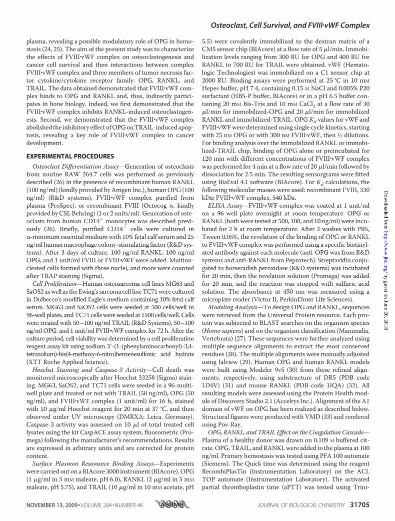
FVIII�vWF Inhibits Murine and Human Osteoclast Differen-tiation Induced by RANKL—The impact of the FVIII�vWF com-plex on osteoclastogenesis was first examined using the cellularmodelRAW264.7.After5daysof culturewith100ng/mlRANKL,RAW 264.7 cells differentiated into multinucleated cells. Asexpected, 50ng/mlOPGinhibited theRANKL-inducedosteoclas-togenesis by 47% (p � 0.01) (Fig. 1A). Surprisingly, 2 units/mlFVIII�vWF complex inhibited RANKL-induced osteoclastogen-esis by 42% (p� 0.01), whereas 1 unit/ml FVIII�vWF complex hadno effect on RANKL-induced osteoclastogenesis (Fig. 1A). Fur-thermore, when 2 units/ml FVIII�vWFcomplexwere added to theculture medium in the presence of OPG, the inhibition of oste-oclastogenesis was significantly stronger than that observed withOPG alone. Indeed, the inhibition of RANKL-induced osteoclas-togenesis reached 65% in the presence of a mixture OPG,FVIII�vWF complex compared with 47% with OPG alone (p �0.05). The recombinant FVIII alone had no effect on RANKL-in-duced osteoclastogenesis of RAW264.7 cells (data not shown).To ascertain the effect of FVIII�vWF complex on osteoclas-
togenesis, we next generated osteoclasts from human CD14�
purified from total peripheral blood mononuclear cells uponmacrophage colony-stimulating factor and RANKL activation(26). As shown in Fig. 1, B and C, and similarly to RAW 264.7cells, 1 unit/ml FVIII�vWF complex significantly inhibited by30% the RANKL-induced osteoclastogenesis of CD14� cells(p � 0.05) (Fig. 1C). Furthermore, 1 unit/ml FVIII�vWF com-plex reinforced the OPG inhibitory activity on RANKL-in-duced osteoclastogenesis (p � 0.05) (Fig. 1C). In accordancewith the RAW 264.7 cells, the recombinant FVIII alone hadno effect on RANKL-mediated osteoclastogenesis (data notshown).RANKL Binds to FVIII�vWF Complex Similarly to OPG—To
explore the molecular mechanism underlying the effect of theFVIII�vWF complex on RANKL-induced osteoclastogenesis andthe possible synergistic effect ofOPGandFVIII�vWFcomplex, weinvestigated the molecular interactions between RANKL, OPG,FVIII�vWFcomplex, recombinantFVIII, andvWFby surfaceplas-mon resonance. It has been determined that OPG and vWF arephysically associated in Weibel-Palade bodies of endothelial cellsand also in the plasma (24, 25). Thus, we first immobilized vWFand confirmed that the interaction between OPG and vWFdepends on the biochemical environment (25). In fact, the bindingof OPG to immobilized-vWF occurred only with 20mM Bis-Tris,pH 6.5, and not with 10 mM Hepes, pH 7.4 (Fig. 2A), and the dis-sociation constant obtained wasKd � 3.51 10�9 M (Fig. 2B). Then
FIGURE 1. FVIII�vWF complex inhibits RANKL-induced osteoclastogen-esis. A, RAW 264.7 cells were cultured for 5 days in the presence or not of 100ng/ml human RANKL (hRANKL), 100 ng/ml OPG, and 1 or 2 units/ml FVIII�vWFcomplex. After May Grunwald/Giemsa staining. B, purified human CD14�
monocytes were cultured for 15 days in the presence of 25 ng/ml humanmacrophage colony-stimulating factor and 100 ng/ml human RANKL and 1unit/ml FVIII�vWF. TRAP coloration was performed at the end of the cultureperiod. C, purified human CD14� monocytes were cultured for 15 days in thepresence of 25 ng/ml human macrophage colony-stimulating factor and 100ng/ml human RANKL with or without 50 ng/ml OPG and 1 unit/ml FVIII�vWF.Multinucleated TRAP-positive cells were counted under a light microscope. Aand C, results are expressed as the number of multinucleated cells (morethan three nuclei) per well; each value represents the mean � S.D. Allexperiments were performed independently three times in triplicate. *,p � 0.05, **, p � 0.01.
Osteoclast, Cell Survival, and FVIII�vWF Complex
31706 JOURNAL OF BIOLOGICAL CHEMISTRY VOLUME 284 • NUMBER 46 • NOVEMBER 13, 2009
by guest on June 20, 2018http://w
ww
.jbc.org/D
ownloaded from

we revealed that in the pH 7.4 buffer FVIII�vWF complex was alsoable tobindto immobilized-OPG,whereas recombinantFVIIIwasnot (Fig. 2C). Furthermore,usinga singlecyclekinetic assay, theKd
of OPG for FVIII�vWF complex was 7.19 10�8 M (Fig. 2D). Thebinding of OPG to the FVIII�vWF complex was also confirmedby ELISA assay. As shown in Fig. 2E, OPG can bind in adose-dependent manner to the FVIII�vWF complex previ-ously coated. Taken together, these results revealed that the
interaction between OPG and the FVIII�vWF complexoccurred through the vWF.To explore the putative mode of ligand-receptor binding, we
modeled the OPG-RANKL interaction using constructs ob-tained from crystallographic coordinates of homologousproteins TRAIL-DR5 complex as described by Cheng et al.(34). We confirmed that OPG-RANKL binding model isclosely related to TRAIL-DR5 binding mode (data not
FIGURE 2. The interaction between OPG and the FVIII�vWF complex occurred through the vWF. A, OPG binds to immobilized-vWF chip in specificbiochemical conditions. Binding assays were performed using 2 different buffers (pH 7.4 or 6.5) as described under “Experimental Procedures.” B, determina-tion of the Kd of OPG for vWF using a single cycle kinetic assay is shown. OPG was injected over immobilized vWF in pH 6.5 buffer at 25 nM and then 1⁄2 dilutions.C, in the pH 7.4 buffer, FVIII�vWF complex, but not recombinant Factor VIII, binds to OPG. FVIII�vWF complex (50 units/ml) or recombinant Factor VIII (50 units/ml)was injected at a flow rate of 30 �l/min over the immobilized-OPG sensor chip for 5 min, and the dissociation was monitored for 10 min. D, determination ofthe Kd of OPG for FVIII�vWF complex in pH 7.4 buffer using a single cycle kinetic assay is shown. FVIII�vWF complex was injected over immobilized-OPG startingat 300 nM and then 1⁄2 dilutions. E, OPG (500, 100, and 10 ng/ml) bound to the coated FVIII�vWF complex (1 unit/ml) using an ELISA assay is shown. Results areexpressed using arbitrary units. F, modeling of the interactions between OPG (green), RANKL (gray), and the A1 domain of vWF (yellow) is shown. OPG has thesame orientation in the three illustrations. The right illustration is an overlay of left and middle illustrations. G, shown is a representation of the binding domainsinvolved in the interaction OPG-RANKL and OPG-vWF. OPG amino acids involved for the binding with RANKL are schematized in black, and those involved forthe binding with vWF are schematized in yellow.
Osteoclast, Cell Survival, and FVIII�vWF Complex
NOVEMBER 13, 2009 • VOLUME 284 • NUMBER 46 JOURNAL OF BIOLOGICAL CHEMISTRY 31707
by guest on June 20, 2018http://w
ww
.jbc.org/D
ownloaded from

shown). The data obtained clearly showed that the OPGbinding domain to A1 domain of vWF is closely located andpartly overlaps to its binding site to RANKL (Fig. 2F). Indeed,the interface shape consists of two anchoring points on OPG
for RANKL by amino acids 68, 69, 82, and amino acids88–91, 111, and 116–120, whereas the contact surface is acontinuum for A1 domain of vWF to OPG (amino acids62–69 and 82–89) (Fig. 2G).
FIGURE 3. Complex FVIII�vWF can bind to RANKL and OPG prevents its binding. A, in the pH 7.4 buffer, FVIII�vWF complex, but not recombinant Factor VIII,binds to RANKL. FVIII�vWF complex (50 units/ml) or recombinant Factor VIII (50 units/ml) was injected at a flow rate of 10 �l/min over the immobilized RANKLsensor chip for 5 min, and the dissociation was monitored for 10 min. B, shown is RANKL (500, 100, and 10 ng/ml) bound to the coated FVIII�vWF complex (1unit/ml) using an ELISA assay. Results are expressed in arbitrary units. C, FVIII�vWF complex increases the binding of OPG to RANKL. OPG was incubated withincreasing concentrations of FVIII�vWF complex for 2 h before the injection to immobilized RANKL. D, FVIII�vWF complex, OPG, and RANKL can form a tripartitecomplex. Human RANKL (5 �g/ml) was injected to immobilized OPG with a flow rate of 20 �l/min, then the FVIII�vWF complex was injected. Schematicexplanations are represented in c and d.
Osteoclast, Cell Survival, and FVIII�vWF Complex
31708 JOURNAL OF BIOLOGICAL CHEMISTRY VOLUME 284 • NUMBER 46 • NOVEMBER 13, 2009
by guest on June 20, 2018http://w
ww
.jbc.org/D
ownloaded from
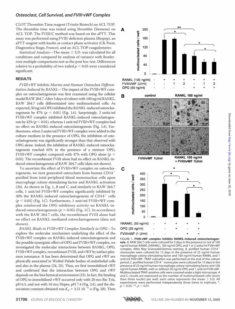
The binding of FVIII�vWF complex to immobilized RANKLwas then investigated in the pH 7.4 buffer. Surprisingly,FVIII�vWF complex was able to bind to immobilized RANKL(response of 150 RU), whereas recombinant FVIII was not (Fig.3A). However, when using an immobilized-vWF sensor chip,no binding was observed whatever the biochemical parametersused (20mMBis-Tris, pH6.5, or 10mMHepes, pH7.4) (data notshown). As for the binding of OPG to the FVIII�vWF complex,we performed an ELISA assay using a coating of FVIII�vWFcomplex. As shown in Fig. 3B, RANKL was able to bind toFVIII�vWF complex in a dose-dependent manner, confirmingthe results obtained by surface plasmon resonance experi-ments. Thus, the present data demonstrated for the first timethe capacity of FVIII�vWF complex to bind RANKL. However,in contrast toOPG, for which the interaction with this complexis done via the vWF, our results suggested that the tridimen-sional structure of the FVIII�vWF complex is mandatory for itsinteractionwithRANKL.To further explore the involvement ofthe FVIII�vWF complex in the RANKL/OPG interactions, theeffect of a preincubation of OPG (100 ng/ml) and increasing
concentrations of FVIII�vWF complex for 2 hwas assessed. Thepre-formed FVIII�vWF complex�OPG complex was theninjected over the immobilized RANKL (Fig. 3C). This experi-ment revealed that the pre-formed complex FVIII�vWF/OPGdid not prevent the binding ofOPG toRANKLor the binding ofFVIII�vWF to RANKL. Furthermore, the binding of OPG washigher in the presence of FVIII�vWF complex than without thiscomplex. These results suggest that the FVIII�vWF complex, bybinding to RANKL or OPG, induced somemodifications in thethree-dimensional structure of OPG, RANKL, or FVIII�vWF,resulting in a higher affinity between OPG and RANKL andthen potentially increasing its biological activity. Such hypoth-esis was supported by the synergistic effect of OPG-FVIII�vWFcomplex observed on RAW 264.7 cells (Fig. 1A). Similarly, Fig.3D showed that FVIII�vWF complex was still able to bindRANKL or OPG even if RANKL had already been bound toimmobilized-OPG, demonstrating that these three moleculescan interact together without interfering the binding of one toanother (see Fig. 3, c and d, for schematic explanations). Thesame results were observed using immobilized vWF; indeed, a
FIGURE 4. TRAIL binds to FVIII without affecting TRAIL/OPG interactions. A, recombinant Factor VIII and FVIII�vWF complex bind to immobilized-TRAIL.Recombinant Factor VIII (50 units/ml) or FVIII�vWF complex (50 units/ml) were injected at a flow rate of 30 �l/min over the immobilized-TRAIL for 2 minassociation. B, recombinant Factor VIII bound to TRAIL does not interfere the binding of TRAIL to immobilized-OPG. 50 units/ml recombinant Factor VIII waspreincubated with TRAIL for 2 h at room temperature. The complex formed was then injected to immobilized-OPG. C, FVIII�vWF complex inhibits the bindingof OPG to TRAIL. FVIII�vWF complex was incubated with OPG for 2 h at room temperature; the new complex formed was then injected at a flow rate of 30 �l/minfor 2 min over immobilized-TRAIL. D, schematic representation of plasmon resonance experiments a, b, and c summarized respectively A, B, and C.
Osteoclast, Cell Survival, and FVIII�vWF Complex
NOVEMBER 13, 2009 • VOLUME 284 • NUMBER 46 JOURNAL OF BIOLOGICAL CHEMISTRY 31709
by guest on June 20, 2018http://w
ww
.jbc.org/D
ownloaded from

pre-formed complex OPG/RANKL was able to bind to immo-bilized vWF in the sameway as OPG alone (see the supplemen-tal figure).TRAIL Binds to FVIII without Affecting TRAIL/OPG
Interactions—OPG is not only a decoy receptor for RANKL butalso acts as soluble receptor for TRAIL and, thus, inhibits itsproapoptotic activity (8, 10, 11). To determine whether or notthe FVIII�vWF complex could affect the complex OPG/TRAIL,TRAIL has been immobilized on a sensor chip, and the capacityof FVIII�vWF complex to bind to TRAIL was analyzed. In con-trast to the previous experiments with OPG and RANKL, bothFVIII�vWF complex and recombinant FVIII were able to bindto immobilized TRAIL (Fig. 4A and summarized in Fig. 4D).Furthermore, as for OPG binding to immobilized-vWF, onlyspecific biochemical conditions of 20 mM Bis-Tris, pH 6.5,allowed the binding of TRAIL to immobilized vWF (no bindingwith 10 mM Hepes, pH 7.4) (data not shown). We confirmedthat TRAIL bound to immobilized-OPG and showed that thecomplex formed byTRAIL and recombinant FVIII was still ableto bind similarly to OPG (Fig. 4B). To further explore theinvolvement of the FVIII�vWF complex in the OPG/TRAILinteractions, the effect of a preincubation of OPG and increas-ing concentrations of FVIII�vWF has been investigated. What-ever the concentration of FVIII�vWF complex used to form acomplex with OPG, all these combinations completely inhib-ited the binding of OPG to TRAIL (Fig. 4C and summarized inFig. 4D). These results suggested that the binding domains ofOPG to vWF and TRAIL is very closed, and one moleculebound to OPG can then block the binding sites of the second.To investigate the relevance of this inhibition in a biological
experiment, we performed a viability assay on the humanosteo-sarcoma cell line MG63 sensitive to TRAIL-induced apoptosis.As shown in Fig. 5A, the ability of TRAIL to induce MG63 celldeath (�75%, p � 0.01) was prevented by the addition of OPG.In contrast, when 1 unit/ml FVIII�vWF complex was added tothe culture medium, OPG was not able to prevent the capacityofTRAIL to induceMG63 cell death (�60%,p� 0.05). Further-more, the apoptotic effect of TRAIL was confirmed even in thepresence of FVIII�vWF complex and OPG. Nucleus fragmenta-tion was observed in MG63 (Fig. 5B). In the same manner of50 ng/ml TRAIL, the combination TRAIL � FVIII�vWF com-plex�OPG induced nucleus fragmentation, as the cells exhibiteda characteristic kidney-like form with condensed chromatinclumps compared with control cells. Moreover, TRAIL induceda significant increase of caspase-3 activity inMG63cells (p�0.01)(Fig. 5C) which was significantly reduced in the presence of OPG.But the caspase-3 activity was not decreased by OPG whenFVIII�vWF complex was added (p � 0.05). The same results ofviability,Hoechst staining, and caspase-3 activationwere obtainedusingothercell lines sensitive toTRAIL-inducedapoptosis suchasthe human osteosarcoma cell line SaOS2 and the human Ewing’ssarcomacell lineTC71 (datanot shown).Thus, thesedata revealedthat the inhibitory effect ofOPGonTRAIL-inducedapoptosis canbe reversed by FVIII�vWF complex and then evidenced the role ofFVIII�vWF complex in the control of cell death.Recombinant Human OPG, RANKL, and TRAIL Do Not
Affect the Coagulation Cascade—Because of the different inter-actions evidenced in our study between OPG, RANKL, TRAIL,
and the FVIII�vWF complex, we evaluated the potential impli-cations of these three molecules on the coagulation cascade.We demonstrated that 100 ng/ml OPG, RANKL, and TRAILhave no effect on the following assays: primary hemostasis,
FIGURE 5. FVIII�vWF complex blocks the inhibitory effect of OPG on TRAIL-induced apoptosis on MG63 cells. A, osteosarcoma cell line MG63 was culturedwith 100 ng/ml TRAIL�100 ng/ml OPG�1 unit/ml FVIII�vWF complex. After 72 hof culture, cell viability was determined by XTT assay. Results were expressed aspercentage of control. Experiments were performed at least three times (*, p �0.05). B, nuclear morphological changes induced by TRAIL, OPG, and FVIII�vWFcomplex were analyzed by Hoechst staining on MG63 cells. C, caspase-3 activitywas assessed on MG63 cells after 16 h of treatment with TRAIL, OPG, andFVIII�vWF complex (*, p � 0.05; **, p � 0.01).
Osteoclast, Cell Survival, and FVIII�vWF Complex
31710 JOURNAL OF BIOLOGICAL CHEMISTRY VOLUME 284 • NUMBER 46 • NOVEMBER 13, 2009
by guest on June 20, 2018http://w
ww
.jbc.org/D
ownloaded from

Quick time, aPTT, thrombin time, and the FVIII/C methodbased on the aPTT (data not shown).
DISCUSSION
FVIII associated with the vWF is a key protagonist of thecoagulation process as evidenced in patients suffering fromhemophilia A. Recent papers revealed the physical interactionbetween vWF and OPG (24, 25), a powerful inhibitor of oste-oclastogenesis and, therefore, of bone resorption (7). Althoughsevere hemophilia patients have also joint diseases, to ourknowledge there is no evidence of the effect of FVIII�vWF com-plex on bone cells and especially on osteoclasts. The presentwork demonstrates that FVIII�vWF complex binds to OPG andRANKL and, thus, indirectly participates to bone biology. Thispaper is, thus, the first evidence that FVIII�vWF complex inhib-its RANKL-induced osteoclastogenesis. Furthermore, in a sec-ond part of the manuscript, we also demonstrated for the firsttime that the FVIII�vWF complex abolishes the inhibitory effectof OPG on TRAIL-induced apoptosis, suggesting a potentialfunction of FVIII�vWF complex in cancer development (Fig. 6).In two different models FVIII�vWF complex regulates oste-
oclastogenesis by inhibiting the pro-osteoclastic activity ofRANKL.Twodifferent effects can be involved in this inhibition;the first way of inhibition occurs through a physical interactionbetween FVIII�vWF complex and RANKL, leading to an inacti-vation of RANKL, and the second potential effect is a synergiceffect of the FVIII�vWF complex with OPG. In fact, both mole-cules inhibit RANKL-induced osteoclastogenesis by them-selves, but their association in the culture medium increasedthis inhibitory effect. However, different mechanisms could beproposed. OPG could bind to the FVIII�vWF complex through
the vWF, and this complex couldincrease the affinity of OPG toRANKL, or the complex FVIII�vWFcould bind to both RANKL andOPG, leading to a stronger inhibi-tion of RANKL activity.These interactions between
FVIII�vWF complex, OPG, andRANKL point out their potentialinvolvement in bone and vascularsystem (7). Indeed, the hallmark ofsevere hemophilia is repeatedbleedings into joints and musclesresulting in a severe and painfulinflammation of synovitis namedhemophilic synovitis (35, 36). How-ever, the exact mechanism relatedto blood-induced joint disease is notprecisely known even if somemech-anisms are now settled. The pro-cesses that occur at the early stagesof blood-induced joint disease asso-ciated with infiltration of inflamma-tory cells releasing high amounts ofinflammatory cytokines, enzymes(36), proteins such as hemoglobin,and an increase of intra-articular
pressure and synovial proliferation. The later stages are charac-terized by a promotion of angiogenesis, cartilage cell apoptosis,and subchondral bone destruction. Thus, hemophilic arthrop-athy shares several biological featureswith rheumatoid arthritis(37). Numerous studies in rheumatoid arthritis models haveproduced evidence for a causal role of excessive RANKL activ-ity in associated-bone loss (38). Indeed, RANKL levels wereconcomitantly increased in inflamed joint leading to anincrease in the RANKL/OPG ratio, which appears positivelycorrelated with bone destruction and osteoclast activity (39).The present data evidenced for the first time that FVIII�vWFcomplex inhibits RANKL-induced osteoclastogenesis. More-over FVIII�vWF complex did not abolish OPG activity on oste-oclastogenesis but reinforced its activity in murine and humanmodels. In this context hemophilic arthritis may be associatedwith an intra-articular inflammatory process concomitantlywith an increased osteoclastogenesis due to a deficiency ofFVIII�vWF complex.OPG/RANK/RANKL triad constitutes a molecular bridge
spanning bonemetabolism, vascular biology, and immunity (7).The first evidence linking the OPG/RANK/RANKL system tothe vessel biology has been provided by the vascular phenotypeofOPG-deficientmice (40). Indeed,OPG-deficientmice exhib-ited medial calcification of the aorta and renal arteries and notof smaller vessels, suggesting that OPG and its molecular part-ners may play a role in the long term-observed associationbetween osteoporosis and vascular calcification (40). OPGphysically associated with the vWF is localized in the Weibel-Palade bodies of endothelial cells and is rapidly secreted inresponse to inflammatory stimuli (24).More recently, in a case-control study Bilora et al. (41) assessed the presence of athero-
FIGURE 6. Schematic representation describing the involvement of FVIII�vWF complex in coagulationcascade, bone, and cancer biology. FVIII�vWF complex is one of the main complex involved in coagulation;FVIII is released from vWF by the action of thrombin and becomes a cofactor for Factor IX to stimulate coagu-lation cascade, whereas vWF is essential in platelet activation. FVIII�vWF complex also plays a major role in otherbiological processes. Indeed, this complex inhibits RANKL-induced osteoclastogenesis by binding to RANKLand also by increasing the anti-osteoclastic activity of OPG. Furthermore, FVIII�vWF complex may be involved incell apoptosis (endothelial, bone, and cancer cells); through its binding to OPG, the FVIII�vWF complex inhibitsthe OPG protective effect on TRAIL-induced apoptosis that occurs in inflammation and cancer disorders.
Osteoclast, Cell Survival, and FVIII�vWF Complex
NOVEMBER 13, 2009 • VOLUME 284 • NUMBER 46 JOURNAL OF BIOLOGICAL CHEMISTRY 31711
by guest on June 20, 2018http://w
ww
.jbc.org/D
ownloaded from

sclerosis in 50 patients suffering from hemophilia and in 50age-matched control individuals. Their results suggest thathemophilia could protect against asymptomatic atherosclero-sis. Overall, these observations strongly support that theOPG/RANK/RANKL and FVIII�vWF systems constitute amolecular cascade essential in the development of athero-sclerotic lesions. Furthermore, our present work gives a bio-logically direct relationship between the FVIII�vWF systemand osteoclastic cells, strengthening the interests of prophy-laxis in young patients suffering from severe hemophilia.Even if prophylaxis seems to be the best therapeutic optionfor severe hemophilia A to prevent joint damages in evi-dence-based medicine (42, 43), these results give a basicexplanation for the effect of prophylaxis in joint damage andsubchondral bone erosion prevention.The second important result reported in our study is the
control of cell apoptosis by the FVIII�vWF complex. Weobserved in vitro that OPG did not inhibit TRAIL-induced cellapoptosis when FVIII�vWF complex was present in the culturemedium. Physical interactions between the FVIII�vWF com-plex, OPG, and TRAIL were confirmed by surface plasmon res-onance experiments. We showed that the FVIII�vWF complexwas able to bind to TRAIL, and then we demonstrated that,when associated to OPG, FVIII�vWF complex prevented thebinding TRAIL/OPG, correlating the in vitro apoptosis exper-iment. To our knowledge, the functional relationship betweenFactor VIII and/or vWF and apoptosis has never been investi-gated. TRAIL is a cytotoxic ligand that binds to type I trans-membrane receptors (DR4 andDR5) possessing death domainsand which ultimately activates the caspase cascade, inducingcell death (44). TRAIL also has two decoy receptors (DcR1,DcR2) that lack a functional death domain and explain in partthe absence ofmassive apoptosis in cells that express functionalmembrane receptor (45). However, normal and cancer cellslacking these decoy receptors can escape to cell death throughthe expression of OPG, which is able to block TRAIL transduc-tion signaling (8). It is well established that the coagulationcascade contributes to cancer development (46), and a clearcorrelation between thrombosis and cancer progression hasbeen established. Indeed, tissue factor is up-regulated on bothtumor and host cells in cancer patients and initiates protease-activated receptor-mediated cell signaling that leads to the pro-duction of soluble cytokines and angiogenic growth factors(47). More recently, Ho-Tin-Noe et al. demonstrated thatplatelets support tumor vascular homeostasis by regulating thestability of tumor vessels (48). Thus, tumor development ap-pears as the equilibrium between cell proliferation and celldeath actively controlled by blood vessels and coagulation cas-cade. By reversing the inhibitory effect of OPG on TRAIL-induced apoptosis, FVIII�vWF may control tumor growth.Hemophilia A has been recently reported after tumor resec-tion in patients suffering from glioblastoma (49), and it hasbeen suggested that cancer cells could produce factor VIII-like tumor antigens. Such hypothesis has been also strength-ened by Franchini et al. (50), who recently reviewed theacquired factor VIII inhibitors in oncohematology. If the originof such nonclassical antibodies against FVIII is not yet defined,these autoantibodies may complicate the clinical course of the
malignancy (51). All these data associated with the presentwork are in favor of a contribution of FVIII�vWF complex dur-ing cancer disorders. Then the interaction between OPG-TRAIL-FVIII�vWF complex may be involved in induced cellapoptosis (endothelial, cartilage, bone, and tumor cells), whichis essential during angiogenesis associated with inflammationand cancer disorders.
REFERENCES1. Kong, Y. Y., Yoshida, H., Sarosi, I., Tan, H. L., Timms, E., Capparelli, C.,
Morony, S., Oliveira-dos-Santos, A. J., Van, G., Itie, A., Khoo, W., Wake-ham, A., Dunstan, C. R., Lacey, D. L., Mak, T. W., Boyle, W. J., and Pen-ninger, J. M. (1999) Nature 397, 315–323
2. Theoleyre, S.,Wittrant, Y., Tat, S. K., Fortun, Y., Redini, F., and Heymann,D. (2004) Cytokine Growth Factor Rev. 15, 457–475
3. Burgess, T. L., Qian, Y., Kaufman, S., Ring, B. D., Van, G., Capparelli, C.,Kelley, M., Hsu, H., Boyle, W. J., Dunstan, C. R., Hu, S., and Lacey, D. L.(1999) J. Cell Biol. 145, 527–538
4. Hsu, H., Lacey, D. L., Dunstan, C. R., Solovyev, I., Colombero, A., Timms,E., Tan, H. L., Elliott, G., Kelley,M. J., Sarosi, I.,Wang, L., Xia, X. Z., Elliott,R., Chiu, L., Black, T., Scully, S., Capparelli, C.,Morony, S., Shimamoto, G.,Bass, M. B., and Boyle, W. J. (1999) Proc. Natl. Acad. Sci. U.S.A. 96,3540–3545
5. Tsuda, E., Goto, M., Mochizuki, S., Yano, K., Kobayashi, F., Morinaga, T.,and Higashio, K. (1997) Biochem. Biophys. Res. Commun. 234, 137–142
6. Simonet, W. S., Lacey, D. L., Dunstan, C. R., Kelley, M., Chang, M. S.,Luthy, R., Nguyen, H. Q., Wooden, S., Bennett, L., Boone, T., Shimamoto,G., DeRose, M., Elliott, R., Colombero, A., Tan, H. L., Trail, G., Sullivan, J.,Davy, E., Bucay, N., Renshaw-Gegg, L., Hughes, T. M., Hill, D., Pattison,W., Campbell, P., Sander, S., Van, G., Tarpley, J., Derby, P., Lee, R., andBoyle, W. J. (1997) Cell 89, 309–319
7. Baud’huin, M., Lamoureux, F., Duplomb, L., Redini, F., and Heymann, D.(2007) Cell. Mol. Life Sci. 64, 2334–2350
8. Emery, J. G., McDonnell, P., Burke, M. B., Deen, K. C., Lyn, S., Silverman,C., Dul, E., Appelbaum, E. R., Eichman, C., DiPrinzio, R., Dodds, R. A.,James, I. E., Rosenberg,M., Lee, J. C., and Young, P. R. (1998) J. Biol. Chem.273, 14363–14367
9. Degli-Esposti, M. (1999) J. Leukocyte Biol. 65, 535–54210. Wiley, S. R., Schooley, K., Smolak, P. J., Din, W. S., Huang, C. P., Nicholl,
J. K., Sutherland, G. R., Smith, T. D., Rauch, C., and Smith, C. A. (1995)Immunity 3, 673–682
11. Pitti, R. M., Marsters, S. A., Ruppert, S., Donahue, C. J., Moore, A., andAshkenazi, A. (1996) J. Biol. Chem. 271, 12687–12690
12. Hollestelle, M. J., Thinnes, T., Crain, K., Stiko, A., Kruijt, J. K., van Berkel,T. J., Loskutoff, D. J., and van Mourik, J. A. (2001) Thromb. Haemost. 86,855–861
13. Bolton-Maggs, P. H., and Pasi, K. J. (2003) Lancet 361, 1801–180914. Sadler, J. E. (2005) Annu. Rev. Med. 56, 173–19115. Nichols, W. L., Hultin, M. B., James, A. H., Manco-Johnson, M. J., Mont-
gomery, R. R., Ortel, T. L., Rick, M. E., Sadler, J. E., Weinstein, M., andYawn, B. P. (2008) Haemophilia 14, 171–232
16. Suva, L. J., Hartman, E., Dilley, J. D., Russell, S., Akel, N. S., Skinner, R. A.,Hogue, W. R., Budde, U., Varughese, K. I., Kanaji, T., and Ware, J. (2008)Am. J. Pathol. 172, 430–439
17. Kovacs, C. S. (2008) Transfus. Apher Sci. 38, 33–4018. Wallny, T. A., Scholz, D. T., Oldenburg, J., Nicolay, C., Ezziddin, S., Pen-
nekamp, P. H., Stoffel-Wagner, B., and Kraft, C. N. (2007) Haemophilia13, 79–84
19. Lollar, P., Hill-Eubanks, D. C., and Parker, C. G. (1988) J. Biol. Chem. 263,10451–10455
20. Dumas, J. J., Kumar, R., McDonagh, T., Sullivan, F., Stahl, M. L., Somers,W. S., and Mosyak, L. (2004) J. Biol. Chem. 279, 23327–23334
21. Adachi, T., Matsushita, T., Dong, Z., Katsumi, A., Nakayama, T., Kojima,T., Saito, H., Sadler, J. E., and Naoe, T. (2006) Biochem. Biophys. Res.Commun. 339, 1178–1183
22. Maita, N., Nishio, K., Nishimoto, E., Matsui, T., Shikamoto, Y., Morita, T.,Sadler, J. E., and Mizuno, H. (2003) J. Biol. Chem. 278, 37777–37781
Osteoclast, Cell Survival, and FVIII�vWF Complex
31712 JOURNAL OF BIOLOGICAL CHEMISTRY VOLUME 284 • NUMBER 46 • NOVEMBER 13, 2009
by guest on June 20, 2018http://w
ww
.jbc.org/D
ownloaded from
23. Fukuda, K., Doggett, T. A., Bankston, L. A., Cruz, M. A., Diacovo, T. G.,and Liddington, R. C. (2002) Structure 10, 943–950
24. Zannettino, A. C., Holding, C. A., Diamond, P., Atkins, G. J., Kostakis, P.,Farrugia, A., Gamble, J., To, L. B., Findlay, D. M., and Haynes, D. R. (2005)J. Cell. Physiol. 204, 714–723
25. Shahbazi, S., Lenting, P. J., Fribourg, C., Terraube, V., Denis, C. V., andChristophe, O. D. (2007) J. Thromb. Haemost. 5, 1956–1962
26. Duplomb, L., Baud’huin, M., Charrier, C., Berreur, M., Trichet, V., Blan-chard, F., and Heymann, D. (2008) Endocrinology 149, 3688–3697
27. Altschul, S. F., Gish,W.,Miller,W.,Myers, E.W., and Lipman, D. J. (1990)J. Mol. Biol. 215, 403–410
28. Larkin, M. A., Blackshields, G., Brown, N. P., Chenna, R., McGettigan,P. A., McWilliam, H., Valentin, F., Wallace, I. M., Wilm, A., Lopez, R.,Thompson, J. D., Gibson, T. J., and Higgins, D. G. (2007) Bioinformatics23, 2947–2948
29. Clamp, M., Cuff, J., Searle, S. M., and Barton, G. J. (2004) Bioinformatics20, 426–427
30. Sali, A., and Blundell, T. L. (1993) J. Mol. Biol. 234, 779–81531. Mongkolsapaya, J., Grimes, J. M., Chen, N., Xu, X. N., Stuart, D. I., Jones,
E. Y., and Screaton, G. R. (1999) Nat. Struct. Biol. 6, 1048–105332. Ito, S., Wakabayashi, K., Ubukata, O., Hayashi, S., Okada, F., and Hata, T.
(2002) J. Biol. Chem. 277, 6631–663633. Humphrey, W., Dalke, A., and Schulten, K. (1996) J. Mol. Graph. 14,
33–38, 27–2834. Cheng, X., Kinosaki, M., Takami, M., Choi, Y., Zhang, H., and Murali, R.
(2004) J. Biol. Chem. 279, 8269–827735. De Palma, A. F., and Cotler, J. M. (1956) AMA Arch. Surg. 72, 247–25036. Valentino, L. A., Hakobyan, N., Rodriguez, N., and Hoots, W. K. (2007)
Haemophilia 13, 10–1337. Lafeber, F. P., Miossec, P., and Valentino, L. A. (2008) Haemophilia 14,
3–938. Kearns, A. E., Khosla, S., and Kostenuik, P. J. (2008) Endocr. Rev. 29,
155–19239. Bolon, B., Campagnuolo, G., and Feige, U. (2002) Cell. Mol. Life Sci. 59,
1569–157640. Bucay, N., Sarosi, I., Dunstan, C. R., Morony, S., Tarpley, J., Capparelli, C.,
Scully, S., Tan, H. L., Xu,W., Lacey, D. L., Boyle, W. J., and Simonet, W. S.(1998) Genes Dev. 12, 1260–1268
41. Bilora, F., Zanon, E., Petrobelli, F., Cavraro, M., Prandoni, P., Pagnan, A.,and Girolami, A. (2006) Clin. Appl. Thromb. Hemost. 12, 193–198
42. Astermark, J., Petrini, P., Tengborn, L., Schulman, S., Ljung, R., and Bern-torp, E. (1999) Br. J. Haematol. 105, 1109–1113
43. Manco-Johnson, M. J., Abshire, T. C., Shapiro, A. D., Riske, B., Hacker,M. R., Kilcoyne, R., Ingram, J. D., Manco-Johnson, M. L., Funk, S., Jacob-son, L., Valentino, L. A., Hoots, W. K., Buchanan, G. R., DiMichele, D.,Recht, M., Brown, D., Leissinger, C., Bleak, S., Cohen, A., Mathew, P.,Matsunaga, A., Medeiros, D., Nugent, D., Thomas, G. A., Thompson,A. A., McRedmond, K., Soucie, J. M., Austin, H., and Evatt, B. L. (2007)N. Engl. J. Med. 357, 535–544
44. Johnstone, R. W., Frew, A. J., and Smyth, M. J. (2008) Nat. Rev. Cancer 8,782–798
45. Pan, G., Ni, J., Wei, Y. F., Yu, G., Gentz, R., and Dixit, V. M. (1997) Science277, 815–818
46. Dogan, M., and Demirkazik, A. (2005) Support Cancer Ther. 3, 28–3447. Pawlinski, R., and Mackman, N. (2008) Semin. Thromb. Hemostasis 34,
182–18648. Ho-Tin-Noe, B., Goerge, T., Cifuni, S. M., Duerschmied, D., andWagner,
D. D. (2008) Cancer Res. 68, 6851–685849. van Durme, C.M., Idema, R. N., and van Guldener, C. (2008)Neth. J. Med.
66, 286–28850. Franchini, M., Targher, G., Manzato, F., and Lippi, G. (2008) Crit. Rev.
Oncol. Hematol. 66, 194–19951. Meeks, S. L., Healey, J. F., Parker, E. T., Barrow, R. T., and Lollar, P. (2008)
Blood 112, 1151–1153
Osteoclast, Cell Survival, and FVIII�vWF Complex
NOVEMBER 13, 2009 • VOLUME 284 • NUMBER 46 JOURNAL OF BIOLOGICAL CHEMISTRY 31713
by guest on June 20, 2018http://w
ww
.jbc.org/D
ownloaded from
Maillasson, Marc Fouassier and Dominique HeymannMarc Baud'huin, Laurence Duplomb, Stéphane Téletchéa, Céline Charrier, Mike
Controls Cell SurvivalFactor VIII-von Willebrand Factor Complex Inhibits Osteoclastogenesis and
doi: 10.1074/jbc.M109.030312 originally published online September 16, 20092009, 284:31704-31713.J. Biol. Chem.
10.1074/jbc.M109.030312Access the most updated version of this article at doi:
Alerts:
When a correction for this article is posted•
When this article is cited•
to choose from all of JBC's e-mail alertsClick here
Supplemental material:
http://www.jbc.org/content/suppl/2009/09/16/M109.030312.DC1
http://www.jbc.org/content/284/46/31704.full.html#ref-list-1
This article cites 51 references, 14 of which can be accessed free at
by guest on June 20, 2018http://w
ww
.jbc.org/D
ownloaded from