factors important for persistence of lactobacillus reuteri ...¥th.pdf · that were induced in heat...
TRANSCRIPT

1
Factors Important for Persistence of Lactobacillus reuteri in the
Gastrointestinal Tract
A Study of Extracellular Proteins, Stress Response and Survival of Mutants in a Model System
Klara Båth Faculty of Natural Resources and Agricultural Sciences
Department of Microbiology Uppsala
Doctoral thesis Swedish University of Agricultural Sciences
Uppsala 2007

2
Acta Universitatis Agriculturae Sueciae 2007:22 ISSN 1652-6880 ISBN 978-91-576-7321-3 © 2007 Klara Båth, Uppsala Tryck: SLU Service/Repro, Uppsala 2007

3
Abstract
Båth, K. 2007. Factors Important for Persistence of Lactobacillus reuteri in the Gastrointestinal Tract - A Study of Extracellular Proteins, Stress Response and Survival of Mutants in a Model System. ISSN:1652-6880, ISBN:978-91-576-7321-3 Lactobacillus reuteri ATCC 55730 is a commercially available probiotic i.e. health promoting bacterium. The interest for lactic acid bacteria (LAB) as probiotics might be due to the tradition of use in food preservation, their non-pathogenic behavior and, a natural occurrence in the human gastrointestinal (GI) tract. Although examples of clinically proven probiotic effects are increasing, the understandings of bacterial-host interactions behind these effects are only beginning to be unravelled. To put this thesis work into context, the field of probiotic research is described and health promoting mechanisms and perspectives of strain selection are discussed. The experimental work has focused on properties of L. reuteri ATCC 55730 that are tentatively important for persistence of this and other probiotic strains in the GI tract. To get an idea of the proteins on the surface of L. reuteri ATCC 55730, a draft genome sequence was scrutinized using bioinformatic tools. This resulted in the identification of a diverse set of 126 putative proteins with different possible means of surface attachment. The bacterium was also exposed to heat and acid stress and the response monitored by DNA microarrays. Acid stress is a well-documented way of investigating probiotic strains, since survival in the acidic GI tract is considered a prerequisite for a probiotic effect. Heat stress could be considered an artificially constructed environment for a probiotic bacterium, but is probably still relevant in elucidating mechanisms of persistence in other stressful conditions such as the passage of the GI tract. To further investigate genes revealed in the bioinformatical and DNA microarray analysis as well as a set of genes from the seven earlier identified two-component systems of L. reuteri ATCC 55730, 14 genes were mutated. These where then evaluated for ecological performance using a three-stage continuous culture system simulating a human colon using a real time PCR approach to monitor the relative abundance of the individual mutants. Two caseinolytic protease (Clp) genes, clpE and clpL that were induced in heat and acid stress respectively, both showed importance for persistence in the colon model. Keywords: Lactobacillus reuteri, probiotics, heat stress, acid stress, surface proteins, gastrointestinal tract, survival, persistence, mutants, Clp Author's address: Klara Båth, Department of Microbiology, SLU, Box 7025, SE-750 07 Uppsala, Sweden

4
Kanske tandkräm också…. man vill ju göra nåt fint eller åtminstone ville jag det något som skulle stå kvar en extra speciell tandborste med tandkräm kvar som människorna i nästa tid skulle vilja gå o titta på titta mamma! Där är bob hanssons tandborste jag ville liksom duga sätta ut något fint i gräset som en sång mellan träden för barn o ungar att nynna med i men det vet ju alla att så kan man inte hålla på /Bob Hansson (Halleluja liksom)

5
Contents
Introduction 7 Objectives and outline of the thesis 8 Microbial ecology of the gastrointestinal tract 9 The gastrointestinal tract and the indigenous microbiota 9 Disorders related to the microbiota of the gastrointestinal tract 11 Lactic acid bacteria & probiotics 13 Effects of lactic acid bacteria 13 Probiotic strain selection 14 Probiotic research: Mechanisms known today and future perspectives 16 Lactobacillus reuteri ATCC 55730 17 Interaction of bacteria with the environment 18 Surface proteins (I) 19 Responses to changes in the environment 23
Heat stress response (II) 27 Acid stress response (III) 29
Biological importance of specific genes 32 Construction of mutants 32 Evaluation of mutants in an artificial colon (IV) 34
Concluding remarks 36 References 38 Acknowledgements 49

6
Appendix
Papers I-IV This thesis is based on the following papers, which are referred to in the text by their Roman numerals:
I. Båth, K., Roos, S., Wall, T. & Jonsson, H. 2005. The cell surface of Lactobacillus reuteri ATCC 55730 highlighted by identification of 126 extracellular proteins from the genome sequence. FEMS Microbiology Letters 253, 75-82.
II. Båth, K., Wall, T. Jonsson, H. & Roos, S. Heat stress response in
Lactobacillus reuteri ATCC 55730. Submitted III. Wall, T., Båth, K., Britton, R., Jonsson, H., Versalovic, J. & Roos, S.
The early response to acid shock in Lactobacillus reuteri involves the ClpL chaperone and a putative cell wall altering esterase. Submitted
IV. Båth, K., Costabile, A., Goulas, T., Jonsson, H., & Roos, S. Fourteen
genes of Lactobacillus reuteri ATCC55730 putatively important for persistence in the intestine mutated and analysed in a artificial colon. Manuscript.
My contribution to the papers included in this thesis has been as follows:
I. Took part in bioinformatical analysis and writing of the paper. II. Major part in planning, laboratory work and writing of the paper. III. Took part in planning and performance. IV. Major part in planning and laboratory work. Major contribution to
writing together with S. Roos and H. Jonsson.

7
Introduction
Once upon a time, before food was easily accessible in grocery stores, people were more frequently exposed in their every day life to bacteria, both the pathogens and those that live in harmony with us. Since, e.g. of the oldest ways of preserving food is through naturally occurring fermentation (Ross, Morgan & Hill, 2002). However, in the industrialised part of the world we are now more prone to throw away food, since we can afford to buy new fresh groceries. Thus people in the past provided their gastrointestinal (GI) tract with vast amounts of microorganisms, both good and bad, on a daily basis. Today our surroundings are far more sterile and we encounter fewer microorganisms in our everyday life (Isolauri et al., 2002). Many of us no longer consume as much naturally fermented foods such as sauerkraut, fermented milks, pickles, sourdough bread etc. Instead we consume a great deal of white bread and pasta, made from cereals, and a lot of sugar. Cereals did not become part of the staple diet until cultivation of crops begun (about 10 000 years ago). Prior to that, we consumed more meat, fish, eggs, fruit, vegetables and roots. Diet differs greatly between different areas of the world due to climate, culture and the economy. In Sweden cereals along with beans, dairy products, cooking fat and sugar currently constitute more than 70% of the daily calorie intake (Lindeberg, 2005). A question is whether this influences our health today and also if our digestive system is influenced. Could we possibly benefit from a dietary supplement to correct a disturbance of this system? In the Western society today different diets are frequently introduced, which we are told we benefit from in different ways. There is e.g. the paleolithic diet (or the hunter/gatherer diet) that for instance is claimed to give a lower blood pressure and higher insulin sensitivity than the today normal cereal based diet (Jönsson et al., 2006). There are also people with an anthroposophic lifestyle, with a diet comprising vegetables fermented naturally by lactobacilli (Alm et al., 2002). A lot of studies show that by adding a daily dose of a bacterial culture to our diet we can promote health. An anthroposophic lifestyle has e.g. shown to reduce the risk for allergy in children (Alm et al., 1999; Flöistrup et al., 2006). The interest in microorganisms as health-promoting additives to our diet began already 100 years ago (Metchnikoff, 1907), and live microorganisms that show positive health-promoting properties are today termed probiotics (Fuller, 1986). The possibility that certain microbes might be beneficial to human health is highlighted by numerous consumer products containing probiotic bacteria (Marco, Pavan & Kleerebezem, 2006). This thesis focuses on the lactic acid bacterium (LAB) Lactobacillus reuteri ATCC 55730 used in various probiotic products and shown to have beneficial effects on human health (Tubelius, Stan & Zachrisson, 2005; Weizman, Asli & Alsheikh, 2005). How can a single bacterial strain have an impact on our health and well-being even though the microbiota of our gut is very complex, consisting of approximately 800 species and more than 7000 strains (Bäckhed et al., 2005)? An important factor could be that the gut mucosa is the major sites for induction and regulation of our immune system (Guarner, 2006). In humans about 60% of the total immunoglobin produced is secreted into the GI tract (Brandtzaeg et al., 1989), and about 70% of our immune system is estimated

8
to be located in the gut (Bengmark, 2002). Perhaps this provides an opportunity for a small change in the microbial composition to have a large impact. However, many of the details behind the beneficial effects of a single probiotic strain are still unknown, and basic knowledge of intestinal bacteria and their interactions with other microorganisms and with the host is a prerequisite for successful probiotic research and development (Parvez et al., 2006).
Objectives and outline of the thesis
The work described in this thesis employs basic research to investigate properties of L. reuteri ATCC 55730. The main objective was to identify factors important for gastrointestinal persistence in this commercially available probiotic bacterium. A secondary objective was also to write a thesis that in a, read wise, pleasant way describe the field of probiotic research, discuss health-promoting mechanisms and provide a perspective of strain selection in order to place the research results in a context. The starting point of this thesis is the draft genome sequence of L. reuteri ATCC 55730. In Paper I bioinformatical methods were used to give a putative picture of the extracellular proteins of this bacterium, in order to identify possible factors for host interaction. In Paper II and III the bacterium was exposed to heat and acid stress respectively, and DNA micro arrays were used to investigate the cellular stress response. Acid stress is a well-documented way of investigating probiotic strains, since survival in the acidic upper GI tract is considered a prerequisite for a probiotic effect. Heat stress, on the other hand, is an artificially constructed situation normally not encountered by L. reuteri but can still be used to identify persistence mechanisms used in other possibly stressful environments such as the GI tract. In Paper IV the importance of genes revealed in Papers I-III for ecological performance in an artificial colon was investigated. A schematic view of the work of this thesis is presented in Fig. 1.
Fig. 1. Outline of thesis work. A schematic picture of how Papers I-IV in this thesis are interconnected.

9
Microbial ecology of the gastrointestinal tract
The gastrointestinal tract and the indigenous microbiota The GI tract is specialised to carry out digestion of food (stomach and upper small intestine), absorption of nutrients (small intestine) and water salvage and waste storage (large intestine) (Tannock, 1995). In addition, the intestine is a complex ecological system. The microbiota is in constant communication with the mucosal surface of the intestine and thus with the immune system of its host. The normal interaction between the abundant, diverse and active microbiota of the GI tract and the host is a symbiotic relationship from which both partners benefit (Hooper, Midtvedt & Gordon, 2002). At birth, the GI tract is sterile, but colonization occurs directly after delivery and within a few months a relatively stable microbial population is established (Ewaschuk & Dieleman, 2006; Fanaro et al., 2003). Each individual has a genetic predisposition to being colonized by different organisms. The delivery mode at birth, the environment in which the child is reared, the diet etc. are other factors that determines this individual microbiota. Thus, each person has a particular combination of predominant and subdominant species (Guarner, 2006). The intestine of an adult individual contain 500-800 species, of which 99% is comprised of 30-40 predominant species (Bäckhed, et al., 2005; Hooper, Midtvedt & Gordon, 2002). Actually the number of microbial cells within the gut lumen exceeds the number of cells in the body by a factor of 10 (Bengmark, 2002; Guarner & Malagelada, 2003) and has been estimated to account for 35-50% of the volume of the human colon content (Isolauri, Salminen & Ouwehand, 2004). Current knowledge about the human microbiota is far from complete. Earlier culture-based investigations and more recent 16S rRNA-based studies show temporal and individual variations in the microbial composition of our microbiota (Dethlefsen et al., 2006). The human stomach harbours a relatively small number of microbes. The number is restricted because of the high acidity in the stomach (pH as low as 2). A maximum of 104 microbes per millilitre gastric juice can be detected. The only undisputed inhabitant is Helicobacter pylori, present in the gastric mucosa in some individuals. (Dethlefsen, et al., 2006). However, a recent 16s rRNA study showed a diverse picture, with 128 bacterial species possibly representing both ingested and resident species of the stomach (Bik et al., 2006). Interestingly, a number of lactobacilli, including for new species, were identified from a study of gastric biopsies (Roos, Engstrand & Jonsson, 2005). The transit time from the mouth to the ileum is short, about 4-6 h (Bourlioux et al., 2003), which is also a limiting factor for colonization. However, the number of bacteria increases from about 104 up to about 107 per ml intestinal content in the jejunum. In the ileum the passage of the material for digestion is slower and microbial colonization is facilitated. The microbiota here is diverse and harbours e.g. lactobacilli and enterococci but also gram-negative facultative anaerobic bacteria and obligate anaerobes. A total of about 108 bacteria per millilitre of content is found in this niche (Metchnikoff, 1907). The large intestine is the main site of microbial colonization of the human GI tract. The transit time here is slow, normally about 55 hours (Bourlioux, et al., 2003), and a large number of bacteria are present. The amount of bacteria increase from about 108 to 1011-1012 per ml

10
stool (Dethlefsen, et al., 2006), representing about 800 species (Bäckhed, et al., 2005) (Fig. 2). However, it is worth considering that there is a difference between the bacterial composition of the lumen and the mucosal membrane (Zoetendal et al., 2002), which means that our picture of the GI colonization is even more incomplete.
Fig 2. An illustration of the intestine and the major bacterial groups found in different parts. The microbiota of the GI tract is far from completely described. Data generated so far show temporal and individual variations in the microbial composition. (Illustration of data from Dethlefsen et al. (2006)). By studying animals bred under germ-free conditions important information about the effect of the microbial community on its host can be obtained. The functions of the enteric microbiota can be divided into three groups: metabolic, protective and trophic (Fig. 3.) (Guarner, 2006). It has e.g. observed seen that germ-free animals require 30% more energy in their diet and that supplementation of vitamins K and B is necessary to maintain their body weight (Isolauri, Salminen & Ouwehand, 2004). The mucosal surface of our GI tract serves as a barrier between the microbiota and the host, and as mentioned above the major part of our immune system is situated there. The non-pathogenic bacteria, whether just passing or colonizing, could interact with the host through this mucosal surface and possibly prime or modulate our immune system in a positive manner. The balanced normal intestinal microbiota also strengthens the barrier effect of the mucosal surface, blocking the passage of bacteria and antigens into the blood stream (Parvez, et al., 2006).
Fig. 3. Effect of the microbiota on its host (Source: (Guarner, 2006)).

11
The microbes in our GI tract have the potential to act in a favourable, neutral or deleterious manner in relation to their host. Pathogenic bacteria are often considered to use a highly sophisticated mode of action during infection and the action of pathogenesis has already been thoroughly investigated, probably since when the pathogens strike they cause obvious medical problems. However, it would perhaps be a better survival strategy for a bacterial species to live in symbiosis with its host, i.e. giving something in return instead of merely exploiting. A group of indigenous inhabitants of the human GI tract, thought to be among the dominant inhabitants of the small intestine, are members of lactic acid bacteria (LAB). The most dominant common feature of the LAB is that a major proportion of their metabolic end-products is lactic acid. The organic acids produced tend to lower the pH of the gut (at least locally) and thus create a less desirable environment for harmful bacteria. The LAB also produces antimicrobial compounds (e.g. bacteriocins) with activity against closely related bacteria, but some strains also produce substances such as hydrogen peroxide, reuterin etc. with effects against gastric and intestinal pathogens and other microbes (Ljungh & Wadström, 2006).
Disorders related to the microbiota of the gastrointestinal tract The human GI tract with its enteric microbiota is generally able to respond rapidly to invading pathogens and then return to balance after an infection. When this balanced interaction is disturbed, a chronic intestinal inflammation may occur. Diet, stress and side-effects of modern medical practices have been implicated as factors capable of exerting a negative influence on human health and nutrition (Buret, 2006; Collins, 2001; Kiliaan et al., 1998; McKay, 2005). A possible outcome of these factors could be a deficiency or shift in the equilibrium of the gut microbiota, leading to an increase in chronic intestinal illness in the industrialised world of today. It is well accepted among the general public that emotional stress is a factor that can result in disturbances in intestinal health. Evidence in scientific studies also suggests that stress is a contributing factor in intestinal inflammatory disease, but the mechanisms involved remain to be identified. It is evident from animal work however, that stress is a potent modulator of inflammatory response (Collins, 2001), a cause of disturbance of the barrier effect (Kiliaan, et al., 1998) and it has also been shown that stressful life events often precede disease relapses in patients with intestinal inflammatory disease (Collins, 2001; Kiliaan, et al., 1998). It has been known for about 50 years that people treated with antibiotics have an increased susceptibility to new infections (Bengmark, 2002). Antibiotic-associated diarrhoea is a frequent side-effect of antibiotics, and can affect as many as 25% of patients treated depending on the antibiotic used (Katz, 2006). In the past decade there has been a major increase in cases of antibiotic-associated diarrhoea, but in most cases the cause of the diarrhoea is unclear (Meier, 2005). One of the mechanisms behind these cases of diarrhoea is thought to relate to alteration of the microbiota, leading to a loss of colonization resistance. This predisposes the gut to colonization by potential pathogens such as Clostridium difficile, causing a

12
potentially severe form of toxin-induced colitis that accounts for 15-25% of cases of antibiotic-associated diarrhoea (Katz, 2006). A number of GI disorders connected to the microbiota and their symptoms are presented in Fig. 4.
Fig. 4. A summary of some gastrointestinal disorders connected to the microbiota of the GI tract, based on data from (Broekaert & Walker, 2006; Collins, 2001; Felley & Michetti, 2003). As mentioned above, the enteric microbiota differs greatly between individuals and to date it has been difficult to get a complete picture of the microorganisms in the GI tract since only 60-80% of them have so far been cultivated (Suau et al., 1999). In the current era of genetic techniques we may be able to expand our knowledge about the composition of our microbiota. Molecular biological techniques have shown that the microbial structure of the microbiota differs greatly between individuals (Eckburg et al., 2005). Interestingly, connections between certain microbes or structures of microbial community e.g. and inflammatory disorders of the intestine have been observed (Seksik et al., 2003). This connection makes it interesting to speculate whether it is possible to somehow manipulate the microbiota in a positive manner.

13
Lactic acid bacteria & probiotics
Effects of lactic acid bacteria The idea of yoghurt promoting gut health was raised already by the Ancient Roman historian Pliny the Elder, who wrote '…sour milk is being added to fresh milk which is wanted to curdle. This preparation is extremely wholesome to the stomach…' in Naturalis historiae (Plinius Secundus). In more modern times (100 years ago) Metchnikoff (Metchnikoff, 1907) also declared yoghurt to be a health-promoting additive to the human diet. The health-promoting organisms (responsible for the souring of milk) are today known to be lactic acid bacteria (LAB) and some of them are defined as probiotics. The definition of probiotics is a living microbial food ingredient that has a beneficial effect on human health (Fuller, 1986), and currently there are a large number of probiotic products on the market. The most extensively used and studied probiotic bacteria are the LAB, particularly Lactobacillus but also Bifidobacterium spp. Some species of other genera are also used e.g. Bacillus spp. (Guo et al., 2006; Kim et al., 2006), E. coli Nissle (Kokesova et al., 2006) and the yeast Saccharomyces boulardii (Billoo et al., 2006). The interest in LAB as probiotics might be due to their traditional use in preservation of food and the fact that they are normally not associated with pathogenesis and occur naturally all through the GI tract. It has been shown that probiotics can impart a number of health benefits (modified from Parvez (2006) and Rastall (2005)), such as an ability to:
• synthesise and enhance the bioavailability of nutrients. • modulate the immune system. • reduce symptoms of lactose intolerance. • decrease the prevalence of allergy. • reduce the risk of certain cancers. • reduce the risk of infectious diseases.
Originally, the probiotic effects were primarily thought to be a result of the improvement of the intestinal microbiota. However, many probiotic effects are in fact unlikely to be related to intestinal microbiota changes alone. Other factors include production of vitamins and a possible induction of host mucus production. Indeed, an important part of the beneficial effects of probiotics relates to their immunomodulatory effects; immune-enhancing as well as anti-inflammatory activity (Isolauri, Salminen & Ouwehand, 2004). An example of an interesting probiotic effect is the ability to reduce symptoms of atopic disease. Studies have shown that atopic children have a different microbial community structure than healthy children, in which the level of lactobacilli (Björkstén et al., 1999) and bifidobacteria (Watanabe et al., 2003) is significantly reduced. There is evidence that the incidence of atopic diseases is increasing in industrialised countries with a market economy. The reason is unknown, but it has been suggested that the increase in allergies is connected with a decreased microbial pressure in early childhood (Björkstén, et al., 1999). There is evidence that some probiotic strains prevent allergies. Lactobacillus rhamnosus strain GG supplied to pregnant mothers and in a follow up treatment to the children themselves for 6 months gave

14
a 50% reduction in atopic disease among the children examined at 2 and 4 years of age (Kalliomäki et al., 2001; Kalliomäki et al., 2003). In another study, children with existing, moderate or severe, atopic symptoms were supplied with L. rhamnosus 19070-2 and L. reuteri DSM 12246. The severity of eczema and the frequency of GI symptoms were decreased after 6 weeks of consumption (Rosenfeldt et al., 2004). Lately, evidence that probiotics also play a role in oral health has increased (Meurman, 2005). It has e.g. been demonstrated that consumption of yoghurt containing L. reuteri reduces oral carriage of mutans streptococci (Nikawa et al., 2004) and L. reuteri has also proven to be effective in reducing both gingivitis (inflammation of the gums) and plaque in patients with moderate to severe gingivitis (Krasse et al., 2006). In another recent study oral administration of L. reuteri also improved the symptoms of infantile colic within just a week in 95 % of patients (Savino et al., 2007). LAB has been administered to individuals with inflammatory disorders such as atopic and Crohn's disease as well as those with HIV and immunosuppression. Treatment resulted in a down regulation of over-expressed inflammatory responses and stabilisation of gut mucosal barrier (Rastall, et al., 2005).
Probiotic strain selection Previous criteria when selecting probiotic strains that are still used to some extent today include some or all of the following properties (Chermesh & Eliakim, 2006; Dunne et al., 1999; Ewaschuk & Dieleman, 2006; Parvez, et al., 2006). A probiotic has to be able to:
• withstand processing of the foodstuff and remain viable through the shelf life.
• withstand transit through the GI tract. • adhere to intestinal epithelial cells. • produce antimicrobial substances • modulate the mucosal immune system. • have a beneficial effect on the host. • be of human origin. • be safe (i.e. it must be non-pathogenic, free of transferable antibiotic
resistance determinants, not degrade mucin etc. ). Most probiotic strains on the market today were originally selected on the basis of easily measurable parameters such as the ability to tolerate low pH or large amounts of bile salt or to survive passage through the GI tract. However, the selection of probiotics is perhaps not as straightforward as usually presented. In a human trial on L. plantarum 299v it was shown that irrespective of the subject's gastric acidity, the faecal count of supplemented bacteria reached the same level (Goossens et al., 2005). A higher pH of the GI tract, might not, as one would assume, lead to better survival or colonization of a probiotic in the GI tract. Another question raised is whether it is really true that probiotics have to be alive to exercise their beneficial properties. In an animal model it was indicated that the effect of a probiotic could be, at least partly, mediated by its DNA (Katakura et

15
al., 2005). Thus it is important to bear in mind, that it might not only be live microorganisms that can exert a beneficial effect, but possibly also non-viable (dead) cells. Adherence and colonization are considered to be important for the probiotic effect, but one has to consider that long-term intestinal colonization may only be possible during very early infancy and immediately after birth. No proof of a need for colonization has been reported and probiotics mostly reside only transiently in faecal samples from humans following dietary intake (Isolauri, Salminen & Ouwehand, 2004). A transient effect could still be of interest during e.g. acute diarrhoea or after an antibiotic treatment that disrupts the microbial community of the gut. Studies on lactose intolerance, diarrhoea and colon cancer shows that a daily dose of probiotics is needed for any measurable effect (Parvez, et al., 2006; Rembacken et al., 1999). The effect of antimicrobial compounds in combating pathogens might also be discussed. Most compounds produced by the probiotic LAB are actually not directed specifically towards the pathogens but rather towards closely related species (Ljungh & Wadström, 2006; Ocana & Nader-Macias, 2004). The effects of LAB as a probiotic in various individuals might differ due to the original composition of the individuals' microbiota. In an extensive study with 11 commercial probiotic lactobacilli tested against 1079 freshly isolated indigenous Lactobacillus strains in joint pair cultivation, it was shown that the probiotic strains were incompatible with (i.e. suppressed the growth of) 60% of the indigenous isolates. Application of incompatible strains to mice (one dose) was shown to decrease the number of host-resident LAB in the short term (Shenderov & Glushanova, 2006). Whether the inhibition of host-resident LAB is good or bad might depend on the situation. It might just be a side-effect of no importance if the probiotic supplied is in balance with the resident microbiota and is still beneficial to the host. However, it could be a disadvantage to disturb the resident LAB in a GI tract in balance. Host-specific persistence behaviour was also shown in a human study, where a mix of five Lactobacillus strains was administered to 17 healthy volunteers. Non of the volunteers harboured any of the strains before administration, but after a 22 days wash out period one of the volunteers still harboured one of the strains administered (L. plantarum DC13) (Klingberg & Budde, 2006). Thus it is tempting to speculate that in the future one could use a specific selection of a probiotic strain depending on the microbial composition of an individual.

16
Probiotic research: Mechanisms known today and future perspectives At present, interest is being directed more towards specific effects of clearly defined strains and the focus is not on finding new probiotic strains but instead on determining what makes them beneficial (Fig. 5).
Fig. 5. A schematic view of the field of probiotic science. The focus today is not solely on screening for new strains according to existing criteria such as survival in acid, or production of antimicrobial compounds, but on understanding the mechanisms behind the probiotic effects so as to perform more targeted and specialised strain selection in the future.
Since it is still believed that a probiotic has to be viable at the time of consumption and persist passage through the GI tract to be effective, a well-documented way of investigating probiotics is by testing the survival in acid and bile (Bron et al., 2006; Chou & Weimer, 1999; Lin et al., 2006; Pieterse et al., 2005; Taranto, Perez-Martinez & de Valdez, 2006). Also studies have been performed looking at the response of bacteria in e.g. a mouse intestine (Bron et al., 2004; Walter et al., 2003). This has to date resulted in a number of genes known to be involved in persistence of the bacterium during these conditions. The challenge today is to evaluate the importance of these genes for the probiotic effect. This would preferably be done by construction of knock-out mutants and comparison of their effect to the wild type in clinical studies. This is however not that easily accomplish due to e.g. practical and ethical issues regarding studies on humans and with legalisation regulating the use of genetically modified organisms (GMO). An alternative to the testing of GMO could be to compare effects of strains with different genetic composition and properties. This could give information about what is required for persistence and probiotic effects, but it would not target the importance of specific genes, since there is always more than one gene differing between two strains. As mentioned, part of the probiotic effect might be mediated by bacterial modulation of host cell function. The mechanisms underlying such modulations are currently being investigated in vitro. A number of studies on immune cell response and intestinal integrity have been performed (a recent summary can bee found in Marco et al (2006)) and observed responses and derived hypothesises now has to be confirmed in animal and human studies. Adherence has so far been studied in vitro, using different methods and cell lines (Riedel et al., 2006; Roselli et al., 2006). Different genomic approaches are also used to predict cell surface associated proteins (Broekaert & Walker, 2006; van Pijkeren et al., 2006) and homologues to proteins already known to adhere to

17
intestinal cells and mucus (Boekhorst et al., 2006a). However, it is important to reflect on genes of unknown function and possibly unknown motifs for binding that will not be detected in these studies. As mentioned above the probiotic strains on the market today are selected on the basis of easily measurable parameters such as tolerance to acid and bile. Hopefully, the future perspective will be selection for strains possessing well-defined health benefits.
Lactobacillus reuteri ATCC 55730 L. reuteri ATCC 55730, isolated from the breast milk of a woman living in the Peruvian Andes, is a bacterium used today in various probiotic products. L. reuteri are Gram-positive, rod-shaped, hetero-fermentative bacteria that commonly inhabits the gut of mice, poultry, and pigs as a member of the normal microbiota (Hammes & Hertel, 2005), and has also been found in the human GI tract (Ahrné, Molin & Axelsson, 1992). It was recently shown in samples from 226 women in Sweden, Denmark, Israel, South Africa, South Korea, Japan and Peru that 12% of mothers had L. reuteri in their breast milk (Sinkiewicz & Nordström, 2005). L. reuteri has been shown in several clinical studies to promote health and improve defence against illness, such as GI tract problems (Rosenfeldt et al., 2002; Shornikova et al., 1997), atopic disease (Rosenfeldt et al., 2003), colic in babies (Savino, et al., 2007) and oral gingivitis (Krasse, et al., 2006). There are products manufactured specifically for ingestion but also for oral health (e.g. chewing gum). The bacteria also produce a broad-spectrum antimicrobial compound called reuterin when using glycerol as an electron-acceptor (Talarico et al., 1988). For production of reuterin this bacterium needs vitamin B12. The production of B12 uses a large set of genes and L. reuteri is the only commercially available probiotic strain known to possess this property (Taranto et al., 2003). B12 has been shown to affect inflammation (Bottiglieri, 1996; Scalabrino et al., 2003) and also the nervous system (Ahluwalia, 2004) and thus, could partially be responsible for the effects of L. reuteri as a probiotic. In a large double blind study involving 14 child care centres in Israel infants where supplied L. reuteri ATCC 55730. The L. reuteri group had significant decrease of days with fever, clinic visits, child care absence, and antibiotic prescription compared to the control group (Weizman, Asli & Alsheikh, 2005). In another double blind study of 262 employees at TetraPack in Sweden it was shown that after 80 days consumption of L. reuteri ATCC 55730 there was a remarkable decrease in reported sick-leave of the L. reuteri group compared to the control (Tubelius, Stan & Zachrisson, 2005). Hence, L. reuteri has shown to be a diversely effective probiotic strain and it is thus interesting to further investigate properties of this bacterium.

18
Interaction of probiotic bacteria with the environment All living organisms have to interact with the environment and cope with the changes that can occur there. As indicated above, a reasonable prerequisite for a bacterium to benefit our health lies in its interaction with the host. For the bacterium to achieve this interaction it has to persist in the GI tract for some time, even though permanent colonization is not needed. A vast number of properties could be of importance for this persistence, and the nature and outcome of the bacterial-host interactions are certainly worthy of study. In this work, the probiotic bacterium itself is the focus of attention and the perspective is its ability to live and perform in the host intestine. There are different approaches to investigate the interactions taking place in the intestinal ecosystem. For example, they can be investigated from the host perspective, looking at the eukaryotic (human or animal) cells and how they respond to the bacteria by e.g. immunological responses. Another way of elucidating mechanisms important for the probiotic affect is by looking at the response when the bacterium is exposed to an environment where it is assumed to be active. This can take the form of comparison of cultures exposed to different environments, using e.g. two-dimensional gel electrophoresis to study the response on the protein level (De Angelis & Gobbetti, 2004) or DNA microarrays investigating the global gene expression (Pieterse, et al., 2005). These methods can also be used to look at the response in eukaryotic cells exposed to a probiotic bacterium (Di Caro et al., 2005). Another approach that could be employed here is the in vivo expression technology (IVET), a genetic system designed to be of general use in a wide variety of bacterial-host systems (Mahan, Slauch & Mekalanos, 1993). This system has been used to find genes of lactobacilli specifically induced in the mouse GI tract (Bron, et al., 2004; Walter, et al., 2003). These are all methods that can be used for screening for interesting genes and proteins for further investigation through e.g. analysis of knock-out mutants, over expression of the genes or possibly recombinant production of the proteins. A growing opportunity at present is the sequencing of a vast number of bacterial genomes (Gordon et al., 2005). This gives rise to new possibilities for learning more about bacteria and their potential probiotic properties. The genomes are currently being screened for genes involved in housekeeping functions, metabolism, transport, regulatory systems, stress responses and production of surface proteins, bacteriocins and antimicrobial compounds. However, it is significant that 30-40% of the genes determined are of unknown function (Rastall, et al., 2005). Let us not forget that these might also be important and their biological functions have to be investigated.

19
Surface proteins (I) A conceivable way of interaction with the host is via the bacterium's outer surface, more specifically via its extracellular proteins. Hence it was deemed interesting to predict what the surface of a probiotic bacterium might look like and to identify some features common among probiotics or unique for a certain strain. To get a conceivable picture of possible host-interaction of L. reuteri ATCC 55730, the draft genome sequence was used to bioinformatically predict an image of the proteinaceous compounds on its surface. Predicting the function of surface proteins is quite a challenge, since they can be large and have a complex set of domains and since the sequence conservation of functionally overlapping proteins from different species of bacteria can be relatively low. Still, the identification and characterisation of domains and repeats can play an important role in elucidating
the function of the extracellular proteins (Boekhorst et al., 2006b). Surface proteins are known to be involved in transport (both import and export), signal recognition and transduction, virulence and binding and many extracellular proteins are enzymes involved in e.g. cell wall biogenesis and metabolic processes. A crucial function of the bacteria is of course transport, which is needed for import of nutrients and export of metabolites and other compounds with e.g. antimicrobial effect. A large class is the ABC transporters, known to import e.g. ions and amino acids and export e.g. bacteriocins (Nes et al., 1996). An example of signal recognition and transduction is the two-component systems discussed below, where the signal recognition component is localised in the cell membrane. Many surface proteins involved in virulence have been well characterised in pathogens (Palumbo & Wang, 2006; Schmidt, Riley & Benz, 2003; Schwarz-Linek, Höök & Potts, 2006; Tuomanen, 1999) and they have also been shown to be involved in adhesion of LAB to Caco-2 cells (Buck et al., 2005) and mucus (Roos & Jonsson, 2002). Many proteins on the surface of bacteria have demonstrated moonlighting properties. Again, one example is the ABC transporters, of which several have also been shown to be involved in adherence of streptococci (Kolenbrander et al., 1998) and campylobacter (del Rocio Leon-Kempis et al., 2006) and in L. reuteri there is the collagen binding protein CnbP (Roos & Jonsson, 2002). Using a variety of tools available on the Internet, the genome sequence of L. reuteri ATCC 55730 was screened for genes encoding a signal peptide that would enable transport of the protein through the cell membrane (Nielsen et al., 1997) or transmembrane (TM) helices directing the proteins to the membrane of the cell. Several predicted membrane proteins possessed only one TM helix, most often located close to the amino (N) terminal of the protein. Since these did not contain a cleavable signal peptide, they could be considered to be cell membrane-anchored proteins attached via an N-terminal TM helix. The bioinformatical screening revealed 126 putative extracellular proteins, 94 with an N-terminal signal sequence and 32 with a putatively uncleaved N-terminal TM anchor. A functional categorisation of these putative proteins showed that 47% were of unknown function and 35% coded for enzymes. Other functional categories identified were transport and regulator or signal components (Fig. 6). Even though closely related species have been sequenced and their extracellular proteins have

20
been studied, 24 genes were found only in L. reuteri ATCC 55730, indicating that this bacterium has some unique properties. In line with several papers dealing with a prediction of surface proteins we excluded proteins predicted to have more then one TM helix from Paper I. However, in a biological perspective there is no good reason to exclude these, since among these there are also membrane-bound proteins with extracellular domains and proteins involved in transport. The contribution of membrane proteins to colonization and other interactions of lactobacilli is unknown. However, some membrane proteins in other bacteria have been assigned such functions. For example, OmpA from E. coli has been ascribed important functions in host-bacterial interactions and its surface exposed loops interact with host components (Shin et al., 2005). Thus, further studies of this group of proteins is relevant.
Fig. 6. Function categorisation of the predicted extracellular proteins of L. reuteri ATCC 55730. As mentioned, in order to
be secreted across the membrane, proteins have to have a signal peptide, but there are then different means of attaching the protein to the cell surface (Fig. 7). Proteins with a signal sequence but with no putative domains indicating attachment to the cell wall are predicted to be secreted out of the cell. A well-documented group of surface proteins attached to the cell wall have a carboxyl (C) terminal LPXTG motif and were first described in Gram-positive cocci (Fischetti, Pancholi & Schneewind, 1990). The protein is cleaved between the threonine and glycine residues and the carboxyl group of the threonine residue is linked to the peptidoglycan of the cell wall. Another group of cell wall-associated proteins is the lipoproteins, attached at their N-terminus via the cysteine residue of the so-called lipobox to the long chain fatty acids of the plasma membrane. As mentioned earlier, there are also hydrophobic tail proteins anchored in the membrane by a C- or N- terminal TM helix followed by positively charged amino acids (Krogh et al., 2001). Lys-M is a domain that attaches the protein to the cell surface by interacting with the peptidoglycan in the cell wall (Steen et al., 2003). This domain is often found in a variety of enzymes involved in cell wall degradation (Bateman & Bycroft, 2000; Carroll et al., 2003; Fukushima et al., 2006).
Miscellaneous5%
Regulator or signal component
3%
Unknown unconserved
19%
Unknown conserved
28%
Transport10%
Enzyme35%

21
Cytoplasmic membrane
T XP L -N
Cys. -C
-C/N
-C/N
GW
GWGW
Lys-MLys-M
-C/N
LPXTG protein
Lipoprotein
N-terminally transmembrane anchored protein
Secreted protein
Re-associated protein
GW protein
Lys-M protein
Membrane protein
+ + + -C
+ + + -NC-terminally transmembrane anchored protein
Cell wallCytoplasmic membrane
T XP L -NT XP LT XP L -N
Cys. -CCys. -C
-C/N-C/N
-C/N
GW
GWGW
-C/N
GW
GWGW
GW
GWGW
Lys-MLys-M
-C/N
Lys-MLys-M
Lys-MLys-M
-C/N
LPXTG protein
Lipoprotein
N-terminally transmembrane anchored protein
Secreted protein
Re-associated protein
GW protein
Lys-M protein
Membrane protein
+ + + -C+ + + -C
+ + + -N+ + + -NC-terminally transmembrane anchored protein
Cell wall
Fig. 7. Mechanisms of anchoring proteins to the cell surface identified in L. reuteri. The 126 predicted extracellular proteins include, LPXTG proteins, lipoproteins, N-terminal TM anchored proteins, C-terminal TM anchored proteins, secreted proteins, secreted and re-associated proteins, GW proteins and LysM proteins. In addition to extracellular proteins, membrane proteins can be important for interactions with the host, other microbes and the environment. When predicting the surface-associated proteins of L. reuteri, a much more diverse picture than first envisaged emerged. Among the 126 predicted extracellular proteins we found 32 possessing an N-terminal anchor, this is a group of proteins not always considered when discussing surface associated proteins but it has also been noticed in the genome project of Lactobacillus plantarum (Boekhorst, et al., 2006b; Kleerebezem et al., 2003). In addition, during the process six genes encoding proteins highly similar to known lipoproteins but lacking the cysteine residue of the lipobox were detected. These proteins were all positively charged, having a high isoelectric point (pI>9), and we predicted these to be secreted by means of their signal sequence and then re-associated with the cell surface via electrostatic forces. The collagen binding protein CnBP earlier characterized in L. reuteri (Roos et al., 1996) was found to be among these. The hypothesis of re-associated proteins is strengthened by the fact that CnBP can be released in large amounts from the cell surface of L. reuteri by extraction with LiCl solution. Only five genes encoding true LPXTG motifs were identified, however L. reuteri ATCC 55730 seems to possess some defect genes encoding proteins otherwise similar to known LPXTG proteins. This apparent genome decay has earlier been described in Streptococcus thermophilus (Hols et al., 2005) and more recently in L. salivarius (van Pijkeren, et al., 2006) and it could be speculated that the specific

22
loss of LPXTG proteins reflects an adaptation to a new environment where these proteins are not selected for. We also found genes encoding proteins similar to known lipoproteins with a cystine residue close to the signal sequence and since many of these contained an atypical lipobox, we suggested a modified, broader consensus of the lipobox in L. reuteri ATCC 55730 than previously described ([LIVFAGTM] - [AISVTLF] - [AGSIVTFLYC] - C - [GLSIAFETW]) (Sutcliffe & Harrington, 2002). Another group contained a set of eight genes, all being similar to each other, encoding a signal peptide but no means of anchoring. Several of them contained enzymatic domains suggesting a surface location. These proteins where also found to contain similar repeats which all harboured the dipeptide glycine-tryptophane (GW). Repeats containing this peptide have been found previously in Listeria, where they are shown to be attached to the cell wall. In this organism proteins with GW repeats are large surface proteins known to be important for interaction and virulence (Cabanes et al., 2002). The GW motif is also found in repeats in a number of Streptococcus pneumoniae proteins (Yother & White, 1994). No proteins with GW repeats had been identified previously in Lactobacillus. However, recently genes encoding such repeats were also found in Lactobacillus salivarius (van Pijkeren, et al., 2006). Several of the extracellular proteins described in Paper I could be speculated to be involved in interactions with the host, but from among these, two genes containing GW proteins were chosen to be mutated for further characterization (Table. 1A). The procedure for construction of mutants and investigation of their ecological performance will be discussed later on (in Paper IV). Table 1A. L. reuteri ATCC 55730 genes mutated in order to investigate their biological importance for ecological performance. (This table will expand with more genes during this thesis, the complete table is Table 1D.) Gene Predicted product Predicted surface protein Lr1610 Gw1 Lr1611 Gw2
23
Responses to changes in the environment The response in a bacterium when exposed to challenging environmental change is often termed a "stress response". It can be interesting to consider bacterial stress in the light of emotional stress in humans. In behavioural science the "biopsychosocial model of stress" is defined (Selye, 1982). This model includes three parts (i) an external component, (ii) an internal component and (iii) an interaction between the external and the internal component. In short, the external component is the environmental events that precede the stress response. The internal component is the neurological and physiological reaction, which includes three stages: (a) the alarm stage, (b) the resistance or adaptive stage and (c) the exhaustion stage. The interaction is the ongoing relationship between the individual and its environment, and here the emphasis is on the meaning an event has for the individual, i.e. not on the physiological response (Lazarus, 1984). In an attempt to compare research concerning the stress response in bacteria to Selyes biopsychosocial model (Selye, 1982), a schematic diagram was drawn up (Fig. 8). It can be seen that most studies concentrate on the effect of one external component when investigating the internal response, e.g. by using a pure culture, exposing bacteria to a stress and evaluating the response at a couple of time points. Selye (1982) states that with so many factors contributing to emotional stress it can be difficult to define the concept of "stress" and few people actually define stress in the same way or even attempt to make a clear-cut definition. The same is true for bacteria; the level of "stress" differs for different bacteria. This can partly be explained by the environment from where it is isolated, as what is considered a stress condition for a bacterium isolated from e.g. soil is most often different from that for a bacterium isolated from e.g. the GI tract. The level of the external component can also differ, i.e. the intensity of the stress can be changed by using e.g. a higher temperature or a lower pH. Of course, the time during which the bacterium is exposed to stress is also important. Thus giving a homogeneous definition of stress is clearly difficult, since the natural environment for a bacterium differs greatly, just as different individuals respond to different extents to the same emotional stress depending on their background, i.e. their upbringing.

24
Fig. 8. A definition of stress illustrated using a model for human emotional stress. The grey boxes represent the different stages in Seyle's (1982) biopsychosocial model of stress, while suggestions of different stages of bacterial stress are written underneath. It is generally considered that live bacteria are a prerequisite for a probiotic effect. Consequently, a bacterium used as a probiotic has to be able to survive the process of product formulation and then persist in the challenging environment of the gut and intestine. Functional properties and also robustness, reflecting the stability of a probiotic strain, is thus an important field of study. More specifically, it is crucial to understand how the bacteria respond to changes in the environment. The exposure to different types of stress can be a tool in revealing genes and proteins involved in biological performance of the bacterium. A type of system that is important for sensing the environment and possibly responding to stress is the two-component signal transduction system. Such a system is classically characterised by a sensor histidine protein kinase (HPK) that senses conditions in the environment and thereafter transfers a signal to a response regulator (RR) that elicits the response by effecting a number of genes (West & Stock, 2001) (Fig. 9). This type of two-component system is known to control e.g. phosphate and nitrogen availability, osmotic response, chemotaxis, respiration, transport, autolysis, haemolysis, haemagglutination, toxicity, encapsulation and surface proteins (Hoch, 2002). Such systems are considered to be the predominant means by which bacteria sense and respond to their environment (Skerker et al., 2005), by detecting external cues and responding by changing the gene expression. In agreement with this, bacteria with a complex life style (e.g. Myxococcus xanthus), bacteria found in varied environments (e.g. Pseudomonas) or bacteria with numerous alternative metabolic strategies (e.g. δ- and ε-proteobacteria) have a large number of two component signal transduction

25
systems while few have been identified in the reduced genomes of parasitic bacteria likely to have a relatively constant external environment (Alm, Huang & Arkin, 2006).
Fig. 9. Two-component systems. When exposed to an appropriate stimulus, the HPK is activated to autophosphorylate a specific histidine residue. The phosphorylated histidine serves as a high-energy intermediate and the phosphoryl group is subsequently transferred to an aspartic residue on the RR. Phosphorylation of the RR usually creates an active form of the protein (probably by causing conformational changes), which mediates the output response. (Picture modified after (Morel-Deville, Fauvel & Morel, 1998).) Seven complete two-component systems have been identified in L. reuteri ATCC 55730 and one RR has also been mutated (rr2) (Wall, 2005). In addition to this, one more RR and five HPK's were successfully mutated in this thesis work (Table. 1B). As mentioned above, the procedure for construction of mutants and investigation of their ecological performance will be discussed later on (in Paper IV). Table 1B. L. reuteri ATCC 55730 genes mutated in order to investigate their biological importance for ecological performance. Gene Predicted product Predicted surface protein genes lr1610 Gw1 lr1611 Gw2 RR of two-component system Lr677 Rr2 Lr1057 Rr5 HPK of two-component system lr0392 Hpk1 lr0679 Hpk2 lr0819 Hpk3 lr0856 Hpk4 lr1749 Hpk6

26
A way of studying the regulation of genes in response to different stress conditions is by the use of DNA microarrays. With this technique it is possible to compare mRNA levels of each individual gene in a genome or a large set of genes between two samples.
RNA is extracted from the samples, reversely transcribed into complementary DNA (cDNA) and labelled with fluorescent dyes (either Cy3 or Cy5). The cDNA is then hybridized to the microarray. The microarray used in the present case was a glass-slide spotted with 60-mer oligonucleotides representing each of more than 1800 genes from a draft genome sequence of L. reuteri ATCC 55730. The hybridized microarray slides are then scanned for fluorescence and data are extracted from the image (Fig. 10). To get reliable results, it is important to use both biological and technical replicate including dye swaps, where the different dyes are used for both the treated and the control sample (Zhou & Yang, 2006).
Fig. 10. Outline of a DNA microarray experiment. DNA microarray is a good technique for investigation of changes in gene expression of all or a large set of genes in a cell at a certain point. This information could be used for formulation of hypotheses and e.g. for further characterisation of the biological function of certain genes. Looking at the mRNA levels provides the major picture of regulation, but it must be borne in mind that RNA is short-lived and that its production does not necessarily result in production of a protein because regulation can be controlled at other levels such as the translational level. DNA microarrays are currently used to study gene expression in a large range of organisms but the use in studies of Lactobacilllus is so far relatively limited to, e.g. studies on response to bile in L. plantarum (Bron, et al., 2006) and the carbohydrate utilization in L. acidophilus (Barrangou et al., 2006).

27
Heat stress response (II) A bacterium exposed to heat primarily suffers from denaturation of proteins. However, the cell wall and membrane of the bacterium are also influenced by a melting of lipids and a change in the state of the peptidoglycan. At higher temperatures, the ribosomes and DNA start to denature (Teixeira et al., 1997). At the cellular level, the response to stress is the production of specific gene products. These products are variously termed heat shock proteins (HSPs) or cell stress proteins and were originally identified as molecules produced in response to the presence of unfolded proteins within the cell. They are called molecular chaperones and function by interacting with denatured or only partially folded proteins, facilitating correct folding. In both prokaryotes and eukaryotes molecular chaperones are classified as essential proteins, and there is significant conservation of sequences between prokaryotic and eukaryotic chaperones (Henderson, Allan & Coates, 2006). It has been shown that the classical HSPs are not produced solely as a response to stress in order to protect the bacteria. These chaperones have also been shown to be involved in other mechanisms. One example is the GroEL heat shock protein of Lactobacillus johnsonii La1, which has been shown to be cell surface-associated, bind to mucin and epithelial cells and mediate aggregation of Helicobacter pylori (Bergonzelli et al., 2006). Another more extreme example is found in Enterobacte aerogenes that lives in the mouth of the antlion (Myrmeleon bore). The GroEL of this bacterium is actually a toxin that paralyses the prey of these insect-consuming larvae. What is even more fascinating about this is that GroEL of Enterobacter aerogenes differs from the GroEL of E. coli by only 11 amino acid residues, and it has been shown that a substitution of a single residue can make the difference between being toxic or not (Yoshida et al., 2001). In experimental disease models for e.g. arthritis, type 1 diabetes and allergies, microbial HSPs or peptides derived from HSPs have been shown to have protective effects (van Eden, van der Zee & Prakken, 2005). The immunoregulatory qualities of HSPs might stem from their evolutionary conservation, which enables the cross-reactivity between microbial HSPs and self-HSPs (van Eden, van der Zee & Prakken, 2005). Highly speculative, could the HSPs of the probiotic bacteria in fact be a means to modulate and prepare our immune system for encountering pathogens with HSPs highly similar to those of the probiotic? The relevance of heat stress studies for improved understanding of the action of lactobacilli in the GI tract could be argued. Heat stress is a condition that bacteria living in the GI tract normally never encounter. The genes induced during a heat stress response e.g. in a probiotic LAB are most probably not evolved for this specific situation but might instead be used in other stressful conditions such as the acidity, bile or enzymes in the GI tract or in encountering the immune system. In fact, there are numerous reports on classical HSPs with important roles in virulence, colonization and interaction with the host (Lemos, Luzardo & Burne, 2007; Michel et al., 2006).

28
The heat stress experiment described in this thesis (Paper II) was performed by exposing a culture of L. reuteri ATCC 55730 at late stationary phase to 50 oC for 5, 15 and 30 minutes. The expression profile was investigated using DNA microarrays (described above) comparing heat-stressed cells with cells grown at 37 oC. The genes induced during heat stress were mainly involved in replication, recombination and repair of DNA, but several genes encoding classical HSPs (i.e. GroEL, GroES, DnaK and ClpE) were also induced as a response to elevated temperature. In addition, relA was induced and the expression of many metabolic systems was turned down, indicating activation of the stringent response (Braeken et al., 2006). The regulators of the classical HSPs, ctsR and hrcA, were induced and a possible dual regulation by both HrcA and CtsR of the groESL operon was identified. HrcA is known to recognise the so-called CIRCE motif and regulate the dnaK and the groESL operones. CtsR has been shown to regulate the caseinolytic proteases (Clp's) by binding to the so-called CtsR box. A dual regulation of the groESL and the dnaK operon has previously been identified in Staphylococcus aureus (Chastanet, Fert & Msadek, 2003). In total there are six Clps in the L. reuteri genome, only one of which (clpE) was induced as a response to an elevated temperature. This is also the only one of the Clps harbouring a CtsR box upstream of the gene. As discussed above, two-component signal transduction systems are important in sensing and responding to changes in the environment and one of the response regulators described earlier (rr5) was induced in this study. We also encountered genes with a specific function usually not related to stress (e.g. a L-ldh). The function of the proteins encoded by these genes during heat stress remains to be determined. This is also true for the many genes of unknown function induced during heat stress. The genes rr5, clpE, ldhL1 and the gene encoding a RelA/SpoT domain protein (rdpR) were all mutated for further analysis (Table 1C). The procedure for construction of mutants and investigation of their ecological performance is discussed below (in Paper IV).

29
Table 1C. L. reuteri ATCC 55730 genes mutated in order to investigate their importance for ecological performance. Gene Predicted product Predicted surface protein genes lr1610 Gw1 lr1611 Gw2 RR of a two-component system lr1057* Rr5 lr0677 Rr2 HPK of a two-component system lr0679 Hpk2 lr0392 Hpk1 lr0819 Hpk3 lr0856 Hpk4 lr1749 Hpk6 Genes induced in response to heat lr1057* Rr5 lr0004 ClpE lr0167 L-ldh lr1056 RdpR
*Same mutant Acid stress response (III) The acidity or alkalinity of an environment has profound effects on the activity and stability of macromolecules such as enzymes, so it is not surprising that the growth and metabolism of bacteria are influenced by pH. Drastic variations in pH can harm bacteria by disrupting the plasma membrane or inhibiting the activity of enzymes and membrane transport proteins. Changes in external acidity might also alter the ionisation of nutrient molecules and thus reduce their availability to the bacterium. The ability of a probiotic culture to persist in an acidic environment is crucial when passing through the gut and also for stability of a fermented food product. Generally microorganisms have their maximum growth between pH 6-8 while LAB have a maximum between 6-6.5(Konings, 2002). Acid stress can often be defined as a combined biological effect of low pH and weak organic acids present in the environment. Weak acids in their uncharged protonated forms can diffuse across the cell membrane and dissociate inside the cell and lower the internal pH. The lower the external pH, the more undissociated weak acids will be available to cross the membrane and affect the internal pH (Bearson, Bearson & Foster, 1997). Bacteria have developed different mechanisms to tolerate a sudden drop in external pH. Gram positive bacteria control an acidic environment by utilising proton pumps (e.g. the F1F0 ATPase proton pump), proteins involved in repair or degradation of damaged cell components, alkalisation of the external environment and alterations in the cell envelope (Cotter & Hill, 2003; De Angelis et al., 2001; van de Guchte et al., 2002). Neutrophilic bacteria (such E. coli) maintain their internal pH close to neutral, producing a large proton gradient. Acid stress response may be particularly important in LAB,

30
whose growth is accompanied by the production of organic acid, which results in acidification of the media, arrest of cell multiplication, and possible cell death. Fermentative bacteria have developed an alternative mechanism. In these organisms the internal pH decreases as the surrounding pH drops during growth, thus maintaining a constant pH gradient instead of a constant internal pH (Siegumfeldt, Rechinger & Jakobsen, 2000), supposedly making these bacteria more acid-tolerant.
Fig. 11. Outline of the acid stress DNA microarray experiment. The experiment was designed considering three different factors; pH, time and dilution (D). Using a linear model, genes only affected by D (illustrated in black in the schematic Venn-diagram) could be distinguished from genes affected by pH and time specifically. This illustration was modified from a picture kindly supplied by Dr. Wall. DNA microarray experiments were also performed to investigate acid stress in L. reuteri ATCC 55730 (Paper III) (for set-up se Fig. 11). Similarly to heat stress a drop in pH normally results in denaturation of proteins and an increased expression of chaperones and classical stress proteins such as the HSPs. However, in contrast to the heat shock experiment, in this study only few of the classical HSPs or other well-characterized stress proteins were induced as a response to a drop in external pH. In fact only one previously known classical stress protein gene was induced, namely clpL. This Clp is related to clpE induced in the heat stress experiment (Paper II) but since no CtsR box was found upstream of clpL, it

31
could be suggested that the Clps are regulated differently and are specialised in their function in L. reuteri ATCC 55730. The objective of this study (PaperIII) was to mimic the acidity of the stomach and, thus a very low pH (2.7) was chosen. The response to this level of acidity was different from the response to a milder and more long-term acid stress used in most experiments dealing with acid stress (Lim, Ehrlich & Maguin, 2000; Lorca, Font de Valdez & Ljungh, 2002). See Fig. 8 for the reasoning about different conditions of stress above. The same is true for the heat stress experiment, where the stress was also relatively severe. It could be speculated that milder conditions would have resulted in a greater overlap in response between heat (Paper II) and acid (Paper III) stress. Genes putatively involved in cell envelope biogenesis were also induced in response to the acidic environment. These genes could be important for bacterial persistence in the acidity. One of these genes, a cell wall-altering esterase (lr1516) contains a putative penicillin-binding domain although it is not a classical penicillin-binding protein (PBP). PBPs are associated with peptidglycan synthesis (Spratt & Cromie, 1988), and thus a change in the cell wall of the acid stressed bacteria can be postulated. The genes clpL and lr1516 were mutated for further characterisation (Table 1D). The procedure for construction of mutants and investigation of their ecological performance will is discussed below (in Paper IV). Table 1D. L. reuteri ATCC 55730 genes mutated in order to investigate their biological importance for ecological performance. Gene Predicted product Predicted surface protein genes lr1610 Gw1 lr1611 Gw2 RR of two-component system lr1057* Rr5 lr0677 Rr2 HPK of two-component system lr0679 Hpk2 lr0392 Hpk1 lr0819 Hpk3 lr0856 Hpk4 lr1749 Hpk6 Genes induced in response to heat lr1057* Rr5 lr0004 ClpE lr0167 L-ldh lr1056 RdpR Genes induced in response to acid lr1864 ClpL lr1516 Cell wall altering esterase
*Same mutant

32
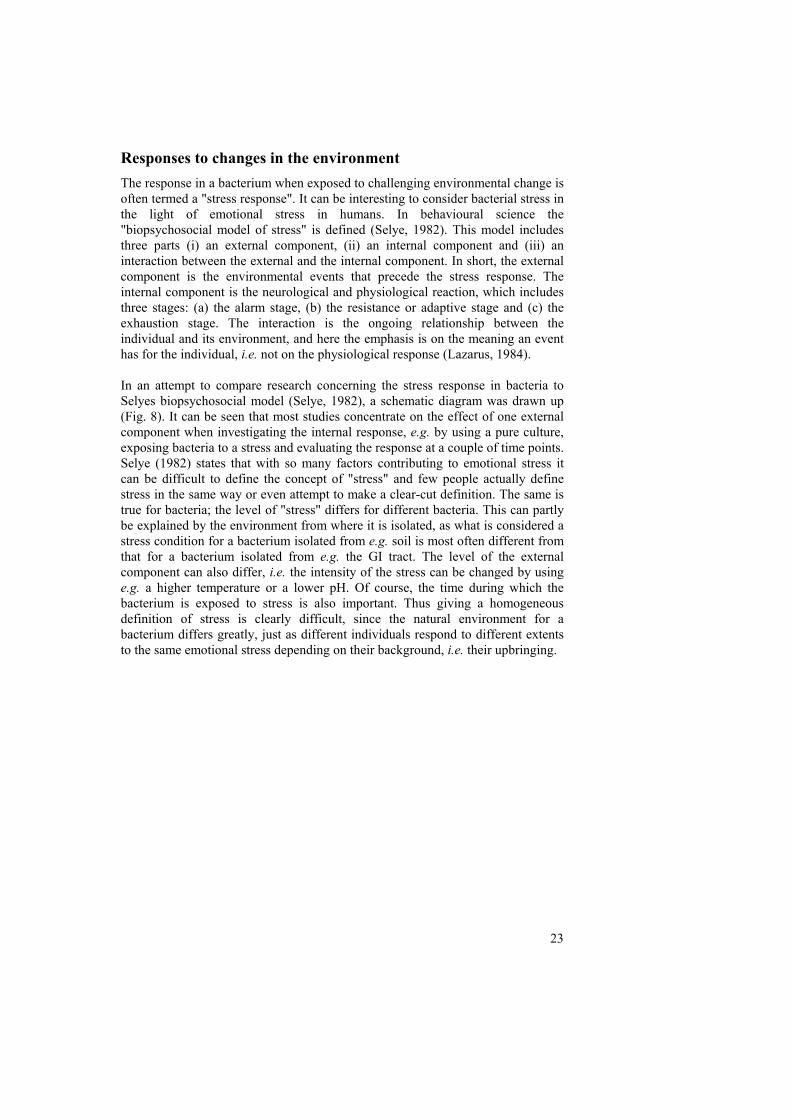
Biological importance of specific genes The biological importance of specific genes for survival, persistence, competition etc. can be investigated using different approaches and different models ranging from a simple laboratory culture experiment to a complex human/animal study. There are advantages and disadvantages with both extremes. Using the more limited laboratory culture is cheaper and the interpretation of the results might be more straightforward. On the other hand more unexpected effects and the importance of the complex system might be overlooked. Animal models are obviously good when it comes to natural similarities although human trials are of course the optimal way of studying probiotic effects. However, both animal and human trials are expensive, sampling can be difficult and ethics are also an issue. Considering these facts, the use of the three stage continuous culture system mimicking the human large intestine (here termed an artificial colon), introduced by Gibson et al. (1988) and validated by Macfarlane et al. (1998), was considered to be a good alternative for an evaluation of the L. reuteri mutants. The three-stage continuous culture system allowed us to study the persistence and competitive ability of the mutants in a complex human-like ecosystem. Construction of mutants In order to investigate the biological importance of different previously described genes of interest, a number of L. reuteri ATCC 55730 genes were mutated. Two of the putative surface proteins with GW repeats (gw1 and gw2), two genes induced as a response to acid stress (clpL and lr1516, a putative cell wall-altering esterase) and four genes induced as a response to an elevated temperature (clpE, ldhL1, rdpR and rr5) were all mutated. In addition to rr5 one more RR had previously been mutated (rr2) (Wall, 2005) In addition the HPK of the same two-component system (hpk2) and four other HPK's (hpk1, hpk3, hpk4 and hpk6) were also mutated (Table X.). Mutants were constructed by interruption of the target gene through site-specific integration of a plasmid harbouring an antibiotic resistance gene (for details see Fig. 12 and Fig. 13).
33
Fig. 12. Plasmids involved in construction of mutants. (A) pORI28 with a fragment of the target gene(lr fragment in pink) inserted, pORI28 harbours a erythromycin resistance gene but lacks repA and thus need this gene from another plasmid to be able to replicate. (B) The helper plasmid pVE6007 which carries repA and a chloramphenicol resistance gene, the replication of this plasmid is temperature sensitive.
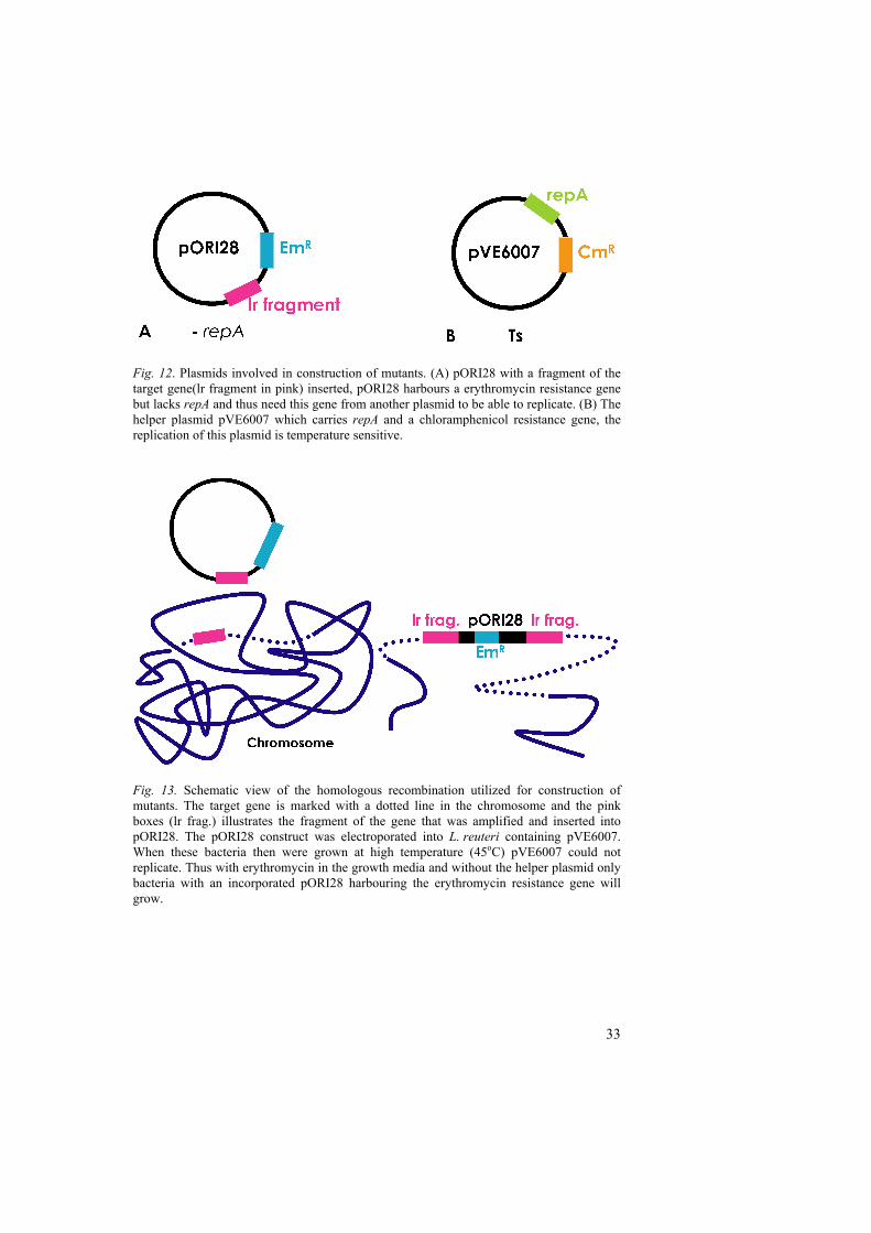
Fig. 13. Schematic view of the homologous recombination utilized for construction of mutants. The target gene is marked with a dotted line in the chromosome and the pink boxes (lr frag.) illustrates the fragment of the gene that was amplified and inserted into pORI28. The pORI28 construct was electroporated into L. reuteri containing pVE6007. When these bacteria then were grown at high temperature (45oC) pVE6007 could not replicate. Thus with erythromycin in the growth media and without the helper plasmid only bacteria with an incorporated pORI28 harbouring the erythromycin resistance gene will grow.

34
When testing the mutants from the heat and acid stress experiments, the first obvious approach was to test their survival at an elevated temperature or in an acidic environment. When the survival of the mutants from the heat stress experiment was compared with that of the wild type, the clpE and rdpR mutants both showed decreased survival at 50 oC after 2.5 and 3 h incubation. The clpE mutant had a decreased survival already after 2 h. The ldhL1 mutant was also affected after 3 h (Paper II). When the wild type and some of the mutants were grown at 45 oC, the clpE mutant exhibited unexpected behaviour. After 12 h the wild type and the other mutants had established a stationary phase, but in contrast, the cell density of the clpE mutant had instead dropped. Since no aggregation of the bacteria was observed under the microscope, autolysis is a possible explanation for this phenotype. However, this mutant did show a disturbed cell morphology with a mycelial like growth under the microscope (Paper II). A change in morphology was also observed already in 37 oC in a double mutant of clpE and the associated proteolytic domain clpP of Listeria monocytogenes. This mutant was also sensitive to higher temperatures and did not survive at 42 oC (Nair et al., 1999). There were no clear differences between the wild type and the clpL and esterase mutants from the acid stress experiment when diluted in MRS with pH 2.7. However, the survival was also examined in synthetic gastric juice (lacking enzyme and bile) with pH 2.0. After 50 minutes, the survival of the clpL mutant and the esterase mutant was significantly lower than that of the wild type (paper III). Evaluation of mutants in a colon model (IV) Paper I, II and III of this thesis all resulted in the identification of a set of genes possibly of importance for ecological performance. Some of these genes were mutated and evaluated in the respective paper. The selection of mutants from the previous works was increased by mutants of genes from two-component signal transduction systems originally identified by Wall (2005) (all genes mutated can be found in Table 1D). In paper IV the whole collection of mutants were evaluated in a colon model.
This three-stage continuous culture fermentation system was designed to mimic the spatial, temporal, nutritional and physiochemical characteristics of the microbiota in the proximal (vessel 1) and distal (vessel 2 and 3) colon (Macfarlane, Macfarlane & Gibson, 1998). The temperature was set to 37oC and the pH of vessels 1, 2 and 3 were set to 5.5, 6.2 and 6.8 respectively. The gut model was set up and inoculated with a fresh human faecal sample (Fig. 14).
Fig. 14. The three-stage continues culture system.

35
Fig. 15. Outline of the experiment evaluating performance of mutants in an artificial colon.
After equilibration of the system a mix of fifteen strains, the fourteen mutants described above and a strain with a mutated inactive transposase (lr0849) (termed the control-mutant), were inoculated in the top vessel. This inoculation was repeated after 24 h. Samples were then taken from each vessel from 8 h up to 7 days after the first inoculation to collectively follow the persistence of all mutants in the system (Fig. 15). The bacteria were grown on plates with erythromycin and vancomycin to select for L. reuteri mutants. Samples taken before inoculation of the mutants showed no growth of L. reuteri (Fig. 16). The total L. reuteri mutant count dropped from the initial value of 107 to around 103 after five days. Thereafter, a tendency to stabilisation could be seen. The fact that the total number of mutants was rapidly reduced could be explained by the fact that they multiply at a rate that is to slow to compensate for the dilution effect.
Fig. 16. Growth of samples from the artificial colon. (A) 10-2 dilution of sample before inoculation of the L. reuteri mutants and (B) 10-5 dilution of a sample taken 8 h after first inoculation The appearance of L. reuteri compared to bacteria from the indigenous microflora of the artificial colon was clearly visible. There was no growth on the plate from the 10-5 dilution of the sample taken before inoculation of L. reuteri mutants.
Fig. 17. Schematic diagram of the design of specific real time PCR primers. The primers were used to individually detect each mutant in samples taken from the artificial colon experiment.

36
A real time PCR base approach was used to determine the relative abundance of individual mutants. Since specific detection of the wild type was not possible, a control-mutant was constructed to facilitate comparison of persistence between the mutants. Using primers specific for each mutant (Fig. 17) the ratio of mutants on days four and five after the first inoculation was investigated. The data from this experiment are so far only based on samples from one artificial colon experiment and so it is difficult to draw conclusions on the definite importance for survival of the individual genes. It would therefore be interesting to perform the same experiment again. Furthermore, a large amount of samples were taken and not all have been investigated so far. Even though the conditions and composition in the different vessels were a somewhat different, the result from each can to some extent be considered as replicates. Comparisons show that three mutants were clearly less persistent than the control-mutant in all three vessels, indicating that clpE, rr2 and hpk1 are important for persistence in the human colon. There were also tendencies for low persistence of other mutants, e.g. ldhL1, clpL, gw2 and hpk4 mutants. It is interesting to see that it is the same mutants that are sensitive to acid and heat that are influenced by the environment and competition in the artificial colon. However, it is clear that not all genes induced in separate response to heat or acid are important for persistence of the bacterium in the colon. The bacterium has a complex matrix of signals and protective strategies all working together to perform in the GI tract. To our knowledge, this way of using a real time based PCR approach to detect the relative abundance of a mix of mutants and evaluate their persistence in a complex model has not been published previously, and hopefully opens up a new perspective on how to evaluate mutant survival in a biological system. This way of using real time PCR could also possibly provide new opportunities for evaluation of mutants in important in vivo studies.
Concluding remarks
From the work described in this thesis it can be concluded that some of the classical HSPs such as the Clps are important for survival during stress and for ecological performance. Genes encoding Clps were induced in response both to heat and acid stress, though the response seems to be specialised since different Clps were induced and also differences in regulatory motifs were observed. However, both these chaperones were important for the persistence of L. reuteri ATCC 55730 in a colon model and thus probably for the ecological performance in the GI tract. The importance of these genes for the health promoting effects still has to be unravelled. The bioinformatics study of extracellular proteins described in this thesis revealed a divers set of proteins with different means of attachment. Both stress response and identification of surface proteins exposed a complex picture where most of the genes actually have an unknown function. To be able to understand the intricate mechanism enabling a bacterium to persist in the GI tract

37
and interact with the host much effort has to be put in studies of these unknown genes. Another issue that complicates the determination of the function behind genes is the possible moonlighting properties that sometimes give a false realisation of the gene function. When looking at previously used criteria for strain selection of probiotics, the only things being undoubtedly important are beneficial effects and safety. However, it is of interest to understand the mechanisms underlying the promotion of health to be able to find new ways of selecting strains with more defined effects. This could eventually lead to new probiotics with a more justified and accepted role in treatment of gastrointestinal derived disorders. The field of probiotic science is now moving from selecting strains by these previously described criteria based on readily measurable parameters such as survival in acidic conditions, towards understanding the underlying mechanisms behind GI tract persistence and in the long run the health-promoting effects. By screening genomes in different ways, e.g. by predicting surface proteins or gene homologues to already characterised proteins, or by studying genes and proteins involved in stress response, using e.g. 2D gel electrophoresis and DNA microarrays, we can get a glimpse of the bacterium's strategy for ecological performance. However, it is important that we do not end up only identifying more and more genes that are potentially important for persistence. To move further, we also need to evaluate these genes by studies of the probiotic effects of mutants compared to their wild type.
"Foxes have holes and birds of the air have nests, but the Son of Man has no place to lay his head."
(Luke 9:58)

38
References
Ahluwalia, N. 2004. Aging, nutrition and immune function. The Journal of Nutrtition, Health and Aging 8, 2-6.
Ahrné, S., Molin, G. & Axelsson, L. 1992. Transformation of Lactobacillus reuteri with electroporation: studies on the erythromycin resistance plasmid pLUL631. Current Microbiology
Alm, E., Huang, K. & Arkin, A. 2006. The evolution of two-component systems in bacteria reveals different strategies for niche adaptation. Public Library of Science Computional Biology 2, 1329-1342.
Alm, J.S., Swartz, J., Björkstén, B., Engstrand, L., Engström, J., Kühn, I., Lilja, G., Möllby, R., Norin, E., Pershagen, G., Reinders, C., Wreiber, K. & Scheynius, A. 2002. An anthroposophic lifestyle and intestinal microflora in infancy. Pediatric Allergy and Immunology 13, 402-411.
Alm, J.S., Swartz, J., Lilja, G., Scheynius, A. & Pershagen, G. 1999. Atopy in children of families with an anthroposophic lifestyle. The Lancet 353, 1485-1488.
Barrangou, R., Azcarate-Peril, M.A., Duong, T., Conners, S.B., Kelly, R.M. & Klaenhammer, T.R. 2006. Global analysis of carbohydrate utilization by Lactobacillus acidophilus using cDNA microarrays. Proceedings of the National Academy of Sciences of the USA 103, 3816-3821.
Bateman, A. & Bycroft, M. 2000. The structure of a LysM domain from E. coli membrane-bound lytic murein transglycosylase D (MltD). Journal of Molecular Biology 299, 1113-1119.
Bearson, S., Bearson, B. & Foster, J.W. 1997. Acid stress responses in enterobacteria. FEMS Microbiology Letters 147, 173-180.
Bengmark, S. 2002. Gut microbial ecology in critical illness: is there a role for prebiotics, probiotics, and synbiotics? Current Opinion in Critical Care 8, 145-151.
Bergonzelli, G.E., Granato, D., Pridmore, R.D., Marvin-Guy, L.F., Donnicola, D. & Corthésy-Theulaz, I.E. 2006. GroEL of Lactobacillus johnsonii La1 (NCC 533) is cell surface associated: potential role in interactions with the host and the gastric pathogen Helicobacter pylori. Infection and Immunity 74, 425-434.
Bik, E.M., Eckburg, P.B., Gill, S.R., Nelson, K.E., Purdom, E.A., Francois, F., Perez-Perez, G., Blaser, M.J. & Relman, D.A. 2006. Molecular analysis of the bacterial microbiota in the human stomach. Proceedings of the National Academy of Sciences of the USA 103, 732-737.
Billoo, A.G., Memon, M.A., Khaskheli, S.A., Murtaza, G., Iqbal, K., Shekhani, M.S. & Siddiqi, A.Q. 2006. Role of a probiotic (Saccharomyces boulardii) in management and prevention of diarrhoea. World Journal of Gastroenterology 12, 4557-4560.
Björkstén, B., Naaber, P., Sepp, E. & Mikelsaar, M. 1999. The intestinal microflora in allergic Estonian and Swedish 2-year-old children. Clinical and Experimental Allergy 29, 342-346.

39
Boekhorst, J., Helmer, Q., Kleerebezem, M. & Siezen, R.J. 2006a. Comparative analysis of proteins with a mucus-binding domain found exclusively in lactic acid bacteria. Microbiology 152, 273-280.
Boekhorst, J., Wels, M., Kleerebezem, M. & Siezen, R.J. 2006b. The predicted secretome of Lactobacillus plantarum WCFS1 sheds light on interactions with its environment. Microbiology 152, 3175-3183.
Bottiglieri, T. 1996. Folate, vitamin B12, and neuropsychiatric disorders. Nutrition Reviews 54, 382-390.
Bourlioux, P., Koletzko, B., Guarner, F. & Braesco, V. 2003. The intestine and its microflora are partners for the protection of the host: report on the Danone Symposium "The Intelligent Intestine," held in Paris, June 14, 2002. The American Journal of Clincal Nutrition 78, 675-683.
Braeken, K., Moris, M., Daniels, R., Vanderleyden, J. & Michiels, J. 2006. New horizons for (p)ppGpp in bacterial and plant physiology. Trends in Microbiology 14, 45-54.
Brandtzaeg, P., Halstensen, T.S., Kett, K., Krajči, P., Kvale, D., Rognum, T.O., Scott, H. & Sollid, L.M. 1989. Immunobiology and immunopathology of human gut mucosa: humoral immunity and intraepithelial lymphocytes. Gastroenterology 97, 1562-1584.
Broekaert, I.J. & Walker, W.A. 2006. Probiotics and chronic disease. Journal of Clinical Gastroenterology 40, 270-274.
Bron, P.A., Grangette, C., Mercenier, A., de Vos, W.M. & Kleerebezem, M. 2004. Identification of Lactobacillus plantarum genes that are induced in the gastrointestinal tract of mice. Journal of Bacteriology 186, 5721-5729.
Bron, P.A., Molenaar, D., de Vos, W.M. & Kleerebezem, M. 2006. DNA micro-array-based identification of bile-responsive genes in Lactobacillus plantarum. Journal of Applied Microbiology 100, 728-738.
Buck, B.L., Altermann, E., Svingerud, T. & Klaenhammer, T.R. 2005. Functional analysis of putative adhesion factors in Lactobacillus acidophilus NCFM. Applied and Environmental Microbiology 71, 8344-8351.
Buret, A.G. 2006. How stress induces intestinal hypersensitivity. American Journal of Pathology 168, 3-5.
Bäckhed, F., Ley, R.E., Sonnenburg, J.L., Peterson, D.A. & Gordon, J.I. 2005. Host-bacterial mutualism in the human intestine. Science 307, 1915-1920.
Cabanes, D., Dehoux, P., Dussurget, O., Frangeul, L. & Cossart, P. 2002. Surface proteins and the pathogenic potential of Listeria monocytogenes. Trends in Microbiology 10, 238-245.
Carroll, S.A., Hain, T., Technow, U., Darji, A., Pashalidis, P., Joseph, S.W. & Chakraborty, T. 2003. Identification and characterization of a peptidoglycan hydrolase, MurA, of Listeria monocytogenes, a muramidase needed for cell separation. Journal of Bacteriology 185, 6801-6808.
Chastanet, A., Fert, J. & Msadek, T. 2003. Comparative genomics reveal novel heat shock regulatory mechanisms in Staphylococcus aureus and other Gram-positive bacteria. Molecular Microbiology 47, 1061-1073.
Chermesh, I. & Eliakim, R. 2006. Probiotics and the gastrointestinal tract: where are we in 2005? World Journal of Gastroenterology 12, 853-857.

40
Chou, L.-S. & Weimer, B. 1999. Isolation and characterization of acid- and bile-tolerant isolates from strains of Lactobacillus acidophilus. Journal of Dairy Science 82, 23-31.
Collins, S.M. 2001. Stress and the Gastrointestinal Tract IV. Modulation of intestinal inflammation by stress: basic mechanisms and clinical relevance. American Journal of Physiology. Gastrointestinal and Liver Physiology 280, G315-G318.
Cotter, P.D. & Hill, C. 2003. Surviving the acid test: responses of gram-positive bacteria to low pH. Microbiology and Molecular Biology Reviews 67, 429-453.
De Angelis, M., Bini, L., Pallini, V., Cocconcelli, P.S. & Gobbetti, M. 2001. The acid-stress response in Lactobacillus sanfranciscensis CB1. Microbiology 147, 1863-1873.
De Angelis, M. & Gobbetti, M. 2004. Environmental stress responses in Lactobacillus: a review. Proteomics 4, 106-122.
del Rocio Leon-Kempis, M., Guccione, E., Mulholland, F., Williamson, M.P. & Kelly, D.J. 2006. The Campylobacter jejuni PEB1a adhesin is an aspartate/glutamate-binding protein of an ABC transporter essential for microaerobic growth on dicarboxylic amino acids. Molecular Microbiology 60, 1262-1275.
Dethlefsen, L., Eckburg, P.B., Bik, E.M. & Relman, D.A. 2006. Assembly of the human intestinal microbiota. Trends in Ecology and Evolution 21, 517-523.
Di Caro, S., Tao, H., Grillo, A., Elia, C., Gasbarrini, G., Sepulveda, A.R. & Gasbarrini, A. 2005. Effects of Lactobacillus GG on genes expression pattern in small bowel mucosa. Digestive and Liver Disease 37, 320-329.
Dunne, C., Murphy, L., Flynn, S., O'Mahony, L., O'Halloran, S., Feeney, M., Morrissey, D., Thornton, G., Fitzgerald, G., Daly, C., Kiely, B., Quigley, E.M.M., O'Sullivan, G.C., Shanahan, F. & Collins, J.K. 1999. Probiotics: from myth to reality. Demonstration of functionality in animal models of disease and in human clinical trials. Antonie Van Leeuwenhoek 76, 279-292.
Eckburg, P.B., Bik, E.M., Bernstein, C.N., Purdom, E., Dethlefsen, L., Sargent, M., Gill, S.R., Nelson, K.E. & Relman, D.A. 2005. Diversity of the human intestinal microbial flora. Science 308, 1635-1638.
Ewaschuk, J.B. & Dieleman, L.A. 2006. Probiotics and prebiotics in chronic inflammatory bowel diseases. World Journal of Gastroenterology 12, 5941-5950.
Fanaro, S., Chierici, R., Guerrini, P. & Vigi, V. 2003. Intestinal microflora in early infancy: composition and development. Acta Paediatrica. Supplementum 91, 48-55.
Felley, C. & Michetti, P. 2003. Probiotics and Helicobacter pylori. Best Practice and Research Clinical Gastroenterology 17, 785-791.
Fischetti, V.A., Pancholi, V. & Schneewind, O. 1990. Conservation of a hexapeptide sequence in the anchor region of surface proteins from Gram-positive cocci. Molecular Microbiology 4, 1603-1605.
Flöistrup, H., Swartz, J., Bergström, A., Alm, J.S., Scheynius, A., van Hage, M., Waser, M., Braun-Fahrländer, C., Schram-Bijkerk, D., Huber, M.,

41
Zutavern, A., von Mutius, E., Üblagger, E., Riedler, J., Michaels, K.B. & Pershagen, G. 2006. Allergic disease and sensitization in Steiner school children. Journal of Allergy and Clinical Immunology 117, 59-66.
Fukushima, T., Afkham, A., Kurosawa, S., Tanabe, T., Yamamoto, H. & Sekiguchi, J. 2006. A new D,L-endopeptidase gene product, YojL (renamed CwlS), plays a role in cell separation with LytE and LytF in Bacillus subtilis. Journal of Bacteriology 188, 5541-5550.
Fuller, R. 1986. Probiotics. Journal of Applied Bacteriology Symposium Supplements, 1S-7S.
Gibson, G.R., Cummings, J.H. & Macfarlane, G.T. 1988. Use of a three-stage continuous culture system to study the effect of mucin on dissimilatory sulfate reduction and methanogenesis by mixed populations of human gut bacteria. Applied and Environmental Microbiology 54, 2750-2755.
Goossens, D., Jonkers, D., Russel, M., Thijs, A., van den Bogaard, A., Stobberingh, E. & Stockbrügger, R. 2005. Survival of the probiotic, L. plantarum 299v and its effects on the faecal bacterial flora, with and without gastric acid inhibition. Digestive and Liver Disease 37, 44-50.
Gordon, J.I., Ley, R.E., Wilson, R., Mardis, E., Xu, J., Fraser, C.M. & Relman, D.A. 2005. Extending our view of self: the human gut microbiome initiative (HGMI). Bethesda, Maryland: National Human Genome Research Institute.
Guarner, F. 2006. Enteric flora in health and disease. Digestion 73 (suppl 1), 5-12. Guarner, F. & Malagelada, J.R. 2003. Gut flora in health and disease. The Lancet
361, 512-519. Guo, X., Li, D., Lu, W., Piao, X. & Chen, X. 2006. Screening of Bacillus strains
as potential probiotics and subsequent confirmation of the in vivo effectiveness of Bacillus subtilis MA139 in pigs. Antonie Van Leeuwenhoek 90, 139-146.
Hammes, W.P. & Hertel, C. 2005. The genera Lactobacillus and Carnobacterium. In The Prokaryotes. Edited by M. Dworkin. Springer-Verlag. New York.
Henderson, B., Allan, E. & Coates, A.R.M. 2006. Stress wars: the direct role of host and bacterial molecular chaperones in bacterial infection. Infection and Immunity 74, 3693-3706.
Hoch, J.A. 2002. Control of signal transduction in the sporulation phosphorelay. In Signals, switches, regulons and caascades: control of bacterial gene expression. Edited by D.A. Hodgson & C.M. Thomas. Cambridge University Press. Cambridge. 199-212. pp.
Hols, P., Hancy, F., Fontaine, L., Grossiord, B., Prozzi, D., Leblond-Bourget, N., Decaris, B., Bolotin, A., Delorme, C., Dusko Ehrlich, S., Guedon, E., Monnet, V., Renault, P. & Kleerebezem, M. 2005. New insights in the molecular biology and physiology of Streptococcus thermophilus revealed by comparative genomics. FEMS Microbiology Review 29, 435-463.
Hooper, L.V., Midtvedt, T. & Gordon, J.I. 2002. How host-microbial interactions shape the nutrient environment of the mammalian intestine. Annual Review of Nutrition 22, 283-307.

42
Isolauri, E., Rautava, S., Kalliomäki, M., Kirjavainen, P. & Salminen, S. 2002. Role of probiotics in food hypersensitivity. Current Opinion in Allergy and Clinical Immunology 2, 263-271.
Isolauri, E., Salminen, S. & Ouwehand, A.C. 2004. Probiotics. Best Practice and Research Clinical Gastroenterology 18, 299-313.
Jönsson, T., Ahrén, B., Pacini, G., Sundler, F., Wierup, N., Steen, S., Sjöberg, T., Ugander, M., Frostegård, J., Göransson, L. & Lindeberg, S. 2006. A Paleolithic diet confers higher insulin sensitivity, lower C-reactive protein and lower blood pressure than a cereal-based diet in domestic pigs. Nutrition and Metabolism 3, 39-48.
Kalliomäki, M., Salminen, S., Arvilommi, H., Kero, P., Koskinen, P. & Isolauri, E. 2001. Probiotics in primary prevention of atopic disease: a randomised placebo-controlled trial. The Lancet 357, 1076-1079.
Kalliomäki, M., Salminen, S., Poussa, T., Arvilommi, H. & Isolauri, E. 2003. Probiotics and prevention of atopic disease: 4-year follow-up of a randomised placebo-controlled trial. The Lancet 361, 1869-1871.
Katakura, K., Lee, J., Rachmilewitz, D., Li, G., Eckmann, L. & Raz, E. 2005. Toll-like receptor 9-induced type I IFN protects mice from experimental colitis. The Journal of Clinical Investigation 115, 695-702.
Katz, J.A. 2006. Probiotics for the prevention of antibiotic-associated diarrhea and Clostridium difficile diarrhea. Journal of Clinical Gastroenterology 40, 249-255.
Kiliaan, A.J., Saunders, P.R., Bijlsma, P.B., Berin, M.C., Taminiau, J.A., Groot, J.A. & Perdue, M.H. 1998. Stress stimulates transepithelial macromolecular uptake in rat jejunum. American Journal of Physiology. Gastrointestinal and Liver Physiology 275, 1037-1044.
Kim, H.S., Park, H., Cho, I.Y., Paik, H.D. & Park, E. 2006. Dietary supplementation of probiotic Bacillus polyfermenticus, Bispan strain, modulates natural killer cell and T cell subset populations and immunoglobulin G levels in human subjects. Journal of Medicinal Food 9, 321-327.
Kleerebezem, M., Boekhorst, J., van Kranenburg, R., Molenaar, D., Kuipers, O.P., Leer, R., Tarchini, R., Peters, S.A., Sandbrink, H.M., Fiers, M.W.E.J., Stiekema, W., Lankhorst, R.M.K., Bron, P.A., Hoffer, S.M., Groot, M.N.N., Kerkhoven, R., de Vries, M., Ursing, B., de Vos, W.M. & Siezen, R.J. 2003. Complete genome sequence of Lactobacillus plantarum WCFS1. Proceedings of the National Academy of Sciences of the USA 100, 1990-1995.
Klingberg, T.D. & Budde, B.B. 2006. The survival and persistence in the human gastrointestinal tract of five potential probiotic lactobacilli consumed as freeze-dried cultures or as probiotic sausage. International Journal of Food Microbiology 109, 157-159.
Kokesova, A., Frolova, L., Kverka, M., Sokol, D., Rossmann, P., Bartova, J. & Tlaskalova-Hogenova, H. 2006. Oral administration of probiotic bacteria (E. coli Nissle, E. coli O83, Lactobacillus casei) influences the severity of dextran sodium sulfate-induced colitis in BALB/c mice. Folia Microbiologica (Praha) 51, 478-484.

43
Kolenbrander, P.E., Andersen, R.N., Baker, R.A. & Jenkinson, H.F. 1998. The adhesion-associated sca operon in Streptococcus gordonii encodes an inducible high-affinity ABC transporter for Mn2+ uptake. Journal of Bacteriology 180, 290-295.
Konings, W.N. 2002. The cell membrane and the struggle for life of lactic acid bacteria. Antonie Van Leeuwenhoek 82, 3-27.
Krasse, P., Carlsson, B., Dahl, C., Paulsson, A., Nilsson, A. & Sinkiewicz, G. 2006. Decreased gum bleeding and reduced gingivitis by the probiotic Lactobacillus reuteri. Swedish Dental Journal 30, 55-60.
Krogh, A., Larsson, B., von Heijne, G. & Sonnhammer, E.L.L. 2001. Predicting transmembrane protein topology with a hidden Markov model: application to complete genomes. Journal of Molecular Biology 305, 567-580.
Lazarus, R.S. 1984. Stress, Appraisal and Coping. Guilford. New York pp. Lemos, J.A., Luzardo, Y. & Burne, R.A. 2007. Physiologic effects of forced
down-regulation of dnaK and groEL expression in Streptococcus mutans. Journal of Bacteriology (In Press, Accepted Manuscript) 189
Lim, E.M., Ehrlich, S.D. & Maguin, E. 2000. Identification of stress-inducible proteins in Lactobacillus delbrueckii subsp. bulgaricus. Electrophoresis 21, 2557-2561.
Lin, W.-H., Hwang, C.-F., Chen, L.-W. & Tsen, H.-Y. 2006. Viable counts, characteristic evaluation for commercial lactic acid bacteria products. Food Microbiology 23, 74-81.
Lindeberg, S. 2005. Paleolitisk kost och evolutionsmedicin: nyckel till västvärldens sjukdomar. Läkartidningen 102, 26-27.
Ljungh, A. & Wadström, T. 2006. Lactic acid bacteria as probiotics. Current Issues in Intestinal Microbiology 7, 73-89.
Lorca, G.L., Font de Valdez, G. & Ljungh, A. 2002. Characterization of the protein-synthesis dependent adaptive acid tolerance response in Lactobacillus acidophilus. Journal of Molecular Microbiology and Biotechnology 4, 525-532.
Macfarlane, G.T., Macfarlane, S. & Gibson, G.R. 1998. Validation of a Three-Stage Compound Continuous Culture System for Investigating the Effect of Retention Time on the Ecology and Metabolism of Bacteria in the Human Colon. Microbial Ecology 35, 180-187.
Mahan, M.J., Slauch, J.M. & Mekalanos, J.J. 1993. Selection of bacterial virulence genes that are specifically induced in host tissues. Science 259, 686-688.
Marco, M.L., Pavan, S. & Kleerebezem, M. 2006. Towards understanding molecular modes of probiotic action. Current Opinion in Biotechnology 17, 204-210.
McKay, D.M. 2005. Good bug, bad bug: in the case of enteric inflammatory disease does the epithelium decide? Memórias do Instituto Oswaldo Cruz 100 Suppl 1, 205-210.
Meier, R.F. 2005. Probiotics: a new treatment for antibiotic-associated diarrhea? Digestion 72, 49-50.
Metchnikoff, E. 1907. Prolongation of Life. William Heinemann. London pp.

44
Meurman, J.H. 2005. Probiotics: do they have a role in oral medicine and dentistry? European Journal of Oral Sciences 113, 188-196.
Michel, A., Agerer, F., Hauck, C.R., Herrmann, M., Ullrich, J., Hacker, J. & Ohlsen, K. 2006. Global regulatory impact of ClpP protease of Staphylococcus aureus on regulons involved in virulence, oxidative stress response, autolysis, and DNA repair. Journal of Bacteriology 188, 5783-5796.
Morel-Deville, F., Fauvel, F. & Morel, P. 1998. Two-component signal-transducing systems involved in stress responses and vancomycin susceptibility in Lactobacillus sakei. Microbiology 144, 2873-2883.
Nair, S., Frehel, C., Nguyen, L., Escuyer, V. & Berche, P. 1999. ClpE, a novel member of the HSP100 family, is involved in cell division and virulence of Listeria monocytogenes. Molecular Microbiology 31, 185-196.
Nes, I.F., Diep, D.B., Havarstein, L.S., Brurberg, M.B., Eijsink, V. & Holo, H. 1996. Biosynthesis of bacteriocins in lactic acid bacteria. Antonie Van Leeuwenhoek 70, 113-128.
Nielsen, H., Engelbrecht, J., Brunak, S. & von Heijne, G. 1997. A neural network method for identification of prokaryotic and eukaryotic signal peptides and prediction of their cleavage sites. International Journal of Neural Systems 8, 581-599.
Nikawa, H., Makihira, S., Fukushima, H., Nishimura, H., Ozaki, Y., Ishida, K., Darmawan, S., Hamada, T., Hara, K., Matsumoto, A., Takemoto, T. & Aimi, R. 2004. Lactobacillus reuteri in bovine milk fermented decreases the oral carriage of mutans streptococci. International Journal of Food Microbiology 95, 219-223.
Ocana, V.S. & Nader-Macias, E.M. 2004. Production of antimicrobial substances by lactic acid bacteria II: screening bacteriocin-producing strains with probiotic purposes and characterization of a Lactobacillus bacteriocin. Methods in Molecular Biology 268, 347-353.
Palumbo, R.N. & Wang, C. 2006. Bacterial invasin: structure, function, and implication for targeted oral gene delivery. Current Drug Delivery 3, 47-53.
Parvez, S., Malik, K.A., Ah Kang, S. & Kim, H.-Y. 2006. Probiotics and their fermented food products are beneficial for health. Journal of Applied Microbiology 100, 1171-1185.
Pieterse, B., Leer, R.J., Schuren, F.H.J. & van der Werf, M.J. 2005. Unravelling the multiple effects of lactic acid stress on Lactobacillus plantarum by transcription profiling. Microbiology 151, 3881-3894.
Plinius Secundus, C. Naturalis historiae XXVIII: 36, 135. Rastall, R.A., Gibson, G.R., Gill, H.S., Guarner, F., Klaenhammer, T.R., Pot, B.,
Reid, G., Rowland, I.R. & Sanders, M.E. 2005. Modulation of the microbial ecology of the human colon by probiotics, prebiotics and synbiotics to enhance human health: an overview of enabling science and potential applications. FEMS Microbiology Ecology 52, 145-152.
Rembacken, B.J., Snelling, A.M., Hawkey, P.M., Chalmers, D.M. & Axon, A.T.R. 1999. Non-pathogenic Escherichia coli versus mesalazine for the treatment of ulcerative colitis: a randomised trial. The Lancet 354, 635-639.

45
Riedel, C.U., Foata, F., Philippe, D., Adolfsson, O., Eikmanns, B.J. & Blum, S. 2006. Anti-inflammatory effects of bifidobacteria by inhibition of LPS-induced NF-κB activation. World Journal of Gastroenterology 12, 3729-3735.
Roos, S., Aleljung, P., Robert, N., Lee, B., Wadstrom, T., Lindberg, M. & Jonsson, H. 1996. A collagen binding protein from Lactobacillus reuteri is part of an ABC transporter system? FEMS Microbiology Letters 144, 33-38.
Roos, S., Engstrand, L. & Jonsson, H. 2005. Lactobacillus gastricus sp. nov., Lactobacillus antri sp. nov., Lactobacillus kalixensis sp. nov. and Lactobacillus ultunensis sp. nov., isolated from human stomach mucosa. International journal of systematic and evolutionary microbiology 55, 77-82.
Roos, S. & Jonsson, H. 2002. A high-molecular-mass cell-surface protein from Lactobacillus reuteri 1063 adheres to mucus components. Microbiology 148, 433-442.
Roselli, M., Finamore, A., Britti, M.S. & Mengheri, E. 2006. Probiotic bacteria Bifidobacterium animalis MB5 and Lactobacillus rhamnosus GG protect intestinal Caco-2 cells from the inflammation-associated response induced by enterotoxigenic Escherichia coli K88. British Journal of Nutrition 95, 1177-1184.
Rosenfeldt, V., Benfeldt, E., Nielsen, S.D., Michaelsen, K.F., Jeppesen, D.L., Valerius, N.H. & Pærregaard, A. 2003. Effect of probiotic Lactobacillus strains in children with atopic dermatitis. Journal of Allergy and Clinical Immunology 111, 389-395.
Rosenfeldt, V., Benfeldt, E., Valerius, N.H., Pærregaard, A. & Michaelsen, K.F. 2004. Effect of probiotics on gastrointestinal symptoms and small intestinal permeability in children with atopic dermatitis. The Journal of Pediatrics 145, 612-616.
Rosenfeldt, V., Michaelsen, K.F., Jakobsen, M., Nexman Larsen, C., Lange Møller, P., Tvede, M., Weyrehter, H., Valerius, N.H. & Pærregaard, A. 2002. Effect of probiotic Lactobacillus strains on acute diarrhea in a cohort of nonhospitalized children attending day-care centers. The Pediatric Infectious Disease Journal 21, 417-419.
Ross, R.P., Morgan, S. & Hill, C. 2002. Preservation and fermentation: Past, present and future. International Journal of Food Microbiology 79, 3-16.
Savino, F., Pelle, E., Palumeri, E., Oggero, R. & Miniero, R. 2007. Lactobacillus reuteri (American Type Culture Collection Strain 55730) versus simethicone in the treatment of infantile colic: a prospective randomized study. Pediatrics 119, 124-130.
Scalabrino, G., Buccellato, F.R., Veber, D. & Mutti, E. 2003. New basis of the neurotrophic action of vitamin B12. Clinical Chemistry and Laboratory Medicine 41, 1435-7.
Schmidt, M.A., Riley, L.W. & Benz, I. 2003. Sweet new world: glycoproteins in bacterial pathogens. Trends in Microbiology 11, 554-561.
Schwarz-Linek, U., Höök, M. & Potts, J.R. 2006. Fibronectin-binding proteins of gram-positive cocci. Microbes and Infection 8, 2291-2298.

46
Seksik, P., Rigottier-Gois, L., Gramet, G., Sutren, M., Pochart, P., Marteau, P., Jian, R. & Doré, J. 2003. Alterations of the dominant faecal bacterial groups in patients with Crohn's disease of the colon. Gut 52, 237-242.
Selye, H. 1982. History and present status of the stress concept. In Handbook of Stress: Theoretical and Clinical Aspects. Edited by L. Goldberger & S. Breznitz. The Free Press. New York. 7-17. pp.
Shenderov, B.A. & Glushanova, N.A. 2006. Lecture 2.1.4 Experimental substantiation of new approaches to intestinal microbial ecology correction by means of probiotics (In: Abstracts from AG/SOMED 2006). Microbial Ecology in Health and Disease 18, 85-108.
Shin, S., Lu, G., Cai, M. & Kim, K.-S. 2005. Escherichia coli outer membrane protein A adheres to human brain microvascular endothelial cells. Biochemical and Biophysical Research Communications 330, 1199-1204.
Shornikova, A.-V., Casas, I.A., Mykkänen, H., Salo, E. & Vesikari, T. 1997. Bacteriotherapy with Lactobacillus reuteri in rotavirus gastroenteritis. The Pediatric Infectious Disease Journal 16, 1103-1107.
Siegumfeldt, H., Rechinger, K.B. & Jakobsen, M. 2000. Dynamic changes of intracellular pH in individual lactic acid bacterium cells in response to a rapid drop in extracellular pH. Applied and Environmental Microbiology 66, 2330-2335.
Sinkiewicz, G. & Nordström, E.A. 2005. Occurrence of Lactobacillus reuteri, Lactobacilli and bifidobacteria in human breast milk: 353. Pediatric Research 58, 415.
Skerker, J.M., Prasol, M.S., Perchuk, B.S., Biondi, E.G. & Laub, M.T. 2005. Two-component signal transduction pathways regulating growth and cell cycle progression in a bacterium: a system-level analysis. Public Library of Science Biology 3, 1770-1788.
Spratt, B.G. & Cromie, K.D. 1988. Penicillin-binding proteins of gram-negative bacteria. Reviews of Infectious Diseases 10, 699-711.
Steen, A., Buist, G., Leenhouts, K.J., El Khattabi, M., Grijpstra, F., Zomer, A.L., Venema, G., Kuipers, O.P. & Kok, J. 2003. Cell wall attachment of a widely distributed peptidoglycan binding domain is hindered by cell wall constituents. The Journal of Biological Chemistry 278, 23874-23881.
Suau, A., Bonnet, R., Sutren, M., Godon, J.-J., Gibson, G.R., Collins, M.D. & Doré, J. 1999. Direct analysis of genes encoding 16S rRNA from complex communities reveals many novel molecular species within the human gut. Applied and Environmental Microbiology 65, 4799-4807.
Sutcliffe, I.C. & Harrington, D.J. 2002. Pattern searches for the identification of putative lipoprotein genes in Gram-positive bacterial genomes. Microbiology 148, 2065-2077.
Talarico, T.L., Casas, I.A., Chung, T.C. & Dobrogosz, W.J. 1988. Production and isolation of reuterin, a growth inhibitor produced by Lactobacillus reuteri. Antimicrobial Agents and Chemotherapy 32, 1854-1858.
Tannock, G.W. 1995. Sticky microbes: the association of microbes with host surfaces. In Normal Microflora. Chapman and Hall. London. 49-62. pp.
Taranto, M.P., Perez-Martinez, G. & de Valdez, G.F. 2006. Effect of bile acid on the cell membrane functionality of lactic acid bacteria for oral administration. Research in Microbiology 157, 720-725.

47
Taranto, M.P., Vera, J.L., Hugenholtz, J., De Valdez, G.F. & Sesma, F. 2003. Lactobacillus reuteri CRL1098 produces cobalamin. Journal of Bacteriology 185, 5643-5647.
Teixeira, P., Castro, H., Mohacsi-Farkas, C. & Kirby, R. 1997. Identification of sites of injury in Lactobacillus bulgaricus during heat stress. Journal of Applied Microbiology 83, 219-226.
Tubelius, P., Stan, V. & Zachrisson, A. 2005. Increasing work-place healthiness with the probiotic Lactobacillus reuteri: a randomised, double-blind placebo-controlled study. Environmental Health: a Global Access Science Source 4, 25-29.
Tuomanen, E. 1999. Molecular and cellular biology of pneumococcal infection. Current Opinion in Microbiology 2, 35-39.
Wall, T. (2005). Environmental Interactions of Lactobacillus reuteri: Signal Transduction, Gene Expression and Extracellular Proteins of a Lactic Acid Bacterium, Swedish University of Agricultural Sciences.
Walter, J., Heng, N.C.K., Hammes, W.P., Loach, D.M., Tannock, G.W. & Hertel, C. 2003. Identification of Lactobacillus reuteri genes specifically induced in the mouse gastrointestinal tract. Applied and Environmental Microbiology 69, 2044-2051.
van de Guchte, M., Serror, P., Chervaux, C., Smokvina, T., Ehrlich, S.D. & Maguin, E. 2002. Stress responses in lactic acid bacteria. Antonie Van Leeuwenhoek 82, 187-216.
van Eden, W., van der Zee, R. & Prakken, B. 2005. Heat-shock proteins induce T-cell regulation of chronic inflammation. Nature Reviews Immunology 5, 318-330.
van Pijkeren, J.-P., Canchaya, C., Ryan, K.A., Li, Y., Claesson, M.J., Sheil, B., Steidler, L., O'Mahony, L., Fitzgerald, G.F., van Sinderen, D. & O'Toole, P.W. 2006. Comparative and functional analysis of sortase-dependent proteins in the predicted secretome of Lactobacillus salivarius UCC118. Applied and Environmental Microbiology 72, 4143-4153.
Watanabe, S., Narisawa, Y., Arase, S., Okamatsu, H., Ikenaga, T., Tajiri, Y. & Kumemura, M. 2003. Differences in fecal microflora between patients with atopic dermatitis and healthy control subjects. Journal of Allergy and Clinical Immunology 111, 587-591.
Weizman, Z., Asli, G. & Alsheikh, A. 2005. Effect of a probiotic infant formula on infections in child care centers: comparison of two probiotic agents. Pediatrics 115, 5-9.
West, A.H. & Stock, A.M. 2001. Histidine kinases and response regulator proteins in two-component signaling systems. Trends in Biochemical Sciences 26, 369-376.
Yoshida, N., Oeda, K., Watanabe, E., Mikami, T., Fukita, Y., Nishimura, K., Komai, K. & Matsuda, K. 2001. Protein function. Chaperonin turned insect toxin. Nature 411, 44.
Yother, J. & White, J.M. 1994. Novel surface attachment mechanism of the Streptococcus pneumoniae protein PspA. Journal of Bacteriology 176, 2976-2985.
Zhou, D. & Yang, R. 2006. Global analysis of gene transcription regulation in prokaryotes. Cellular and Molecular Life Science 63, 2260-2290.

48
Zoetendal, E.G., von Wright, A., Vilpponen-Salmela, T., Ben-Amor, K., Akkermans, A.D.L. & de Vos, W.M. 2002. Mucosa-associated bacteria in the human gastrointestinal tract are uniformly distributed along the colon and differ from the community recovered from feces. Applied and Environmental Microbiology 68, 3401-3407.

49
Acknowledgments Eg har sår frå svik
Eg må ha av bandasjen Eg går over lik
Over din plantasje Opp i din etage
til de informerte Eg har navnet på de involverte
/Kaizers orchestra (De involverte)
This study was supported by the Swedish Research Council for Environment, Agricultural Sciences and Spatial Planning and BioGaia AB, Stockholm, Sweden. Tack till mina handledare, Stefan och Hasse, för alla skratt och allt hårt arbete! Inget är så enkelt att det inte går att göra komplicerat! Jag ska också göra ett försöka att separera duon… ;-) Stefan, du är som ett levande uppslagsverk åtminstone när det kommer till mjölksyrabakterier. Tack för din energi och ditt engagemang! Hasse, du har en stor förmåga att överblicka och sätta saker i sitt sammanhang, speciellt tack för "fluffandet" under skrivprocessen! I have had the opportunity to visit and work together with Rob Britton and his group at Michigan State University, I would like to thank Rob and everybody else in the lab over there for making our stay a good time and of course also for helping us in understanding the art of DNA microarrays! I would also like to thank Prof. Gibson at the School of Food Biosciences at Reading University for letting me visit. I would also like to thank Theo for welcoming me, and Adele for all the time you spent helping me in the lab. I also want to thank all the students, PhD students and Post docs that took me out for shopping and beers. Jag vill också tacka Torun för vår tid tillsammans i probiotikagruppen och för de artiklar och manus vi lyckats få till! Jag skulle vilja tacka alla kollegorna på mikro (både gamla och nya), utan er hade det kanske gått men det hade absolut inte varit lika skoj! Ett speciellt tack till er som spenderar några extra minuter, kvartar, halvtimmar…. i det övre fikarummet, ni vet vilka ni är! Ett extra tack till Sture, Anki, Barbro och Susanne för administrativ assistans och Ingmar för all praktisk hjälp, närhelst man behöver det! Jag vill också tacka för alla små uppmuntrande sakerna under de sista veckorna, blommor, kakor, glada tillrop, klappar på axeln, kramar och inte minst det mystiska ekonomiska tillskottet (vem? vad hände? varför? hur?).

50
Tack också "Mikro's ski-team", den där Åre resan var precis vad jag behövde just då! Min evige rumskompis Johan, ska (trots knäckebrödstuggande) ha ett stort tack för givande diskussioner av alla de slag! Bättre rumskamrat är svårt att hitta… Jag skulle vilja tacka Magnus för att du "supportat" mig i mitt skrivande och Volkmar för hjälpen med den fantastiska metoden real tids PCR. Håkan Larsson förtjänar också ett tack för min första tid som doktorand, då det handlade om proteiner! Attans!! Att det inte blev ngt av alla 2D geler, tankar och funderingar… Jag vill tacka Jonas för alla dina inputs angående estetiken i vetenskapen och speciellt för putsandet av bilden till framsidan! Jag vill även passa på att tacka Andreas Ahlgren som la grunden till bilden, ursäkta min mail-bombning! Anders vill jag tacka nåt enormt mycket. Du är inte bara en rackare på EndNote, du är även en riktig vän! Åsa, Åsa, Åsa…. En fantastisk vän! Vad kan jag säga? Tack för all hjälp vad det gäller jobb, men allra mest för allt roligt vi haft (och kommer att ha) och det stöd du gett i alla lägen! Jag vill uppmärksamma alla mina goda vänner! Ett särskilt tack till Uppsala brudarna (Anette, Sara, Cinna och även numera Göteborgs bruden Alma), tänk va mycket skoj vi haft! Ett tack också till Linköpings brudarna, jag uppskattar att vi under alla år lyckats hålla kontakten, och att ni ibland dykt upp och hälsat på! Ett tack också till Bella, du har alltid några snälla ord på lager… precis när jag behöver få höra dem… Ett stort tack till min familj, där pappa står för ordningen, mamma för känslan och brorsan ser till att håller oss på gott humör med sina komiska bidrag. Tack också mamma och pappa för att ni gav mig vingar, men låtit mig sköta flaxandet själv... Att ni finns känns tryggt!