factors associated with cognitive decline in transient ischemic
TRANSCRIPT
Stroke is the leading cause of prolonged disability in theelderly and the second most common cause of death.1,2 prior tostroke individuals often experience minor cerebrovascularevents such as transient ischemic attacks (tias). according tothe World health organization criteria, a tia is defined as afocal neurological deficit lasting for less than 24 hours andpresumed to be of vascular origin.3 these events have beenconsidered crucial “warning signs” for increased risk of anupcoming stroke. Stroke has been shown to follow a tia in 12to 30% of patients, and the highest risk of recurrent cerebralischemia is within the first 24 hours of the initial event.4 Withoutearly detection and proper treatment, tias can be followed bymore severe ischemic stroke. Cerebrovascular disease is associated with cognitiveimpairment that significantly impacts patients in the long term.up to 25% of stroke survivors meet the criteria for dementiawithin 12 months of an ischemic event.5,6. Chroniccerebrovascular changes, without overt clinical evidence of anischemic stroke, can lead to cognitive decline, ultimatelyresulting in vascular dementia. in tia patients, symptoms andtissue deficits are by definition considered temporary, butcognitive impairments have been identified in some studies afterinitial focal symptoms have resolved.7-9 imaging studies intia/minor stroke patients have identified correlates of cognitivedeficits, but a profile describing how cognitive changes evolveover time has not been established. it is unknown whethercognitive impairments remain stable, worsen, or resolve in time
ABSTRACT: Chronic cerebrovascular disease and large ischemic stroke are both associated with cognitive impairment. Much less isknown about the acute cognitive sequelae of transient ischemic attack (tia). although often overlooked, there is increasing evidencethat cognitive impairment does occur following tia. in some patients, cognitive changes persist after resolution of focal neurologicaldeficits, but the temporal profile of these symptoms is unknown. in addition, clinical and imaging correlates of cognitive impairmentafter tia have not been systematically studied. this under-studied and recognized problem has significant implications for tia patientmanagement. in this review, we summarize the evidence currently available and identify future research priorities.
RÉSUMÉ: Facteurs associés au déclin cognitif chez les patients présentant un accident ischémique transitoire. la maladie vasculaire cérébralechronique et l’accident vasculaire cérébral extensif sont tous deux associés à un déficit cognitif. on connaît moins bien les séquelles cognitives aiguësd’un accident ischémique transitoire (ait). bien que ce soit rarement pris en compte, il y a de plus en plus de données en faveur de la présence d’undéficit cognitif suite à un ait. Chez certains patients, les changements cognitifs persistent après la résolution des déficits neurologiques focaux, mais leprofil temporel de ces symptômes demeure inconnu. de plus, les corrélats cliniques et d’imagerie du déficit cognitif après un ait n’ont pas été étudiéssystématiquement. Ce problème peu étudié comporte des implications importantes pour le traitement des patients ayant présenté un ait. dans cetterevue, nous faisons un sommaire des données disponibles et nous identifions les priorités à considérer dans des études futures.
Can J neurol Sci. 2014; 41: 303-313
the Canadian Journal of neurologiCal SCienCeS 303
Factors Associated with CognitiveDecline in Transient Ischemic AttackPatients Leka Sivakumar, Richard Camicioli, Ken Butcher
from the division of neurology, university of alberta, edmonton, alberta, Canada.reCeived april 30, 2013. final reviSionS SubMitted deCeMber 12, 2013.
Correspondence to: Ken butcher, 2e3 WMC health Sciences Centre, university ofalberta, 8440 112th St., edmonton, alberta, t6g 2b7, Canada. email: [email protected].
REVIEW ARTICLE
and what factors may significantly predict these changes. thesefactors are relevant to managing patient rehabilitation andmaking informed decisions related to return to previousactivities including work and driving. the purpose of this review is to provide an overview ofimaging and clinical factors correlated with impairments incognition function, describe existing temporal profiles ofcognitive change and address current therapeutic strategies fortreating cognitive decline in tia patients.
IMAGING STUDIES IN TIA PATIENTSImaging Modalities Used in TIA it has been estimated that approximately one third of patientsdiagnosed with a tia on clinical grounds alone, actually haveevidence of an infarct on magnetic resonance imaging (Mri)scan.10,11 this has resulted in a paradigm shift in the approach totia/minor stroke, which are now viewed as a spectrum of theacute cerebrovascular presentation, rather than discrete
Copyright ©2014 the Canadian Journal of neurologiCal SCienCeS inC.
https://www.cambridge.org/core/terms. https://doi.org/10.1017/S0317167100017248Downloaded from https://www.cambridge.org/core. IP address: 54.39.17.49, on 13 Apr 2018 at 18:17:51, subject to the Cambridge Core terms of use, available at
entities.12 the fact that patients with transient symptoms oftenhave evidence of parenchymal brain injury indicates that thecondition is not as benign as once believed. transient ischemicattack-related infarcts can be difficult to detect because they areoften very small - generally less than 1ml in volume.13
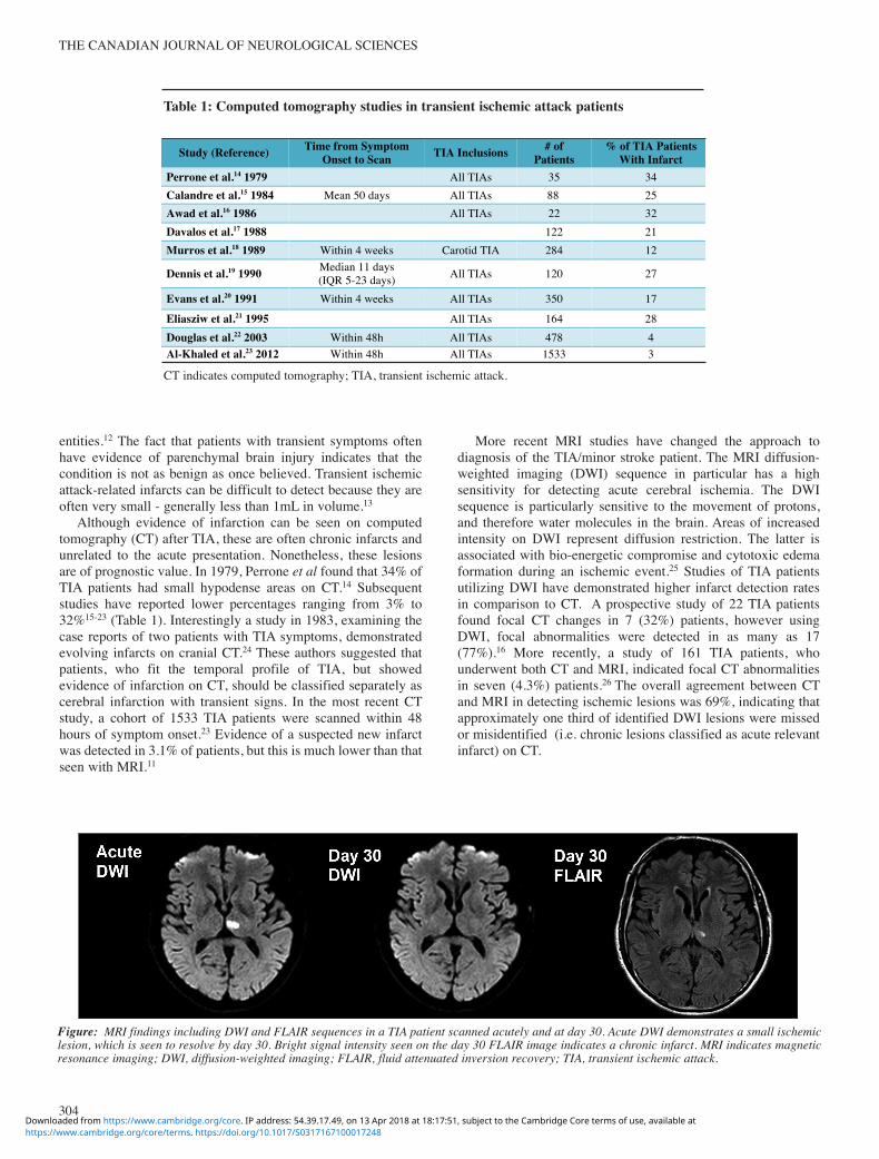
although evidence of infarction can be seen on computedtomography (Ct) after tia, these are often chronic infarcts andunrelated to the acute presentation. nonetheless, these lesionsare of prognostic value. in 1979, perrone et al found that 34% oftia patients had small hypodense areas on Ct.14 Subsequentstudies have reported lower percentages ranging from 3% to32%15-23 (table 1). interestingly a study in 1983, examining thecase reports of two patients with tia symptoms, demonstratedevolving infarcts on cranial Ct.24 these authors suggested thatpatients, who fit the temporal profile of tia, but showedevidence of infarction on Ct, should be classified separately ascerebral infarction with transient signs. in the most recent Ctstudy, a cohort of 1533 tia patients were scanned within 48hours of symptom onset.23 evidence of a suspected new infarctwas detected in 3.1% of patients, but this is much lower than thatseen with Mri.11
More recent Mri studies have changed the approach todiagnosis of the tia/minor stroke patient. the Mri diffusion-weighted imaging (dWi) sequence in particular has a highsensitivity for detecting acute cerebral ischemia. the dWisequence is particularly sensitive to the movement of protons,and therefore water molecules in the brain. areas of increasedintensity on dWi represent diffusion restriction. the latter isassociated with bio-energetic compromise and cytotoxic edemaformation during an ischemic event.25 Studies of tia patientsutilizing dWi have demonstrated higher infarct detection ratesin comparison to Ct. a prospective study of 22 tia patientsfound focal Ct changes in 7 (32%) patients, however usingdWi, focal abnormalities were detected in as many as 17(77%).16 More recently, a study of 161 tia patients, whounderwent both Ct and Mri, indicated focal Ct abnormalitiesin seven (4.3%) patients.26 the overall agreement between Ctand Mri in detecting ischemic lesions was 69%, indicating thatapproximately one third of identified dWi lesions were missedor misidentified (i.e. chronic lesions classified as acute relevantinfarct) on Ct.
the Canadian Journal of neurologiCal SCienCeS
304
Ct indicates computed tomography; tia, transient ischemic attack.
Study (Reference)
Time from Symptom
Onset to Scan
TIA Inclusions
# of
Patients
% of TIA Patients
With Infarct Perrone et al.14 1979 All TIAs 35 34 Calandre et al.15 1984 Mean 50 days All TIAs 88 25 Awad et al.16 1986 All TIAs 22 32 Davalos et al.17 1988 122 21 Murros et al.18 1989 Within 4 weeks Carotid TIA 284 12
Dennis et al.19 1990 Median 11 days (IQR 5-23 days) All TIAs 120 27
Evans et al.20 1991 Within 4 weeks All TIAs 350 17
Eliasziw et al.21 1995 All TIAs 164 28 Douglas et al.22 2003 Within 48h All TIAs 478 4 Al-Khaled et al.23 2012
Within 48h
All TIAs
1533
3
Table 1: Computed tomography studies in transient ischemic attack patients
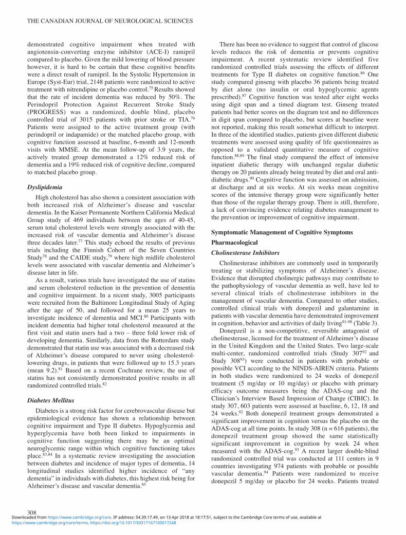
Figure: MRI findings including DWI and FLAIR sequences in a TIA patient scanned acutely and at day 30. Acute DWI demonstrates a small ischemiclesion, which is seen to resolve by day 30. Bright signal intensity seen on the day 30 FLAIR image indicates a chronic infarct. MRI indicates magneticresonance imaging; DWI, diffusion-weighted imaging; FLAIR, fluid attenuated inversion recovery; TIA, transient ischemic attack.
https://www.cambridge.org/core/terms. https://doi.org/10.1017/S0317167100017248Downloaded from https://www.cambridge.org/core. IP address: 54.39.17.49, on 13 Apr 2018 at 18:17:51, subject to the Cambridge Core terms of use, available at

Systematic Mri studies have found that the proportion oftia patients with positive dWi changes ranges from 11% to68%13,27-52 (table 2). the discrepancy in this large range ofreported values may be explained by variations in study design,study population and sample size. for example, time fromsymptom onset to Mri scan varied from within six hours to amedian 17 days later. in studies reporting later time points, dWiinfarcts may have resolved, while acute studies may have morefrequently reported positive dWi scans. additionally, althoughall studies included tia patients, some also included minorstroke and population sizes ranged from 14 to 1862 patients.Serial imaging studies indicate that some dWi lesions do resultin a visible chronic infarct (figure), while others do not. in astudy of 42 tia patients, relevant dWi abnormalities werereported in nearly half of patients.27 of the nine dWi positivepatients who had a follow-up imaging study two to seven monthsafter the event, four did not reveal any infarct relevant to theoriginal abnormality. in another multicenter study, dWi scanswere performed within 24 hours of symptom onset in 458patients and acute ischemic lesions were found in 96 (21%)patients.52 a follow-up Mri was done on 48 patients showingthat in five (10.4%), dWi lesions visible on admission had
disappeared. in a study by oppenheim and colleagues, 21% oftia patients with baseline positive dWi scans showed nopermanent injury when assessed 11.6 months later.44 lesions that‘reversed’ had smaller initial dWi volumes than those thatinfarcted. although this phenomenon has been referred to as‘dWi reversal’ it more likely represents a very small infarct thatis below the resolution of standard Mri.
Imaging Correlates of Cognitive Impairment Studies identifying clinical characteristics associated with thepresence of dWi abnormalities in tia patients focus morestrongly on functional impairments than cognitive deficits. alonger duration of neurological symptoms and the presence ofmotor weakness are both associated with the presence of dWilesions.34,41 the impact of dWi lesion presence, location,volume and number on cognitive performance is unknown.Studies correlating Mri findings in tia patients withneuropsychological testing are lacking. there is an increasedlikelihood of aphasia in tia patients with dWi lesion, but therelationship to other cognitive domains is unknown.45,51 onestudy found that tia patients with a positive dWi scan were 25times more likely to have aphasia than those with negative
le Journal Canadien deS SCienCeS neurologiqueS
Volume 41, No. 3 – May 2014 305
Mri indicated magnetic resonance imaging; dWi, diffusion-weighted imaging; tia, transient ischemic attack.
Study (Reference)
Time from Symptom
Onset to Scan
TIA Inclusions
# of Patients
% of TIA Patients
With Infarct Kidwell et al.27 1999 Mean 17h Cerebral and brainstem 42 48 Engelter et al.28 1999 Mean 36.5h Any focal deficit <24h 40 35 Takayama et al.29 2000 Within 48h All TIAs 19 37 Kamal et al.30 2002 Within 6h All TIAs 28 46 Ay et al.13 2002 Mean 39h Cerebral and brainstem 57 47 Marx et al.31 2002 Mean 10.7h Brainstem 14 29
Kastrup et al.32 2002 Mean 5 days (DWI +) Mean 6 days (DWI -) Carotid TIA 42 45
Rovira et al.33 2002 Mean 5 days Cerebral and brainstem 58 67 Crisostomo et al.34 2003 Mean 23h All TIAs 75 21 Nagura et al.35 2003 Median 17.3h All TIAs 45 31 Nakamura et al.36 2003 Within 48h All TIAS 18 50
Restrepo et al.37 2004 Mean 56 min (DWI +) Mean 33 min (DWI -) All TIAs 22 55
Purroy et al.38 2004 Within 7 days Cerebral and brainstem 83 33 Winbeck et al.39 2004 Within 24h Anterior circulation 60 30 Schulz et al.40 2004 Median 17 days All TIAs 136 13 Inatomi et al.41 2004 Median 4 days Cerebral and brainstem 129 44 Ay et al.42 2005 Mean/SD 22 ± 26h Cerebral and brainstem 87 41 Coutts et al.43 2005 Median 8.5h TIA and MS (NIH <6) 143 68 Oppenheim et al.44 2006 Median 24h All TIAs 103 35 Lamy et al.45 2006 Mean 42.4h Cerebral and brainstem 98 35 Prabhakaran et al.46 2007 Within 48h All TIAs 146 25 Redgrave et al.47 2007 Within 72h All TIAs 200 16 Calvet et al.48 2009 Median 19.5h All TIAs 339 40 Mlynash et al.49 2009 Mean/SD 23.2 ± 12.5h All TIAs 43 35 Adeoye et al.50 2010 Within 48h All TIAs 323 15 Al-Khaled et al.51 2013 Within 48h Time defined TIAs 1862 11 Miyagi et al.52 2013
Within 7 days
All TIAs
458
21
Table 2: MRI (DWI) studies in transient ischemic attack patients
https://www.cambridge.org/core/terms. https://doi.org/10.1017/S0317167100017248Downloaded from https://www.cambridge.org/core. IP address: 54.39.17.49, on 13 Apr 2018 at 18:17:51, subject to the Cambridge Core terms of use, available at

scans.34 in a study of 147 tia patients, disturbance of highercortical function evidence such as aphasia and spatial neglectwas a significant factor associated with dWi abnormalities.41
although dWi lesion characteristics in tia patients such asvolume, location and frequency have been reported, studiesinvestigating the association of these parameters with cognitiveimpairments are very scarce. in the Sydney Stroke Study, 170patients with stroke or tia and 96 age-matched controls wereexamined between three to six months following the event.53
patients were categorized as having vascular dementia, vascularcognitive impairment (vCi) or no cognitive impairment byconsensus and administered detailed neuropsychological testingalong with an Mri scan. although vascular dementia subjectshad larger infarct volumes than vCi subjects, cognitiveimpairments were not significantly correlated with volume ornumber of infarctions. instead, a significant relationship wasobserved between chronic deep white matter hyperintensitiesand cognitive deficits.
COGNITIVE PERFORMANCE TESTING AFTER STROKE AND TIACognitive Screening Assessments in Acute Ischemic Stroke Cognitive function is often compromised after stroke, but israrely assessed in stroke trials. Cognitive changes in acute strokepatients have been assessed quantitatively using differentbatteries of neuropsychological tests. While detailedneuropsychological testing represents the gold standard in termsof identifying cognitive impairment, this is generally impracticalin the acute setting, partially motivating the need for brief yetaccurate instruments. the two most commonly utilized tests incognitive screening for acute stroke are the Mini-Mental Stateexamination (MMSe) and the Montreal Cognitive assessment(MoCa). the MMSe, developed by folstein et al, is a cognitiveassessment of patient mental state and has been considered theclinical standard in stroke.54 the assessment comprises a batteryof individual tests that can be administered in approximately tenminutes. the test is scored out of a total of 30 points, with higherscores indicating higher functioning. items are grouped intocategories to test orientation, registration, recall, calculation andattention, naming, repetition, comprehension, reading, writingand drawing. the MMSe has however been criticized for beinginsensitive to mild cognitive impairment (MCi). this test alsoprimarily focuses on assessing memory and language abilitieswhile it fails to assess executive function, a common impairmentin cerebrovascular disease. the MoCa was a test designed more recently as a cognitivescreening assessment with increased sensitivity to MCi.55 theMoCa can be administered in a short ten minutes, where patientscan score a total of 30 points. the test is divided into eightsections assessing the cognitive domains of visuo-executivefunction, naming, memory, attention, language, abstraction,delayed recall and orientation. Cognitive impairment based onMoCa scoring has been designated by a cutoff score less than26.55,56
a number of factors make accurate assessment of cognitiveimpairment following stroke/tia challenging. depressionpresent following acute stroke has been implicated in worsefunctional outcome and also exacerbates any acute/chroniccognitive impairment.57-59 Cognitive assessments also vary in
their sensitivity for mild impairment. a validation study bynassreddine and colleagues in 2005 compared the sensitivitiesof MMSe and MoCa in 94 patients who met the criteria forMCi.55 the MMSe had a sensitivity of 18% in MCi patients,whereas the MoCa had a sensitivity of 90%. Several studiescomparing MoCa and MMSe in acute stroke and tia patientshave shown that MoCa is superior to the MMSe in detectingMCi.56,60-62 despite this, many studies continue to utilizeMMSe. there is no consensus on the cognitive test most appropriatefor assessment of post stroke cognitive impairment. a number ofstudies have compared MoCa and MMSe after tia with othermethods of evaluation including the addenbrooke’s Cognitiveexamination revised (aCe-r), telephone MoCa (t-MoCa)assessment and specific batteries of neuropsychologicaltests.63-65 in a population-based study of 100 patients assessed ≥one year after tia or stroke, MoCa and MMSe were comparedto the aCe-r for detecting MCi, defined using the neurologicaldisorders and Stroke-Canadian Stroke network vascularCognitive impairment battery.63 both the MoCa and aCe-rhad better sensitivity and specificity for MCi compared to theMMSe, which demonstrated a ceiling effect. a study by thesame group assessed the t-MoCa against face-to-face cognitivetests in 91 patients with tia or stroke.64 after one year, MoCasubtest scores for repetition, abstraction and verbal fluency weresignificantly worse by telephone than face-to-face testing. the t-MoCa was therefore considered limited in its ability to assessvisuoexecutive and complex language tasks. another studyaimed to compare the MoCa with a computerized battery ofneuropsychological tests for memory, attention and executivefunctions to detect mild to moderate cognitive impairments inpatients with tia or stroke.65 in comparison to stroke, tiapatients presented significantly better scores through bothmethods. however, the MoCa was able to identity many moresubjects with low scores (<26) compared to the neuro-psychological battery.
Cognitive Assessments in Vascular Dementia the most widely used criteria for diagnosis of vasculardementia is the national institute of neurological disorders andStroke and association internationale pour la recherché etl’ensignement en neurosciences (nindS-airen) criteria.66
these criteria rely on neuroimaging by Ct or Mri for evidenceof focal brain damage as well as cognitive deficits in at leastthree cognitive domains (one of which must be memory).patients are diagnosed as having probably, possible or definitevascular dementia based on the strength of the associationbetween cerebrovascular disease and cognitive impairment. thealzheimer’s disease assessment Scale (adaS) is used to assesscognitive dysfunction in individuals with alzheimer disease andother dementias.67 its subscale, the alzheimer’s diseaseassessment Scale-cognitive subscale (adaS-cog), is the mostpopular cognitive testing instrument used in clinical trials ofnootropics.68 it consists of 11 tasks measuring the disturbancesof memory, language, praxis, attention and other cognitiveabilities.
the Canadian Journal of neurologiCal SCienCeS
306https://www.cambridge.org/core/terms. https://doi.org/10.1017/S0317167100017248Downloaded from https://www.cambridge.org/core. IP address: 54.39.17.49, on 13 Apr 2018 at 18:17:51, subject to the Cambridge Core terms of use, available at

Cognitive Impairment after TIA Cognitive studies in tia patients have demonstrated thatcognitive impairments in many cases do persist beyond thetransient event. a comparative study of MoCa and MMSe, in 20patients diagnosed with tia or stroke, utilized the MMSe onadmission and the MoCa two weeks later.69 With cutoffs forimpairment set at ≤26, MMSe detected cognitive impairment in10% of patients, while MoCa detected impairment in 55%. in alarger population based study of 413 tia and stroke patients, theMMSe and MoCa were administered at a six month or five yearfollow-up.56 defining cognitive impairment as a score of <27 oneither test, 58% of patients with an MMSe score within normallimits had an abnormal MoCa. the MoCa indicated poorcognitive function in 70% of all patients. in a more recent study,cognitive impairment in 97 first time tia patients was assessedusing MoCa and compared to 100 healthy control patients.70
tia patients exhibited declined cognitive function withimpairments in verbal fluency, memory recall, abstraction, andvisuospatial/executive abilities.
Temporal Pattern of Cognitive Changes after TIA and MinorStroke the temporal pattern of motor, speech and sensory deficits intia/minor stroke is an acute onset and relatively rapid resolutionwith no long-term sequelae. it is unknown to what extent, if anycognition is affected in this population hyperacutely and whetherthese deficits actually resolve with the other neurologicalsymptoms. longitudinal studies that adequately assess thispattern in tia and minor stroke patients are scarce. Most studiesin tia and minor stroke patients lack serial assessment ofcognition at multiple time points, including immediately aftersymptom onset, and only assess deficits several days aftersymptom onset. in a cross-sectional study of 280 tia and minorstroke patients (national institutes of health Stroke Scale ≤ 3),MMSe was administered both at the initial assessment (baseline)and one month later and then repeated at one, two and fiveyears.7 patients were divided into two baseline groups: thoseseen between one and seven days and those seen between 8 and20 days. transient cognitive impairment (tCi) was defined as abaseline MMSe score ≥ 2 points lower than the one monthfollow-up MMSe. the rate of tCi in patients initially assessedwithin seven days (median four days) was 38.9%. this washigher than the rate of 19% seen in those examined between 8and 20 days (median 12 days). patients with tCi did show arecovery in mean MMSe scores from 23.9 ± 3.6 at baseline to27.2 ± 3.0 at one month. Sachdev and colleagues examined 128stroke or tia patients aged 49 to 87 years, with no history ofdementia or aphasia as a limiting factor (<3 on the aphasiaSeverity rating Scale).8 the initial assessment was done withinthree and six months post stroke, and a follow-up assessment 14months later. at baseline, patients were categorized based onseverity into three groups: vascular dementia, vascular cognitiveimpairment no dementia (vCi-nd) and no cognitiveimpairment. Cognitive tests showed a mean decline of 0.83points on MMSe between the two time points. patients impairedat baseline assessment (vascular dementia + vCi-nd), showed agreater decline in visuoconstructive function and abstractiondomains than patients with no cognitive impairment. in thisstudy, higher white matter hyperintensity load was a significant
predictor of cognitive decline. education level emerged as aprotective factor against cognitive decline following thetia/stroke. another study assessed cognitive function in 252patients with tia or non-disabling ischemic stroke at baseline(within six months of symptom onset) and again after one year.9at baseline, after administering MMSe along with the vasculardementia battery, 56% of patients were ‘cognitively intact’, 40%were ‘cognitively impaired but not demented’ and 4% were‘demented’. of the 252 patients, only 155 were reassessed at theone year follow-up. of these patients, 120 remained in the samecategories as at baseline. nineteen patients (12%) cognitivelyimpaired but not demented at baseline, improved to the pointthey were considered cognitively intact at one year. nine patientscognitively intact at baseline, deteriorated to cognitivelyimpaired but not demented at the one year follow-up and sevenpatients who were cognitively impaired but not demented atbaseline were demented at one year. demographic variables thatdifferentiated those who deteriorated from those who remainedstable or improved included hypertension, age, years ofeducation and MMSe score.
Knowledge Gaps Most of these studies included patients with large and oftendisabling strokes. it is therefore unsurprising that cognition isaffected in these patients, particularly as many have pre-existingdeficits. What remains unknown is the extent that tia affectscognition and what the temporal profile of any changes is.accurate predictors of improvement/worsening in cognitivestatus after tia/minor stroke are also unknown.
THERAPEUTICS AND MANAGEMENT OF COGNITIVE DECLINE treatment and management of cognitive decline is critical topatient care and preventing prolonged disability followingstroke. there is currently no standard treatment for vCi.
Prevention of Vascular Dementia: Vascular Risk FactorManagementHypertension hypertension is an established risk factor for cardiovasculardisease and stroke. a number of studies have identified a bloodpressure threshold of 130/80 mmhg above which strokes aremore likely to occur.71,72 hypertension has also been implicatedin affecting cognitive function. a recent systematic reviewinvestigated the association of arterial hypertension withincreased risk of vascular dementia.73 results demonstrated thatpeople with midlife hypertension have a doubled risk ofdeveloping vascular dementia comparing to those withouthypertension. there is some evidence suggesting treatment of hypertensiondecreases the risk of dementia. published studies have found thatlowering blood pressure in the middle aged or younger elderlypopulation can be useful for the prevention of late life dementia.a few major randomized controlled trials have reported positiveeffects of antihypertensive treatment on cognitive function inpatients with cerebrovascular disease. the heart outcomesprevention evaluation (hope) study was a randomized doubleblind study of 1013 high-risk patients with a history of stroke ortia.74 the study showed that significantly fewer patients
le Journal Canadien deS SCienCeS neurologiqueS
Volume 41, No. 3 – May 2014 307https://www.cambridge.org/core/terms. https://doi.org/10.1017/S0317167100017248Downloaded from https://www.cambridge.org/core. IP address: 54.39.17.49, on 13 Apr 2018 at 18:17:51, subject to the Cambridge Core terms of use, available at
demonstrated cognitive impairment when treated withangiotensin-converting enzyme inhibitor (aCe-i) ramiprilcompared to placebo. given the mild lowering of blood pressurehowever, it is hard to be certain that these cognitive benefitswere a direct result of ramipril. in the Systolic hypertension ineurope (Syst-eur) trial, 2148 patients were randomized to activetreatment with nitrendipine or placebo control.75 results showedthat the rate of incident dementia was reduced by 50%. theperindopril protection against recurrent Stroke Study(progreSS) was a randomized, double blind, placebocontrolled trial of 3015 patients with prior stroke or tia.76
patients were assigned to the active treatment group (withperindopril or indapamide) or the matched placebo group, withcognitive function assessed at baseline, 6-month and 12-monthvisits with MMSe. at the mean follow-up of 3.9 years, theactively treated group demonstrated a 12% reduced risk ofdementia and a 19% reduced risk of cognitive decline, comparedto matched placebo group.
Dyslipidemia high cholesterol has also shown a consistent association withboth increased risk of alzheimer’s disease and vasculardementia. in the Kaiser permanente northern California Medicalgroup study of 469 individuals between the ages of 40-45,serum total cholesterol levels were strongly associated with theincreased risk of vascular dementia and alzheimer’s diseasethree decades later.77 this study echoed the results of previoustrials including the finnish Cohort of the Seven CountriesStudy78 and the Caide study,79 where high midlife cholesterollevels were associated with vascular dementia and alzheimer’sdisease later in life. as a result, various trials have investigated the use of statinsand serum cholesterol reduction in the prevention of dementiaand cognitive impairment. in a recent study, 3005 participantswere recruited from the baltimore longitudinal Study of agingafter the age of 50, and followed for a mean 25 years toinvestigate incidence of dementia and MCi.80 participants withincident dementia had higher total cholesterol measured at thefirst visit and statin users had a two – three fold lower risk ofdeveloping dementia. Similarly, data from the rotterdam studydemonstrated that statin use was associated with a decreased riskof alzheimer’s disease compared to never using cholesterol-lowering drugs, in patients that were followed up to 15.3 years(mean 9.2).81 based on a recent Cochrane review, the use ofstatins has not consistently demonstrated positive results in allrandomized controlled trials.82
Diabetes Mellitus diabetes is a strong risk factor for cerebrovascular disease butepidemiological evidence has shown a relationship betweencognitive impairment and type ii diabetes. hypoglycemia andhyperglycemia have both been linked to impairments incognitive function suggesting there may be an optimalneuroglycemic range within which cognitive functioning takesplace.83,84 in a systematic review investigating the associationbetween diabetes and incidence of major types of dementia, 14longitudinal studies identified higher incidence of “anydementia” in individuals with diabetes, this highest risk being foralzheimer’s disease and vascular dementia.85
there has been no evidence to suggest that control of glucoselevels reduces the risk of dementia or prevents cognitiveimpairment. a recent systematic review identified fiverandomized controlled trials assessing the effects of differenttreatments for type ii diabetes on cognitive function.86 onestudy compared ginseng with placebo 36 patients being treatedby diet alone (no insulin or oral hypoglycemic agentsprescribed).87 Cognitive function was tested after eight weeksusing digit span and a timed diagram test. ginseng treatedpatients had better scores on the diagram test and no differencesin digit span compared to placebo, but scores at baseline werenot reported, making this result somewhat difficult to interpret.in three of the identified studies, patients given different diabetictreatments were assessed using quality of life questionnaires asopposed to a validated quantitative measure of cognitivefunction.88,89 the final study compared the effect of intensiveinpatient diabetic therapy with unchanged regular diabetictherapy on 20 patients already being treated by diet and oral anti-diabetic drugs.90 Cognitive function was assessed on admission,at discharge and at six weeks. at six weeks mean cognitivescores of the intensive therapy group were significantly betterthan those of the regular therapy group. there is still, therefore,a lack of convincing evidence relating diabetes management tothe prevention or improvement of cognitive impairment.
Symptomatic Management of Cognitive Symptoms PharmacologicalCholinesterase Inhibitors Cholinesterase inhibitors are commonly used in temporarilytreating or stabilizing symptoms of alzheimer’s disease.evidence that disrupted cholinergic pathways may contribute tothe pathophysiology of vascular dementia as well, have led toseveral clinical trials of cholinesterase inhibitors in themanagement of vascular dementia. Compared to other studies,controlled clinical trials with donepezil and galantamine inpatients with vascular dementia have demonstrated improvementin cognition, behavior and activities of daily living91-98 (table 3). donepzeil is a non-competitive, reversible antagonist ofcholinesterase, licensed for the treatment of alzheimer’s diseasein the united Kingdom and the united States. two large-scalemulti-center, randomized controlled trials (Study 30792 andStudy 30893) were conducted in patients with probable orpossible vCi according to the nindS-airen criteria. patientsin both studies were randomized to 24 weeks of donepeziltreatment (5 mg/day or 10 mg/day) or placebo with primaryefficacy outcome measures being the adaS-cog and theClinician’s interview based impression of Change (CibiC). instudy 307, 603 patients were assessed at baseline, 6, 12, 18 and24 weeks.92 both donepezil treatment groups demonstrated asignificant improvement in cognition versus the placebo on theadaS-cog at all time points. in study 308 (n = 616 patients), thedonepezil treatment group showed the same statisticallysignificant improvement in cognition by week 24 whenmeasured with the adaS-cog.93 a recent larger double-blindrandomized controlled trial was conducted at 111 centers in 9countries investigating 974 patients with probable or possiblevascular dementia.94 patients were randomized to receivedonepezil 5 mg/day or placebo for 24 weeks. patients treated
the Canadian Journal of neurologiCal SCienCeS
308https://www.cambridge.org/core/terms. https://doi.org/10.1017/S0317167100017248Downloaded from https://www.cambridge.org/core. IP address: 54.39.17.49, on 13 Apr 2018 at 18:17:51, subject to the Cambridge Core terms of use, available at

with donepezil showed significant improvement from baseline toend point based on the vascular alzheimer’s disease assessmentScale-Cognitive Subscale (vadaS-cog), compared to therelative stability of the placebo group. it is thereforerecommended by the american heart association/americanStroke association (aha/aSa) that donepezil can be useful forcognitive enhancement in patients with vascular dementia.99
galantamine is a specific, competitive and reversibleacetylcholinesterase inhibitor that has been shown to improvecognition and behavior in patients with alzheimer’s typedementia.100,101 in one randomized, placebo controlled trial, 592patients with probable vascular dementia or alzheimer’s diseasewith cerebrovascular disease were randomized to placebo orgalantamine 24mg/day for six months following a four weekplacebo period.95 according to the adaS-cog scores, treatmentgroup patients significantly improved from baseline to the sixmonth period compared to those assigned placebo, whereasplacebo group deteriorated below baseline. another randomizedplacebo controlled trial investigated galantamine in 788 patientswith probable vascular dementia using slow dose escalation upto 26 weeks.96 after a four week placebo run, patients wererandomized to receive increasing doses of placebo orgalantamine, initiated at 4mg twice daily and escalated to a finaldose of 8 or 12mg twice daily. improvements in adaS-cogscores in those treated with galantamine were significantlygreater compared with placebo after 26 weeks. the aha/aSaguidelines therefore suggest that galantamine can be beneficialto patients with mixed alzheimer’s disease or vasculardementia.99
Glutamate Receptor Antagonists glutamate is the principal excitatory neurotransmitter thatstimulates n-methyl-d-aspartate (ndMa) receptors in corticalneurons. there is some evidence that sustained elevation ofglutamate may underlie the neuronal loss that is observed indementia.102 ischemia has been associated with the repeatedstimulation of nMda receptors; agents that block thestimulation of this receptor may play a protective role,preventing further neurodegeneration leading to cognitivedecline. Memantine is a nMda receptor antagonist, shown tohave neuro-protective effects that help improve cognitiveperformance in vascular dementia patients.97,103 there have beenseveral clinical studies demonstrating memantine’s effects onimproving cognitive performance in dementia patients97,98,104
(table 3). in one multicenter, randomized controlled trial, 321patients with mild to moderate vascular dementia were randomlyallocated to receive placebo or memantine.97 after a two weekplacebo run-in period, patients received doses of 20 mg/day for28 weeks to follow. Mean adaS-cog scores showed thatpatients in the memantine group improved from baseline whilethe placebo group deteriorated in function. in a largerrandomized controlled trial, 579 patients with probable vasculardementia were randomized to placebo or treatment with 20-mg/dmemantine for 28 weeks.98 Memantine resulted in improvementin adaS-cog scores, not seen in placebo treated patients.according to aha/aSa, the benefits of memantine are not wellestablished in vascular dementia.99
le Journal Canadien deS SCienCeS neurologiqueS
Volume 41, No. 3 – May 2014 309
nindS-airen indicates national institute of neurological disorders and Stroke and association internationale pour la recherchéet l'enseignement en neurosciences; adaS-cog, alzheimer’s disease assessment Scale-Cognitive Subscale; CibiC, Clinicianinterview based impression of Change; vadaS-cog, vascular alzheimer’s disease assessment Scale-Cognitive Subscale;ninCdS-adrda, national institute of neurological and Communicative diseases and Stroke/alzheimer's disease and relateddisorders association.
Treatment
Study Reference
Trial Duration
Dosage
Participants
Measured Outcomes
Donepezil Black et al.92 (Donepezil 307)
24 wks 5mg daily and 10 mg daily
603 patients with probable or possible vascular dementia by NINDS-AIREN
ADAS-cog and CIBIC improvement in both treatment group
Donepezil Wilkinson et al.93 (Donepezil 308)
24 wks 5mg daily and 10 mg daily
616 patients with probable or possible vascular dementia by NINDS-AIREN
ADAS-cog and CIBIC improvement in both treatment groups
Donepezil Roman et al.94 24 wks 5mg daily 974 patients with probable or possible vascular dementia by NINDS-AIREN
VADAS-cog improvement in treatment group
Galantamine Erkinjuntti et al.95 24 wks 24mg daily 592 patients with NINCDS-ADRDA Alzheimer’s disease with cerebrovascular disease or vascular dementia by NINDS-AIREN
ADAS-cog improvement in treatment group
Galantamine Auchus et al.96 26 wks Escalated to 8 mg or 12 mg twice daily
788 patients with probable vascular dementia by NINDS-AIREN
ADAS-cog/11 improvement in treatment group
Memantine Orgogozo et al.97 (MMM300)
28 wks 20mg daily 321 patients with probable vascular dementia by NINDS-AIREN
ADAS-cog improvement in treatment group
Memantine Wilcock et al.98 (MMM500)
28 wks 20mg daily 579 patients with probable vascular dementia by NINDS-AIREN
ADAS-cog improvement in treatment group
Table 3: Pharmacological evidence for treatment of vascular dementia
https://www.cambridge.org/core/terms. https://doi.org/10.1017/S0317167100017248Downloaded from https://www.cambridge.org/core. IP address: 54.39.17.49, on 13 Apr 2018 at 18:17:51, subject to the Cambridge Core terms of use, available at

Non-PharamcologicalPhysical Activity physical exercise has been associated with several beneficialeffects including the reduced risk of alzheimer’s disease andcognitive decline. Many longitudinal studies in the health elderlyhave consistently found that regular physical activity wasassociated with better cognitive function and less cognitivedecline later in life. a meta-analysis including 16 prospectivestudies of non-demented patients suggested that physical activityreduced the risk of dementia and alzheimer’s disease by 28%and 45% respectively.105
Studies targeting patients with alzheimer’s disease, vasculardementia and cognitive impairment have reported similarfindings. a recently conducted systematic review included a totalof 24 longitudinal studies of 1378 patients with vasculardementia.106 the meta-analysis technique was used todemonstrate a significant reduced risk for vascular dementia inpeople who were naturally more physically active compared tothose who were not. a four month randomized controlled trialconducted in 40 patients diagnosed with alzheimer’s diseaseassessed the effectiveness of a community based home exerciseprogram on improving cognitive and physical function.107
patients were randomly assigned to usual treatment plus exerciseor usual treatment alone groups. When assessed at baseline andthe four month follow-up, patients who exercised had improvedcognition compared to controls when assessed with MMSe. onemeta-analysis reviewed 30 randomized controlled trialsevaluating exercise in patients with cognitive impairments.108
exercise was associated with statistically significantimprovements in cognitive function as well as physical fitness.
Education higher levels of education have been associated with areduced risk dementia and cognitive impairment during aging. ithas yet to be determined if education protects from developmentof neurodegenerative brain pathology or if it increases the brain’sresilience against dementia related pathology. the cognitivereserve hypothesis suggests that individuals exposed to anenriched environment through higher education will maintainhigher cognitive function in later years by functionallycompensating for any neurological load.109
Studies have examined the relationship between educationlevels and cognitive changes in both normal and dementedadults. a recent systematic review aimed at investigating thecognitive reserve hypothesis in 133 quantitative studiesincluding both healthy and alzheimer’s disease patients.110 ameta-analysis of this data showed that those with lowereducation levels had a higher risk for dementia. a longitudinalstudy of 630 cognitively healthy individuals aged 50 to 80assessed educational level and mental demands at work asrelated to cognitive decline.111 at the three year follow-up,persons with low education (primary education and lowervocational secondary education) and lower mental workloadshowed accelerated cognitive decline in speed (Stroop test)memory (verbal learning test) and general cognitive status(MMSe). there is current evidence that lifelong bilingualism is also afactor that protects against cognitive decline and the onset ofdementia.112 in a recent study of 211 patients diagnosed with
probable alzheimer’s disease, age, education and languagehistory were recorded, classifying 102 patients as bilingual and109 patients as monolingual.113 results demonstrated thatcompared to monolinguals, bilingual patients had beendiagnosed 4.3 years later and had reported the onset ofsymptoms 5.1 years later. a study investigating possible neuralcorrelates of this effect tested the hypothesis that bilingualism isassociated with maintenance of white matter integrity in olderindividuals.114 results from diffusion tensor imaging showedstronger structural and functional connectivity in bilingualscompared to monolinguals. participation in cognitively stimulating activities has beenhypothesized to reduce the risk of dementia cognitive decline,although evidence of this association is scarce. a longitudinalcohort study tested this hypothesis in 801 non-dementedindividuals evaluated at baseline and a mean follow-up of 4.5years.115 results showed that an individual reporting frequentcognitive activity at baseline had a 47% reduced chance ofdeveloping alzheimer’s disease. another interesting study of488 cognitively intact individuals assessed the effect of self-reported cognitive activities on the onset of cognitive decline.116
findings showed that every additional self-reported day ofcognitive activity at baseline, delayed the onset of acceleratedmemory decline by 0.18 years. these findings provide hope thatengaging in cognitively stimulating activities may reduce therisk of dementia.
Smoking Smoking has been associated with both cognitive decline anda significantly increased risk of vascular dementia andalzheimer’s disease. a meta-analysis conducted in 2007identified 19 studies with at least 12 months of follow-upshowing current smokers had an increased risk of dementia andcognitive decline ranging from 40-80% compared with peoplewho have never smoked.117 recently, a population based cohortstudy of 21,123 patients surveyed between 1978 and 1985demonstrated that heavy smoking in midlife was associated witha greater than 100% increase in risk of dementia, alzheimer’sdisease and vascular dementia more than 20 years later.118
evidence that smoking cessation prevents cognitiveimpairment of onset of dementia is limited. the honolulu-asiaaging Study was one of the first to investigate smokingcessation and cognitive function.119 this study found that theodds of cognitive impairment was 36% higher amongcontinuous smokers than never smokers and significantlydeclined in long term quitters. a recent smoking cessation trialrecruited 229 older smokers and 98 never smokers to assess howcessation in chronic smokers would affect rate of change inadaS-cog scores measured over 24 months.120 resultsdemonstrated that chronic smokers who continued to smoke orstopped smoking for less than 18 months experienced greatercognitive decline and greater deterioration of memory scoresover two years when compared with never smokers.
CONCLUSION Cognitive changes are common in cerebrovascular patients.although there is good evidence that patients with disablingstroke also have cognitive symptoms, there are less data relatedto tia patients. arguably, these are the more relevant patients tostudy, as they make functional recoveries, returning to live and
the Canadian Journal of neurologiCal SCienCeS
310https://www.cambridge.org/core/terms. https://doi.org/10.1017/S0317167100017248Downloaded from https://www.cambridge.org/core. IP address: 54.39.17.49, on 13 Apr 2018 at 18:17:51, subject to the Cambridge Core terms of use, available at
work in the community. assessment of cognition in acute strokepatients is challenging, due to the presence of other neurologicaldeficits. this has led to under-recognition of the seriousness ofcognitive changes in acute cerebrovascular disease patients,particularly those with minor or even apparently transientsymptoms. reliable predictors of cognitive impairment have notbeen identified, although imaging correlates may hold somepromise. the temporal pattern of cognitive impairment after tiahas not yet been adequately characterized. given theimplications for patient rehabilitation, return to work, andactivities of daily living, we suggest that this is a researchpriority.
ACKNOWLEDGEMENTS KSb holds a Canada research Chair in Cerebrovasculardisease, an alberta innovates health Solutions new investigatoraward and the heart and Stroke foundation professorship inStroke Medicine at the university of alberta.
REFERENCES1. donnan ga, fisher M, Macleod M, davis SM. Stroke. lancet.
2008;371:1612-23.2. roger vl, go aS, lloyd-Jones dM, et al. heart disease and stroke
statistics--2011 update: a report from the american heartassociation. Circulation. 2011;123:e18-e209.
3. the World health organization MoniCa project (monitoringtrends and determinants in cardiovascular disease): a majorinternational collaboration. Who MoniCa project principalinvestigators. J Clin epidemiol. 1988;41:105-14.
4. rothwell pM, Warlow Cp. timing of tias preceding stroke: timewindow for prevention is very short. neurology. 2005;64:817-20.
5. desmond dW, Moroney Jt, Sano M, Stern y. incidence ofdementia after ischemic stroke: results of a longitudinal study.Stroke. 2002;33:2254-60.
6. tatemichi tK, desmond dW, paik M, et al. Clinical determinantsof dementia related to stroke. ann neurol. 1993;33:568-75.
7. pendlebury St, Wadling S, Silver le, Mehta Z, rothwell pM.transient cognitive impairment in tia and minor stroke. Stroke.2011;42:3116-21.
8. Sachdev pS, brodaty h, valenzuela MJ, lorentz lM, Koschera a.progression of cognitive impairment in stroke patients.neurology. 2004;63:1618-23.
9. tham W, auchus ap, thong M, et al. progression of cognitiveimpairment after stroke: one year results from a longitudinalstudy of Singaporean stroke patients. J neurol Sci. 2002;203-204:49-52.
10. ovbiagele b, Kidwell CS, Saver Jl. epidemiological impact in theunited States of a tissue-based definition of transient ischemicattack. Stroke. 2003;34:919-24.
11. fazekas f, fazekas g, Schmidt r, Kapeller p, offenbacher h.Magnetic resonance imaging correlates of transient cerebralischemic attacks. Stroke. 1996;27:607-11.
12. Kidwell CS, Warach S. acute ischemic cerebrovascular syndrome:diagnostic criteria. Stroke. 2003;34:2995-8.
13. ay h, oliveira-filho J, buonanno fS, et al. 'footprints' of transientischemic attacks: a diffusion-weighted Mri study. Cerebrovascdis. 2002;14:177-86.
14. perrone p, Candelise l, Scotti g, de grandi C, Scialfa g. Ctevaluation in patients with transient ischemic attack. Correlationbetween clinical and angiographic findings. eur neurol. 1979;18:217-21.
15. Calandre l, gomara S, bermejo f, Millan JM, del pozo g. Clinical-Ct correlations in tia, rind, and strokes with minimumresiduum. Stroke. 1984;15:663-6.
16. awad i, Modic M, little Jr, furlan aJ, Weinstein M. focalparenchymal lesions in transient ischemic attacks: correlation of
computed tomography and magnetic resonance imaging. Stroke.1986;17:399-403.
17. davalos a, Matias-guiu J, torrent o, vilaseca J, Codina a.Computed tomography in reversible ischaemic attacks: clinicaland prognostic correlations in a prospective study. J neurol.1988;235:155-8.
18. Murros Ke, evans gW, toole Jf, howard g, rose la. Cerebralinfarction in patients with transient ischemic attacks. J neurol.1989;236:182-4.
19. dennis M, bamford J, Sandercock p, Molyneux a, Warlow C.Computed tomography in patients with transient ischaemicattacks: when is a transient ischaemic attack not a transientischaemic attack but a stroke? J neurol. 1990;237:257-61.
20. evans gW, howard g, Murros Ke, rose la, toole Jf. Cerebralinfarction verified by cranial computed tomography andprognosis for survival following transient ischemic attack.Stroke. 1991;22:431-6.
21. eliasziw M, Streifler Jy, Spence Jd, fox aJ, hachinski vC,barnett hJ. prognosis for patients following a transient ischemicattack with and without a cerebral infarction on brain Ct. northamerican Symptomatic Carotid endarterectomy trial(naSCet) group. neurology. 1995;45:428-31.
22. douglas vC, Johnston CM, elkins J, Sidney S, gress dr, JohnstonSC. head computed tomography findings predict short-termstroke risk after transient ischemic attack. Stroke. 2003;34:2894-8.
23. al-Khaled M, Matthis C, Munte tf, eggers J. use of cranial Ct toidentify a new infarct in patients with a transient ischemic attack.brain behav. 2012;2:377-81.
24. Waxman Sg, toole Jf. temporal profile resembling tia in thesetting of cerebral infarction. Stroke. 1983;14:433-7.
25. nentwich lM, veloz W. neuroimaging in acute stroke. emerg MedClin north am. 2012;30:659-80.
26. forster a, gass a, Kern r, et al. brain imaging in patients withtransient ischemic attack: a comparison of computedtomography and magnetic resonance imaging. eur neurol. 2012;67:136-41.
27. Kidwell CS, alger Jr, di Salle f, et al. diffusion Mri in patientswith transient ischemic attacks. Stroke. 1999;30:1174-80.
28. engelter St, provenzale JM, petrella Jr, alberts MJ. diffusion Mrimaging and transient ischemic attacks. Stroke. 1999;30:2762-3.
29. takayama h, Mihara b, Kobayashi M, hozumi a, Sadanaga h,gomi S. [usefulness of diffusion-weighted Mri in the diagnosisof transient ischemic attacks]. no to Shinkei. 2000;52:919-23.
30. Kamal aK, Segal aZ, ulug aM. quantitative diffusion-weightedMr imaging in transient ischemic attacks. aJnr am Jneuroradiol. 2002;23:1533-8.
31. Marx JJ, Mika-gruettner a, thoemke f, et al. diffusion weightedmagnetic resonance imaging in the diagnosis of reversibleischaemic deficits of the brainstem. J neurol neurosurgpsychiatry. 2002;72:572-5.
32. Kastrup a, Schulz Jb, Mader i, dichgans J, Kuker W. diffusion-weighted Mri in patients with symptomatic internal carotidartery disease. J neurol. 2002;249:1168-74.
33. rovira a, rovira-gols a, pedraza S, grive e, Molina C, alvarez-Sabin J. diffusion-weighted Mr imaging in the acute phase oftransient ischemic attacks. aJnr am J neuroradiol. 2002;23:77-83.
34. Crisostomo ra, garcia MM, tong dC. detection of diffusion-weighted Mri abnormalities in patients with transient ischemicattack: correlation with clinical characteristics. Stroke. 2003;34:932-7.
35. nagura J, Suzuki K, Johnston SC, et al. diffusion-weighted Mri inevaluation of transient ischemic attack. J Stroke Cerebrovascdis. 2003;12:137-42.
36. nakamura t, uchiyama S, Shibagaki y, iwata M. [abnormalitieson diffusion-weighted magnetic resonance imaging in patientswith transient ischemic attack]. rinsho Shinkeigaku. 2003;43:122-5.
37. restrepo l, Jacobs Ma, barker pb, Wityk rJ. assessment oftransient ischemic attack with diffusion- and perfusion-weightedimaging. aJnr am J neuroradiol. 2004;25:1645-52.
le Journal Canadien deS SCienCeS neurologiqueS
Volume 41, No. 3 – May 2014 311https://www.cambridge.org/core/terms. https://doi.org/10.1017/S0317167100017248Downloaded from https://www.cambridge.org/core. IP address: 54.39.17.49, on 13 Apr 2018 at 18:17:51, subject to the Cambridge Core terms of use, available at

38. purroy f, Montaner J, rovira a, delgado p, quintana M, alvarez-Sabin J. higher risk of further vascular events among transientischemic attack patients with diffusion-weighted imaging acuteischemic lesions. Stroke. 2004;35:2313-19.
39. Winbeck K, bruckmaier K, etgen t, von einsiedel hg, rottingerM, Sander d. transient ischemic attack and stroke can bedifferentiated by analyzing early diffusion-weighted imagingsignal intensity changes. Stroke. 2004;35:1095-9.
40. Schulz ug, briley d, Meagher t, Molyneux a, rothwell pM.diffusion-weighted Mri in 300 patients presenting late withsubacute transient ischemic attack or minor stroke. Stroke. 2004;35:2459-65.
41. inatomi y, Kimura K, yonehara t, fujioka S, uchino M. dWiabnormalities and clinical characteristics in tia patients.neurology. 2004;62:376-80.
42. ay h, Koroshetz WJ, benner t, et al. transient ischemic attack withinfarction: a unique syndrome? ann neurol. 2005;57:679-86.
43. Coutts Sb, Simon Je, eliasziw M, et al. triaging transient ischemicattack and minor stroke patients using acute magnetic resonanceimaging. ann neurol. 2005;57:848-54.
44. oppenheim C, lamy C, touze e, et al. do transient ischemicattacks with diffusion-weighted imaging abnormalitiescorrespond to brain infarctions? aJnr am J neuroradiol. 2006;27:1782-7.
45. lamy C, oppenheim C, Calvet d, et al. diffusion-weighted Mrimaging in transient ischaemic attacks. eur radiol. 2006;16:1090-5.
46. prabhakaran S. reversible brain ischemia: lessons from transientischemic attack. Curr opin neurol. 2007;20:65-70.
47. redgrave Jn, Schulz ug, briley d, Meagher t, rothwell pM.presence of acute ischaemic lesions on diffusion-weightedimaging is associated with clinical predictors of early risk ofstroke after transient ischaemic attack. Cerebrovasc dis. 2007;24:86-90.
48. Calvet d, touze e, oppenheim C, turc g, Meder Jf, Mas Jl. dWilesions and tia etiology improve the prediction of stroke aftertia. Stroke. 2009;40:187-92.
49. Mlynash M, olivot JM, tong dC, et al. yield of combinedperfusion and diffusion Mr imaging in hemispheric tia.neurology. 2009;72:1127-33.
50. adeoye o, heitsch l, Moomaw CJ, et al. how much wouldperforming diffusion-weighted imaging for all transientischemic attacks increase Mri utilization? Stroke. 2010;41:2218-22.
51. al-Khaled M, Matthis C, Munte tf, eggers J. the incidence andclinical predictors of acute infarction in patients with transientischemic attack using Mri including dWi. neuroradiology.2013;55:157-63.
52. Miyagi t, uehara t, Kimura K, et al. examination timing and lesionpatterns in diffusion-weighted magnetic resonance imaging ofpatients with classically defined transient ischemic attack. JStroke Cerebrovasc dis. 2013;22:e310-16.
53. Sachdev pS, brodaty h, valenzuela MJ, et al. the neuro-psychological profile of vascular cognitive impairment in strokeand tia patients. neurology. 2004;62:912-19.
54. folstein Mf, robins ln, helzer Je. the Mini-Mental Stateexamination. arch gen psychiatry. 1983;40:812.
55. nasreddine ZS, phillips na, bedirian v, et al. the MontrealCognitive assessment, MoCa: a brief screening tool for mildcognitive impairment. J am geriatr Soc. 2005;53:695-9.
56. pendlebury St, Cuthbertson fC, Welch SJ, Mehta Z, rothwell pM.underestimation of cognitive impairment by Mini-Mental Stateexamination versus the Montreal Cognitive assessment inpatients with transient ischemic attack and stroke: a population-based study. Stroke. 2010;41:1290-3.
57. lenzi gl, altieri M, Maestrini i. post-stroke depression. revneurol (paris). 2008;164:837-40.
58. Kimura M, robinson rg, Kosier Jt. treatment of cognitiveimpairment after poststroke depression: a double-blind treatmenttrial. Stroke. 2000;31:1482-6.
59. Kouwenhoven Se, Kirkevold M, engedal K, Kim hS. depressionin acute stroke: prevalence, dominant symptoms and associated
factors. a systematic literature review. disabil rehabil. 2011;33:539-56.
60. dong y, Sharma vK, Chan bp, et al. the Montreal Cognitiveassessment (MoCa) is superior to the Mini-Mental Stateexamination (MMSe) for the detection of vascular cognitiveimpairment after acute stroke. J neurol Sci. 2010;299:15-18.
61. toglia J, fitzgerald Ka, o'dell MW, Mastrogiovanni ar, lin Cd.the Mini-Mental State examination and Montreal Cognitiveassessment in persons with mild subacute stroke: relationship tofunctional outcome. arch phys Med rehabil. 2011;92:792-8.
62. godefroy o, fickl a, roussel M, et al. is the Montreal Cognitiveassessment superior to the Mini-Mental State examination todetect poststroke cognitive impairment? a study withneuropsychological evaluation. Stroke. 2011;42:1712-16.
63. pendlebury St, Mariz J, bull l, Mehta Z, rothwell pM. MoCa,aCe-r, and MMSe versus the national institute ofneurological disorders and Stroke-Canadian Stroke networkvascular Cognitive impairment harmonization Standardsneuropsychological battery after tia and stroke. Stroke. 2012;43:464-9.
64. pendlebury St, Welch SJ, Cuthbertson fC, Mariz J, Mehta Z,rothwell pM. telephone assessment of cognition after transientischemic attack and stroke: modified telephone interview ofcognitive status and telephone Montreal Cognitive assessmentversus face-to-face Montreal Cognitive assessment andneuropsychological battery. Stroke. 2013;44:227-9.
65. Shopin l, Shenhar-tsarfaty S, ben assayag e, et al. Cognitiveassessment in proximity to acute ischemic Stroke/transientischemic attack: Comparison of the Montreal Cognitiveassessment test and MindStreams Computerized Cognitiveassessment battery. dement geriatr Cogn disord. 2013;36:36-42.
66. roman gC, tatemichi tK, erkinjuntti t, et al. vascular dementia:diagnostic criteria for research studies. report of the nindS-airen international Workshop. neurology. 1993;43:250-60.
67. rosen Wg, Mohs rC, davis Kl. a new rating scale foralzheimer's disease. am J psychiatry. 1984;141:1356-64.
68. Connor dJ, Sabbagh Mn. administration and scoring variance onthe adaS-Cog. J alzheimers dis. 2008;15:461-4.
69. MacKenzie g, gould l, ireland S, leblanc K, Sahlas d. detectingcognitive impairment in clients with mild stroke or transientischemic attack attending a stroke prevention clinic. Can Jneurosci nurs. 2011;33:47-50.
70. Wang l, Jia J, Wu l. the relationship between cognitiveimpairment and cerebral blood flow changes after transientischaemic attack. neurol res. 2013;35:580-5.
71. benavente or, hart rg, McClure la, Szychowski JM, Coffey CS,pearce la. effects of clopidogrel added to aspirin in patientswith recent lacunar stroke. n engl J Med. 2012;367:817-25.
72. Chobanian av, bakris gl, black hr, et al. the Seventh report ofthe Joint national Committee on prevention, detection,evaluation, and treatment of high blood pressure: the JnC 7report. JaMa. 2003;289:2560-72.
73. Sharp Si, aarsland d, day S, Sonnesyn h, ballard C. hypertensionis a potential risk factor for vascular dementia: systematicreview. int J geriatr psychiatry. 2011;26:661-9.
74. bosch J, yusuf S, pogue J, et al. use of ramipril in preventingstroke: double blind randomised trial. bMJ. 2002;324:699-702.
75. forette f, Seux Ml, Staessen Ja, et al. prevention of dementia inrandomised double-blind placebo-controlled Systolichypertension in europe (Syst-eur) trial. lancet. 1998;352:1347-51.
76. tzourio C, anderson C, Chapman n, et al. effects of bloodpressure lowering with perindopril and indapamide therapy ondementia and cognitive decline in patients with cerebrovasculardisease. arch intern Med. 2003;163:1069-75.
77. Solomon a, Kivipelto M, Wolozin b, Zhou J, Whitmer ra. Midlifeserum cholesterol and increased risk of alzheimer's and vasculardementia three decades later. dement geriatr Cogn disord.2009;28:75-80.
78. Menotti a, blackburn h, Kromhout d, et al. Changes in populationcholesterol levels and coronary heart disease deaths in sevencountries. eur heart J. 1997;18:566-71.
the Canadian Journal of neurologiCal SCienCeS
312https://www.cambridge.org/core/terms. https://doi.org/10.1017/S0317167100017248Downloaded from https://www.cambridge.org/core. IP address: 54.39.17.49, on 13 Apr 2018 at 18:17:51, subject to the Cambridge Core terms of use, available at

79. Kivipelto M, helkala el, laakso Mp, et al. apolipoprotein eepsilon4 allele, elevated midlife total cholesterol level, and highmidlife systolic blood pressure are independent risk factors forlate-life alzheimer disease. ann intern Med. 2002;137:149-55.
80. beydoun Ma, beason-held ll, Kitner-triolo Mh, et al. Statinsand serum cholesterol's associations with incident dementia andmild cognitive impairment. J epidemiol Community health.2011;65:949-57.
81. haag Md, hofman a, Koudstaal pJ, Stricker bh, breteler MM.Statins are associated with a reduced risk of alzheimer diseaseregardless of lipophilicity. the rotterdam Study. J neurolneurosurg psychiatry. 2009;80:13-17.
82. Mcguinness b, o'hare J, Craig d, bullock r, Malouf r, passmorep. Cochrane review on 'Statins for the treatment of dementia'. intJ geriatr psychiatry. 2013;28:119-26.
83. Cox dJ, Kovatchev bp, gonder-frederick la, et al. relationshipsbetween hyperglycemia and cognitive performance amongadults with type 1 and type 2 diabetes. diabetes Care. 2005;28:71-7.
84. Mcnay eC. the impact of recurrent hypoglycemia on cognitivefunction in aging. neurobiol aging. 2005;26 Suppl 1:76-9.
85. biessels gJ, Staekenborg S, brunner e, brayne C, Scheltens p. riskof dementia in diabetes mellitus: a systematic review. lancetneurol. 2006;5:64-74.
86. areosa Sa, grimley ev. effect of the treatment of type ii diabetesmellitus on the development of cognitive impairment anddementia. Cochrane database Syst rev. 2002:Cd003804.
87. Sotaniemi ea, haapakoski e, rautio a. ginseng therapy in non-insulin-dependent diabetic patients. diabetes Care. 1995;18:1373-5.
88. testa Ma, Simonson dC. health economic benefits and quality oflife during improved glycemic control in patients with type 2diabetes mellitus: a randomized, controlled, double-blind trial.JaMa. 1998;280:1490-6.
89. quality of life in type 2 diabetic patients is affected bycomplications but not by intensive policies to improve bloodglucose or blood pressure control (uKpdS 37). u.K.prospective diabetes Study group. diabetes Care. 1999;22:1125-36.
90. naor M, Steingruber hJ, Westhoff K, Schottenfeld-naor y, griesaf. Cognitive function in elderly non-insulin-dependent diabeticpatients before and after inpatient treatment for metaboliccontrol. J diabetes Complications. 1997;11:40-6.
91. erkinjuntti t, roman g, gauthier S, feldman h, rockwood K.emerging therapies for vascular dementia and vascular cognitiveimpairment. Stroke. 2004;35:1010-17.
92. black S, roman gC, geldmacher dS, et al. efficacy andtolerability of donepezil in vascular dementia: positive results ofa 24-week, multicenter, international, randomized, placebo-controlled clinical trial. Stroke. 2003;34:2323-30.
93. Wilkinson d, doody r, helme r, et al. donepezil in vasculardementia: a randomized, placebo-controlled study. neurology.2003;61:479-86.
94. roman gC, Salloway S, black Se, et al. randomized, placebo-controlled, clinical trial of donepezil in vascular dementia:differential effects by hippocampal size. Stroke. 2010;41:1213-21.
95. erkinjuntti t, Kurz a, gauthier S, bullock r, lilienfeld S,damaraju Cv. efficacy of galantamine in probable vasculardementia and alzheimer's disease combined withcerebrovascular disease: a randomised trial. lancet. 2002;359:1283-90.
96. auchus ap, brashear hr, Salloway S, Korczyn ad, de deyn pp,gassmann-Mayer C. galantamine treatment of vasculardementia: a randomized trial. neurology. 2007;69:448-58.
97. orgogozo JM, rigaud aS, Stoffler a, Mobius hJ, forette f.efficacy and safety of memantine in patients with mild tomoderate vascular dementia: a randomized, placebo-controlledtrial (MMM 300). Stroke. 2002;33:1834-9.
98. Wilcock g, Mobius hJ, Stoffler a. a double-blind, placebo-controlled multicentre study of memantine in mild to moderatevascular dementia (MMM500). int Clin psychopharmacol.2002;17:297-305.
99. gorelick pb, Scuteri a, black Se, et al. vascular contributions tocognitive impairment and dementia: a statement for healthcareprofessionals from the american heart association/americanStroke association. Stroke. 2011;42:2672-713.
100. Wilcock gK, lilienfeld S, gaens e. efficacy and safety ofgalantamine in patients with mild to moderate alzheimer'sdisease: multicentre randomised controlled trial. galantamineinternational-1 Study group. bMJ. 2000;321:1445-9.
101. raskind Ma, peskind er, Wessel t, yuan W. galantamine in ad:a 6-month randomized, placebo-controlled trial with a 6-monthextension. the galantamine uSa-1 Study group. neurology.2000;54:2261-8.
102. Kornhuber J, Wiltfang J. the role of glutamate in dementia. Jneural transm Suppl. 1998;53:277-87.
103. thomas SJ, grossberg gt. Memantine: a review of studies into itssafety and efficacy in treating alzheimer's disease and otherdementias. Clin interv aging. 2009;4:367-77.
104. Molinuevo Jl, llado a, rami l. Memantine: targeting glutamateexcitotoxicity in alzheimer's disease and other dementias. am Jalzheimers dis other demen. 2005;20:77-85.
105. hamer M, Chida y. physical activity and risk of neurodegenerativedisease: a systematic review of prospective evidence. psycholMed. 2009;39:3-11.
106. aarsland d, Sardahaee fS, anderssen S, ballard C. is physicalactivity a potential preventive factor for vascular dementia? asystematic review. aging Ment health. 2010;14:386-95.
107. vreugdenhil a, Cannell J, davies a, razay g. a community-basedexercise programme to improve functional ability in people withalzheimer's disease: a randomized controlled trial. Scand JCaring Sci. 2012;26:12-19.
108. littbrand h, Stenvall M, rosendahl e. applicability and effects ofphysical exercise on physical and cognitive functions andactivities of daily living among people with dementia: asystematic review. am J phys Med rehabil. 2011;90:495-518.
109. tucker-drob eM, Johnson Ke, Jones rn. the cognitive reservehypothesis: a longitudinal examination of age-associateddeclines in reasoning and processing speed. dev psychol. 2009;45:431-46.
110. Meng X, d'arcy C. education and dementia in the context of thecognitive reserve hypothesis: a systematic review with meta-analyses and qualitative analyses. ploS one. 2012;7:e38268.
111. bosma h, van boxtel Mp, ponds rW, houx pJ, burdorf a, Jolles J.Mental work demands protect against cognitive impairment:MaaS prospective cohort study. exp aging res. 2003;29:33-45.
112. bialystok e, Craik fi, luk g. bilingualism: consequences for mindand brain. trends Cogn Sci. 2012;16:240-50.
113. Craik fi, bialystok e, freedman M. delaying the onset ofalzheimer disease: bilingualism as a form of cognitive reserve.neurology. 2010;75:1726-9.
114. luk g, bialystok e, Craik fi, grady Cl. lifelong bilingualismmaintains white matter integrity in older adults. J neurosci.2011;31:16808-13.
115. Wilson rS, Mendes de leon Cf, barnes ll, et al. participation incognitively stimulating activities and risk of incident alzheimerdisease. JaMa. 2002;287:742-8.
116. hall Cb, lipton rb, Sliwinski M, Katz MJ, derby Ca, verghese J.Cognitive activities delay onset of memory decline in personswho develop dementia. neurology. 2009;73:356-61.
117. anstey KJ, von Sanden C, Salim a, o'Kearney r. Smoking as arisk factor for dementia and cognitive decline: a meta-analysis ofprospective studies. am J epidemiol. 2007;166:367-78.
118. rusanen M, Kivipelto M, quesenberry Cp, Jr., Zhou J, Whitmerra. heavy smoking in midlife and long-term risk of alzheimerdisease and vascular dementia. arch intern Med. 2011;171:333-9.
119. tyas Sl, White lr, petrovitch h, et al. Mid-life smoking and late-life dementia: the honolulu-asia aging Study. neurobiol aging.2003;24:589-96.
120. almeida op, garrido gJ, alfonso h, et al. 24-month effect ofsmoking cessation on cognitive function and brain structure inlater life. neuroimage. 2011;55:1480-9.
le Journal Canadien deS SCienCeS neurologiqueS
Volume 41, No. 3 – May 2014 313https://www.cambridge.org/core/terms. https://doi.org/10.1017/S0317167100017248Downloaded from https://www.cambridge.org/core. IP address: 54.39.17.49, on 13 Apr 2018 at 18:17:51, subject to the Cambridge Core terms of use, available at