factor b structure provides insights into activation of the central protease of the complement...
TRANSCRIPT
Factor B structure provides insights into activationof the central protease of the complement systemFin J Milder1, Lucio Gomes1, Arie Schouten1, Bert J C Janssen1, Eric G Huizinga1, Roland A Romijn2,Wieger Hemrika2, Anja Roos3, Mohamed R Daha3 & Piet Gros1
Factor B is the central protease of the complement system of immune defense. Here, we present the crystal structure of humanfactor B at 2.3-A resolution, which reveals how the five-domain proenzyme is kept securely inactive. The canonical activationhelix of the Von Willebrand factor A (VWA) domain is displaced by a helix from the preceding domain linker. The two helicesconformationally link the scissile-activation peptide and the metal ion–dependent adhesion site required for binding of the ligandC3b. The data suggest that C3b binding displaces the three N-terminal control domains and reshuffles the two central helices.Reshuffling of the helices releases the scissile bond for final proteolytic activation and generates a new interface between theVWA domain and the serine protease domain. This allosteric mechanism is crucial for tight regulation of the complement-amplification step in the immune response.
Factor B is a tightly regulated, highly specific serine protease. In itsactivated form, it catalyzes the central amplification step of comple-ment activation to initiate inflammatory responses, cell lysis, phago-cytosis and B-cell stimulation1,2. Factor B is activated through anassembly process: it binds surface-bound C3b, or its fluid-phasecounterpart C3(H2O), after which it is cleaved by factor D intofragments Ba (residues 1–234) and Bb (residues 235–739)3,4. FragmentBa dissociates from the complex, leaving behind the alternative path-way C3 convertase complex C3b–Bb, which cleaves C3 into C3a andC3b (see Fig. 1a). This protease complex is intrinsically instable. Oncedissociated from the complex, Bb cannot reassociate with C3b5.A similar C3 convertase complex is formed upon activation ofthe classical (antibody-mediated) and lectin-binding pathways, com-prised of C4 and C2, which are homologous to C3 and factor B,respectively. The proenzyme factor B consists of three N-terminalcomplement control protein (CCP) domains, connected by a45-residue linker to a VWA domain and a C-terminal serine protease(SP) domain, which carries the catalytic center (Fig. 1a). The VWAand SP domains form fragment Bb, and CCP1 through CCP3 and thelinker form fragment Ba. Binding of factor B to C3b depends onelements in fragment Ba6 and the Mg2+-dependent metal ion–depen-dent adhesion site (MIDAS) motif in the VWA domain of fragmentBb7. The VWA domain is structurally homologous to inserted (I)domains in integrins. In I domains, ligand binding to the MIDAS iscoupled to a B10-A shift of the a7 activation helix, with concomitantdomain rearrangements that activate the integrins8,9. Structuresof a truncated Bb fragment10 and its full-length homolog C2a11
show variable positions of the a7 activation helix affecting
the orientation of the VWA and SP domains, which indicates that arelated mechanism may occur in convertase formation and dissocia-tion. These structures, however, do not reveal the regulation of theproteolytic activity of factor B. In particular, it is unclear how factor Bis maintained in its inactive, zymogen state and how C3b bindingmakes factor B susceptible to proteolytic activation by factor D.To gain insights into the regulatory mechanisms underlying comple-ment activation, we set out to determine the structure of humanproenzyme factor B.
RESULTSOverall structure of factor BHere we present the crystal structure of human proenzyme factor B at2.3-A resolution (see Methods and Supplementary Fig. 1 online). Theoverall structure (Fig. 1b) consists of three lobes, which is in agree-ment with electron micrographs12. One lobe represents the trypsin-like SP domain with the catalytic site in a fully exposed position. Theother two lobes represent the VWA domain, with its a/b/a fold, andthe CCP1–CCP3 domains, with their typical b-sandwich fold stabi-lized by pairs of disulphide bonds. The long CCP3-VWA linker,containing the Arg234-Lys235 scissile bond, forms a short loop(residues 198–201), an a-helix (residues 202–213) and a long loop(residues 214–242), which is partially disordered (residues 219–232 arenot evident in the electron density). Unexpectedly, the observed linkerhelix, denoted aL, is integrated into the VWA domain and occupiesthe position of the a7 activation helix in fragment Bb10 (Fig. 2a). Thedisplaced a7 helix lies adjacent to aL on the surface of the VWAdomain. These two helices lie at the center of the five-domain
Received 4 December 2006; accepted 26 January 2007; published online 25 February 2007; doi:10.1038/nsmb1210
1Crystal and Structural Chemistry, Bijvoet Center for Biomolecular Research, Faculty of Sciences, and 2ABC Expression Center, Utrecht University, Padualaan 8, 3584CH Utrecht, The Netherlands. 3Department of Nephrology, Leiden University Medical Center, 2300 RC Leiden, The Netherlands. Correspondence should be addressedto P.G. ([email protected]).
22 4 VOLUME 14 NUMBER 3 MARCH 2007 NATURE STRUCTURAL & MOLECULAR BIOLOGY
ART IC L E S©
2007
Nat
ure
Pub
lishi
ng G
roup
ht
tp://
ww
w.n
atur
e.co
m/n
smb
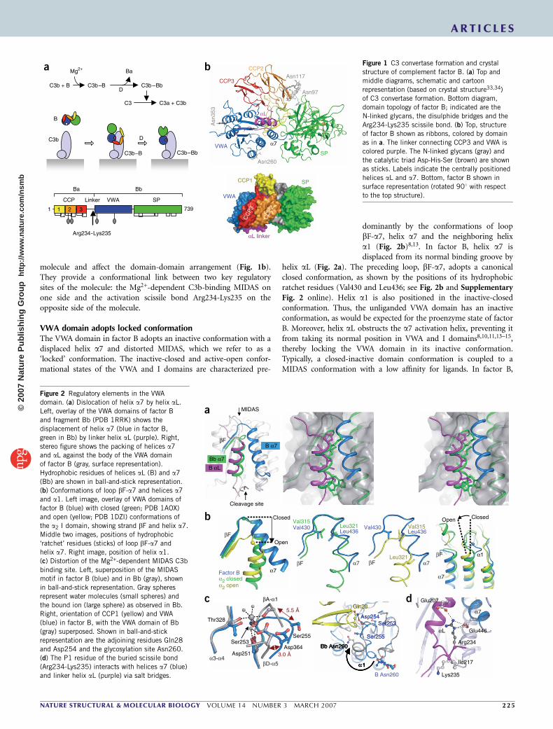
molecule and affect the domain-domain arrangement (Fig. 1b).They provide a conformational link between two key regulatorysites of the molecule: the Mg2+-dependent C3b-binding MIDAS onone side and the activation scissile bond Arg234-Lys235 on theopposite side of the molecule.
VWA domain adopts locked conformationThe VWA domain in factor B adopts an inactive conformation with adisplaced helix a7 and distorted MIDAS, which we refer to as a‘locked’ conformation. The inactive-closed and active-open confor-mational states of the VWA and I domains are characterized pre-
dominantly by the conformations of loopbF-a7, helix a7 and the neighboring helixa1 (Fig. 2b)8,13. In factor B, helix a7 isdisplaced from its normal binding groove by
helix aL (Fig. 2a). The preceding loop, bF-a7, adopts a canonicalclosed conformation, as shown by the positions of its hydrophobicratchet residues (Val430 and Leu436; see Fig. 2b and SupplementaryFig. 2 online). Helix a1 is also positioned in the inactive-closedconformation. Thus, the unliganded VWA domain has an inactiveconformation, as would be expected for the proenzyme state of factorB. Moreover, helix aL obstructs the a7 activation helix, preventing itfrom taking its normal position in VWA and I domains8,10,11,13–15,thereby locking the VWA domain in its inactive conformation.Typically, a closed-inactive domain conformation is coupled to aMIDAS conformation with a low affinity for ligands. In factor B,
C3b + B C3b–B D
Ba
C3b–Bb
C3 C3a + C3b
Mg2+
SP
Asn
353
ba
Arg234-Lys235
CCP VWA SPLinker
1 7392 31
Ba Bb
C3b–Bb
C3b
B
D
C3b–B
CCP1
VWA
SP
αL linker
CC
P3
CC
P2
Asn260
α7
αL
Asn97
Asn117CCP2
CCP3
VWA
B Asn260
Asp251
Ser253
βA-α1
Thr328
Asp364
5.5 Å
3.0 Å
Ser255
Ile217
Lys235
Arg234
αL Glu446
Glu207
α7
MIDAS
Cleavage site
Bb α7
B αL
βFB α7
Open
Leu321Val430Leu436
Val315Val315
Leu321
Val430Leu436
ClosedOpen
α1
b
a
Factor Bα2 closedα2 open
α7
c d
βF
βF α7 βF α7
βF
α7
α3-α4βD-α5
Closed
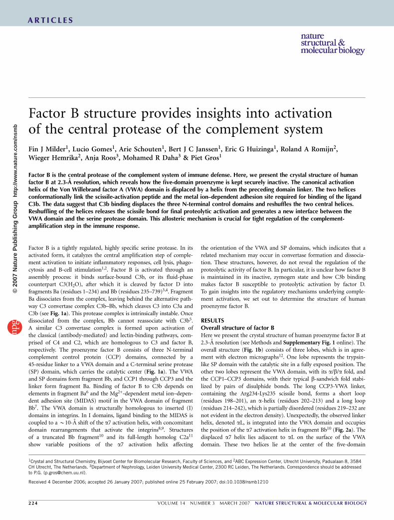
Figure 1 C3 convertase formation and crystal
structure of complement factor B. (a) Top and
middle diagrams, schematic and cartoon
representation (based on crystal structure33,34)
of C3 convertase formation. Bottom diagram,
domain topology of factor B; indicated are the
N-linked glycans, the disulphide bridges and the
Arg234-Lys235 scissile bond. (b) Top, structure
of factor B shown as ribbons, colored by domain
as in a. The linker connecting CCP3 and VWA is
colored purple. The N-linked glycans (gray) and
the catalytic triad Asp-His-Ser (brown) are shown
as sticks. Labels indicate the centrally positioned
helices aL and a7. Bottom, factor B shown in
surface representation (rotated 901 with respectto the top structure).
Figure 2 Regulatory elements in the VWA
domain. (a) Dislocation of helix a7 by helix aL.
Left, overlay of the VWA domains of factor B
and fragment Bb (PDB 1RRK) shows the
displacement of helix a7 (blue in factor B,
green in Bb) by linker helix aL (purple). Right,
stereo figure shows the packing of helices a7and aL against the body of the VWA domain
of factor B (gray, surface representation).
Hydrophobic residues of helices aL (B) and a7
(Bb) are shown in ball-and-stick representation.
(b) Conformations of loop bF-a7 and helices a7
and a1. Left image, overlay of VWA domains of
factor B (blue) with closed (green; PDB 1AOX)
and open (yellow; PDB 1DZI) conformations of
the a2 I domain, showing strand bF and helix a7.
Middle two images, positions of hydrophobic
‘ratchet’ residues (sticks) of loop bF-a7 and
helix a7. Right image, position of helix a1.
(c) Distortion of the Mg2+-dependent MIDAS C3b
binding site. Left, superposition of the MIDAS
motif in factor B (blue) and in Bb (gray), shown
in ball-and-stick representation. Gray spheres
represent water molecules (small spheres) and
the bound ion (large sphere) as observed in Bb.Right, orientation of CCP1 (yellow) and VWA
(blue) in factor B, with the VWA domain of Bb
(gray) superposed. Shown in ball-and-stick
representation are the adjoining residues Gln28
and Asp254 and the glycosylation site Asn260.
(d) The P1 residue of the buried scissile bond
(Arg234-Lys235) interacts with helices a7 (blue)
and linker helix aL (purple) via salt bridges.
ART IC L E S
NATURE STRUCTURAL & MOLECULAR BIOLOGY VOLUME 14 NUMBER 3 MARCH 2007 2 2 5
©20
07 N
atur
e P
ublis
hing
Gro
up
http
://w
ww
.nat
ure.
com
/nsm
b
compared with fragment Bb, displacements up to 5.5 A in loops bA-a1 and bD-a5 distort the MIDAS (Fig. 2c and Supplementary Fig. 2).The distorted conformation indicates reduced divalent-ion binding.Moreover, we did not observe electron density for an ion bound to theMIDAS in factor B. The absence of an ion is consistent with the notionthat fragment Bb, but not factor B, has high affinity for divalent ions16.The neighboring CCP1 domain may affect the MIDAS conformation.Gln28 of loop b2-b3 in CCP1 makes a hydrogen bond to Asp254,located in the displaced VWA bA-a1 loop. Furthermore, CCP1 mayprevent loop bA-a1 from assuming the conformation seen in Bb byobstructing the glycan at Asn260. Instead, in factor B helix a1 isshifted and partially unwound, pointing the glycan on Asn260 awayfrom the neighboring CCP1 domain (Fig. 2c). Factor B with D254Gand N260D single and double mutations (where N260D deletes theN-glycosylation site) forms convertases more readily and morestably than wild-type factor B17,18, indicating that these mutationspromote the transition from an inactive to an active conformation ofthe VWA domain.
Central role for linker regionThe CCP3-VWA linker helix aL occupies the position of helix a7of the VWA domain. It binds Arg234 of the scissile bond,preventing proteolytic activation. By virtue of its short-chain hydro-phobic residues, helix aL is squeezed between helix a7 and the VWAcore, effectively blocking helix a7 (Fig. 2a). After helix aL, the CCP3-VWA linker forms a long loop and folds back to the C-terminal end ofhelix aL (Fig. 2d). The scissile bond Arg234-Lys235 is located here,buried by the preceding flexible loop. The side chain of Arg234,crucial for substrate binding at the S1 pocket (as defined in Schechterand Berger notation) of factor D, is bound to both helix aL (Glu207)and helix a7 (Glu446) by salt bridges (Fig. 2d). Thus, the scissilebond in factor B is protected from proteolysis by factor D and isconformationally linked to helices aL and a7. In contrast, the scissilebond in isolated VWA domains is susceptible to factor D cleavage19,because they lack the aL-a7 arrangement that locks the P1 residue(Arg234). In the formation of the convertase, binding of C3b to factor
B probably induces reshuffling of helices aLand a7 that liberates the P1 arginine residueand makes the scissile bond accessible forproteolysis by factor D.
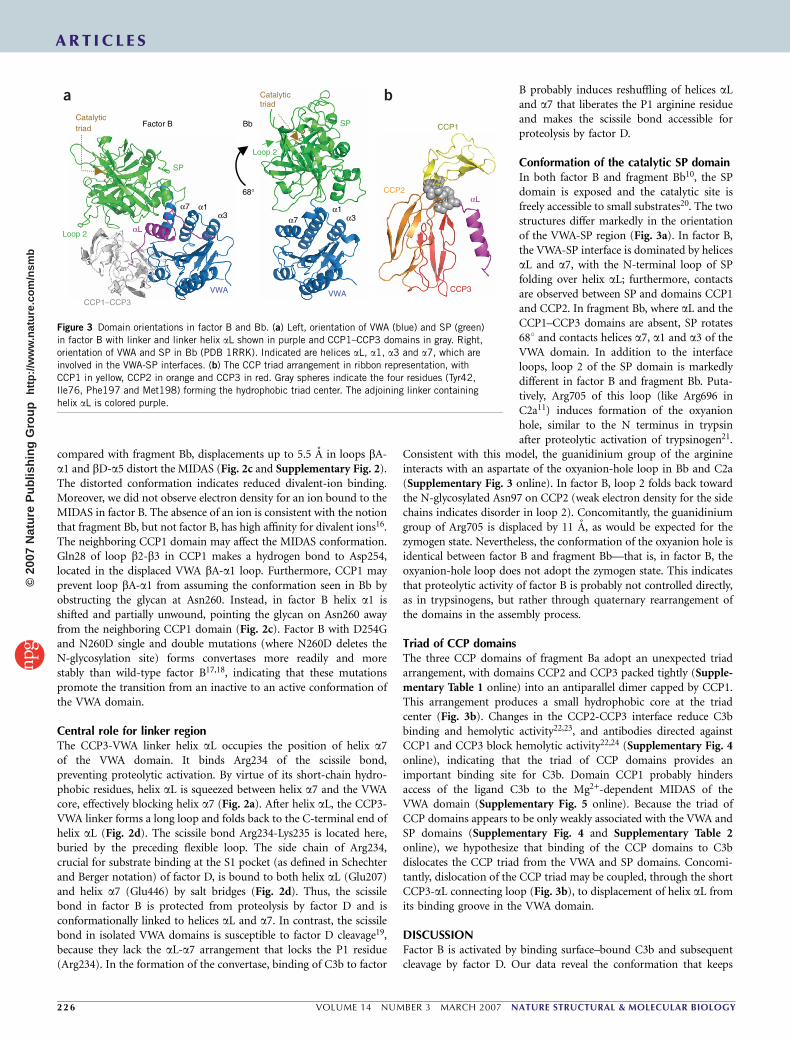
Conformation of the catalytic SP domainIn both factor B and fragment Bb10, the SPdomain is exposed and the catalytic site isfreely accessible to small substrates20. The twostructures differ markedly in the orientationof the VWA-SP region (Fig. 3a). In factor B,the VWA-SP interface is dominated by helicesaL and a7, with the N-terminal loop of SPfolding over helix aL; furthermore, contactsare observed between SP and domains CCP1and CCP2. In fragment Bb, where aL and theCCP1–CCP3 domains are absent, SP rotates681 and contacts helices a7, a1 and a3 of theVWA domain. In addition to the interfaceloops, loop 2 of the SP domain is markedlydifferent in factor B and fragment Bb. Puta-tively, Arg705 of this loop (like Arg696 inC2a11) induces formation of the oxyanionhole, similar to the N terminus in trypsinafter proteolytic activation of trypsinogen21.
Consistent with this model, the guanidinium group of the arginineinteracts with an aspartate of the oxyanion-hole loop in Bb and C2a(Supplementary Fig. 3 online). In factor B, loop 2 folds back towardthe N-glycosylated Asn97 on CCP2 (weak electron density for the sidechains indicates disorder in loop 2). Concomitantly, the guanidiniumgroup of Arg705 is displaced by 11 A, as would be expected for thezymogen state. Nevertheless, the conformation of the oxyanion hole isidentical between factor B and fragment Bb—that is, in factor B, theoxyanion-hole loop does not adopt the zymogen state. This indicatesthat proteolytic activity of factor B is probably not controlled directly,as in trypsinogens, but rather through quaternary rearrangement ofthe domains in the assembly process.
Triad of CCP domainsThe three CCP domains of fragment Ba adopt an unexpected triadarrangement, with domains CCP2 and CCP3 packed tightly (Supple-mentary Table 1 online) into an antiparallel dimer capped by CCP1.This arrangement produces a small hydrophobic core at the triadcenter (Fig. 3b). Changes in the CCP2-CCP3 interface reduce C3bbinding and hemolytic activity22,23, and antibodies directed againstCCP1 and CCP3 block hemolytic activity22,24 (Supplementary Fig. 4online), indicating that the triad of CCP domains provides animportant binding site for C3b. Domain CCP1 probably hindersaccess of the ligand C3b to the Mg2+-dependent MIDAS of theVWA domain (Supplementary Fig. 5 online). Because the triad ofCCP domains appears to be only weakly associated with the VWA andSP domains (Supplementary Fig. 4 and Supplementary Table 2online), we hypothesize that binding of the CCP domains to C3bdislocates the CCP triad from the VWA and SP domains. Concomi-tantly, dislocation of the CCP triad may be coupled, through the shortCCP3-aL connecting loop (Fig. 3b), to displacement of helix aL fromits binding groove in the VWA domain.
DISCUSSIONFactor B is activated by binding surface–bound C3b and subsequentcleavage by factor D. Our data reveal the conformation that keeps
VWA
SP
α7 α1α3
Catalytictriad Factor B
CCP1–CCP3
Loop 2
68°
Bb
VWA
SP
α7α1
α3
Catalytictriad
Loop 2
CCP2αL
CCP3
CCP1
αL
a b
Figure 3 Domain orientations in factor B and Bb. (a) Left, orientation of VWA (blue) and SP (green)
in factor B with linker and linker helix aL shown in purple and CCP1–CCP3 domains in gray. Right,
orientation of VWA and SP in Bb (PDB 1RRK). Indicated are helices aL, a1, a3 and a7, which are
involved in the VWA-SP interfaces. (b) The CCP triad arrangement in ribbon representation, with
CCP1 in yellow, CCP2 in orange and CCP3 in red. Gray spheres indicate the four residues (Tyr42,
Ile76, Phe197 and Met198) forming the hydrophobic triad center. The adjoining linker containing
helix aL is colored purple.
ART IC L E S
22 6 VOLUME 14 NUMBER 3 MARCH 2007 NATURE STRUCTURAL & MOLECULAR BIOLOGY
©20
07 N
atur
e P
ublis
hing
Gro
up
http
://w
ww
.nat
ure.
com
/nsm
b
factor B locked in its proenzyme state. The structure adds this lockedconformation to the open and closed conformations described in paststudies of the VWA and I domains8,9 and provides insights into apreviously uncharacterized mechanism for regulating serine proteaseactivity21. Assembly of an active protease complex probably proceedsthrough a number of steps. We hypothesize that initial binding of C3bdislocates the CCP triad. In turn, dislocation of the CCPs probablyinduces formation of an active MIDAS and allows access of the largeC3b ligand. Concomitantly, the blocking helix aL is displaced from itsposition in the VWA domain, thereby allowing the activation helix a7to move into its normal location. Rearrangement of helices aL and a7liberates the bound scissile peptide, making it accessible for proteolyticcleavage by factor D. Cleavage of the scissile bond results in dissocia-tion of the Ba fragment, yielding the active, short-lived proteasecomplex that cleaves C3 into C3a and C3b. Thus, the tight regulationof complement activation is determined by a series of conformationalchanges that establish the C3 convertase activity required for ampli-fication, which is crucial for the biological functions of the comple-ment system.
METHODSProtein expression, purification and crystallization. Human factor B fused to
an N-terminal Gly-Ser-(His)6-Gly-Ser tag was expressed in human embryonic
kidney 293S GnTI– cells to prevent complex and heterogeneous N-linked
glycosylation25. Secreted factor B was purified from the expression medium via
immobilized metal affinity chromatography followed by size-exclusion chro-
matography. The purified protein showed a single band of 90 kDa on
Coomassie-stained SDS-PAGE gels and on western blots probed with an
antibody to His6. Details of factor B expression and purification are given in
Supplementary Methods online. Crystals were grown by hanging drop vapor
diffusion by mixing 1 ml factor B (13 mg ml–1) in 10 mM tris(hydroxymethyl)
aminomethane (pH 7.4), 25 mM arginine, 25 mM glutamic acid and 1 ml
well solution (50 mM malic acid 2-(N-morpholino)ethanesulfonic acid tris
(hydroxymethyl)aminomethane buffer (pH 6.5) and 12% (w/v) PEG 1,500) at
20 1C. Glycerol (20% v/v) was added to the well solution before flash-cooling of
the crystal in liquid nitrogen. The crystal diffracted to 2.3-A resolution at the
European Synchrotron Radiation Facility beamline ID14-EH4. The space group
of the crystal was identified as P3121 (a ¼ b ¼ 104.0 and c ¼ 151.1 A). Data
were processed by MOSFLM and CCP4 (ref. 26). Crystallographic data
collection and refinement statistics are given in Table 1.
Structure determination. The SP and VWA domain were placed by molecular
replacement using Phaser27 with structures of the isolated SP (PDB 1DLE)28
and VWA (PDB 1Q0P)14 domains of factor B as search models. Subsequent
placement of the three CCP domains using various homologous structures, as
well as model completion by automated model building using ARP/wARP29,
failed at this stage. The VWA helix a7 and SP domain loops C and D were
rebuilt and the partial model was refined using COOT30, RESOLVE31 and
REFMAC5 (ref. 32). Using this partial model, ARP/wARP then successfully
completed the model to B80%. Iterative cycles of refinement with REFMAC5
and model building in COOT were used to finalize the model. The refined
model of factor B contains 710 residues; residues 1–8, 218–232, 321–326 and
344–346 were excluded from the model because of poor electron density. R and
Rfree values were 19.5% and 24.1%, respectively (see Table 1 for refinement
statistics). All molecular graphic figures were generated with PyMOL (http://
pymol.sourceforge.net).
Accession codes. Protein Data Bank: Coordinates and structure factors have
been deposited with accession code 2OK5.
Note: Supplementary information is available on the Nature Structural & MolecularBiology website.
ACKNOWLEDGMENTSWe thank the European Synchrotron Radiation Facility for providing synchrotronradiation facilities and the beamline scientists at ID-14-EH4 for their help withdata collection. This work was supported by a ‘Pionier’ program grant (P.G.) ofthe Council for Chemical Sciences of the Netherlands Organization for ScientificResearch (NWO-CW).
COMPETING INTERESTS STATEMENTThe authors declare that they have no competing financial interests.
Published online at http://www.nature.com/nsmb/
Reprints and permissions information is available online at http://npg.nature.com/
reprintsandpermissions
1. Carroll, M.C. The complement system in regulation of adaptive immunity. Nat.Immunol. 5, 981–986 (2004).
2. Walport, M.J. Complement. First of two parts. N. Engl. J. Med. 344, 1058–1066(2001).
3. Fishelson, Z., Pangburn, M.K. & Muller-Eberhard, H.J. Characterization of the initialC3 convertase of the alternative pathway of human complement. J. Immunol. 132,1430–1434 (1984).
4. Xu, Y., Narayana, S.V. & Volanakis, J.E. Structural biology of the alternative pathwayconvertase. Immunol. Rev. 180, 123–135 (2001).
5. Pangburn, M.K. & Muller-Eberhard, H.J. The C3 convertase of the alternative pathwayof human complement. Enzymic properties of the bimolecular proteinase. Biochem.J. 235, 723–730 (1986).
6. Pryzdial, E.L. & Isenman, D.E. Alternative complement pathway activation fragment Babinds to C3b. Evidence that formation of the factor B-C3b complex involves twodiscrete points of contact. J. Biol. Chem. 262, 1519–1525 (1987).
7. Horiuchi, T., Macon, K.J., Engler, J.A. & Volanakis, J.E. Site-directed mutagenesis ofthe region around Cys-241 of complement component C2. Evidence for a C4b bindingsite. J. Immunol. 147, 584–589 (1991).
8. Emsley, J., Knight, C.G., Farndale, R.W., Barnes, M.J. & Liddington, R.C. Struc-tural basis of collagen recognition by integrin alpha2beta1. Cell 101, 47–56(2000).
9. Springer, T.A. Complement and the multifaceted functions of VWA and integrin Idomains. Structure 14, 1611–1616 (2006).
10. Ponnuraj, K. et al. Structural analysis of engineered Bb fragment of complement factorB: insights into the activation mechanism of the alternative pathway C3-convertase.Mol. Cell 14, 17–28 (2004).
11. Milder, F.J. et al. Structure of complement component C2a: implications for convertaseformation and substrate binding. Structure 14, 1587–1597 (2006).
Table 1 Data collection and refinement statistics
Factor B
Data collection
Space group P3121
Cell dimensions
a, b, c (A) 104.0, 104.0, 151.1
a, b, g (1) 90, 90, 120
Resolution (A) 60.0–2.3 (2.42–2.30)
Rmerge 10.8 (56.1)
I / sI 10.4 (2.6)
Completeness (%) 99.8 (100)
Redundancy 4.1 (4.1)
Refinement
Resolution (A) 60.0–2.30
No. reflections 40,409
Rwork / Rfree 0.195 / 0.241
No. atoms
Protein 5,745
Ligand/ion 78
Water 295
B-factors (A2)
Protein 33.5
Ligand/ion 58.1
Water 31.6
R.m.s. deviations
Bond lengths (A) 0.010
Bond angles (1) 1.30
Values in parentheses are for highest-resolution shell.
ART IC L E S
NATURE STRUCTURAL & MOLECULAR BIOLOGY VOLUME 14 NUMBER 3 MARCH 2007 2 2 7
©20
07 N
atur
e P
ublis
hing
Gro
up
http
://w
ww
.nat
ure.
com
/nsm
b
12. Smith, C.A., Vogel, C.W. & Muller-Eberhard, H.J. MHC Class III products: an electronmicroscopic study of the C3 convertases of human complement. J. Exp. Med. 159,324–329 (1984).
13. Shimaoka, M. et al. Structures of the alpha L I domain and its complex with ICAM-1reveal a shape-shifting pathway for integrin regulation. Cell 112, 99–111 (2003).
14. Bhattacharya, A.A., Lupher, M.L., Jr., Staunton, D.E. & Liddington, R.C. Crystalstructure of the A domain from complement factor B reveals an integrin-like openconformation. Structure 12, 371–378 (2004).
15. Emsley, J., King, S.L., Bergelson, J.M. & Liddington, R.C. Crystal structure ofthe I domain from integrin alpha2beta1. J. Biol. Chem. 272, 28512–28517(1997).
16. Fishelson, Z., Pangburn, M.K. & Muller-Eberhard, H.J. C3 convertase of the alternativecomplement pathway. Demonstration of an active, stable C3b, Bb (Ni) complex.J. Biol. Chem. 258, 7411–7415 (1983).
17. Hourcade, D.E., Mitchell, L., Kuttner-Kondo, L.A., Atkinson, J.P. & Medof, M.E. Decay-accelerating factor (DAF), complement receptor 1 (CR1), and factor H dissociate thecomplement AP C3 convertase (C3bBb) via sites on the type A domain of Bb. J. Biol.Chem. 277, 1107–1112 (2002).
18. Hourcade, D.E., Mitchell, L.M. & Oglesby, T.J. Mutations of the type A domain ofcomplement factor B that promote high-affinity C3b-binding. J. Immunol. 162,2906–2911 (1999).
19. Williams, S.C., Hinshelwood, J., Perkins, S.J. & Sim, R.B. Production and functionalactivity of a recombinant von Willebrand factor-A domain from human complementfactor B. Biochem. J. 342, 625–632 (1999).
20. Kam, C.M. et al. Human complement proteins D, C2, and B. Active site mapping withpeptide thioester substrates. J. Biol. Chem. 262, 3444–3451 (1987).
21. Khan, A.R. & James, M.N. Molecular mechanisms for the conversion of zymogens toactive proteolytic enzymes. Protein Sci. 7, 815–836 (1998).
22. Hourcade, D.E., Wagner, L.M. & Oglesby, T.J. Analysis of the short consensus repeatsof human complement factor B by site-directed mutagenesis. J. Biol. Chem. 270,19716–19722 (1995).
23. Xu, Y. & Volanakis, J.E. Contribution of the complement control protein modules ofC2 in C4b binding assessed by analysis of C2/factor B chimeras. J. Immunol. 158,5958–5965 (1997).
24. Thurman, J.M. et al. A novel inhibitor of the alternative complement pathway preventsantiphospholipid antibody-induced pregnancy loss in mice. Mol. Immunol. 42, 87–97(2005).
25. Reeves, P.J., Callewaert, N., Contreras, R. & Khorana, H.G. Structure and function inrhodopsin: high-level expression of rhodopsin with restricted and homogeneousN-glycosylation by a tetracycline-inducible N-acetylglucosaminyltransferase I-negativeHEK293S stable mammalian cell line. Proc. Natl. Acad. Sci. USA 99, 13419–13424(2002).
26. Collaborative Computational Project, Number 4. The CCP4 suite: programs for proteincrystallography. Acta Crystallogr. D Biol. Crystallogr. 50, 760–763 (1994).
27. Storoni, L.C., McCoy, A.J. & Read, R.J. Likelihood-enhanced fast rotation functions.Acta Crystallogr. D Biol. Crystallogr. 60, 432–438 (2004).
28. Jing, H. et al. New structural motifs on the chymotrypsin fold and their potential rolesin complement factor B. EMBO J. 19, 164–173 (2000).
29. Perrakis, A., Morris, R. & Lamzin, V.S. Automated protein model building combinedwith iterative structure refinement. Nat. Struct. Biol. 6, 458–463 (1999).
30. Emsley, P. & Cowtan, K. Coot: model-building tools for molecular graphics. ActaCrystallogr. D Biol. Crystallogr. 60, 2126–2132 (2004).
31. Terwilliger, T.C. Automated main-chain model building by template matching anditerative fragment extension. Acta Crystallogr. D Biol. Crystallogr. 59, 38–44 (2003).
32. Winn, M.D., Isupov, M.N. & Murshudov, G.N. Use of TLS parameters to modelanisotropic displacements in macromolecular refinement. Acta Crystallogr. D Biol.Crystallogr. 57, 122–133 (2001).
33. Janssen, B.J., Christodoulidou, A., McCarthy, A., Lambris, J.D. & Gros, P. Structure ofC3b reveals conformational changes that underlie complement activity. Nature 444,213–216 (2006).
34. Wiesmann, C. et al. Structure of C3b in complex with CRIg gives insights intoregulation of complement activation. Nature 444, 217–220 (2006).
ART IC L E S
22 8 VOLUME 14 NUMBER 3 MARCH 2007 NATURE STRUCTURAL & MOLECULAR BIOLOGY
©20
07 N
atur
e P
ublis
hing
Gro
up
http
://w
ww
.nat
ure.
com
/nsm
b