facilitating the safety evaluation of manufactured
TRANSCRIPT

Facilitating the safety evaluation of manufactured nanomaterials by characterising their potential genotoxic hazard
Project Coordinator French Agency for Food, Environmental and Occupational Health & Safety (ANSES)

This document arises from the NANOGENOTOX Joint Action which has received funding from the European Union, in the framework of the Health Programme under Grant Agreement n°2009 21.This publication reflects only the author’s views and the Community is not liable for any use that may be made of the information contained therein.
3
ummaryContributing to increasing the safe use of manufactured nanomaterials in the Eu ������������������������������������������������������4
Working together towards a method for detecting the genotoxicity of manufactured nanomaterials �������������������5
Good practice for the implementation of a Joint action �����������8
Coordination ������������������������������������������������������������� 8
Reaching the stakeholders ������������������������������������������ 9
Evaluation �����������������������������������������������������������������11
Characterising manufactured nanomaterials (mNs) and exposure media ���������������������������������������������������������13
In vitro methods for genotoxicity testing ������������������������������ 23
Toxicokinetics and tissue distribution of mNs and organs at risk for genotoxicity testing �������������������������������������������� 34
In vivo genotoxicity testing ������������������������������������������������ 39
Considerations to achieve a robust method for genotoxicity testing of mNs ������������������������������������������ 43
recommendations and perspectives ���������������������������������� 44
List of Deliverables ����������������������������������������������������������� 46
Glossary �������������������������������������������������������������������������� 47
acknowledgments ����������������������������������������������������������� 52
associated partners ��������������������������������������������������������� 54
Collaborating partners ����������������������������������������������������� 58
S

4 5
FranceANSESIPLINRSCEA
BelgiumIPH
CODA-CERVA
PortugalINSA
SpainUAB
ItalyISS
BulgariaIMC-BASIMB-BAS
PolandNIOM
GermanyBfR
NetherlandsRIVM
DenmarkNRCWE
FinlandFIOH
Associated Partners of the NANOGENOTOX Joint Action
Towards a method for detecting the potential genotoxicity of nanomaterials
Nanotechnology is a highly strategic industrial and economic sector showing enormous potential benefits for many societal and environmen-tal domains. Human exposure to manufactured nanomaterials (MNs) present in consumer products may occur during several phases of their life cycle, from synthesis, production and inclusion in the products to the release of MNs into the environment.The lack of scientific knowledge and the absence of evidence demons-trating the safety of some nanomaterials make regulation a challenge. In 2009 the Executive Agency for Health and Consumers (EAHC) awarded a
grant through the second programme of Community action in the field of health (2008-2013) for a Joint Action (JA) on the “Safety of nanomaterials”.The NaNOGENOTOX Joint Action started in March 2010 for a period of 3 years and had a total budget of over 6.2 million euros, 46% co-funded by the European Commission’s Health
Programme. It was coordinated by the French Agency for Food, Environ-mental and Occupational Health & Safety (ANSES). Sixteen associated partners from 11 member states and 13 collaborating partners came together to pool their expertise and competences.
CONTrIbuTING TO INCrEaSING ThE SaFE uSE OF maNuFaCTurED NaNOmaTErIaLS IN ThE Eu

4 5
Facilitating the saFety evaluation oF manuFactured nanomaterials
Forecast work plan of the NANOGENOTOX Joint Action
The objective of the Joint Action (JA) was to work together towards establishing a robust (specific and sensitive) methodology to assess the potential genotoxicity (i.e. capacity to induce DNA damage) of MNs and to generate data on the genotoxic effect of certain commonly used MNs materials.
The strategy applied to reach the aim of the JA included a state-of-the-art assessment in order to identify gaps and fill these gaps, as far as pos-sible, through testing a selected group of MNs and therefore providing generic Standard Operating Procedures (SOPs) and protocols.The work plan consisted of four scientific work packages (WPs) and three transversal WPs devoted to the coordination, dissemination and scientific evaluation of the JA.
OrkING TOGEThEr TOWarDS a mEThOD FOr DETECTING ThE GENOTOXICITy OF maNuFaCTurED NaNOmaTErIaLS
W
Primary Characteristics
Analytical and dispersion protocols
SOPs for characterisation of selected MNs
MN data sets with requested physico-chemical properties
WP4 - characterisation
In vitro genotoxicity studies (comet and micronucleus on intestinal, lung and skin cells)
In vitro ring test
Evaluation of the results from the in vitro and in vivo tests for correlation and used to formulate a strategy for genotoxicity testing of MNs
WP5 - in vitro
In vivo genotoxicity assays (oral and instillation exposure)
Qualitative analysis of the correalation between in vitro and in vivo genotoxicity data
WP6 - in vivo
Analytical techniques for determination of MNs in blood and tissue (with WP4)
Pilot dose range studies (ADME)
Pivotal biodistribution studies
WP7 - toxicokinetic
1Months
6 8 12 15 18 20 24 30 36
6 7
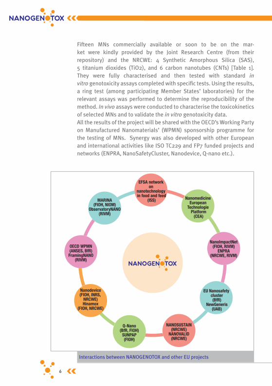
Fifteen MNs commercially available or soon to be on the mar-ket were kindly provided by the Joint Research Centre (from their repository) and the NRCWE: 4 Synthetic Amorphous Silica (SAS), 5 titanium dioxides (TiO2), and 6 carbon nanotubes (CNTs) [Table 1]. They were fully characterised and then tested with standard in vitro genotoxicity assays completed with specific tests. Using the results, a ring test (among participating Member States’ laboratories) for the relevant assays was performed to determine the reproducibility of the method. In vivo assays were conducted to characterise the toxicokinetics of selected MNs and to validate the in vitro genotoxicity data.All the results of the project will be shared with the OECD’s Working Party on Manufactured Nanomaterials’ (WPMN) sponsorship programme for the testing of MNs. Synergy was also developed with other European and international activities like ISO TC229 and FP7 funded projects and networks (ENPRA, NanoSafetyCluster, Nanodevice, Q-nano etc.).
Interactions between NANOGENOTOX and other EU projects
OECD WPMN(ANSES, BfR)FramingNANO
(RIVM)
MARINA(FIOH, NIOM)
ObservatoryNANO(RIVM)
EFSA network on
nanotechnology in food and feed
(ISS) Nanomedicine European
Technologie Platform
(CEA)
NanoImpactNet(FIOH, RIVM)
ENPRA(NRCWE, RIVM)
EU Nanosafety cluster(BfR)
NewGeneris(UAB)
NANOSUSTAIN(NRCWE)
NANOVALID(NRCWE)
Q-Nano(BfR, FIOH)
SUNPAP(FIOH)
Nanodevice(FIOH, INRS,
NRCWE)Hinamox
(FIOH, NRCWE)

6 7
Facilitating the saFety evaluation oF manuFactured nanomaterials
Table 1 – Manufactured nanomaterials selected for the JA
Ref. Type Major use Tested in WP
TiO2
NM-100 Anatase paper loadings, rubber, cosmetics, adhesives, low
cost interior paints
WP4, WP7
NM-101 Anatase semiconductor catalyst for use in photocatalytic
processes
WP4, WP7
NM-102 Anatase photocatalytic WP4, WP5 (Round Robin), WP6, WP7
NM-103 Rutilecosmetics (sun care,
colour), pharmaceuticals, food
WP4, WP5, WP6, WP7
NM-104 Rutile WP4, WP5, WP6, WP7
NM-105 Anatase/rutile catalysis, heat stabilizer WP4, WP5, WP6, WP7
SAS
NM-200 Precipitated food processing WP4, WP5, WP6, WP7
NM-201 Precipitated reinforcement, mechanical and optical properties and
process
WP4, WP5, WP6
NM-202 Pyrolise inks, adhesives, cosmetics, reinforcement, powder
process, food, pharmaceuticals
WP4, WP5, WP6
NM-203 Pyrolise food, cosmetics, pharma-ceuticals, reinforcement
WP4, WP5 (Round Robin), WP6, WP7
CNT
NM-400 MWCNT Catalytic Chemical Vapour
Deposition
structural composite and energy applications
WP4, WP5, WP6, WP7
NM-401 WP4, WP5, WP6, WP7
NM-402 WP4, WP5, WP6, WP7
NM-403 WP4, WP5 (Round Robin)
NRCWE-006 lithium/ion battery WP4, WP5, WP6, WP7
NRCWE-007 structural composite WP4, WP5
8 9
Efficient project management refers to the planning and organisation of the activities to ensure that the Consortium delivers the expected out-puts in due time and budget. This requires in particular a sufficient level of organisational capacity by the participants, a strong partnership cou-pled with the implementation of a targeted communication strategy and a systematic evaluation of the activities. The three horizontal Work Packages (Coordination, Dissemination and Evaluation) implemented a set of tools to improve the chances of success as well as a series of processes to monitor time, costs, quality and scope of the project.
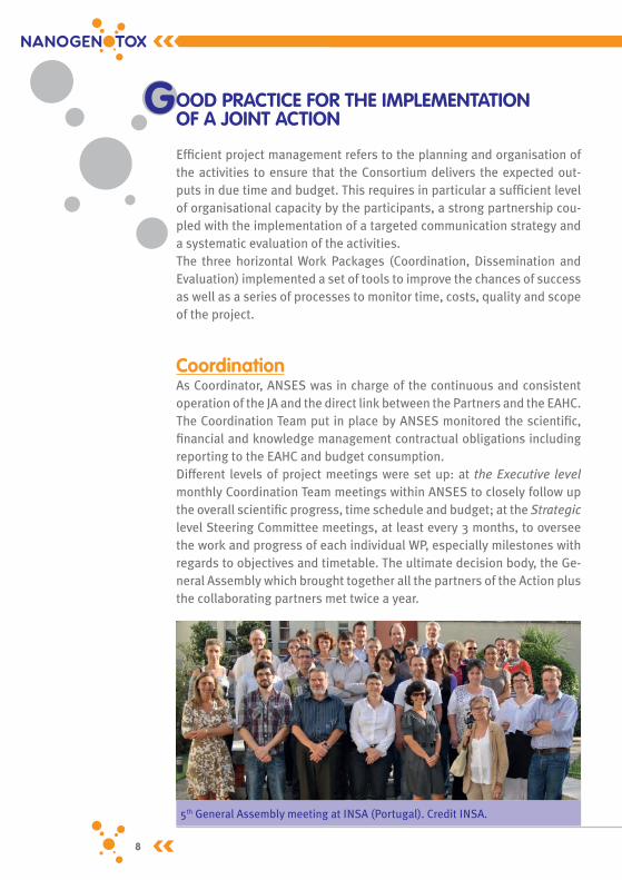
CoordinationAs Coordinator, ANSES was in charge of the continuous and consistent operation of the JA and the direct link between the Partners and the EAHC. The Coordination Team put in place by ANSES monitored the scientific, financial and knowledge management contractual obligations including reporting to the EAHC and budget consumption. Different levels of project meetings were set up: at the Executive level monthly Coordination Team meetings within ANSES to closely follow up the overall scientific progress, time schedule and budget; at the Strategic level Steering Committee meetings, at least every 3 months, to oversee the work and progress of each individual WP, especially milestones with regards to objectives and timetable. The ultimate decision body, the Ge-neral Assembly which brought together all the partners of the Action plus the collaborating partners met twice a year.
GOOD praCTICE FOr ThE ImpLEmENTaTION OF a JOINT aCTION
5th General Assembly meeting at INSA (Portugal). Credit INSA.
8 9
Facilitating the saFety evaluation oF manuFactured nanomaterials
The rules of the Consortium were precisely defined in a Consortium Agreement established by the Coordinator in order to secure the part-nership by a formally signed agreement contractualising the coopera-tion, organising the legal and operational framework of the Joint Action, the access rights and defining the rights and obligations of the partners.
reaching the stakeholdersDissemination is the process of engaging with, and making the results and deliverables available to, the stakeholders and a wider audience. The key elements for appropriate targeting are to carry out a stakeholder analysis, ensuing consultations and adapting the dissemination and sus-tainability strategy to the expectations of the stakeholders.Stakeholders selected for the consultation had to be involved at an EU (or international) level, recognised in their domain and exercising a cer-tain influence, recognised for their ability to relay information to a wider audience, possess technical and scientific knowledge regarding nano-toxicology and be willing to engage in technical discussions about nano-toxicology.Five categories of stakeholders were identified: • EU risk assessors and policy-makers,• members of the scientific community,• professional federations representing companies,• non-governmental organisations (NGOs),• trade unions.
The stakeholders’ input is invaluable in helping to identify the concerns and needs of the various groups of stakeholders, for example, the needs of industry regarding to safe design or for the preliminary testing of a nanomaterial that is still under development before investing important efforts and money. Although the objective was not to directly look into risk assessment and risk management concerns, policy-makers were involved in at least two ways: through certain partners which are risk assessment institutes with strong links, as knowledge brokers, to ministries, and directly as minis-tries of several Member States were collaborating partners.
OOD praCTICE FOr ThE ImpLEmENTaTION OF a JOINT aCTION

10 11
Figure 1 – Stakeholders consultation process
The final conference of NANOGENOTOX took place on Friday 22 February 2013 and was hosted by the French Ministry of Social Affairs and Health, in Paris, France. It gathered around 200 participants from all over Europe and beyond, including partners of the Joint Action.The results of each WP were presented and discussed in the morning ses-sions. In the afternoon, a presentation summarising the considerations to achieve a robust method for testing the genotoxicity of MNs was made, followed by discussions with the audience. Particular efforts were made during the panel discussion and in the conclusions and perspectives by policy-makers to examine how this method can be taken up and followed upon within REACH or other regulatory mechanisms.
ON-g
OiNg
STA
kehO
lder
CON
SulT
ATiO
N
• Inform stakeholders• Establish contacts• Identify concerns
• Present final results• Discuss results &
impact• Panel discussion
• Present preliminary results
• Share & discuss critical issues on genotoxicity assessment
M8 (Oct. 2010)
Stakeholder survey
M36 (Feb. 2013)
Final conference
M24 (Feb. 2011)
Stakeholders workshop
• Positive perception of the JA• Key issues include:
- Translation of the results into policy orientations,- Validity and harmonisation of protocols and the testing approaches,- Dissemination of the results,- Coordination with other EU projects
• Discussion on the scientific and technical aspects
• Knowledge transfer & dissemination of the results• Key issues include:
- Availability of the protocols and SOPs- Concerns on the cellular uptake of the particles and the positive controls chosen- Questions on genotoxicity mechanisms & role of the inflammatory cells- Questions on the inhalation route and the consequence of the bolus administration
• Discussion on the results that can be taken up by regulators & other stakeholders for hazard identification purposes
• Discussion and exchanges on strengths and limits of the JA results
• Key issues include:- Use of the results- Difficulties encountered- Translation of the results into policy
recommendations- Interaction with REACH, OECD guidelines, etc.
Synthesis report
May 2011
Publishable summary
report April 2013
Synthesis report
June 2012

10 11
Facilitating the saFety evaluation oF manuFactured nanomaterials
As policy-makers, the OECD WPMN and the EC DG SANCO representatives confirmed that the results and lessons coming out from NANOGENOTOX can be built upon for risk assessment and risk management purposes. Existing frameworks and guidelines are applicable for nanomaterials but some particular test guidelines have to be examined closely and may need to be updated or replaced. The concerns identified in the NANOGE-NOTOX findings are shared by the EC Scientific Committee on Consumer Safety (SCCS).
Like most collaborative projects, NANOGENOTOX developed classical tools for raising awareness: a web site (www.nanogenotox.eu) and a leaflet, giving an identity and explaining the objectives and planned acti-vities of the JA. Newsletters focusing on the results were published every 6 months. Partners participated in national and international conferences to present their results, and agreements on publications have been reached in each scientific WP.
Evaluation of the Joint actionEvaluation is an important process in order to assess if the project objectives have been achieved and whether the needs of the stake- holders have been met. Therefore, the quality of the work and the ability of the JA to respond to the requirements of the EU Health Programme were systematically evaluated during the course of the JA. An Internal Evaluation Team, composed of one representative from each Work Package, monitored and analysed the quantitative (number of
Final conference, 22 February 2013, Paris (France). Credit ANSES.

12 13
datasets) and qualitative (robustness and reliability of the tests) speci-fic objective indicators according to an approved evaluation plan defined early in the project by the Evaluation Work Package (WP3).Following the Evaluation plan which included defined templates, ques-tionnaires and indicators as well as timelines, confidential “cruise mode” evaluation reports were completed every 6 months reviewing the JA data generation and knowledge sharing actions. In these reports, recommen-dations were made by the Evaluation Team in order to keep up with the work plan and to take corrective actions.In addition, in cooperation with the Coordination Team, an external aca-demic reviewer panel was created to evaluate the Joint Action’s delive-rables and to respond to any specific issues that the internal Evaluation Team might have. The external reviewers were selected for their excellent scientific record in the respective fields of expertise. They participated in a final review meeting in Berlin in November 2012 with the Internal Evaluation Team and the work package leaders to discuss in depth the preliminary results as well as the Deliverables (as available at that point).A number of them participated in the Final conference to share with the conference participants their evaluation of the JA’s scientific outputs.
3rd GA meeting at NRCWE (Denmark), October 2011. Credit ANSES.

12 13
Facilitating the saFety evaluation oF manuFactured nanomaterials
CObjectivesVerification and detailed information on test materials are essential for proper interpretation of experimental results. Reliable data on physi-co-chemical characteristics of MNs become especially crucial when such results are applied in furthering a new understanding of nanomaterial toxicity and in producing advice for regulatory use. Therefore, reliable high-quality methods for characterisation of the materials as such, but also their exposure characteristics in both air and liquid dispersions are highly necessary and currently under continuous development. In response to the requirements in the NANOGENOTOX project, the main objectives of WP4 were to:
• test and develop suitable methods and Standard Operating Procedures (SOPs) for analysis and characterisation of MNs and dispersions thereof,• determine the intrinsic characteristics of MNs selected for toxicological studies, • test the homogeneity of the MN batches distributed,• develop, test and verify highly suitable MN dispersion protocols to be used in toxicity testing.
materialsAll selected MNs were characterised and the primary characterisation data constituted part of the selection criteria for the toxicological studies. Table 2 shows the complete material list and selected material characteristics obtained in the JA.
methods and SOp developmentsNumerous different methods and procedures could be applied for cha-racterisation of the MNs, their reactivity, exposure characteristics and emission potential. Therefore, the WP had to focus on a number of key characteristics to be studied. These characteristics included the primary and secondary (aggregate) average particle sizes (or distribution), mor-phologies of particles and fibers, their atomic structure, chemical com-positions, contaminants, catalysts and associated organic matter, as well as primary surface charge given by the zeta-potential as a function of pH. Another task was aimed at characterising the biologically relevant hydrochemical reactivity (OH radical formation capacity, causticity, O2 or redox-activity) and short-term solubility of the individual MNs. Finally,
haraCTErISING maNuFaCTurED NaNOmaTErIaLS aND EXpOSurE mEDIa

14 15
Table 2 – Summary of samples and average data on key analytical results
PowderBatch
dispersion for toxicology
Sample Phase
Average XRD
crystallite size
Average TEM particle size
Average BET & SAXS
SSA£
TGA mass-loss
Main elemental impurities
Average SAXS
aggregate size
Average DLS
Zeta-size
NM-100 anatase56.7 - >100
nm110 nm 9 m2/g nd K,P NA 215 nm
NM-101 anatase 7 nm 6 nm 316 m2/g 8 wt%€ Al,Na,P,S,Zr NA 483 nm
NM-102 anatase 21 nm 22 nm 78 m2/g nd S 560 nm 545 nm
NM-103 rutile 23 nm 25 nm 51 m2/g 2 wt%€ Al,Si,Na,S 140 nm 194 nm
NM-104 rutile 23 nm 25 nm 56 m2/g 2 wt%€ Al,Si,Ca,Na,S 160 nm 234 nm
NM-105anatase
rutile23 nm60 nm
24 nm15 nm
46 m2/g nd nd 130 nm 155 nm
NM-200 SAS NA 18 nm 189 m2/g 3 wt%€ Na,Al 440 nm 185 nm
NM-201 SAS NA 18 nm 140 m2/g 3 wt%€ Na,Al 180 nm 176 nm
NM-202 SAS NA 18 nm 204 m2/g nd Ca,Al 100 nm 134 nm
NM-203 SAS NA 25 nm 204 m2/g nd Al NA* 127 nm
NM-204 SAS NA 16 nm 137 m2/g <1 wt%€ Na,Al NA 174 nm
NM-400 MWCNT NA d=14nm; L<1µm 254 m2/g 84 wt% Al,Fe,Co,Zn,Na NA 55 nm$
NM-401 MWCNT NA d=64nm; L <5µm 18 m2/g 82 wt% Fe,Zn,Na NA 710 nm$
NM-402 MWCNT NA d=13nm;L<5µm 226 m2/g 89 wt% Fe,Al,Na NA NA*
NM-403 MWCNT NA d=12nm;L<0.5µm 135 m2/g 97 wt% Co,Mn,Mg,Al,Na NA NA*
NRCWE-006 MWCNT NA d=74nm;L<10µm 26 m2/g 82 wt% Co,Fe,Mg,Al,Na NA 682 nm$
NRCWE-007 MWCNT NA d=17nm;L<0.5µm 96 m2/g 94 wt% Ni,Fe,Cr,Co,Na NA 223 nm$
NA: Not applicable/Not available; € ascribed to organic coating/functionalisation; £ The average of one BET and one SAXS determination. $Note that DLS size of CNT is an apparent number; * Not sizeable; nd: not detected
the size-distributions and potential of the powder MNs to release dust during handling was assessed by dustiness testing using two fundamen-tally different approaches. A complete list of the specific characterisation items and applied methods addressed in WP4 are shown below.
Regarding the methods, several international (e.g. ISO, CEN and OECD) particle and material characterisation SOPs may in principle be appli-cable to MNs. However, in many cases, method validation on different materials, adjustments for nanomaterials or final consensus on proce-dures has yet to be made. All participants in WP4 had previous expe-rience in MN characterisation, and some had already been in the process of improving or establishing new procedures for characterisation of MNs.

14 15
Facilitating the saFety evaluation oF manuFactured nanomaterials
Therefore, it was decided to follow or further develop internal best pro-cedures in the characterisation work and thereby propose first drafts of SOPs to be further developed or used for validation in other internatio-nal collaborations. It is already planned that some of the NANOGENOTOX procedures will be further investigated in the EU FP7 co-funded project NANoREG “Common European approach to the regulatory testing of nanomaterials”, which specifically aims at developing SOPs for regula-tory purposes. In the following section, the conclusions of the primary NANOGENOTOX characterisation work, which includes the analysis of the primary physico-chemical characteristics of the test materials and development of the generic NANOGENOTOX dispersion protocol, will be discussed.
Figure 2 – Summary list of characteristics and analytical methods applied in NANOGENOTOX
Abbreviations: XRD (X-ray diffraction); Raman (Raman Spectroscopy); TEM (Transmission Electron Micros-copy); AFM (Atomic Force Microscopy); DLS (Dynamic Light Scattering); SAXS and uSAXS (Small Angle X-ray Scattering and ultrafine SAXS); BET (Brünauer, Emmett and Teller gas adsorption); TGA (Thermogravio-metric analysis); DTA (Differential Thermal Analysis); GC (Gas-Chromatography); HPLC (High-Performance Liquid Chromatography); ICP-MS (Inductively Coupled Plasma Mass Spectrometry); FMPS (Fast Mobility Particle Sizer); APS (Aerodynamic Particle Sizer); ELPI (Electrical Low-Pressure Impactor).
| XRD| RAMAN| AFM| TEM| DLS| SAXS| BET| TGA / DTA| GC / HPLC ….| ICP-OES/MS| Zeta-potential
| Sensor Dish Reader
| Rotating Dustiness drum| Vortex Shaker| FMPS + APS / ELPI
Size (distribution) Morphology Surface area
Atomic structure Organic coating
Chemical composition Catalyst impurities
Surface charge
OH-radical formation capacity pH and O2 reactivity
Solubility/Biodurability
Dustiness Dust particle size-distributions
16 17
The primary mN characteristics
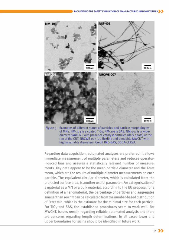
Particle sizeThe primary particle size (-distribution) is the key criteria for defining a MN, in accordance with the proposed EC nanomaterial definition, and it may be analysed by a number of different methods. In NANOGENOTOX, XRD, TEM, AFM, DLS and SAXS were used as complementary methods for describing the particle size. XRD is only applicable to crystalline materials and therefore has limited general use in size-analyses. However, XRD is an excellent method for analyses of the mineralogy and significant crystalline impurities in bulk powder samples. Advanced mathematical analyses of XRD data enable quantification of the average crystallite size, preferred orientations, in a nanocrystalline material as well as estimates of the proportion between phases in a sample. However, the project data also indicate that the size and proportions obtained may be dependent on both instruments and the data-analyses method applied in the calculations.TEM and AFM are generally considered to be among the most precise typical methods for measurements of the particle size-dimensions in the nanoscale. In TEM, the particle size is measured on highly magnified digital images and it may be completed following automated, semi-auto-mated and manual methods. Some of the challenges in TEM size-distri-bution measurements include reliable sample preparation that captures and maintains the real particle size-distribution of the samples, as well as the very large span in size dimensions observed for some MNs. The latter is evident in the case of MWCNT, where the thin diameter of the CNT may range from ca. 1 nm to more than 100 nm while the lengths may vary from nm-scale to several tens of µm’s. In addition MWCNTs may be very flexible causing complex structures and agglomerates and therefore dif-ficulties in sizing the tube length [Figure 3]. Another issue related to mor-phology is primary particle size-distribution measurements of dispersed and non aggregated particles to nanostructured and fused particles or structures such as the synthetic amorphous silica [Figure 3]. For sample preparation, the study concluded that powder samples should be pre-sonicated in a suitable dispersion medium to disperse large ag-glomerates and aggregates before adding them to TEM-grids. The exact medium, the sonication power and duration must be optimised for each specific MN. For example, pure double-distilled water is sufficient to dis-perse most SAS, whereas acidified water may be used for TiO2. For CNT addition of bovine serum albumin was proven to be applicable. However, other alternatives certainly exist.
16 17
Facilitating the saFety evaluation oF manuFactured nanomaterials
Regarding data acquisition, automated analyses are preferred. It allows immediate measurement of multiple parameters and reduces operator-induced bias and assures a statistically relevant number of measure-ments. Key data appear to be the mean particle diameter and the Feret mean, which are the results of multiple diameter measurements on each particle. The equivalent circular diameter, which is calculated from the projected surface area, is another useful parameter. For categorisation of a material as a MN or a bulk material, according to the EU proposal for a definition of a nanomaterial, the percentage of particles and aggregates smaller than 100 nm can be calculated from the number-based distribution of Feret min, which is the estimate for the minimal size for each particle. For TiO2 and SAS, the established procedures seem to work well. For MWCNT, issues remain regarding reliable automated analysis and there are concerns regarding length determinations. In all cases lower and upper boundaries for sizing should be identified in future work.
Figure 3 – Examples of different states of particles and particle morphologies of MNs. NM-103 is a coated TiO2, NM-201 is SAS, NM-401 is a wide-diameter MWCNT with presence catalyst particles (dark spots) at the rim of the CNT. NRCWE-007 is a flexible and bendable MWCNT with highly variable diameters. Credit IMC-BAS, CODA-CERVA.
18 19
analysis of particle size in liquid dispersionsSAXS and DLS are two very different techniques and were used to mea-sure the size-distribution of particles in liquid suspensions. Whereas the DLS measures the particles by their Brownian motion, SAXS measures the particles according to mathematical treatment of scattered X-ray data. Both methods may be applicable for measurement of aggregate size-distributions after appropriate dispersion in a liquid. However, SAXS can also be used to derive information on the aggregate/agglomerate structure, the average primary particle size and the average shape factor and can also be used on dry powders. Both methods were assumed to be applicable both for SAS and TiO2 samples, but uncertainties are still raised regarding the applicability for measurements on CNT. Comparison between aggregate sizes determined in batch dispersion media prepared according to the generic NANOGENOTOX dispersion protocol for toxicity testing suggests that SAXS gives smaller average aggregate sizes than DLS, when the intensity-derived hydrodynamic zeta-average values are used from the DLS.
analyses of particle size in dustiness testingA last method for investigating the particle size-distributions of the powder samples was applied as part of dustiness testing. We measu-red the dustiness and dust size-distributions generated by two different methods: a downscaled standard EN15051 rotating drum and a Vortex shaker. Both methods are candidates for a new nanopowder dustiness standard in EC Mandate 461 for standardisation activities regarding na-notechnologies and nanomaterials. Sizing with a Fast Mobility Particle Sizer plus an Aerodynamic Particle Sizer (FMPS+APS) in the rotating drum test and an Electrical Low-Pres-sure Impactor (ELPI) in the Vortex shaker tests showed that the powder dusts all had bi- to multimodal number size-distributions. The dust par-ticle size-ranges were very wide ranging from less than 100 nm and into the µm size-range. However, it was also evident that either the two types of tests produce dusts with different size-distributions or the different instruments give different method-dependent size-distributions. The FMPS+APS measurements of TiO2 and SAS dusts showed peak num-ber concentrations for particle sizes around 200 and 300 nm, respecti-vely, but size-modes were also observed around 40-60 nm for some pow-ders and in the µm-range for all powders. ELPI measurements showed more variation in the number peak-size where both TiO2 and SAS usually had a primary or secondary peak-size around or below the 100 nm size

18 19
Facilitating the saFety evaluation oF manuFactured nanomaterials
range. In all cases the majority of dusts from TiO2 and SAS were released as aggregates and/or agglomerates. The MWCNT (NM-400, 401, 402, and 403) were only analysed using the Vortex shaker method. The expe-riments revealed that both free and agglomerated/aggregated CNT “fi-bers” were released during this agitation procedure. The fraction of dust particles smaller than 100 nm was very significant in tests of NM-400, NM-402 and NM-403. In NM-401, the peak size in particle number was located between 200 and 300 nm.
Specific surface area (SSa)Specific surface area analyses were performed by BET and SAXS. Expe-rimentally, SSA was also determined by TEM tomography on single samples. BET is surface area measurement based on quantification of the amount of nitrogen gas that was adsorbed to the powders, whereas SAXS relies on mathematical analyses of the X-ray scattering signal from the particle surfaces in the powder sample. Determination of SSA or VSSA (Volume-Specific Surface Area) by TEM tomography is based on 3D morphological analysis of each powder particle in a sample. The results showed a wide range in the specific surface areas of the MNs analyzed with BET and there was a general linear correlation between
data obtained by BET and data obtai-ned by SAXS. Above ca. 130 m2/g the SAXS data appeared to underscore the specific surface area determined by BET. However, more samples with a wide range in SSA are required to ful-ly assess the comparability between these two methods. The tomographic approach also appears to give reliable values, but particle-specific data have not been obtained on standards or by other methods in this project. In addi-tion the technology is not yet ready for high-throughput analyses on all types of materials therefore a final conclusion cannot be made on the applicability of this procedure.
Tecnai spirit transmission electron microscope with biotwin lens configuration operating at 120 kV. Credit CODA-CERVA.

20 21
Chemical compositions, impurities and coatingsThe elemental chemical composition of the nanomaterials and associa-ted organic compounds are obviously of high interest, beyond the iden-tification and categorisation of the MN. The presence of inorganic minor elements, either as substitutions of constituent elements in the MN ato-mic structure or due to an inorganic coating, may change the toxicolo-gical effect of a MN. Similar effects have been observed due to organic coatings and functionalisations of MN as well due to the presence of dif-ferent catalyst materials e.g., in MWCNT.
Clearly different techniques are required to identify and quantify inor-ganic elements and associated organic compounds as contaminants, coatings and functionalisations. Even though extensive development of chemical analysis was not part of this project, procedures were refined to improve extraction for elemental analysis of inorganic MWCNT cata-lysts by ICP. In addition, a procedure was established in synergy with two EU FP7 projects, NANODEVICE and ENPRA, using thermogravimetric analysis to identify whether a MN may be constituted of or associated with organic compounds. Mass losses due to compounds evaporating at temperatures greater than 105°C were discriminated from mass-losses occurring at lower temperatures, which could be due to adsorbed water, for example. In the current work it was decided that any inorganic mate-rial with a total mass-loss greater than 1 wt% during combustion in air would be subject to subsequent organic chemical analysis. For MWCNT, the residual mass after combustion was used to determine the amount of inorganic catalyst material in the samples. Inorganic chemical analysis by EDS showed the presence of ca. 4.5 wt% Al and 0.7 wt% Si in NM-103 and NM-104 (TiO2) as well as about 0.5 wt%
Transmission electron tomographic reconstruction of aggregated SAS nanoparticles. Bar is 50 nm. Credit CODA-CERVA.

20 21
Facilitating the saFety evaluation oF manuFactured nanomaterials
Fe in NM-400. Al was known to be present in inorganic coatings in NM-103 and NM-104. All other TiO2 samples contained less than 0.2 wt% Si. Other minor impurities were due to salts. NM-101, NM-104 and NM-104 had a TGA mass-loss of 2 to 8 wt% that could be ascribed to organic coatings.
EDS and ICP analysis both showed that the SAS MNs all contained minor amounts of Al. Other minor impurities were due to salts. TGA showed a mass-loss of 1 to 3 wt% in NM-200, NM-201 and NM-204.
Quantitative elemental analysis of MWCNT was found to be a greater challenge than may be generally anticipated. There was poor agreement between the results coming from different laboratories. None of the ele-mental analysis reached the 3 to 18 wt% impurities indicated by TGA. Combining results from EDS-analysis and different ICP methods as well as XRD on residuals after combustion confirmed the major impurities to be various combinations of transition metal catalysts. However, a signifi-cant fraction may also sometimes be ascribed to salts, which are thought to be residuals after purification of the CNT.
Focus on protocol for producing suitable manufactured nanomaterial exposure media
As a major and very early deliverable, WP4 was requested to develop Standard Operating Proce-dures (SOPs) for preparing MN batch dispersions suitable for application in in vitro and in vivo toxi-city testing. It was agreed that WP4 would produce a generic dispersion protocol aiming to:
1. use the biologically relevant serum albumin for particle stabilisation (surfactant),2. reduce the albumin concentrations as much as possible to limit poten-tial unwanted toxicological side-effects and3. adopt the batch dispersion MN concentration and probe sonication conditions already established in the EU FP7 project, ENPRA.
Based on these boundary requirements, a series of range-finding tests were conducted to identify applicable BSA (Bovine Serum Albumin) concentrations to achieve relatively stable MN dispersions, procedures to also enable dispersion of hydrophobic MNs, detailed adjustments and harmonisation of the sonication vials, preparation
Dispersion of mNs for toxicity testing

22 23
volumes, and sonication conditions, including selection of proper im-mersion depth of the sonicator probe, and adjustments of sonication times for sonicators with different sonication energies and amplitudes. The work finally resulted in a common generic dispersion protocol using a 0.05% w/v sterile-filtered BSA-water solution as dispersion medium for 2.56 mg MN per ml, total batch dispersion volume 6-10 ml, generic pre-wetting of the MN powder with 0.5% (v/v) ethanol to also enable dispersion of hydrophobic MN, and probe-sonication for 16 min at 400W and 10% probe-amplitude while keeping the sample vial cooled in an ice-water bath. Sonicators with higher energy output should apply reduced sonication times (e.g., 12 min at 500 W and 20% amplitude). The detailed dispersion protocol is available on the JA web site.It is important to note that NANOGENOTOX protocol is a procedure which is generally applicable to all powder MN. It will not always, and probably rarely, disperse MNs into their primary particles or aggregates. However, in most cases, the protocol produced metastable dispersions with de-rived DLS number peak-sizes within the lower and upper TEM size-range found for the MN and less than 10% sedimentation within the first 1 hour. Introduction of mandatory brief vortex shaking of the batch dispersions immediately before use ensured re-establishment of the original charac-teristics of the batch dispersions. It should be noted that the MN dis-persions should always be used immediately after preparation in order to minimise potential artefacts induced by particle dissolution and / or exhausted surface reactivity.

22 23
Facilitating the saFety evaluation oF manuFactured nanomaterials
I N vITrO mEThODS FOr GENOTOXICITy TESTING
ObjectivesThe basic questions of in vitro genotoxicity testing of manufactured nanomaterials (MNs) include how well in vitro assays can be used for revealing the genotoxic potential of MNs, which assays are suitable for this task, and which modifications are needed in the tests when MNs are studied. The main aim of WP5 was to establish robust methodology to screen in vitro genotoxicity of MNs in pulmonary, intestinal and dermal cell systems. The first objective was to generate in vitro genotoxicity data on the chosen MNs, using standard tests and modified assays utilising specific cell models. The second objective was to evaluate the robustness of the methodology. Based on in vitro genotoxicity test results, a round robin test was carried out, using the most promising in vitro assays.
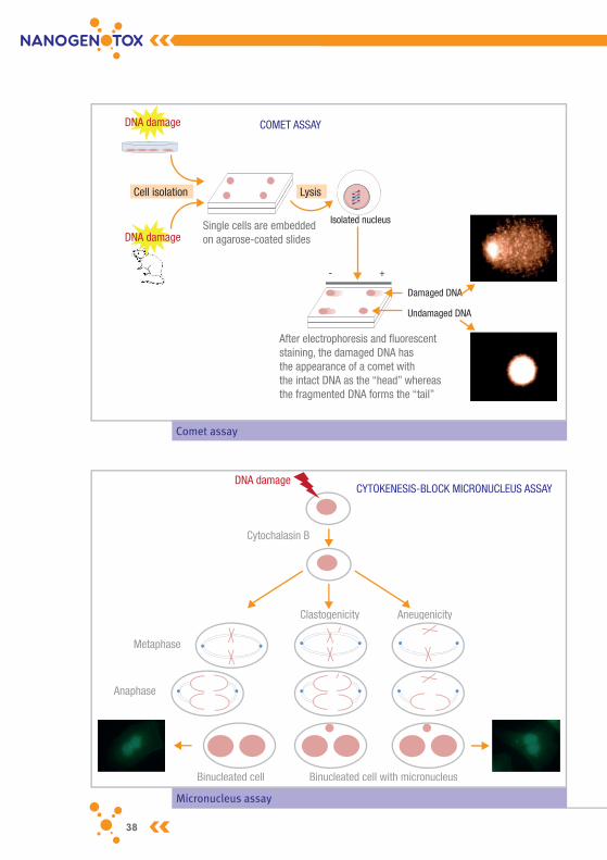
methodsThree genotoxicity endpoints were chosen for the first part of WP5: DNA damage, micronuclei formation, and mutations.Alkaline and FpG-modified comet assays were used for assessing DNA damage. The alkaline comet assay is a simple and sensitive method for the detection of DNA strand breaks (single- and double-strand breaks) and alkali-labile sites. The FpG-modified assay allows the detection of oxidative DNA damage. The micronucleus assay was used to detect agents that modify chromo-some structure or their segregation, leading to the formation of an addi-tional nucleus (micronucleus) during cellular division. The cytokinesis block micronucleus assay, using cytochalasin B to prevent cytokinesis, was performed in all cell lines, except 16 HBE cells where cytochalasin B was not used.The mouse lymphoma assay was carried out to detect mutations.Various human cell lines of different origin were used: pulmonary (bronchial epithelial BEAS 2B and 16 HBE; alveolar A549), intestinal (Caco-2, prima-rily undifferentiated cells used) and epidermal (NHEK keratinocytes). Reconstructed full-thickness skin models were applied only for testing of TiO2 and ZnO nanoparticles. The micronucleus assay was performed also in human primary lymphocytes. The mouse lymphoma mutation assay was carried out in mouse lymphoma L5178Y TK+/- cells.
In vitro comet and micronucleus assays, complemented with the mouse lymphoma assay, were applied to all MNs assessed (except in the der-mal systems, where only TiO2 was tested) using the dispersion protocol
24 25
provided by WP4. Most series included nanosized ZnO as a nanoparticle control, in addition to assay-specific (chemical) positive controls: mito-mycin C in the micronucleus assay, ethyl methane sulphonate or methyl methane sulphonate (MMS) in the comet assay, and MMS in the mouse lymphoma assay.
The protocols were harmonised, following the general principles des-cribed below. One experiment was performed per MN per genotoxicity endpoint (two experiments when time allowed it). Each treatment was conducted as duplicate cultures. No metabolic activation system was utilised. The comet assay was carried out with two treatment times, 3 h or 24 h. For the micronucleus assay, a longer-term treatment covering 1.5-2 cell cycles was used; cytochalasin B was added 6 h after the start of the treatment, in Caco-2 cells at 24 h. The treatments were performed in the same medium that was used for the culture. The doses of the MNs tested were chosen on the basis of cytotoxicity measurements using mostly cell count relative to control, relative increase in cell counts (RICC), or relative population doubling (RPD). The highest dose was either at the cytotoxi-city limit of 55% +/-5% or as otherwise justified. For each nanomaterial, 4-6 doses were included in the genotoxicity assays to obtain a minimum of 3 analysable doses. In the case of MNs with low cytotoxicity, the maxi-mum dose was derived from the WP4 dispersion protocol (256 µg/ml) or was based on technical limitations (e.g. inhibition of analysis because cells were covered with MNs). Doses were given in µg/ml and (for cells growing on surface) also in µg/cm2. The results were related to specific surface area (if possible) and other characteristics considered important, to find out if they correlated with genotoxicity.
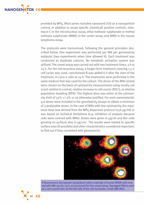
A fluorescence micrograph (propidium iodine staining) of human NHEK cells trea-ted with NM-104 for 24 h, as processed for the comet assay. Damaged DNA is seen as a red «comet tail» on the left side of the cell nucleoids. Credit IMB-BAS.

24 25
Facilitating the saFety evaluation oF manuFactured nanomaterials
Full thickness skin models (EpiDermFTTM). Credit BfR.
In the context of the development of experimental methodologies, a round robin study (an inter-laboratory test performed independently in 12 different laboratories), was carried out to assess the reproducibi-lity of the genotoxicity tests. The round robin study comprised in vitro genotoxicity testing of one type of each family of studied MNs: TiO2 (NM-102, doses: 0, 64, 128 and 256 µg/ml), SAS (NM-203, doses: 0, 8, 32 and 64 µg/ml), and MWCNT (NM-403, doses: 0, 64, 128 and 256 µg/ml); both the cytokinesis block micronucleus assay and the alkaline comet assay were carried out. ZnO (NM-110, doses: doses used between 1.5 and 8.55 µg/ml) was included in all series, to assess its possible use as a nanoparticle positive control. The partners were divided into two groups, one group of six laboratories using bronchial human epithelial BEAS 2B cells, and the other six laboratories using human epithelial colorectal adenocarcinoma Caco-2 cells.
1st part – in vitro results TiO2
The micronucleus assay was positive1 for each TiO2 in NHEK cells. There was also a positive finding for lymphocytes for NM-102, NM-103 and NM-104. The micronucleus assay was negative for all TiO
2 in other types of cells. The comet assay was positive for all TiO2 in Caco-2 cells after the 24-h treatment except for NM-104 (negative). Results of the comet assay were positive, with the 3-h or 24-h treatment for NM-102 in all cell lines except 16HBE and for NM-105 in all cell lines except BEAS 2B and 16HBE.
1. Positive result: a statistically significant increase with ≥2 doses or a statistically significant increase at high dose and a dose-dependent increase.

26 27
The FpG-modified comet assay was positive for NM-104 and NM-105 in BEAS 2B and Caco-2 cells, and for NM-104 in A549 cells, but negative for all types of TiO2 in 16HBE cells.The mutation assay was negative for all forms of TiO2 tested.In 3D human reconstructed full thickness skin models, all TiO2 nano-materials (NM-102, NM-103, NM-104, and NM-105) investigated for DNA damage were negative in the comet assay. In contrast, the chemical posi-tive control MMS consistently generated a significant increase in DNA-damage. The highest dose studied by this protocol was 246 µg/cm2 skin surface which showed no interference during the analysis. Transmission electron micro scopic analysis by CODA-CERVA could not identify pene-tration of TiO2 through the stratum corneum of reconstructed human full thickness skin models even after 72 h exposure. This points to an undisturbed skin barrier in these 3D models and may explain the lack of positive results as compared to results obtained with NHEK cells. As TiO2 nanoparticles showed no penetration, the in vitro micronucleus assay was not systematically carried out with the 3D human skin models, but a more in depth investi gation was performed using the comet assay. The probability of those nanomaterials reaching dividing cells of an intact 3D skin barrier is close to zero. Furthermore, the full thickness skin mo-dels appear to show only a low cell division rate in the end-differentiated stage. Thus, it is postulated that nanomaterials with a realistic agglo-merate size above 20 nm will not enter viable human skin models and consequently will not exert genotoxic effects in this test system.
SASAll SAS nanomaterials induced micronuclei in Caco-2 cells, but when the experiment was repeated, the initial positive results could not be confirmed. NM-201 and NM-202 induced micronuclei in A549 cells. The micronucleus assay was mostly negative for all SAS in other cells.After the 3-h treatment, the comet assay was mostly positive for the different types of SAS in BEAS 2B cells and for NM-200 in all cell lines. Positive results were also obtained in the comet assay with NM-201, NM-202 and NM-203 in A549 cells after the 3-h or 24-h treatment and with NM-203 in Caco-2 cells (both treatment times). The mutation assay was negative for all types of SAS tested.
26 27
Facilitating the saFety evaluation oF manuFactured nanomaterials
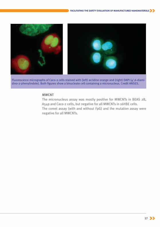
Fluorescence micrographs of Caco-2 cells stained with (left) acridine orange and (right) DAPI (4’,6-diami-dino-2-phenylindole). Both figures show a binucleate cell containing a micronucleus. Credit ANSES.
MWCNT The micronucleus assay was mostly positive for MWCNTs in BEAS 2B, A549 and Caco-2 cells, but negative for all MWCNTs in 16HBE cells.The comet assay (with and without FpG) and the mutation assay were negative for all MWCNTs.

28 29
+ Positive: a statistically significant increase with ≥2 doses or a statistically significant increase at high dose and a dose-dependent increase;
(+) Equivocal: a statistically significant increase with 1 dose, no dose-dependent increase; - Negative; / Used to separate outcome of two experiments. Grey box, Not performed.
Pulmonary Intestinal Lymphatic Dermal
BEAS 2B 16 HBE A549 Caco-2 Lymphocyte L5178Y TK+/- NHEK Skin 3D
Mic
ronu
cleu
s a
Com
et 3
h
Com
et 2
4h
Com
et F
pG 3
h
Com
et F
pG 2
4h
Mic
ronu
cleu
s b
Com
et 3
h
Com
et 2
4h
Com
et F
pG 3
h
Com
et F
pG 2
4h
Mic
ronu
cleu
s a, c
Com
et 3
h
Com
et 2
4h
Com
et F
pG 3
h
Com
et F
pG 2
4h
Mic
ronu
cleu
s d
Com
et 3
h
Com
et 2
4h
Com
et F
pG 3
h
Com
et F
pG 2
4h
Mic
ronu
cleu
s e
MLA
Mic
ronu
cleu
s f
Com
et 3
h
Com
et 2
4h
Com
et 3
h
Com
et 2
4h
TiO2
NM-102 - + + - - - - + - - - + (+) - + (+) (+) - -
NM-103 - - - - - - - - - - - (+) + - + (+) (+) - -
NM-104 - - - - - - - - - - - - + - + (+) (+) - -
NM-105 - - - - - - - + - - - +/+ - - + (+) (+) - -
SAS
NM-200 - + (+) - + - - - -/- (+) - - - +/- + + (+) + - -
NM-201 - (+) - - - - - - +/+ + (+) - (+) +/- - (+) - + - -
NM-202 - + + - - - - - +/+ + (+) + - +/- (+) (+) + - - -
NM-203 (+) + + - - - - - -/(+) - + - + +/- + + + (+) - -
MWCNT
NM-400 (+) - - - - - - - (+) - - (+) - - - - - -
NM-401 + - - - - - - - - - - + - - - - - -
NM-402 + - - - - - - - + - - + - - - - (+) -
NM-403 + - - - - - - - - - - (+) - - - - + -
NRCWE-006 + - - - - - - - + - - - - - - - + -
NRCWE-007 + - - - - - - - + - - + - - - - - -

28 29
Facilitating the saFety evaluation oF manuFactured nanomaterials
Table 3 – Outcome of the in vitro genotoxicity assays in various cell systems (first part of WP5)
Pulmonary Intestinal Lymphatic Dermal
BEAS 2B 16 HBE A549 Caco-2 Lymphocyte L5178Y TK+/- NHEK Skin 3D
Mic
ronu
cleu
s a
Com
et 3
h
Com
et 2
4h
Com
et F
pG 3
h
Com
et F
pG 2
4h
Mic
ronu
cleu
s b
Com
et 3
h
Com
et 2
4h
Com
et F
pG 3
h
Com
et F
pG 2
4h
Mic
ronu
cleu
s a, c
Com
et 3
h
Com
et 2
4h
Com
et F
pG 3
h
Com
et F
pG 2
4h
Mic
ronu
cleu
s d
Com
et 3
h
Com
et 2
4h
Com
et F
pG 3
h
Com
et F
pG 2
4h
Mic
ronu
cleu
s e
MLA
Mic
ronu
cleu
s f
Com
et 3
h
Com
et 2
4h
Com
et 3
h
Com
et 2
4h
TiO2
NM-102 - + + - - - - + - - - + (+) - + (+) (+) - -
NM-103 - - - - - - - - - - - (+) + - + (+) (+) - -
NM-104 - - - - - - - - - - - - + - + (+) (+) - -
NM-105 - - - - - - - + - - - +/+ - - + (+) (+) - -
SAS
NM-200 - + (+) - + - - - -/- (+) - - - +/- + + (+) + - -
NM-201 - (+) - - - - - - +/+ + (+) - (+) +/- - (+) - + - -
NM-202 - + + - - - - - +/+ + (+) + - +/- (+) (+) + - - -
NM-203 (+) + + - - - - - -/(+) - + - + +/- + + + (+) - -
MWCNT
NM-400 (+) - - - - - - - (+) - - (+) - - - - - -
NM-401 + - - - - - - - - - - + - - - - - -
NM-402 + - - - - - - - + - - + - - - - (+) -
NM-403 + - - - - - - - - - - (+) - - - - + -
NRCWE-006 + - - - - - - - + - - - - - - - + -
NRCWE-007 + - - - - - - - + - - + - - - - - -
a Treatment for 48h, Cyt-B added at 6h.b Treatment for 41h, no Cyt-B used.c Treatment for 24 h (TiO2, NRCWE-006 and NRCWE-007), Cyt-B added at 6h.d Treatment for 52 h, Cyt-B added at 24 h.e Treatment for 30 h, Cyt-B added at 6 h.f Treatment for 54 h, Cyt-B added at 6h

30 31
2nd part – Round robin testNM-102 (TiO2)In BEAS 2B cells, the outcome of the comet assay with NM-102 was almost unanimously positive (five out of six laboratories), in accordance with the outcome of the 1st part of WP5. The result of the micronucleus assay in BEAS 2B cells was negative in four laboratories (in agreement with the 1st part), positive in one laboratory and equivocal in another laboratory.In Caco-2 cells, the comet assay with NM-102 (positive in the 1st part) was negative in three laboratories but positive in two. Three laboratories working with Caco-2 cells had problems in reading the micronucleus slides due to presence of particle agglomerates on the microscopical slides; for the remaining three laboratories, the outcome of the micronucleus assay with NM-102 (negative in the 1st part) was negative in two laboratories and positive in one.
NM-203 (SAS)In BEAS 2B cells, the result of the comet assay with NM-203 was negative (similarly to the results in the 1st part) in three laboratories but positive in three others. The outcome of the micronucleus assay with NM-203 in BEAS 2B cells was positive in three laboratories and negative in three laboratories (results were equivocal in the 1st part).In the comet assay with Caco-2 cells, NM-203 (positive in the 1st part) was negative in three and positive in two laboratories. The results of the micronucleus assay with NM-203 in Caco-2 cells were split, with three positives and three negatives; conflicting results had also been obtained in the 1st part.
NM-403 (MWCNT) The comet assay in BEAS 2B cells with NM-403 (negative in the 1st part) showed a split outcome, with three negative and three positive results. The outcome of the micronucleus assay with NM-403 in BEAS 2B cells was almost unanimous: a negative result was obtained in all laboratories except one - despite the positive result in the 1st part.In Caco-2 cells, NM-403 yielded four negatives and one positive in the comet assay (negative in the 1st part) and three positive, one equivocal, and two negative results in the micronucleus assay (equivocal in the 1st part).

30 31
Facilitating the saFety evaluation oF manuFactured nanomaterials
NM-110 (ZnO)NM-110 (ZnO), tested as a candidate positive nanoparticle control, was unanimously positive in the micronucleus assay with Caco-2 cells and yielded 3 positives and 3 negatives in BEAS 2B cells. In the comet assay, 3 negatives and 2 positives were recorded in Caco-2 cells and 4 positives and 2 negatives in BEAS 2B cells.The outcome of the round robin test is presented in the Table 3.
Table 3 – Outcome of the round robin test
Partner No.
TiO2 NM-102 SAS NM-203 MWCNT MN-403 ZnO NM-110
Comet CBMN Comet CBMN Comet CBMN Comet CBMN
Caco-2 cells
A - - - + - + - +
B + ANP + + + + + +
C - + - - - + - +
D - - + - - - - +
E ANP - - +
F + ANP - + - (+) + +
BeAS 2B cells
G + + - + + + + +
H - - + - - - + +
I + (+) + + + - + -
J + - + - - - - +
K + - - + - - - -
L + - - - + - + -
+: Positive; (+): Equivocal; -: NegativeGrey box: not performed; ANP: Analysis could not be performed.

32 33
ConclusionsIn the first part of WP5, data on the genotoxicity of 15 MNs (4 TiO2, 4 SAS, 6 MWCNTs, and ZnO as a candidate positive nanoparticle control) were generated from the comet assay and the micronucleus assay using a number of different human cell lines of pulmonary, intestinal, and epider-mal origins. In addition, micronuclei were also studied in human primary lymphocytes, and mutations in mouse lymphoma cells. While the mouse lymphoma assay was uniformly negative, the outcome of the comet as-say and the micronucleus assay varied greatly among the different cell systems. It is presently unclear how much of this variation represented true differences among the cell systems and how much could be explai-ned by experimental variations. Although dose-dependent effects could be seen in many experiments, the genotoxicity of the MNs studied was generally relatively low; in such a situation, experimental variation may determine if the result will turn out positive or negative. Variation may occur among experiments, e.g. in MN dispersions, the agglomerate size of the MN in the cell culture, MN sedimentation on the cells, and thereby cellular uptake and intracellular dose. Agglomerates of different size and shape may have differential effects on cells.
In the round robin study, relatively reproducible results were obtained for the comet assay with NM-102 in BEAS 2B cells (mostly positive) and with NM-403 in Caco-2 cells (mostly negative), and for the micronucleus assay with NM-403 (mostly negative) in BEAS 2B cells and NM-110 with Caco-2 cells (all positive). When a positive response was seen, it was low (simi-larly to the 1st part of WP5), which probably contributed to the situation where identical outcomes were not systematically obtained. Although ZnO may be applicable as a nanoparticle positive control in some in vitro cell systems such as the micronucleus assay in Caco-2 cells, it does not appear to be universally suitable for this purpose because of the narrow dose range of its genotoxicity in some cell systems.In summary, the present studies suggest that many MNs have some genotoxic potential detectable in human cells in vitro using the comet assay or the micronucleus assay. On the other hand, the mutation assay with mouse lymphoma cells appears to give only negative results. The in vitro genotoxic effect of the MNs studied was mostly low, which possibly contributed to the variation observed in outcome among the cell systems. It is technically feasible to perform such genotoxicity assays

32 33
Facilitating the saFety evaluation oF manuFactured nanomaterials
with dispersed MNs in cultured cells, but the predictive value of these in vitro tests in identifying MNs that are genotoxic in vivo that could be carcinogenic is presently unclear. More information on the mecha-nisms of (i) the detected in vitro genotoxicity and of (ii) the MNs that are genotoxic in vivo or carcinogenic is needed before this question can be answered.

34 35
ObjectivesThe aim of WP7 is to identify relevant organs for genotoxicity testing based on the determination of organ exposure to nanomaterials. The dis-tribution of the nanomaterials into the various organs is an indication for the organs at risk for nanomaterial toxicity and thus also genotoxicity, based on the interaction and activity of the nanomaterials with the cells of the organ. In WP7, the kinetic parameters and tissue distribution are determined for selected nanomaterials: titanium dioxide (TiO2), silicon dioxide as synthetic amorphous silica (SAS), and carbon nanotubes (CNTs) after oral and intravenous administration.
routes investigatedThe oral route of exposure was chosen as this is a common route of expo-sure for consumers. However, after oral exposure the absorption of MNs may vary greatly. After inhalation and dermal exposure in general the dis-tribution and absorption of MNs were demonstrated previously to be low, if any. In addition to the oral route, the intravenous route (IV) of exposure was also investigated as this route of administration circumvents the biological barriers present and results in direct systemic availability of the nanomaterials in the blood circulation and thus in the internal organs. Organ and blood samples were collected and evaluated for detection for Ti, Si, and CNTs. For SAS and TiO2 MNs, it was not the MNs themselves but the elements silica (Si) and titanium (Ti) which were determined using different inductively coupled plasma equipment: Inductively Coupled Plasma-Mass Spectrometry (ICP-MS), Inductively Coupled Plasma Optical Emission Spectroscopy (ICP-OES), High Resolution Inductively Coupled Plasma-Mass Spectrometry (HR-ICP-MS). For SAS, a fit-for-purpose ana-lytical method was developed using ion-molecule chemistry to eliminate polyatomic interferences and enable interference free-detection of Si. For the CNTs the determination of C was not an option to study the tissue distribution as C is present in all tissues. Therefore, CNTs were radiolabe-led with 14C atoms to allow detection in the body.
T OXICOkINETICS aND TISSuE DISTrIbuTION OF mNS aND IDENTIFICaTION OF OrGaNS aT rISk FOr GENOTOXICITy TESTING

34 35
Facilitating the saFety evaluation oF manuFactured nanomaterials
resultsFor the investigated MNs the oral administration resulted in a low uptake from the gastro-intestinal (GI) tract even after 5 repeated oral administra-tions. The infrequent and incidental demonstration of some Ti in tissues beyond the GI-tract may indicate that uptake of TiO2 is possible. In addi-tion, it was demonstrated that the faeces of control rats already contained an amount of Ti well above the detection limit. Similar Ti levels were obser-ved in the GI-tract of control and IV exposed rats which led to the conclu-sion that excretion via GI-tract after IV injections is not occurring. For SAS, the levels determined in liver and spleen as indicator organs for systemic uptake were similar to control levels or close to the detection limit indica-ting a very low absorption from the GI tract. The organ levels after repeated oral administration suggest minor differences between male and female animals and between the SAS nanomaterials (NM-200 and NM-203) inves-tigated, although the limited absorption makes it difficult to draw any firm conclusions. Translocation of the MWCNT from the GI-tract into the syste-mic circulation or any of the organs investigated was not demonstrated.For the single and repeated IV administrations, the results indicate that the TiO2 MNs can remain in the body for a prolonged period of time, the exception being NM-105 (an anatase-rutile mixture). For all TiO2, there was a rapid decrease in blood concentrations after the IV administration with most of the Ti cleared from the blood 2 hours after administration. A similar pattern was observed for both the single and repeated IV adminis-tration. The liver was the major organ for the Ti distribution followed by the spleen and the lung, while other organs investigated (brain, kidney, thymus, reproductive organs etc.) had a distribution below 0.1% of the dose administered. For some TiO2 redistribution was noted with spleen levels increasing and liver levels decreasing. However, the liver remains the organ with the highest uptake in view of its size and total weight. It should be noted that not only the levels expressed as a percentage of the dose are important in the tissue distribution of nanomaterials, the levels
Radioimaging: slice of kidney after IV injection of 14C-NRCWE-006. Scale CPM. Credit CEA.

36 37
Radioimaging: Slice of liver after IV injection of 14C-NRCWE-006. Scale CPM. Credit CEA.
expressed as µg/g organ can give a direct indication of the possible expo-sure of the organ and might be indicative for the induction of toxicity. All five TiO2 MNs were still present in various organs at day 90 after the IV administrations, only for one (NM-105) out of five TiO2 was a major decline in organ levels noted. The results indicate that after the IV admi-nistration of manufactured TiO2 there is no excretion of Ti via the faeces.
For the SAS, a decrease in tissue concentrations was observed between day 2/day 6 and day 90 after the repeated and single dosing indicating a clearance from the body. Major organs for the distribution of the MNs are liver, spleen and lung and to a lesser extent the kidney. For SAS, fol-lowing single dose IV administration, measurable concentrations slightly above the limit of quantification of Si were detected in the liver of male and female rats up until 90 days after administration. After repeated IV administration, a considerable concentration of Si is present in liver and spleen of males at day 6, with marked particle- and gender-related dif-ferences, and detectable concentrations found in other organs as well. After a single IV SAS administration, the highest level of NM-203 was observed in the spleen of male rats, while for NM-200 in male and female rats the highest concentration was noted in the liver. In female rats the concentration of NM-203 was similar for liver and spleen. After repea-ted IV SAS administration NM-203 showed the highest concentrations in the spleen of male rats, while for NM-200 the highest concentration was in the liver. In female rats, similar concentrations were observed in liver and spleen both at day 6 and day 90 for NM-203, while for NM-200 the highest concentration was present in the liver both at day 6 and day 90. The meaning of such differences is not clear. However, although there is a clear decrease in Si concentration in liver and spleen at day 90 after intra-venous administration, Si concentrations were still distinctly higher than in controls suggesting that a longer time period than 90 days is required for complete elimination of administered SAS from the body.

36 37
Facilitating the saFety evaluation oF manuFactured nanomaterials
Each type of the four MWCNTs investigated was found to display particu-lar bioaccumulation and biopersistence properties in the various organs evaluated. NM-400 and NRCWE-006 showed much higher bioaccumula-tion than NM-401 and NM-402. In liver and spleen, at day 90, a marked reduction in the MWCNT level expressed as percentage of injected dose, was only observed for NM-400. For NM-401, NM-402 and NRCWE-006 no significant decrease was observed between day 6 and day 90. In lung, a significant reduction in the MWCNT level expressed as percentage of injected dose was only observed for NM-401. Although a decrease was observed between day 6 and day 90 for NM-400, all MWCNTs investiga-ted including NM-400, were still observed in the various organs (liver, spleen, lung) at day 90 after the administration. For NM-401, NM-402 and NRCWE-006, the data suggest a significant biopersistence of these MWCNTs in most organs beyond 3 months after administration.Although the detection of the radiolabel is indicative for the presence of the CNT in organs it could also be that a detached label not associated with the CNTs was measured. In additional experiments it was demons-trated unambiguously that the radioactive signal indeed corresponds to the presence of CNTs carbon nanotubes, with a direct visualisation of the CNT walls, the walls inside, and finally measurement of the CNT diameter.
ConclusionFor all TiO2 and SAS nanomaterials, oral administration resulted in a rather low uptake via the GI-tract after repeated oral administration, whereas for MWCNT no uptake from the GI-tract was demonstrated.After IV administration, most MNs showed a rapid clearance from the blood indicating a quick distribution to, and uptake by, the various or-gans. For the single and repeated IV administrations, the results indicate that especially some TiO2 and MWCNT are still present in the organs at day 90 after the last administration. For SAS in general, a decrease in Si level was noted between day2/day 6 and day 90, although at day 90 Si could still be detected.Major organs for the biodistribution of the investigated MNs are liver, spleen and lung and to a limited extent the kidney. Although the IV admi-nistration can be considered an artificial route of exposure, the results obtained clearly demonstrate that some MNs can persist in organs for a prolonged period of time until at least 90 days, the last time point inves-tigated in these studies.
38 39
Micronucleus assay
DNA damage
Cytochalasin B
Clastogenicity
Binucleated cell with micronucleusBinucleated cell
Anaphase
Metaphase
Aneugenicity
CYTOKENESIS-BLOCK MICRONUCLEUS ASSAY
Comet assay
COMET ASSAY
After electrophoresis and fluorescent staining, the damaged DNA has the appearance of a comet with the intact DNA as the “head” whereas the fragmented DNA forms the “tail”
Single cells are embedded on agarose-coated slidesDNA damage
Damaged DNA
+-
Isolated nucleus
Undamaged DNA
DNA damage
Cell isolation Lysis

38 39
Facilitating the saFety evaluation oF manuFactured nanomaterials
I N vIvO GENOTOXICITy TESTING
Toxicological studies require experimental models, in vitro (organs or cell cultures) or in vivo (animals, mostly rodents). In vitro methods are increa-singly developed as an alternative approach to animal experimentation due to simplicity and rapidity, along with cost effectiveness and animal welfare considerations. In vitro models are also useful to elucidate the mechanisms of toxicity induced by xenobiotics. However, in vivo studies in rodents will reflect the toxicokinetics (uptake and behaviour of a xeno-biotic in the whole body), taking into account the complexity of a whole organism. For risk assessment, in vivo results still remain unavoidable.
ObjectivesThe aim of WP6 was to complete the results obtained on in vitro models by in vivo genotoxicity testing, using comet and micronucleus assays in rodent models. Correlation between in vivo and in vitro results should be assessed taking into account the toxicokinetic results.
methodsIn vivo studies were conducted on male rats exposed to three doses [the highest concentration being the non toxic dose used in WP7 plus 2 lowest doses (dilution/2)] of nanoparticles suspensions (up to 5 animals per dose). Two routes were investigated: instillation and gavage. In order to detect genotoxic effects, an administration schedule of 3 consecutive days with tissue sample collection 3 hours after the last administration was chosen. The doses were selected according to the toxicokinetics data from WP7, where no death or obvious adverse effect was induced: up to 20 mg/kg/day SAS, 12.8 mg/kg/day TiO2 and 51.2 mg/kg/day CNTs for gavage and up to 12 mg/kg/day SAS, 4.6 mg/kg/day TiO2 and 0.48 mg/kg/day CNTs for instillation. Depending on the route of exposure, up to 6 organs/tissues were collected for comet genotoxicity testing: liver, kidney, blood, bone marrow, intestine and colon for the oral route while intestine and colon were replaced by lung and bronchoalveolar lavage (BAL) fluid for the instillation route. From the same animals, bone marrow (according to the OECD guideline n°4742) and colon samples (embedded in paraffin) were also studied by the micronucleus assay. A piece of organs was also kept for further histology analysis in case
2. OECD Test Guideline No. 474: Mammalian Erythrocyte Micronucleus Test

40 41
of positive results in the comet assay. To measure the inflammation following instillation of nanoparticles, BAL fluid cytology was also perfor-med. Oxidative DNA damage was also investigated using a modified co-met assay with FpG enzyme recognising some specific oxidative lesions. Methylmethane sulfonate (MMS) and N-ethyl-N-nitrosourea were used as chemical positive controls.
Various methods for cell isolation (enzymatic, mechanical) were used according to the protocol set up in each laboratory. At least one hundred cells per organ per animal were scored for comet assay (parameters: % tail DNA, tail moment). For micronucleus assays, at least 2000 immature erythro-cytes or 1000 colon crypt cells were scored per animal. Statistical analysis was per-formed with the non-parametric Kruskall- Wallis test for the comet assay and the chi-square test for micronucleus assays.
A gene mutation assay on LacZ mice was also performed with NM-102 (TiO2) according to the transgenic rodent mutation assay OECD guideline n° 488. It was selected according to the in vitro genotoxicity and toxico-kinetics results. Animals were treated intravenously with NM-102 (10 and 15 mg/kg b.w.) for 2 consecutive days. Following 28 days, the DNA of the target organs (liver and spleen) was extracted and the mutant frequen-cies were determined. In this integrative mouse study, comet assay on liver and spleen as well as bone marrow micronucleus assay were also performed. Furthermore, to verify that the exposure of the mice in the organs was effective, samples from liver tissue were also collected for Transmission Electron Microscopy (TEM) and histopathology analyses.
Results TiO2
After instillation, only one (NM-105) out of the four TiO2 nanomaterials induced DNA damage in BAL cells. Two other TiO2 (NM-102 and -103) gave equivocal dose responses in liver. None of the TiO2 nanomaterials studied showed genotoxic effects in lung, spleen, and kidney.
Immunohistochemical localisation of a metallothionein (in brown) in the liver of Wistar rats treated i.v. with 11 mg/kg b.w NM-105 for 5 consecutive days. (Magnification x200). Credit IMB-BAS.

40 41
Facilitating the saFety evaluation oF manuFactured nanomaterials
Following gavage, some genotoxic effects were observed with the comet assay with TiO2 in spleen, intestine (NM-103), colon (NM-102 and -105). However, all TiO2 nanomaterials studied showed no genotoxic effects in liver samples. Additional studies were included from both after single and repeated (5 times) intravenous administrations for NM-103 and NM-104 in order to increase the MNs potency to reach systemic organs, no increase in micronuclei could be detected in bone marrow. Similar negative results for bone marrow micronucleus assay after repeated IV (2 times) expo-sure to NM-102 were obtained from the lacZ mice assay. Moreover, no genotoxic effects (comet assay) could be disclosed for NM-102 in liver and spleen and no mutagenic effect was observed in liver and spleen from lacZ transgenic mice.
SASNo obvious DNA damage was detected with the comet assay for the four SAS tested whether after oral or instillation exposure. Moreover, no spe-cific oxidative DNA damage was detected using the modified FpG comet assay. The genotoxicity of one SAS (NM-203) was also investigated after intravenous exposure in order to increase the bioavailability of MNs to systemic organs but no DNA damage was induced irrespective of the organ or tissue, even when using FpG.None of the four SAS induced micronucleus formation in bone marrow after gavage. For instillation, no induction of micronuclei in bone mar-row was detected irrespective of the SAS. For intravenous administra-tion (NM-203 only), results were also negative, even though a slight increase in micronucleus formation was observed at the highest dose tested (20 mg/kg), but which induced also some animal death (three out of six). Oral administration of two (NM-202 and -203) out of the four SAS induced an increase of micronuclei in colon samples but only at the lowest dose (5 mg/kg).
CNTsAfter gavage, some equivocal dose-responses from the comet assay were obtained for NM-401 in liver and kidney while the results were negative for the other organs collected. Results from FpG modified assay did not show any specific oxidative damage irrespective of the CNT.After instillation, an obvious induction of DNA damage was only noticed in kidney for NM-400 and in BAL for NM-401. An equivocal dose-response was obtained in spleen for NM-401 and in BAL, lung and kidney for NM-403. Results from FpG modified assay indicated some oxidative DNA

42 43
damage principally in kidney samples for NM-401 and NM-402.No induction of micronuclei was reported in the bone marrow after instil-lation exposure for the four CNTs.
ConclusionWith the comet assay, the responses were largely negative for most of the MNs tested and the organs considered. In most cases, when positive results were obtained, no dose response relationship could be established which makes it difficult to conclude on the in vivo genotoxi-city of the MNs tested.The WP6 results showed that no mutation damage was observed in bone marrow after gavage with either of four SAS, which may be explained by the low bioavailability of SAS after gavage (as observed in the toxicokine-tics studies) or by SAS dissolution. None of the tested TiO2, SAS and CNT nanomaterials induced micronuclei formation in bone marrow after instil-lation and gavage while two SAS (NM-202 and -203) induced an increase of micronuclei in colon samples but only at the lowest dose.
recommendationsIt should be noted that, in order to reduce the number of animals within this WP, the two genotoxic assays (Comet and micronucleus assay) were performed together. As MNs were administered on 3 consecutive days and more than 5 tissues from the same animal were collected, a good organization was required. Moreover, performing non-OECD genotoxi-city assays can be challenging and would require agreeing on criteria for data acceptability. In order to reduce the large intra- and inter-laboratory variabilities observed, it is recommended that only experienced labora-tories would conduct this specific in vivo genotoxicity testing. Finally, the internalization of MNs in some key organs must be confirmed to correlate those data with the genotoxic ones.
Big comet in kidney (with FpG). Credit ANSES.

42 43
Facilitating the saFety evaluation oF manuFactured nanomaterials
ONSIDEraTIONS TO aChIEvE a rObuST mEThOD FOr GENOTOXICITy TESTING OF mNs
In the risk assessment process, the first step of hazard identification is the process of determining whether exposure to a stressor can cause an increase in the incidence of specific adverse health effects, it is therefore necessary to study the effects associated with different routes of expo-sure. In NANOGENOTOX, the effects on cell lines corresponding to the different routes were studied.The JA demonstrated that the tests used for chemical genotoxicity testing (i.e., OECD TG 487 Guideline for the testing of chemicals in vitro mam-malian cell micronucleus test) are applicable for nanomaterials but may need some adaptation in order to provide predictive results in vivo.The JA demonstrated the need to conduct a complete and reliable phy-sico-chemical characterisation both of the bulk and the dispersed ma-terial. The JA demonstrated that it is not possible to classify as “mono-substance” the families of MN studied (TiO2, SAS and MWCNTs) as non negligible differences were observed in the physico chemical characteri-sation, in the results of the in vitro and in vivo genotoxicity as well as in the toxicokinetic behaviour.It was agreed that though several physico-chemical characterisation methods are available, some perform better than others depending on the parameters studied. For example to determine the primary MN size-distribution, TEM was strongly recommended, however SAXS may be ap-plied for average size while DLS is very useful for evaluation of dispersion quality and stability in MN suspensions.The MNs investigated in NANOGENOTOX did not so far show strong geno-toxicity in vivo or in vitro; neither in exposed target cells following gavage (colon), nor after instillation (lung), nor in vitro on 3D reconstructed hu-man skin models. However, in several cases, even at the lowest tested doses, some genotoxic effects were detectable in vitro and in vivo. For hazard identification of substance-related genotoxicity, the OECD test guideline TG 487 can be used but with target cells corresponding to the route of exposure. However, particle uptake into the cells of the chosen test system should be demonstrated, otherwise negative results might occur due to lack of exposure, hence not describing the potential hazard.The in vivo mammalian erythrocyte micronucleus OECD test guideline (TG 474) can also be used, however, similarly, it must be demonstrated that the test item reaches the target cells in vivo. NANOGENOTOX also highlighted the necessity of toxicokinetic studies in this context.The results of this JA also suggest that other in vivo tests might be ap-
C

44 45
r
plicable for genotoxicity investigation of MNs, for example the in vivo micronucleus assay on lung, or intestine cells or colon as some genotoxic effects were observed in vivo on those organs.
ECOmmENDaTIONS aND pErSpECTIvES
• In general and according to the behaviour of the MNs and their specifi-cities, any genotoxic test guideline should be amended to include some toxicokinetic testing as there is a critical need to always investigate whe-ther the tested nanomaterials reach the target cells and not just rely on genotoxicity methods commonly used. • The variability in the results observed in the outcome of the tests used in in vitro and in vivo studies highlighted the need to include historical data or criteria of acceptability and reproducibility of testing, especially for non-OECD tests and with cell models which are not commonly used for genotoxicity investigations. However it was highlighted that this variability of the results is also seen for chemicals, particularly in geno-toxicity testing.• Even if these tests are commonly used for genotoxicity it was demons-trated that a sufficient level of skills is required to perform the testing (including for instance, preparation and scoring of the slides as well as the results interpretation).• In addition, any genotoxicity assessment in vitro should specify the dispersion protocol used to prepare the MNs and the characterisation of the resulting dispersion, and provide information on the availability of the MNs to reach the cells/tissues and their uptake. The most sensitive and relevant cell type according to the relevant exposure route should be used, and appropriate positive and negative controls should be included.
The participation of a large number of scientific teams from various EU Member States enabled the development of a common methodology and should contribute to its uptake and implementation. In the insti-tutes participating in the JA, researchers, post-doctoral fellows and PhD students were trained in the experimental procedures developed for the JA, and will in the future contribute to the dissemination of SOPs and protocols. Several ministries of different EU Member States participated

44 45
Facilitating the saFety evaluation oF manuFactured nanomaterials
in the JA as collaborating partners and followed the progression of the JA’s work as well as its outcomes thus contributing to the sustainability of the action.
The JA has contributed to the creation of a network of laboratories within the partner institutes that will hopefully continue to work together. This should allow results from genotoxicity testing to be shared and accep-ted by different Member States thereby avoiding unnecessary duplica-tion. The JA accelerated the exchange of best practices in in vitro and in vivo genotoxicity as well as in the physico-chemical characterisation of manufactured nanomaterials. The developed method (data and results obtained will be publically available) may be used by the Member States and EU human health risk assessment and regulatory bodies, industries, consumer or worker protection associations and others, thereby impro-ving public health in the EU.

46 47
L IST OF DELIvErabLES
Deliverable no. Deliverable title WP Dissemination level
1 Evaluation reports 3 CO
2 SOPs for characterisation of the selected MN types
4 PU
3 Final protocol for producing suitable MN exposure media
4 PU
4 MN data sets with requested physico-chemical properties
4 PU
5 In vitro genotoxicity testing strategy for nanomaterials
including database
5 PU
6 Characterisation of MNs for their clastogenic/aneugenic
effects or DNA damage potentials and correlation
analysis
6 PU
7 Identification of target organs and biodistribution including
ADME parameters
7 PU
8 Report for stakeholders on the JA results and policy
recommendations
2 PU
9 Interim and final technical and financial reports
1 CO
CO: Confidential
PU: Public

46 47
Facilitating the saFety evaluation oF manuFactured nanomaterials
LOSSaryabbreviationsADME: Absorption, Distribution, Metabolism, Elimination
ANSES: Agence nationale de sécurité sanitaire de l’alimentation, de l’environnement et du travail – French Agency for Food, Environmental and Occupational Health & Safety
BAL: Bronchoalveolar lavage
BfR: Bundesinstitut fur Risikobewertung - Federal Institute for Risk Assessment
BSA: Bovine Serum Albumin
CEA: Commissariat à l’énergie atomique et aux énergies alternatives
CEN: European Committee for Standardization
CNT: Carbon Nanotube
CODA-CERVA: Centrum voor Onderzoek in Diergeneeskunde en Agrochemie – Centre d’étude et de recherche vétérinaire et agrochimique
DG SANCO: European Commission’s Directorate General for Health and Consumers
DLS: Dynamic Light Scattering
DNA: DeoxyriboNucleic Acid
EC: European Commission
EAHC: Executive Agency for Health and Consumers
EU: European Union
FIOH: Finnish Institute of Occupational Health
FpG: formamido–pyrimidine–DNA–glycosylase
GI: Gastro Intestinal
HPLC-ICP-MS: High-performance liquid chromatography
HR-ICP-MS: High Resolution Inductively Coupled Plasma Mass Spectroscopy
ICP-MS: Inductively Coupled Plasma Mass Spectroscopy
ICP-OES: Inductively Coupled Plasma Optical Emission Spectroscopy
IMB-BAS: Institute of Molecular Biology “Roumen Tsanev” – Bulgarian Academy of Sciences
IMC-BAS: Institute of Mineralogy and Crystallography – Bulgarian Academy of Sciences
INRS: Institut national de recherche et de sécurité pour la prévention des accidents du travail et des maladies professionnelles
G

48 49
INSA: Instituto Nacional de Saúde Dr. Ricardo Jorge
IPH: Scientific Institute of Public Health
IPL: Institut Pasteur de Lille
ISO: International Standardization Organisation
ISS: Istituto Superiore di Sanità - Italian National Health Institute
IV: Intra Venous
JA: Joint Action
JRC: Joint Research Centre
LNE: Laboratoire national de métrologie et d’essais
MN: Manufactured Nanomaterial
MWCNT: Multi-Walled Carbon Nanotubes
NGO: Non–Governmental Organisation
NIOM: Nofer Institute of Occupational Medicine
nm: nanometer
NRCWE: National Research Centre for the Working Environment
OECD: Organisation for Economic Co-operation and Development
WPMN: Working Party on Manufactured Nanomaterials
REACH: Registration, Evaluation, Authorization of Chemicals
RICC: relative increase in cell counts
RIVM: Rijksinstituut voor Volksgezondheid en Milieu – National Institute for Public Health and Environment
RPD: relative population doubling
RSA: Rat Serum Albumin
SAS: Synthetic Amorphous Silica
SAXS: Small Angle X-ray Scattering
SWCNT: Single-Walled Carbon Nanotubes
SiO2: Silicon Dioxide
SOP: Standard Operating Procedure
TEM: Transmission Electron Microscopy
TiO2: Titanium Dioxide
UAB: Universitat Autonoma de Barcelona
UCD: University College Dublin
USAXS: Ultra Small Angle X-ray Scattering
WP: Work Package

48 49
Facilitating the saFety evaluation oF manuFactured nanomaterials
DefinitionsADME parameters: set of parameters used in pharmacokinetics, a branch of pharmacology dedicated to the determination of the fate of substances administered to a living organism. Pharmacokinetics is divided into several areas which includes the extent and rate of Absorption, Distribution, Meta-bolism and Excretion. This is commonly referred to as the ADME scheme. Pharmacokinetics describe how the body affects a specific drug after admi-nistration. The site of administration and the concentration in which the drug is administered could affect its pharmacokinetic properties.
Agglomerate: collection of weakly bound particles or aggregates or mix-tures of the two where the resulting external surface area is similar to the sum of the surface areas of the individual components.Note 1 The forces holding an agglomerate together are weak forces, for example van der Waals forces, or simple physical entanglement.
Note 2: Agglomerates are also termed secondary particles and the original source particles are termed primary particles. (ISO TS 27687:2008).
Aggregate particle: comprising strongly bonded or fused particles where the resulting external surface area may be significantly smaller than the sum of calculated surface areas of the individual components.Note 1: The forces holding an aggregate together are strong forces, for example covalent bonds, or those resulting from sintering or complex physical entan-glement.
Note 2: Aggregates are also termed secondary particles and the original source particles are termed primary particles. (ISO TS 27687:2008).
Aneugenic effect: action affecting cellular division and inducing an abnor-mal separation of the chromosomes, resulting in the formation of cells with an abnormal number of chromosomes (aneuploidy).
Carbon nanotubes: cylindrical tube-like structures elaborated from gra-phite sheets. Some exhibit remarkable properties including: mechanical properties (strength, rigidity, flexibility, etc.), physio chemical properties (good thermal or electrical conductivity, etc.). Carbon nanotubes are typi-cally a few nanometres in diameter and several micrometres to centimetres long. Different types of CNT are produced according to the wrapping of the graphite sheet(s). A carbon nanotube that has only one layer of graphite is called a single walled carbon nanotube (SWCNT) and nanotubes that consist of multiple layers (concentric tubes) of graphite are called multiwalled carbon nanotubes (MWCNT).
Carcinogenic: capable to induce, promote or aggravate cancers.
Clastogenic effect: effect that can cause breaks in the chromosomes.

50 51
Comet assay: also called Single Cell Gel Electrophoresis (SCGE) is a rapid, simple, visual, and sensitive technique for measuring and analysing DNA breakage in individual cells.
Manufactured nanomaterial (MN): nanomaterial intentionally produced to have specific properties or composition (ISO/CD TS 80004-1, under publi-cation).
Mutagenic: that can cause modifications in the nucleic acid sequence (DNA).Mutations can be natural, but can also be caused by mutagenic agents and can be responsible for cancers.
Nanomaterial: material with any external dimension in the nanoscale or having internal or surface structure in the nanoscale. Note: Generic term covering both nano-object and nanostructured material. (ISO/CD TS 80004-1, under publication).
Nanometer: one billionth of a meter (1/1,000,000,000 or 1. 10-9 m). Abbre-viation for nanometer: nm.
Nanoparticle: nano-object with all three external dimensions in the nanos-cale.Note: If the lengths of the longest to the shortest axes of the nano-object differ significantly (typically by more than three times), the terms nanofibre or nanoplate are intended to be used instead of the term nanoparticle. (ISO TS 27687:2008).
Nanoscale: size range from approximately 1 nm to 100 nm.Note 1: Properties that are not extrapolations from a larger size will typically, but not exclusively, be exhibited in this size range. For such properties the size limits are considered approximate.
Note 2: The lower limit in this definition (approximately 1 nm) is introduced to avoid single and small groups of atoms from being designated as nano-objects or elements of nanostructures, which might be implied by the absence of a lower limit. (ISO TS 27687:2008)
REACh: Registration, Evaluation, Authorisation of Chemicals. REACh is the new EC regulation no. 1907/2006 that has been in force since June 1, 2007.
Genotoxicity: describes a harmful action on a cell’s genetic material af-fecting its integrity. Genotoxic substances are known to be potentially mutagenic or carcinogenic, specifically those capable of causing genetic mutations and of contributing to the development of tumors. This includes certain chemical compounds but also certain types of radiation.
Round robin test: also called ring test, is a test (measurement, analy-sis, or experiment) performed independently several times. For example in the NANOGENOTOX JA, it involves multiple independent scientists

50 51
Facilitating the saFety evaluation oF manuFactured nanomaterials
performing the same test with the use of the same protocols in different la-boratories. The aim of the round robin tests is to evaluate the robustness of the developed methodology by using different equipment and apparatus
SOPs: Standard Operating Procedures: A set of instructions covering those features of operations which lend themselves to a definite or standardized procedure without loss of effectiveness.
Silicon dioxide: also known as silica, is an oxide of silicon (chemical for-mula SiO2). Crystalline silica is naturally widely abundant in sand and soils, rocks (sandstone, granite), minerals (quartz). Amorphous silica is far less abundant in nature (this form is called diatomite) but is commonly manu-factured and called SAS by industry for Synthetic Amorphous Silica. In the NANOGENOTOX Joint Action, SAS or silica only refers to the nanometric SAS (micronised agglomerates of SAS nanoparticles). Nanometric SAS have been mass produced since 1950 and widely used for a large range of industrial applications (flow agents, anti-cacking agents and flavor car-riers in food, polishing agents in toothpastes, flattening agents and thicke-ners in paints, etc.).
Titanium dioxide: is the naturally occurring oxide of titanium (chemical for-mula TiO2). Three natural crystalline structures are identified: rutile, ana-tase and brookite. Among them only rutile and anatase are commonly used due to their stability and their natural abundance. Anatase is mainly used at the nano-scale for its remarkable photocatalytic properties for anti-mi-crobial and self cleaning material applications while rutile nanomaterials are mainly used for their optical properties (e.g.: UV-block for sunscreen and paint).
Stakeholders: in the JA, this term describes an individual or a group that is concerned or stands to be affected – directly or indirectly – by NANOGENOTOX’s work.

52 53
aCkNOWLEDGEmENTS
Thanks to all partners who participated developing NANOGENOTOX
ANSESAnthony Cadene, Fabrice Coutureau, Isabelle Domain, Salma Elreedy, Florence Etoré (Coordination Team Leader), Valerie Fessard (WP6 Leader), Thierry Guerin, Kevin Hogeveen, Sylvie Huet, Jean-Guy Rolland, Gérard Jarry, Rachelle Lanceleur, Michel Laurentie, Ludovic Le Hegarat, Sandrine Millour, Annick Mourot, Yacine Nia, Laurent Noël, Adrienne Pittman, Martine Poul, Nathalie Ruaux, Liliya Stefanova, Adeline Tarantini, Nathalie Thieriet (WP1 Leader), Benoit Vergriette (WP2 Leader).
BFRDenise Barthel, Tanja Burgdorf, Dagmar Fieblinger, Mario Götz (WP3 Leader), Annika-Mareike Gramatke, Anja Köth, Andreas Luch, Katrin Maul, Philipp Reichardt, Dorothé Storm, Jutta Tentschert, Julian Tharmann, Susanne Trappe, Doreen Wittke.
CEAFannely Berthon, Bertrand Czarny, Laetitia Da Costa, Vincent Dive, Dominique Georgin, Camille Guiot, Gaelle Kerric, Olivier Spalla, Olivier Taché, Frederic Taran.
CODA-CERVALudwig De Temmerman, Pieter-Jan De Temmerman, Nadine Dubois, Michel Abi Daoud Francisco, Marina Ledecq, Jan Mast, Elke Van Doren, Eveline Verleysen, Nadia Waegeneers.
FIOHJulia Catalán, Ghita Falck, Hilkka Järventaus, Hanna Lindberg, Hannu Norppa (WP5 Leader), Lea Pylkkänen, Kirsi Siivola, Satu Suhonen, Esa Vanhala.
IMB-BASMargarita Apostolova, Margarita Dubrovina, Lyudmila Ivanova, Nadya Ivanova, Lyubomir Jerahov, Nina Kaneva, Irina Karadjova, Julian Kirilov, Iliyan Kolev, Rahila Kozarova, Leni Markova, Iva Ugrinova.
IMC-BAS Vergil Dimov, Louiza Dimova, Diana Nihtianova, Rositsa Nikolova, Iskra Piroeva, Lotchezar Petrov, Ognyan Petrov, Boris Shivachev, Mihail Tarassov.

52 53
Facilitating the saFety evaluation oF manuFactured nanomaterials
INRS Sebastien Bau, Bernard BianchiI, Stéphane Binet, Christian Darne, Marie Defrance Eric Drais, Caroline Fontana, Laurent Gate, Yves Guichard, Cristina Langlais, Marie-Aline Maire, Stéphane Malard, Jean-Claude Micillino, Martine Reynier, Myriam Ricaud, Sylvie Sébillaud, Olivier Witschger, Richard Wrobel.
INSASusana Antunes, João Lavinha, Henriqueta Louro, Ana Tavares, Maria João Silva, Nadia Vital
IPHRoel Anthonissen, Hind Moukhliss, Paul Troisfontaines, Luc Verschaeve.
IPLAlizée Brient, Céline Defrain, Alexandre Delobel, Gwendoline Dewaele, Gonzague Dourdin, Séverine Heumel, Doris Lagache, Franck Le Curieux, Lauren Nakab, Fabrice Nesslany, Stéphanie Quarré, Sophie Simar, Aurélie Stachowiak, Smaïl Talahari, Eric Vercauteren.
ISSFederica Aureli, Maria Cristina Barea Toscan, Paolo Coluccio, Francesco Cubadda, Marilena D’Amato, Antonio Di Virgilio, Agostino Eusepi, Alber-to Mantovani, Francesca Maranghi, Andrea Martinelli, Gabriele Moracci, Andrea Raggi, Roberta Tassinari, Mauro Valeri.
NIOMMagdalena Ferlińska, Katarzyna Kalska, Anna Najder, Marek Nocuń, Barbara Pawlak, Joanna Roszak, Konrad Rydzyński, Anna Smok-Pieniążek, Maciej Stępnik, Kamila Szcześniak , Wojciech Wąsowicz, Mariola Włodarek.
NRCWE Renie Kristina Birkedal Nielsen, Elzbieta Christensen, Per Axel Clausen, Michael Guldbrandsen, Petra Jackson, Nicklas Raun Jacobsen, Anne-Karin Jensen, Keld Alstrup Jensen (WP4 Leader), Yahia Kembouche, Vivi Kofoed-Sørensen, Ismo K Koponen, Zdenka Orabi Kyjovská, Signe H. Nielsen, Lourdes Pedersen, Lisbeth M Petersen, Håkan Wallin.
RIVMEsther Brandon, Wim de Jong (WP7 Leader), Liset de la Fonteyne-Blarkestijn, Christiaan Delmaar, Conelle Noorlander, Agnes Oomen, Adrienne Sips, Jan van Benthem, Henny Verharen, Bert Verlaan.
UAB Anna Corral, Amadeu Creus, Alba Hernandez, Ricard Marcos, Gerard Vales, Noel Xamena.
54 55
SSOCIaTED parTNErSaPartner’s acronym Logo Unit/laboratory WP implication
ANSeS
Risk Assessment Department WP6, WP7
European and International Affairs Department WP1, WP2
Department of Information, Communication and Dialogue with Society WP2
Fougères Laboratory - Toxicology of Contaminants Unit WP5, WP6
Fougères Laboratory - Pharmacokinetic-pharmacodynamic Unit WP7
Maisons-Alfort Laboratory for Food Safety - Environmental Inorganic Contaminants and Mineral Unit
WP7
Bfr Molecular toxicology unit of the Department of Safety of Consumer Products WP3, WP5
CeA
Materials Sciences Division - Saclay Institute of Matter and Radiation (IRAMIS) WP4
Life Sciences Division - Institute of biology and technology of Saclay (iBiTec-S) WP7
COdA-CerVA Electron Microscopy unit WP4
FiOh New Technologies and Risks laboratory WP5, WP6
iMB-BAS Medical and Biological Research Laboratory WP5, WP6, WP7
iMC-BAS Institute of Mineralogy and Crystallography WP4
iNrS
Aerosol Metrology Laboratory and the Inorganic Analysis and Aerosol Characterization Laboratory
WP4
Carcinogenesis and Developmental Toxicology Laboratory WP5, WP6
Technical expertise and consulting division WP2

54 55
Facilitating the saFety evaluation oF manuFactured nanomaterials
Partner’s acronym Logo Unit/laboratory WP implication
ANSeS
Risk Assessment Department WP6, WP7
European and International Affairs Department WP1, WP2
Department of Information, Communication and Dialogue with Society WP2
Fougères Laboratory - Toxicology of Contaminants Unit WP5, WP6
Fougères Laboratory - Pharmacokinetic-pharmacodynamic Unit WP7
Maisons-Alfort Laboratory for Food Safety - Environmental Inorganic Contaminants and Mineral Unit
WP7
Bfr Molecular toxicology unit of the Department of Safety of Consumer Products WP3, WP5
CeA
Materials Sciences Division - Saclay Institute of Matter and Radiation (IRAMIS) WP4
Life Sciences Division - Institute of biology and technology of Saclay (iBiTec-S) WP7
COdA-CerVA Electron Microscopy unit WP4
FiOh New Technologies and Risks laboratory WP5, WP6
iMB-BAS Medical and Biological Research Laboratory WP5, WP6, WP7
iMC-BAS Institute of Mineralogy and Crystallography WP4
iNrS
Aerosol Metrology Laboratory and the Inorganic Analysis and Aerosol Characterization Laboratory
WP4
Carcinogenesis and Developmental Toxicology Laboratory WP5, WP6
Technical expertise and consulting division WP2

56 57
Partner’s acronym Logo Unit/laboratory WP implication
iNSA Group of Genetic Toxicology, R&D Unit WP5, WP6
iPh Laboratory of toxicology WP5
iPl Genetic Toxicology Laboratory WP5, WP6
iSS Food and Veterinary Toxicology Unit WP7
NiOM Laboratory of Molecular Toxicology WP5, WP6
NrCWe National Research Centre for the Working Environment WP4, WP5, WP6, WP7
riVM Laboratory for Health Protection Research WP7, WP5, WP6
uAB Group of Mutagenesis WP5

56 57
Facilitating the saFety evaluation oF manuFactured nanomaterials
Partner’s acronym Logo Unit/laboratory WP implication
iNSA Group of Genetic Toxicology, R&D Unit WP5, WP6
iPh Laboratory of toxicology WP5
iPl Genetic Toxicology Laboratory WP5, WP6
iSS Food and Veterinary Toxicology Unit WP7
NiOM Laboratory of Molecular Toxicology WP5, WP6
NrCWe National Research Centre for the Working Environment WP4, WP5, WP6, WP7
riVM Laboratory for Health Protection Research WP7, WP5, WP6
uAB Group of Mutagenesis WP5

5858
COLLabOraTING parTNErS
Institution
Ministry of Social Affairs and Health – FRANCE
Joint Research Centre (JRC) – EUROPEAN COMMISSION
Finland Ministry of Social Affairs and Health – FINLAND
Federal public service for health, food chain safety and the environment – BELGIUM
Ministerie van Infrastructuur en Milieu / Ministry of Infrastructure and the Environment – THE NETHERLANDS
Health Protection Agency – UNITED KINGDOM
Ministerio de Sanidad y Politica Social – SPAIN
Ministero della salute – ITALY
Bundesministerium für Umwelt, Naturschutz und Reaktorsicherheit – GERMANY
University College Dublin – IRELAND
French Health Products Safety Agency (ANSM) – FRANCE
Laboratoire National de Métrologie et d'Essais (LNE) – FRANCE
Institut National de l’EnviRonnement industriel et des rISques (INERIS) – FRANCE
Université de Rennes 1 – FRANCE
Duke University – USA

Publication details :
Content: NANOGENOTOX Partners
Editing and layout: Parimage
Printed by Bialec
March 2013
ISBN : 978-2-11-138272-5
