face-specific impairment in holistic perception following ... · d université de toulouse, ......
TRANSCRIPT

Face-specific impairment in holistic perception following focal lesionof the right anterior temporal lobe
Thomas Busigny a,d,n, Goedele Van Belle a, Boutheina Jemel b, Anthony Hosein b,Sven Joubert c, Bruno Rossion a
a Institute of Research in Psychology and Institute of Neuroscience, Université Catholique de Louvain, Louvain-la-Neuve, Belgiumb Neuroscience and Cognitive Electrophysiology Research Lab, Hôpital Rivière-des-Prairies, Montréal, Canadac Département de Psychologie, Université de Montreal & Centre de Recherche Institut Universitaire de Gériatrie de Montréal, Canadad Université de Toulouse, UPS, Centre de Recherche Cerveau et Cognition (CNRS, Cerco), Toulouse, France
a r t i c l e i n f o
Article history:Received 29 May 2013Received in revised form21 January 2014Accepted 24 January 2014Available online 4 February 2014
Keywords:Acquired prosopagnosiaAnterior temporal lobeRight hemisphereFace perceptionHolistic perception
a b s t r a c t
Recent studies have provided solid evidence for pure cases of prosopagnosia following brain damage.The patients reported so far have posterior lesions encompassing either or both the right inferior occipitalcortex and fusiform gyrus, and exhibit a critical impairment in generating a sufficiently detailed holisticpercept to individualize faces. Here, we extended these observations to include the prosopagnosic patient LR(Bukach, Bub, Gauthier, & Tarr, 2006), whose damage is restricted to the anterior region of the righttemporal lobe. First, we report that LR is able to discriminate parametrically defined individual exemplars ofnonface object categories as accurately and quickly as typical observers, which suggests that the visualsimilarity account of prosopagnosia does not explain his impairments. Then, we show that LR does notpresent with the typical face inversion effect, whole-part advantage, or composite face effect and, therefore,has impaired holistic perception of individual faces. Moreover, the patient is more impaired at matchingfaces when the facial part he fixates is masked than when it is selectively revealed by means of gazecontingency. Altogether these observations support the view that the nature of the critical face impairmentdoes not differ qualitatively across patients with acquired prosopagnosia, regardless of the localization ofbrain damage: all these patients appear to be impaired to some extent at what constitutes the heart of ourvisual expertise with faces, namely holistic perception at a sufficiently fine-grained level of resolution todiscriminate exemplars of the face class efficiently. This conclusion raises issues regarding the existingcriteria for diagnosis/classification of patients as cases of apperceptive or associative prosopagnosia.
& 2014 Elsevier Ltd. All rights reserved.
1. Introduction
1.1. Apperceptive and associative prosopagnosia
For most of us, recognizing a friend by his/her face is so easyand natural that we are usually unaware of the complexity of theoperations at play during face recognition, a function that has beenthe topic of many investigations in experimental (neuro)psychol-ogy over the past decades (Calder, Rhodes, Johnson, & Haxby,2011; Bruce & Young, 2012). Traditionally, experimental psychol-ogists and cognitive neuropsychologists have divided the processof face recognition into several sub-functions or sub-processes(Bruce & Young, 1986). These processes are thought to be carriedout by distinct brain structures, organized in a network (e.g.,
Sergent, Otha, & MacDonald, 1992; Allison, Puce, Spencer, & McCarthy,1999; Haxby, Hoffman, & Gobbini, 2000; Gobbini & Haxby,2007; Fox, Iaria, & Barton, 2009; Weiner & Grill-Spector, 2010;Rossion, Hanseeuw, & Dricot, 2012; Pyles, Verstynen, Schneider, &Tarr, 2013).
A major distinction in the field is made between the perceptualand mnesic aspects of face recognition. For instance, severalauthors have applied the apperceptive/associative classificationof visual agnosia, called “psychic blindness” by Lissauer (1890), tothe domain of faces (De Renzi, 1986; De Renzi, Faglioni, Grossi, &Nichelli, 1991; Sergent & Signoret, 1992a, 1992b; Barton, 2003).These authors argued that there are at least two separate forms ofthe inability to recognize faces following brain damage: anapperceptive and an associative form of prosopagnosia. Patientswith apperceptive prosopagnosia are unable to perceive facesproperly, while patients with associative prosopagnosia are unableto associate a correct percept of a face with stored memories aboutthis face. Apperceptive prosopagnosia has been linked to posterioroccipito-temporal lesions, whereas associative prosopagnosia has
Contents lists available at ScienceDirect
journal homepage: www.elsevier.com/locate/neuropsychologia
Neuropsychologia
http://dx.doi.org/10.1016/j.neuropsychologia.2014.01.0180028-3932 & 2014 Elsevier Ltd. All rights reserved.
n Corresponding author at: Université Catholique de Louvain (UCL), Institut dePyschologie (IPSY), Place du Cardinal Mercier, 10, B-1348 Louvain-La-Neuve,Belgium. Tel: þ32 10 479 260; fax:þ32 10 473 774.
E-mail address: [email protected] (T. Busigny).
Neuropsychologia 56 (2014) 312–333

been associated with lesions of the anterior part of the temporallobe, in particular in the right hemisphere (Gross & Sergent, 1992;Sergent & Signoret, 1992b; Barton, Cherkasova, & Hefter, 2004;Pancaroglu et al. 2011).
The vast majority of impairments in face recognition followingdamage to the anterior temporal lobe has been reported in thecontext of neurodegenerative diseases, such as the right temporalvariant of frontotemporal dementia (Tyrrell, Warrington,Frackowiak, & Rossor, 1990; Barbarotto, Capitani, Spinnler, &Trivelli, 1995; Evans, Heggs, Antoun, & Hodges, 1995; Gentileschi,Sperber, & Spinnler, 1999, 2001; Gainotti, Barbier, & Marra, 2003;Gainotti, Ferraccioli, Quaranta, & Marra, 2008; Gorno-Tempiniet al., 2004; Thompson et al., 2004; Joubert et al., 2006; Busigny,Robaye, Dricot, & Rossion, 2009). However, these patients areusually also impaired in recognizing individuals by their namesand voices. Since face recognition impairment is only one symp-tom of (somewhat diffuse) damage in the anterior temporal lobe,it might be more appropriate to characterize these patients ashaving “multimodal person recognition disorder” rather than“associative prosopagnosia” (Gainotti, 2013).
More rarely, sudden focal brain damage to the anterior tem-poral lobe due to herpes simplex encephalitis (Warrington &McCarthy, 1988; Hanley, Young, & Pearson, 1989; Sergent &Poncet, 1990; Haslam, Cook, & Coltheart, 2001; Barton, Hanif, &Ashraf, 2009; Dalrymple et al., 2011; Pancaroglu et al., 2011),closed head injury (Kapur, Ellison, Smith, McLellan, & Burrows,1992; Barton, Zhao, & Keenan, 2003), or following anteriortemporal lobe resection in the context of epileptic seizuresresistant to medication (Ellis, Young, & Critchley, 1989; Tippett,Miller, & Farah, 2000; Glosser, Salvucci, & Chiaravalloti, 2003;Chiaravalloti & Glosser, 2004; Drane et al., 2008; Pancaroglu,Johnston, Sekunova, Duchaine, & Barton, 2012) (see Table 1) canall result in impairments in face recognition. However, themajority of these patients are also impaired at recognizingindividuals by their names and voices.
1.2. Perceptual impairment in associative prosopagnosia
The studies listed above suggest that patients with rightanterior temporal lobe damage are impaired at memorizingnew faces, identifying familiar faces and retrieving semantic
information about familiar faces. In general, these patients havebeen reported as being unimpaired in face perception, i.e., theability to build a proper visual representation – an internal image– of a face. For instance, most patients were able to matchsimultaneously presented pictures of unfamiliar faces, as in theBenton Face Recognition Test (BFRT, Benton & van Allen, 1968) orsimilar tests (Warrington & McCarthy, 1988; Ellis et al., 1989;Hanley et al., 1989; Sergent & Poncet, 1990; Kapur et al., 1992;Tippet et al., 2000; Haslam et al., 2001; Glosser et al., 2003;Chiaravalloti & Glosser, 2004; Barton et al., 2009; Dalrymple et al.,2011; see Table 1). However, importantly, with the exception of asingle-patient study (Sergent & Poncet, 1990), response times werenever reported. Consequently, whether these patients relied on aslow feature-by-feature strategy to match faces, as reported inmany cases of prosopagnosia with face perception impairment (e.g. Newcombe, 1979; McNeil & Warrington, 1991; Young, Flude,Hay, & Ellis, 1993; Mattson, Levin, & Grafman, 2000; Rossion et al.,2003; Delvenne, Seron, Coyette, & Rossion, 2004), remainsunknown. Moreover, there are good reasons to doubt that percep-tion of faces is intact in these patients. In a review of 99 cases ofassociative visual agnosia, Farah (1990/2004) reported that mostpatients characterized as being of the ‘associative’ form wereabnormally sensitive to the visual quality of the stimuli, andperformed poorly when recognizing line drawing stimuli orstimuli presented tachistoscopically. Most of these patients' recog-nition errors were visual in nature. When patients did copydrawings reasonably well, they were described as using a “slavish,line-by-line, and piecemeal” strategy (Farah, 1990/2004, p. 74).Although these observations concern object rather than facerecognition, they raise issues around the diagnosis criteria of apurely associative form of prosopagnosia.
1.3. Nature of the perceptual impairment in apperceptiveprosopagnosia
There is now converging evidence supporting the view thatpatients with acquired apperceptive prosopagnosia present withimpaired configural/holistic face perception (for a review seeBusigny, Joubert, Felician, Ceccaldi, & Rossion, 2010a). While theirperception of face parts is relatively well preserved, these patientsseem unable to integrate these different parts into a single, unified
Table 1Face perception results of patients reported with anterior temporal lesions and presenting with face recognition difficulties.
Patient Etiology Face perception Results
RFR (Warrington & McCarthy, 1988) HSE OK 18/20 (same/diff matching task)BD (Hanley et al., 1989) HSE OK 46/54 (BFRT)PV (Sergent & Poncet, 1990) HSE OK 46/54, 6min21 (BFRT)TG (Haslam et al., 2001) HSE OK 43/54 (BFRT)B-AT1 (Barton et al., 2009; Dalrymple et al., 2011) HSE OK 45/54 (BFRT) 100% (same/diff discrimination, FAB) 48% of
errors (CFPT, mean¼36.7; SD¼12.2)R-AT2 (Barton et al., 2009) HSE OK 47/54 (BFRT) 90% (same/diff discrimination, FAB) 40% of
errors (CFPT, mean¼36.7; SD¼12.2)LT (Kapur et al., 1992) CHI OK Preserved (BFRT)TS/008 (Barton et al., 2003; Barton, 2008) CHI impaired 24/54 (BFRT) No face geometry effectKS (Ellis et al., 1989) RATL OK 45/54 (BFRT)CT (Tippett et al., 2000) RATL OK 43/54 (BFRT) 91% (1581 ms) upright faces 78% (1641 ms)
inverted faces Normal performanceþnormal FIER-AT1 (Barton et al., 2009) RATL OK 41/54 (BFRT) 58% of errors (CFPT, mean¼36.7; SD¼12.2)26 patients (Glosser et al., 2003) RATL OK Z¼�0.71 (0,22) (BFRT) compared to controls38 patients (Chiaravalloti & Glosser, 2004) RATL OK 744/54 (before surgery) (BFRT) 743/54 (after surgery)
(BFRT)
HSE¼Herpes Simplex Encephalitis.CHI¼Closed Head Injury.ATL¼Right Anterior Temporal Lobectomy.BFRT¼Benton Face Recognition Test (Benton & Van Allen, 1968).FAB¼Florida Affect Battery (Bowers et al., 1991).CFPT¼California Face Perception Test (Duchaine et al., 2007).
T. Busigny et al. / Neuropsychologia 56 (2014) 312–333 313

representation (Davidoff, Matthews, & Newcombe, 1986; Levine &Calvanio, 1989; Sergent & Villemure, 1989; Spillmann, Laskowski,Lange, Kasper, & Schmidt, 2000; Saumier, Arguin, & Lassonde,2001; Boutsen & Humphreys, 2002; Barton, Press, Keenan, &O’Connor, 2002; Barton, 2008; Riddoch, Johnston, Bracewell,Boutsen, & Humphreys, 2008; Wilkinson et al., 2009; Busignyet al., 2010a; Ramon, Busigny, & Rossion, 2010).
A wide variety of methodological paradigms have been used totest holistic/configural face perception in prosopagnosia. In moststudies, the patients tested were also impaired at object recogni-tion (e.g., Boutsen & Humphreys, 2002; Levine & Calvanio, 1989).More recently, classical paradigms developed for testing holistic/configural face perception in the normal population have beenused to assess patients with intact object recognition (‘pureprosopagnosia’). More specifically, impairment in holistic percep-tion was evidenced in prosopagnosia by the absence or reductionof the face inversion effect (Busigny & Rossion, 2010a; Busignyet al., 2010a), a lack of advantage in processing parts embedded inwhole faces as compared to isolated parts (Busigny et al., 2010a;Ramon et al., 2010) and an abnormal composite face effect(Busigny et al., 2010a; Ramon et al., 2010). A novel approach tounderstand difficulties in holistic perception of individual faceshas also been developed using gaze-contingency (Van Belle, deGraef, Verfaillie, Busigny, & Rossion, 2010a; Van Belle et al., 2011), amethod traditionally used to investigate the perceptual span inreading by selectively revealing/masking a portion of the visualfield (Rayner, 1998). Introducing this approach in face perceptionresearch, Van Belle et al. (2010a, 2011) showed that the perfor-mance pattern of patients with acquired prosopagnosia in a facematching task was reversed in comparison to normal observers.That is, compared to their performance with faces presented in fullview, the patients showed almost no decrease in performancewhen only one facial part (eye, mouth, nose, etc.) was available ata time (forcing part-based analysis). In contrast, when the fixatedpart was selectively masked (promoting holistic perception)patients showed an increased impairment.
Altogether, these observations support a generalized account ofacquired prosopagnosia in which the critical impairment concernsholistic perception of an individual face. Among all visual categories,faces would be the only one for which fine-grained differentiation(i.e., individualization) would require holistic perception. Thiswould lead to the observation of rare cases of pure prosopagnosiafollowing brain damage (Busigny et al., 2010a).
So far, this latter approach, as well as the other detailedinvestigations testing the holistic face perception hypothesis, haveonly been applied to cases of prosopagnosia presenting with clearperceptual impairments with faces, following posterior (occipitalinferior and fusiform) brain damage. In the present study, wetested a case of acquired prosopagnosia with damage restricted tothe right anterior temporal lobe, in order to test the hypothesisthat even in such cases there is impairment in holistic perceptionof individual faces.
This hypothesis was prompted by two further observations.First, Barton and colleagues have shown that the patient TS/008,with bilateral anterior temporal lobe lesions (sparing the lingualand fusiform gyri bilaterally), showed some difficulties in faceperception in addition to his strong impairment in face memory,face familiarity and famous face recognition (Barton et al., 2003;Barton, 2008). More precisely, TS was severely impaired at theBenton Face Recognition Test (25/54) and showed reduced sensi-tivity to the global relations between facial parts with simulta-neously presented faces (Barton et al., 2003). Second, patient LR(Bukach, Bub, Gauthier, & Tarr, 2006), presenting with rightanterior temporal damage and memory deficits for faces, alsoseemed to be impaired at face perception. Although LR performedwithin the normal range at the Benton Face Recognition Test
(49/54), he was described as being particularly slow, as evidencedby a dramatically low score of 12/54 when a 17 s cutoff wasadministered for each trial (Bukach et al., 2006). In addition, thepatient was impaired at extracting diagnostic information at thelevel of the eyes, a characteristic shared by several cases of acquiredprosopagnosia with an impairment of perceptual nature (Caldaraet al., 2005; Busigny et al., 2010a). Bukach et al. (2006) concludedtheir case study by stating that LR had preserved holistic processingof faces, because his matching of two top halves of faces (same ordifferent) was influenced by the congruency (same or different) ofthe bottom halves. However, this conclusion needs to be qualifiedbecause it was based on a single test, and with only two controlparticipants. Moreover, the congruency paradigm used to infernormality of holistic face processing lacked a control condition(misaligned face halves) and presented other significant methodo-logical limitations (Rossion, 2013).
Here, we aimed to study holistic face perception more exten-sively in the same patient LR. First, we made a novel assessment ofLR's recognition impairment and its specificity to faces vs. otherfamiliar object categories. Specifically, we asked the patient todiscriminate exemplars of both face and nonface objects at a fine-grained level, using a series of tasks developed recently in thecontext of apperceptive prosopagnosia (Busigny, Graf, Mayer, &Rossion, 2010b). Next, we assessed LR's holistic face perceptionusing the following tests: face inversion, whole-part advantageand composite face paradigms, and gaze-contingency during facematching.
2. Methods
2.1. LR’s case description
LR is a male born in 1953 who received a head wound in a motor vehicleaccident at 19 years of age. As described in Bukach et al. (2006, 2008), LR wasthrown from the front passenger seat of a truck onto the gearshift. The gear leverwas missing the usual plastic cap covering the top, and LR received a penetratinghead wound when the hollow metal tube of the uncapped gearshift impaled hislower left cheek in front of the jaw, passing through the left intracranial cavity andsphenoid sinus. The shaft then entered the right cavernous sinus, clipping the rightinternal carotid artery and injuring the abducens nerve and the ophthalmic andmaxillary divisions of the trigeminal nerve. It pierced the right temporal lobe,leaving a bone fragment in the superficial aspect of the middle temporal gyrus. LRsubsequently developed a right temporal intracerebral hematoma, which wasrelieved through a surgeryrequiring clipping of the right internal carotid artery(Fig. 1A). CT scans revealed ablation of the anterior and inferior sections of the righttemporal lobe affecting the amygdala, but sparing posterior regions including thefusiform gyrus (Fig. 1B–D). As a result of the clip, LR is not able to undergo magneticresonance imaging (MRI). Visual acuity a year following the accident was 20/20 inboth eyes with corrective lenses, and visual fields were full. However, following theaccident, LR was no longer able to recognize faces, including highly familiarindividuals like his own daughter. To recognize people, he claims to rely primarilyon distinctive features and context.
LR performed above average on all tests of memory, object naming, reading andperception, excluding a general cognitive disorder as a cause of his face recognitionproblems (see Bukach et al., 2006). His only abnormalities were on tests involvingfaces. As mentioned above, LR showed an extremely slow and feature-basedstrategy when carrying out the Benton Face Recognition Test (49/54 but 55.18 sper trial). Regarding face memory, LR obtained an impaired score of 38/50 (5thpercentile) in the Warrington Recognition Memory Test (Warrington, 1984). Whenpresented with 121 photos of famous persons, he was able to provide correctnames for only 23 famous faces. In another task requiring identifying 35 famouspeople from their face or their name, LR was able to correctly identify all of them bytheir names by providing correct semantic information, but in contrast, he was onlyable to identify 15 people from their faces (Bukach et al., 2008).
A new assessment of face processing abilities, conducted in 2010 by the authorsof the present paper, confirmed LR's face recognition impairment. In a first task, LRrecognized 13 out of 40 famous faces (of individuals from the US and UK, includingactors, singers and politicians). When provided with their names, he knew 33 outof 40 of these faces. Excluding the 7 individuals unknown by name, his facerecognition score remains very low (13/33, or 39.4%). LR's score was significantlylower (Chi21¼38.34, po0.001) than his partner's (55 years old), who recognized31 faces out of 40 (his partner knew 34 of them when provided with their names).
T. Busigny et al. / Neuropsychologia 56 (2014) 312–333314

In the next tasks, LR's face memory impairment was confirmed by the CambridgeFace Memory Test of Duchaine and Nakayama (2006; LR'score: 42/72; Z¼�2.01)and on an old/new face recognition task (Busigny et al., 2010b, task 3), in which heobtained a score of 30/50 (not different from chance; Chi2¼2, p¼0.16). Altogether,these results clearly illustrate LR's massive impairment in face recognition.
2.2. General methodological considerations
LR was administered a set of seven behavioral tasks, including one experimentwith gaze-contingency. These experiments were conducted during three timeperiods, in February 2008, May 2009, and June 2010. Ten healthy male controlparticipants were also tested in each experiment (7 in the last experiment withgaze contingency), matched to LR for socio-economic background and age. Some ofthe control data were collected in previous studies (Busigny, Joubert et al., 2010;Busigny, Graf et al., 2010), and these data were included in the present study tohave ten controls for each experiment. Control participants had no history ofneurological or vascular disease, head injury or alcohol abuse, and did not havecognitive complaints. All participants gave informed consent.
In the first six behavioral experiments, the stimuli were presented using E-Prime 1.1 (Schneider, Eschman, & Zuccolotto, 2002). The patient was positioned atabout 40 cm from the screen. He was asked to provide a binary response using thekeyboard of the laptop computer. In the seventh experiment, concerning gaze-contingency, eye movements were recorded using an SR Eyelink 1000 monoculardesktop mount, tracking the dominant (the non-dominant eye was covered).Stimuli were displayed on a 210 0 CRT Viewsonic Graphic series G225f monitor witha spatial resolution of 1024�768 pixels, and a temporal resolution of 75 Hz at adistance of 62 cm.
Percentages of correct responses and response times (RTs) on correct trialswere calculated. Outlier response times were discarded when they were more thantwo standard deviation of the average response time of each participant in thecondition considered.
Statistical analyses were conducted with SPSS 18.0 within the framework ofone-tailed hypothesis (0.05 p-value), except when mentioned otherwise in the text.For intra-subject statistical analyses (conducted on patient LR), Chi-square testswere conducted on accuracy scores and independent sample T-tests wereperformed on response times. For across subject statistical analyses (conducted
on the normal controls considered as a group), paired sample t-tests were used onaccuracy rates and response times. To compare the results of LR to the controlparticipants, the modified t-test of Crawford and Howell (1998) for single-casestudies was used with a 0.05 p-value within the framework of a unilateralhypothesis, because we expected worse performance for the patient than thecontrols. Consequently, for all scores associated with a p-value under 0.05, LR wasconsidered as being out of normal range. Analyses were conducted with acomputerized version of the Crawford & Howell's method: SINGLIMS.EXE: Pointestimate and confidence limits on the abnormality of a test score (Crawford &Garthwaite, 2002). Experiments 6 and 7 only required comparison of multiplevariables. Thus, for both intra-subject and across subject analyses we used ANOVAs.we used ANOVAs. Furthermore, for intra-subject analyses we used a non-parametrical bootstrapping procedure on accuracy rates. The bootstrap procedurewas done with 1000 iterations, in which N random numbers (N¼the number oftrials) were generated from a binomial distribution, with a probability distributionequal to the accuracy in the condition under consideration. For each iteration, theaccuracy rate was calculated and sorted: the 95% confidence interval correspondsto the 25th and 975th generated accuracy rate.
3. Experiments
3.1. Is LR's Visual recognition impairment limited to faces?
3.1.1. Experiment 1. face and object discrimination at the individuallevel3.1.1.1. Material and procedure. The patient and control partici-pants were shown individual pictures from different objectcategories: birds, boats, cars, chairs and faces (the task wasoriginally described in Schiltz et al., 2006 and later in Busignyet al., 2010b). In a delayed two-alternative forced choice decisiontask, participants were first presented with a target stimulusbelonging to one of the five categories for one second. Following
Fig. 1. LR's lesions located in the anterior portion of the right temporal lobe. (A) Clips, (B) Coronal view (from Bukach et al., 2006), (C) Multiple 2D transversal views and (D)3D view created from 7 transversal views.
T. Busigny et al. / Neuropsychologia 56 (2014) 312–333 315

a brief delay (1000 ms), they have to discriminate the target from adistractor. The distractor belongs to the same (intra-categorydiscrimination, half of the trials, Fig. 2A) or to another visualcategory (inter-category discrimination). To encode the response,participants are asked to press a key corresponding to the positionof the stimulus (i.e., right-key if right-stimulus; left-key if left-stimulus); no time constraints were applied but the participantswere instructed to respond as accurately and as quickly aspossible. Photographs of faces were cropped (i.e., external cueswere removed) and for all objects any “external” cue was alsoremoved (e.g., license plates of cars). Twenty-four grayscalepictures of each category were used in the two conditions (inter-and intra-category). The experiment was divided into four blocksof sixty randomized trials. The stimuli subtended approximatelythe following sizes, respectively in height and width: birds (6.41�9.91), boats (8.51�9.91), cars (51�9.91), chairs (9.91�5.71)and faces (9.21�7.11). The pictures were displayed on a whitebackground.
3.1.1.2. Control participants. Ten healthy controls were tested(mean age: 57; SD: 7.12).
3.1.1.3. Results and discussion. For the between-category discrimi-nation (distractor from another category), performance was at ceiling
for all participants and categories (global performance of LR: 100%;average global performance of controls: 99.6%, SD: 0.29; t9¼1.315,p¼0.11). In the within-category discrimination (distractor from thesame category), LR performed in the normal range for the fivecategories, including faces (all p-values above 0.18) (Fig. 2B).Regarding RTs, LR performed in the normal range for the four non-face categories (all p-values above 0.12). However, he was significantlyslowed down for faces (LR: 1533ms; controls' mean: 991 ms, SD: 158;t9¼3.271, po0.01), even though faces were not the most difficultitems to discriminate for normal controls (Fig. 2C). These results showthat the patient probably uses an abnormal strategy to process faces.His impairment does not seem to extend to other visual categories.
3.1.2. Experiment 2. discrimination of similar items: cars and faces3.1.2.1. Material and procedure. This task was aimed at assessingLR's ability to discriminate pictures of cars and faces presented atdifferent levels of physical similarity (a task described in Busigny,Joubert et al., 2010; Busigny, Graf et al., 2010). Twenty photographsof cars were selected and were morphed two-by-two withMorph™. We extracted 5 distractors in increasing order ofdissimilarity from each original car photograph (20, 40, 60, 80and 100%). For faces, 32 color laser scanned pictures of faces (fromthe Max-Planck Institute, Germany) were used (half female) andwere morphed two-by-two (Morphable Model For The Synthesis
Fig. 2. Experiment 1: Face and object discrimination at the individual level. (A) Examples of targets, probes and distractors in the intra-category discrimination. (B) Accuracyrates for LR and controls in intra-category discrimination. (C) Response times on correct trials for LR and controls in intra-category discrimination. Error bars for controlsindicate standard errors. Asterisks refer to the significance of the t-value provided by the modified t-test of Crawford and Howell (1998), performed between LR and controls.
T. Busigny et al. / Neuropsychologia 56 (2014) 312–333316

Of 3D Faces; Blanz & Vetter, 1999). As for pictures of cars, we used5 levels of (dis)similarity for the distractors (20, 40, 60, 80 and100%). Overall, we used 32 trials for each level. The car stimulisubtended approximately 5.71�12.71 and the face stimuli 7.81�6.41, at 40 cm from the monitor. They were displayed on a whitebackground. The participants had to perform a 2-alternativeforced-choice (2AFC) matching task. The target was presentedfirst during 2000 ms, followed by an ISI (1000 ms) and then ascreen appeared showing the target together with a distractor.This distractor consisted in one of five levels of morphing ofthe target item. The patient had to decide which of the twoprobe pictures was the same as the previous one by pressing acorresponding key. The experiment was divided into four blocks of80 trials (blocks 1 & 3 displayed faces and blocks 2 & 4 displayedcars, and the order was kept identical for every control and thepatient).
Typical participants are expected to perform better and fasterwith the most dissimilar distractor, with a progressive increase oferror rates and RTs as the visual similarity between the target anddistractor increases. If LR's face processing impairment is due to anincreased difficulty with visually similar items, then the slope ofincrease of error rates and correct RTs should be steeper for LRthan for normal controls.
3.1.2.2. Control participants. Ten healthy controls were tested(mean age: 59.9; SD: 6.96).
3.1.2.3. Results and discussion3.1.2.3.1. Car pictures. Overall, LR performed in the normal
range in accuracy and correct RTs (see Table 2). In accuracy, LRwas in the normal range for the 5 levels (all p-values above 0.21;see Table 2 and Fig. 3B). For the most difficult level, in which thedissimilarity between the target and the distractor is only of 20%,LR scored at chance level, as did four of the controls. We alsocompared the regression slopes of LR and controls (see Armitage,1980): LR's slope did not differ significantly from controls(t6¼0.284, p¼0.79).
For correct RTs, LR was also in the normal range for the 5 levels(all p-values above 0.08; see Table 2 and Fig. 3B) and his slope didnot differ from that of the controls (t6¼0.724, p¼0.49).
As expected, control participants showed decreased of accuracyand increased response time with the degree of dissimilarity: themore similar the distractor was to the target (from 100% differenceto 20% difference), the less efficient were the controls. Thedecrease in performance with decreasing levels of dissimilaritywas also noticeable for accuracy and RTs for LR. He obtainedexactly the same results as controls: at each level, his accuracy andcorrect response times were in the normal range, his pattern ofperformance following exactly the same slope as the controls.
3.1.2.3.2. Face pictures. Control participants' performancedecreased progressively as similarity between the target face andits distractor increased, just like their pattern of performance with
pictures of cars. However, LR's pattern of results with faces wasstrikingly different from his performance with pictures of cars.First, although LR's overall performance did not differ significantlyfrom normal controls (p¼0.08), he was significantly impaired inaccuracy for the three first levels of dissimilarity, the three easiestones, that is 100% (po0.001), 80% (po0.05), and 60% (po0.05)(Table 3). His accuracy rates were in the range of normal controlsfor the last two levels (see Table 3 and Fig. 4B). He was sloweddown overall (po0.01), and significantly slowed down relative tocontrols for the first four levels of dissimilarity, at 100% (po0.001),80% (po0.001), 60% (po0.01) and 40% (po0.01). He performedin the normal range at the fifth level of dissimilarity – the mostdifficult one (see Table 3 and Fig. 4B). LR's regression slopes did notdiffer from controls, neither for accuracy rates (t6¼0.116, p¼0.91),nor correct response times (t6¼0.994, p¼0.36).
Altogether, these observations yet again contradict the accountof prosopagnosia in terms of impairment in processing visuallysimilar items (Faust, 1955; Damasio, Damasio, & Van Hoesen, 1982;Gauthier, Behrmann, & Tarr, 1999) and replicate the recent findingsobtained with two other cases of acquired prosopagnosia (Busigny,Joubert et al., 2010; Busigny, Graf et al., 2010). Like these two cases,LR's slope of accuracy and RTs at discriminating visually similarpictures of cars was entirely normal. With faces, LR's decreasedperformance relative to controls was the largest when the faces todiscriminate were clearly dissimilar, showing a very similarpattern of performance as the previous cases (albeit with a lesssevere impairment). This finding directly contradicts the view thatprosopagnosia is due to a difficulty in processing items that arevisually similar because under this hypothesis one would haveexpected LR's difficulties to rather increase more than controls assimilarity increases. Admittedly, we cannot exclude that theabsence of a difference for the most difficult visual discriminationlevel reflects a floor effect (the control participants performingrelatively poorly with highly similar faces), even though there wasstill room for a decrease of performance even with highly similarfaces (40% of dissimilarity: 79% of correct responses; 20% ofdissimilarity: 66% of correct responses for normal participants)and increase of RTs.
If LR's prosopagnosia is not a problem at disambiguating itemsthat are visually similar, alternative explanations need to beconsidered. In the next section, we directly test LR's holisticperception of individual faces.
3.2. Holistic perception of the individual face
3.2.1. Face inversion effectInversion is perhaps the most widely used transformation applied
to face stimuli in the scientific literature, following the work of Yin(1969), in which it was found that this manipulation affected therecognition of faces much more than other mono-oriented objectcategories. While the reason(s) underlying the detrimental effectof face inversion continues to be a matter a debate in the literature
Table 2LR' accuracy rates and response times for the experiment 2: Discrimination of visually similar pictures of cars. Standard deviations are provided in parentheses. T-valuescorrespond to the modified single case t-test of Crawford and Howell (1998).
Accuracy(%) RTs (ms)
Controls LR t p (one-tailed) Controls LR t p (one-tailed)
100% diff. 97.8 (3.24) 96.9 0.265 0.40 1271 (155) 1403 0.812 0.2280% 95.8 (4.66) 100 0.859 0.21 1413 (232) 1366 0.193 0.4360% 90.1 (6.42) 93.8 0.550 0.30 1527 (306) 1620 0.290 0.3940% 83.1 (9.09) 84.4 0.136 0.45 1910 (426) 2612 1.571 0.0820% 61.3 (14.43) 56.3 0.330 0.37 2807 (1273) 3067 0.195 0.43Overall 85.6 (5.74) 86.3 0.116 0.46 1853 (529) 2014 0.290 0.39
T. Busigny et al. / Neuropsychologia 56 (2014) 312–333 317

Fig. 3. Experiment 2: discrimination of visually similar items: Cars. (A) Examples of car stimuli. (B) Accuracy and response times of LR and controls for the car category. Errorbars for controls indicate standard errors.
Table 3LR' accuracy rates and response times for the experiment 2: Discrimination of gradually similar faces (npo0.05; nnpo0.01; nnnpo0.001). Standard deviations are provided inparentheses. T-values correspond to the modified single case t-test of Crawford and Howell (1998).
Accuracy(%) RTs (ms)
Controls LR t p (one-tailed) Controls LR t p (one-tailed)
100% diff. 99.5 (0.75) 90.6 11.314 0.000nnn 999 (93) 1533 5.475 0.000nnn
80% 98.1 (1.44) 93.8 2.847 0.010n 1094 (122) 1715 4.853 0.000nnn
60% 94.1 (3.19) 87.5 1.973 0.040n 1174 (133) 1704 3.800 0.002nn
40% 79.1 (6.76) 78.1 0.141 0.45 1404 (187) 2252 4.324 0.001nn
20% 66.6 (11.30) 57.8 0.743 0.24 1921 (718) 2760 1.114 0.15Overall 87.5 (3.64) 81.6 1.548 0.08 1278 (191) 1993 3.569 0.003nn
T. Busigny et al. / Neuropsychologia 56 (2014) 312–333318

(for recent reviews, see Rossion, 2008, 2009), most authors agree thatinversion affects our ability to perceive a face holistically/configurally(Farah, Wilson, Drain, & Tanaka, 1998; Maurer, Le Grand, & Mondloch,2002; Rossion, 2008, 2009). Several cases of prosopagnosia have beentested with upright and inverted faces (e.g., McNeil & Warrington,1991; Farah, Wilson, Drain, & Tanaka, 1995; Marotta, McKeeff, &Behrmann, 2002; Riddoch et al., 2008). According to a recent review,the face inversion effect is present abnormally in cases of acquiredprosopagnosia, being generally reduced or even abolished (Busigny &Rossion, 2010a).
Bukach et al. (2006) already tested LR with upright vs. invertedfaces in a sequential-matching paradigm. However, the stimuliwere also modified in terms of spatial relations between parts(position of the eyes and the mouth) and the parts themselves(identity of the eyes and the mouth). The authors reported adecrease of sensitivity to the eye region, as already mentioned, butthere was no direct comparison of LR's performance for invertedfaces vs. upright faces relative to controls, so that the presence andmagnitude of LR's inversion effect for faces remain unclear. More-over, there were no other visual stimuli included in the paradigm
Fig. 4. Experiment 2: discrimination of visually similar items: Faces. (A) Examples of face stimuli. (B) Accuracy and response times of LR and controls for the face category.Error bars for controls indicate standard errors. Asterisks refer to the significance of the t-values provided by the modified t-test of Crawford and Howell (1998), performedbetween LR and controls.
T. Busigny et al. / Neuropsychologia 56 (2014) 312–333 319
to test whether a potential disruption of performance by inversionwould be specific to faces. Here, we conducted two new experi-ments dedicated to assess face inversion effect in LR, the firstexperiment (Experiment 3) simply comparing results in theBenton Face Recognition Test (BFRT) in the upright and theinverted orientations, the second experiment (Experiment 4)comparing inversion effect for faces to inversion effect for anotherclass of mono-oriented stimuli (cars).
3.2.2. Experiment 3. BFRT upright and inverted3.2.2.1. Material and procedure. LR was administered an electronicversion of the BFRT, with faces in upright (day 1) and inverted (day2) orientations. Control participants also performed the uprightorientation first and the inverted orientation one day later.
3.2.2.2. Control participants. Ten healthy controls were tested(mean age: 56.4; SD: 7.06).
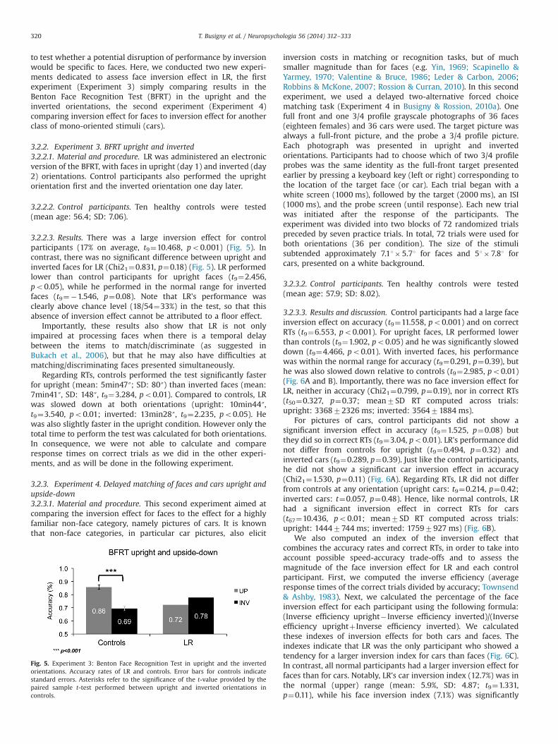
3.2.2.3. Results. There was a large inversion effect for controlparticipants (17% on average, t9¼10.468, po0.001) (Fig. 5). Incontrast, there was no significant difference between upright andinverted faces for LR (Chi21¼0.831, p¼0.18) (Fig. 5). LR performedlower than control participants for upright faces (t9¼2.456,po0.05), while he performed in the normal range for invertedfaces (t9¼�1.546, p¼0.08). Note that LR's performance wasclearly above chance level (18/54¼33%) in the test, so that thisabsence of inversion effect cannot be attributed to a floor effect.
Importantly, these results also show that LR is not onlyimpaired at processing faces when there is a temporal delaybetween the items to match/discriminate (as suggested inBukach et al., 2006), but that he may also have difficulties atmatching/discriminating faces presented simultaneously.
Regarding RTs, controls performed the test significantly fasterfor upright (mean: 5min47″; SD: 80″) than inverted faces (mean:7min41″, SD: 148″, t9¼3.284, po0.01). Compared to controls, LRwas slowed down at both orientations (upright: 10min44″,t9¼3.540, po0.01; inverted: 13min28″, t9¼2.235, po0.05). Hewas also slightly faster in the upright condition. However only thetotal time to perform the test was calculated for both orientations.In consequence, we were not able to calculate and compareresponse times on correct trials as we did in the other experi-ments, and as will be done in the following experiment.
3.2.3. Experiment 4. Delayed matching of faces and cars upright andupside-down3.2.3.1. Material and procedure. This second experiment aimed atcomparing the inversion effect for faces to the effect for a highlyfamiliar non-face category, namely pictures of cars. It is knownthat non-face categories, in particular car pictures, also elicit
inversion costs in matching or recognition tasks, but of muchsmaller magnitude than for faces (e.g. Yin, 1969; Scapinello &Yarmey, 1970; Valentine & Bruce, 1986; Leder & Carbon, 2006;Robbins & McKone, 2007; Rossion & Curran, 2010). In this secondexperiment, we used a delayed two-alternative forced choicematching task (Experiment 4 in Busigny & Rossion, 2010a). Onefull front and one 3/4 profile grayscale photographs of 36 faces(eighteen females) and 36 cars were used. The target picture wasalways a full-front picture, and the probe a 3/4 profile picture.Each photograph was presented in upright and invertedorientations. Participants had to choose which of two 3/4 profileprobes was the same identity as the full-front target presentedearlier by pressing a keyboard key (left or right) corresponding tothe location of the target face (or car). Each trial began with awhite screen (1000 ms), followed by the target (2000 ms), an ISI(1000 ms), and the probe screen (until response). Each new trialwas initiated after the response of the participants. Theexperiment was divided into two blocks of 72 randomized trialspreceded by seven practice trials. In total, 72 trials were used forboth orientations (36 per condition). The size of the stimulisubtended approximately 7.11�5.71 for faces and 51�7.81 forcars, presented on a white background.
3.2.3.2. Control participants. Ten healthy controls were tested(mean age: 57.9; SD: 8.02).
3.2.3.3. Results and discussion. Control participants had a large faceinversion effect on accuracy (t9¼11.558, po0.001) and on correctRTs (t9¼6.553, po0.001). For upright faces, LR performed lowerthan controls (t9¼1.902, po0.05) and he was significantly sloweddown (t9¼4.466, po0.01). With inverted faces, his performancewas within the normal range for accuracy (t9¼0.291, p¼0.39), buthe was also slowed down relative to controls (t9¼2.985, po0.01)(Fig. 6A and B). Importantly, there was no face inversion effect forLR, neither in accuracy (Chi21¼0.799, p¼0.19), nor in correct RTs(t50¼0.327, p¼0.37; mean7SD RT computed across trials:upright: 336872326 ms; inverted: 356471884 ms).
For pictures of cars, control participants did not show asignificant inversion effect in accuracy (t9¼1.525, p¼0.08) butthey did so in correct RTs (t9¼3.04, po0.01). LR's performance didnot differ from controls for upright (t9¼0.494, p¼0.32) andinverted cars (t9¼0.289, p¼0.39). Just like the control participants,he did not show a significant car inversion effect in accuracy(Chi21¼1.530, p¼0.11) (Fig. 6A). Regarding RTs, LR did not differfrom controls at any orientation (upright cars: t9¼0.214, p¼0.42;inverted cars: t¼0.057, p¼0.48). Hence, like normal controls, LRhad a significant inversion effect in correct RTs for cars(t67¼10.436, po0.01; mean7SD RT computed across trials:upright: 14447744 ms; inverted: 17597927 ms) (Fig. 6B).
We also computed an index of the inversion effect thatcombines the accuracy rates and correct RTs, in order to take intoaccount possible speed-accuracy trade-offs and to assess themagnitude of the face inversion effect for LR and each controlparticipant. First, we computed the inverse efficiency (averageresponse times of the correct trials divided by accuracy; Townsend& Ashby, 1983). Next, we calculated the percentage of the faceinversion effect for each participant using the following formula:(Inverse efficiency upright� Inverse efficiency inverted)/(Inverseefficiency uprightþ Inverse efficiency inverted). We calculatedthese indexes of inversion effects for both cars and faces. Theindexes indicate that LR was the only participant who showed atendency for a larger inversion index for cars than faces (Fig. 6C).In contrast, all normal participants had a larger inversion effect forfaces than for cars. Notably, LR's car inversion index (12.7%) was inthe normal (upper) range (mean: 5.9%, SD: 4.87; t9¼1.331,p¼0.11), while his face inversion index (7.1%) was significantly
Fig. 5. Experiment 3: Benton Face Recognition Test in upright and the invertedorientations. Accuracy rates of LR and controls. Error bars for controls indicatestandard errors. Asterisks refer to the significance of the t-value provided by thepaired sample t-test performed between upright and inverted orientations incontrols.
T. Busigny et al. / Neuropsychologia 56 (2014) 312–333320

below normal controls (mean: 22.5%, SD: 6.23; t¼2.357, po0.05).Moreover, results from applying the Revised Standardized Differ-ence Test (Crawford & Garthwaite, 2005), to compare a singlepatient's discrepancy with the control sample, showed that LR hada profile significantly different from controls (t9¼2.802, po0.05).While the controls presented with a stronger inversion effect forfaces than for cars, LR did not show this dissociation.
In conclusion, LR's profile of performance was comparable tonormal observers when matching pictures of upright cars acrossviewpoint changes. In contrast, LR performed lower than controlswhen matching faces. This observation reinforces the selectivity ofhis impairment for faces. In addition, LR did not present with anormal effect of inversion for faces. Since his inversion effect forphotographs of cars was in the normal range, this absence of aninversion effect cannot be explained by a general factor. Hence,LR's acquired prosopagnosia seems to affect primarily a processthat is specific to upright faces. These observations are in line withthe absence of inversion effect for the prosopagnosic patient PStested extensively with upright and inverted faces (Busigny &Rossion, 2010a), as well as for other such patients (e.g., McNeil &Warrington, 1991; Boutsen & Humphreys, 2002; Delvenne et al.,2004; Busigny et al., 2010b).
3.2.4. Experiment 5. whole-to-part effect3.2.4.1. Rationale. We aimed to provide more direct evidence forthe patient LR's deficient holistic processing of individual faces.
A paradigm that is classically used is the face superiority effect(Tanaka & Farah, 1993), also called the whole-part advantage (e.g.,Delvenne et al, 2004). It refers to the superior discrimination oftwo whole faces differing by one part (e.g., the eyes) in comparisonto the discrimination of these parts when they are shown inisolation. In other words, the discrimination of the diagnostic partis facilitated by the presence (and correct organization) of theremaining facial parts. This effect is thought to reflect the fact thata change of one diagnostic part affects the whole face, thus makingthe discrimination easier (Tanaka & Farah, 1993). Different kinds ofwhole-part advantage paradigms have been used withprosopagnosic patients: two-alternative forced choice matchingtasks (Davidoff & Landis, 1990; Delvenne et al., 2004; Wilkinsonet al., 2009) or variants with “Thatcherized” faces (Boutsen &Humphreys, 2002; Riddoch et al., 2008). All these studies showedthat patients with acquired prosopagnosia were relativelyimpaired at showing an advantage for processing whole faces vs.isolated parts.
Here we assessed this effect in a delayed face matching task, asin most studies with normal observers (e.g., Michel, Caldara, &Rossion, 2006a; Goffaux & Rossion, 2006). We presented wholesand parts of faces preceded by whole faces, and we completed theparadigm by also including trials in which the first stimulus couldbe part of a face followed by wholes or parts of faces (see Ramonet al., 2010). The diagnostic cue for analysis was the eyes, but therewere foil trials for mouth and nose. The prediction was that,for normal observers, the performance would be better (higher
Fig. 6. Experiment 4: Delayed matching of faces and cars upright and upside-down. (A) Accuracy rates for LR and controls. (B) Response times on correct trials for LR andcontrols. (C) Individual indexes of inversion calculated with the following formula: (Upright� Inverted)/(Uprightþ Inverted). Error bars for controls indicate standard errorscomputed across subjects. In (A and B), asterisks refer to the significance of the t-values provided by the paired sample t-tests for controls and the independent sample t-testfor LR. In (C), the asterisk refers to the significance of the t-value provided by the modified t-test of Crawford and Howell (1998) performed between LR and controls.
T. Busigny et al. / Neuropsychologia 56 (2014) 312–333 321

accuracy, slower RTs) when the format of the encoding (target)stimuli and format of the subsequently presented test (probe)stimuli were identical. That is, the conditions “part-part” and“whole-whole” should be associated with superior performanceas compared to “part-whole” and “whole-part”, respectively. Thus,the effect would not necessarily be a whole/part advantage, but ademonstration that the processing of facial parts, at least innormal participants, is influenced by the presence of the otherfeatures (see Leder & Carbon, 2005).
3.2.4.2. Material and procedure. Thirty grayscale full-front picturesof unfamiliar faces (half female) posing with a neutral expression,cropped without external features and free of facial hair or glassesserved as stimuli. Using Adobe Photoshop, we created 20 eye-foilsby swapping the eye region amongst 20 of the original faces. Theremaining ten original faces were used to generate five nose-foilsand five mouth-foils using the same feature swapping procedure.Isolated features (eyes, nose or mouth) were generated byisolating the relevant feature, resulting in a total of 30 featurestimuli (20 isolated eyes, five noses, five mouths). Nose and mouthfoil face parts and whole faces were used as catch trials (one thirdof the trials, 40/120) in the experiment to avoid participantsexclusively focusing on the eyes, and these trials were notanalyzed (as in our previous studies with this paradigm: e.g.,Michel et al., 2006a; Ramon et al., 2010).
The task was delayed two-alternative forced choice identitymatching, as described previously in Ramon et al. (2010) andBusigny et al. (2010a). Trials began with a target face stimulipresented centrally for 2000 ms. Following a blank screen of500 ms, two juxtaposed probe stimuli remained on the screenuntil a response was made. Participants were instructed to selectthe probe that matched the target stimulus by pressing the keycorresponding with probe location (right versus left) on thescreen. The next trial started 1000 ms after each response. Thetarget stimulus was either an original face (whole-whole andwhole-part conditions) or a single part (part-part and part-wholeconditions). Each target item was slightly larger in size than theprobe stimuli.
In the whole display condition, the probes were whole faces,one identical to the target, the other one (i.e., the foil) differingfrom the target by a single part (eyes in experimental trials, noseor mouth in catch trials). In the part display condition, the probesdepicted isolated face parts (eyes in experimental trials, nose ormouth in catch trials) (see Fig. 7A). There were 40 trials perexperimental condition, and 160 trials in total. Each target andprobe stimulus appeared four times. The location of foil stimuli(right versus left) was counterbalanced. Eighty catch trials (mouthand nose whole and part foils) were added, giving a total of 240(four blocks of 60 trials). Trial order was at random and varied foreach participant. Six practice trials were completed before theexperiment commenced. The target stimulus subtended 5�61 ofvisual angle, while size of the subsequently presented probesdiffered depending on probe type (whole faces: 4.1�4.11; eyefeature stimuli: 0.7�41; nose features: 1.4�1.41; mouth features:1�21). All stimuli were presented on a gray background.
3.2.4.3. Control participants. Ten healthy controls were tested(mean age: 59.4; SD: 6.8).
3.2.4.4. Results and discussion. Normal controls showed a signi-ficant advantage in the whole-whole as compared to the whole-partcondition (“Whole-part advantage”) with respect to accuracy(t9¼2.587, po0.05) and correct RTs (t9¼12.852, po0.001)(Fig. 7B & C). They also showed a significant “Part-wholedisadvantage” when comparing the part-part and the part-whole
conditions, both in terms of accuracy (t9¼3.399, po0.01), andcorrect RTs (t9¼5.387, po0.001). These results show that thecontrol participants are better and faster when the encoding andretrieval face context is the same.
In contrast, LR showed no difference between the whole-partand the whole-whole conditions, neither in accuracy nor incorrect RTs (t60¼0.684, p¼0.25; mean7SD RT computed acrosstrials: whole-whole: 21037684 ms; whole-part: 22187639 ms)(Fig. 7B & C).
Considering the part-part and the part-whole conditions, LRshowed no significant effect in accuracy (Chi21¼2.057, p¼0.08),but there was a significant difference in the expected directionin RTs (t64¼2.749, po0.01; mean7SD RT computed across trials:part-part: 15677361 ms; part-whole: 19707771 ms) (Fig. 7C).
Indexes were computed using the same formula that was usedfor the inversion effect (see above). Thus we calculated an indexfor the condition in which the whole face is presented first(Inverse efficiency whole-part� Inverse efficiency whole-whole)/(Inverse efficiency whole-partþ Inverse efficiency whole-whole)and for the condition in which the part is presented first (Inverseefficiency part-whole� Inverse efficiency part-part)/(Inverse effi-ciency part-wholeþ Inverse efficiency part-part). In the wholedisplay condition, each single control participant obtained a highindex of whole-part advantage (mean¼16.7%, SD: 6.86), but thiswas not the case for LR, who obtained an index close to zero(2.66%), significantly lower than the controls (t9¼1.954, po0.05)(Fig. 7D). In the part display condition, each participant showed ahigh index of part-whole disadvantage (mean¼14.7%, SD: 4.91).This time, LR did not show a significantly reduced effect comparedto controls (LR: 15.4%, t9¼0.150, p¼0.44) (Fig. 7E).
In conclusion, normal controls show both a whole-part advan-tage and a part-whole disadvantage, while LR shows only thesecond effect. That is, he does not perform better at discriminatinga part embedded in a whole face than when presented in isolation.However, when he is presented with a face part at encoding, herecognizes the part better if it is isolated than when it is embeddedin a whole face. This effect could be the consequence of residualface holistic processing. This residual face holistic processing couldbe insufficient to produce a positive effect (an advantage of theface configuration), but could be sufficient to produce a negativeeffect (the disadvantage of supplementary facial information). Wewill come back to this issue in the discussion section.
3.2.5. Experiment 6. Composite face effect: top composite(alignment� identity)3.2.5.1. Rationale. The composite face effect was originally describedby Young, Hellawell, and Hay (1987) as the difficulty to identifythe top half (or bottom half) of a famous face when it is alignedwith the bottom half (or top half) of another person. It is thought toreflect the fact that one half of a face cannot be perceived in isolation,but is integrated into a whole face representation. This paradigmwaslater applied to the matching of individual unfamiliar faces, in whichtwo identical top halves of faces are perceived as being slightlydifferent if they are aligned with distinct bottom halves (e.g., Hole,1994; Le Grand, Mondloch, Maurer, & Brent, 2004; Michel, Rossion,Han, Chung, & Caldara, 2006b; Goffaux & Rossion, 2006; Rossion,2013 for a review). As mentioned in the introduction, LR has alreadybeen tested with composite faces by Bukach et al. (2006), whoconcluded that he did not differ from normal controls at this task.However, critically, these authors did not include misaligned facehalves in their paradigm, and thus merely tested a Stroop-likecongruency/interference effect (whether an irrelevant face half inter-feres with a matching/discrimination decision on target face half),not a composite face effect (see Rossion, 2013).
T. Busigny et al. / Neuropsychologia 56 (2014) 312–333322

Fig. 7. Experiment 5: Whole-to-Part effect. (A) Examples of stimuli in each condition. (B) Accuracy rates for LR and controls. (C) Response times on correct trials for LR andcontrols. (D) Individual indexes of the whole-part advantage calculated with the following formula: (whole-part�whole-whole)/(whole-partþwhole-whole). (E) Individualindexes of part-whole disadvantage calculated with the following formula: (part-whole�part-part)/(part-wholeþpart-part). Error bars for controls indicate standard errors.In Figs. 7B and C, asterisks refer to the significance of the t-values provided by the paired sample t-tests for controls and the independent sample t-test for LR. In (D), theasterisk refers to the significance of the t-value provided by the modified t-test of Crawford and Howell (1998), performed between LR and controls.
T. Busigny et al. / Neuropsychologia 56 (2014) 312–333 323

Given these observations, we used the last experimentdescribed in Ramon et al. (2010), experiment 5; see also Busignyet al., 2010a, experiment 24), in which there were four criticalconditions for which the top half of the faces was the same(Aligned/Same Bottom; Aligned/Different Bottom; Misaligned/Same Bottom; Misaligned/Different Bottom). For normal controls,we expected a composite effect, that is an effect of alignment(misaligned trials performed better than aligned trials), whichshould be observed only when the bottom halves differ betweenthe faces to match. In contrast, this effect was not expected for LR.
3.2.5.2. Material and procedure. The stimuli used in thisexperiment were full-front color pictures of 23 unfamiliar faces(neutral expression, 16 female, no glasses or facial hair) that werecropped so that neither hair nor external features were depicted.The resulting faces, subtending approximately 160 pixels in widthand 230 pixels in height, were fitted onto a white background.Using Adobe Photoshop, the original faces were separated byinserting a 1.76 mm gap located above the nostril upper limit.The gap was used so that the border separating the top andbottom halves could be well identified, even in the alignedcondition (see Rossion, 2013 for the rationale for using such agap). Each original face was transformed into two stimuli: the firstone (“original”) differed from the original merely by the insertedgap (Same Top/Same Bottom), the second one (“composite”) had agiven top part combined with a different bottom part from arandomly selected other face (i.e., a composite face). Misalignedversions were created by laterally offsetting the lower parts so thatthe top part's right edge of the nose was aligned with the bottompart's left side of the nose.
Participants performed a two-alternative forced choice decisiontask (see Ramon et al., 2010; Busigny et al., 2010a). Each trialinvolved the consecutive presentation of two stimuli (both eitheraligned or misaligned), which had to be judged with regard to theidentity of the top part (i.e., same or different) (see Fig. 8A). Therewere four possible trials requiring a “same” response: the originalface paired with itself (‘Aligned/Same Bottom’; ‘Misaligned/SameBottom’) and different composite faces that had the same top halfbut a different bottom half (‘Aligned/Different Bottom’; ‘Misa-ligned/Different Bottom’). There were also real “different” trials,in which a face was paired with another randomly selected face sothat both face halves were different (see Rossion, 2013; Figure 26for a display of this paradigm). This paradigm was used in severalprevious studies and is highly sensitive to disclose a compositeface effect in normal participants (e.g., Ramon et al., 2010; Busignyet al., 2010b).
Trials started with a fixation cross presented centrally for300 ms. Following a 200 ms blank, a target face was presentedfor 600 ms. After a 300 ms ISI, the probe face appeared until aresponse was provided. The next trial was initiated 1000 ms after agiven response. In order to restrict the possibility of participantsengaging in comparing merely a specific location of the displaywhile performing the matching task, the target and sample facesappeared at slightly different screen locations. Participants wereasked to attend only to the top parts and to respond whether thesewere the same or different (by pressing a right or left key,respectively). In line with previous studies (e.g., Goffaux &Rossion, 2006; Michel et al., 2006b), trials in which both halveswere different were used only as catch trials, and not considered inthe analysis (Rossion, 2013). The experiment consisted of a total of138 trials, which were divided into two blocks of equal length.There were 92 “same” trials (23 per condition) and 46 “different”trials (23 per condition). Prior to the beginning of the experiment,participants completed four practice trials. Aligned stimuli sub-tended approximately 5.7�3.81 of visual angle and misalignedstimuli 5.7�5.21.
3.2.5.3. Control participants. Ten healthy controls were tested(mean age: 62; SD: 6.68).
3.2.5.4. Results. The data of age-matched normal controls areillustrated in Fig. 8(B & C). The ANOVA for accuracy rates showeda significant main effect of alignment (F(1,9)¼5.000, po0.05), and asignificant main effect of identity of the bottom half (F(1,9)¼7.653,po0.05). Moreover, there was a significant interaction betweenthe two factors (F(1,9)¼4.048, po0.05): performance decreasedonly when the two parts were aligned and when the bottom facehalf was different. The ANOVA for RTs showed a main effect ofalignment (F(1,9)¼3.844, po0.05), and a main effect of identity ofthe bottom half (F(1,9)¼9.985, po0.01). Most importantly, in linewith the results for accuracy, there was a highly significantinteraction between the two factors (F(1,9)¼26.963, po0.001).These findings are a hallmark of holistic face perception forcontrol participants.
A bootstrap procedure with 10,000 iterations for each condition onthe accuracy rates of LR indicated the same profile as the normalcontrols: lower performance for the aligned stimuli with differentbottom halves (‘Aligned/Different Bottom’ trials,M¼0.78; CI¼[0.6087,0.95652]) than for all other conditions (po0.01). Performance in bothmisaligned conditions was at ceiling (M¼1; CI¼[1, 1]) and did notdiffer significantly from the aligned condition with same bottomhalves (‘Aligned/Same Bottom’ trials, M¼0.96; CI¼[0.86957, 1];p¼0.40) (Fig. 8B). However, in correct RTs, LR's profile was different.He showed no significant main effect of alignment (F(1,85)¼2.115,p¼0.08), and only a main effect of identity (F(1,85)¼6.905, po0.01).There was no interaction between the two factors (F(1,85)¼0.272,p¼0.30) because LR showed an unexpected increase of RTs in the‘Misaligned/Different Bottom’ condition (Fig. 8B & C).
To compare LR's magnitude of composite effect with the controls,indexes were computed individually by using the formula: (‘Aligned/Same Bottom’� ‘Aligned/Different Bottom’)�(‘Misaligned/Same Bot-tom’� ‘Misaligned/Different Bottom’). As in our previous study(Busigny et al., 2010b), given the inter-subject variance in themagnitude of the effect in accuracy rates, the indexes were calculatedon the normalized correct response times. There was a high index ofcomposite effect (mean¼18.39%, SD: 5.7) for every participant. LR'scomposite face effect index was in the lower range but did not differfrom controls (LR: 11.08%; t¼1.223, p¼0.13) (Fig. 8D). However, acareful look at the data suggests that LR has a different profile ofresponse in this task than normal controls. Indeed, while LR'sperformance was influenced by the identity of the bottom halfsimilarly to the normal controls in the aligned condition (LR: 33.5%,controls mean: 19.08%, SD: 9.45, t9¼1.455, p¼0.09) (Fig. 8E), he alsoshowed an influence of the bottom half in the misaligned condition(LR: 22.4%), contrary to normal controls (mean: 0.68%, SD: 6.44;t9¼3.216, po0.01) (Fig. 8F).
Interestingly, these results do not contradict the resultsobtained by Bukach et al. (2006; Experiment 2), who reportedthat the identity of the bottom half of the face influenced LR'sjudgment on the top half. However, thanks to the inclusion ofmisaligned trials in the present study, a necessary baseline tomeasure the composite face effect (Rossion, 2013), we found thatthis influence generalizes to misaligned trials to some extent onlyfor LR, leading to an uncharacteristic profile of response. Thisinfluence may be due to LR's well-documented tendency to usethe lower part of the face, the mouth in particular, to match faces(Bukach et al., 2006, 2008). Importantly, such a strategy cannot beassociated with preserved holistic processing.
3.2.6. Experiment 7. Gaze contingent individual face discrimination3.2.6.1. Rationale. Gaze-contingently revealing/masking a portionof the visual field in a face matching task provides a way to
T. Busigny et al. / Neuropsychologia 56 (2014) 312–333324

Fig. 8. Experiment 6: Composite face effect. (A) Examples of stimuli in each condition. (B) Accuracy rates for LR and controls in the four conditions for which the top half isthe same. (C) Response times on correct trials for LR and controls (LR0 variance computed on trials is: ‘aligned/same bottom’: 10967543; ‘aligned/different bottom’:14997589; ‘misaligned/same bottom’: 9767246; ‘misaligned/different bottom’: 12467689). (D) Indexes of composite effect calculated with the following formula:(‘aligned/same bottom’� ‘aligned/different bottom’)�(‘misaligned/same bottom’� ‘misaligned/different bottom’). (E) Indexes of the composite effect for the aligned trialscalculated with the following formula: ‘aligned/same bottom’� ‘aligned/different bottom’. (F) Indexes of the composite effect for the misaligned trials calculated with thefollowing formula: ‘misaligned/same bottom’� ‘misaligned/different bottom’. Error bars for controls indicate standard errors. In (F), asterisks refer to the significance of the t-value provided by the modified t-test of Crawford and Howell (1998), performed between LR and controls.
T. Busigny et al. / Neuropsychologia 56 (2014) 312–333 325

selectively impair holistic or analytical processing. In a ‘window’
condition, only the fixated part is visible in a gaze-contingentwindow, preventing perception of multiple parts in the face atonce, and impairing holistic perception. On the contrary, a maskcondition, in which a mask covers the fixated part only, impairsanalytical perception by preventing the use of highly detailedfoveal information necessary for analytical processing (Fig. 9). Thismethod demonstrated impairment in holistic face perception intwo cases of acquired prosopagnosia: PS (Van Belle et al., 2010a)and GG (Van Belle et al., 2011). Compared to full view, PS' and GG'sperformance was almost unaffected when they could see only onepart of the faces at a time (window condition), while they werelargely impaired when the whole face but the fixated part wasvisible (mask condition). These findings were opposite to theobservations made on normal controls. Gaze-contingency wasalso used to show that the face inversion effect increases inthe mask condition and decreases in the window condition,reinforcing the link between these manipulations and theselective disruption of holistic vs. analytical processing (VanBelle, de Graef, Verfaillie, Rossion, & Lefèvre, 2010b).
Here, if LR has difficulties with holistic perception of individualfaces, we expect his performance to differ from that of the controlparticipants, especially in the mask condition.
3.2.6.2. Material and procedure. The methods are similar to therecent study of Van Belle et al. (2011) and will be briefly describedhere. For this experiment we used a stimulus set of 10 male and 10female faces (KDEF database, Lundqvist, Flykt, & Ohman, 1998) with aheight of 151, fromwhich the external features were cropped but headshape was largely preserved. A drift correction with a central fixation
cross was followed by a centrally presented face. After 1 s, thisreference face was replaced by two faces presented side-by-side,with a distance between the faces' inner borders of approximately101. The participant's task was to indicate which of the two facescorresponded to the reference face by pressing the assigned left orright key on the keyboard. The faces were randomly combined in pairsof two males or two females.
Participants could freely explore the side-by-side presentedfaces. In one third of the trials the faces were completely visible(full view). In another third of the trials, however, a gazecontingent mask covered the fixated feature in the central partof the visual field (mask condition). In the remaining third of thetrials only the fixated feature in the central part of the visual fieldwas visible through a limited spatial window (window condition)(Fig. 9).1 The elliptical window and mask were 6�51. During theexploration of the pair of faces, the face that was not fixated wasreplaced by a grayscale average of all faces in order to provide anequal reference frame for saccade planning, and an equal amountof information from the non-fixated face in all viewing conditions(Fig. 9). There were 75 trials per viewing condition, resulting in atotal of 225 trials, presented in 5 blocks of 45 trials each. Viewingcondition order was randomized across trials.
3.2.6.3. Control participants. Seven healthy controls were tested(mean age: 57; SD: 2.93).
Fig. 9. Experiment 7: Gaze-contingent individual face discrimination (from Van Belle et al., 2010a). (A) Full view: the whole face is visible, central mask: only the non-fixatedpart of the face is visible, central window: only the fixated area of the face is visible. (B) experimental design: drift correction was followed by a fixation outside of the facearea, a 1 s presentation of the reference face and two faces side by side until response.
1 Movies of the gaze-contingency experiment as performed by the patient PSin a previous study are available at http://face-categorization-lab.webnode.com/pictures/.
T. Busigny et al. / Neuropsychologia 56 (2014) 312–333326

3.2.6.4. Results. Control participants showed a main effect ofviewing condition on accuracy (F(2, 12)¼6.51, po0.05). Individualcontrasts were Tukey corrected for multiple comparisons (two-tailed). Compared to full view (M¼0.97), accuracy significantlydecreased with a window (M¼0.87, t(12)¼3.54, po0.05) but notwith a mask (M¼0.90, t(12)¼2.38, p¼0.08). Performance with amask did not differ significantly from that with a window(t(12)¼1.16, p¼0.50). The main effect in response times was alsosignificant, (F(2, 12)¼4.30, po0.05). Again, participants were onlysignificantly slower with a window (M¼2.34 s) than in full view(M¼1.27 s, t(12)¼2.93, po0.05). The difference between responsetimes in full view and with a mask was not significant (M¼1.56 s,t(12)¼2.29, p¼0.29); additionally, response times with a mask didnot differ significantly from those with a window (t(12)¼1.34,p¼0.40).
A bootstrap procedure with 1000 repetitions showed that,compared to full view (M¼0.96; 95% CI¼[0.91; 1.00]), the accu-racy of LR was equally affected by the window (M¼0.87; 95% CI¼[0.79; 0.93]) and the mask (M¼0.88; 95% CI¼[0.80; 0.95])(Fig. 10A). For RTs, an ANOVA on the correct response trialsrevealed a main effect of the viewing condition for LR (LR: F(2,179)¼57.40, po0.0001). Contrary to the control participants, LRwas faster in full view than with a mask (LR: t(179)¼10.70,po0.0001) or with a window (LR: t(179)¼5.44, po0.0001)(Fig. 10B). Importantly, LR was also faster with a window thanwith a mask (t(179)¼5.18, po0.0001), contrary to normal controls.
Crawford and Howell's (Crawford & Howell, 1998) method forthe analysis of single case neuropsychological data was used tocompare LR's performance directly to that of the control partici-pants. LR's accuracy did not differ significantly from that of the agematched control participants in any of the conditions (full view:t(6)¼0.47, p¼0.33 ; window: t(6)¼0.15, p¼0.44; mask: t(6)¼0.16,p¼0.44). His response times, on the contrary, were slower than theage matched control participants in full view (t(6)¼2.017, po0.05)and with a mask (t(6)¼3.55, po0.01). However, with a window, LRperformed within the normal range (t(6)¼0.80, p¼0.23) (Fig. 10).
Finally, in order to assess the differential effect of the window andmask on control participants vs. LR, we calculated the index of the RTincrease due to window and mask (Fig. 11) as the difference betweenthe performance in full view and each experimental condition, dividedby the sum of these performances. LR was significantly more sloweddown by the mask than the control participants (t(6)¼3.867, po0.01),while the effect of the window did not significantly differ for LR andfor the control participants (t(6)¼0.090, p¼0.47).
In summary, LR is undoubtedly less severely impaired than thetwo cases of acquired prosopagnosia previously tested in thisexperiment (PS: Van Belle et al., 2010a; GG: Van Belle et al., 2011),showing accuracy rates in the normal range. However, the difficultyof the discrimination task was adjusted initially for the patient PS, inorder to obtain a reasonably high performance in the full view facecondition. Here, the most interesting pattern is observed in responsetimes only, for which LR's results are completely in line with theobservations made on these two cases, and opposite to normalobservers: for LR, the mask condition appears to be the most difficultviewing conditions. Hence, compared to normal observers, LR'sperformance is affected the most when he is prevented from usinga part-based process; when forced to use a part-based process, onthe contrary, his performance does not differ from that of normalobservers. These results support the view that, like PS and GG, LR isimpaired at holistic perception of the individual face.
Fig. 10. Experiment 7: Gaze-contingent individual face discrimination. (A) Accuracy rates for LR and controls, including individual participants' performance. (B) Responsetimes on correct trials for LR and controls, including individual participants' performance. Error bars indicate standard errors.
Fig. 11. Experiment 7: Gaze-contingent individual face discrimination. Index of RTincrease due to window or mask compared to full view ((RTcond�RTfull)/(RTcondþRTfull). Error bars indicate standard errors.
T. Busigny et al. / Neuropsychologia 56 (2014) 312–333 327

4. General discussion
Several experiments performed on the patient LR show thatface perception can be specifically impaired in prosopagnosiafollowing brain damage to the right anterior temporal lobe,sparing posterior regions. The nature of LR's impairment appearsto concern holistic perception of individual faces, as is observed incases of prosopagnosia with posterior brain damage. These resultsextend previous observations obtained using the same tests withtwo other cases of acquired prosopagnosia, PS and GG. Notably, inthese two patients, brain damage concerned posterior brain areas,namely the right inferior occipital cortex and left middle fusiformgyrus for PS and the right lingual, fusiform and parahippocampicgyrus for GG, sparing the anterior temporal lobe region (see inparticular Rossion et al., 2003; Ramon et al., 2010; Sorger, Goebel,Schiltz, & Rossion, 2007 for PS; Busigny et al., 2010a; Van Belleet al., 2011 for GG). Compared to these patients, the patient LR isless severely impaired in the same face discrimination tasks,whose parameters were adjusted initially for studying the patientPS. Moreover, contrary to these cases, LR's ability to processindividual faces holistically may not be completely abolished, asindicated by a significant part-whole advantage in one version ofthe experiment 5 and a significant – albeit weaker than in controls– composite face effect in experiment 6. Nevertheless, consideredaltogether, these observations support the view that the nature ofthe critical functional impairment does not fundamentally differacross patients with acquired prosopagnosia, regardless of thelocalization of brain damage (Busigny et al., 2010a): all thesepatients appear to be impaired at what constitutes the heart of ourvisual expertise with faces, namely holistic perception at asufficiently fine-grained level of resolution to discriminate exem-plars of the face class efficiently (for a direct comparison of LR, PSand GG, see Busigny & Rossion, 2010b, and Supplementarymaterial). Such a conclusion challenges the view that there areseveral subtypes of prosopagnosia (Damasio, Tranel, & Damasio,1990; Schweich & Bruyer, 1993) casting doubts in particular on thedichotomy between apperceptive and associative forms of proso-pagnosia (e.g., De Renzi, 1986; De Renzi et al., 1991; Barton, 2003).
4.1. Face-specificity
Experiments 1 and 2 show that LR, who never complained ofany object recognition problems in real life, is able to identifyobjects rapidly when tested, and is not impaired at fine-graineddiscrimination of non-face exemplars. In a delayed matching task,he was able to match individual exemplars of various nonfacecategories (birds, cars, chairs, houses) as well as controls. Further-more, his pattern of performance (i.e., slope) in a parametricindividual car discrimination task mimicked normal observers'performance. However, LR was impaired at discriminating indivi-dual exemplars of faces in the exact same paradigm, despite thefact that, if anything, individual face discrimination was easier forfaces than nonface objects for normal observers.2 This pattern ofperformance is very similar to what was found for the patients PS(Busigny et al., 2010b) and GG (Busigny et al., 2010a), ruling out thevisual similarity account of prosopagnosia (Faust, 1955; Damasioet al., 1982; Gauthier et al., 1999), and indicating that brain damageto vastly different brain structures can lead to a pure form ofprosopagnosia (see also other cases, as listed in Table 1 in Busignyet al., 2010b).
4.2. Impairment in holistic face perception
Although LR's impairment in holistic face perception is not asclear-cut as the patients PS and GG, our experiments 3 to 7 showthat LR is impaired at holistic perception of individual faces.Indeed, the patient does not discriminate upright faces betterthan inverted faces, and he does not present with a typical whole-part face advantage. Interestingly, we replicated the finding thathis processing of the top half of a face is influenced by the bottomhalf, an observation which prompted Bukach et al. (2006) to claimthat LR processed faces holistically. However, this effect wasqualified here by a substantial influence observed also for mis-aligned faces – which were not included in this previous study –
specifically for LR. Most importantly, LR's pattern of performancein the gaze-contingency experiment with selective revealing/masking of a portion of the face stimulus replicates previousobservations on PS and GG (Van Belle et al., 2010a; Van Belleet al. 2011, respectively) and demonstrates LR's difficulties inholistic perception of individual faces relative to normal observers.
Although Bukach et al. (2006) concluded that LR has preservedholistic processing, a careful look at the patient's data in theseauthor's other experiments rather support the opposite view. Forinstance, when required to divide his attention over multiple facefeatures, LR was able to determine the identity of only a singlefeature. Moreover, in a face matching task (Experiment 3, Bukachet al., 2006), LR was impaired at processing relative distances atthe level of the eyes of a face, and he did not have an inversioneffect in that condition. These results were replicated in a laterstudy where LR exhibited an impaired ability to discriminatediagnostic cues in the eye region (Bukach et al., 2008). Similarobservations of a severe impairment at extracting diagnosticinformation from the eyes have been made for the patients PSand GG (Caldara et al., 2005; Orban de Xivry, Ramon, Lefèvre, &Rossion, 2008; Rossion, Kaiser, Bub, & Tanaka, 2009; Busigny et al.,2010a) reinforcing the parallel made between the functionalimpairment of these patients and LR's impairment in holistic faceperception. According to this view, the eye region loses itsdiagnosticity in acquired prosopagnosia because rapidly extractingdiagnostic information from the eye region of a face requiressimultaneous integration of information from multiple sources(pupils, iris, eyelids, eyebrows, distance between eyes, distancebetween eyes and forehead, etc.) (Caldara et al., 2005; Orban deXivry et al., 2008; Rossion et al., 2009).
In the large majority of tests of holistic face perception, LRshowed an atypical pattern of performance, and this pattern ofperformance was qualitatively similar to previous patients withpure prosopagnosia following brain damage tested with the sameexperiments (i.e., PS and GG). However, contrary to these patients,LR showed evidence for holistic face perception effects in anunusual version of the Whole-to-Part Effect (superior performancefor matching parts to parts than parts to wholes). Moreover, eventhough LR showed an abnormal performance for misaligned trialsin the composite face paradigm, his composite face effect was stillsignificant. These observations raise the question of whether LRwould be affected at a finer-grained level of holistic perceptionthan these other patients with prosopagnosia. Indeed, holisticperception is useful not only to individualize faces, but also todetect faces, for instance when the parts of the stimulus are notface-like, as in Mooney faces (Mooney, 1957; Moore & Cavanagh,1998; McKone, 2004) or Arcimboldo paintings (Hulten, 1987).However, face detection abilities in these three patients, as testedwith Mooney and Arcimboldo face stimuli, are well preserved (SeeRossion, Dricot, Goebel, & Busigny, 2011 for PS; Busigny et al.,2010a for GG; supplemental material for LR's data comparedto these two patients’ data). These observations indicate that allthree patients are able to perceive faces holistically at a coarse
2 In a recent study, Bukach et al. (2012) claimed that LR was impaired atlearning exemplars of novel objects (“Greebles”). However, there is currently noevidence that learning these stimuli requires holistic processing at individuatingmembers of this category (Robbins & McKone, 2007; Rossion, 2013).
T. Busigny et al. / Neuropsychologia 56 (2014) 312–333328

level – sufficient to categorize a stimulus as a face – but not at thefinest grained level (i.e. to individualize a face). However, in theabsence of comparative data for other face categorizations (e.g.,gender, expression, age, etc.) requiring intermediary levels ofholistic representation, we cannot exclude that the patients differin terms of the level at which they can process a face holistically.Specifically, LR may differ from patients with more posterior braindamage such as PS and GG in being able to reach a finer-grainedlevel of holistic representation than these patients. Whether suchobservations would be taken as quantitative or qualitative differ-ences between the functional impairments of the patients wouldcertainly be a matter of debate, but at this stage the availableevidence as provided here and in our previous studies point onlyto quantitative differences between these patients with acquiredprosopagnosia.
4.3. Revisiting the concept of associative prosopagnosia
The term “holistic” has been used by many authors for faceprocessing, and it may have different meanings in the literature(e.g., Farah et al., 1998; Maurer et al., 2002; McKone, Martini, &Nakayama, 2003; Rossion, 2008, 2009, 2013; Tanaka & Farah,1993). However, it refers undoubtedly to a perceptual process, thatis, the (presumably simultaneous) perceptual integration of themultiple parts of a face into a single representation. The mostcompelling evidence that this is a perceptual process is that it canbe illustrated as a visual illusion, for instance in the well-knowncomposite face illusion (Young et al., 1987; Rossion & Boremanse,2008, Rossion, 2013). Hence, if LR is impaired at holistic processingof faces, this means somehow that he is unable to derive a properpercept of an individual face. His impaired performance at indivi-dual face discrimination tasks supports this claim, although mostof the tasks involved a minimal short-term memory component(delayed matching in experiments 1, 2, 4, 5). LR's impairedperformance at the Benton Face Recognition Test, in which hehad to match simultaneously presented pictures of unfamiliarfaces, further supports the view that he is impaired at perceivingindividual faces.
The observation of a perceptual face impairment in a patientlike LR, whose ventral occipito-temporal cortex up to the anteriorfusiform gyrus appears entirely preserved form brain damage,challenges the very existence of pure associative forms of proso-pagnosia, which has been postulated by several authors(Meadows, 1974; Benton, 1980; Damasio et al., 1982, 1990; DeRenzi, 1986; De Renzi et al., 1991; Barton, 2003). Typically, thecriterion for associative prosopagnosia is precisely that the patientperforms normally at unfamiliar face matching tasks, such as inthe Benton Face Recognition Test (Benton & Van Allen, 1968). Forinstance, De Renzi (1986) and De Renzi et al. (1991) claimed thattheir brain-damaged patients represented pure forms of associa-tive prosopagnosia, because they were able to achieve normalrange performance at this test. However, the evidence collected inthese studies was often limited to a single test, with few items, nocontrol data, and without any measure of response times. Forinstance, the patient 4 of De Renzi (1986) presented a score of21/27 at the short form of the BFRT,3 a score that was judged ashigh enough to exclude an impairment of perceptual nature.
Most importantly, there is substantial evidence that evenprosopagnosic patients with clear perceptual impairments canuse idiosyncratic strategies to compensate for their problems inunfamiliar face matching tasks and are able to reach normal levelsof performance on accuracy scores at these tasks (e.g., Davidoffet al., 1986; Davidoff & Landis, 1990; Farah, 1990/2004; McNeil &Warrington, 1991; Sergent & Signoret, 1992a, 1992b; Delvenneet al., 2004). In these situations, measuring correct RTs, forinstance at BFRT, systematically reveals that the patients are muchslower than all normal controls. This was also the case for LR in thepresent study, who sometimes showed a normal performance atunfamiliar face matching tasks when considering accuracy ratesonly (experiments 1, 6 and 7), but who was considerably sloweddown at these tasks relative to controls. Importantly, this slowingdownwas not found for other categories of objects (experiments 1,2 and 4). Moreover, across all experiments, LR' slowing downrelative to controls was not observed in the most difficult condi-tions for the controls (inverted faces, similar faces, faces revealedthrough a window only) or even in the conditions that were themost difficult for him in absolute terms (e.g., morphed faces thatwere very similar). In all these experiments, the trials of thedifferent conditions were presented randomly. These observationsexclude an explanation in terms of a general slowing down of thepatient or a lack of confidence. Rather, they show that so-calledcases of associative prosopagnosia can reach normal performanceat a single matching test of unfamiliar faces by abnormal means,and that a perceptual impairment cannot be excluded withoutperforming several tests and taking complementary RT measures.This is particularly the case for measurements of holistic faceperception, because these effects are often revealed in responsetimes rather than accuracy rates in normal observers (e.g., Younget al., 1987; see Rossion, 2013).
Besides the lack of stringent testing, there are other reasonswhy there is so little evidence of perceptual impairment inprosopagnosic patients with right anterior temporal lobe damage.First, cases of prosopagnosia with anterior temporal lesions aremuch less frequent than cases with more posterior lesions. Amongabout 120 prosopagnosic patients reported in the literature, wefound only 11 patients with a lesion in the right anterior temporallobe, while the vast majority of the patients show lesions in theright fusiform gyrus and in the right inferior occipital gyrus (seealso Bouvier & Engel, 2006). One reason for that is that theprincipal etiology of prosopagnosia is an ischemic stroke, specifi-cally in the territory of the right posterior cerebral artery. Bycontrast, lesions of the right anterior temporal lobe are principallyreported in the context of a degenerative disorder, i.e., the righttemporal lobe variant of frontotemporal dementia (e.g., Joubertet al., 2006; Gainotti, 2007). Most of the time, these patients havemultimodal and multicategorical impairment, preventing them torecognize faces, but also other categories (e.g., famous places), andvia different modalities (pictures, names, voices,…) (see Gainotti,2007, 2013; Busigny et al., 2009). In these cases, the nature of thedefect is generally semantic, and the patients cannot be consideredstrictly as prosopagnosic, since they do not present with arecognition defect limited to the visual modality (see Gainotti,2013). Thus, prosopagnosic patients after a lesion of the anteriortemporal lobe are rare, most patients with this kind of damagehaving multimodal person recognition defects. Given that he hasvisual impairment in face recognition, but does not have anydifficulties in recognizing people from their names and providingsemantic information about them, LR is one of these rare patients.
Nevertheless, these observations on LR are in line with thedescription of two cases of prosopagnosia, TS (Barton et al., 2003)and BD (Williams, Savage, & Halmagyl, 2006), who had damagerestricted to the right anterior temporal lobe and were impairedat perceiving faces. Interestingly, both of these patients were
3 While such a performance could presumably be equated to a score of 42/54 atthe long form of the test, slightly above a score within the norms, it should bequalified because the patients are usually faultless at the first 6 items of this test, allincluded in the short form, where only one target face has to be found among 6,without any change of viewing conditions. If these items are excluded, De Renzi(1986)'s patient had a score of 15/21 (70%) at a single unfamiliar face matching task,a performance that is insufficient to exclude a perceptual impairment with faces.
T. Busigny et al. / Neuropsychologia 56 (2014) 312–333 329

probably impaired at holistic face processing, TS being insensitiveto the global spatial configuration that differs between faces, andBD having a disrupted composite face effect. Furthermore, a studyof anterior temporal lobectomized patients showed a correlationbetween these patients’ face naming impairment and their per-ceptual discrimination of unfamiliar faces (Glosser et al., 2003).Together with our results, these observations support the claimthat anterior temporal lobe structures of the right hemipshere playa role in binding together information relevant to the perceptualidentification of unique entities such as individual faces.
To summarize this point, we argue that the apperceptive/associative distinction used to refer to cases of prosopagnosia ismisleading and should perhaps be abandoned. We hypothesizethat when tested stringently, other cases of prosopagnosia follow-ing focal brain damage, even those with damage restricted to the(right) temporal pole, may be impaired at face perception. Asmentioned in the introduction, these observations are in line withthe claim of Farah (1990/2004), that “perception is at fault in allcases of associative agnosia so far studied” (Farah, 1990/2004, p. 75).In fact, Lissauer himself qualified his own distinction betweenapperceptive and associative visual agnosia because he noted thatthe full perception of complex visual stimuli such as facial figuresrequires an appropriate linkage with stored representations(Lissauer, 1890; Shallice & Jackson, 1988). Hence, according to thislatter view, the cause of prosopagnosia cannot be strictly dividedinto a perceptual vs. a mnesic impairment. The case studyof LR illustrates the difficulty of building a proper perceptualrepresentation of an individual face following damage to neuralstructures that are important in linking perceptual and associativerepresentations.
4.4. The right anterior temporal lobe: a critical node in a faceperception network
Although we must remain careful in the absence of detailedanatomical and functional data (i.e., (f)MRI)) of the patient LR, ourobservations in this patient and the description of other such casesas mentioned above point to a role of the right anterior temporallobe in face perception, and specifically in holistic perception ofindividual faces.
Face perception is supported by an extensive neural networkthat encompasses regions of the ventral occipito-temporal path-way, with a right hemispheric dominance (Sergent et al., 1992;Haxby et al., 2000; Fox et al., 2009; Weiner & Grill-Spector, 2010;Davies-Thompson & Andrews, 2012; Rossion et al., 2012; Pyleset al., 2013). This network involves, at least, a face-selectivefunctional region in the lateral middle fusiform gyrus (called the“Fusiform Face Area”, “FFA”, Kanwisher, McDermott, & Chun, 1997;McCarthy, Puce, Gore, & Allison, 1997), another in the lateralinferior occipital cortex (“Occipital Face Area”, “OFA”, Gauthieret al., 2000; Rossion et al., 2003) and yet another one in theposterior part of the superior temporal sulcus (Puce, Allison,Bentin, Gore, & McCarthy, 1998; Allison, Puce, & McCarthy, 2000;Hoffman & Haxby, 2000), all three regions being dominant in theright hemisphere. These regions have been described as the coreface-perceptual network (Haxby et al., 2000), with the FFA andOFA at least showing clear sensitivity to individual faces discrimi-nation (e.g., Gauthier et al., 2000; Grill-Spector & Malach, 2001;Schiltz & Rossion, 2006; Andrews, Davies-Thompson, Kingstone, &Young, 2010).
In this context, what would be the role of the right anteriortemporal lobe in face perception? One possibility is that thisregion also plays a direct role in individual face perception, butthat the strong emphasis on the FFA (and to a lesser extent theOFA), coupled with the presence of strong magnetic susceptibilityartifacts in the anterior temporal lobe (ATL, Devlin et al., 2000;
Bellgowan, Bandettini, van Gelderen, Martin, & Bodurka, 2006),has hidden its contributing role. Nevertheless, face-specific activa-tion has been reported in the human anterior temporal lobe,particularly in the right hemisphere (e.g., Rajimehr, Young, &Tootell, 2009; Rossion et al., 2012). Moreover, recent neuroimagingstudies suggested that the right anterior temporal lobe is alsoinvolved in individual face discrimination, even when the stimuliare not explicitly associated with any biographical or semanticinformation (Kriegeskorte, Formisano, Sorger, & Goebel, 2007;Furl, van Rijsbergen, Treves, & Dolan, 2007; Pinsk et al., 2009).
However, the right ATL has also been described as having acrucial function in face memory and semantic knowledge aboutpeople (e.g., Gorno-Tempini & Price, 2001; Snowden, Thompson,& Neary, 2004; Thompson et al., 2004; Rogers et al., 2006; Olson,Plotzker, & Ezzyat, 2007; Drane et al., 2008; Tsukiura, Suzuki,Shigemune, & Mochizuki-Kawai, 2008; Simmons, Reddish, Bellgowan,& Martin, 2010). Hence, along the lines of Lissauer (1890)'sinsightful view, one may also consider that this region plays a rolein the storage, consolidation and linking of perceptual representa-tions, and that such dynamic associations are necessary to achievea fully refined perceptual representation of an individual face.That is, rather than seeing the perceptual impairment as beingcausal, one would see it the other way around: damage to areasprimarily involved in associative memory could cause a defectin face perception. These observations are in line with theEmergent Memory Account (EMA; Barense, Gaffan, & Graham,2007; Graham, Barense, & Lee, 2010), which suggests that percep-tion and memory depend on an overlapping network of wide-spread representations, and that memory is the outcome of adynamic interplay between hierarchically organized perceptualrepresentations distributed throughout the brain. This complexentanglement between perception and memory implies that theway we perceive visually is influenced by our stored representa-tions. In other words, complex (multidimensional) perceptsemerge from the automatic associations of simple, non category-specific representations, through the matching with internalholistic representations. Considering this, in the case of LR forinstance, a lesion in the anterior temporal lobe appears to havenegative effects on perceptual processing of faces, suggesting thatthe right anterior temporal cortex plays a crucial role in theinterplay between memory and perception of faces.
5. Conclusions
In conclusion, we report a case of prosopagnosia followingright anterior temporal damage who is specifically impaired withthe holistic perception of individual faces. Taken together withresults obtained from other cases of acquired prosopagnosia, theseobservations suggest that complex (multidimensional) face per-cepts emerge from the automatic associations of simple, noncategory-specific representations, through the matching withinternal face representations. A crucial process involved at thatlevel is the holistic/configural perception of individual faces. Anindividual face can be fully perceived as a single entity only if itcan be successfully associated with an internal holistic facerepresentation. This process may be successfully completed onlyif a whole network of posterior and anterior cerebral regions isfunctionally intact. In this framework, the notion of pure associa-tive prosopagnosia, namely, a patient impaired at face recognitionwho would be able to derive a correct percept of a face, should bereconsidered. At the functional level, although the degree ofseverity of the face recognition impairment and the kind ofassociated deficits can greatly vary among patients, our findingsraise the possibility that there is a single form of acquiredprosopagnosia.
T. Busigny et al. / Neuropsychologia 56 (2014) 312–333330

Acknowledgments
We are grateful to LR for the numerous hours that he spentperforming these experiments, as well as to his spouse and all thecontrols who took part in the study. We also thank Dan Bub andJim Tanaka for having introduced us to LR, and for their help andtheir relevant comments about this study. We thank JayalakshmiViswanathan, Talia Retter and two anonymous reviewers for theircommentaries and corrections on a previous version of the manu-script. This research was supported by the Belgian National Fundfor Scientific Research (FRSM 3.4601.12) and a PAI/UIAP grant noP7/33 (Pôles d'attraction interuniversitaires, phase 7).
Appendix A. Supporting information
Supplementary data associated with this article can be found inthe online version at http://dx.doi.org/10.1016/j.neuropsychologia.2014.01.018.
References
Allison, T., Puce, A., & McCarthy, G. (2000). Social perception from visual cues: Roleof the STS region. Trends in Cognitive Sciences, 4, 267–278.
Allison, T., Puce, A., Spencer, D. D., & McCarthy, G. (1999). Electrophysiologicalstudies of human face perception. I: Potentials generated in occipitotemporalcortex by face and non-face stimuli. Cerebral Cortex, 9, 415–430.
Andrews, T. J., Davies-Thompson, J., Kingstone, A., & Young, A. W. (2010). Internaland external features of the face are represented holistically in face-selectiveregions of visual cortex. The Journal of Neuroscience, 30(9), 3544–3552.
Armitage, P. (1980). Statistical methods in medical research. Oxford, UK: BlackwellScientific Publications.
Barbarotto, R., Capitani, E., Spinnler, H., & Trivelli, C. (1995). Slowly progressivesemantic impairment with category specificity. Neurocase, 1, 107–119.
Barense, M. D., Gaffan, D., & Graham, K. S. (2007). The medial temporal lobeprocesses online representations of complex objects. Neuropsychologia, 45,2963–2974.
Barton, J. J. S. (2003). Disorders of face perception and recognition. NeurologicClinics, 21(2), 521–548.
Barton, J. J. S. (2008). Structure and function in acquired prosopagnosia: Lessonsfrom a series of 10 patients with brain damage. Journal of Neuropsychology, 2,197–225.
Barton, J. J. S., Cherkasova, M. V., & Hefter, R. (2004). The covert priming effect offaces in prosopagnosia. Neurology, 63(11), 2062–2068.
Barton, J. J. S., Hanif, H., & Ashraf, S. (2009). Relating visual to verbal semanticknowledge: The evaluation of object recognition in prosopagnosia. Brain, 132,3456–3466.
Barton, J. J. S., Press, D. Z., Keenan, J. P., & O’Connor, M. (2002). Lesions of thefusiform face area impair perception of facial configuration in prosopagnosia.Neurology, 58, 71–78.
Barton, J. J. S., Zhao, J., & Keenan, J. P. (2003). Perception of global facial geometry inthe inversion effect and prosopagnosia. Neuropsychologia, 41, 1703–1711.
Bellgowan, P. S., Bandettini, P. A., van Gelderen, P., Martin, A., & Bodurka, J. (2006).Improved BOLD detection in the medial temporal region using parallel imagingand voxel volume reduction. Neuroimage, 29(4), 1244–1251.
Benton, A. L. (1980). The neuropsychology of facial recognition. The AmericanPsychologist, 35(2), 176–186.
Benton, A. L., & Van Allen, M. W. (1968). Impairment in facial recognition in patientswith cerebral disease. Transactions of the American Neurological Association, 93,38–42.
Blanz, V., & Vetter, T., (1999). A morphable model for the synthesis of 3D faces. InProceedings of the 26th annual conference on computer graphics and interactivetechniques (pp. 187–194). New York, USA: ACM Press/Addison-Wesley Publish-ing Co.
Boutsen, L., & Humphreys, G. W. (2002). Face context interferes with local partprocessing in a prosopagnosic patient. Neuropsychologia, 40, 2305–2313.
Bouvier, S. E., & Engel, S. A. (2006). Behavioral deficits and cortical damage loci incerebral achromatopsia. Cerebral Cortex, 16(2), 183–191.
Bowers, D., Blonder, L., & Heilman, K. M. (1991). The Florida affect battery. Gainesville,FL: Center for Neuropsychological Studies, University of Florida.
Bruce, V., & Young, A. (1986). Understanding face recognition. British Journal ofPsychology, 77, 305–327.
Bruce, V., & Young, A. (2012). Face perception. Hove: Psychology Press.Bukach, C., Gauthier, I., Tarr, M. J., Kadlec, H., Barth, S., Ryan, E., et al. (2012). Does
acquisition of Greeble expertise in prosopagnosia rule out a domain-generaldeficit? Neuropsychologia, 50(2), 289–304.
Bukach, C., Le Grand, R., Kaiser, M., Bub, D., & Tanaka, J. W. (2008). Preservation ofmouth region processing in two cases of prosopagnosia. Journal of Neuropsy-chology, 2, 227–244.
Bukach, C. M., Bub, D. N., Gauthier, I., & Tarr, M. J. (2006). Perceptual expertiseeffects are not all or none: Spatially limited perceptual expertise for faces in acase of prosopagnosia. Journal of Cognitive Neurosciences, 18(1), 48–63.
Busigny, T., Graf, M., Mayer, E., & Rossion, B. (2010). Acquired prosopagnosia as aface-specific disorder: Ruling out the general visual similarity account. Neu-ropsychologia, 48, 2051–2067.
Busigny, T., Joubert, S., Felician, O., Ceccaldi, M., & Rossion, B. (2010). Holisticperception of the individual face is specific and necessary: evidence from anextensive case study of acquired prosopagnosia. Neuropsychologia, 48,4057–4092.
Busigny, T., Robaye, L., Dricot, L., & Rossion, B (2009). Right anterior temporal lobeatrophy and person-based semantic defect: A detailed case study. Neurocase, 15(6), 485–508.
Busigny, T., & Rossion, B. (2010a). Acquired prosopagnosia abolishes the faceinversion effect. Cortex, 46, 965–981.
Busigny, T., & Rossion, B. (2010b). Acquired prosopagnosia is not due to a generalimpairment in fine-grained recognition of exemplars of a visually homoge-neous category. Behavioural Neurology, 23, 229–231.
Caldara, R., Schyns, P., Mayer, E., Smith, M. L., Gosselin, F., & Rossion, B. (2005). Doesprosopagnosia take the eyes out from faces? Evidence for a defect in the use ofdiagnostic facial information in a brain-damaged patient. Journal of CognitiveNeuroscience, 17, 1652–1666.
Calder, A. J., Rhodes, G., Johnson, M., & Haxby, J. V. (2011). The oxford handbook offace perception. Oxford: Oxford University Press.
Chiaravalloti, N. D., & Glosser, G. (2004). Memory for faces dissociates from memoryfor location following anterior temporal lobectomy. Brain and Cognition, 54,35–42.
Crawford, J. R., & Garthwaite, P. H. (2002). Investigation of the single case inneuropsychology: Confidence limits on the abnormality of test scores and testscore differences. Neuropsychologia, 40, 1196–1208.
Crawford, J. R., & Garthwaite, P. H. (2005). Testing for suspected impairments anddissociations in single-case studies in neuropsychology: Evaluation of alter-natives using Monte Carlo simulations and revised tests for dissociations.Neuropsychology, 19, 318–331.
Crawford, J. R., & Howell, D. C. (1998). Comparing an individual's test score againstnorms derived from small samples. The Clinical Neuropsychologist, 12(4),482–486.
Dalrymple, K. A., Oruç, I., Duchaine, B., Pancaroglu, R., Fox, C. J., Iaria, G., et al. (2011).The anatomic basis of the right face-selective N170 in acquired prosopagnosia:A combined ERP/fMRI study. Neuropsychologia, 49(9), 2553–2563.
Damasio, A. R., Damasio, H., & Van Hoesen, G. W. (1982). Prosopagnosia: Anatomicbasis and behavioural mechanisms. Neurology, 32, 331–341.
Damasio, A. R., Tranel., D., & Damasio, H. (1990). Face agnosia and the neuralsubstrates of memory. Annual Review of Neuroscience, 13, 89–109.
Davidoff, J., & Landis, T. (1990). Recognition of unfamiliar faces in prosopagnosia.Neuropsychologia, 28(11), 1143–1161.
Davidoff, J., Matthews, B., & Newcombe, F. (1986). Observations on a case ofprosopagnosia. In: H. Ellis, M. Jeeves, F. Newcombe, & A. Young (Eds.), Aspects offace processing (pp. 279–290). Dordrecht: Martinus Nijhoff.
Davies-Thompson, J., & Andrews, T. J. (2012). Intra- and interhemispheric con-nectivity between face-selective regions in the human brain. Journal ofNeurophysiology, 108, 3087–3095.
De Renzi, E. (1986). Current issues on prosopagnosia. In: H. D. Ellis, M. A. Jeeves,F. Newcombe, & A. Young (Eds.), Aspects of face processing (pp. 243–252).Dordrecht, Netherlands: Martinus Nijhoff.
De Renzi, E., Faglioni, P., Grossi, D., & Nichelli, P. (1991). Apperceptive andassociative forms of prosopagnosia. Cortex, 27(2), 213–221.
Delvenne, J.-F., Seron, X., Coyette, F., & Rossion, B. (2004). Evidence for perceptualdeficits in associative visual (prosop)agnosia: A single-case study. Neuropsy-chologia, 42, 597–612.
Devlin, J. T., Russell, R. P., Davis, M. H., Price, C. J., Wilson, J., Moss, HE, et al. (2000).Susceptibility-induced loss of signal: comparing PET and fMRI on a semantictask. Neuroimage, 11, 589–600.
Drane, D. L., Ojemann, G. A., Aylward, E., Ojemann, J. G., Johnson, L. C., Silbergeld, D.L., et al. (2008). Category-specific naming and recognition deficits in temporallobe epilepsy surgical patients. Neuropsychologia, 46(5), 1242–1255.
Duchaine, B. C., & Nakayama, K. (2006). The Cambridge Face Memory Test: Resultsfor neurologically intact individuals and an investigation of its validity usinginverted face stimuli and prosopagnosic participants. Neuropsychologia, 44,576–585.
Duchaine, B. C., Germine, L., & Nakayama, K. (2007). Family resemblance: Ten familymembers with prosopagnosia and within-class object agnosia. Cognitive Neu-ropsychology, 24(4), 419–430.
Ellis, A. W., Young, A. W., & Critchley, E. M. (1989). Loss of memory for peoplefollowing temporal lobe damage. Brain, 112, 1469–1483.
Evans, J. J., Heggs, A. J., Antoun, N., & Hodges, J. R. (1995). Progressive prosopagnosiaassociated with selective right temporal lobe atrophy. A new syndrome? Brain,118(1), 1–13.
Farah, M. J. (1990/2004). Visual agnosia: Disorders of object recognition and what theytell us about normal vision. Cambridge, MA: MIT Press.
Farah, M. J., Wilson, K. D., Drain, H. M., & Tanaka, J. R. (1995). The inverted faceinversion effect in prosopagnosia: Evidence for mandatory, face-specific per-ceptual mechanisms. Vision Research, 35(14), 2089–2093.
Farah, M. J., Wilson, K. D., Drain, H. M., & Tanaka, J. R. (1998). What is “special” aboutface perception? Psychological Review, 105, 482–498.
T. Busigny et al. / Neuropsychologia 56 (2014) 312–333 331

Faust, C. (1955). Evaluation of posttraumatic organic personality disorders. DeutscheMedizinische Wochenschrift, 80, 1237–1239.
Fox, C. J., Iaria, G., & Barton, J. J. (2009). Defining the face processing network:optimization of the functional localizer in fMRI. Human Brain Mapping, 30(5),1637–1651.
Furl, N., van Rijsbergen, N. J., Treves, A., & Dolan, R. J. (2007). Face adaptationaftereffects reveal anterior medial temporal cortex role in high level categoryrepresentation. Neuroimage, 37(1), 300–310.
Gainotti, G. (2007). Different patterns of famous people recognition disorders inpatients with right and left anterior temporal lesions: A systematic review.Neuropsychologia, 45, 1591–1607.
Gainotti, G. (2013). Is the right anterior temporal variant of prosopagnosia a form of‘associative prosopagnosia’ or a form of ‘multimodal person recognitiondisorder’? Neuropsycholy Review, 23(2), 99–110.
Gainotti, G., Barbier, A., & Marra, C. (2003). Slowly progressive defect in recognitionof familiar people in a patient with right anterior temporal atrophy. Brain, 126,792–803.
Gainotti, G., Ferraccioli, M., Quaranta, D., & Marra, C. (2008). Cross-modal recogni-tion disorders for persons and other unique entities in a patient with rightfronto-temporal degeneration. Cortex, 44, 238–248.
Gauthier, I., Behrmann, M., & Tarr, M. J. (1999). Can face recognition really bedissociated from object recognition? Journal of Cognitive Neuroscience, 11(4),349–370.
Gauthier, I., Tarr, M. J., Moylan, J., Skudlarski, P., Gore, J. C., & Anderson, A. W. (2000).The fusiform “face area” is part of a network that processes faces at theindividual level. Journal of Cognitive Neuroscience, 12, 495–504.
Gentileschi, V., Sperber, S., & Spinnler, H. (1999). Progressive defective recognitionof familiar people. Neurocase, 5, 407–424.
Gentileschi, V., Sperber, S., & Spinnler, H. (2001). Crossmodal agnosia for familiarpeople as a consequence of right infero-polar temporal atrophy. CognitiveNeuropsychology, 18(5), 439–463.
Glosser, G., Salvucci, A. E., & Chiaravalloti, N. D. (2003). Naming and recognizingfamous faces in temporal lobe epilepsy. Neurology, 61(1), 81–86.
Gobbini, M. I., & Haxby, J. V. (2007). Neural systems for recognition of familiar faces.Neuropsychologia, 45(1), 32–41.
Goffaux, V., & Rossion, B. (2006). Faces are “spatial” – Holistic face perception issupported by low spatial frequencies. Journal of Experimental Psychology:Human Perception and Performance, 32(4), 1023–1039.
Gorno-Tempini, M. L., & Price, C. J. (2001). Identification of famous faces andbuildings: a functional neuroimaging study of semantically unique items. Brain,124(10), 2087–2097.
Gorno-Tempini, M. L., Rankin, K. P., Woolley, J. D., Rosen, H. J., Phengrasamy, L., &Miller, B. L. (2004). Cognitive and behavioural profile in a case of right anteriortemporal lobe neurodegeneration. Cortex, 40, 631–644.
Graham, K. S., Barense, M. D., & Lee, A. C. H. (2010). Going beyond LTM in the MLT: Asynthesis of neuropsychological and neuroimaging findings on the role of themedial temporal lobe in memory and perception. Neuropsychologia, 48, 831–853.
Grill-Spector, K., & Malach, R. (2001). fMR-adaptation: A tool for studying thefunctional properties of human cortical neurons. Acta Psychologica, 107(1–3),293–321.
Gross, C. G., & Sergent, J. (1992). Face recognition. Current Opinion in Neurobiology,2, 156–161.
Hanley, J. R., Young, A. W., & Pearson, N. A. (1989). Defective recognition of familiarpeople. Cognitive Neuropsychology, 6(2), 179–210.
Haslam, C., Cook, M., & Coltheart, M. (2001). ‘I know your name but not your face’:Explaining modality-based differences in access to biographical knowledge in apatient with retrograde amnesia. Neurocase, 7, 189–199.
Haxby, J. V., Hoffman, E. A., & Gobbini, M. I. (2000). The distributed human neuralsystem for face perception. Trends in Cognitive Sciences, 4(6), 223–233.
Hoffman, E., & Haxby, J. (2000). Distinct representations of eye gaze and identity inthe distributed human neural system for face perception. Nature Neuroscience,3, 80–84.
Hole, G. J. (1994). Configural factors in the perception of unfamiliar faces.Perception, 23(1), 65–74.
Hulten, P. (1987). The arcimboldo effect. London: Thames & Hudson.Joubert, S., Felician, O., Barbeau, E., Ranjeva, J. P., Christophe, M., Didic, M., et al.
(2006). The right temporal lobe variant of frontotemporal dementia: Cognitiveand neuroanatomical profile of three patients. Journal of Neurology, 253(11),1447–1458.
Kanwisher, N., McDermott, J., & Chun, M. (1997). The fusiform face area: A modulein human extrastriate cortex specialized for face perception. The Journal ofNeuroscience, 17, 4302–4311.
Kapur, N., Ellison, D., Smith, M. P., McLellan, D. L., & Burrows, E. H. (1992). Focalretrograde amnesia following bilateral temporal lobe pathology. Brain, 115,73–85.
Kriegeskorte, N., Formisano, E., Sorger, B., & Goebel, R. (2007). Individual faces elicitdistinct response patterns in human anterior temporal cortex. Proceedings ofthe National Academy of Sciences of the United States of America, 104(51),20600–20605.
Le Grand, R., Mondloch, C. J., Maurer, D., & Brent, H. P. (2004). Impairment inholistic face processing following early visual deprivation. Psychological Science,15, 762–768.
Leder, H., & Carbon, C. C. (2005). When context hinders! Learn-test compatibility inface recognition. The Quarterly Journal of Experimental Psychology. A, HumanExperimental Psychology, 58, 235–250.
Leder, H., & Carbon, C. C. (2006). Face-specific configural processing of relationalinformation. British Journal of Psychology, 97, 19–29.
Levine, D. N., & Calvanio, R. (1989). Prosopagnosia: A defect in visual configuralprocessing. Brain and Cognition, 10, 149–170.
Lissauer, H. (1890). Ein fall von seelenblindheit nebst einem beitrage zur theoriederselben [English translation by Shallice, T., & Jackson, M. (1988). Lissaueron agnosia]. Cognitive Neuropsychology, 5, 153–192(Archiv für Psychiatrie, 21,222–270).
Lundqvist, D., Flykt, A., & Ohman, A. (1998). The Karolinska directed emotional faces.Sweden: The Karolinska Institute.
Marotta, J. J., McKeeff, T. J., & Behrmann, M. (2002). The effects of rotation andinversion on face processing in prosopagnosia. Cognitive Neuropsychology, 19(1), 31–47.
Mattson, A. J., Levin, H. S., & Grafman, J. (2000). A case of prosopagnosia followingmoderate closed head injury with left hemisphere focal lesion. Cortex, 36,125–137.
Maurer, D., Le Grand, R., & Mondloch, C. J. (2002). The many faces of configuralprocessing. Trends in Cognitive Sciences, 6(6), 255–260.
McCarthy, G., Puce, A., Gore, J. C., & Allison, T. (1997). Face-specific processing in thehuman fusiform gyrus. Journal of Cognitive Neuroscience, 9, 605–610.
McKone, E. (2004). Isolating the special component of face recognition, peripheralidentification and a Mooney face. Journal of Experimental Psychology: Learning,Memory, and Cognition, 30, 181–197.
McKone, E., Martini, P., & Nakayama, K. (2003). Isolating holistic processing in faces(and perhaps objects). In: M. Peterson, & G. Rhodes (Eds.), Perception of Faces,Objects, and Scenes: Analytic and Holistic Processes (pp. 92–119). Oxford, UK:Oxford University Press.
McNeil, J. E., & Warrington, E. K. (1991). Prosopagnosia: A reclassification. TheQuarterly Journal of Experimental Psychology A, 43(2), 267–287.
Meadows, J. C. (1974). The anatomical basis of prosopagnosia. Journal of Neurology,Neurosurgery, and Psychiatry, 37, 489–501.
Michel, C., Caldara, R., & Rossion, B. (2006a). Same-race faces are perceived moreholistically than other-race faces. Visual Cognition, 14(1), 55–73.
Michel, C., Rossion, B., Han, J., Chung, C-S., & Caldara, R. (2006b). Holisticprocessinsg is finely tuned for faces of our own race. Psychological Science, 17,608–615.
Mooney, C. M. (1957). Age in the development of closure ability in children.Canadian Journal of Psychology, 11, 219–226.
Moore, C., & Cavanagh, P. (1998). Recovery of 3D volume from 2-tone images ofnovel objects. Cognition, 67, 45–71.
Newcombe, F. (1979). The processing of visual information in prosopagnosia andacquired dyslexia: Functional versus physiological interpretation. In: D.J. Oborne, M. M. Gruneberg, & J. R. Eiser (Eds.), Research in psychology andmedecine, Vol 1). London: Academic Press.
Olson, I. R., Plotzker, A., & Ezzyat, Y. (2007). The enigmatic temporal pole: A reviewof findings on social and emotional processing. Brain, 130, 1718–1731.
Orban de Xivry, J. J., Ramon, M., Lefèvre, P., & Rossion, B. (2008). Reduced fixation ofthe eyes area of personally familiar faces following acquired prosopagnosia.Journal of Neuropsychology, 2, 245–268.
Pancaroglu, R., Busigny, T., Johnston, S., Sekunova, A., Duchaine, B., & Barton, J. J. S.(2011). The right anterior temporal lobe variant of prosopagnosia. Journal ofVision, 11(11), 573.
Pancaroglu, R., Johnston, S., Sekunova, A., Duchaine, B., & Barton, J. J. S. (2012).Prosopagnosia following epilepsy surgery: What you see is not all they have.Journal of Vision, 12(9), 489.
Pinsk, M. A., Arcaro, M., Weiner, K. S., Kalkus, J. F., Inati, S. J., Gross, C. G., et al.(2009). Neural representations of faces and body parts in macaque and humancortex: A comparative fMRI study. Journal of Neurophysiology, 101(5),2581–2600.
Puce, A., Allison, T., Bentin, S., Gore, J. C., & McCarthy, G. (1998). Temporal cortexactivation in humans viewing eye and mouth movements. The Journal ofNeuroscience, 18, 2188–2199.
Pyles, J. A., Verstynen, T. D., Schneider, W., & Tarr, M. J. (2013). Explicating the faceperception network with white matter connectivity. PLoS One, 8(4), e61611.
Rajimehr, R., Young, J. C., & Tootell, R. B. (2009). An anterior temporal face patch inhuman cortex, predicted by macaque maps. Proceedings of the National Academyof Sciences of the United States of America, 106(6), 1995–2000.
Ramon, M., Busigny, T., & Rossion, B. (2010). Impaired holistic processing ofunfamiliar individual faces in acquired prosopagnosia. Neuropsychologia, 48(4), 933–944.
Rayner, K. (1998). Eye movements in reading and information processing: 20 yearsof research. Psychological Bulletin, 124, 372–422.
Riddoch, M. J., Johnston, R. A., Bracewell, R. M., Boutsen, L., & Humphreys, G. W.(2008). Are faces special? A case of pure prosopagnosia. Cognitive Neuropsy-chology, 25(1), 3–26.
Robbins, R., & McKone, E. (2007). No face-like processing for objects-of-expertise inthree behavioural tasks. Cognition, 103, 34–79.
Rogers, T. T., Hocking, J., Noppeney, U., Mechelli, A., Gorno-Tempini, M. L., Patterson,K., et al. (2006). Anterior temporal cortex and semantic memory: reconcilingfindings from neuropsychology and functional imaging. Cognitive, Affective, andBehavioural Neuroscience, 6(3), 201–213.
Rossion, B. (2008). Picture-plane inversion leads to qualitative changes of faceperception. Acta Psychologica, 128, 274–289.
Rossion, B. (2009). Distinguishing the cause and consequence of face inversion: theperceptual field hypothesis. Acta Psychologica, 132, 300–312.
T. Busigny et al. / Neuropsychologia 56 (2014) 312–333332

Rossion, B. (2013). The composite face illusion: A whole window into our under-standing of holistic face perception. Visual Cognition, 21(2), 139–253.
Rossion, B., & Boremanse, A. (2008). Nonlinear relationship between holisticprocessing of individual faces and picture-plane rotation: Evidence from theface composite illusion. Journal of Vision, 8, 1–13.
Rossion, B., Caldara, R., Seghier, M., Schuller, A.-M., Lazeyras, F., & Mayer, E. (2003).A network of occipito-temporal face-sensitive areas besides the right middlefusiform gyrus is necessary for normal face processing. Brain, 126, 2381–2395.
Rossion, B., & Curran, T. (2010). Visual expertise with pictures of cars correlates withRT magnitude of the car inversion effect. Perception, 39(2), 173–183.
Rossion, B., Dricot, L., Goebel, R., & Busigny, T. (2011). Holistic face categorization inhigher-level cortical visual areas of the normal and prosopagnosic brain:Towards a non-hierarchical view of face perception. Frontiers in HumanNeuroscience, 4, 225, http://dx.doi.org/10.3389/fnhum.2010.00225.
Rossion, B., Hanseeuw, B., & Dricot, L. (2012). Defining face perception areas in thehuman brain: A large-scale factorial fMRI face localizer analysis. Brain andCognition, 79(2), 138–157.
Rossion, B., Kaiser, M. D., Bub, D., & Tanaka, J. W. (2009). Is the loss of diagnosticityof the eye region of the face a common aspect of acquired prosopagnosia?Journal of Neuropsychology, 3, 69–78.
Saumier, D., Arguin, M., & Lassonde, M. (2001). Prosopagnosia: A case studyinvolving problems in processing configurale information. Brain and Cognition,46, 255–316.
Scapinello, K. F., & Yarmey, A. D. (1970). The role of familiarity and orientation inimmediate and delayed recognition of pictorial stimuli. Psychonomic Science, 21,329–331.
Schiltz, C., & Rossion, B. (2006). Faces are represented holistically in the humanoccipito-temporal cortex. Neuroimage, 32(3), 1385–1394.
Schiltz, C., Sorger, B., Caldara, R., Ahmed, F., Mayer, E., Goebel, R., et al. (2006).Impaired face discrimination in acquired prosopagnosia is associated withabnormal response to individual faces in the right middle fusiform gyrus.Cerebral Cortex, 16, 574–586.
Schneider, W., Eschman, A., & Zuccolotto, A. (2002). E-prime reference guide.Pittsburgh: Psychology Software Tools Inc.
Schweich, M., & Bruyer, R. (1993). Heterogeneity in the cognitive manifestations ofprosopagnosia: The study of a group of single cases. Cognitive Neuropsychology,10, 529–547.
Sergent, J., Otha, S., & MacDonald, B. (1992). Functional neuroanatomy of face andobject processing. A positron emission tomography study. Brain, 115, 15–36.
Sergent, J., & Poncet, M. (1990). From covert to overt recognition of faces in aprosopagnosic patient. Brain, 113(4), 989–1004.
Sergent, J., & Signoret, J.-L. (1992a). Varieties of functional deficits in prosopagnosia.Cerebral Cortex, 2, 375–388.
Sergent, J., & Signoret, J.-L. (1992b). Functional and anatomical decomposition offace processing: Evidence from prosopagnosia and PET study of normalsubjects. Philosophical Transactions of the Royal Society of London. Serie B, 335,55–62.
Sergent, J., & Villemure, J. G. (1989). Prosopagnosia in a right hemispherectomizedpatient. Brain, 112(4), 975–995.
Shallice, T., & Jackson, M. (1988). Lissauer on agnosia. Cognitive Neuropsychology, 5,153–192.
Simmons, W. K., Reddish, M., Bellgowan, P. S., & Martin, A. (2010). The selectivityand functional connectivity of the anterior temporal lobes. Cerebral Cortex, 20(4), 813–825.
Snowden, J. S., Thompson, J. C., & Neary, D. (2004). Knowledge of famous faces andnames in semantic dementia. Brain, 127, 860–872.
Sorger, B., Goebel, R., Schiltz, C., & Rossion, B. (2007). Understanding the functionalneuroanatomy of prosopagnosia. NeuroImage, 35, 856–862.
Spillmann, L., Laskowski, W., Lange, K. W., Kasper, E., & Schmidt, D. (2000). Stroke-blind for colors, faces and locations: Partial recovery after three years.Restorative Neurology and Neuroscience, 17, 89–103.
Tanaka, J. W., & Farah, M. J. (1993). Parts and wholes in face recognition. TheQuarterly Journal of Experimental Psychology, 46A(2), 225–245.
Thompson, S. A., Graham, K. S., Williams, G., Patterson, K., Kapur, N., & Hodges, J. R.(2004). Dissociating person-specific from general semantic knowledge: Roles ofthe left and right temporal lobes. Neuropsychologia, 42, 359–370.
Tippett, L. J., Miller, L. A., & Farah, M. J. (2000). Prosopamnesia: A selectiveimpairment in face learning. Cognitive Neuropsychology, 17, 241–255.
Townsend, J. T., & Ashby, F. G. (1983). Stochastic modelling of elementary psycholo-gical processes. London: Cambridge University Press.
Tsukiura, T., Suzuki, C., Shigemune, Y., & Mochizuki-Kawai, H. (2008). Differentialcontributions of the anterior temporal and medial temporal lobe to theretrieval of memory for person identity information. Human Brain Mapping,29(12), 1343–1354.
Tyrrell, P. J., Warrington, E. K., Frackowiak, R. S. J., & Rossor, M. N. (1990). Progressivedegeneration of the right temporal lobe studied with positron emissiontomography. Journal of Neurology, Neurosurgery, and Psychiatry, 53, 1046–1050.
Valentine, T., & Bruce, V. (1986). The effect of race, inversion and encoding activityupon face recognition. Acta Psychologica, 61, 259–273.
Van Belle, G., Busigny, T., Lefèvre, P., Joubert, S., Felician, O, Gentile, F., et al. (2011).Impairment of holistic face perception following right occipito-temporaldamage in prosopagnosia: converging evidence from gaze-contingency. Neu-ropsychologia, 49(11), 3145–3150.
Van Belle, G., de Graef, P., Verfaillie, K., Busigny, T., & Rossion, B. (2010a). Whole nothole: Expert face recognition requires holistic perception. Neuropsychologia, 48,2609–2620.
Van Belle, G., de Graef, P., Verfaillie, K., Rossion, B., & Lefèvre, P. (2010b). Faceinversion impairs holistic perception: Evidence from gaze-contingent stimula-tion. Journal of Vision., 10(5), 1–13.
Warrington, E. K. (1984). Recognition memory test. Windsor, Berkshire: NFER.Warrington, E. K., & McCarthy, R. A. (1988). The fractionation of retrograde amnesia.
Brain and Cognition, 7, 184–200.Weiner, K. S., & Grill-Spector, K. (2010). Sparsely-distributed organization of face
and limb activations in human ventral temporal cortex. Neuroimage, 52(4),1559–1573.
Wilkinson, D., Ko, P., Wiriadjaja, A., Kilduff, P., McGlinchey, R., & Milberg, W. (2009).Unilateral damage to the right cerebral hemisphere disrupts the apprehensionof whole faces and their component parts. Neuropsychologia, 47, 1701–1711.
Williams, M. A., Savage, G., & Halmagyl, M. (2006). Abnormal configural faceperception in a patient with right anterior temporal lobe atrophy. Neurocase,12, 286–291.
Yin, R. K. (1969). Looking at upside-down faces. Journal of Experimental Psychology,81, 141–145.
Young, A. W., Flude, B. M., Hay, D. C., & Ellis, A. W. (1993). Impaired discriminationof familiar from unfamiliar faces. Cortex, 29, 65–75.
Young, A. W., Hellawell, D., & Hay, D. C. (1987). Configural information in faceperception. Perception, 16, 747–759.
T. Busigny et al. / Neuropsychologia 56 (2014) 312–333 333