fabella fractures after total knee arthroplasty with
TRANSCRIPT
Case ReportFabella Fractures after Total Knee Arthroplasty withCorrection of Valgus Malalignment
Thomas Christian Kwee,1,2 Ben Heggelman,2 Robert Gaasbeek,3 and Maarten Nix2
1Department of Radiology and Nuclear Medicine, University Medical Center Utrecht, Heidelberglaan 100,3584 CX Utrecht, Netherlands2Department of Radiology, Meander Medical Center, Amersfoort, Netherlands3Department of Orthopedics and Trauma, Meander Medical Center, Amersfoort, Netherlands
Correspondence should be addressed toThomas Christian Kwee; [email protected]
Received 22 October 2015; Accepted 27 April 2016
Academic Editor: Paul E. Di Cesare
Copyright © 2016 Thomas Christian Kwee et al.This is an open access article distributed under the Creative Commons AttributionLicense, which permits unrestricted use, distribution, and reproduction in anymedium, provided the originalwork is properly cited.
The incidence of fabella fractures is considered to be extremely low.This report presents two patientswith femorotibial osteoarthritisand considerable preoperative valgus malalignment, who developed a fracture of the fabella (as demonstrated by radiography) aftertotal knee arthroplasty with intraoperative correction of the valgus malalignment. Special attention should be paid to the fabellafor not missing a fabella fracture in these patients.
1. Introduction
The fabella (Latin for “little bean”) is a sesamoid bone thatis visible at radiography in approximately 10–30% of thegeneral population and is found bilaterally in most cases [1].However, according to human cadaver studies, the frequencyof the fabella is higher, up to 66% [2], due to the fact thatpurely fibrocartilaginous fabellae are invisible at radiography.Mechanical stresses and intrinsic genetic factors are thoughtto play a role in the development of the fabella [1, 3]. Thevast majority of fabellae are located in the tendon of thelateral head of the gastrocnemius muscle and often directlyarticulate with the posterior surface of the lateral femoralcondyle [1]. This sesamoid bone, when present, also has acentral position in the posterolateral ligamentous complex(Figure 1) [2, 4]. At present, the main clinical relevance ofthe fabella is to recognize its presence and to not confuse itwith a loose body. However, the fabella may also be involvedin or affected by several pathological conditions, includingosteoarthritis of the knee involving the fabella, isolatedfabellofemoral osteoarthritis, fabella syndrome, commonfibular nerve impingement, fabella dislocation, and fractures[5]. Fractures of the fabella are very rare and have only beensporadically reported in the literature [6–13]. We report twocases of fabella fractures in patients who have (very) recently
undergone total knee replacement with correction of valgusmalalignment.
2. Case Presentation
Case 1. The first patient who was diagnosed with a fabellafracture was a 68-year-old woman with a history of deepvenous thrombosis and pulmonary embolism due to factor VLeiden deficiency and open lateral meniscectomy of the rightknee. She presented with recurrent right-sided knee pain andhydrops, and her knee radiographs demonstrated signs offemorotibial osteoarthritis on both sides with approximately10∘ valgus malalignment of the right knee and approximately14∘ valgus malalignment of the left knee. She underwenttotal knee arthroplasty of the right knee, using a standardventral midline incision, medial arthrotomy, and placementof both cemented femoral and tibial and patellar components.Radiography of the right knee immediately after surgery, onthe same day, showed not only a good position of the totalknee arthroplasty and corrected valgusmalalignment but alsoa fracture of the fabella (Figure 2). Her postoperative coursewas otherwise uncomplicated, and she was dismissed fromhospital seven days after surgery. Further follow-up was alsounremarkable.
Hindawi Publishing CorporationCase Reports in OrthopedicsVolume 2016, Article ID 4749871, 5 pageshttp://dx.doi.org/10.1155/2016/4749871

2 Case Reports in Orthopedics
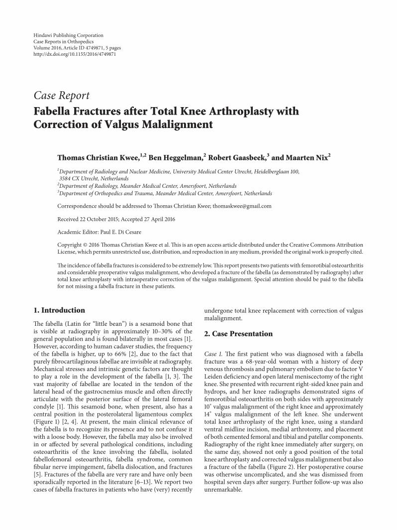
Knee capsule
Semimembranosus insertion
Oblique popliteal ligament
Popliteus muscle
FabellaLateral collateral lig
Biceps femoris tendonFabellofibular lig
Arcuate ligamentPopliteal fibular lig
Figure 1: Posterolateral ligamentous complex of the knee including arcuate ligament, fabellofibular ligament, popliteal-fibular ligament,lateral collateral ligament, and biceps femoris tendon (case courtesy of Dr. Frank Gaillard, http://radiopaedia.org/). The fabella is located inthe posterior aspect of the knee where lines of tensile stress intersect [14]. It articulates with the posterior part of the articular surface of thelateral femoral condyle and is embedded in the muscular fibres of the gastrocnemius muscle [14]. Anteriorly the fabella is bordered by theposterior capsule of the knee joint and posteriorly it is situated at the endpoint of the oblique popliteal ligament and the lateral gastrocnemiustendon [14]. In addition, the fabellofibular ligament runs to its distal insertion at the styloid process of the fibular head [14].
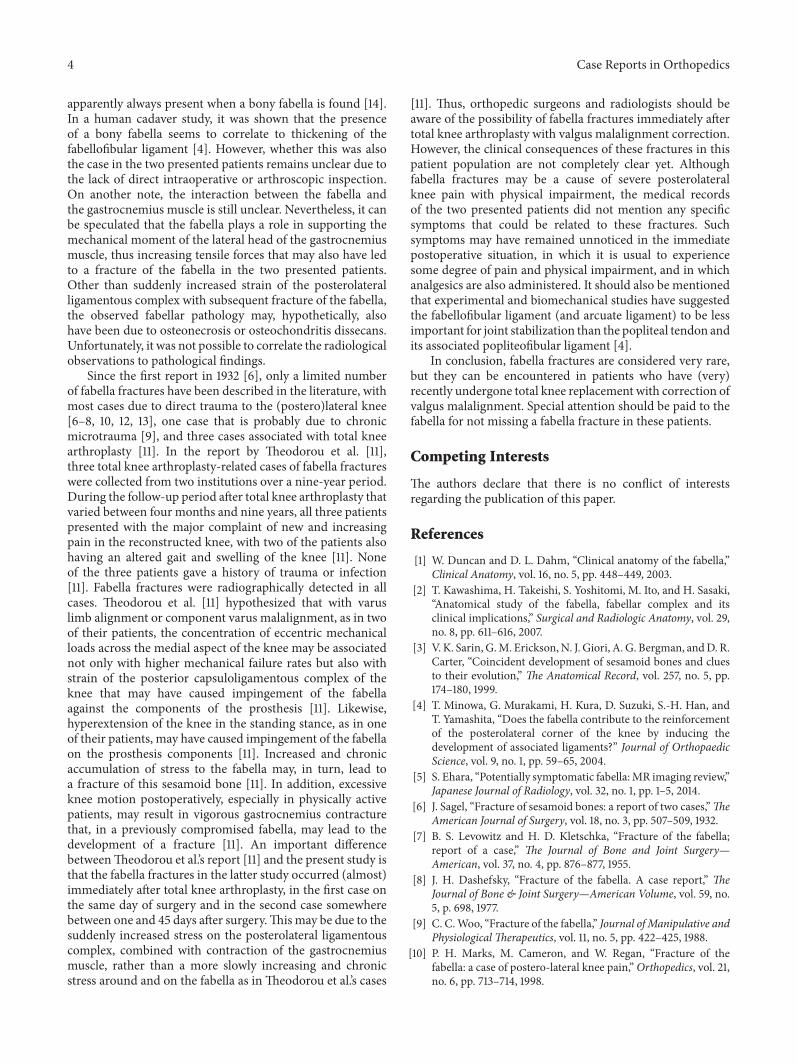
(a) (b) (c) (d)
Figure 2: Preoperative (a, b) and immediate postoperative (c, d) knee radiographs in a 68-year-old woman who underwent total kneearthroplasty of the right knee because of symptomatic knee osteoarthritis with valgusmalalignment. Preoperative knee radiographs (anterior-posterior view of both knees (a) and lateral view of the right knee (b)) show osteophytes, sclerotic changes, and some degree of generalizedjoint space narrowing on both sides, in keeping with femorotibial osteoarthritis. There is approximately 10∘ valgus malalignment of the rightknee and approximately 14∘ valgusmalalignment of the left knee. Note the normal appearing fabella ((b) arrowhead). Immediate postoperativeradiographs of the right knee on the same day of surgery (anterior-posterior view (c) and lateral view (d)) show not only a good position ofthe total knee arthroplasty with a well-aligned knee joint but also a horizontal fracture through the fabella with only minimal dislocation ((d)including magnified view of the fabella).
Case 2. The second patient who was diagnosed with afabella fracture was a 63-year-old woman with a history ofobesity, dyslipidemia, hypertension, cardiac failure, gout, andMeniere disease, who suffered from progressive right-sidedknee pain. Radiography demonstrated signs of femorotibialosteoarthritis on both sides, with approximately 23∘ valgusmalalignment of the right knee and approximately 7∘ varus
malalignment of the left knee. She underwent total kneearthroplasty of the right knee, using a standard ventralmidline incision, medial arthrotomy, and placement of bothcemented femoral and tibial components, but without a patel-lar component. Radiography of the right knee immediatelyafter surgery, on the same day, showed a good position of thetotal knee arthroplasty and corrected valgus malalignment,

Case Reports in Orthopedics 3
(a) (b) (c)
(d) (e) (f) (g)
Figure 3: Preoperative (a–c), immediate postoperative (d, e), and 45-day postoperative (f, g) knee radiographs in a 63-year-old woman whounderwent total knee arthroplasty of the right knee because of symptomatic knee osteoarthritis with valgus malalignment. Preoperative kneeradiographs (anterior-posterior (a) and lateral views (b, c) of both knees) showosteophytes, sclerotic changes, and severe joint space narrowingof right-sided lateral and left-sidedmedial femorotibial compartments, in keeping with femorotibial osteoarthritis.There is approximately 23∘valgus malalignment of the right knee and approximately 7∘ varus malalignment of the left knee. Note the normal appearing fabellae ((b) and(c) arrowheads). Immediate postoperative radiographs of the right knee on the same day of surgery (anterior-posterior view (d) and lateralview (e)) show a good position of the total knee arthroplasty with a well-aligned knee joint and with a still normal appearing fabella ((e)arrowhead). However, 45-day postoperative radiographs of the right knee (anterior-posterior view (f) and lateral view (g)) show a shatteredpatella ((g) including magnified view of the fabella).
with a normal appearing fabella. Three days later she wasdismissed from hospital. A repeated radiograph of the rightknee 45 days after surgery, however, showed a fracturedfabella (Figure 3). Her medical records did not mention anyspecific circumstances or symptoms that could be related tothis fracture. No additional treatment was given because ofthis fabella fracture. Further follow-up was unremarkable.
3. Discussion
The two patients with fabella fractures who have beenpresented in this report had very recently undergone totalknee arthroplasty and had considerable preoperative valgusmalalignment of the knee that was corrected intraoperatively.Anatomical and biomechanical factors could play a role inthe development of these fractures. The fabella is situatedat the endpoint of the oblique popliteal ligament and thelateral gastrocnemius tendon and is connected with the
styloid process of the fibular head through the fabellofibularligament (Figure 1) [15], with all of these structures exposingthe fabella to tensile forces. Both patients had preoperativevalgusmalalignment of the knee, and it can be speculated thatits sudden intraoperative correction stresses the abovemen-tioned tendoligamentous structures beyond their adaptednormal physiological ranges. Since the fabella is located atthe intersection point of these tensile forces, fracture ofthis sesamoid bone may occur. Particularly the fabellofibularligament, which is a static stabilizer that tenses when theknee is fully extended [12], is thought to expose the fabella toconsiderable stress after correction of knee valgus alignment,due to the nearly parallel orientation of its long axis tothe main direction of knee motion (flexion extension). Thehorizontal orientation of the fracture plane through thefabella in the first case (Figure 2) also indicates tensile forcesin the craniocaudal direction, which supports this theory. Ofinterest, the fabellofibular ligament has been reported to be
4 Case Reports in Orthopedics
apparently always present when a bony fabella is found [14].In a human cadaver study, it was shown that the presenceof a bony fabella seems to correlate to thickening of thefabellofibular ligament [4]. However, whether this was alsothe case in the two presented patients remains unclear due tothe lack of direct intraoperative or arthroscopic inspection.On another note, the interaction between the fabella andthe gastrocnemius muscle is still unclear. Nevertheless, it canbe speculated that the fabella plays a role in supporting themechanical moment of the lateral head of the gastrocnemiusmuscle, thus increasing tensile forces that may also have ledto a fracture of the fabella in the two presented patients.Other than suddenly increased strain of the posterolateralligamentous complex with subsequent fracture of the fabella,the observed fabellar pathology may, hypothetically, alsohave been due to osteonecrosis or osteochondritis dissecans.Unfortunately, it was not possible to correlate the radiologicalobservations to pathological findings.
Since the first report in 1932 [6], only a limited numberof fabella fractures have been described in the literature, withmost cases due to direct trauma to the (postero)lateral knee[6–8, 10, 12, 13], one case that is probably due to chronicmicrotrauma [9], and three cases associated with total kneearthroplasty [11]. In the report by Theodorou et al. [11],three total knee arthroplasty-related cases of fabella fractureswere collected from two institutions over a nine-year period.During the follow-up period after total knee arthroplasty thatvaried between four months and nine years, all three patientspresented with the major complaint of new and increasingpain in the reconstructed knee, with two of the patients alsohaving an altered gait and swelling of the knee [11]. Noneof the three patients gave a history of trauma or infection[11]. Fabella fractures were radiographically detected in allcases. Theodorou et al. [11] hypothesized that with varuslimb alignment or component varus malalignment, as in twoof their patients, the concentration of eccentric mechanicalloads across the medial aspect of the knee may be associatednot only with higher mechanical failure rates but also withstrain of the posterior capsuloligamentous complex of theknee that may have caused impingement of the fabellaagainst the components of the prosthesis [11]. Likewise,hyperextension of the knee in the standing stance, as in oneof their patients, may have caused impingement of the fabellaon the prosthesis components [11]. Increased and chronicaccumulation of stress to the fabella may, in turn, lead toa fracture of this sesamoid bone [11]. In addition, excessiveknee motion postoperatively, especially in physically activepatients, may result in vigorous gastrocnemius contracturethat, in a previously compromised fabella, may lead to thedevelopment of a fracture [11]. An important differencebetweenTheodorou et al.’s report [11] and the present study isthat the fabella fractures in the latter study occurred (almost)immediately after total knee arthroplasty, in the first case onthe same day of surgery and in the second case somewherebetween one and 45 days after surgery.Thismay be due to thesuddenly increased stress on the posterolateral ligamentouscomplex, combined with contraction of the gastrocnemiusmuscle, rather than a more slowly increasing and chronicstress around and on the fabella as in Theodorou et al.’s cases
[11]. Thus, orthopedic surgeons and radiologists should beaware of the possibility of fabella fractures immediately aftertotal knee arthroplasty with valgus malalignment correction.However, the clinical consequences of these fractures in thispatient population are not completely clear yet. Althoughfabella fractures may be a cause of severe posterolateralknee pain with physical impairment, the medical recordsof the two presented patients did not mention any specificsymptoms that could be related to these fractures. Suchsymptoms may have remained unnoticed in the immediatepostoperative situation, in which it is usual to experiencesome degree of pain and physical impairment, and in whichanalgesics are also administered. It should also be mentionedthat experimental and biomechanical studies have suggestedthe fabellofibular ligament (and arcuate ligament) to be lessimportant for joint stabilization than the popliteal tendon andits associated popliteofibular ligament [4].
In conclusion, fabella fractures are considered very rare,but they can be encountered in patients who have (very)recently undergone total knee replacement with correction ofvalgus malalignment. Special attention should be paid to thefabella for not missing a fabella fracture in these patients.
Competing Interests
The authors declare that there is no conflict of interestsregarding the publication of this paper.
References
[1] W. Duncan and D. L. Dahm, “Clinical anatomy of the fabella,”Clinical Anatomy, vol. 16, no. 5, pp. 448–449, 2003.
[2] T. Kawashima, H. Takeishi, S. Yoshitomi, M. Ito, and H. Sasaki,“Anatomical study of the fabella, fabellar complex and itsclinical implications,” Surgical and Radiologic Anatomy, vol. 29,no. 8, pp. 611–616, 2007.
[3] V. K. Sarin, G.M. Erickson,N. J. Giori, A. G. Bergman, andD. R.Carter, “Coincident development of sesamoid bones and cluesto their evolution,” The Anatomical Record, vol. 257, no. 5, pp.174–180, 1999.
[4] T. Minowa, G. Murakami, H. Kura, D. Suzuki, S.-H. Han, andT. Yamashita, “Does the fabella contribute to the reinforcementof the posterolateral corner of the knee by inducing thedevelopment of associated ligaments?” Journal of OrthopaedicScience, vol. 9, no. 1, pp. 59–65, 2004.
[5] S. Ehara, “Potentially symptomatic fabella:MR imaging review,”Japanese Journal of Radiology, vol. 32, no. 1, pp. 1–5, 2014.
[6] J. Sagel, “Fracture of sesamoid bones: a report of two cases,”TheAmerican Journal of Surgery, vol. 18, no. 3, pp. 507–509, 1932.
[7] B. S. Levowitz and H. D. Kletschka, “Fracture of the fabella;report of a case,” The Journal of Bone and Joint Surgery—American, vol. 37, no. 4, pp. 876–877, 1955.
[8] J. H. Dashefsky, “Fracture of the fabella. A case report,” TheJournal of Bone & Joint Surgery—American Volume, vol. 59, no.5, p. 698, 1977.
[9] C. C.Woo, “Fracture of the fabella,” Journal of Manipulative andPhysiological Therapeutics, vol. 11, no. 5, pp. 422–425, 1988.
[10] P. H. Marks, M. Cameron, and W. Regan, “Fracture of thefabella: a case of postero-lateral knee pain,”Orthopedics, vol. 21,no. 6, pp. 713–714, 1998.

Case Reports in Orthopedics 5
[11] S. J.Theodorou, D. J.Theodorou, andD. Resnick, “Painful stressfractures of the fabella in patients with total knee arthroplasty,”American Journal of Roentgenology, vol. 185, no. 5, pp. 1141–1144,2005.
[12] G. M. Heideman, K. E. Baynes, A. P. Mautz, M. S. DuBois, andJ. W. Roberts, “Fabella fracture with CT imaging: a case report,”Emergency Radiology, vol. 18, no. 4, pp. 357–361, 2011.
[13] A. R. Barreto, F. A. Chagas-Neto, M. D. Crema et al., “Fractureof the fabella: a rare injury in knee trauma,” Case Reports inRadiology, vol. 2012, Article ID 390150, 3 pages, 2012.
[14] E. B. Kaplan, “The fabellofibular and short lateral ligaments ofthe knee joint,”The Journal of Bone & Joint Surgery—AmericanVolume, vol. 43, pp. 169–179, 1961.
[15] A. Driessen, M. Balke, C. Offerhaus et al., “The fabellasyndrome—a rare cause of posterolateral knee pain: a reviewof the literature and two case reports,” BMC MusculoskeletalDisorders, vol. 15, article 100, 2014.