f1000researchdata.s3.amazonaws.com · web viewa single peak at 280 nm can be observed for the pure...
TRANSCRIPT

Extraction, purification, and activity of protease from the leaves of
Moringa oleiferaSwarnali Banik1, Shrutidhara Biswas2 and Srabani Karmakar1*
1Department of Biotechnology, Techno India University, Kolkata-700064, India2Department of Biotechnology, Indian Institute of Technology, Guwahati-781039, India
*Corresponding author: [email protected]
Abstract
Background: Proteases cleave proteins, thereby providing essential amino acids for protein synthesis, and
degrade misfolded and damaged proteins to maintain homeostasis. Proteases also serve as signaling molecules,
therapeutic agents and find wide applications in biotechnology and pharmaceutical industry. Plant-derived
proteases are suitable for many biomedical applications due to their easy availability and activity over a wide
range of pH, temperature, and substrates. Moringa oleifera Lam (Moringaceae) is a very common food plant
with medicinal property and geographically distributed in tropical countries. Here, we isolate proteases from
the leaves of Moringa oleifera and characterize its enzymatic activity.
Methods: Proteases were isolated from the aqueous leaf extract of Moringa oleifera by ammonium sulfate
precipitation and purified by ion exchange chromatography. Subsequently, the enzyme kinetics was
determined using casein as a substrate and calibrated over different pH and temperature range for maximal
activity.
Results: We obtained purified fraction of the protease having a molecular weight of 51 kDa. We observed that
for the maximal caseinolytic activity of the protease, a pH of 8 and temperature of 37ºC was found to be most
effective.
Conclusion: The plant-derived proteolytic enzymes are finding increasing clinical and industrial applications.
We could extract, purify and characterize the enzymatic activity of proteases from the leaves of Moringa
oleifera. Further molecular characterization, substrate specificity and activity of the extracted protease are
required for determining its suitability as a proteolytic enzyme for various applications.
Keywords: Casein, Enzyme activity, Leaf extract, Moringa Oleifera, Plant-derived proteases, Protein
purification.
1

Introduction
All organisms contain proteases that hydrolyze peptide bonds in order to maintain systemic homeostasis
and for its normal growth and development1, 2. Proteases derived from plants, animals and microbes find
wide industrial applications including in the leather, food, brewery and pharmaceutical industry 2-4
corresponding to approximately 60% of the total worldwide enzyme sales5.
Moringa oleifera is one of the best known medicinal plants widely distributed in the tropical regions6. It
contains a mixture of several hydrolytic enzymes, in which proteases are the key enzymes reported to
show pharmacological activity7. We attempted to investigate the protease activity of aqueous extracts
of Moringa oleifera leaf. Here, we have isolated and purified the protease from Moringa leaves and
carried out enzyme kinetics study and find that the protease exhibited optimal caseinolytic activity in
alkaline pH.
Methods
Preparation of crude enzyme extract
Mature Moringa Oleifera leaves were collected from a plant located near TIU campus, Salt Lake Kolkata
and crushed along with 20mM phosphate buffer (pH 7.5) and 0.1% tween 20 detergent and protease
cocktail inhibitor followed by centrifugation with plastocraft table top refrigerated centrifuge machine
(Rota 4RV/FM) at 10000 rpm for 10 mins at 4ºC. The crude soup was mixed with 40% ammonium
sulphate to obtain the protein precipitate, which was then dissolved in 20 mM tris buffer for further
evaluation.
Determination of protein content
The total protein content of the solutions at different stages of protein purification was determined by
Lowry’s and Bradford methods.
The total protein content of the solutions at different stages of protein purification was determined by
Bradford methods8 using Sigma’s Bradford reagent (B6916). In this assay, a series of BSA standard
solutions (0.1 - 1.2mg/ml) were used to prepare the standard curve. Bradford assay was
performed by adding 1 mL of Bradford reagent to 20 µl of each standard solutions or unknown
solution, and homogenized by using vortex mixer. The samples were incubated in dark
conditions for 10 minutes and the absorbance was read at 595 nm.
2

SDS PAGE
We performed 12% sodium dodecyl sulfate (SDS) polyacrylamide gel electrophoresis (PAGE) for
separating proteins. We followed the Lamelle’s method for gel electrophoresis and stained the gel with
Coomassie Brilliant Blue for band analysis.
We performed sodium dodecyl sulfate (SDS) polyacrylamide gel electrophoresis (PAGE) using 12 %
resolving and 5 % stacking gels for separating proteins. We followed the Laemmli’s method 9 for gel
electrophoresis. The samples were mixed with equal volume of gel loading buffer and heated at 95 0 C in
dry heating bath for 2 mins. The electrophoresis process was run with 90 V for first 10 mins and then run
at 150 V with Biorad mini protean gel electrophoresis system. After complete run the gel was stained
with Coomassie Brilliant Blue . We have used protein marker (10kD to 250 kD) from GCC biotech (Pre-
stained protein marker GCR-P4B) for determination of molecular weight. We imaged the gels in Biorad
gel documentation system. Acrylamide, bis acrylamide, Tris and TEMED (T9281) are from Sigma
Aldrich. Coomassie Brilliant Blue R250 (93473) and Ammonium per sulphate (28575) was from SRL
(Sisco Research Laboratories)
Purification of protein
Dialysis: The pellet dissolved in Tris buffer as obtained above was then dialyzed in 3.5cm/ml
dialysis tubing (SIGMA Aldrich D6066 overnight in a magnetic stirrer by immersing the tubing
in a buffer containing Tris (pH 8) and phenylmethylsulfonyl fluoride (PMSF) SRL , which was
repeated thrice for complete exchange of buffer.
Diethylaminoethyl (DEAE) cellulose ion exchange chromatography: The protein sample was
loaded in the DEAE cellulose (SIGMA Aldrich 30477) column. Ion exchange column
chromatography was carried out by using an assembly of Biorad’s Econo pump model EP-1, UV
monitor and chart recorder from Atto, Japan and aBiorad’s fraction collector model 2110. A
gradient of 0.05 M to 0.5 M NaCl was used to elute the protein from the column. The gradient
was run for 150 min with a flow rate of 2ml1ml/min. Optical density (OD) of all the fractions
were taken at 280 nm with Schimadzu 2401 UV Vis Spectrophotometer.
Bovine serum albumin (BSA) digestion
3

Samples at different stages of purification were tested for albuminolytic property of protease by using
BSA SIGMA as substrate. BSA digestion was performed at 37ºC and pH 7.5 for 1 hour. Further, each of
the samples were mixed with Lameli dye protein gel loading dye in 1:1 ratio and loaded in SDS PAGE
and examinedthe gel was imaged with Biorad gel documentation system.
Protease activity assay
In this assay, β-casein was used as substrate. If protease digests casein, the amino acid tyrosine is
liberated along with other peptide fragments. Folin’s reagent reacts with free tyrosine to generate a blue
colored product, which is quantifiable and measured as an absorbance value on the Schimadzu UV 2401
spectrophotometer at 660 nm. A tyrosine standard calibration curve is constructed to determine the
amount of tyrosine released after the proteolytic activity. A series of tyrosine standard solutions at
different concentrations (5 - 50 μg/mL) were prepared from the 0.18mg/mL L-tyrosine stock solution
with deionized water. L-tyrosine was purchased from Himedia, Fohlin’s reagent was obtained from SRL
and β-casein from SIGMA.
Effect of pH & temperature on the protease activity
We have assayed the protease activity in terms of caseinolytic activity with plant leaf extracts at different
stages of purification (crude soup, 40% ammonium soup and pooled soup) and at different pH range 4-8
is the initial supernatant after homogenization and centrifugation, 40% ammonium soup is the phosphate
dissolved pellet after 40% ammonium sulphate fractionation and pooled soup is the final collection of
pure fractions came from DEAE cellulose column). All the three samples were dialysed to remove
protease inhibitr and EDTA before the protease assay. The protease activity of pure protein was examined
at different pH range 4-9 and temperature range 4-70ºC.
Enzyme kinetics assay at different β-casein concentration
The enzyme activity assay for protease was conducted with different concentrations of β-casein as
substrate, at pH-8 in 37°C respective optimum conditions as determined with the previous experiments
described above (optimum temperature and pH conditions). Here the substrate concentration (β-casein)
varied in the range (0.81, 1.6, 2.4, 4.03, 5.2) mg/ml keeping the enzyme concentration fixed.
Results
4

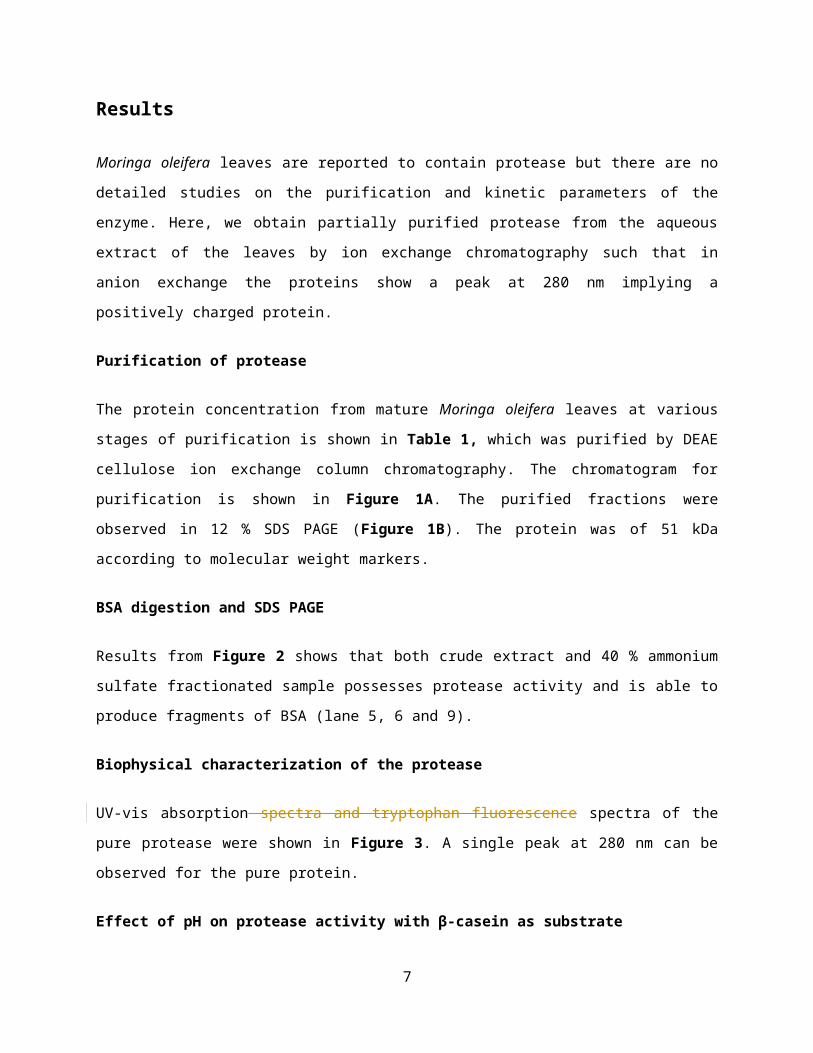
Moringa oleifera leaves are reported to contain protease but there are no detailed studies on the
purification and kinetic parameters of the enzyme. Here, we obtain partially purified protease from the
aqueous extract of the leaves by ion exchange chromatography such that in anion exchange the proteins
show a peak at 280 nm implying a positively charged protein.
Purification of protease
The protein concentration from mature Moringa oleifera leaves at various stages of purification is shown
in Table 1, which was purified by DEAE cellulose ion exchange column chromatography. The
chromatogram for purification is shown in Figure 1A. The purified fractions were observed in 12 % SDS
PAGE (Figure 1B). The protein was of 51 kDa according to molecular weight markers.
BSA digestion and SDS PAGE
Results from Figure 2 shows that both crude extract and 40 % ammonium sulfate fractionated sample
possesses protease activity and is able to produce fragments of BSA (lane 5, 6 and 9).
Biophysical characterization of the protease
UV-vis absorption spectra and tryptophan fluorescence spectra of the pure protease were shown in Figure
3. A single peak at 280 nm can be observed for the pure protein.
Effect of pH on protease activity with β-casein as substrate
In both crude extract and purified protein, protease activity was measured as described in methods.
Reactions in different pH 4, 5, 6, 7, 8 and 9 were done (Figure 4). The results showed maximum activity
at the pH 8.0. Therefore, the enzyme is an alkaline protease.
Effect of temperature on protease activity with β-casein as substrate.
The protease assay with β-casein as substrate was performed at a range of temperatures; 4 0C, 25 0C 37 0C, 55 0C and 70 0C (Figure 5) according to the methods described above. The enzyme activity was found
to be maximum at 37 0C.
Enzyme kinetics
5

Specific activity of the protease was calculated by enzyme activity from the protease assay using β-
casein as substrate and the total protein content of the protease solution. We can see a large increase in
specific activity after the final purification (Figure 6).
IncreasingWe have seen increasing protease activity in the initial substrate concentration range and
purifiedthen saturation of protease reaction shows Michaelis Menten kinetics plot foractivity above
concentration of 4.03 to 5.2 mg/ml β- casein (Figure 7). The graph as a result follows conventional
Michaelis Menten kinetics. We calculated KM and Vmax from thisthe corresponding double reciprocal plot
i.e. Lineweaver Burk plot as shown in the inset graph (Table 2). KM is 105.23 µg5.47 mg/ml and Vmax is
2059.94 µg/ml588.23 µM/min.
Discussion
Our study concludes that mature leaves from Moringa oleifera contains a protease with an approximate
molecular weight of 51kD, with an optimum temperature of 37 0C and optimum pH of 8.0 for its
caseinolytic property. This is the first report of purification of a protease from Moringa oleifera to our
knowledge. Further determination of molecular characterization, substrate specificity and activity of the
protease are required to determine its suitability for industrial applications.
Competing interests
No competing interests were disclosed.
Grant information
The authors declare that no grants were involved in supporting this work
Acknowledgements
We would like to acknowledge Mr Dipak Chandra Konar of Department of Chemistry, Bose Institute, for
his help in using the protein purification set up, dynamic light scattering and spectrophotometric assays.
Special thanks go to Prof. K. P Das, Bose Institute, for his support in this project.
6
REFERENCES
[1] Lopez-Otin, C., and Overall, C. M. (2002) Protease degradomics: a new challenge for
proteomics, Nat Rev Mol Cell Biol 3, 509-519.
[2] Turk, B. (2006) Targeting proteases: successes, failures and future prospects, Nat Rev Drug
Discov 5, 785-799.
[3] Li, Q., Yi, L., Marek, P., and Iverson, B. L. (2013) Commercial proteases: present and future,
FEBS Lett 587, 1155-1163.
[4] Lopez-Otin, C., and Bond, J. S. (2008) Proteases: multifunctional enzymes in life and
disease, J Biol Chem 283, 30433-30437.
[5] Li, S., Yang, X., Yang, S., Zhu, M., and Wang, X. (2012) Technology prospecting on
enzymes: application, marketing and engineering, Comput Struct Biotechnol J 2, e201209017.
[6] Stohs, S. J., and Hartman, M. J. (2015) Review of the Safety and Efficacy of Moringa
oleifera, Phytother Res 29, 796-804.
[7] Satish, A., Sairam, S., Ahmed, F., and Urooj, A. (2012) Moringa oleifera Lam.: Protease
activity against blood coagulation cascade, Pharmacognosy Res 4, 44-49.
[1] Lopez-Otin, C., and Overall, C. M. (2002) Protease degradomics: a new challenge for proteomics, Nat Rev Mol Cell Biol 3, 509-519.
[2] Turk, B. (2006) Targeting proteases: successes, failures and future prospects, Nat Rev Drug Discov 5, 785-799.
[3] Li, Q., Yi, L., Marek, P., and Iverson, B. L. (2013) Commercial proteases: present and future, FEBS Lett 587, 1155-1163.
7

[4] Lopez-Otin, C., and Bond, J. S. (2008) Proteases: multifunctional enzymes in life and disease, J Biol Chem 283, 30433-30437.
[5] Li, S., Yang, X., Yang, S., Zhu, M., and Wang, X. (2012) Technology prospecting on enzymes: application, marketing and engineering, Comput Struct Biotechnol J 2, e201209017.
[6] Stohs, S. J., and Hartman, M. J. (2015) Review of the Safety and Efficacy of Moringa oleifera, Phytother Res 29, 796-804.
[7] Satish, A., Sairam, S., Ahmed, F., and Urooj, A. (2012) Moringa oleifera Lam.: Protease activity against blood coagulation cascade, Pharmacognosy Res 4, 44-49.
[8] Bradford, M. M. (1976) A rapid and sensitive method for the quantitation of microgram quantities of protein utilizing the principle of protein-dye binding, Analytical Biochemistry 72, 248-254.
[9] Laemmli, U. K. (1970) Cleavage of Structural Proteins during the Assembly of the Head of Bacteriophage T4, Nature 227, 680.
Tables:
Table 1: Total protein content in different stages of purification
8
SamplesProtein
concentration(mg/ml)
Crude 0.56
40% 0.55
Pooled 0.22

Table 2: KM and Vmax from enzyme kinetics
KM
µgmg/ml
Vmax
µg/mlµM/min
105.23625.47 2059.945588.235
Figure legends:
Figure 1: A. Chromatogram showingfor the purification of protein from Moringa oleifera shows
the elution time versus absorbance at 280 nm and the saltcorresponding NaCl gradient. profile
(ranging from 0.04M to 0.25M) for maximal elution B. SDS PAGE of the pure crude extract and
fractions after and before purification by DEAE cellulose ion exchange chromatography. Lane 1
shows extract after 40% ammonium sulphate precipitation, lane 2 shows the prestained
molecular weight marker from GCC biotech marking 140, 100, 91, 71, 51, 25 and 10 kDa bands,
lanes 3 to 8 represent fractions after column purification, lanes 9 and10 show the bands from
crude leaf extract.
Figure 2: SDS PAGE showing BSA fragmentation by Moringa oleifera crude enzyme as
compared to with trypsin as positive control. Lane 1 is BSA and trypsin, lane 2 is BSA + trypsin
+ PMSF, lane 3 is BSA, lane 4 is BSA +40% Ammonium Sulphate cut + PMSF, lane 5 is BSA +
40% Ammonium Sulphate cut, lane 6 is BSA + Pooled pure protein, lane 7 is BSA, lane 8 is
prestained molecular weight marker from GCC biotech showing 140, 100, 91, 71, 51, 25 and 10
kDa bands and lane 9 is BSA + crude leaf extract.
9

Figure 3: Biophysical characterizationUV-Vis absorbance spectra from (230 nm to 400 nm) of
the purified protein from Moringa oleifera leaf extract is shown.
Figure 4: Effect of pH on the caseinolytic property of the protease: protease. Protease activity of
the pooled pure fractions on beta β-casein degradation wasis plotted against different pH and
fixed temperature(4, 5, 6, 7 and 8) at 37ºC. Free tyrosine liberated due to β-casein degradation
was monitored with Folin-Ciocalteau reagent at 660 nm and the corresponding amount was
measured from the tyrosine standard curve.
Figure 5: Effect of temperature on the caseinolytic property of the protease: prootease. Protease
activity of the pooled pure fractions on beta β-casein degradation wasis plotted against different
temperature (4, 25, 37, 55 and fixed700 C) at pH 8. Free tyrosine liberated due to β-casein
degradation was measured as described earlier.
Figure 6: Comparison of proteasespecific activity of crude extract, 40 % ammonium sulfate
fraction and pure proteasepurified protease at optimum pH 8 and optimum temperature 37ºC is
represented as a bar diagram. Free tyrosine liberated due to β-casein degradation was measured
as described earlier. Enzyme activity present per amount of enzyme is calculated as specific
activity of the protease.
Figure 7: Protease activity with different betaβ- casein concentrations wasis plotted to get
Michaelis Menten curve and the double reciprocal plot was(1/substrate concentration versus 1/
enzyme activity) is shown on the inset. KM and Vmax was obtained from thisthe double reciprocal
plot. are 5.47 mg/ml and 588.23 µM/min respectively.
10
Figure 1
A
11

Figure 2
12

13

Figure 3
14

Figure 4
15

Figure 5
16

Figure 6
17

Figure 7
18

19