eznettm phage display cdna library - maxim ivd and molecular products
TRANSCRIPT

1
EZnetTM Phage Display cDNA Library
Construction Kit (PDL-5001)Screening Kit (PDL-3001)
Instruction Manual
version 3.2
Maxim Biotech, Inc.
780 Dubuque Avenue Tel: (800) 989-6296So. San Francisco, CA 94080 Fax: (650) 871-2857U.S.A. http://www.maximbio.comApril, 1999 PDL-10005001
M B I

2
Introduction 3-6
Product Information 7-10
Library Construction 11-13
Panning & Screening Preparation 14-21
I Preparation 14,16II Titering 15,17,18III Helper phage 19IV Bait Coating 20-21
Panning, Rescue & Superinfection 22-27
ELISA 28-31
Trouble-Shooting 32-34
References 35-37
Appendix I: Vector pHage3.2 Map 38
Appendix II: Prepare TG1 competent cells 39
Ta b l e o f C o n t e n t s

3
IntroductionMaxim’s EZnetTM Phage Display Libraries, Library Screening, and Detection/Expression kits
provide a convenient and sensitive method to isolate rare and low-copy- number cDNA. Our systemmaximizes the benefits of in vivo and in vitro technologies to ensure you success in locating the cDNAtranscripts of your interest. We offer a complete, high-titer phage display library which is ready to bescreeened with our Library Screening kit or you may choose to create your own library. The protein-ligandcomplex can be detected by screening the library with MBI's Screening kit. Screening of phage displaylibraries provides a means to locate the cDNA which encodes the protein. EZnetTM cDNA Librariesare constructed in pHage3.2 vector. The cDNA/gene III fusion products are displayed on the filamen-tous phage particle tip, which makes interaction with an immobilized bait ligand easy. Several roundsof biopanning enrich for the interacting clones and then ELISA can be used to identify individualpositive clones and confirm positive interactions.
EZnetTM Phage Display cDNA Libraries are ready-to- screen by panning. With their high complexity(1010 pfu/ml), EZnet Libraries enhance the possibility of finding cDNAs for rare or low-copy-numbertranscripts. You can conveniently screen up to 109-1010 phage clones in one panning experiment.Premade EZnet Libraries are made from poly A+RNA isolated from a wide selection of human tissuesand cell lines
EZnetTM Phage Display Libraries Screening Kit includes all the necessary materials you need forperforming the library panning and ELISA procedures. We also include control reagents so you canfamiliarize yourself with the screening procedure before you try it with the library and your bait. Thecontrol system includes microtiter wells precoated with a control bait protein (the bcl-2 fusion proteins), apositive control phage clone known to interact with the control bait, and a negative control (noninteracting)phage clone. The EZnetTM Library Screening Kit and protocol have been optimized for use withEZnetTMLibraries.
EZnetTM Phage Display Expression Kit provides the HB2151 nonsuppressor E. coli host strain for invivo expression of Myc-tagged proteins without the pIII (gene III coat protein) fusion moiety. This kitalso provides the materials you need for detecting Myc-tagged proteins, including a titered anti-Mycmonoclonal antibody (mAb), and myc-tag based protein purification system. This kit has been opti-mized for use with EZnet Phage Display Libraries.
Panning
Panning is used to enrich libraries for specific clones by exposing pooled phge clones to a bait-coated well followed by a series of washings to remove unbound phage particles (Kay et al, 1996). Allphages that express a protein that can interact with the bait will adhere to the well. The TG1 host cells arethen applied to the well so they can become infected by the bound particles. Since the presence ofampicillan favors plasmid amplification, recombinant phagemids are then amplified by growing transformantson an ampicillan-containing medium. To maximaze enrichment M13 phage particles are superinfected withhelper phage and phagemid DNA. The entire panning process can be executed in one well of a 96-wellmicrotiter plate and can be completed in only one day.

4
IntroductionPositive clones and interactions can be confirmed by screening individual clones by ELISA; using
the same bait ligand as in the panning. Detection of bound phae is acheived by applying an antibody againstthe pVIII protein. Additionally, positive protein-ligand interactions can be further verified by using an anti-myc monoclonal antibodies in ELISA and molecular weight of fusion proteins can be dermined by WesternBlot. Then, myc-tagged proteins can be over-expressed from the original vector and purified with an anti-myc mAB affinity column. It is also possible to perform additional studies on the purified Myc-taggedproteins.
Vectors:
Maxim has designed a multifaceted, M13 -derived phagemid vector: pHage 3.2. Vectorcarries the pUC replication origin and the ampicillan resistance gene which permits growth andselection. They also contain an amber stop codon between the myc-tag sequence and the pIII genewhich allows for heproduction of geneIII fusion proteins. The gene III leader sequence is strategi-cally fused to the N-terminus so that the gene III fusion proteins can be secreted intothe periplasmicspace (Kang et al., 1991).
Helper phage:
The helper phge M13K07 conveniently infects TG1cells, that have been transformed with phage2.1or pHage 3.2, at room temperature or 37. The helper phage express wild-type gene III proteins andsecrete them into the periplasmic space where they are packaged into phage particles with pIII fusionproteins. Transcription is under the control of the lac promoter which keeps fusion gene transcription levelslow in the absence of IPTG. It is important to maintain low transcription levels so that only one or twocopies of the pIII fusion proteins are displayed on each phage particle surface. The presence of multiplecopies of wild-type pIII coat proteins ensures efficient packaging and infectivity. Each vector contains anM13 replication origin so in the presence of M13K07 helper phage the phagemid is replicated as a normalM13 phage.
After a positive clone has been identified and isolated, the foreign protein can be directlyoverexpressed from pHage3.2 without the pIII fusion moiety in the nonsuppressing E.coli host strainHB2151. High-level expression is inducible with IPTG. No subcloning is necessary, and no proteolysis isrequired to separate the cloned protein form pIII. In addition, because the foreign protein is still fused to thegene III leader sequence, the expressed protein is secreted into the periplasm and, in some cases, can bepurified directly from the culture medium without lysing the cells.
Low-valency, gene III fusion system:
pIII is a minor coat proteinthat is usually located in groups of five at one end of the filamentous rod.pIII is an integral component in the adsorption of the virus particle to the bacterial pilus (Sambrook et al,1989). Since only a few copies of the hybrid pIII molecules are produced by each phage particle, largefusion moieities can be put onto pIII without causing steric interference with protein-lignd interactions.

5
The low valency of pIII hybrids tends to select for high-affinity interactions (e.g.,Kd’s in themicromolar range; Kay et al., 1996; Cesarini,1992). Thus, low–affinity interactions may be missed in thissystem unless the binding and/or washing conditions are modified to decrease the stringency (see Section VIII,Troubleshooting Guide). Another possible limitation of this system is the full-length cDNA containing in-framestop codons TAA and/or TGA will not be expressed as pIII fusions and thus will not be displayed or found inthe screening. However, if the only stop codon is an amber stop (i.e., TAG), then the entire cDNA may beexpressed as a pIII fusion in the amber suppressor host strain TG1.
Applications:
An important application of phage display cloning technology is in receptor/ligand research(Wrighton etal., 1996). In fact, the display of proteins on the surface of a phage is one of the mosteffective methods for identifying cell-surface proteins that recognize specific baits. In addition, becauseit is not necessary for the interacting proteins to be localized to the cell’s nucleus, phage display technol-ogy is suitable for identifying interacting cytoplasmic proteins that may be overlooked in a two-hybridlibrary screening. Signal transduction research, new drug development and screening, and antibodyengineering are areas of proven application of phage display technology.
Phage display peptide libraries made with synthetic oligonucleotides have been used success-fully to identify peptides that interact with many different kinds of bait ligands, including proteins,peptides, RNAs, and oligonucleotides (reviewed bait ligands, including proteins, peptides, RNAs, andoligonucleotides (reviewed in Kay et al., 1996; Dunn, 1996; Smith & Scott, 1993;) Cesarini, 1992). Forexample, phage display peptide libraries using geneIII as the fusion moiety have been used to screen theepitopes of antibodies (Devlin et al., 1990; Cwirla et al., 1990; Scott & Smith, 1990; Oldenburg et al.,1992; Scott et al., 1992).
The recent development of phage display cDNA libraries enables researchers to use this power-ful technology to identify sequences that are expressed in nature and presumably encode proteins withspecific biological functions. For example, an allergenic protein form Aspergillus fumigatus was iden-tified (and its cDNA cloned ) by panning an A. fumigatus cDNA phage display library against immobi-lized serum immunoglobulin E from sensitized individuals (Crameri et al. 1996). Other examples arethe specialized libraries constructed using cDNAs for single-chain antibodies (scFv) and Fab antibodyfragments (Griffiths et al., 1993; Nissim et al., 1994; Mc Cafferty et al., 1990; Clackson et al., 1991;Winter et al., 1994). Phage display technology offers an advantage for identifying rare cDNAs becauseit allows screening of up to 1 x 10¹⁰ independent clones in small aliquots (Marks et al., 1992; Bass etal., 1990; Jacobsson & Fryberg, 1995; Gram et al., 1993; McCafferty et al., 1991; Crameri et al., 1994;Crameri & Suter, 1993). Phage display and yeast two-hybrid library screening are complementary approachesfor identifying novel proteins that interact with known bait. The EZnet Phage Display Library Screening Kitand Libraries are the system of choice if your application involves:
Introduction

6
Introduction
√ looking for protein-protein interactions that normally occur on the cell surface or in the cytoplasm;√ using a nonproteinaceous bait or very small amounts of bait material;√ screening a very large library quickly.
However, if you are looking for protein-protein interactions that depend on post-translational modifica-tions unique to eukaryotes, or that are particularly weak or transient, you should use a MATCHMAKERTwo-Hybrid System (Allen et al., 1995). Phage display library screening requires a small amount ofbait material (preferably purified), which is not required for a two-hybrid library screening. However,phage display library screening requires much less bait than plaque screening on filters using a conven-tional immunoscreening procedure.

7
EZnetTM Library Screening Kit (Cat. No.: PDL-3001)
Components
The materials provided are sufficient for probing 88 bait wells and8 total control wells in 96-well microtiter plates.
Component Description Volume/Amount
Positive control phage clone Interacts with the bait protein bcl-2 0.2 ml(BAD) (109 cfu/ml in PBS/20% glycerol)
Negative control phage clone Does not interact with bait 0.2 ml(1010 cfu/ml in PBS/20% glycerol)
M13K07 helper phage 1010 pfu/ml in PBS/20% glycerol 0.5 ml
Strips coated with bcl-2 Interacts with provided positive phage 1 strippositive control protein
Anti-pVIII polyclonal antibody Raised in goats (50 ug/ml) 0.5 ml
HRP-conjugated antibody Rabbit anti-goat; 50X 0.5 ml
HRP substrate A Includes H2O2 6 ml
HRP substrateB Solution B; includes 3,3',5,5'- 6 mltetramethyl-benzidine [TMB]
Product Information

8
EZnetTM Library Construction Kit (Cat. No.: PDL-5001)
Components:
Materials necessary are provided to construct phage-display library.
Component Description Volume/Amount
pHage 3.2 Vector Phage-display Vector (0.1 ug/ul) 0.1 ml
TG1 E. coli host cells in Stab agar 1 vial
HB2151 E. coli host cells in Stab agar 1 vial
M13K07 helper phage 1x10¹⁰ pfu/ml in PBS/20% glycerol 0.5 ml
Manual one copy
Kit Storage:Short-term storage: Less than 3 months. Store components at 4ºC.Long-term storage: Greater than 3 months. Store host cells at 4ºC.
Store all other components at -70ºC.
Recommendations:
Host strain: TG1 can also be used to titer the library and the helper phage. Upon arrival, prepare a TG1stock culture in 20% glycerol and store at -70ºC. Also, maintain a working stock plate onan M9/+ Thi minimal medium to keep selection on the F’ episome. The TG1 genotype:supEthi-1((lac-proAB) hsd(5[F’ traD36+ proAB+ lacIq lacZ(M15] (Sambrook et al., 1989).
Host strain: HB2151Prepare a stock culture in 20% glycerol for long-term storage and maintain a workingstock plate on an M9/+ Thi minimal medium.The HB2151 genotype: nalr thi-1 ara ( (lac-proAB [F’ proAB+ laciq lacZ(M15]
Helper Phage:M13K07. Prepare a fresh stock of helper phage before procedure.
Product Information

9
EZnet Phage Display Libraries
Components:
Materials are sufficient for up to 20 primary library pannings using 20 bait-coated wells in a 96-wellmicrotiter plate (or an equivalent surface area).
Component Description Volume/Amount
Phage library Dissolved in PBS/20% glycerol 0.2ml
TG1 E. coli host cells; lyophilized 1 vialfrom 1 ml of saturated culture
M13K07 helper phage 1x10¹⁰ pfu/ml in PBS/20% glycerol 0.5ml
PEG solution 1 vial
Library Storage:Short-term storage: Less than 3 months. Store components at 4ºC.
Long-term storage:
Recommendations:
Titer : The titer is the number of colony-forming units (cfu) per ml of culture. Retiter the library upon arrival to verify the titer since the titer value stated on the label is the titer at the time of construction. Titers are usually stable at-70ºC for at least one year. but may drop slightly when stored at 4ºC. If the library titer is at least108 pfu/ml, it is representative. However, if it drops below 108 cfu/ml, repeat the titer. If the titer is still low, please contact our Technical Support Department immediately.
Host strain: TG1 can also be used to titer the library and the helper phage. Upon arrival, prepare a TG1stock culture in 20% glycerol and store at -70ºC. Also, maintain a working stock plate onan M9/+ Thi minimal medium to keep selection on the F’ episome. The TG1 genotype:supEthi-1((lac-proAB) hsd(5[F’ traD36+ proAB+ lacIq lacZ(M15] (Sambrook et al., 1989).
Helper Phage:M13K07.Prepare a fresh stock of helper phage before each panning procedure.
Greater than 3 months. Divide the library into working aliquots (~20µl each) andstore at -70ºC. Store all other components at -20ºC. Avoid feeze/thaw cyles.
Product Information

10
Library InformationLibrary Construction:
cDNA is synthesized using random primers and a modified Gubler & Hoffman procedure (1983).The cDNA is size-fractionated by gel electrophoresis; the 0.3-3.0 kb fraction is extracted. The cDNA isblunt-ended and ligated into the EcoRV site of pHage3.2. Recombinant phagemids are packaged into infec-tious M13 phage particles upon superinfection with helper phage M13K07. The phage is precipitated fromsupernatants and resuspended in PBS/20%/glycerol at a high titer (>1010 cfu/ml).
The primary library (see Figure 2) is amplified by growth of the E. coli culture in liquid medium for 3 hrbefore it is frozen or superinfected with M13K07 helper phage. The superinfection and packaging in M13also amplify the library. No further amplification of the library is performed or recommended.
Library Quality Control:
Independent clones:The number of independent clones is equal to the number of recombinant ampicillan resistant colonies
that were produced when the vector was transformed into TG1.
Library titer:Library titer (generally >1010 cfu/ml) is determined after the phage particles are precipitated with PEG
and resuspended in PBS/20% glycerol. Although the library is provided in the form of M13 phage particles,the titer is given in units of cfu/ml (rather than pfu/ml) because pHage3.2 phagemids are not packaged and donot form plaques in the absence of helper phage .
Insert:The average insert size and range are estimated by running the cDNA on a gel prior to cloning and
comparing the profile to MW size markers. The insert size of 15 randomly selected clones is determined byPCR using primers designed for this vector.
Note that even though the libraries are constructed using size-selected cDNA, some of the inserts maybe <300 bp.
11
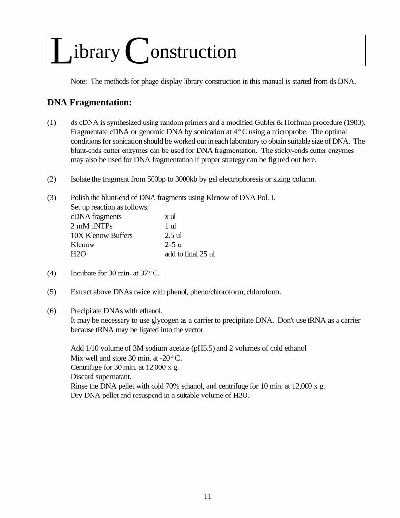
Library ConstructionNote: The methods for phage-display library construction in this manual is started from ds DNA.
DNA Fragmentation:
(1) ds cDNA is synthesized using random primers and a modified Gubler & Hoffman procedure (1983).Fragmentate cDNA or genomic DNA by sonication at 4ºC using a microprobe. The optimalconditions for sonication should be worked out in each laboratory to obtain suitable size of DNA. Theblunt-ends cutter enzymes can be used for DNA fragmentation. The sticky-ends cutter enzymesmay also be used for DNA fragmentation if proper strategy can be figured out here.
(2) Isolate the fragment from 500bp to 3000kb by gel electrophoresis or sizing column.
(3) Polish the blunt-end of DNA fragments using Klenow of DNA Pol. I.Set up reaction as follows:cDNA fragments x ul2 mM dNTPs 1 ul10X Klenow Buffers 2.5 ulKlenow 2-5 uH2O add to final 25 ul
(4) Incubate for 30 min. at 37ºC.
(5) Extract above DNAs twice with phenol, pheno/chloroform, chloroform.
(6) Precipitate DNAs with ethanol.It may be necessary to use glycogen as a carrier to precipitate DNA. Don't use tRNA as a carrierbecause tRNA may be ligated into the vector.
Add 1/10 volume of 3M sodium acetate (pH5.5) and 2 volumes of cold ethanolMix well and store 30 min. at -20ºC.Centrifuge for 30 min. at 12,000 x g.Discard supernatant.Rinse the DNA pellet with cold 70% ethanol, and centrifuge for 10 min. at 12,000 x g.Dry DNA pellet and resuspend in a suitable volume of H2O.

12
(continue)Ligate DNA Fragments into Vector:
(1) Digeste pHage 3.2 vector with EcoRV to generate blunt-ended vector.Or proper sticky-end enzymes if applicable.
(2) Dephosphorylate linear vector with alkaline phosphatase under recommended condition.
(3) Extract the digested vector DNAs twice with phenol, pheno/chloroform, chloroform.
(4) Incubate for 30 min. at 37ºC.
(5) Extract above DNAs twice with phenol, pheno/chloroform, chloroform.
(6) Precipitate DNAs with ethanol.Add 1/10 volume of 3M sodium acetate (pH5.5) and 2 volumes of cold ethanolMix well and store 30 min. at -20ºC.Centrifuge for 30 min. at 12,000 x g.Discard supernatant.Rinse the DNA pellet with cold 70% ethanol, and centrifuge for 10 min. at 12,000 x g.Dry DNA pellet and resuspend in a suitable volume of H2O.
(7) Ligate DNA fragments into the vector at ratio 2:1.(We recommend to use high concentration of T4 ligase from Bio-lab)
(8) Transform the ligation reaction into TG1 cells by electroporation.
Transformation (electroporation)
(1) Thaw competent TG1 cells on ice or use freshly made competent cells.
(2) Add 1 ul DNA vector (salt-free) in 50 ul of cells and transfer to a pre-chilled 0.2 cm cuvette. Place onice for 1 min.
(3) Program electroporator according to Manual. Dry outside of the curette with Kimwipes. Place it inthe electroporator chamber. Pulse once.
(4) Immediately add 0.95 ml 2x YT medium containing 2% glucose to the curette and cover to resuspendthe cells. Transfer the transformed cells to a 10 ml disposable culture tube.
(5) Grow the cells for 1 hour at 37ºC with shaking at 225 rpm.
Library Construction
13
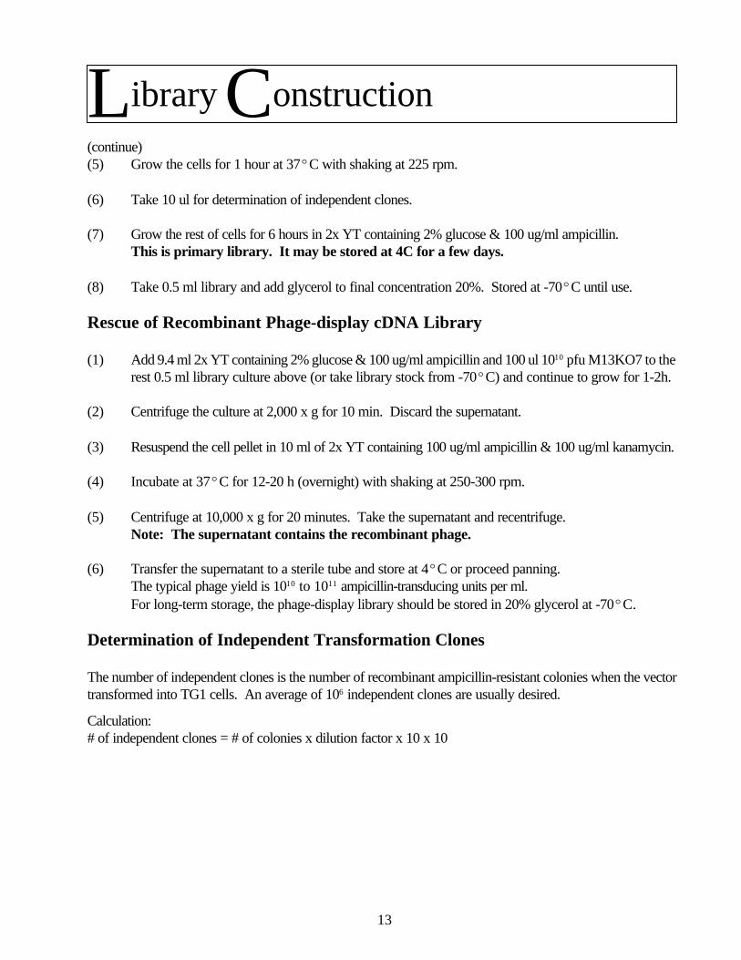
(continue)(5) Grow the cells for 1 hour at 37ºC with shaking at 225 rpm.
(6) Take 10 ul for determination of independent clones.
(7) Grow the rest of cells for 6 hours in 2x YT containing 2% glucose & 100 ug/ml ampicillin.This is primary library. It may be stored at 4C for a few days.
(8) Take 0.5 ml library and add glycerol to final concentration 20%. Stored at -70ºC until use.
Rescue of Recombinant Phage-display cDNA Library
(1) Add 9.4 ml 2x YT containing 2% glucose & 100 ug/ml ampicillin and 100 ul 1010 pfu M13KO7 to therest 0.5 ml library culture above (or take library stock from -70ºC) and continue to grow for 1-2h.
(2) Centrifuge the culture at 2,000 x g for 10 min. Discard the supernatant.
(3) Resuspend the cell pellet in 10 ml of 2x YT containing 100 ug/ml ampicillin & 100 ug/ml kanamycin.
(4) Incubate at 37ºC for 12-20 h (overnight) with shaking at 250-300 rpm.
(5) Centrifuge at 10,000 x g for 20 minutes. Take the supernatant and recentrifuge.Note: The supernatant contains the recombinant phage.
(6) Transfer the supernatant to a sterile tube and store at 4ºC or proceed panning.The typical phage yield is 1010 to 1011 ampicillin-transducing units per ml.For long-term storage, the phage-display library should be stored in 20% glycerol at -70ºC.
Determination of Independent Transformation Clones
The number of independent clones is the number of recombinant ampicillin-resistant colonies when the vectortransformed into TG1 cells. An average of 106 independent clones are usually desired.
Library Construction
Calculation:# of independent clones = # of colonies x dilution factor x 10 x 10

14
I. Plating, Storing and CulturingA. Reconstitution
Reconstitute the lyophilized culture of E. coli provided with your EZnetTM Kit or Library.To reconstitute lyophilized cultures:
1. Add 1 ml of 2X YT2. Incubate on shaker (250rpm) overnight at 37ºC.3. Use this culture to prepare a glycerol stock culture for freezing and a primary streak plate.
B. Glycerol Stock CultureTo prepare a glycerol stock culture, remove 0.75 ml of the overnight-reconstituted culture from Step Iabove to a fresh, pre-labeled tube. Add glycerol to a final concentration of 20% and store at -70ºC.
C. Primary streak plateUsing a sterile loop, streak out the overnight culture from Step I above on a 2X YT agar plate for singlecolonies. Incubate at 37ºC overnight.
Wrap the primary streak plate in Parafilm and store it at 4ºC. Prepare a fresh primary plate from the frozenglycerol stock at 3-month intervals (or sooner if contamination is evident).
To recover the frozen cells, streak a small portion (~5ul) of the frozen glycerol stock onto a 2X YT plate.Incubate at 37ºC overnight and proceed as in StepIII above.
D. Working Stock PlateTo prepare a working stock plate, pick a single, isolated colony from the primary streak plate and streak iton an M9/+ Thi plate. Incubate at 37ºC overnight. Wrap the plate in Parafilm and store at 4ºC for up to2 weeks. This plate will be your source of fresh colonies for inoculating liquid cultures and for preparingyour next fresh working stock plate.
Be sure to prepare a fresh working stock plate each week from the previous working stock plate, so youwill always have a source of fresh colonies. If you suspect contamination on your current working stockplate, prepare a new primary streak plate from the frozen culture.
E. Log-phage liquid cultureChoose an isolated colony from the working stock plate and use it to inoculate 5 ml of 2X YT (or LB)broth in a 50-ml test tube and incubate at 37ºC with shaking at 250 rpm until the OD600 reaches 0.4-0.6which is mid-log phase (approx. 6-8hr). After 4 hours of incubation, check the OD every half hour.Note: Stationary-phase bacterial cultures tend to lose F’ episomes (Sambrook et al., 1989). Therefore, culture should bechilled to 4ºC and placed in storage at 4ºC before it reaches saturation.
Immediately chill the culture on ice and then store it at 4ºC for up to one week. This chilled, log-phaseculture may be used for phage titering and plating.Additional Information:
The genes coding for enzymes involved in proline biosynthesis have been deleted from the chromosomes of TG1 and B2251.Thus, only those bacteria carrying the F’ plasmid [F’ traD36 proAB+ lacq lacZ(M15] or [F’ proAB+ lacIq lacZ(M15] willform colonies on medium lacking proline.
Bacteria grown on mimimal medium plates will not survive long at 4ºC. Also, bacteria grow much slower on minimal plates.
Panning & Screeening Prep
15
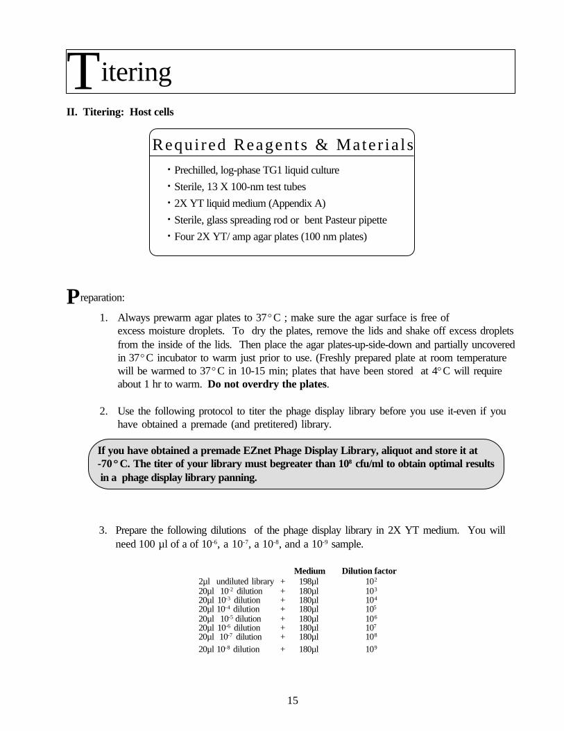
II. Titering: Host cells
Prechilled, log-phase TG1 liquid culture
Sterile, 13 X 100-nm test tubes
2X YT liquid medium (Appendix A)
Sterile, glass spreading rod or bent Pasteur pipette
Four 2X YT/ amp agar plates (100 nm plates)
Preparation:
1. Always prewarm agar plates to 37ºC ; make sure the agar surface is free ofexcess moisture droplets. To dry the plates, remove the lids and shake off excess dropletsfrom the inside of the lids. Then place the agar plates-up-side-down and partially uncoveredin 37ºC incubator to warm just prior to use. (Freshly prepared plate at room temperaturewill be warmed to 37ºC in 10-15 min; plates that have been stored at 4ºC will requireabout 1 hr to warm. Do not overdry the plates.
2. Use the following protocol to titer the phage display library before you use it-even if youhave obtained a premade (and pretitered) library.
If you have obtained a premade EZnet Phage Display Library, aliquot and store it at-70ººC. The titer of your library must begreater than 108 cfu/ml to obtain optimal results in a phage display library panning.
3. Prepare the following dilutions of the phage display library in 2X YT medium. You willneed 100 µl of a of 10-6, a 10-7, a 10-8, and a 10-9 sample.
Medium Dilution factor2µl undiluted library + 198µl 102
20µl 10-2 dilution + 180µl 103
20µl 10-3 dilution + 180µl 104
20µl 10-4 dilution + 180µl 105
20µl 10-5 dilution + 180µl 106
20µl 10-6 dilution + 180µl 107
20µl 10-7 dilution + 180µl 108
20µl 10-8 dilution + 180µl 109
Titering
Required Reagents & Mater ia l s•
•
•
•
•

16
4. Transfer 100µl of the 10-6, 10-7, 10-8, and 10-9 dilutions of the phage library to fresh, prelabeledtubes.
5. Add 100 µl of prechilled log-phase TG1 cells to each tube from Step 2 above. Mix gently andincubate at room temperature for 5 min to allow infection; M13 phage adsorbs rapidly to TG1cells.
6. Using a sterile glass spreading rod or bent Pasteur pipette, spread each phage/cell mixture on a2X YT/amp plate. Set plates at room temperature for 5 min to allow the inoculum to beabsorbed by the agar.
7. Incubate plates up-side-down at 37ºC overnight.
8. Count the number of the colonies on the plates that have between 30 and 300 colonies.
9. Worksheet
WORKSHEET 1
Plating the infected cells on ampicillin-containing medium ensures that onlycells harboring a pHage3.2 phagemid will grow. The low multiplicity of infectionensures that a host cell will be infected by only one phage.Thus, the titer (cfu/ml) of infected cells = the titer (pfu/ml) of the library.
Calculate the titer (cfu/ml) of the infected cells and hence of the phage displaycDNA library:
cfu/ml (≡ pfu/ml) = (# colonies on the plate) x dilution factor0.5 x 0.1 ml
cfu/ml = colonies x 10
0.5x 0.1ml
Panning & Screeening Preparation

17
II. Titering: M13K07 Helper Phage Stock
Prechilled, log-phase TG1 cells in 2X YT (Section III.A.6)
Melted 0.7% top agarose (in 2X YT) equilibrated to 47ºCin a water bath or heating block; allow 3 ml per dilution to beplated When plating bacteria/phage mixtures using melted topagar, the melted top agar should be at 45- 47ºC ; highertemperatures will kill the bacteria.Sterile, 13 X 100-mm test tubesThree 2X YT agar plates (100-mm plates)
Preparation:
1. The helper phage titer sometimes drops during prolonged storage. Therefore, titer the helperphage M13K07 stock provided before you use it to prepare the fresh helper phage stock.
2. Prepare ten-fold serial dilutions of the helper phage stock provided in 2X YT medium. You willneed 100(l of a 10-7, a 10-8, and a 10-9 dilution.
Medium Dilution factor2µl undiluted library + 198µl 102
20µl 10-2 dilution + 180µl 103
20µl 10-3 dilution + 180µl 104
20µl 10-4 dilution + 180µl 105
20µl 10-5 dilution + 180µl 106
20µl 10-6 dilution + 180µl 107
20µl 10-7 dilution + 180µl 108
20µl 10-8 dilution + 180µl 109
3. Transfer 100µl of the 10-7, 10-8, and 10-9 dilutions of the helper phage to sterile, 13 X 100-mmtubes.
4. Add 100µl of prechilled, log-phase TG1 culture to each tube from Step 2 above. Mix gentlyand incubate at room temperature for 5 min (infection; M13 phage adsorbs rapidly to TG1cells).
Titering
•
•
•
•
Requ i red Reagen t s & Mate r i a l s

18
II. Titering: M13K07 Helper Phage (Cont.)
5. To each tube of infected TG1, add 3 ml of melted top agarose (47ºC), mix gently, and immediately pour on a 2X YT plate. Gently rock the plate to evenly distribute the melted top agarose.
6. Allow the top agarose to harden at room temperature for 5 min. Then, invert the plates andincubate them at 37ºC for 8-10 hrs or overnight.
7. Count the plaques to determine titer (pfu/ml)Note: Because M13K07 does not lyse the host cells, the plaques are cloudy rather then clear.They are small (1-mm) circular areas of less dense bacterial growth, as seen against a bacteriallawn.
Pfu/ml = (# plaques on the plate) x dilution factor
0.5 x 0.1 ml
8. Use Parafilm to seal one of the plates having distinct plaques and store the plate at 4ºC. Thisis your working stock plate for helper phage preparation.
Titering

19
Fresh working stock plate of M13K07
Prechilled, log-phase TG1 cells
2X YT/kan medium
Preparation:
1. You will need 3 ml of fresh helper phage stock (109 pfu/ml) to carry out a complete phagedisplay library screening experiment.
2. Use a sterile, yellow plastic pipette tip to pick up the agar plug containing a single, isolatedplaque of helper phage M13K07 from the working stock plate. Transfer the plaque to 3 ml of2X YT/kan medium in a sterile 17 X 100-mm tube.
Fresh plaques that are less than 1 month old will give best results.
3. To ensure that you will obtain a successful phage culture, start two or three such cultures fromisolated plaques.
4. Incubate at 37ºC for 12-16 hr (overnight) with shaking at 250 rpm.
5. Transfer the cells to a sterile centrifuge tube. Centrifuge at 12,000 x g for 2 min at 4ºC.6. Transfer the supernatant to a fresh, labeled tube and store at 4ºC for up to 1 year.
7. Measure the titer of the M13K07 stock by plaque formation on a TG1 lawn .NOTE: The titer of particles containing single-stranded plasmid DNA is normally >5 x 10¹⁰ pfuper ml of bacterial culture. During propagation of M13K07, there is selection for bacteriophagegenomes that have lost the p15A origin and the Tn 903 transposon. Therefore do not pass thebacteriophage serially. Use stocks of M13K07 derived directly from a single plaque forsuperinfection.
Helper Phage Stock Preparation
•
•
•
Required Reagents & Mater ia l s
20
Bait Coating
Required Reagents & Materials
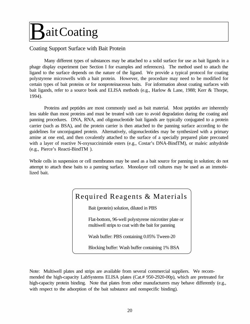
Coating Support Surface with Bait Protein
Many different types of substances may be attached to a solid surface for use as bait ligands in aphage display experiment (see Section I for examples and references). The method used to attach theligand to the surface depends on the nature of the ligand. We provide a typical protocol for coatingpolystyrene microwells with a bait protein. However, the procedure may need to be modified forcertain types of bait proteins or for nonproteinaceous baits. For information about coating surfaces withbait ligands, refer to a source book and ELISA methods (e.g., Harlow & Lane, 1988; Kerr & Thorpe,1994).
Proteins and peptides are most commonly used as bait material. Most peptides are inherentlyless stable than most proteins and must be treated with care to avoid degradation during the coating andpanning procedures. DNA, RNA, and oligonucleotide bait ligands are typically conjugated to a proteincarrier (such as BSA), and the protein carrier is then attached to the panning surface according to theguidelines for unconjugated protein. Alternatively, oligonucleotides may be synthesized with a primaryamine at one end, and then covalently attached to the surface of a specially prepared plate precoatedwith a layer of reactive N-oxysuccinimide esters (e.g., Costar’s DNA-BindTM), or maleic anhydride(e.g., Pierce’s Reacti-BindTM ).
Whole cells in suspension or cell membranes may be used as a bait source for panning in solution; do notattempt to attach these baits to a panning surface. Monolayer cell cultures may be used as an immobi-lized bait.
Bait (protein) solution, diluted in PBS
Flat-bottom, 96-well polystyrene microtiter plate ormultiwell strips to coat with the bait for panning
Wash buffer: PBS containing 0.05% Tween-20
Blocking buffer: Wash buffer containing 1% BSA
Note: Multiwell plates and strips are available from several commercial suppliers. We recom-mended the high-capacity LabSystems ELISA plates (Cat.# 950-2920-00p), which are pretreated forhigh-capacity protein binding. Note that plates from other manufacturers may behave differently (e.g.,with respect to the adsorption of the bait substance and nonspecific binding).

21
Panning & Screeening Preparation
Procedure:
1. If the bait protein is more concentrated than 10 ug/ml, dilute it to this concentration in 1X PBS.
2. Coat each well with 50-200 µl of solution (equivalent to 0.1-1µg of protein).
3. Incubate at room temperature for 2 hr, or at 4ºC overnight (16hr).Note: Use a 4ººC Incubation if the bait protein is unstable at room temperature.
4. Discard or save the coating solution and wash the wells three times with wash buffer.
5. Add 200 µl of blocking buffer to each well.
6. Incubate at room temperature for 2 hr, or overnight (16hr).Note: Use a 4ººC Incubation if the bait protein is unstable at room temperature.
7. Discard blocking buffer and wash the wells three times with wash buffer.
8. Dry the plate and wrap it in plastic wrap. Store at 4ºC for up to 1 month.Note: Most peptides and some bait proteins are not stable enough to store and should be used
immediately.

22
Please review the following guidelines before starting this procedure.
Guidelines:
1. For best results, repeat the panning procedure at least three times on each library aliquot.ELISA screening is generally not recommended before the third round of panning because thepercentage of positives in the enriched population will still be too low (<1%) in most cases.
2. For each subsequent panning, use a fresh bait-coated well; a well may not be reusedsince the phage coats remain attached to the bait in this procedure.Note: “spent” library aliquots may be saved after panning for use in future studies. However,due to possible loss of clones that cross-react with different baits, it is not recommended toreuse the spent library to pan against different bait.
3. In this protocol, the bound phage is gently and efficiently rescued by simply allowing them toinfect TG1 host cells. However, if you prefer, the bound phage can also be eluted using bufferswith decreasing pH, as described in Wrington et al. (1996). Be aware that extremes of pH(pH2) can adversely effect phage viability.
4. After each panning cycle, you must amplify and superinfect a portion of the infected, enrichedTG1 cells if you plan to perform a subsequent panning.
5. After the third panning, you have the option of screening individual clones for positives byELISA using the Anti-pVIII polyclonal antibody provided in the EZnet Library Screening Kit. Alternatively, you may wish to directly repeat the amplification/panning/plating processfor one or two more cycles before you perform the ELISA screening. The extra cycles willrequire 2-3 more days, but may save time overall if it turns out that additional panning cycles arerequired to identify positives.
6. If >30% of the clones are positive after three rounds of panning, many of them may be duplicated of the same insert; bias may be introduced into the population due to differences ininfectivity, relative growth rates, or both. In this case, you may find a wider variety of clonesafter fewer rounds of enrichment. For this reason, it is prudent to store the enriched library at -20ºC after each panning. If necessary, you can use them as a source of individual clones for
` ELISA screening.
Controls:
1. After each panning, plate out a diluted sample of the enriched TG1 library for single colonies onappropriate selection medium and count the resulting colonies . This will indicate whether youhave successfully recovered phagemid DNA in the host cells. In addition, the number ofphage trapped on the plate and recovered, assuming one phage will infect one host cell.
Panning,Rescue&Superinfection
23
Controls (cont):
[Recommended]Titer the enriched phage library. Although you need not wait for this information beforeproceeding to the next panning cycle, it will be necessary if you wish to calculate thepercent of phage trapped in the panning .
[Optional]To estimate the percent of enriched library clones that have been selected because of nonspecificbinding to the surface, pan a 10µl aliquot of the enriched library against a well precoated with BSA (bovineserum albumin; Sigma, Fraction V).
EZnet Phage Display Library
The following materials are included in the EZnet LibraryScreening kit and with EZnet Phage Display libraries:
M13K07 helper phage
TG1 cells (prechilled, log phage cells)
Microtiter plate wells precoated with the desired bait material
PBS (Appendix)
Wash buffer: PBS containing 0.05% Tween-20
Blocking buffer: wash buffer containing 1% BSA.
2X YT/amp/kan broth (Appendix )
2X YT/glucose broth (Appendix )
Ampicillin stock solution (50 mg/ml)
Sterile 20% PEG 8000 solution
For estimating recovery of trapped phage: 2X YT broth and2X YT/amp/glucose/MgCl2 agar plates
For titering the purified, enriched phage library: 2X YT/amp platesor long-term storage of enriched TG1 library and enriched purifiedphage library: Sterile glycerol solution (at least 50% v/v)
Panning,Rescue&Superinfection
R e q u i r e d R e a g e n t s & M a t e r i a l s
•
•
•
•
•
•
•
•
•
•
•
•
•
•
24
Panning:
1. Add library aliquot to the bait-coated well.
2. First panning cycle: use an aliquot of the original library. Usually, panning 109 libraryclones against one coated well (1 cm2) is sufficient to identify a cDNA for a rare ormoderately abundant transcript. For example, for an EZnet Phage Display Library with atiter of 1010 cfu/ml, dilute 10 µl of the library in 90 µl of blocking buffer and apply this to thewell.Subsequent rounds of panning: use upto 50 µl of the purified, enriched phage library from
the previous panning.
3. Incubate the library on the coated surface at 37ºC for 1hr. alternatively, for less stableproteins, incubate at room temperature or 4ºC for up to 4 hr.
4. Remove the spent library and save it if you wish. Wash the coated surface at least 5 timeswith wash buffer (~200 µl each wash).Note: Use more washes (up to 20) for higher-stringency binding conditions.
Rescuing Trapped Phage:
1. Add 0.1 ml of prechilled, log-phase TG1 cells to the panning well
2. Incubate at 37ºC for 30 min. Immediately place the plate on ice or at 4ºC.Note: Longer incubation at 37ºCwill allow the infected TG1 cells to duplicate and will make it moredifficult to use cell number to estimate percent of phage rescued .
If you have just completed the first round of panning, this is the 1stround enriched TG1 library. If you have just completed the secondround of panning, this is the 2nd round enriched TG1 library, etc.
3. Dilute a small aliquot of the enriched TG1 library and plate it out to determine the titer andestimate recovery of trapped phage.(Recommended)
4. Transfer the enriched TG1 library to fresh microcentrifuge tubes. Store at 4ºC for up to 3days, or add sterile glycerol to a final concentration of 15%, and store at -70ºC for up to1 yr.
Helpful Tip: If you plan to repeat the panning within days, keep 50 µl of the enriched TG1library at 4ºC and store the rest at -70ºC.
Panning,Rescue&Superinfection

25
If you just finished your 1st or 2ndround of panning, repeat the panning process.If you just finshed your 3rd round of panning, proceed to the ELISA screening.
To estimate recovery of trapped phage:
1. Prepare 10-1, 10-2, and 10-3 dilutions of the enriched TG1 library from Step E.4 above in2X YT medium.
2. Plate 100 µl of each cell dilution onto a 2X YT/amp/glucose/MgCl2 plate. Invert the platesand incubate at 37ºC overnight.
3. Count colonies, calculate the total number of cells in the enriched TG1 library, and estimatepercent of phage recovered in panning.
cfu/ml = (#colonies on plate) x dilution factor
Ml
Total # cfu = cfu/ml x total vol. of enriched TG1 library
Percent of phage recovered = total # cfu
# cfu* applied to panning surface
*Note that the library titer is expressed in cfu/ml (see Section III.B).
Expected recovery rates
Because the TG1 cells have time, at most , to undergo one generation before they are placed at 4ºC (StepE.2 above), the total number of cells in the enriched TG1 library is a rough estimate of the number ofphage rescued.
Panning,Rescue&Superinfection

26
Panning,Rescue&SuperinfectionAfter only one or two panning rounds, the percentage of phage trapped is expected to be very low((1%) for most rare-to-moderately abundant transcripts. The percent recovery is an indication of thestringency of selection. Thus, if your recovery is >1%, the panning conditions may not be stringentenough for your particular bait (see Troubleshooting Guide, Section VIII).
Repeat Panning:
Transfer 50 µµl of the enriched TG1 library to a sterile, 17 X 100-mm tube. Store the remainder at -70ººC.
1. Add 1 ml of 2X YT/glucose to the 50 µl aliquot of enriched TG1 library.
2. Incubate at 37ºC for 1 hr with shaking at 250 rpm.
3. Add ampicillin to a final concentration of 100 ug/ml (e.g., 2 µl of a 50 mg/ml amp stock).Immediately add 5 x 10⁹ pfu of M13K07 (~0.1 ml of fresh phage stock) to the cell culture(superinfection step).Note: The multiplicity of infection (MOI) should be about 20:1 (phage:cells).
4. Incubate at 37ºC for 1 hr with shaking at 250 rpm.
5. Transfer culture to a centrifuge tube. Centrifuge at 2,000 x g for 10 min at 4ºC or roomtemperature. Remove and discard the supernatant.Note: Be sure to remove all the supernatant. Traces of glucose will repress theexpression and hence, display, of fusion proteins.
6. Resuspend the cell pellet in 10 ml of 2X YT/amp/kan.
7. Incubate at 37ºC overnight with shaking at 250 rpm.
8. Centrifuge culture at 10,000 x g for 20 min at 4ºC. Transfer the supernatant (whichcontains the enriched phage library) to a fresh tube.
9. Recentrifuge the supernatant at 10,000 x g for 10 min to further clarify it. Transfer thesupernatant to a sterile polypropylene tube: this is the enriched library in a crude phagepreparation.Note: If necessary, the enriched crude phage library may be store at 4ºC for up to 1month. However, this is not recommended because of potential reduction of titer.
27
10. Purify the enriched phage library as follows:
Note: Because the suppression of the amber stop codon is only about 80% efficient in TG1 (Sambrook etal., 1989), some cDNA-coding proteins with only signal sequences will be expressed and secreted tothe medium. If not removed from the phage preparation, these soluble proteins will compete forbinding sites in thee bait-coated wells and interfere with the next panning.
a. Add 10ml of blocking solution to 10ml of the enriched (crude) phage library. (Use an equalvolume of blocking solution and phage library.)
b. Incubate at 4ºC for 10 min.
c. Add 4 ml of 20% sterile PEG solution. Incubate at 4ºC for 30-60 min to precipitatethe phage.
d. Centrifuge at 10,000 x g for 20 min at 4ºC. Discard the supernatant and resuspend thepellet in 1 ml of PBS.
e. Transfer the resuspended pellet to a sterile microcentrifuge tube and centrifuge at 14,000rpm (top speed) for 10 min at 4ºC. This time, save the supernatant: this is the enriched(purified) phage library.
11. Titer the enriched phage library on 2X YT/amp plates (Section III.B). Titering will provideimportant information, as described in Section IV. B.
12. The enriched (purified) phage library may be stored at 4ºC for up to 1 month. If youplan to keep the enriched (purified) phage library for >1 month, add sterile glycerol toa final concentration of 20%, and store it at -70ºC for up to 1 yr.Note: You will need 50 µl of the enriched (purified) phage library for the next round of
panning.
If you have performed only one or two rounds ofpanning,then return to the panning procedure.
Panning,Rescue&Superinfection

28
• M13K07 helper phage
• Control protein and phage clones
• Anti-pVIII polyclonal antibody (affinity purified)
• HRP-conjugated secondary antibody
• HRP substrates A & B
• Enriched phage display library in TG1
• ELISA plates precoated with the desired bait
• Notes:If you have purchased the EZnetExpression and
Detection Kit, use the 96-well microtiter plates provided.
• 2X YT/amp/glucose/MgCl2 agar plates (Appendix )
• 2X YT/amp/glucose broth (Appendix )
• 2X YT/amp/kan broth (Appendix )
• Ample supply of 1.5-ml microcentrifuge tubes or deep-well
microtiter array tubes and holder
• Sterile glycerol solution (at least 50%)
• Sterile 1X PBS (Appendix A)
• Wash buffer: PBS containing 0.05% Tween-20
• Blocking buffer: Wash buffer containing 1% BSA.
• Sterile 20% PEG 8000 solution
Optional: for quantitation of results1 N H2SO4 and an ELISA plate reader, such as the ThermoMax Plate Reader from Molecular Devices.
ELISA
R e q u i r e d R e a g e n t s & M a t e r i a l s
29
Preparation of Candidate Phage Clones for ELISA:
Dilute an aliquot of the enriched TG1 library after the third panning and plateout 100 µl of each dilution on 2X YT/amp/glucose/MgCl2 for single colonies.Incubate at 37ºC overnight.
1. Add 400 µl of 2X YT/amp/glucose medium to prelabeled 1.5-ml microcentrifuge tubes orseparate tubes of a 96-deep-well microtiter array. Label these tubes “PC1” + a number todesignate the individual clone.
2. Using sterile pipette tips, pick 44-88 well-isolated, single colonies from the plates aboveand use each to separately inoculate the medium in the small tubes.
3. Incubate tubes at 37ºC overnight shaking at 250 rpm. These are the master stocks of theindividual phagemid clones in TG1; we refer to these culture as “PC1”
4. Transfer 50 µl of each overnight PC1 culture to a fresh tube prelabeled “PC2” + thenumber designating the individual clone. To the remaining 350 (l of PC1 culture, add sterileglycerol (final concentration 15%) and store at -70ºC. Save all PC1 cultures until you haveobtained the ELISA results.
5. Add 400 µl of 2X YT/amp/glucose medium containing 5 x 10⁸ pfu of M13K07 (~10 µl offresh phage stock to each tube labeled PC2.
6. Incubate tubes at 37ºCfor 1-2 hr with shaking at 200-250 rpm.
7. Centrifuge samples at room temperature:
8. Centrifuge 1.5-ml microcentrifuge tubes at 14,000 rpm for 5 min.
9. For a 96-deep-well microtiter array, centrifuge at 2,000 x g for 10 min using a centrifugeadapted for multi-well plates.
10. Discard the supernatants. Resuspend each cell pellet in 400 µl of 2X YT/amp/kan.Note: Be sure to remove all the supernatant. Traces of glucose will repress the expressionand, hence, display of fusion proteins.
11. Incubate the wells or tubes at 37ºC overnight with shaking at 250 rpm. These are thecrude supernatants of the superinfected individual phagemid clones; we refer to thesecultures as “PC2”.
ELISA
30
12. Purify phage clones
13. Centrifuge PC2 cultures as in Step 8 above.
14. Transfer 300 µl of each supernatant (which contains the phage) to fresh tubes prelabeled“PC3” + the number designating the individual clone.
15. Add 300 µl of blocking buffer.
16. Incubate at 4ºC for 10 min
17. Add 120 µl of sterile 20 PEG solution to each tube.
18. Incubate at 4ºC for 30 min.
19. Centrifuge at 10,000 x g for 20 min at 4ºC.
20. Discard the supernatant and resuspend the phage pellet in 100 µl of PBS. These are theindividual purified phage clones; we refer to these preparations as “PC3” PC3 phage prepsmay be stored at 4ºC for 1 month. For storage > month, add glycerol to 20% final concentration and place at -70ºC.
Screening:
1. Apply 50 µl of each PC3 phage prep to a well precoated with your bait protein.We strongly recommend to apply 20-50 µl of each positive control phage clone to a wellin a positive trip precoated with control bait protein (provided in kit) at same time.
2. Incubate at room temperature for 2 hr.Note: Use a 4ºC incubation overnight if bait is unstable.
3. Remove the phage solution and wash the wells three times with wash buffer (~200 µl eachwash).
4. Dilute the Anti-pVIII polyclonal antibody to 2 µg/ml in blocking buffer. Add 100 µl ofdiluted anti-pVIII antibody to each well of the plate.Note: The anti-pVIII antibody can be used at a higher concentration (up to 10 µg/ml) ifnecessary, to boost a weak signal.
ELISA

31
ELISA
Screening (cont):
5. Incubate the plate at room temperature for 1 hr. Remove the primary antibody anddiscard it.
6. Wash the plate three times with wash buffer (~200 µl each wash).
7. Dilute the HRP-conjugated secondary antibody 1:50 in blocking solutionNote: Do not use sodium azide in the blocking solution; it inhibits HRP activity.
8. Add 100 µl of the diluted HRP conjugate to each well.
9. Incubate the plate at room temperature for 1 hr. Wash the plate three times with wash buffer(~200 µl each wash).
10. Prepare the HRP substrate solution: mix 1 part of Solution A and 1 part Solution B. Add100 µl of the substrate solution into each well.
11. Incubate the plate at room temperature until a suitable blue color has developed (i.e., whenthe color intensity in the positive control wells appears to be approx. 2X background. Forthe positive control clone provided in the kit, color development should take 30 min. Forlibrary clones, color development can take from 30 min. to 12 hr, depending on the relativeabundance of the transcript and on the affinity of the displayed protein for the bait. At thispoint, take a photograph if desired and make a note of the color intensity in the positive wellsrelative to the positive controls. Proceed to Step 12 if you wish to obtain quantitative results;otherwise, proceed to Step 13.
12. Stop the color reaction in all the wells at the same time by adding 10 µl of 1 N H2SO4 toeach well. (Blue wells will turn yellow.) Read the absorbance at 405 or 410 nm (preferably) on a plate-reader.
13. On the basis of the ELISA results, decide which of the PC1 and PC3 clones you wish tokeep and discard the rest. Keep all of the positive clones-and a few of the negative clonesfor later use as controls.
32
No Positive Library Clones Obtained After Panning & Screening
If not positive clones are obtained in the ELISA screening of the enriched library, it could be due toproblems with the panning procedure, the ELISA, or both. Problems with the ELISA are generallyeasier to pinpoint, especially if you are using the control materials provided with the EZnet PhageDisplay Library Screening Kit . This is contains control protein and positive and negative controlphage clones to help you ascertain if the ELISA screening is working properly. In addition, the anti-pVIII antibody produced with the EZnet Library Screening Kit has been affinity purified and pretestedfor performance in the ELISA assays.
Elisa Tips
If you have not done so already, be sure to perform ELISA assays on at least 88 clones. Re-member, the more clones you screen, the greater your chances of finding displayed proteins encodedby rare transcripts.
Try using a higher concentration of the pVIII polyclonal antibody and/or purified phage clone in theELISA assays . The purified anit-pVIII antibody provided with the EZnet Library Screening Kit maybe used within a range of 1-10 µg/ml; the higher end of the range may be more suitable for displayedproteins that have a relatively weak affinity for the bait.
The wells may not be optimally coated with the bait protein. Check the concentration of the proteinsolution used to coat the wells; it should be approximately 10mg/ml. If available, use can antibody specificfor the bait to confirm that a sample well is evenly coated with bait.
Panning Tips
If you have not already done so, be sure to check the titer of the library used in the panning. If the titer islower tan expected, you may be starting the panning experiment with a suboptimal number of library clones.Use a larger volume of library if necessary to provide at least 109 clones.
If you have not done so already, perform at least three rounds of panning before you prepare individualclones for ELISA screening. If you have performed three rounds, perform one or two more rounds andrescreen individual clones.
The wells may not be optimally coated with bait protein .Check the viability of the TG1 culture used in the phage rescue. Check for the presence of
the F’ episome by plating on M/9+Thi minimal medium. Remember that the host cells must contain an F’episome to be efficiently infected by M13, and they can lose the episome if they are grown to saturationunder nonselective conditions.
Troubleshooting

33
If you have not done so already, check the titer of the M13K07 helper phage; if the titer is < 109 pfu/ml,prepare a fresh stock of helper phage .
If you have no don so already, check the percent phage recovery . Checking the percent page recoverywill also tell you the titer of thee enriched library.
If phage recovery was poor (i.e., <0.001%), you may not have used the optimal number of phage (i.e.,~109 particles) in the subsequent panning. Use more of the enriched library when you repeat the panning.Alternatively, decrease the stringency of the washing conditions . For example, if you used more thanfive washes, reduce the number to five. (However, do not use fewer than five washes, or else you mayhave problems with nonspecific binding.) You may want to try changing the formulation of the washingbuffer to decrease its stringency; please refer to a laboratory manual on immunoassays for suggestions.
If phage recovery was very high (i.e., >1%), but the number of positive clones still very low, increasethe number of washes (up to a maximum of 20). You may want to try changing the formulation of thewashing buffer to increase its stringency. However, be aware that most measures that will increasestringency (such as using a higher detergent concentration or more extreme pH) will have a deleteriouseffect on phage viability. Therefore, if you change the formulation of the washing buffer, be sure to testthe viability of the phage in the new buffer before you use it in a panning experiment.
If phage recovery was >0.001%, but <1%, use more panning rounds (at least five total).
For rare transcripts, you can use more library clones and a correspondingly larger panning surface.
Many of the Positive Clones Do Not Bind Bait Specifically
Upon further analysis, if clones do not bind specifically to the bait, it could be due to nonspecificinteractions with the wells, or the specific interactions with other components in the bait preparation.
To reduce nonspecific binding to wells, check the percent phage recovery . if recovery is >1%, increasethe stringency of the wash conditions . If recovery is <1%, use more panning rounds (at least five total).Check your blocking agent, such as 1% nonfat dry milk instead of BSA, or try adding both BSA andnonfat dry milk to the buffer. (Note that nonfat dry milk contains biotin, which will interfere with biotin-based detection systems.)
It may be possible to prevent phage particles from binding to non-bait components in the panning pro-cess. However, use of proper controls in the initial analysis of candidate positives will eliminate thenon-bait binding clones from further analysis .
Troubleshooting

34
Background too High in the ELISA Assay
Try a different blocking buffer
Use a higher dilution (i.e., less concentrated) solution of the secondary antibody conjugate.
Do not let the HRP color reaction go longer than 20 min; in most cases, 10 min should be sufficient.
Cannot Find Overexpressed Protein
If you have not done so already, look for the overexpressed protein in the medium, the periplasmic extract,and a whole-cell extract .
If you have not done so already, optimize the induction conditions by taking several time points over aperiod of at least 20 hr. if the overexpressed protein is particularly unstable or insoluble, it may help tolower the incubation temperature. Temperature as low as 4(C may be used, but you may need a corre-spondingly longer induction period (at least 20 hr).
Troubleshooting

35
Allen, J.B., Walberg, M.W., Edwards, M. C. & Elledge, S. J. (1995) Finding prospective partners in the library:the two-hybrid system and phage display find a match. TIBS 20:511-516.
Anderson, S.K., Gibbs, C.P., Tanaka, A., Kung, H.-J. & Fujita, D.J. (1989) Human Cellular src gene: nucle-otide sequence and derived amino acid sequence of the region coding for the carboxy-terminal two-thirds ofpp60c-src. Mol. Cell. Biol. 5:1122-1129.
Ausubel, F.M., Brent, R., Kingston, r.E., Moore, D.D., Seidman, J. G., Smith, J. A. & Struhl, K.(1994) InCurrent Protocols in Molecular biology (John Wiley &Sons, Inc.)
Bass, S.H., Greene, R. & Wells, J.A. (1990) Hormone phage: An enrichment method for variant proteins withaltered binding properties. Proteins 8:309-314.
Cesareni, G. (1992) Peptide display on filamentous phage capsids: A new powerful tool to study protein-ligandinteraction. FEBS 307: 66-70.
Chuang, S.-E., Chen, A.-I., & Chao, C.-C. (1995) Growth of E. coli at low temperature dramatically increasesthe transformation frequency by electroporation. Nucleic Acids Res. 23:1641.
Clackson, T., Hoogenboom, H.R., Griffiths, A.D. & Winter, G. (1991) Making antibody fragments using phagedisplay libraries. Nature 352:624-628.
Corey, D.R., Shiau, A.K., Yang, Q., Janowski, B.A. & Craik, C.S. (1993) Trypsin display on the surfce ofbacteriophage, Gene 128:129-134.
Crameri, R., Jaussi, R., Menz, G. & Blaser, K. (1994) Display of expression products of cDNA libraries onphage surfaces-a versatile screening system for selective isolation of genes by specific gene-product/ligandinteraction. Eur. J. Biochem. 226:53-58.
Crameri, R., Faith, A. Hemmann, S., Jaussi, R., Ismail, C., Menz, G. & Blaser, K. (1996) Humoral and cell-mediated autoimmunity in allergy to Aspergillus fumigatus. J. Exp. Med. 184:265-270.
Crameri, R. & Suter, M. (1993) Display of biologically active proteins on the surface of filamentous phages: AcDNA cloning system for selection of functional gene products linked to the genetic information responsible fortheir production. Gene 137:69-75.
Cwirla, S.E., Peters, E.A., Barrett, R.W. & Dower, W.J. (1990) Peptides of phage: a vast library of peptides foridentifying ligands. Proc. Natl. Acad. Sci. U.S.A. 87:6378-6382.
Devlin, J.J., Pnaganiban, L.C. & Devlin, P.E. (1990) Random peptide libraries: A source of specific protein-binding molecules. Science 249:404-406.
Dower, W.J., Miller, J.F. & Ragsdale, W.W. (1988) High-efficiency transformation of E. coli by high-voltageelectroporation. Nucleic Acids Res. 16:6127-6145.
References

36
Dunn, I.S. (1996_ Phage display of proteins. Curr. Op. Biotechnology 7:547-553.
EasyMATCHTM Phage Display Kits (January 1997) CLONTECHniques XII(1):7-11.
Gram, H., Strittmatter, U., Lorenz, M., Gluck, D. & Zenke, G. (1993) Phage display as a rapid gene expressionsystem: Production of bioactive cytokine-phage and generation of neutralizing monoclonal antibodies. J.Immunol. Methods 16:169-176.
Gubler, U. & Hoffman, B.J. (1983) A simple and very efficient method for generating cDNA libraries. Gene25:263-269.
Griffiths, A.D., Williams, S.C., Hartley, O., tomlinson, I.M., Waterhouse, P., Crosby, W.L., Kontermann, r.E.,Jones, P.T., Low. N. M., Allison, T.J., Prospero, T.D., Hoogenboom, H.R., Nissim, A., Cox, J.P. L., Harrison,J.L., Zaccolo, M., Gheradi, E., & Winter, G. (1994) Isolation of high-affinity human antibodies directly fromlarge synthetic repertoires. EMBO J 13:3245-3260.
Harlow, E. & Jane, E. (1988) Antibodies: A Laboratory Manual (Cold Spring Harbor Press, Cold Spring Harbor,NY), Chapter 14.
Hoogenboom, H.R., Griffiths, A.D., Johnson, D.S., Chiswell, D.J., Hudson, P. & Winter, G. (1991) Multi-subunit proteins on the surface of filamentous phage: methodologies for displaying antibody (Fab) heavy andlight chains. Nucleic Acids Res. 19:41333-4137.
Jacobsson, D. & Frykberg, L. (1995) Cloning of ligand-binding domains of bacterial receptors by phage display.Biotechniques 18:878-885.
Kang, A.S., Barbas, C.F., Janda, K.D. Benkovic, S.J. & Lerner, R.A. (1991) Linkage of recognition andreplication functions by assembling combinatorial antibody Fab libraries along phage surfaces. Proc. Natl. Acad.Sci. U.S.A. 88:43633-4366.
Kay, B., Winter, J. & McCafferty, J. (eds) (1996) Phage Display of Peptides and Proteins: A LaboratoryManual (Academic Press, San Diego, CA).
Kerr, M.A. & Thorpe, R., eds. (1994) Immunochemistry LabFax, Bios Scientific Publishers Academic Press.
McCafferty, J., Griffiths, A.D., Winter, G. & Chiswell, D.J. (1990) Phage antibodies: Filamentous phagedisplaying antibody variable domains. Nature 348:552-554.
McCafferty, J., Jackson, r.H. & chiswell, D. J. (1991) Phage-ezymes: Expression and affinity chromatography of functional alkaline phsphatase on the surface of bacteriophage. Protein Eng. 4:955-961.
Marks, J. D., Hoogenboom, H.R., Griffiths, A.D. & Winter, G. (1992) Molecular evolution of proteins onfilamentous phage. J. Biol. Chem. 267:16007-160010.
References

37
Nissim, A., Hoogenboom, H.R., Tomlinson, I.M., Flynn, G., Midgley, C., Lan, D. & Winter, G. (1994) Anti-body fragments from a ‘single pot’ phage display library as immunochemical reagents. EMBO J. 13:692-698.
Oldenburg, D., Loganathan, D., Goldstein, I., Schultz, P. & Gallop, M. (1992) Peptide ligands for a supar-binding protein isolated from a random peptide library. Proc. Natl. Acad. Sci. U.S.A. 89:5393-5397.
O’Neil, K.T., DeGrado, W.F., Mousa, S.A., Ramachandran, N & Hoess, R.H. (1994) Identification of recogni-tion sequences of adhesion molecules using phage display technology. Methods Enzymol. 2455:370-386.
Parker, R.C., Mardon, G., Lebo, R.V., Varmus, H.E. & Bishop, J.M. (1985) Isolation of duplicated human c-src genes located on chromosomes 1 and 20. Mol. Cell, Biol. 5:831-838.
Parmley, S.F. & Smith, G.P. (1988) Antibody-selectable filamentous fd phage vector: Affinity purification oftarget genes. Gene 73:305-318.
Sambrook, J., Fritsch, E.F. & Maniatis, T. (1989) Molecular Cloning: A Laboratory Manual (Cold SpringHarbor Laboratory, Cold Spring Harbor, NY).
Scott, J.K. & Smith, G. P. (1990) Searching for peptide ligands with an epitope library. Science 249:386-390.
Scott, J.K., Loganathan, D., Easley, R., Gong, X. & Goldstein, I. (1992) A family of concanavalin A-bindingpeptides from a gexapeptide epitope library. Proc. Natl. Acad. Sci. U.S.A. 89:5398-5402.
Smith, G.P. (1985) Filamentous fusion phage: Novel expression vectors that display cloned antigens on thesurface of the virion. Science 228:1315-1317.
Smith, G. D. & Scott, J. K. (1993) Libraries of peptides and proteins displayed on filamentous phage. InMethods in Enzymol. (R.Wu., ed.), Vol. 217, pp. 228-257. Academic Press, San Diego, CA.
Tanaka, A., Gibbs, C.P., Arthur, R. R., Anderson, S.K., Kung, H.-J. & Fujita, D.J. (1987) DNA Sequenceencoding the amino-terminal region of the human c-src protein: implications of sequence divergence amoung src-type kinase oncogenes. Mo. Cell. Biol. 7:1978-1983.
Winter, G., Griffiths, A.D., Hawkins, R.E. & Hoogenboom, H.R. (1994) Making antibodies by phage displaytechnology, Annu. Rev. Immunol. 12:433-
Wrington, N.C. Farrell, F.X., Chang, R., Kashyap, A.K., Barbone, F.P., Mulcahy, L.S., Johnson, D.L., Barrett,R.W., Jolliffe, L.K. & Dower, W.J. (1996) Small peptides as potent mimetics of the protein hormone erythropoi-etin, Science 273:458-463.
References
38
pHage 3.2
4.505 kb
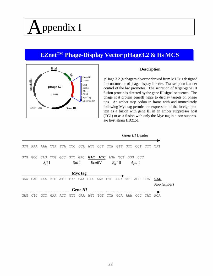
Description
pHage 3.2 (a phagemid vector derived from M13) is designedfor construction of phage-display libraries. Transcription is undercontrol of the lac promoter. The secretion of target-gene IIIfusion protein is directed by the gene III signal sequence. Thephage coat protein geneIII helps to display targets on phagetips. An amber stop codon in frame with and immediatelyfollowing Myc-tag permits the expression of the foreign pro-tein as a fusion with gene III in an amber suppressor host(TG1) or as a fusion with only the Myc-tag in a non-suppres-sor host strain HB2151.
EZnetTM Phage-Display Vector pHage3.2 & Its MCS
ColE1 ori
Am
pici
llin
Gene III
lac
fi ori
Gene IIILeader
Sal IEcoRVBgl IIApa I
myc-Tagamber codon
Gene III Leader
GTG AAA AAA TTA TTA TTC GCA ATT CCT TTA GTT GTT CCT TTC TAT
GCG GCC CAG CCG GCC GTC GAC GAT ATC AGA TCT GGG CCCSfi I Sal I EcoRV Bgl II Apa I
Myc tagGAA CAG AAA CTG ATC TCT GAA GAA AAC CTG AAC GGT ACC GCA TAG
Stop (amber)Gene III
GAG CTC GCT GAA ACT GTT GAA AGT TGT TTA GCA AAA CCC CAT ACA
Appendix I

39
Appendix II
Molecular Cloning Manual by Sambrook et al. contains useful information on how to grow TG1 cells andprepare competent cells properly. Following is a procedure we recommend:
Preparation of Competent Cells
1. Grow E.coli host TG1 cells in LB Broth medium (500ml- 1litre) with single colony from a minimal plate.Incubate at 37oC overnight with shaking at 250 rpm.
2. Dilute the overnight culture 1:50 in LB-Broth medium. Incubate at 37oC with shaking at 250 rpm untila A600 of 0.4-0.6 is reached (approximately 2-2.5 hours).
3. Chill the cells on ice for 10 minutes. Spin at 4,000 x g for 15 minutes at 4oC. Discard the supernatant andresuspend the cell pellet in 1 litre of ice-cold sterile 1mM Hepes (pH 7.0). (Wash Step 1)
4. Spin as described above, Discard the supernatant and resuspend the cell pellet in 500 ml of ice-coldsterile 1mM Hepes (pH 7.0). (Wash Step 2)
5. Spin as described above, Discard the supernatant and resuspend the cell pellet in 500 ml of ice-coldsterile 1mM Hepes (pH 7.0). (Wash Step 3)
6. Spin as described above, Discard the supernatant and resuspend the cell pellet in 20 ml of sterile 10%glycerol in water. (Wash Step 4)
7. Spin as described above, Discard the supernatant and resuspend the cell pellet in a total volume of 2-3 ml10% glycerol in water. Dispense in 50-100 ul aliquots, freeze on dry ice and store at -70oC until use.