eyeblink conditioning during an interstimulus interval switch in … · 2018-12-07 · initially...
TRANSCRIPT

Eyeblink Conditioning During an Interstimulus Interval Switch in Rabbits(Oryctolagus cuniculus) Using Picrotoxin to Disrupt Cerebellar Cortical
Input to the Interpositus Nucleus
Richard W. VogelIndiana University and University of Kansas
Jeffrey C. Amundson, Derick H. Lindquist,and Joseph E. Steinmetz
University of Kansas
The role of the cerebellar cortex in eyeblink classical conditioning remains unclear. Experimentalmanipulations that disrupt the normal function impair learning to various degrees, and task parametersmay be important factors in determining the severity of impairment. This study examined the role ofcerebellar cortex in eyeblink conditioning under conditioned stimulus–unconditioned stimulus intervalsknown to be optimal or nonoptimal for learning. Using infusions of picrotoxin to the interpositus nucleusof the rabbit cerebellum, the authors pharmacologically disrupted input from the cerebellar cortex whiletraining with an interstimulus interval (ISI)-switch procedure. One group of rabbits (Oryctolaguscuniculus) was 1st trained with a 250-ms ISI (optimal) and then switched to a 750-ms ISI (nonoptimal).A 2nd group was trained in the opposite order. The most striking effect was that picrotoxin-treated rabbitsinitially trained with a 250-ms ISI learned comparably to controls, but those initially trained with a750-ms ISI were severely impaired. These results suggest that functional input from cerebellar cortexbecomes increasingly important for the interpositus nucleus to learn delay eyeblink conditioning as theISI departs from an optimal interval.
Keywords: cerebellum, eyeblink classical conditioning, interstimulus interval, GABA, picrotoxin
Classical conditioning has proven to be an important tool forstudying brain–behavior relationships, and significant progresshas been made in the endeavor to understand the neural mecha-nisms that govern learning and memory. Indeed, arguably weknow more about the structures and functions that underlie basicassociative memory than any other form of experience-dependentbehavior. The eyeblink classical conditioning preparation hasmade critical contributions to our current knowledge base in thisarea.
A single eyeblink conditioning trial usually consists of an au-ditory conditioned stimulus (CS) that is presented to an organismshortly before a blink-eliciting unconditioned stimulus (US), suchas a puff of air directed at the cornea. In the most basic procedure,delay conditioning, the CS and US overlap and coterminate. Ini-tially, naıve subjects produce a reflexive, unconditioned blinkresponse that follows US onset. After many trials, however, or-ganisms associate the stimuli, and a CS-triggered response actu-alizes in the form of a blink that begins before US onset. This is theclassically conditioned eyeblink response (CR). Under optimalconditions, well-trained organisms execute CRs on a high percent-age of trials.
Rabbits are commonly used in eyeblink conditioning studies,and extensive data have been collected to define the stimulus andtiming parameters that promote optimal learning, as defined by thefastest rate of acquisition, the greatest percentage of CRs, and themost extinction-resistant learning. One important factor is the timebetween CS onset and US onset—the interstimulus interval (ISI).Classic studies have demonstrated that learning in delay eyeblinkconditioning is most robust in rabbits when the ISI is 200–500 ms(Coleman & Gormezano, 1971; Gormezano, Kehoe, & Marshall,1983; Schneiderman, 1966; Schneiderman & Gormezano, 1964;Smith, 1968; Smith, Coleman, & Gormezano, 1969). It can thus beargued that an ISI of 200–500 ms is optimal for rabbit eyeblinkconditioning, and longer or shorter ISIs are nonoptimal.
The neural substrates for eyeblink conditioning have been ex-tensively summarized (see Christian & Thompson, 2003). Briefly,CS and US signals converge on cells in two regions of thecerebellum: the cortex and the deep nucleus. At the level of cortex,these signals ultimately converge on Purkinje cells, which in turn
Richard W. Vogel, Program in Neural Science and Department ofPsychological and Brain Sciences, Indiana University, and Department ofPsychology, University of Kansas; Jeffrey C. Amundson and Derick H.Lindquist, Department of Molecular Biosciences, University of Kansas;Joseph E. Steinmetz, Departments of Psychology and Molecular Bio-sciences, University of Kansas.
This research represents a portion of Richard W. Vogel’s dissertationwork and was previously presented in the form of a poster at the annualmeeting of the Pavlovian Society in Austin, Texas, in 2007.
We thank Luke Mahoney, Jordan Pack, Andy Rellihan, Rehaan Shaffie,Sheila Tsau, and Tony Wu for their help in training rabbits and DaleSengelaub (Indiana University) for assistance with histology. This researchwas supported by the National Institute of Mental Health Grant MH074983to Joseph E. Steinmetz) and an National Institute of Neurological Disordersand Stroke Training Grant 5 T32-NS007487 awarded to Richard W. Vogelby the Program in Neural Science at Indiana University.
Correspondence concerning this article should be addressed to Joseph E.Steinmetz, College of Liberal Arts and Sciences, University of Kansas,Strong Hall 200, 1450 Jayhawk Boulevard, Lawrence, KS 66045. E-mail:[email protected]
Behavioral Neuroscience © 2009 American Psychological Association2009, Vol. 123, No. 1, 62–74 0735-7044/09/$12.00 DOI: 10.1037/a0014082
62

inhibit cerebellar output cells located in the deep nucleus. Both thecortex and deep nucleus of the cerebellum have been studiedextensively in an effort to delineate the neural mechanisms thatunderlie learning and memory in eyeblink conditioning. Signifi-cant progress has been made in that the cerebellar interpositusnucleus has been identified as the critical locus for all forms ofeyeblink conditioning, but the function of cerebellar cortex re-mains poorly understood.
Various methodologies have been successfully used in an effortto study the role of cerebellar cortex in eyeblink conditioning. Oneapproach is to interfere with the cortico-nuclear connection, andthis can be done three ways: (a) the cerebellar cortex can beablated or electrolytically lesioned (e.g., Lavond & Steinmetz,1989), (b) Purkinje cells can be selectively destroyed (Chen, Bao,Lockard, Kim, & Thompson, 1996; Nolan & Freeman, 2006), or(c) compounds that alter activity can be infused into discreteregions of the cerebellum (e.g., Bao, Chen, Kim, & Thompson,2002). Although each of these approaches has their advantages anddrawbacks, we chose the last approach for this study because thedrug effects are temporary and reversible.
Many cerebellar pharmacological perturbations take advantageof the fact that Purkinje cells represent the sole output of thecerebellar cortex, and they modulate neurons in the interpositusnucleus through release of �-aminobutyric acid (GABA). Thus,temporary disruption of cerebellar cortico-nuclear function can beaccomplished by infusing the noncompetitive GABA receptorantagonist, picrotoxin, into the interpositus nucleus (Mamounas,Thompson, & Madden, 1987). This procedure produces a rapidand reversible block of GABA-mediated Cl� flux at the GABAA
receptor complex on interpositus cells. Although this manipulationhas been presented as a reversible disconnection of cerebellarcortex (e.g., Garcia & Mauk, 1998), alternative pharmacologicalmanipulations that may represent a more complete disconnectionof cerebellar cortex have been described (Bao et al., 2002). Thus,picrotoxin infusion to the interpositus nucleus may best be char-acterized as a disruption of the cerebellar cortico-nuclear connec-tion rather than a complete decortication of the cerebellum.
To our knowledge, only one study has examined the effects ofpicrotoxin infusions to the interpositus nucleus on acquisition ofeyeblink CRs in naıve animals. Bao et al. (2002) found thatpicrotoxin alone, or in combination with the GABA agonist mus-cimol, prevented acquisition (but not retention) of CRs. The au-thors argue that the primary memory trace for eyeblink condition-ing occurs in the interpositus nucleus and relies on patternedinhibition from the cortex where secondary memory traces existfor shaping CR timing and amplitude. In accordance with this idea,numerous studies have found that picrotoxin infusions to theinterpositus nucleus of previously trained animals can result in theemergence of improperly timed, short-latency eyeblink CRs(Aksenov, Serdyukova, Irwin, & Bracha, 2003; Bao et al., 2002;Garcia & Mauk, 1998; Ohyama & Mauk, 2001; Ohyama, Nores,Medina, Riusech, & Mauk, 2006). These data fit well with lesion(Perrett, Ruiz, & Mauk, 1993) and electrophysiological (Green &Steinmetz, 2005) studies, which suggest that the anterior lobe ofthe cerebellar cortex regulates proper CR timing via preciselytimed inhibition and disinhibition of the interpositus nucleus.
It is of interest to us that cerebellar cortex may contribute socritically to CR timing, and we are interested in determiningwhether there is a differential role for this region in learning the
CR at different ISIs. Recent evidence from aging rabbits(Woodruff-Pak, Seta, Roker, & Lehr, 2007) and developing rats(Brown, Pagani, & Stanton, 2006) suggests this is so. Becausehigher doses of picrotoxin appear to block initial acquisition (Baoet al., 2002), we decided to use a lower dose that has a demon-strated efficacy in unmasking short-latency responses in well-trained rabbits for a period of time that is approximately equal tothe length of our training session (Garcia & Mauk, 1998).
We undertook the present investigation, in part, to determinewhether cerebellar cortex plays a dissociative role in learningeyeblink conditioning when optimal or nonoptimal ISIs are used.To this end, we examined the effects of picrotoxin-induced dis-ruption of the cerebellar cortico-nuclear function on initial acqui-sition with a 250- or a 750-ms ISI. We hypothesized that normalinput from cerebellar cortex to interpositus nucleus may becomemore important for CR learning and timing as task demands areincreased in eyeblink conditioning (e.g., learning a nonoptimalISI). Additionally, we switched the ISI midtraining, making itlonger or shorter, and examined the rabbit’s ability to alter thelatency of the CR. Considering the importance of cerebellar cortexin CR timing, we further hypothesized that adapting the CR to thenew ISI would be problematic in the absence of normal modula-tory input from cerebellar cortex.
Method
Subjects
Subjects were 40 New Zealand white rabbits (Oryctolagus cu-niculus; Myrtle’s Rabbitry, Thompson Station, TN) weighing aminimum of 2.0 kg before use. All subjects were housed instainless steel cages in the Animal Care Unit at the University ofKansas, which is fully accredited by the Association for Assess-ment and Accreditation of Laboratory Animal Care. On arrival,rabbits acclimated to the housing milieu for at least 1 week. Allrabbits were housed individually with 24-hr access to food andwater and a 12-hr light–dark cycle. The research presented herewas approved by the Institutional Animal Care and Use Committeeat the University of Kansas.
Surgery
A guide cannula for drug infusion was chronically implantedinto the left interpositus nucleus of the cerebellum. Surgery wasperformed under aseptic conditions. Anesthesia was induced withxylazine (6–8 mg/kg im) and ketamine (80–120 mg/kg im). Onceanesthetized, puralube opthalmic ointment (Pharmaderm, Mel-leville, NY) was applied to the eyes to prevent irritation, and therabbit’s head was shaved, cleaned, and secured in a standardstereotaxic apparatus. Anesthesia was maintained throughout thesurgical procedure with administration of 30 mg/kg ketamine (im)every 30–45 min.
The skull was exposed with a midline incision, three anchorscrews were inserted into the bone, and the head was positionedsuch that bregma was 1.5 mm higher than lambda in the dorsal-ventral plane. A four-pin socket assembly, which served as aground, was wired to two of the anchor screws and cemented inplace. Next, a hole was drilled over the interpositus nucleus andfilled with bone wax. Using a stereotaxic arm, a 22-gauge, stain-
63EYEBLINK CONDITIONING DURING ISI SWITCH

less steel guide cannula was lowered into the brain (0.7 mmanterior, 5.2 mm left lateral, and 14.0 mm ventral to lambda) andcemented into place. A stainless steel insect pin (00) was theninserted into the cannula to prevent clogging, and an 18-mmdiameter PVC pipe was cemented around the cannula to protect itfrom bending or breaking.
After cannula implantation, a base was formed on the skull withdental cement. A single bolt was cemented into the anterior portionof the base so that an air nozzle could be secured to the head duringthe training phase. The scalp was then sutured around the base, andan antiseptic ointment was applied to the incision site. Rabbitswere carefully monitored until they fully recovered from anesthe-sia. Postoperative treatment included buprenorphine (0.05–0.1mg/kg sc) twice each day for 2 days or meloxicam (0.2–0.4 mg/kgsc) daily for 2 days. One week of recovery was allowed beforetraining was started.
Drug Infusions
Before each of the classical conditioning sessions describedbelow, rabbits were infused with either phosphate-buffered saline(0.9% NaCl, 0.1 M phosphate buffer, 7.2 pH) or picrotoxin(Sigma-Aldrich, St. Louis, MO; 200 �mol/L solution inphosphate-buffered saline). All drug infusions were deliveredthrough the guide cannula with a 10-�l syringe (Hamilton; Reno,NV), which was equipped with a stopper on the needle to ensurethat the blunted tip did not go more than 0.5 mm beyond the tip ofthe guide cannula. The total volume for all infusions was 2.0 �l,injected at a rate of 0.25 �l/15 s. After the infusion was complete,the syringe was left in place for exactly 2 min. Rabbits weretrained two at a time, and the classical conditioning session startedafter both rabbits were infused and placed in separate sound-attenuating chambers. The maximum time between the end of drugdelivery and the beginning of the conditioning session was 7 min.
At the end of the study, all rabbits underwent retention testing inwhich muscimol (Sigma/Aldrich, 6.0 nM) was infused to theinterpositus nucleus just before a single eyeblink conditioningsession. Because muscimol infusions in this region are known toabolish the eyeblink CR (Krupa, Thompson, & Thompson, 1993),the purpose of this test was to ensure that previous infusionsreached the critical region of the interpositus nucleus.
Behavioral Training
Naıve rabbits were first adapted to the Plexiglas restraint appa-ratus and the conditioning chamber for approximately 45 min oneach of the 2 days before initial training in eyeblink classicalconditioning. The conditioning chamber housed a speaker fordelivering the tone CS, an air nozzle and tube assembly fordelivering the US, and wires for recording eyeblink electromyo-gram (EMG) activity. On the 2nd day of adaptation, two stainlesssteel wires were implanted under local anesthetic (lidocaine im)into the Orbicularis oculi muscle above the left eye for recordingblink-related EMG activity.
A classical conditioning session consisted of 84 trials organizedinto seven blocks of 12 trials. Each block consisted of nine pairedCS–US presentations, two CS-only presentations, and one US-only presentation. Each session, therefore, consisted of 63 paired,14 CS-alone, and 7 US-alone trials. The CS consisted of a 1-kHz,
85-dB tone, and the US consisted of a 3-psi puff of air directed atthe cornea. Trials were separated by a 30-s (�5 s) intertrialinterval. Both a long and a short interstimulus interval (ISI) wereincorporated into the design of the experiment. The short ISI (250ms) consisted of a 350-ms CS that overlapped and coterminatedwith a 100-ms airpuff US. The long ISI (750 ms) consisted of an850-ms CS that overlapped and coterminated with a 100-ms air-puff US. CS and US intensities remained constant across allgroups.
Rabbits were randomly assigned to one of two ISI-switchgroups. One group, 2503750 (n � 21), was first trained for 10sessions with a 250-ms ISI, then switched to a 750-ms ISI andtrained for an additional 10 sessions. The second group, 7503250(n � 19), received training in the opposite order. Thus, all groupsreceived 20 days of training. Within each ISI-switch group, rabbitswere further divided into one of two groups, a picrotoxin group ora saline control group.
Data Acquisition and Planned Analyses
Acquisition of eyelid EMG data was accomplished with a Micro1401 unit (Neuralynx, Inc., Bozeman, MT) that was interfacedwith a computer running Spike2 software (CED Ltd., London,England). For each session, individual trials were extracted fromthe continuous EMG feed beginning 500 ms before CS onset andcontinuing for a total of 2,000 ms. All EMG activity was ampli-fied, rectified, and stored for subsequent offline analysis. The datawere analyzed with custom software.
For each trial, baseline EMG amplitude was established bycalculating the average voltage across the first 400 ms of thepre-CS period. A trial was discarded if the amplitude of the EMGexceeded 3.0 times the average baseline amplitude during the badtrial window, which extended from 100 ms before CS onset to 25ms after CS onset. Responses were recorded as blinks if theamplitude of the EMG exceeded 2.5 times the average baselineamplitude. Responses were scored as CRs if the blink occurredanywhere from 26 ms after CS onset to US onset or to the end ofthe 1,500-ms trial on CS-only trials. Unconditioned blink re-sponses were scored if a blink was initiated after US onset.
Our method for scoring short-latency CRs was determined in theanalysis phase of the experiment. Previously, Ohyama et al. (2006)defined the short-latency CR as any blink that occurred before 200ms after CS onset. This criterion had to be altered for this studybecause the ISIs that we used in training were much shorter orlonger than Ohyama et al.’s 500-ms ISI, and we required a windowthat could be used with both 250- and 750-ms ISIs. Most impor-tant, we required a window that accurately detected short-latencyresponses without rejecting normally timed CRs as defined by thecontrol rabbits. Thus, we used a rather restricted definition forshort-latency CRs: those behavioral responses that had onsets thatoccurred 26–80 ms after CS onset.
During each session, several behavioral response parameterswere measured, including percentage of CR (percentage of pairedor CS-alone trials that resulted in a CR), CR amplitude (averagedeflection from baseline), CR onset latency (time from CS onset tothe point at which a CR was executed), and CR peak latency (timefrom CS onset to maximum eyelid CR closure). For each of theseresponse parameters, sessionwide averages were computed. Per-centage of paired CR was computed for CS–US trials, and per-
64 VOGEL, AMUNDSON, LINDQUIST, AND STEINMETZ

centage of CS-alone trials was computed for test trials in whichonly a tone CS was presented. Because the topography of CRs canbe contaminated by the presence of the unconditioned blink re-sponse on paired trials, CR amplitude and peak latency measureswere computed for CS-alone trials only, except where indicated.Mixed-design ANOVAs were used to analyze response parame-ters. For each ISI-switch group (2503750 or 7503250), drug(saline control or picrotoxin) served as the between-subjects factorand session (1–10 or 11–20) served as the repeated measure.
Histology
At the end of the experiment, to estimate the spread of picro-toxin, all subjects received infusions of 0.25% cresyl violet dye(2.0 �l) to the interpositus nucleus. After a minimum period of 15min following dye injection, rabbits were euthanized with an overdoseof Euthasol (4.0 cc iv), and the infusion site was marked in somerabbits by passing electrical current (500 �A DC for 7–10 s)through the cannula. Rabbits were then immediately perfused asdescribed below.
Transcardial perfusion was accomplished with a peristalticpump. Approximately 1 L of 0.9% saline was circulated, followedby a similar volume of 10% formalin (37% formaldehyde solutionin 0.9% saline solution). Following perfusion, brains were re-moved and stored in 10% formalin/30% sucrose solution for atleast 1 week before sectioning. In preparation for sectioning, thecerebellum was isolated, embedded in albumin-gelatin, and frozen.The cerebellum was positioned coronally on a microtome stageand sliced into 80-�m sections. Slices were saved in a bath of 0.2M phosphate-buffered solution and mounted on gelatin-subbedslides from a bath of 0.2 M acetate buffer solution. To identifyneural structures and to provide contrast to cresyl violet infusions,most sections were stained with neutral red (Sigma-Aldrich[N7005, � 90%]). In some cases, however, every other slice wasstained with cresyl violet, and photomicrographs were taken ofsections where cresyl violet infusions could be easily identified.Additionally, in the subset of brains that were marked by passingcurrent through the cannula, slides were counterstained with po-tassium ferrocyanide.
Inclusion Criteria
Rabbits were included in analyses if they met the followingthree criteria: (a) histology revealed a cannula mark or cresyl stainin the region of the interpositus nucleus, (b) the interpositusnucleus was not damaged, and (c) muscimol infusions in retentionresulted in CR abolition, which we initially defined as less than20% CR on all paired trials (but in actuality, all rabbits included inthe analyses had less than 10% CRs after muscimol infusion). Withregard to the control group, acquisition data from Sessions 1–20were included as long as the interpositus nucleus was not found tobe damaged.
Results
Histology
Figure 1 depicts cannula placements for all rabbits included inthe analyses of Sessions 1–20 for all conditions. Of the 40 originalrabbits trained, 34 met our requirements for inclusion in the
analyses. Two of the excluded rabbits, both control, never learnedthe CR and were found to have suffered damage to the interpositusnucleus. Additionally, 4 rabbits treated with picrotoxin were ex-cluded because a posttraining muscimol infusion did not effec-tively abolish CRs, and cresyl violet stain was not present in theinterpositus nucleus. As a result, all analyses include the followingnumbers of rabbits: 2503750, 9 control and 9 picrotoxin;7503250, 8 control and 8 picrotoxin. Figure 2 depicts stainedcoronal brain sections that provide examples of cannula placementin the interpositus nucleus and cresyl violet spread in the region.
The 2503750-Ms ISI Switch: Effect of Picrotoxin onLearning and Performance of the Eyeblink CR
Figure 3 depicts eyeblink CR performance in the 2503750-msISI-switch group. Control and picrotoxin-treated rabbits acquiredthe CR comparably when initially trained with a 250-ms ISI(Figure 3A). Using the dependent measure of percentage of pairedCRs, a 2 (drug treatment: control vs. picrotoxin) � 10 (trainingsession) repeated measures analysis of variance (ANOVA) wasconducted. This analysis revealed a significant main effect fortraining session, F(9, 144) � 67.739, p � .001, but there were nodifferences between the drug treatment conditions, and no inter-action. The same analysis was performed on the dependent mea-sure of percentage of CS-only CRs. Similarly, this analysis re-vealed a significant main effect for training session, F(9, 144) �50.16, p � .001. We performed these same statistical analyses onCR amplitude, CR peak latency, and CR onset latency, and therewere no statistical differences between control and picrotoxin onany of these behavioral measures ( ps � .05). Thus, when theinitially trained ISI is 250 ms, picrotoxin infusions to interpositusnucleus do not affect acquisition or performance of the CR acrosstraining sessions.
Timing of CR onset and peak did not differ significantly ( ps �.05) between control (onset: M � 175.42, SD � 49.76; peak: M �241.43, SD � 51.1) and picrotoxin (onset: M � 175.00, SD �63.43; peak: M � 230.50, SD � 63.98). To graphically summarizethese data, we constructed histograms that depict the frequencywith which CRs began and peaked, within 10-ms-long bins, fromCS onset through the end of the trial. For control (Figure 3B) orpicrotoxin (Figure 3C) groups, these data were collapsed across thelast four sessions with the 250-ms ISI. Because of the disparity insome groups between the total number of CRs executed by controland picrotoxin rabbits, we normalized the data by calculating CRonset or peak frequency relative to the total number of CRsexecuted. This was accomplished by dividing the number of CRtrials with onsets or peaks that fall within each 10-ms bin by thetotal number of CRs executed over the last four sessions. Thus, thefrequency of CRs that occur in each bin is relative to the totalnumber of CRs executed by all rabbits in that group over the lastfour sessions. This format is used for all histograms that appear inthis article.
Figure 3C depicts the presence of a few short-latency CRs inpicrotoxin-treated rabbits, but their occurrence was relatively in-frequent. We address the issue of short-latency responses in theDiscussion section.
When the ISI was switched to 750 ms, both control and picro-toxin groups exhibited an immediate decline in performance thatwas slowly recovered over training (Figure 3D). This initial de-
65EYEBLINK CONDITIONING DURING ISI SWITCH

crease in performance after switching from a short to a long ISI hasbeen demonstrated previously (Coleman & Gormezano, 1971). Amixed design ANOVA on percentage of paired CRs revealed asignificant main effect for training session, F(9, 144) � 3.443, p �.005. Additionally, the drug treatment effect approached signifi-cance ( p � .088); the interaction was not significant. When thesame analysis was conducted using the dependent measure ofpercentage of CS-only CRs, a significant main effect for trainingsession was revealed, F(9, 144) � 5.521, p � .001, but there wasno effect of drug, and no interaction. Thus, picrotoxin may have aslightly detrimental effect on eyeblink conditioning performance,as measured by percentage of paired CRs, when the ISI is shiftedfrom 250 to 750 ms, but this effect was not significant. This maybe because of either the relatively low CR rate in both groups orthe variability in response frequency exhibited by control rabbitswithin sessions. Again, we examined all other behavioral measuresand found no significant differences between control and picro-toxin ( ps � .05).
Figure 3 (E–F) depicts CR timing histograms for the 750-ms ISI.Onset and peak latency of the CR did not differ significantlybetween control (onset: M � 505.67, SD � 198.62; peak: M �668.22, SD � 166.11) and picrotoxin (onset: M � 517.30, SD �188.46; peak: M � 646.20, SD � 175.91).
In summary, when the initially trained ISI was optimal forlearning, pharmacological disruption of cerebellar cortico-nuclearfunction had little effect on eyeblink conditioning, even after theISI was switched to a longer, nonoptimal interval.
The 7503250-Ms ISI Switch: Effects of Picrotoxin onLearning and Performance of the Eyeblink CR
Figure 4 depicts eyeblink CR performance in the 7503250-msISI-switch group. As compared with control rabbits, learning wasprofoundly impaired in picrotoxin-treated rabbits initially trainedwith a 750-ms ISI (Figure 4A). Using the dependent measure ofpercentage of paired CRs, a 2 (drug) � 10 (training session)repeated measures ANOVA was conducted. This analysis revealedsignificant main effects for training session, F(9, 126) � 24.297,p � .001, and drug, F(1, 14) � 75.867, p � .005, as well as aSession � Drug interaction, F(9, 126) � 4.104, p � .001. Inde-pendent samples t tests confirmed that control outperformed pic-rotoxin on training Sessions 5–10, ts(14) � 2.91, 2.45, 3.95, 2.24,3.15, and 2.38, respectively ( ps are listed in Figure 4’s caption).These analyses were repeated for the dependent measure of per-centage of CS-only CRs. Significant main effects were revealedfor training session, F(9, 126) � 128.044, p � .001, and drug
Figure 1. Schematic drawings of sections through the stereotaxic plane of the rabbit cerebellum depicting theplacement of cannula tips for all control rabbits (left; n � 17) or picrotoxin-treated rabbits (right; n � 17).Numbers indicate anterior distance (millimeters) from lambda. Rabbits excluded from analyses not shown.
66 VOGEL, AMUNDSON, LINDQUIST, AND STEINMETZ
treatment, F(1, 14) � 120.428, p � .001, and there was a signif-icant Session � Drug interaction, F(9, 126) � 4.831, p � .001.Independent samples t tests indicated that control outperformedpicrotoxin on training Sessions 4–10, ts(14) � 2.44, 3.13, 2.74,4.39, 2.25, 5.07, and 3.23, respectively ( ps are listed in Figure 4’scaption). In essence, the CS-alone trial data were similar to thepaired trial data, which is important. This indicates that duringtraining with the 750-ms ISI, late CRs were not present in therabbits that were injected with picrotoxin as sampling for CRs istaken to the end of the US period.
We next examined CR amplitude across Sessions 1–10. Bothcontrol and picrotoxin demonstrated increased amplitudes acrosstraining, F(9, 126) � 13.983, p � .001. Additionally, there was asignificant main effect for drug, F(1, 14) � 6.381, p � .05, and aSession � Drug interaction, F(9, 126) � 3.386, p � .005. Thus,picrotoxin-treated rabbits were slower to learn the CR, and to alower asymptotic level of performance, as compared with controlrabbits. Despite this, there were no differences in CR timingbetween control (onset: M � 489.79, SD � 162.39; peak: M �688.26, SD � 128.13) and picrotoxin (onset: M � 444.64, SD �238.04; peak: M � 605.90, SD � 225.50; ps � .05). Timinghistograms for CR onset and peak with a 750-ms ISI are depictedin Figure 4B for control and Figure 4C for picrotoxin.
When the ISI was switched from 750 to 250 ms, percentage ofCR performance initially declined, but control rabbits quicklycompensated, whereas picrotoxin-treated rabbits required signifi-cant training before they reached performance levels comparableto those of control rabbits (Figure 4D). A 2 � 10 repeatedmeasures ANOVA using the dependent measure of percentage ofpaired CRs revealed significant main effects for session, F(9,126) � 15.686, p � .001, and drug, F(1, 14) � 5.992, p � .05. TheSession � Drug interaction was not significant. Independent sam-ples t tests confirmed that control rabbits outperformed picrotoxin-treated rabbits on Sessions 13, 15, 16, 18, and 19, ts(14) � 2.91,2.45, 3.95, 2.24, 3.15, and 2.38, respectively, ps � .05. The sameanalysis was conducted on percentage of CS-only CRs, and sig-
nificant main effects were revealed for training session, F(9,126) � 10.585, p � .001, and drug treatment, F(1, 14) � 5.796,p � .05. Again, the Session � Drug interaction was not signifi-cant. Independent samples t tests confirmed that control rabbitsoutperformed picrotoxin-treated rabbits on Sessions 12, 13, 15, 16,and 19, ts(14) � 2.27, 2.15, 2.36, 2.43, and 2.39, respectively,ps � .05.
We next examined CR amplitude across Sessions 11–20. Al-though control and picrotoxin groups both increased CR ampli-tudes across training, F(9, 126) � 2.385, p � .05, neither the drugeffect nor the Session � Drug interaction effect was significant.Additionally, there were no significant differences in measures ofCR timing between control rabbits (onset: M � 158.55, SD �49.37; peak: M � 251.44, SD � 54.32) and picrotoxin-treatedrabbits (onset: M � 176.74, SD � 51.69; peak: M � 244.89, SD �67.63), ps � .05. Timing histograms for CR onset and peak witha 250-ms ISI are depicted in Figure 4E for control rabbits andFigure 4F for picrotoxin-treated rabbits.
Taken together, these results indicate that pharmacological dis-ruption of the cerebellar cortico-nuclear connection impairs eye-blink conditioning when the initially trained ISI is not optimal forlearning, and these impairments continue, even after the ISI isswitched to an optimal interval. However, after extended trainingat the 250-ms ISI, picrotoxin-treated rabbits eventually attain per-formance levels that are comparable to those of control rabbits.
Discussion
We pharmacologically disrupted the cerebellar cortico-nuclearconnection with picrotoxin infusions into the interpositus nucleusand examined delay eyeblink conditioning performance in rabbitstrained with ISIs that are known to be optimal or nonoptimal forlearning. When the initially trained ISI was 250 ms (optimal), CRacquisition was comparable for control and picrotoxin-treated rab-bits. However, when the initially trained ISI was 750 ms, picro-toxin treatment severely impaired the rabbits’ ability to learn the
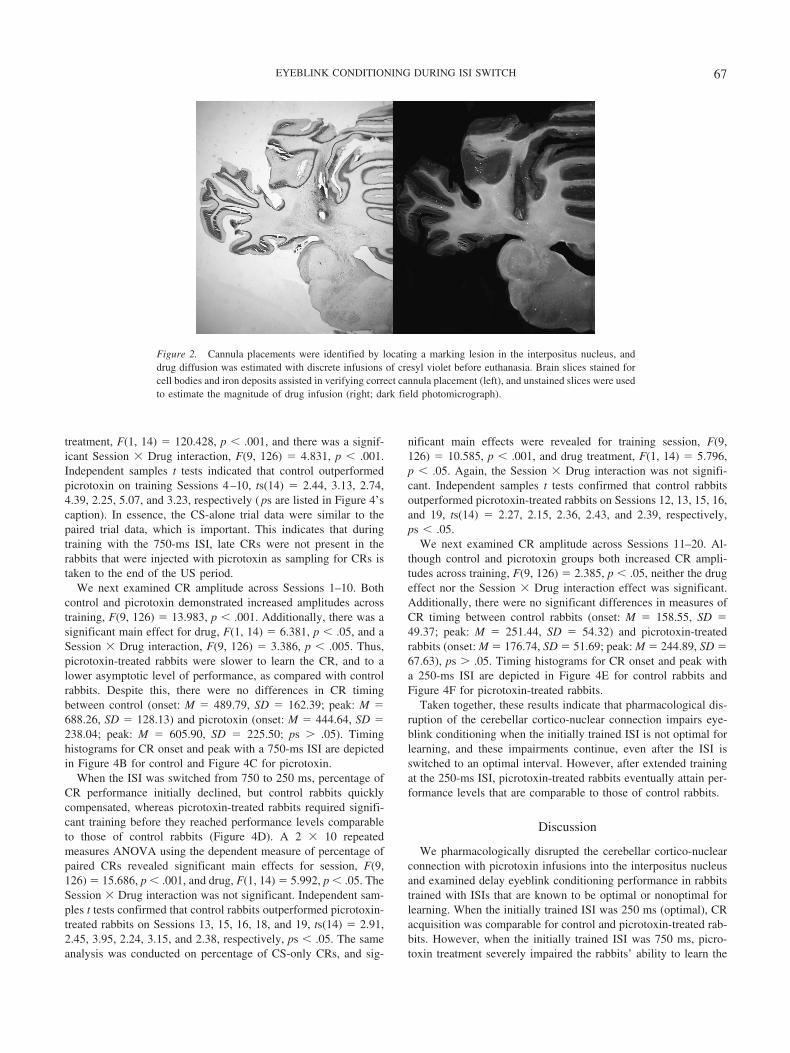
Figure 2. Cannula placements were identified by locating a marking lesion in the interpositus nucleus, anddrug diffusion was estimated with discrete infusions of cresyl violet before euthanasia. Brain slices stained forcell bodies and iron deposits assisted in verifying correct cannula placement (left), and unstained slices were usedto estimate the magnitude of drug infusion (right; dark field photomicrograph).
67EYEBLINK CONDITIONING DURING ISI SWITCH

Figure 3. Conditioned response (CR) learning and timing for rabbits in the 2503750-ms interstimulus interval(ISI)-switch condition. A: Percentage of CR over the first 10 training sessions with a 250-ms ISI (all error barsindicate standard error of the mean). Circles indicate control rabbits (n � 9), triangles indicate picrotoxin-treatedrabbits (n � 9), gray indicates paired conditioned stimulus–unconditioned stimulus (CS–US) trials, and blackindicates CS-alone trials. B and C: Relative frequency of CR onset and peak for control and picrotoxin groups,respectively, over the last 4 sessions with a 250-ms ISI. Dotted lines indicate time of US presentation. D:Percentage of CR over 10 sessions of training with a 750-ms ISI, after the switch from a 250-ms ISI (all errorbars indicate standard error of the mean). M � muscimol infusion to interpositus nucleus on Session 21. E andF: Relative frequency of CR onset and peak for control and picrotoxin groups, respectively, over the last 4sessions with a 750-ms ISI.
68 VOGEL, AMUNDSON, LINDQUIST, AND STEINMETZ

Figure 4. Conditioned response (CR) learning and timing for rabbits in the 7503250-ms interstimulus interval(ISI)-switch condition. A: Percentage of CR over the first 10 training sessions with a 750-ms ISI (all error barsindicate standard error of the mean; statistical differences indicated for paired conditioned stimulus–unconditioned stimulus [CS–US] trials only). Circles indicate control rabbits (n � 8), triangles indicatepicrotoxin-treated rabbits (n � 8), gray indicates paired CS–US trials, and black indicates CS-alone trials. B andC: Relative frequency of CR onset and peak for control and picrotoxin groups, respectively, over the last 4sessions with a 750-ms ISI. Dotted lines indicate time of US presentation. D: Percentage of CR over 10 sessionsof training with a 250-ms ISI, after the switch from a 750-ms ISI (all error bars indicate standard error of themean). M � muscimol infusion to interpositus nucleus on Session 21. E and F: Relative frequency of CR onsetand peak for control and picrotoxin groups, respectively, over the last 4 sessions with a 250-ms ISI. � p � .05.�� p � .01.
69EYEBLINK CONDITIONING DURING ISI SWITCH

task, as compared with controls. These findings support our hy-pothesis that cerebellar cortical modulation of the interpositusnucleus plays a greater role in eyeblink conditioning when the ISIis not optimal for learning (�500 ms).
Data from control rabbits corresponds well with a substantialbody of previous research demonstrating that eyeblink CR learningis most robust with an ISI that is approximately 200–500 ms(Coleman & Gormezano, 1971; Gormezano et al., 1983; Gor-mezano, Schneiderman, Deaux, & Fuentes, 1962; Schneiderman,1966; Schneiderman, Fuentes, & Gormezano, 1962; Schneiderman& Gormezano, 1964; Smith, 1968; Smith et al., 1969). Naıvecontrol rabbits that were trained with a 250-ms ISI (Figure 3A)learned faster and produced a higher overall percentage of CRsthan those trained with a 750-ms ISI (Figure 4A). Also, after 10training sessions, we switched the ISI, making it shorter or longer,and observed an initial decline in performance in both ISI-switchgroups (Figures 3D and 4D). In each case, in the training sessionsfollowing the ISI switch, double-peaked CRs were often observed,and the CR peak associated with the initially trained ISI dimin-ished over subsequent training. Thus, after the ISI switch, the CRpeak associated with ISI-1 (Sessions 1–10) was extinguished as theCR peak associated with ISI-2 (Sessions 11–20) was acquired(data not shown). Rabbits switched from the 750- to the 250-msISI acquired the new CR peak more rapidly than those switchedfrom the 250- to the 750-ms ISI. Similar performance trends havebeen reported by others who have studied the ISI switch in bothhumans (Boneau, 1958; Ebel & Prokasy, 1963; McAllister, 1953;Prokasy, Ebel, & Thompson, 1963) and rabbits (Coleman & Gor-mezano, 1971; Leonard & Theios, 1967; Prokasy & Papsdorf,1965). Thus, data from our control rabbits provide further supportfor the idea that ISIs longer than 500 ms do not produce optimaleyeblink CR learning in rabbits.
Normal input from the cerebellar cortex to the interpositusnucleus appears to be differentially important for eyeblink CRacquisition with optimal versus nonoptimal ISIs. When we initiallytrained rabbits with a 250-ms ISI, control and picrotoxin-treatedrabbits had comparable rates of CR acquisition and asymptoticlevels of performance as measured by percentage of CR (Figure3A). Also, there were no differences on measures of CR amplitudeor latency. In contrast, significant learning impairments were ob-served in picrotoxin-treated rabbits initially trained with a 750-msISI. Indeed, after 10 sessions of training, these rabbits never fullyacquired the CR to the same level as control rabbits (Figure 4A).Furthermore, the rarely executed CR was low in amplitude andhighly variable in latency (Figure 4C). These results suggest thatmodulatory input from cerebellar cortex becomes increasinglyimportant for delay eyeblink conditioning if the initially trained ISIis longer than that which is optimal for learning.
One of the questions that we addressed in this study was howpharmacological disruption of the cerebellar cortico-nuclear con-nection affects the ability of rabbits to learn a new ISI after anabrupt switch from previous training with a longer or shorter ISI.After fixed ISI training, we switched the ISI and found thatpicrotoxin-treated rabbits generally produced lower CR percent-ages than control rabbits. In the 2503750 ISI-switch condition(Figure 3D), this effect was not quite statistically significant. In the7503250 ISI-switch condition (Figure 4D), the cause of thisimpairment is unclear, but one likely explanation is that the rela-tively reduced CR performance was because the picrotoxin-treated
rabbits never fully learned the CR before the ISI switch (Figure4A). These results may indicate that modulatory input from cere-bellar cortex is important for learning and adapting the CR underchanging conditions. Alternatively, cerebellar cortex may be im-portant for new learning after previous training has occurred ineyeblink conditioning (Garcia, Steele, & Mauk, 1999; Perrett &Mauk, 1995; Yeo, Lobo, & Baum, 1997).
Given the growing body of evidence that suggests a role forcerebellar cortex in CR timing (and specifically the role of theanterior lobe), we were interested in evaluating the ability ofpicrotoxin-treated rabbits to adapt their CR latency over training.The frequency histograms in Figures 3 and 4 indicate that rabbitsin all conditions seemed to learn adaptively timed CRs before andafter the ISI switch. In this respect, the overall performance ofpicrotoxin-treated rabbits indicated that normal input from cere-bellar cortex is not critical for CR timing in the interpositusnucleus. However, we noted considerable variability in responsetiming in picrotoxin-treated rabbits. And we did observe that a fewrabbits developed short-latency CRs, as previously described byGarcia and Mauk (1998). Given the literature describing the effectsof picrotoxin infusions in the interpositus nucleus (Bao et al.,2002; Garcia & Mauk, 1998; Ohyama et al., 2006), we weresurprised by the infrequency and variability with which shortlatency CRs were observed in this study.
There are two technical explanations for why this discrepancymay have occurred. First, picrotoxin infusions may not have thepotential to be effective throughout the entire session. To test forthis possibility, we examined the behavior of individual rabbitsthat expressed short-latency CRs to determine whether thesepicrotoxin-mediated responses were present at the end of thesession. Figure 5 depicts blink activity over the last 10 paired trialsin a single session for 1 rabbit that was first trained with a 250-msISI (Figure 5A) and 1 rabbit that was first trained with a 750-msISI (Figure 5B). In each case, short-latency CRs were robustlyexpressed through the end of the conditioning session, as definedby the presence of short-latency responses (see Garcia & Mauk,1998). These data indicate that picrotoxin has the potential to beeffective throughout the duration of a classical conditioning ses-sion. A second reason why our results may differ from thosedescribed previously is that our criterion for scoring short-latencyCRs was much more restrictive (e.g., Garcia & Mauk, 1998). Weused the performance of the control rabbits with the 250- and750-ms ISIs to define the window of time during which a normalCR could be seen, and we scored short-latency CRs when anyresponse onset preceded that window. This resulted in a short-latency CR criterion defined as any response that occurred 26–80ms after CS onset, a window of time that is much narrower than inprevious studies. This may ultimately account for a lower numberof responses that meet the criterion for short-latency CR.
Other reasons why our results may differ from those describedpreviously may lie in the methodology. For example, short-latencyCRs have been unmasked primarily in CR retention studies thatfollow a cerebellar cortex lesion (Perrett et al., 1993) or disruptionof cerebellar cortical input to the interpositus nucleus (Garcia &Mauk, 1998). In these studies, a CR has been established throughtraining, and removal of cerebellar cortex, or disrupting interposi-tus activity, presumably disrupts expression of previously estab-lished plasticity in the form of parallel fiber–Purkinje cell long-term depression, which ultimately modulates CRs via well-timed
70 VOGEL, AMUNDSON, LINDQUIST, AND STEINMETZ

disinhibition of the interpositus nucleus. Interestingly, it has beendemonstrated that overtraining in eyeblink conditioning beforedisruption of cortico-nuclear function can rescue CR timing andendow the interpositus with plasticity sufficient to execute a well-timed CR (Christian & Thompson, 2005). Similarly, although weexamined the effect of cerebellar disruption over the course ofacquisition, the extensive training that the rabbits received mayallow for timing-related plasticity to develop in the interpositus,even in the absence of normal input from the cerebellar cortex.Thus, the variability in response timing that we observed inpicrotoxin-treated rabbits suggests that the cerebellar cortex isimportant for consistently producing well-timed CRs in initialacquisition, but the ability of rabbits to accurately time most CRswith disrupted cortical input to the interpositus nucleus suggeststhat compensatory mechanisms may be at play. In essence, ourhypothesis that cerebellar cortico-nuclear disruption would impairthe rabbits’ ability to adapt the latency of the CR over training,including after an ISI switch, was not conclusively supported.
A consensus has yet to be reached regarding the origin(s) ofneural processes that regulate CR timing in eyeblink conditioning.The most parsimonious hypothesis holds that Purkinje cells regu-late timing via coordinated disinhibition of the interpositus nucleusjust before US onset (e.g., Mauk & Donegan, 1997). Some elec-trophysiological recording data from lobule HVI of cerebellarcortex do not fit this model, as Purkinje cells tend to increase theirdischarge with CS–US pairing (Berthier & Moore, 1986; Gould &Steinmetz, 1996; Kotani, Kawahara, & Kirino, 2003). However,Green and Steinmetz (2005) recently provided support for this
hypothesis by demonstrating that Purkinje cells in the anterior lobetend to decrease their firing rate with CS–US pairings. This sug-gests that important timing-related plasticity may be confined to arather discrete region of cerebellar cortex (but see Nolan & Free-man, 2006).
Although cerebellar cortex may play an important role in CRtiming, our results argue that normal cerebellar cortical input to theinterpositus nucleus is not critical for CR timing, at least in initialacquisition. Indeed, although timing-related neural activity maydevelop in cerebellar cortex, it does not necessarily follow thattiming mechanisms originate in cerebellar cortex. It could be thecase, for example, that CR timing relies on a distributed networkof processes in regions of the brain that ultimately send importanttiming-related input to areas of the cerebellum where CS–USassociations are made. Furthermore, although both the interpositusnucleus and the cerebellar cortex appear to be important for timing,our picrotoxin data argue that adaptive timing of CRs may beaccomplished by the interpositus nucleus, even in the absence ofnormal modulatory input from cerebellar cortex. This compensa-tory mechanism may stand alone in the interpositus nucleus or relyon timing-related input from extracerebellar structures, such as thehippocampus (Hoehler & Thompson, 1980; Port, Mikhail, &Patterson, 1985; Port & Patterson, 1985; but see Poulos & Thomp-son, 2004).
There exists a rather significant body of literature on the role ofcerebellar cortex in eyeblink conditioning (see Christian &Thompson, 2003). The most frequent methodological approachhas been to examine CR acquisition or retention after cerebellarcortex lesions. Although this manipulation does not typically pro-hibit learning or block performance, a myriad behavioral deficitshave been observed, even with an optimal ISI, including severeimpairments in CR acquisition (Lavond & Steinmetz, 1989;McCormick & Thompson, 1984). This is difficult to reconcile withthe normal CR learning that we observed in picrotoxin-treatedrabbits trained with the 250-ms ISI (Figure 3A–C). However, onepossible explanation is that lesion and other chronic preparationsmay represent a more substantial disruption to the cerebellarnetwork. The chronic effects of lesions often include loss ofneurons in afferent and efferent structures, and these losses canaffect conditioning. For example, loss of cells in the inferior olivewould affect US projections to the cerebellum, potentially impair-ing acquisition. Thus, chronic preparations may have wider spreadeffects on neural structure and function than intended. A feature ofacute preparations, such as the drug infusion method used here, isthat the effect is temporary, and this approach allows one tophysically alter neural function without compromising gross neu-ral structure. Thus, chronic and acute preparations are fundamen-tally different, and for this reason, it may be difficult to makedirect comparisons between their effects on behavior. Taken to-gether, however, data generated from the chronic and acute ap-proaches should help delineate the role of cerebellar cortex ineyeblink conditioning.
Although the literature on the subject is rather sparse, pharma-cological perturbations of the cerebellar network may ultimatelyaffect neural activity in the nucleus and produce a multitude ofbehavioral deficits, even with an optimal ISI. For example, variouseyeblink conditioning deficits emerge when picrotoxin is infusedto the interpositus nucleus during tests of CR retention with a500-ms ISI (Attwell, Ivarsson, Millar, & Yeo, 2002; Garcia &
Figure 5. Picrotoxin infusions were effective throughout the entire train-ing session. Each graph represents electromyogram activity recorded fromthe Orbiculus oculi muscle of a single rabbit over the last 10 paired(conditioned stimulus–unconditioned stimulus) trials in a single eyeblinkconditioning session. Upward deflections indicate eyelid closure. Thisgraph demonstrates that when short-latency conditioned responses areobserved, they can persist throughout the entirety of a conditioning session.A: Data from a rabbit first trained with a 250-ms interstimulus interval(ISI). B: Data from a rabbit first trained with a 750-ms ISI.
71EYEBLINK CONDITIONING DURING ISI SWITCH

Mauk, 1998; Medina, Garcia, & Mauk, 2001; Ohyama et al.,2006). These deficits include reductions in the CR latency, CRamplitude, and percentage of CRs. Using a 250-ms ISI, Bao et al.(2002) reported that complete pharmacological decortication of thecerebellum—with infusions of muscimol and then picrotoxin intothe interpositus nucleus—blocked acquisition of CRs in naıveanimals, but CR expression was retained in well-trained animals.Interestingly, CR expression in retention was critically dependenton the level of excitability in the nucleus. Although Bao et al.’sresults may appear to contradict our findings, a resolution to thediscrepancy is that cerebellar decortication produces more pro-found deficits than those observed during disruption of cerebellarcortico-nuclear activity. The critical difference may lie in the basallevel of activity that is maintained in the interpositus nucleus.
Evidence for this idea comes from other studies that have usedacute drug injections into discrete regions of the cerebellar cortex.For example, Yeo and colleagues have found that CNQX (AMPA/kainite receptor antagonist) injections to cortical area HVI blockedboth acquisition (Attwell, Rahman, & Yeo, 1999) and retention(Attwell, Rahman, Ivarsson, & Yeo, 2001) of the eyeblink CR witha 350-ms ISI. This effect makes sense in light of the fact thatglutamate antagonists in the cerebellar cortex increase the sponta-neous discharge of Purkinje cells (Hausser and Clark, 1997) andthus potentially increase basal inhibition in the nucleus (Christian& Thompson, 2003). Thus, increased inhibition in the interpositusnucleus would be expected to prohibit normal acquisition andexpression of CRs. Similarly, Mamounas et al. (1987) reportedtemporary abolition of CRs following microinjections of picro-toxin to lobule HVI, which also has the effect of increasingPurkinje cell output (Hausser & Clark, 1997). When muscimol wasinfused into cortical lobule HVI (see Larsell, 1970), which coulddecrease Purkinje cell output, David Krupa (1993; cited in Chris-tian & Thompson, 2003) found that this manipulation did notdisrupt CR acquisition with similar task parameters (i.e., 250-msISI). Taken together, these results may suggest that neurotransmit-ter systems in the cerebellum may work together to maintain acritical level of activity in the interpositus nucleus, and behavioralimpairments observed in eyeblink conditioning may be related, inpart, to the degree to which interpositus nucleus activity is altered.
An equally viable and nonexclusive hypothesis is that ISI-learning-related plasticity is distributed throughout the brain, withthe most critical and fundamental component being the interposi-tus nucleus. ISIs longer than 500 ms do not promote optimallearning in delay eyeblink conditioning, and one possibility is thatnormal learning requires input from other brain regions. If this isthe case, then it is not unreasonable to extrapolate that higher brainregions may need to contribute more in learning increasinglydifficult tasks. For example, trace eyeblink conditioning, which ischaracterized by a brief stimulus-free period between CS offsetand US onset, requires both the hippocampus (Solomon, VanderSchaaf, Thompson, & Weisz, 1986) and the cerebellum(Woodruff-Pak, Lavond, & Thompson, 1985), but the cerebellarcortex does not appear to be important (Woodruff-Pak, Green,Levin, & Meisler, 2006; Woodruff-Pak et al., 1985). Because thereis no temporal overlap between the CS and US, the trace condi-tioning procedure may be a more difficult learning task than delay.
Walker and Steinmetz (2008) recently demonstrated that ibo-tenic acid lesions to hippocampus were more detrimental to ac-quisition and performance of trace eyeblink conditioning with a
long trace interval than with a short trace interval. They concludedthat performance in trace conditioning was, in part, dependent ontask parameters such as the duration of the CS and the traceperiods. Thus, the hippocampus may play the same important rolein learning nonoptimal task parameters in trace conditioning as thecerebellar cortex plays in learning nonoptimal task parameters indelay conditioning. In other words, the importance of hippocam-pus (trace conditioning) or cerebellar cortex (delay conditioning)may increase significantly as task demands become more difficult,and perturbing these regions, or their inputs to the interpositusnucleus, may restrict the ability of the organism to learn moredifficult tasks without impairing learning in more basic tasks.Indeed, Clark, McCormick, Lavond, and Thompson (1984) sug-gested that different memory systems may be hierarchically orga-nized and that one may abolish higher forms of learning, whilesparing more elementary association components. It will be inter-esting to further elucidate roles for various brain regions in eye-blink conditioning, and especially for the cerebellar cortex in shorttrace conditioning and the hippocampus in long delay condition-ing. We may learn much about the neural substrates for learningand interval timing in eyeblink classical conditioning.
In summary, our picrotoxin data, along with trace conditioningdata from Walker and Steinmetz (2008), suggest that learningeyeblink conditioning with nonoptimal task parameters may re-quire convergence of stimulus information in the interpositusnucleus from multiple brain regions, including cerebellar cortexand hippocampus. When this modulatory input is disrupted, itappears that CR acquisition occurs, albeit with deficiencies in CRamplitude, CR timing, rate of learning, and asymptotic level ofresponding. The magnitude of these deficiencies is likely depen-dent on a complex interaction between the severity of the neuralperturbation and the difficulty of the conditioning task. Futurestudies in eyeblink conditioning will need to examine more closelythe relationship between task parameters, neurotransmitter sys-tems, and the importance of basal activation in the interpositusnucleus.
References
Aksenov, D., Serdyukova, N., Irwin, K., & Bracha, V. (2003). GABAneurotransmission in the cerebellar interposed nuclei: Involvement inclassically conditioned eyeblinks and neuronal activity. Journal of Neu-rophysiology, 91, 719–27.
Attwell, P. J. E., Ivarsson, M., Millar, L., & Yeo, C. H. (2002). Cerebellarmechanisms in eyeblink conditioning. Annals of the New York Academyof Sciences, 978, 79–92.
Attwell, P. J. E., Rahman, S., & Yeo, C. H. (2001). Acquisition of eyeblinkconditioning is critically dependent on normal function in cerebellarcortical lobule HVI. Journal of Neuroscience, 21, 5715–5722.
Attwell, P. J. E., Rahman, S. Ivarsson, M., & Yeo, C. H. (2001). Cerebellarcortical AMPA/kainate receptor blockade prevents performance of clas-sically conditioned nictitating membrane responses. Journal of Neuro-science, 19, 1–6.
Bao, S., Chen, L., Kim, J. J., & Thompson, R. F. (2002). Cerebellar corticalinhibition and classical conditioning. Proceedings of the National Acad-emy of Sciences of the United States of America, 99, 1592–1597.
Berthier, N. E., & Moore, J. W. (1986). Cerebellar Purkinje cell activityrelated to the classically conditioned nictitating membrane response.Experimental Brain Research, 63, 341–350.
Boneau, C. A. (1958). The interstimulus interval and the latency of the
72 VOGEL, AMUNDSON, LINDQUIST, AND STEINMETZ

conditioned eyelid response. Journal of Experimental Psychology, 56,464–471.
Brown, K. L., Pagani, J. H., & Stanton, M. E. (2006). The ontogeny ofinterstimulus interval (ISI) discrimination of the conditioned eyeblinkresponse in rats. Behavioral Neuroscience, 120, 1057–1070.
Chen, L., Bao, S., Lockard, J. M., Kim, J. K., & Thompson, R. F. (1996).Impaired classical eyeblink conditioning in cerebellar-lesioned and Pur-kinje cell degeneration (pcd) mutant mice. Journal of Neuroscience, 16,2829–2838.
Christian, K. M., & Thompson, R. F. (2003). Neural substrates of eyeblinkconditioning: Acquisition and retention. Learning & Memory, 11, 427–455.
Christian, K. M., & Thompson, R. F. (2005). Long-term storage of anassociative memory trace in the cerebellum. Behavioral Neuroscience,119, 526–537.
Clark, G. A., McCormick, D. A., Lavond, D. G., & Thompson, R. F.(1984). Effects of lesions of cerebellar nuclei on conditioned behavioraland hippocampal neuronal responses. Brain Research, 291, 125–136.
Coleman, S. R., & Gormezano, I. (1971). Classical conditioning of therabbit’s (Oryctolagus cuniculus) nictitating membrane response undersymmetrical CS-US interval shifts. Journal of Comparative and Physi-ological Psychology, 77, 447–455.
Ebel, H. C., & Prokasy, W. F. (1963). Classical eyelid conditioning as afunction of sustained and shifted interstimulus intervals. Journal ofExperimental Psychology, 65, 52–58.
Garcia, K. S., & Mauk, M. D. (1998). Pharmacological analysis of cere-bellar contributions to the timing and expression of conditioned eyelidresponses. Neuropharmacology, 37, 471–480.
Garcia, K. S., Steele, P. M., & Mauk, M. D. (1999). Cerebellar cortexlesions prevent acquisition of conditioned eyelid responses. Journal ofNeuroscience, 19, 10940–10947.
Gormezano, I., Kehoe, E. J., & Marshall, B. S. (1983). Twenty years ofclassical conditioning with the rabbit. Progress in Psychobiology andPhysiological Psychology, 10, 197–275.
Gormezano, I., Schneiderman, N., Deaux, E., & Fuentes, I. (1962, October5). Nictitating membrane: Classical conditioning and extinction in thealbino rabbit. Science, 138, 33–34.
Gould, T. J., & Steinmetz, J. E. (1996). Changes in rabbit cerebellarcortical and interpositus nucleus activity during acquisition, extinctionand backward classical conditioning. Neurobiology of Learning andMemory, 65, 17–34.
Green, J. T., & Steinmetz, J. E. (2005). Purkinje cell activity in thecerebellar anterior lobe after rabbit eyeblink conditioning. Learning &Memory, 12, 260–269.
Hausser, M., & Clark, B. A. (1997). Tonic synaptic inhibition modulatesneuronal output pattern and spatiotemporal synaptic integration. Neuron,19, 665–678.
Hoehler, F. K., & Thompson, R. F. (1980). Effect of the interstimulus(CS-UCS) interval on hippocampal unit activity during classical condi-tioning of the nictitating membrane response of the rabbit (Oryctolaguscuniculus). Journal of Comparative and Physiological Psychology, 94,201–215.
Kotani, S., Kawahara, S., & Kirino, Y. (2003). Purkinje cell activity duringlearning a new timing in classical eyeblink conditioning. Brain Re-search, 994, 193–202.
Krupa, D. J., Thompson, J. K., & Thompson, R. F. (1993, May 14).Localization of a memory trace in the mammalian brain. Science, 260,989–991.
Larsell, O. (1970). The comparative anatomy and histology of the cere-bellum from monotremems through apes. Minneapolis, MN: Universityof Minnesota Press.
Lavond, D. G., & Steinmetz, J. E. (1989). Acquisition of classical condi-tioning without cerebellar cortex. Behavioral Brain Research, 33, 113–164.
Leonard, D. W., & Theios, J. (1967). Classical conditioning of the nicti-tating membrane in the rabbit: Effect of CS-US interval shift. Journal ofComparative and Physiological Psychology, 63, 355–358.
Mamounas, L. A., Thompson, R. F., & Madden, J., IV. (1987). CerebellarGABAergic processes: Evidence for critical involvement in a form ofsimple associative learning in the rabbit. Proceedings of the NationalAcademy of Sciences of the United States of America, 84, 2101–2105.
Mauk, M. D., & Donegen, N. H. (1997). A model of Pavlovian eyelidconditioning based on the synaptic organization of the cerebellum.Learning & Memory, 4, 130–158.
McAllister, W. R. (1953). Eyelid conditioning as a function of the CS-USinterval. Journal of Experimental Psychology, 45, 417–422.
McCormick, D. A., & Thompson, R. F. (1984, January 20). Cerebellum:Essential involvement in the classically conditioned eyelid response.Science, 223, 296–299.
Medina, J. F., Garcia, K. S., & Mauk, M. D. (2001). A mechanism forsavings in the cerebellum. Journal of Neuroscience, 21, 4081–4089.
Nolan, B. C., & Freeman, J. H. (2006). Purkinje cell loss by OX7-saporinimpairs acquisition and extinction of eyeblink conditioning. Learning &Memory, 13, 359–365.
Ohyama, T., & Mauk, M. D. (2001). Latent acquisition of timed responsesin cerebellar cortex. Journal of Neuroscience, 21, 682–690.
Ohyama, T., Nores, W. L., Medina, J. F., Riusech, F. A., & Mauk, M. D.(2006). Learning-induced plasticity in deep cerebellar nucleus. Journalof Neuroscience, 26, 12656–12663.
Perrett, S. P., & Mauk, M. D. (1995). Extinction of conditioned eyelidresponses requires the anterior lobe of cerebellar cortex. Journal ofNeuroscience, 15(3, Pt. 1), 2074–2080.
Perrett, S. P., Ruiz, B. P., & Mauk, M. D. (1993). Cerebellar cortex lesionsdisrupt learning-dependent timing of conditioned eyelid responses. Jour-nal of Neuroscience, 13, 1708–1718.
Port, R. L., Mikhail, A. A., & Patterson, M. M. (1985). Differential effectsof hippocampectomy on classically conditioned rabbit nictitating mem-brane response related to interstimulus interval. Behavioral Neuro-science, 99, 200–208.
Port, R. L., & Patterson, M. M. (1985). Sensory preconditioning in therabbit following ACTH injections. Physiology & Behavior, 35, 443–445.
Poulos, A. M., & Thompson, R. F. (2004). Timing of conditioned re-sponses utilizing electrical stimulation in the region of the interpositusnucleus as a CS. Integrative Physiological and Behavioral Science, 39,83–94.
Prokasy, W. F., Ebel, H. C., & Thompson, D. D. (1963). Response shapingat long interstimulus intervals in classical eyelid conditioning. Journal ofExperimental Psychology, 66, 138–141.
Prokasy, W. F., & Papsdorf, J. D. (1965). Effects of increasing theinterstimulus interval during classical conditioning of the albino rabbit.Journal of Comparative and Physiological Psychology, 60, 249–252.
Schneiderman, N. (1966). Interstimulus interval function of the nictitatingmembrane response of the rabbit under delay versus trace conditioning.Journal of Comparative and Physiological Psychology, 62, 397–402.
Schneiderman, N., Fuentes, I., & Gormezano, I. (1962, May 18). Acqui-sition and extinction of the classically conditioned eyelid response in thealbino rabbit. Science, 136, 650–652.
Schneiderman, N., & Gormezano, I. (1964). Conditioning of the nictitatingmembrane of the rabbit as a function of the CS-US interval. Journal ofComparative and Physiological Psychology, 57, 188–195.
Smith, M. C. (1968). CS-US interval and US intensity in classical condi-tioning of the rabbit’s nictitating membrane response. Journal of Com-parative and Physiological Psychology, 66, 679–687.
Smith, M. C., Coleman, S. R., & Gormezano, I. (1969). Classical condi-tioning of the rabbit’s nictitating membrane response at backward,simultaneous and forward CS-US intervals. Journal of Comparative andPhysiological Psychology, 69, 226–231.
73EYEBLINK CONDITIONING DURING ISI SWITCH

Solomon, P. R., Vander Schaaf, E. R., Thompson, R. F., & Weisz, D. J.(1986). Hippocampus and trace conditioning of the rabbit’s classicallyconditioned nictitating membrane response. Behavioral Neuroscience,100, 729–744.
Walker, A. G., & Steinmetz, J. E. (2008). Hippocampal lesions in ratsdifferentially affect long- and short-trace eyeblink conditioning. Physi-ology & Behavior, 93, 570–580.
Woodruff-Pak, D. S., Green, J. T., Levin, S. I., & Meisler, M. H. (2006).Inactivation of sodium channel Scn8A (Na-sub(v)1.6) in Purkinje neu-rons impairs learning in Morris water maze and delay but not traceeyeblink classical conditioning. Behavioral Neuroscience, 120, 229–240.
Woodruff-Pak, D. S., Lavond, D. G., & Thompson, R. F. (1985). Trace
conditioning: Abolished by cerebellar nuclear lesions but not lateralcerebellar cortex aspirations. Brain Research, 348, 249–260.
Woodruff-Pak, D. S., Seta, S. E., Roker, L. A., & Lehr, M. A. (2007).Effects of paradigm and inter-stimulus interval on age differences ineyeblink classical conditioning in rabbits. Learning & Memory, 14,287–294.
Yeo, C. H., Lobo, D. H., & Baum, A. (1997). Acquisition of a new-latencyconditioned nictitating membrane response–Major, but not complete,dependence on the ipsilateral cerebellum. Learning and Memory, 3,557–577.
Received April 21, 2008Revision received September 9, 2008
Accepted September 13, 2008 �
74 VOGEL, AMUNDSON, LINDQUIST, AND STEINMETZ