eye movement disturbances in children - omlab€¦ · centrate on the differences between eye...
TRANSCRIPT
CHAPTER 2
Eye Movement Disturbances in Children Robert L. Tomsak and Louis F. Dell'Osso
The subject of eye movement disturbances has been dealt with in a number of forums, some simpler than others,l-4 Our goal in this chapter is to concentrate on the differences between eye movement disturbances in children and adults.
Isolated Cranial Nerve Palsies
Isolated cranial nerve palsies can be viewed by the anatomic site of disruption of function or by cause. In adults, the causes for cranial nerve palsies are summarized in Table 2- 1. Beware that ocular myasthenia can mimic any of the conditions described in this section.
Third Cranial Nerve ( Oculomotor Nerve) Palsies
The third cranial nerve can be damaged anywhere from its nucleus in the midbrain to its ramifications in the orbit (Figure 2- 1). The classic anatomical types of third nerve palsies are as follows: ( 1) nuclear, (2) fasicular, (3) interpeduncular (subarachnoid), (4) cavernous sinus and superior orbital fissure, and (5) orbital.? The oculomotor nerve innervates the medial, inferior, and superior recti, the inferior oblique, the levator of the lid, and the parasympathetically innervated pupillary sphincter muscle. Therefore, third nerve dysfunction is usually manifested by some degree of ptosis and vertical and/or divergent strabismus. The pupil in third nerve pareses may be completely spared, partially involved, or totally affected.s Thus, it may be normal in size and reactive to light and accommodation, somewhat large and sluggishly reactive, or fully dilated and fixed to light (Le., internal
1 1

12 Eye Movement Disturbances in Children
TABLE 2-1 Determined Causes of Acquired Cranial Nerve Palsies in Adults
Cranial Nerve
III
Vascular (20%) Aneurysm (19%) Trauma (14%) Tumor (12%)
IV
Trauma (34 %) Vascular (22%) Tumor (7%) Aneurysm (1 %)
Data from Tomsak.5
VI
Tumor (21%) Vascular (15%) Trauma (13 %) Aneurysm (3 %)
Multiple
Tumor (38%) Trauma (23%) Aneurysm (12 %) Vascular (5%)
ophthalmoplegia). In general, ischemic lesions tend to spare the pupil or affect it minimally, whereas compressive or traumatic lesions cause significant pupillary dysfunction.S
The causes of third nerve palsies in children differ from adults and are listed in Table 2-2. Anomalous posterior insertion of the medial rectus muscle has also been reported to simulate congenital third nerve palsy.10
Note that aneurysmal third nerve palsies are exceptionally uncommon in children. First, saccular aneurysms in children are rare, representing between 0.5% and 4.6% of all cases; they predominate in males.ll Second, these aneurysms tend to arise at peripheral locations in the arterial tree.12 Gabianelli and colleagues9 reviewed this problem and concluded that formal
Sylvian Aqueduct and ,---.---�------ Superior Colliculus
Periaqueductal Gray Matter ----_'-----'to.l
Posterior Cerebral Artery _--:. ...
Superior Cerebellar Artery ----1 ..... Iiii;�!!!!!!1� Dorsum Sellae and Clivus __ __
'---\------ III Nerve Nucleus
�+---- Red Nucleus
�---- Cortico-Spinal (Pyramidal) Tract in the Cerebral Peduncle
�------ Basilar Artery ������::J Petrous Ridge Sella Turcica -----H-lH
Superior Orbital Fissure --�-
Optic Nerve _...."L..--__ � I
I
, , ,
�----;--- Cavernous Sinus and Middle �,---_�--,Cranial Fossa
Ridge
Superior and Inferior Divisions of III
FIGURE 2-1 Schematic view of third cranial nerve anatomy from midbrain to orbit . (Reprinted by permission of the publisher from Bajandas FJ, Kline LB. Neuroophthalmology review manual, 3rd ed. Thorofare, NJ: Slack, 1988.)
TABLE 2-2
Isolated Cranial Nerve Palsies 13
Types or Causes of Third Nerve Palsies in Childhood
Congenital (45%) Traumatic (20%) Inflammatory (10%) Neoplastic (10%) Ophthalmoplegic migraine (5%) Cryptogenic (0%-3%) Aneurysm (3%)
Data from Gabianelli et al.9
intraarterial angiography is not warranted in children younger than 10 years of age with oculomotor palsy and without signs of subarachnoid hemorrhage. The rapid improvements in magnetic resonance angiography, however, may make the significance of this issue moot in the near future.1 3
Ophthalmoplegic migraine in a rare condition that usually has its first onset in the preteen years and presents as a third nerve palsy, usually with the pupil involved, after a nonthrobbing orbital and hemicranial headache that lasts 1 to 4 days. The problem can be recurrent, and involvement of cranial nerves 4 and 6 has been reported. The duration of the ophthalmoplegia is usually less than 2 months, and complete resolution is the rule. The site of cranial nerve involvement is presumed to be the cavernous sinus and possibly relates to swelling of the intracavernous carotid artery after the attack. Aberrant regeneration of the third cranial nerve with ophthalmoplegic migraine is unusual, but has been reported.l4
Aberrant regeneration, or oculomotor synkinesis, is seen after third nerve damage from a variety of causes (Table 2-3; Figure 2-2). It is usually characterized by some combination of the following signs: (1) elevation of the involved lid on adduction, (2) retraction (elevation) of the involved lid on downgaze, (3) limitation of vertical gaze with occasional retraction of the in-
TABLE 2-3 Causes of Aberrant Regeneration of the Third Nerve
Intracranial aneurysms Basilar skull fractures Tumors Congenital Cavernous sinus thrombosis Ophthalmoplegic migraine Idiopathic third nerve palsies Cyclic oculomotor palsies Basilar meningitis
Data from Sibony et a1.15

14 Eye Movement Disturbances in Children
A
B
FIGURE 2-2 Aberrant regeneration of the left third cranial nerve following head trauma. (A) In· ability to fully elevate left eye. (B) Elevation of left upper lid on attempted adduction of left eye . (e) Elevation of left upper lid on attempted downgaze.
volved eye, (4) adduction of the affected eye on attempted vertical gaze, (5) light-near dissociation of the pupil, and (6) subnormal vertical optokinetic responses on the side of the palsy.I6
An important clinical point is that if the signs of aberrant regene.ration are present, but there is no history of a prior third nerve palsy, neuroimaging should be performed to exclude occult mass lesions in the cavernous sinus regionY A familial form of levator-medial rectus synkinesis has been reported. IS The pathophysiology of oculomotor synkinesis probably involves misrouting of regenerating axons. Ephaptic transmission (neural "cross talk") has also been given as an explanation. IS
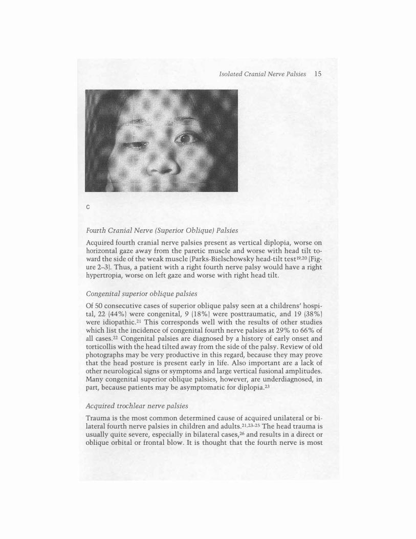
c
Isolated Cranial Nerve Palsies 15
Fourth Cranial Nerve (Superior Oblique) Palsies
Acquired fourth cranial nerve palsies present as vertical diplopia, worse on horizontal gaze away from the paretic muscle and worse with head tilt toward the side of the weak muscle (Parks-Bielschowsky head-tilt test19,20 (Figure 2-3). Thus, a patient with a right fourth nerve palsy would have a right hypertropia, worse on left gaze and worse with right head tilt.
Congenital superior oblique palsies
Of 50 consecutive cases of superior oblique palsy seen at a childrens' hospital, 22 (44%) were congenital, 9 (18%) were posttraumatic, and 19 (38%) were idiopathic.21 This corresponds well with the results of other studies which list the incidence of congenital fourth nerve palsies at 29% to 66% of all cases.22 Congenital palsies are diagnosed by a history of early onset and torticollis with the head tilted away from the side of the palsy. Review of old photographs may be very productive in this regard, because they may prove that the head posture is present early in life. Also important are a lack of other neurological signs or symptoms and large vertical fusional amplitudes. Many congenital superior oblique palsies, however, are underdiagnosed, in part, because patients may be asymptomatic for diplopia.23
Acquired trochlear nerve palsies
Trauma is the most common determined cause of acquired unilateral or bilateral fourth nerve palsies in children and adults.21 ,23-25 The head trauma is usually quite severe, especially in bilateral cases,26 and results in a direct or oblique orbital or frontal blow. It is thought that the fourth nerve is most

16 Eye Movement Disturbances in Children
A
B
FIGURE 2-3 Congenital right fourth nerve paresis. (A) Inability to fully depress right eye in adduction. (S) Elevation of right eye on right head tilt. Note that sclera can be seen between the right lower limbus and the right lower lid . Left lower lid position is normal.
sensitive to injury as it exits the back of the brain stem and crosses near the tentorial edge (Figure 2-4). Minimal trauma, however, may cause decompensation of a preexisting congenital superior oblique weakness.24
Traumatic fourth nerve palsies often do not improve spontaneously. If needed, eye muscle surgery may be performed after a waiting period of 6 months to 1 year if there is no sign of resolution.23-26
Brain stem lesions may cause unilateral or bilateral trochlear nerve palsies,27 and contralateral fourth nerve pareses. Ipsilateral Homer's syndrome has also been described.28 Parinaud's syndrome due to aqueductal stenosis, pineal tumors, and hydrocephalus may have associated unilateral or bilateral superior oblique palsies.29,3o Trochlear nerve palsy also has been

Isolated Cranial Nerve Palsies 17
Inferior COIIIC.JIUS-------......
IV Nerve Nucleus Sylvian Aqueduct -----�f'---tf'�.v�_Medial Longitudinal----fH--� Fasciculus
Posterior Cerebral Artery
Superior Cerebellar Artery ---��� Dorsum Sellae ---l������ Sella Turcica ----------+l�
---- Basilar Artery
���� Petrous Ridge
4------=--- Cavernous Sinus
4------ Sphenoid Ridge
Superior Orbital ------r,''-'JJ �7 ': F;ssu," /,/ dr 5, i :�' --.. , "
Opt;c Ne""
" ( ( : : l " , I I
\
I I I \
FIGURE 2-4 Schematic view of fourth cranial nerve anatomy from midbrain to orbit, (Reprinted by permission of the publisher from Bajandas FJ, Kline LB, Neuroophthalmology review manual, 3rd ed, Thorofare, NJ: Slack, 1988,)
reported as a false localizing sign in a child with pseudotumor cerebri31 and in two siblings with familial periodic cerebellar ataxia.32 Bilateral superior oblique muscle aplasia has accompanied Apert's syndrome,33
Sixth Nerve (Abducens Nerve) Palsies
The classic presentation of sixth nerve palsy is horizontal double vision, worse at distance, and worse on gaze to the affected side. In children, as in adults, a face tum that places the weak lateral rectus muscle out of its normal field of action is the norm. Thus, a youngster with a right lateral rectus palsy, who has normal binocular fusion, will tum his or her head to the right,
Sixth nerve palsies in children often have an etiology different from those that occur in adults. Of all the isolated cranial nerve palsies affecting eye movement, problems with the sixth cranial nerve in children are the most potentially serious and the most vexing (Figures 2-5 and 2-6).
Benign transient unilateral sixth nerve palsies occur, although rarely, in normal neonates34,35 and seem to be associated with forceps deliveries.36

18 Eye Movement Disturbances in Children
Medial Longitudinal Fasciculus
+--- Fourth Ventricle
0-- Spinal Tract of V
Pyramidal Tract
0-- Central Sympathetic Neuron (?)
'IJ---"�+- VII Nucleus
VII
Paramedian Pontine Reticular
Formation (PPRF)
FIGURE 2-5 Schematic view of cross-section of the pons at the level of the sixth cranial nerve nuclei and fascicles . (Reprinted by permission of the publisher from Ba· jandas FJ, Kline LB. Neuro-ophthalmology review manual, 3rd ed. Thorofare, NT: Slack, 1988.)
Other transient ocular motor phenomena during the first 6 weeks of life in normal neonates have been described.37
Acquired persistent sixth nerve palsies pose a very different diagnostic dilemma. It is well known that sixth nerve palsies in children can be benign.38 As such, they are usually blamed on a viral illness or immunization, although no specific virus that causes this syndrome has been reliably identified.38,38 These palsies routinely resolve in days to months, but may be recurrent.39-42 Many times, however, structural lesions must be excluded by
III
J�--IV
��� Ophthalmic Division of V
Maxillary Division of V.
Mandibular Division of V
VI
v--\---f--_VII J-+--+--VIII
FIGURE 2-6 Schematic saggital view of brain stem and cavernous sinus region showing relationships of cranial nerves III-VIII. (Reprinted by permission of the publisher from Bajandas FT, Kline LB. Neuro-ophthalmology review manual, 3rd ed. Thorofare, NT: Slack, 1988 .)

A
B
Isolated Cranial Nerve Palsies 19
FIGURE 2-7 Left sixth nerve palsy from increased intracranial pressure (13-year-old girl with ependymoblastoma). (A) Inability to fully abduct left eye. (B) Left fundus photograph showing early papilledema.
neuroimaging studies (Figure 2-7). For example, when a child has a sixth nerve palsy and other systemic complaints, such as headache, lethargy, or motor symptoms, imaging the brain and posterior fossa is essential. At present, magnetic resonance imaging (MRI) with gadolinium enhancement is the preferred technique. Afifi and co-workers43 found that a majority (71 %) of primary tumors causing sixth nerve palsies in their series were located in the posterior fossa, with pontine gliomas, cerebellar astrocytomas, and ependymomas as the top three. Of the supratentorial tumors, astrocytoma was the more common (43%). (See also chapter 5.)
Acute acquired comitant esotropia, which can be mistaken for sixth nerve palsy, may occur in normal children44 as well as in those with posterior fossa tumors.45 Divergence insufficiency or paralysis, a related condi-

20 Eye Movement Disturbances in Children
tion, has been associated with inflammatory, infectious, and toxic disorders of the cerebrum, brain tumors, and cerebrovascular disease.46 Accommodative esotropia has been reported to follow head trauma.47 Also, an unusual case of transient, exercise-induced esotropia in an otherwise normal 13-yearold boy has been published.48
The differential diagnosis of sixth nerve palsies is shown in Tables 2-4 through 2-7, contrasting adults with children. The "Ts" of Dr. Robert Daroff (personal communication, 1987) are presented in Table 2-7 for ease of recollection, and for a respite of humor.
TABLE 2-4 Determined Causes of Sixth Nerve Palsies in Infants and Children
Cause Number of Patients Percent
Trauma 121 32 Tumor 112 30 Inflammatory 50 13 Miscellaneous 77 20 Congenital 17 5
Data from Kodsi and Younge,25 Afi£i et al.,43 Robertson et al.,49 and Harley.50
TABLE 2-5 Other Causes of Sixth Nerve Palsies in Infants and Children
Hydrocephalus Metastatic neuroblastoma (under age 3) Infection (Gradenigo's syndrome and meningitis) Multiple sclerosis Amold-Chiari malformation
Data from Tay1or36 and Nixon et aPI
TABLE 2-6 Other Causes of Apparent Abduction Defects in Children
Transient strabismus in neonates Duane syndrome Mobius syndrome and related Spasm of near reflex Congenital esotropia with cross-fixation Accomodative esotropia Acute acquired comitant esotropia Congenital ocular fibrosis Ocular myasthenia
Data from Nixon et al.,51 Miller et al.,52 Kishore and Kumar,53 Holmes and Cronin,54 and Bixenman and Laguna.55

Ocular Myasthenia 2 1
TABLE 2-7 Differential Diagnosis of Abduction Defects: Daroff's "Ts"
Myasthenia Tight medial rectus Wernicke's disease Functional (e.g., spasm of the near reflex) Medial orbital wall blow-out fracture Pseudotumor cerebri Old strabismus
TABLE 2-8 Causes of Cavernous Sinus Syndromes in Childhood
Rhabdomyosarcoma Intrasellar germinoma Histiocytosis X Burkitt's lymphoma Craniopharyngioma Tolosa-Hunt syndrome Ophthalmoplegic migraine
Data from Taylor (p. 649)36 and Kandt and Goldstein.56
Multiple Cranial Nerve Palsies
Tensilon Thyroid eye disease Thiamine Tricks (Turkey) Trauma Pseudo Tumor Tropia
Multiple cranial nerve palsies affecting ocular movements are rare in childhood. They may represent cavernous sinus syndromes (Table 2-8; Figure 2--6) or may be mimicked by other ophthalmoplegia syndromes, such as ocular myasthenia, Fisher's syndrome (ataxia, ophthalmoplegia, and areflexia),57,58 or chronic progressive external ophthalmoplegia (see next section; also see foreword).
Ocular Myasthenia
Ocular myasthenia seems to have identical presentations in children and adults.29 Strabismus and/or ptosis, usually variable and worse toward the end ofthe day, are the common findings. Remember that ocular myasthenia can mimic any acquired nonrhythmic eye movement disorder and therefore should be kept near the top of the differential diagnostic list in these cases. It is estimated that eye signs occur in up to 90% of myasthenics at presentation and that eye signs only are present in 15% to 59% of patients. 59 A number of office tests can be done to diagnose ocular myasthenia, the most important being the intravenous edrophonium chloride (Tensilon) test or the intramuscular neostigmine bromide (Prostigmin) test. The latter is often
22 Eye Movement Disturbances in Children
A
B
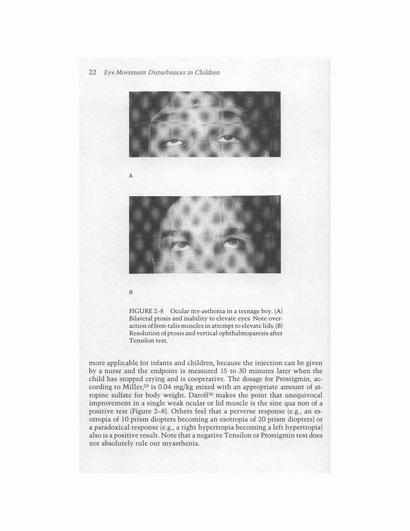
FIGURE 2-8 Ocular my-asthenia in a teenage boy. (A) Bilateral ptosis and inability to elevate eyes. Note overaction of fron-talis muscles in attempt to elevate lids. (B) Resolution of ptosis and vertical ophthalmoparesis after Tensilon test.
more applicable for infants and children, because the injection can be given by a nurse and the endpoint is measured 15 to 30 minutes later when the child has stopped crying and is cooperative. The dosage for Prostigmin, according to Miller,29 is 0.04 mg/kg mixed with an appropriate amount of atropine sulfate for body weight. Daroff59 makes the point that unequivocal improvement in a single weak ocular or lid muscle is the sine qua non of a positive test (Figure 2-8). Others feel that a perverse response (e.g., an esotropia of 10 prism diopters becoming an esotropia of 20 prism diopters) or a paradoxical response (e.g., a right hypertropia becoming a left hypertropia) also is a positive result. Note that a negative Tensilon or Prostigmin test does not absolutely rule out myasthenia.

Chronic Progressive External Ophthalmoplegia (Ophthalmoplegia Plus) 23
Neonatal forms of myasthenia occur in about 10% to 15% of children born of myasthenic mothers and are due to placental transfer of maternal acetylcholine receptor antibodies.6o About 10% of all myasthenia occurs in children and young adults, with a female preponderance.61
The treatment of ocular myasthenia includes patching one eye, the use of anticholinesterase agents and/or immunosuppressants, plasmapheresis, and thymectomy.62 Although thymectomy seems to give the best results, few physicians prescribe this treatment for isolated ocular disease.
Duane's Syndrome and Related Conditions
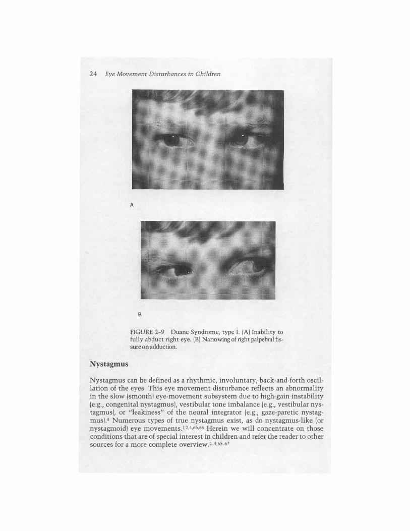
Duane's syndrome is a congenital condition in which there is aplasia of one or both sixth cranial nerve nuclei with innervation of the atrophic lateral rectus from branches of the third nerve.29 There are three clinical presentations: Type I has limited abduction and often narrowing of the palpebral fissure on adduction; type II has limited adduction with reasonably normal abduction; and type III is characterized by limitation of both abduction and adduction. Patients with Duane's syndrome almost never complain of diplopia but, as noted, this condition often mimics unilateral or bilateral sixth nerve palsies (Figure 2-9).
Mobius syndrome is distinguished by bilateral sixth and seventh nerve weakness, also related to aplasia of these brain stem nuclei.52 Esotropia in primary gaze is present in about 50% of patients. Numerous other defects are present, including mental retardation, craniofacial malformations, and limb anomalies.
Chronic Progressive External Ophthalmoplegia (Ophthalmoplegia Plus)
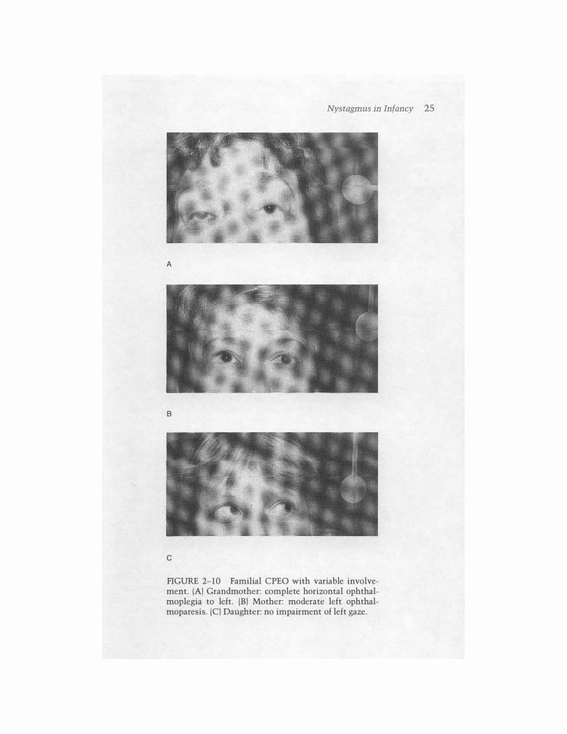
Chronic progressive external ophthalmoplegia (CPEO) is a controversial subject. It is characterized by progressive nonmyasthenic ophthalmoplegia, ptosis, and orbicularis oculi weakness.63 The pupils are not affected. It may begin in infancy or later in life. Familial occurrence is about 40% (Figure 2- 10). It is associated with a wide variety of other problems, the most common being olivopontocerebellar degeneration (Table 2-9).
The Kearns-Sayre-Daroff (Keams syndrome) variant has pigmentary retinopathy, cardiac conduction defects, elevated spinal fluid protein, and spongiform encephalopathy. Endocrine disorders and deafness are also seen. It begins before the age of 15 years and is now known to be due to heteroplasmic mitochondrial DNA deletions.64 It is maternally inherited and genetically lethal. Skeletal muscle biopsies show "ragged red" fibers when stained with the Gomori trichrome stain. There is no adequate treatment, although coenzyme Q 10 has been used. Steroids should be avoided because of reports of hyperglycemic coma and death.63
24 Eye Movement Disturbances in Children
Nystagmus
A
B
FIGURE 2-9 Duane Syndrome, type I. (A) Inability to fully abduct right eye. (B) Narrowing of right palpebral fissure on adduction.
Nystagmus can be defined as a rhythmic, involuntary, back-and-forth oscillation of the eyes. This eye movement disturbance reflects an abnormality in the slow (smooth) eye-movement subsystem due to high-gain instability (e.g., congenital nystagmus), vestibular tone imbalance (e.g., vestibular nystagmus), or "leakiness" of the neural integrator (e.g., gaze-paretic nystagmus).4 Numerous types of true nystagmus exist, as do nystagmus-like (or nystagmoid) eye movements.1,2,4,6S,66 Herein we will concentrate on those conditions that are of special interest in children and refer the reader to other sources for a more complete overview.2-4,6S-67
Nystagmus in Infancy 25
A
B
c
FIGURE 2-10 Familial CPEO with variable involvement. (A) Grandmother: complete horizontal ophthalmoplegia to left. (B) Mother: moderate left ophthalmoparesis. (e) Daughter: no impairment of left gaze.
26 Eye Movement Disturbances in Children
TABLE 2-9 Diseases Associated with Chronic Progressive External Ophthalmoplegia
Spinocerebellar degenerations Myotonic dystrophy Kugelberg-W elander disease Werdnig-Hoffman disease Myotubular myopathy Cystic fibrosis Vitamin E deficiency states
Nystagmus in Infancy
Infantile nystagmus, with onset within the first 6 months of life, may be idiopathic or hereditary (congenital nystagmus [CN]), associated with strabismus and monocular fixation (latent/manifest latent nystagmus [LMLN]), benign and self-limiting (the dissociated pendular nystagmus of spasmus nutans), associated with anterior visual sensory impairment (CN or LMLN), or due to neurologic illness.68,69
Congenital nystagmus
Congenital nystagmus is usually observed at birth or shortly after and continues throughout life. However, it may appear later in life.7° The term congenital in the name should not be taken literally and reflects the congenital predisposition for ocular motor instability present in those who develop CN. Attempts to substitute the term infantile for it are counterproductive, given the variability in time of onset. We use infantile to include all types of nystagmus usually seen in infancy (i.e., CN [with or without sensory defectsl, LMLN, the nystagmus of spasmus nutans, or nystagmus related to neurologic disease in this age group).
Congenital nystagmus reflects a high-gain instability in the smooth eye-movement subsystem, and the effort to see or direct the eyes are the primary actuating factors. Thus, CN is a motor instability and the use of the redundant term motor CN is discouraged because it also incorrectly implies that there is another type of CN (i.e., sensory defect CN). The waveforms of CN result from attempts by the ocular motor control system to increase foveation time in the face of an unstable fixation mechanism.4
Congenital nystagmus is defined best by eye-movement recordings,71 but there are a number of clinical features that are useful in diagnosis (Table 2- 10). Congenital nystagmus is always bilateral and appears conjugate. It may be pendular or jerk. It remains horizontal in vertical gaze, unlike neurologic forms of nystagmus which tend to convert to the same direction as

Nystagmus in Infancy 27
TABLE 2-10 Characteristics of Congenital Nystagmus
Binocular Both eyes in phase (but can exhibit some amplitude dissociation) Usually horizontal (clinically often appears uniplanar; eye-movement recordings
frequently show bi- or triplanar characteristics) Distinctive waveforms May be damped by convergence May have gaze-angle null Increased by fixation attempt Abolished in sleep Optokinetic stimuli may cause eN reversal ("inverted" optokinetic response) Head oscillation may be associated No oscillopsia
Data from Dell'Ossa et a1.4
gaze (see forewordJ. It is usually damped by convergence. There may be a null position of gaze where the nystagmus is least, and in these patients a head tum is frequently present. Perception of movement of the environment, or oscillopsia, is not present with CN. Associated head movements may be present. These have been shown not to be compensatory movements, but rather are another reflection of motor instability.4 Thus, the head movements do not improve visual acuity. Optokinetic stimuli result in inverted CN in about two thirds of cases. For example, in a case of right-beating CN, one would predict that if an optokinetic stimulus was taken to the subject's left, the optokinetic nystagmus elicited would summate with the CN to give a more pronounced right-beating response. What is observed is that the nystagmus is either damped or reverses direction in persons with CN. This socalled optokinetic inversion is diagnostic for CN4 and is due to a shift in the dynamic neutral zone of the CN in the direction opposite to the stimulus motion.
Visual acuity in persons with CN is usually 20/80 or betterJ2 Strabismus is present in about 15% of patients,2 and familial occurrences are well documented. The hereditary forms may be autosomal recessive, autosomal dominant, X-linked recessive, or dominant. Familial forms of CN do not seem to be associated with neurodeve1opmental abnormalities such as delayed development, learning disabilities, mental retardation, coordination difficulties, and epilepsy.72
The treatment of congenital nystagmus is beyond the scope of this discussion and must be individualized to take advantage of certain factors, such as the presence of a gaze null angle and the effect of convergence damping on the CN. Prisms (with extra minus spheres if base outJ, contact lenses, or eye muscle surgery may be of use.65,73,74
28 Eye Movement Disturbances in Children
Latent/manifest latent nystagmus
This form of infantile jerk nystagmus is distinct from CN when eye movement recordings are compared and is always associated with strabismus. The term latent (or occlusion) nystagmus2 has been used for the case where nystagmus is not clinically observed until one eye is covered. Nystagmus then develops with the fast phase toward the viewing eye. Evidence has been presented that suggests that LMLN is not caused by a nasotemporal optokinetic asymmetry,75 Although congenital in etiology, it may not be discovered until monocular vision is first evaluated during a school screening or driver's license examination.l
Manifest latent nystagmus presents with strabismus, usually esotropia, and observable nystagmus with the LMLN waveform. Dissociated vertical divergence and torsional pendular nystagmus may be seen.2 Although the patients view binocularly, they fixate only with one eye. In most cases, latent nystagmus is actually MLN, but the nystagmus under binocular viewing conditions may be subclinical and require the use of eye-movement recordings for documentation;4 it may be visible during direct ophthalmoscopy. Congenital nystagmus and LMLN may coexist in the same person.4 Treatment of LMLN also must be individualized and is somewhat more limited than is treatment of CN.65,76
Infantile nystagmus associated with anterior visual pathway disease
This type of nystagmus is identical to idiopathic or hereditary CN and occurs in association with bilateral abnormalities of the anterior visual system. Whereas idiopathic CN may be noted at birth, CN associated with sensory defects is said to develop in the first 2 or 3 months of lifel; this has not been well studied with ocular motility recordings. Congenital nystagmus associated with sensory defects is said to be more common than idiopathic CN in a ratio of about 10: 1.77 Episodic head shaking occurs in about 7% of these children and is associated with intense visual fixation,78
The role of visual impairment in the genesis of CN is a facilitatory one and the intensity of the nystagmus may be increased in these patients.4 The waveforms of CN associated with sensory defects are identical to those of idiopathic CN.4 It was once thought that the nystagmus in these cases was more often pendular than jerk, but recordings show that this is not the case.4
Conditions often associated with nystagmus include albinism, achromatopsia, aniridia, maculopathies, corneal or lens opacities, optic nerve hypoplasia, and high refractive errors,79 Because CN can be present in the absence of all of these defects and because CN does not result necessarily from any one of them, the sensory defect is not the direct cause of CN. Thus, the terms sensory defect nystagmus and sensory defect eN are misleading; they imply both a causal relationship and two different types of CN (Le., sensory and motor). Both implications are false and the use of these terms is dis-
Nystagmus in Infancy 29
couraged. Because many patients with CN also exhibit some sensory defect, however, the presence of a visual abnormality in the infant may facilitate instability in the developing ocular motor system. It is said that children who lose vision bilaterally before the age of 2 years usually develop nystagmus, whereas loss of vision after the age of 6 years rarely results in this eyemovement disturbance.l
Long-standing monocular visual loss may result in slow vertical drifts, which may have an associated horizontal nystagmus component.80 This phenomenon has been termed monocular vertical oscillations of amblyopia, or the Heimann -Bielschowsky phenomenon.
True nystagmus is rare in cortical visual loss in children, although roving eye movements have been noted in these patients.8! The reason for this observation is not understood but might reflect sparing of visual inputs to cerebellum or brain stem.2
Spasmus Nutans
This condition is classically characterized by the triad of pendular nystagmus, head nodding, and head tilt, but two of the three findings are sufficient to suspect the diagnosis. The nystagmus is of rapid frequency and low amplitude and is often more noticeable in one eye. Recordings show a variable phase difference in the oscillations of the two eyes; this characteristic is diagnostic for spasmus nutans. This condition begins between birth and 14 months of age and usually has a duration of 12 to 24 months,4,82 but may remain subclinical later in life. The head nodding in this condition, unlike in CN, is compensatory for the nystagmus by blocking the oscillation and leaving the normal vestibuloocular reflex.83 It is hypothesized that spasmus nutans is a yoking abnormality due to delayed development, which would explain the variable phase differences between the movements of each eye.84
True spasmus nutans is a benign, self-limited entity. Identical eye movements, however, have been seen in children with optic nerve, chiasmal, or parachiasmal gliomas, third ventricular tumors, Down's syndrome, and possibly the fetal alcohol syndrome.84-88 A 1990 study has shown that clinical findings and eye- and head-movement recordings cannot differentiate the benign spasmus nutans from the sinister form of dissociated pendular nystagmus that mimics it.89 Thus, it is recommended that children with suspected spasmus nutans be evaluated by a pediatric neurologist and have computerized tomography or MRI of the head as well (see foreword).
Other Acquired Nystagmus
Downbeat nystagmus
Downbeat nystagmus (DBN) is a type of primary-position vertical nystagmus with the fast phase beating down and is often more pronounced on
30 Eye Movement Disturbances in Children
eccentric downgaze. Oscillopsia is a prominent complaint. Its presence usually indicates a lesion at the level of the cervicomedullary junction (foramen magnum). In children, Arnold-Chiari malformations, spinocerebellar degenerations, syringobulbia, and hydrocephalus belong on the differential diagnostic list. Posterior fossa tumors affecting the cerbellum and brain stem are also causes90,91 (see chapter 5). Downbeat nystagmus is probably a form of central vestibular nystagmus.2
Convergence-retraction "nystagmus"
This eye movement disturbance is part of the dorsal midbrain syndrome (Sylvian aqueduct syndrome; Parinaud's syndrome; Koerber-Salus-Elschnig syndrome; pretectal syndrome). This consists of upgaze paresis, which may be associated with lid retraction (Collier's sign), light-near dissociated (tectal) pupils, and nystagmus retractorius.92 Convergence and retraction of the eyes is due to co-contraction of the extraocular muscles during an attempted vertical saccade.2 This is actually a disorder of the saccadic system rather than true nystagmus.2 Common etiologies in children are pineal region tumors and hydrocephalus secondary to aqueductal stenosis.
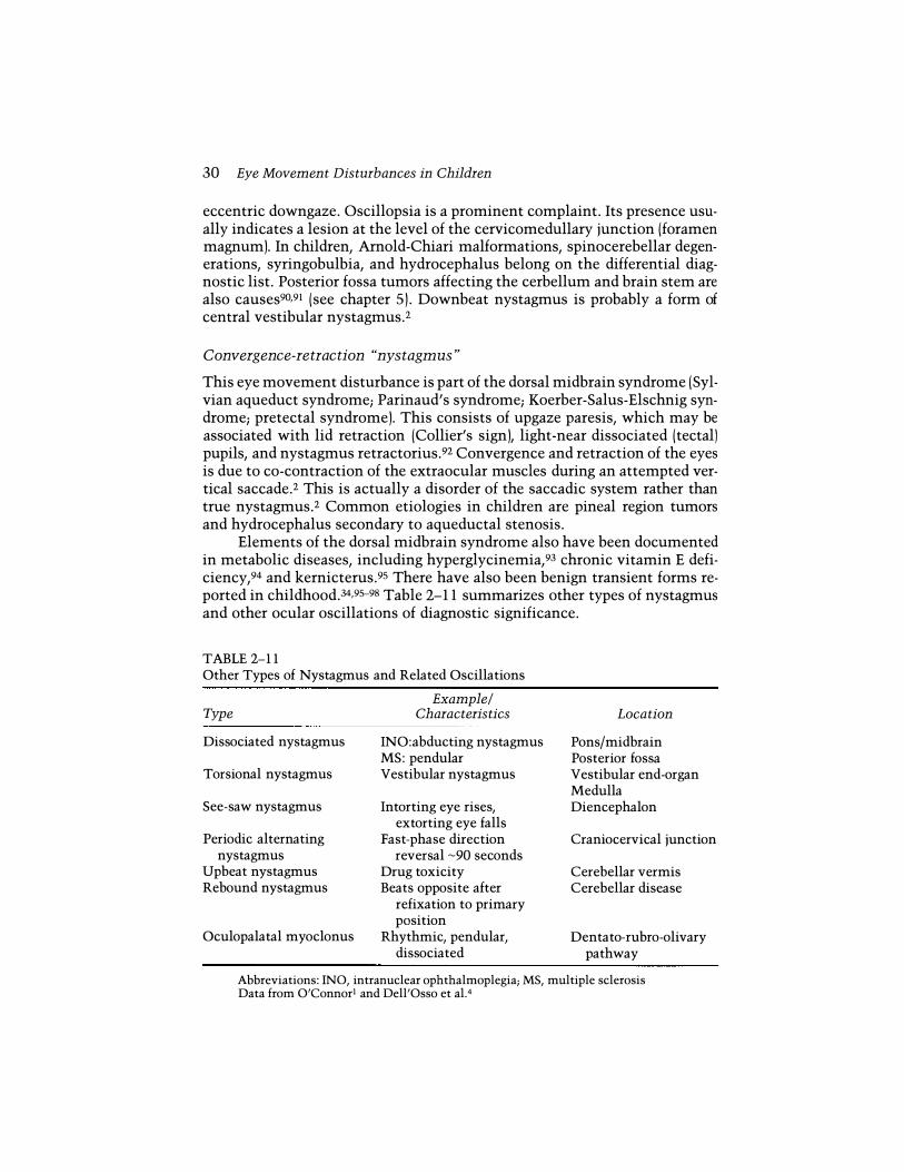
Elements of the dorsal midbrain syndrome also have been documented in metabolic diseases, including hyperglycinemia,93 chronic vitamin E deficiency,94 and kernicterus.95 There have also been benign transient forms reported in childhood.34,95-98 Table 2- 1 1 summarizes other types of nystagmus and other ocular oscillations of diagnostic significance.
TABLE 2-11 Other Types of Nystagmus and Related Oscillations
Example/ Type Characteristics
Dissociated nystagmus
Torsional nystagmus
See-saw nystagmus
Periodic alternating nystagmus
Upbeat nystagmus Rebound nystagmus
Oculopalatal myoclonus
INO:abducting nystagmus MS: pendular Vestibular nystagmus
Intorting eye rises, extorting eye falls
Fast-phase direction reversal -90 seconds
Drug toxicity Beats opposite after
refixation to primary position
Rhythmic, pendular, dissociated
Location
Pons/midbrain Posterior fossa Vestibular end-organ Medulla Diencephalon
Craniocervical junction
Cerebellar vermis Cerebellar disease
Dentato-rubro-olivary pathway
Abbreviations: INa, intranuclear ophthalmoplegia; MS, multiple sclerosis Data from O'Connori and Dell'Osso et a1.4
Saccadic Intrusions and Oscillations 3 1
Saccadic Intrusions and Oscillations
Ocular Dysmetria
This condition is reflected by the eyes overshooting (or undershooting) their intended target after saccadic refixational movements. Cerebellar disease, usually affecting the vermis, is the cause.2,4
Opsoclonus
This bizarre disorder of uncontrolled fast eye movements is also called saccadomania. It consists of conjugate uncontrollable multidirectional saccadic oscillations (without an intersaccadic interval) which persist in sleep.2A It is caused by pontine-pause cell dysfunction of as yet undetermined etiology. In children opsoclonus may be a paraneoplastic effect of occult neuroblastoma and may be associated with limb myoclonus (dancing eyes/dancing feet syndrome). Another common cause is viral encephalitis. Adrenocorticotropic hormone (ACTH) may improve this eye-movement disturbance.
Ocular Flutter
Ocular flutter and opsoclonus are part of a continuum and have the same pathogenesis and localizing significance. Flutter is represented by intermittent bouts of back-to-back saccades in the horizontal direction.
Voluntary "Nystagmus" (Psychogenic Flutter)
This is a volitionally induced "parlor trick," or functional eye movement disturbance, sometimes performed to feign organic illness. The movements consist of back-to-back horizontal sacca des (like ocular flutter) which can be sustained for no longer than 30 seconds.4 Facial grimacing is often associated. Because convergence is needed to initiate this phenomenon,2 voluntary "nystagmus" cannot be sustained by the subject and often stops on refixation or after one eye is occluded.
Other Saccadic Oscillations
A number of other saccadic oscillations include square-wave jerks, squarewave oscillations, macro square-wave jerks, macro saccadic oscillations, and saccadic pulses. They are defined by the characteristics noted by eyemovement recordings and all reflect cerebellar outflow dysfunction.2,4
Superior oblique myokymia (benign intermittent uniocular microtremor)
This condition presents as intermittent monocular oscillopsia, sometimes associated with transient vertical diplopia.2,4,99 Attacks usually last less than 10 seconds but may occur many times a day. The attacks may be brought on

32 Eye Movement Disturbances in Children
by looking down, tilting the head toward the side of the affected eye, and by blinking. Most of the time superior oblique myokymia is idiopathic, although occasional reports of associated neurologic abnormalities exist.lOO Until recently, controversy existed as to the cause of this condition. It is now quite likely that these eye movements are not centrally generated saccades, but are the result of superior oblique muscle fibers discharging at an irregular rate, probably in response to regenerative phenomena in mildly damaged trochlear motoneurons99 (see foreword).
Disorders of Saccadic Initiation
Congenital Oculomotor Apraxia
Congenital ocular motor apraxia (COMA) was first described by CoganlOl and consists of the inability to generate voluntary fast eye movements (saccades). This is compensated for by characteristic head thrusts, often accompanied by blinking.lOl ,102 The head thrusting serves to move the eyes passively by employing the vestibular ocular reflex; once the eyes are locked on target, the head is then straightened as gaze is maintained.2 Congenital oculomotor apraxia usually improves with age. Some children with COMA are neurologically normal while others have psychomotor retardation or developmental neurologic defects.2 It is presumed that cortical, rather than brain stem, ocular motor circuits involved in fast eye movements are abnormal in COMA. 102 The eye movement defects are usually restricted to the horizontal plane, although a case of pure vertical COMA has been reported.l03 By contrast, in acquired ocular motor apraxias, vertical saccades are usually abnormal as well,2,102,1 04 and these patients may have bilateral cortical or brain stem lesions. ! Os
Cyclic or Recurrent Oculomotor Dysfunction
Recurrences in ophthalmoplegic migraine are the rule, and the third cranial nerve is most often involved (discussed previously). Cyclic esotropia is a rare condition in which 24-hour periods of orthophoria alternate with 24-hour periods of esotropia. 106,107 This condition usually occurs in strabismic children between the ages of 4 and 10 years, but has been reported in an adult.l07 The mechanism of this eye-movement disturbance is not clear, but it does not seem to be related to underlying neurologic disease.l°6,107
Periodic alternating gaze deviation (PAGD) is a related abnormality consisting of conjugate lateral deviation of the eyes with contraversive head turning for 2 to 41 h minutes, followed by a brief transition period to opposite gaze and head position.108 The majority of these patients have un-
Miscellaneous Problems 33
derlying esotropia and all have posterior fossa abnormalities, usually involving the cerebellum.108
Periodic alternating esotropia is often associated with PAGD or periodic alternating nystagmus and has been reported with cerebellar vermis hypoplasia. 1 09
Other periodic eye movements include periodic alternating nystagmus, ocular tilt reaction, cyclic oculomotor paralysis, periodic alternating skew deviation, periodic alternating ping pong gaze, pretectal pseudobobbing, ocular neuromyotonia2-4,108 and spasm of the near reflex (see next section).
Miscellaneous Problems
Spasm of the Near Reflex
Spasm of the near reflex (spasm of accommodation; spasm of convergence) is an intermittent, sometimes uncomfortable, triad of convergence, accommodation, and pupillary miosis that can mimic unilateral or bilateral sixth nerve palsyl lO,1 1 1 (Figure 2- 1 1) and may provoke an extensive, expensive, and futile evaluation. I I I Episodic brow ache, variable diplopia, and blurred vision at distance are the direct results of the hyperactive near response. Spasm of the near reflex occurs most frequently in the late adolescent period. It is usually considered a functional disturbance, but can occur along with other organic conditions.1 1 2-1 1 4 The esotropic deviation is variable and cannot be sustained.
The diagnosis of spasm of the near reflex is made by the symptoms just noted and by the combination of intermittently crossed eyes accompanied by pupillary miosis. Also, if gaze is limited on binocular testing, the eyes will fully abduct if tested one at a time (Figure 2- 1 1).
The treatment of this condition is frustrating, but fortunately the problem is self-limited. Cycloplegic eye drops, such as homatropine, can be used to paralyze the accommodative response. Prisms, bifocals, and various other optical aids have been suggested, but rarely work.
Convergence Insufficiency
Convergence insufficiency reflects fatigue of the near response. As such, it is the opposite of convergence spasm. Divergence of the eyes at near (exophoria, exotropia) leads to symptoms of eye strain or double vision.ll5 Convergence insufficiency is common in otherwise healthy teenagers and young adults, but may accompany other states of debility.1 16 Somnolence after short periods of reading is a frequent complaint, and these individuals often arise early to do their reading as opposed to reading late into the evening. Diagnosis requires testing the power of the convergence system and is best done by the ophthalmologist. Treatment often utilizes eye exercises to strengthen the ability to converge.

34 Eye Movement Disturbances in Children
A
B
Dissociated Vertical Deviation
FIGURE 2-11 Spasm of convergence. (Note: pupils were pharmacologically dilated.) (A) Inability to abduct right eye on attempted gaze right. (B) Inability to abduct left eye on attempted gaze left. (e) Normal abduction of right eye with left eye covered. (0) Normal abduction of left eye with right eye covered.
This puzzling condition is reflected by turning of either eye upward when binocular fusion is interrupted, as when one eye is covered. On removal of the cover, the eye returns to primary position. In unilateral deviations, there is no movement of the fellow eye on alternate cover testing, thus ruling out a phoria or tropia. Downward movement of the covered eye may occur when filters of increasing density are held before the fixating eye (Bielschowsky phenomenon). Dissociated vertical deviation (DVD) is most often seen in children with other forms of strabismus, especially infantile esotropia.ll7 Lack of stereopsis is the rule, and latent nystagmus may be present.llB-1 20 Overaction of the ipsilateral inferior oblique muscle may be linked and thus

Miscellaneous Problems 35
c
D
confuse the examiner into the misdiagnosis of a fourth nerve palsy. However, as in Duane's syndrome, the majority of patients with DVD do not have diplopia because they supress the hypertropic eye.l l7
The cause of DVD is unknown (see forewordl. Initially it was thought that optic pathway misrouting akin to that seen in oculocutaneous albinism was present in DVD patients, but apparently this is not SO.t18,1 1 9 Dissociated horizontal deviations have been seen with DVD as well. 120,1 21
Double Elevator Palsy
In double elevator palsy (DEPl, one eye will not elevate in abduction, primary position, or in adduction, thus appearing as if there is a palsy of the inferior

36 Eye Movement Disturbances in Children
A
B
FIGURE 2-12 Double elevator palsy in a young man with microphthalmos. (A) Inability of elevation of left eye on adduction. (B) Inability of elevation of left eye on abduction.
oblique and superior rectus muscles. 1 22 A mild ptosis may be present, which usually disappears when the affected eye is forced to fixate. An apparent hypertropia of the unaffected eye then results and can confuse the examiner. In some cases Bell's phenomenon is present, suggesting a supranuclear cause. In others, Bell's phenomenon is absent on the affected side, suggesting a lesion in peripheral nerve or muscle.
In children, DEP usually is of congenital origin and diplopia is not a complaint.1 22 Restrictive causes such as ocular fibrosis syndromes and orbital floor fractures need to be considered, but can be eliminated from the diagnostic list by forced duction testing. Ocular myasthenia needs to be weighed in acquired cases of DEP.

References 37
Acquired monocular elevation paralysis has been seen in adults with unilateral contralateral pretectall23 or ipsilateral mesodiencephalic junction124 lesions.
Eye Movement Tics
Eye movement tics in children are seen between the ages of 6 and 12 years and are characterized by a stereotyped conjugate deviation of the eyes upward and outward. This movement disorder is sometimes preceded by discomfort in the eyes and seems to stop after a blink or two. Eye-movement tics may be idiopathic or a part of Tourette's syndrome. They are differentiated from nystagmus, ocular flutter, and ocular myoclonus by their lack of rhythmicity. Also, ocular gyric crisis is more sustained than are eye movement tics. They are usually self-limited with about two thirds showing spontaneous remission.125
References
1. O'Connor PS. Nystagmus. In: Farris BK (ed.), The basics of neuro-ophthalmology, St. Louis: Mosby Yearbook, 1991, pp. 209-235.
2. Leigh RL Zee DS. The neurology of eye movements, 2nd ed. Philadelphia: F.A. Davis, 1991.
3. Daroff RB, Troost BT, Leigh RJ. Supranuclear disorders of eye movement. In: Glaser JS (ed.), Neuro-ophthalmology, 2nd ed. Philadelphia: Lippincott, 1990, pp. 299-324.
4. Dell'Osso LF, Daroff RB, Troost BT. Nystagmus and saccadic intrusions and oscillations. In: Glaser JS (ed.), Neuro-ophthalmology, 2nd Ed. Philadelphia: Lippincott, 1990, pp. 325-356.
5. Tomsak RL. Geriatric neuro-ophthalmology. In: Kwitko ML, Weinstock FJ (eds.), Geriatric ophthalmology. Orlando: Grune and Stratton, 1985, p. 310.
6. Bajandas FL Kline LB. Neuro-ophthalmology review manual, 3rd ed. Thorofare, NJ: Slack, 1988.
7. Braziz PW. Localization of lesions of the oculomotor nerve: Recent concepts. Mayo Clin Proc 1991;66:1029-1035.
8. Trobe JD. Third nerve palsy and the pupil: Footnotes to the rule. Arch OphthalmolI988;106:601-602.
9. Gabianelli EB, Klingele TG, Burden RM. Acute oculomotor nerve palsy in childhood: Is arteriography necessary? J Clin Neuro OphthalmolI989;9:33-36.
10. Okano M, Matsuo T, Konishi H, et al. Anomalous posterior insertion of medial rectus muscle simulating congenital oculomotor palsy. Jpn J Ophthalmol 1990;34:275-279.
11. Herman JM, Rekate HL, Spetzler RF. Pediatric intracranial aneurysms: Simple and complex cases. Pediatr Neurosurg 1991-1992;17:66-73.
12. Ostergaard JR. Aetiology of intracranial saccular aneurysms in childhood. Br J Neurosurg 1991;5:575-580.
13. Ross JS, Masaryk TJ, Modic MT, et al. Intracranial aneurysms: Evaluation by MR angiography. AJNR 1990;11:449.
38 Eye Movement Disturbances in Children
14 . O'Day 1, Billson F, King J. Ophthalmoplegic migraine and aberrant regeneration of the oculomotor nerve . Br J OphthalmoI 1980;64:534-536.
15. Sibony PA, Lessell S, Gittinger JW. Acquired oculomotor synkinesis . Surv Oph· thalmoI 1984;28 :382-390.
16. Forster RK, Schatz NT, Smith JL . A subtle eyelid sign in aberrant regeneration of the third nerve. Am J OphthalmoI 1969;67:696-698 .
17. Boghen D, Chartrand J-p, Laflamme P, et al . Primary aberrant third nerve regeneration. Ann NeuroI 1979;6:415-418 .
18 . Pang MP, Zweifach PH, Goodwin J. Inherited levator-medial rectus synkinesis. Arch OphthalmoI 1986; l04 :1489-1491.
19. Bielschowsky A. Lectures on motor anomalies of the eyes . II. Paralysis of individual eye muscles . Arch OphthalmoI 1935;13:33.
20. Parks MM. Isolated cyclovertical muscle palsy. Arch Ophthalmol 1958;60: 1027-1035.
21. Flanders M, Draper J . Superior oblique palsy: Diagnosis and treatment. Can J OphthalmoI 1990;25: 17-24.
22. Harris D1, Memmen JE, Katz NNK, Parks MM. Familial congenital superior oblique palsy. Ophthalmology 1986;93:88-90.
23. Younge BR, Sutula F. Analysis of trochlear nerve palsies: Diagnosis, etiology and treatment. Mayo Clin Proc 1977;52:11-18 .
24 . Burger L, Kalvin N, Smith JL. Acquired lesions of the fourth cranial nerve. Brain 1970;93:567.
25. Kodsi SR, Younge BR. Acquired oculomotor, trochlear, and abducent cranial nerve palsies in pediatric patients. Am J OphthalmoI 1992;114:568-574.
26. Lee 1, Flynn JT. Bilateral superior oblique palsies. Br J Ophthalmol 1985;69: 508-513.
27. Keane JR. Trochlear nerve pareses with brainstem lesions. J Clin Neuro OphthalmoI 1986;6:242-246.
28 . Guy J, Day AL, Mickle JP, Schatz NJ. Contralateral trochlear nerve paresis and ipsilateral Horner's syndrome. Am J OphthalmoI 1989;107:73-76.
29. Miller NR. Walsh and Hoyt's Clinical Neuro-ophthalmology, 4th ed, Vol 2. Baltimore: Williams and Wilkins, 1985, p. 685.
30. Guy JR, Friedman WF, Mickle JP. Bilateral trochlear nerve palsy in hydrocephalus. J Clin Neuro OphthalmoI 1989;9 : 105-111.
31. Halpern JI, Gordon WHo Trochlear nerve palsy as a false localizing sign . Ann OphthalmoI 1981(Jan):53-56.
32. Bain PG, Larkin GBR, Calver DM, O'Brien MD. Persistent superior oblique paresis as a manifestation of familial periodic cerebellar ataxia. Br J Ophthalmol 1991; 75:619-621.
33. Bilateral superior oblique muscle palsy associated with Apert's syndrome. Am J OphthalmoI 1988;106:337-340.
34 . Reisner SH, Perlman M, Ben-Tovim N, et al . Transient lateral rectus muscle paresis in the newborn infant . J Pediatr 1971;78 :461-465.
35. Nixon RB, Helveston EM, Miller K, Archer SM, Ellis FD. Incidence of strabismus in neonates. Am J OphthalmoI 1985; l OO:798-801.
36. Taylor D. Pediatric ophthalmology. Cambridge, MA: Blackwell Scientific, 1990, p. 645.
37. Hoyt CS, Mousel DK, Weber AA. Transient supranuclear disturbances of gaze in healthy neonates. Am J OphthalmoI 1980;89:708-713.
References 39
38. Knox DL, Clark DB, Schuster FF. Benign sixth nerve palsies in children. Pediatrics 1967;40:560.
39. Boger WP III, Puliafito CA, Magoon EA, et al . Rucurrent isolated sixth nerve palsy in children. Ann OphthalmoI 1984; l6:237-244.
40. Reinecke RD, Thompson WE. Childhood recurrent idiopathic paralysis of the lateral rectus. Ann OphthalmoI 1981;13:1037-1039 .
41. Werner DB, Savino PJ, Schatz NJ. Benign recurrent sixth nerve palsies in childhood. Arch OphthalmoI 1983;101:607-608 .
42. Metz HS. Benign sixth nerve palsy of childhood. Am Orthoptic J 1983;33:42-47. 43 . Afifi AK, Bell WE, Menezes AH. Etiology of lateral rectus palsy in infancy and
childhood. J Child NeuroI 1992;7:295-299 . 44. Clark AC, Nelson LB, Simon JW, et al. Acute acquired comitant esotropia. Br J
Ophthalmol 1989; 73:636-638 . 45. Willaims AS, Hoyt CS. Acute comitant esotropia in children with brain tumors.
Arch OphthalmoI 1989; l07:376-378 . 46. Stem RM, Tomsak RL. Magnetic resonance images in a case of "divergence
paralysis. " Surv OphthalmoI 1986;30:397-401. 47. Pollard ZF. Accommodative esotropia after ocular and head injury. Am J Oph
thalmoI 1990; l09:195-198 . 48. Oesterle CS. Exercise induced esotropia. J Pediatr Ophthalmol Strabismus 1989;
26:150-151. 49 . Robertson DM, Hines JD, Rucker JW. Acquired sixth nerve palsy in children .
Arch OphthalmoI 1970;83:574-579. 50. Harley RD. Paralytic strabismus in children. Etiologic incidence and management
of the third, fourth and sixth nerve palsies. Ophthalmology 1980;87: 24-43. 51. Nixon RB, Helveston EM, Miller K, Archer SM, Ellis FD. Incidence of strabis
mus in neonates. Am J OphthalmoI 1985; lOO:798-801. 52. Miller MT, Ray V, Owens P, Chen F. Mobius and Mobius-like syndromes (TTV
OFM, OMLH). J Pediatr Ophthalmol Strabismus 1989;26:176-188. 53. Kishore K, Kumar H. Congenital ocular fibrosis with musculoskeletal abnor
mality: A new association. J Pediatr Ophthalmol Strabismus 1991;28:283-286. 54. Holmes JM, Cronin CM. Duane syndrome associated with oculocutaneous al
binism. J Ped Ophthalmol Strabismus 1991;28:32-34 . 55. Bixenman WW, Laguna JF. Acquired esotropia as initial manifestation of
Amold-Chiari malformation. J Pediatr Ophthalmol Strabismus 1981;18 :29-34. 56. Kandt RS, Goldstein GW. Steroid-responsive ophthalmoplegia in a child: Diag
nostic considerations. Arch NeuroI 1985;42:589-591. 57. Zasorin NL, Yee RD, Baloh RW. Eye-movement abnormalities in ophthalmo
plegia, ataxia and areflexia (fisher'S syndrome). Arch Ophthalmol 1985;103: 55-58.
58 . Price RL, O'Connor PS, Rothner AD. Acute ophthalmoplegia, ataxia, and areflexia ( Fisher syndrome) in childhood. Cleve Clin 1978;45:247-252.
59 . Daroff RB. Ocular myasthenia: Diagnosis and therapy. In: Glaser JS (ed.), Neuroophthalmology, Vol. 10. St. Louis: Mosby, 1980, pp. 62-71.
60. Morel E, Eymard B, Garabedian V, et al . Neonatal myasthenia gravis: A new clinical and immunologic appraisal on 30 cases . Neurology 1988;38: 38-142.
61. Spiro AJ, Pack DR. Disorders of immunologic dysfunction. In: Berg BO (ed.), Neurologic aspects of pediatrics. Boston: Butterworth-Heinemann, 1992, pp. 512-516.
40 Eye Movement Disturbances in Children
62. Goodwin JA. Ocular manifestations of myasthenia gravis. Ophthalmol Clin North Am 1992;5:495-512.
63. Daroff RE. Chronic progressive external ophthalmoplegia. Neurol Neurosurg Update 1983;4:2-7.
64. Kosmorsky GS, Johns DR. Neuro-ophthalmologic manifestations of mitochondrial DNA disorders: Chronic progressive external ophthalmoplegia, KearnsSayre syndrome and Leber's hereditary optic neuropathy. Neurol Clin 1991;9: 147-161.
65. Dell'Osso LF. Nystagmus, saccadic intrusions/oscillations and oscillopsia. In: van Dalen JTW, Lessell S (eds. l, Current neuro-ophthalmology, Vol. 3. St. Louis: �osby, 1991, pp. 153-192.
66. Abel LA. Ocular oscillations (congenital and acquiredl. In: Daroff RB, Neetens A (eds. l, Neurological organization of ocular movement. Berkley: Kugler, 1990, pp. 163-190.
67. Daroff RB, Neetens A (eds. l, Neurological organization of ocular movement. Berkley:Kugler, 1990.
68. Casteels I, Harris C�, Shawkat F, Taylor D. Nystagmus in infancy. Br J Ophthalmol 1992;76:434-437.
69. Trobe JO, Sharpe JA, Hirsh DK, Gebarski SS. Nystagmus of Pelizaeus�erzhacher disease: A magnetic search-coil study. Arch Neurol 1991;48: 87-91.
70. Gresty �, Bronstein �, Page NG, et al. Congenital-type nystagmus emerging later in life. Neurology 1991;44:653-656.
71. Dell'Osso LF, Daroff RB. Eye movement characteristics and recording techniques. In: Glaser JS (ed. 1t Neuro-ophthalmology. 2nd ed. Philadelphia: Lippincott, 1990, pp. 279-298.
72. Jan JE, Carruthers JDA, Tilson G. Neurodevelopmental criteria in the classification of congenital motor nystagmus. Can J Neurol Sci 1992;19:76-79.
73. Dell'Osso LF, Flynn JT. Congenitial nystagmus surgery: A quantitative evaluation of the effects. Arch Ophthalmol 1979;97:462-469.
74. Helveston E�, Sprunger DT. Strabismus and nystagmus surgery. Current Opinion Ophthalmol 1991;2:702-707.
75. Gresty �, �etcalfe T, Timms C, et al. Neurology of latent nystagmus. Brain 1992;115:1303-1321.
76. Zubcov AA, Reinecke RD, Gottlob I, et al. Treatment of manifest latent nystagmus. Am J Ophthalmol 1990;110:160-167.
77. Gelbart SSt Hoyt CS. Congenital nystagmus: A clinical perspective in infancy. In: Smith JL, Katz RS (eds. l, Neuro-ophthalmology enters the nineties. Hialeah, FL: Dutton, 1990, pp. 397-404.
78. Jan JE, Groenveld �, Connolly �B. Head shaking by visually impaired children: A voluntary neuro-visual adaptation which can be confused with spasmus nutans. Dev �ed Child Neurol 1990;32:1061-1066.
79. Cogan DG. Congenital nystagmus. Can J Ophthalmol l967;2:4-1O. 80. Leigh RJ, Thurston SEt Tomsak RL, Grossman GE, Lanska DJ. Effect of monoc
ular visual loss upon stability of gaze. Invest Ophthalmol Vis Sci 1989;30: 288-292.
81. Borchert �S. Nystagmus in childhood. Focal points: Clinical modules for ophthalmologists. San Francisco: American Academy of Ophthalmology, 1991, pp. 1-11.
References 41
82. Weissman BM, Dell'Osso LF, Abel LA, Leigh RJ. Spasmus nutans: A quantitative prospective study. Arch OphthalmolI987;105:525-528 .
83. Gottlob I, Zubcov AA, Wizov SS, Reinecke RD. Head nodding is compensatory in spasmus nutans. Ophthalmology 1992:99:1024-1031.
84. Lavery MA, O'Neill JF, Chu FC, Martyn LJ. Acquired nystagmus in early childhood: A presenting sign of intracranial tumor. Ophthalmology 1984;91: 425-435.
85. Donin JF. Acquired monocular nystagmus in children. Can J Ophthalmol 1967;2:212-215.
86. Wagner RS, Caputo AR, Reynolds RD. Nystagmus in Down's syndrome. Ophthalmology 1990;97:1439-1444.
87. Newman SA. Spasmus nutans-or is it? Surv OphthalmoI 1990;34:453-456. 88 . Bray PF. Can maternal alcoholism cause spasmus nutans in offspring? (letter).
N Engl J Med 1990:322:554. 89. Gottlob I, Zubcov A, Catalano RA, et al . Signs distinguishing spasmus nutans
(with and without central nervous system lesions) from infantile nystagmus. Ophthalmology 1990;97:1166-1175.
90. Schmidt D. Downbeat nystagmus. A clinical review. Neuro Ophthalmology 1991;11:247-263.
91. Yee RD. Downbeat nystagmus: Characteristics and localization of lesions. Trans Am Ophthalmol Soc 1989;87:984-1034.
92. Keane JR. The pretectal syndrome: 206 patients . Neurology 1990;40:684-690. 93. Nightingale S, Barton ME. Intermittent vertical supranuclear ophthalmople
gia and ataxia. Movement Disord 1991;6:76-78 . 94. Rosenblum JL, Keating JP, Prensky AL, Nelson JS. A progressive neurologic
syndrome in children with chronic liver disease. N Engl J Med 1981;304: 503-508 .
95. Hoyt CS, Billson FA, Alpins N. The supranuclear disturbances of gaze in kernicterus. Ann OphthalmoI 1978;(November):1487-1492.
96. Ahn JC, Hoyt WF, Hoyt CS. Tonic upgaze in infancy: A report of three cases. Arch OphthalmoI 1989;107:57-58 .
97. Mets MB. Tonic upgaze in infancy. Arch OphthalmoI 1990;108:482-483. 98 . Deonna T, Roulet E, Meyer HU. Benign paroxysmal tonic upgaze of child
hood-A new syndrome. Neuropediatrics 1990;21:213-214. 99. Leigh RJ, Tomsak RL, Seidman SH, Dell'Osso LF. Superior oblique myokymia:
Quantitative characteristics of the eye movements in three patients. Arch OphthalmoI 1991:109:1710-1713.
100. Morrow MJ, Sharpe JA, Ranalli PJ. Superior oblique myokymia associated with a posterior fossa tumor: Oculographic correlation with an idiopathic case. Neurology 1990;40:367-370.
101. Cogan DG. A type of congenital ocular motor apraxia presenting jerky head movements. Trans Am Acad Ophthalmol OtolaryngoI 1952;56:853-862.
102. Tusa RJ. Saccadic eye movements: Supranuclear control . In: Daroff RB, Neetens A (eds .), Neurological organization of ocular movement. Berkley:Kugler 1990, pp. 67-111.
103. Ebner R, Lopez L, Ochoa S, Crovetto L. Vertical ocular motor apraxia. Neurology 1990;40:712-713.
104. Aicardi J, Barbosa C, Andermann E, et al . Ataxia-ocular motor apraxia: A syndrome mimicking ataxia-telangiectasia. Ann NeuroI 1988;24:497-502.

42 Eye Movement Disturbances in Children
105. Hanson MR, Hamid MA, Tomsak RL, Chou SS, Leigh RJ. Selective saccadic palsy caused by pontine lesions: Clinical, physiological, and pathological correlations. Ann Neurol 1986;20:209-217.
106. Friendly OS, Manson RA, Albert DG. Cyclic strabismus-A case study. Doc Ophthalmol 1973;34:189-202.
107. Troost BT, Abel A, Noreika J, Genovese FM. Acquired cyclic esotropia in an adult. Am J Ophthalmol 1981;91:8-13.
108. Legge RH, Weiss HS, Hedges TH III, Anderson ML. Periodic alternating gaze deviation in infancy. Neurology 1992;42:1740-1743.
109. Hamed LF, Silbiger J. Periodic alternating esotropia. J Pediatr Ophthalmol Strabismus 1992;29:240-242.
110. Cogan DG, Freese CG. Spasm of the near reflex. Arch Ophthalmol 1955;54: 752-759.
111. Griffin JF, Wray SH, Anderson DP. Misdiagnosis of spasm of the near reflex. Neurology 1976;26:1018-1020.
112. Dagi LR, Chrousos GA, Cogan DG. Spasm of the near reflex associated with organic disease. Am J Ophthalmol 1987;103:582-585.
113. Moster ML, Hoenig EM. Spasm of the near reflex associated with metabolic encephalopathy. Neurology 1989;38 :150.
114. Tomsak RL. Spasm of the near reflex following myelography. Neurology 1991;41:460.
115. Owens DA, Wolf-Kelly K. Near work, visual fatigue, and variations of oculomotor tonus. Invest Ophthalmol Vis Sci 1987;28 : 743-749.
116. Krohel GB, Kristan RW, Simon JW, Barrows NA. Posttraumatic convergence insufficiency. Ann Ophthalmol 1986; l8 :101-104.
117. van Noorden GK. Burian-von Noorden's binocular vision and ocular motility: Theory and management of strabismus, 3rd ed. St. Louis: Mosby, 1985, pp. 320-323.
118. Zubcov AA, Fendick MG, Gottolb I, et al. Visual-evoked cortical potentials in dissociated vertical deviation. Am J Ophthalmol 1991;1 l2 : 714-722.
119. Boylan C, Clement RA, Howrie A. Normal visual pathway routing in dissociated vertical deviation. Invest Ophthalmol Vis Sci 1988;29 :1165-1167.
120. Zubcov AA, Reinecke RD, Calhoun JH. Asymmetric horizontal tropias, DVD, and manifest latent nystagmus: An explanation of dissociated horizontal deviation. J Pediatr Ophthalmol Strabismus 1990;27:59-64.
121. Wilson ME, McClatchye SK. Dissociated horizontal deviation. J Pediatr OphthaI mol Strabismus 1991;28 :90-95.
122. von Noorden GK. Burian-von Noorden's binocular vision and ocular motility: Theory and management of strabismus, 3rd ed. St. Louis: Mosby, 1985, pp. 362.
123. Jampel RS, Fells P. Monocular elevation paresis caused by a central nervous system lesion. Arch Ophthalmol 1968;80:45-57.
124. Ford CS, Schwartze GM, Weaver RG, Troost BT. Monocular elevation paresis caused by an ipsilateral lesion. Neurology 1984;34:1264-1267.
125. Binyon S, Prendergast M. Eye-movement tics in children. Dev Med Child Neurol 1991;33:343-345.

Pediatric N euroophthalmology
Edited by Robert L. Tomsak, M.D ., Ph.D. Head, Section of Neuro-ophthalmology The Mount Sinai Medical Center Clinical Associate Professor of
Ophthalmology and Neurology Case Western Reserve University School of Medicine Cleveland, Ohio
With 1 2 contributors
Foreword by J. Lawton Smith, M.D . Professor Emeritus, Department of Ophthalmology, University of Miami School of Medicine; Editor, Journal of Neuro-ophthalmology
Butterworth-Heinemann Boston London Oxford Singapore Sydney Toronto Wellington