extracorporeal shockwave therapy for … shockwave therapy for musculoskeletal indications march 29,...
TRANSCRIPT

Extracorporeal Shockwave Therapy for Musculoskeletal Indications
March 29, 2004
Technology Assessment Update Office of the Medical Director
Department of Labor and Industries
Table of Contents
Topic Page Background and FDA Status 1 ESWT for the Treatment of Plantar Fasciitis 2 High energy 2 Low energy 4 ESWT for the Treatment of Lateral Epicondylitis 10 ESWT for the Treatment of Calcifying Tendonitis of the Shoulder 13 Insurers in Washington 20 Conclusions 21
References 22
Last modified March 29, 2004
FDA Device Status
Background In January 2003, the Department of Labor and Industries conducted a technology assessment of Extracorporeal Shockwave Therapy (ESWT) for musculoskeletal indications, including plantar fasciitis, lateral epicondylitis, and tendonitis. At that time, ESWT was deemed a noncovered therapy due to the controversial nature of the evidence. The results from randomized trials were conflicting, and study protocols indicated uncertainty concerning treatment parameters. (LNI 2003) The department has been presented with several new studies in order to reconsider its noncoverage decision. This second review assesses studies that were published in English between January 2003 and March 2004.
FDA Device Status The FDA granted a Pre-Market Approval to HealthTronics for their OssaTron system in October 2000. The system received a classification of “Generator, Shockwave, (For Pain Relief)”. OssaTron uses high-energy electrohydraulic technology to generate shockwaves for the treatment of proximal plantar fasciitis that has failed to respond to conservative treatment. Chronic proximal plantar fasciitis is defined as “pain in the area of the insertion of the plantar fascia on the medial calcaneal tuberosity that has persisted for more than six months.” (FDA 2000) The FDA granted a second Pre-Market Approval to HealthTronics for their OssaTron system in March 2003. The approval allows marketing of the OssaTron for the treatment of chronic lateral epicondylitis (FDA 2003) In January 2002, the FDA granted a Pre-Market approval to Dornier for its EPOS Ultra Device. The EPOS Ultra treats plantar fasciitis, which is defined as the “traction degeneration of the plantar fascial band at its origin on the medial tubercle of the calcaneus.” (FDA 2002) The FDA approved in 2002 the Siemens Sonocur for patients with chronic lateral epicondylitis for 6 months or more and a history of unsuccessful conservative treatments. (FDA 2002a)
Last modified March 29, 2004 Page 1
ESWT for Plantar Fasciitis
Extracorporeal Shockwave Therapy for Plantar Fasciitis Plantar fasciitis is often characterized by inflammation of the plantar fascia, thickening of the proximal fascia, decreased vascularity, peritendinous inflammation, loss of normal elasticity, and alteration of nociceptor physiology. (Rompe 2003) Advocates suggest that ESWT acts by creating a controlled acute injury or trauma. Hemorrhage, clotting, granulation tissue, and neovascularity with release of local growth factors may result. Therefore, the treatment stimulates healing by creating a wound environment. (Perez 2003) ESWT may be administered with high energy or low energy. However, no consensus exists concerning the repeated use of low energy shock waves without anesthesia versus single use of high-energy shock waves requiring local or regional anesthesia. (Rompe 2003) High Energy ESWT I. Secondary data analysis from FDA trials
Two publications on the high energy OssaTron involve patients from an FDA trial. The patient cohort included randomized and nonrandomized patients. Following FDA approval, additional patients were entered into a nonrandomized study. All patients experienced chronic pain in the inferior heel at the proximal insertion of the plantar fascia for 6 months. Pre-FDA approval patients received 1 or 2 treatments under conscious sedation or ankle block anesthesia. The second treatment occurred at least 3 months after initial treatment. 1500 shocks (0.22 mJ/mm2) were delivered at a rate of 2 Hz for a total energy of 330 J. Patients received treatment over a 2-cm area around the predetermined point of maximal tenderness. Post FDA study heels received 2000 shocks (0.27 mJ/mm2) delivered at a rate of 4 Hz. Total energy was 540 J.
The FDA study used several measurement tools: the Roles and Maudsley system, physician exam, pain testing with a dolorimeter, morning pain, and pain with activities of daily living. A satisfactory outcome was defined as 50% improvement from baseline. a. Lee analyzed radiographic data to determine whether the presence of a calcaneal
bone spur affects symptoms after ESWT and whether ESWT changes heel spur morphology. Pretreatment, 3 months, and 1 year radiographs were examined. (Lee 2003)
Last modified March 29, 2004 Page 2
ESWT for Plantar Fasciitis
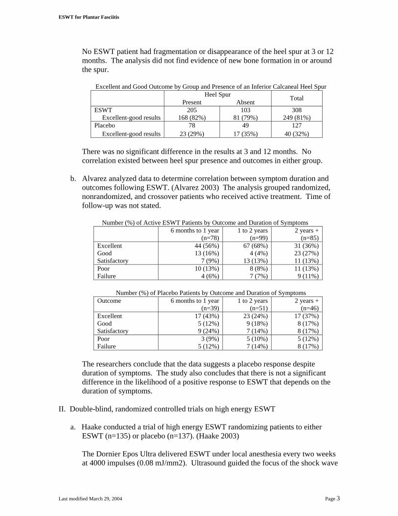
No ESWT patient had fragmentation or disappearance of the heel spur at 3 or 12 months. The analysis did not find evidence of new bone formation in or around the spur.
Excellent and Good Outcome by Group and Presence of an Inferior Calcaneal Heel Spur Heel Spur Present Absent Total
ESWT 205 103 308 Excellent-good results 168 (82%) 81 (79%) 249 (81%)
Placebo 78 49 127 Excellent-good results 23 (29%) 17 (35%) 40 (32%)
There was no significant difference in the results at 3 and 12 months. No correlation existed between heel spur presence and outcomes in either group.
b. Alvarez analyzed data to determine correlation between symptom duration and
outcomes following ESWT. (Alvarez 2003) The analysis grouped randomized, nonrandomized, and crossover patients who received active treatment. Time of follow-up was not stated.
Number (%) of Active ESWT Patients by Outcome and Duration of Symptoms
6 months to 1 year (n=78)
1 to 2 years (n=99)
2 years + (n=85)
Excellent 44 (56%) 67 (68%) 31 (36%) Good 13 (16%) 4 (4%) 23 (27%) Satisfactory 7 (9%) 13 (13%) 11 (13%) Poor 10 (13%) 8 (8%) 11 (13%) Failure 4 (6%) 7 (7%) 9 (11%)
Number (%) of Placebo Patients by Outcome and Duration of Symptoms
Outcome 6 months to 1 year (n=39)
1 to 2 years (n=51)
2 years + (n=46)
Excellent 17 (43%) 23 (24%) 17 (37%) Good 5 (12%) 9 (18%) 8 (17%) Satisfactory 9 (24%) 7 (14%) 8 (17%) Poor 3 (9%) 5 (10%) 5 (12%) Failure 5 (12%) 7 (14%) 8 (17%)
The researchers conclude that the data suggests a placebo response despite duration of symptoms. The study also concludes that there is not a significant difference in the likelihood of a positive response to ESWT that depends on the duration of symptoms.
II. Double-blind, randomized controlled trials on high energy ESWT
a. Haake conducted a trial of high energy ESWT randomizing patients to either ESWT (n=135) or placebo (n=137). (Haake 2003)
The Dornier Epos Ultra delivered ESWT under local anesthesia every two weeks at 4000 impulses (0.08 mJ/mm2). Ultrasound guided the focus of the shock wave
Last modified March 29, 2004 Page 3

ESWT for Plantar Fasciitis
at the heel spur at the insertion of the fascia. The total positive dose was 0.96 J/mm2, the EFD was 0.22 mJ/mm2, and the positive pressure was 13.7 MPa.
Placebo consisted of treatment with a polyethylene foil filled with air fixed with ultrasound gel in front of the coupling cushion to reflect the shock waves.
The primary endpoint was the success rate after 12 weeks. Success was defined as a Roles and Maudsley score of 1 or 2 and no additional treatment. Additional treatment was allowed after assessment of the primary endpoint. Secondary endpoints included pain at rest, night, pressure, and morning measured with a 10-point VAS, walking ability, and need for additional treatments at 1 year.
Follow-up occurred at 6 and 12 weeks and 1 year.
A study population of 272 patients had adequate power to detect a 20% difference in success rates, allowing for a 20% dropout.
Study population: The study included 272 patients with unilateral plantar fasciitis and heel spur. Patients failed 6 months of conservative treatment (minimum of two local injections plus six sessions of physical therapy plus custom orthotics), but had not received therapy for 4 weeks before referral.
Patients were excluded due to dysfunction of foot or ankle, arthrosis, arthritis, neurological abnormality, nerve entrapment, vascular abnormality, operative treatment of the heel spur, or hemorrhagic disorders.
Patient Demographic
ESWT (n=135) Placebo (n=136) Mean age 53.1 years 52.9 years Median history of heel pain 13 months 13 months Median conservative treatment 12 months 11 months
Results: The primary endpoint was assessed in 94% of the patients. The difference in success rates at 12 weeks between groups was 3.6% (43/127 ESWT, 39/129 placebo). At one year follow-up, 91 of 113 (81%) and 87 of 115 (76%) in the placebo group had a Roles Maudsley score of 1 or 2. 41 ESWT patients and 64 placebo patients sought additional treatment.
Conclusion: The researchers found no meaningful improvement of clinical outcome in patients treated with ESWT for plantar fasciitis compared with placebo.
Low Energy ESWT I. Double-blind, randomized, controlled trial on low energy ESWT
Last modified March 29, 2004 Page 4

ESWT for Plantar Fasciitis
a. Speed conducted an intention-to-treat trial using moderate dose ESWT with the Sonocur Plus in the management of plantar fasciitis. (Speed 2003)
According to randomization tables, subjects received at monthly intervals: 1. ESWT with an inflated treatment head propagating 1500 pulses at 0.12
mJ/mm2 2. Sham treatment with a deflated treatment head and no coupling gel. Minimal
energy (0.04 mJ/mm2) was generated, but without contact to the skin.
The study did not use local anesthesia, but used 2 parameters to focus treatment: ultrasonographic localization and focus alteration according to the site of maximum local pain.
A blinded observer assessed patients using a 100-point VAS prior to treatment and 1 and 4 months after therapy. A positive response was defined as a 50% improvement from baseline at 3 months (1 month following final treatment).
No other treatments were permitted during study period.
Study population: The study included 88 subjects with unilateral plantar heel pain for at least 3 months. Patients experienced point tenderness at or near the medial calcaneal insertion.
Patients were excluded due to additional pathology such as instability, arthritis, or diffuse heel pad tenderness. Patients who had undergone treatment within the previous 6 weeks were also excluded.
Patient Demographics ESWT (n=46) Placebo (n=42) Mean age 51.7 years 52.5 years Mean duration 16.7 months 13.6 months Did not complete study 4 subjects 8 subjects
Results:
Subjects with 50% Improvement from Baseline at 3 months
ESWT Placebo Pain 17 (37%) 10 (24%) Night pain 19 (41%) 13 (31%) Start-up pain 19 (41%) 15 (36%)
Both groups showed significant improvement over the course of the study, which was maintained at the 6 month follow-up assessment.
Conclusion: Results indicate that moderate dose ESWT delivered using a electromagnetic generator has no significant benefit over placebo.
Last modified March 29, 2004 Page 5
ESWT for Plantar Fasciitis
II. Single-blind, randomized controlled trials on low energy ESWT
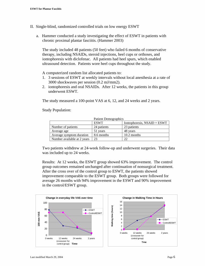
a. Hammer conducted a study investigating the effect of ESWT in patients with chronic proximal plantar fasciitis. (Hammer 2003)
The study included 48 patients (50 feet) who failed 6 months of conservative therapy, including NSAIDs, steroid injections, heel cups or orthoses, and iontophoresis with diclofenac. All patients had heel spurs, which enabled ultrasound detection. Patients wore heel cups throughout the study.
A computerized random list allocated patients to: 1. 3 sessions of ESWT at weekly intervals without local anesthesia at a rate of
3000 shockwaves per session (0.2 mJ/mm2). 2. iontophoresis and oral NSAIDs. After 12 weeks, the patients in this group
underwent ESWT.
The study measured a 100-point VAS at 6, 12, and 24 weeks and 2 years.
Study Population:
Patient Demographics ESWT Iontophoresis, NSAID + ESWT Number of patients 24 patients 23 patients Average age 51 years 48 years Average symptom duration 8.6 months 10.2 months Number available at 2 years 23 22
Two patients withdrew at 24-week follow-up and underwent surgeries. Their data was included up to 24 weeks.
Results: At 12 weeks, the ESWT group showed 63% improvement. The control group outcomes remained unchanged after continuation of nonsurgical treatment. After the cross over of the control group to ESWT, the patients showed improvement comparable to the ESWT group. Both groups were followed for average 26 months with 94% improvement in the ESWT and 90% improvement in the control/ESWT group.
Change in Walking Time in Hours
02468
10121416
0 weeks 12 weeks(crossover forcontrol group)
24 weeks 2 years
Time
Wal
king
tim
e (h
ours
)
ESWTControl/ESWT
Change in everyday life VAS over time
0
20
40
60
80
100
0 weeks 12 weeks(crossover forcontrol group)
24 weeks 2 years
Time
100
mm
VA
S
ESWTControl/ESWT
Last modified March 29, 2004 Page 6

ESWT for Plantar Fasciitis
b. Rompe conducted a trial of electromagnetic ESWT to bring about pain relief for chronic plantar fasciitis in recreational athletes who ran more than 30 miles per week. Subjects with plantar fasciitis for more than 12 months were randomized to 3 applications of 2100 impulses or 3 placebo applications. ESWT was delivered without anesthesia using a total of 6300 shocks in 3 treatment sessions (EFD 0.16 mJ/mm2, 4 Hz). (Rompe 2003) The primary outcome was a reduction on the 10-point pain VAS on first walking in the morning. Secondary outcome included a 50% reduction of pain in the morning, VAS less than 4, and improvement on the100-point American Orthopaedic Foot and Ankle Society’s Ankle Hindfoot Scale. Follow-up occurred at 6 months.
No other treatment was permitted until 6 weeks after shockwave application with the exception of shoe inserts.
The study had power to detect a 3-point difference on the 10-point pain VAS.
Study Population: The study included 45 patients with chronic heel pain defined as symptoms of moderate to severe heel pain in the involved foot at the origin of the proximal plantar fascia on the medial calcaneal tuberosity. Patients failed 3 attempts at nonoperative treatment, including at least 2 of the following: PT, orthotics, or pharmacological treatment.
Patients were excluded due to arthritis, ankylosing spondylitis, Reiter’s syndrome, neurologic abnormalities, nerve entrapment, plantar fascial surgery, bilateral heel pain, workers’ compensation, or NSAIDs for other chronic conditions.
Patient Demographic
ESWT Sham ESWT Number of patients 22 patients 23 patients Average age 43 years 40 years Average symptom duration 20 months 18 months
Results:
Mean Pain VAS on First Walking in the Morning
Initial rating 6 months 1 year ESWT 6.9 (n=22) 2.1 (n=19) 1.5 (n=16) Sham 7.0 (n=23) 4.7 (n=20) 4.4 (n=19)
Number (%) of patients with more than 50%
improvement in pain of first walking in the morning 6 months 1 year ESWT 12 of 20 patients (60%) 13 of 18 patients (72%) Sham 6 of 22 patients (27%) 7 of 20 patients (35%)
One patient in each group underwent surgery.
Last modified March 29, 2004 Page 7

ESWT for Plantar Fasciitis
Conclusion: Results of the current study revealed beneficial effects of low-energy ESWT in long distance runners with chronic plantar fasciitis.
III. Case Series with Comparison Group
a. Hammer investigated the effect of ESWT on the ultrasonographic appearance of plantar fasciitis. Three sessions of ESWT on a piezoelectric system delivered 3000 shock waves per session (0.2 mJ/mm2) without local anesthesia at weekly intervals. (Hammer 2003a) Patients underwent ultrasound before ESWT and at 6, 12, and 24 weeks. Pain was measured with a 100-point VAS in different situations (rest, activities of daily living, one leg stance). Comfortable walking time was also assessed.
Study Population: The study included 22 patients with unilateral plantar fasciitis with heel spurs who failed conservative therapy for 6 months. The contralateral plantar fascia was used as a comparison. Patients had an average age of 51.6 years and an average duration of symptoms of 8.8 months.
Two patients were lost at 12-weeks, and 4 patients were lost at 24 weeks.
Results: Before ESWT, the mean thickness of the plantar fascia was significantly greater on the symptomatic side (5.2 mm) than on the asymptomatic side (4.3 mm). However, no significant differences existed at 6, 12, and 24-week follow-up. Six months after ESWT, the decrease in thickness (5.2 mm) was significant on the plantar fasciitis side whereas no significant change occurred on the comparison side.
Pain on the VAS and walking time before and after ESWT
Baseline 6 weeks 12 weeks 24 weeks VAS
Rest 42.5 34.6 7.5 7.5 Daily activity 78.2 30.0 25.5 16.3
Walking time 0.1 6.7 7.5 9.8
Thickness of the plantar fascia (ultrasonographic measurement) before and after ESWT
0
1
2
3
4
5
6
baseline (n=22) 6 weeks (n=22) 12 weeks (n=20) 24 weeks (n=16)time
thic
knes
s (m
m)
plantar fasciitis (mm)pain-free, control (mm)
Last modified March 29, 2004 Page 8

ESWT for Plantar Fasciitis
Six months after ESWT, patients with little pain had significantly thinner plantar fascias compared to patients with persistent pain. The thickness of the plantar fascia seemed to be related to the response to treatment.
Conclusion: Painful plantar fascia was ultrasonographically thicker than pain-free comparison heels. After ESWT, decreases in thickness as well as pain improvement were significant.
Last modified March 29, 2004 Page 9
ESWT for Lateral Epicondylitis
Lateral Epicondylitis I. Double-blind RCT
a. In March 2003, HealthTronics submitted to the Food and Drug Administration a pre-market approval study. The randomized, double-blind, sham controlled trial evaluated the OssaTron for lateral epicondylitis. (FDA 2003)
After receiving local anesthetic or bier block, 1500 shocks at a power setting of 18kV were delivered to subjects. For the sham group, a styrofoam block was placed against the coupling membrane of the shock head to absorb waves. A fluid-filled IV bag was then placed between the styrofoam block and the elbow to mimic the feel of the membrane. The study defined primary effectiveness as meeting 3 criteria at 8-weeks: 1. Minimum 50% improvement on investigator assessment of pain and score less
than 4 2. Minimum 50% improvement on pain VAS and score less than 4 3. No more than 3 doses of medication during the week prior to evaluation
Follow-up occurred at 4 and 8 weeks.
Study Population: The study included 183 randomized to either active ESWT treatment or sham. Patients had lateral epicondylitis for 6 months that failed conservative therapy. Patients' investigator assessment and VAS scores were also greater than or equal to 5 out of 10.
The study excluded patients due to vascular insufficiency, neuropathy, osteoarthritis, rheumatoid arthritis, osteoporosis, metabolic disorders, Paget’s disease, osteomyelitis, or fracture.
Patient Demographics
ESWT (n=93)
Placebo (n=90)
Mean age 44 years 46 years Mean duration of symptoms 684 days 784 days
18 randomized subjects withdrew or were lost to follow-up. Therefore, 165 randomized subjects were assigned success or failure status.
Results:
Last modified March 29, 2004 Page 10

ESWT for Lateral Epicondylitis
Investigator assessment by study group over time
0
2
4
6
8
10
Baseline Week 4 Week 8Time
Inve
stig
ator
as
sess
men
t
ESWT
Placebo
Patient VAS by study group over time
0
2
4
6
8
10
Baseline Week 4 Week 8Time
VAS
ESWT
Placebo
Number of patients not using pain medications
0153045607590
Baseline Week 4 Week 8Time
Num
ber o
f pat
ient
s
ESWT
Placebo
The majority of the treatment effect was due to blinded evaluator’s assessments. Secondary outcomes were not statistically significant.
Response to Treatment at 8 weeks
ESWT (n=93)
Placebo (n=90)
P value
Investigator assessment 45 (48%) 26 (29%) 0.007 VAS 51 (55%) 37 (41%) 0.063 Medication use 75 (81%) 63 (70%) 0.095 All 3 Components 33 (35%) 20 (22%) 0.043
Conclusion: Based on the findings, the FDA granted approval to HealthTronics to market the OssaTron for lateral epicondylitis.
b. Melikyan conducted a double-blinded trial randomizing patients to either ESWT or placebo to examine the effectiveness of ESWT for lateral epicondylitis. All patients previously failed physiotherapy in the form of splintage and exercise and at least one injection of cortisone. (Melikyan 2003)
Ultrasound guided the tangential application of the shock waves to the common extensor origin. Three sessions without local anesthesia applied energy (333 mJ/mm2) for a total of 1000 mJ/mm2. Ultrasound gel acted as a conductive medium between the skin and treatment head for the treatment group. A foam pad acted as a reflective medium between the skin and treatment head for the control group.
Last modified March 29, 2004 Page 11

ESWT for Lateral Epicondylitis
Blinded observers measured the Disabilities of Arm, Shoulder and Hand (DASH) function/symptom score, pain VAS, grip strength and analgesic requirement to assess outcomes at 3 and 12 months. The end point for study participation was either surgery as originally planned or a request to be removed from the surgical waiting list.
Study Population: The study included 86 patients randomized to either treatment or placebo. 75 patients followed the full course of therapy, but one person did not attend follow-up appointments. As a result, 74 patients with a mean age of 43.4 years were included in the final assessment (37 ESWT, 37 control).
All patients had pain localized to the lateral epicondyle and tenderness over the lateral epicondyle, the supracondylar ridge and the first 2 cm of the extensor muscle mass. Patients also experienced increased pain on resisted wrist extension or increased pain on elbow extension with full wrist flexion.
The study excluded patients due to pain over the posterior interosseous nerve, positive resisted supination test, pain over the radiohumeral joint, exacerbation of pain with movement of the neck, sensory disturbance, previous surgery, or history of fracture.
Results: The study showed no significant difference between the groups on DASH functional/symptom score, pain on lifting a 5 kg dumbbell, pain VAS, grip strength, or the proportion of patients needing analgesics at any point, including baseline values.
17of 37 ESWT patients (46%) and 16 of 37 control patients (43%) underwent surgical release of the common extensor origin.
Conclusion: The study showed no evidence that ESWT for tennis elbow is better than placebo.
Last modified March 29, 2004 Page 12

Extracorporeal Shockwave Therapy for Calcific Tendonitis of the Shoulder
Calcific Tendonitis of the Shoulder
Advocates of ESWT for calcific tendonitis of the shoulder suggest that pain relief is achieved through hyperstimulation analgesia. As a result, patient pain threshold increases. After relieving pain, shoulder joint ROM and activity ability increase. ESWT may also produce a knocking force on the tendon that relieves the adhesion resulting from the chronic tendonitis. (Pan 2003) ESWT acts as an alternative to surgical treatment, which aims to remove calcium deposits and to decompress the subacromion space. While many patients with calcific tendonitis remain asymptomatic depending on the position and size of the calcification, ESWT may assist those patients who experience pain and impaired function. (Wang 2003) I. Double blinded RCT
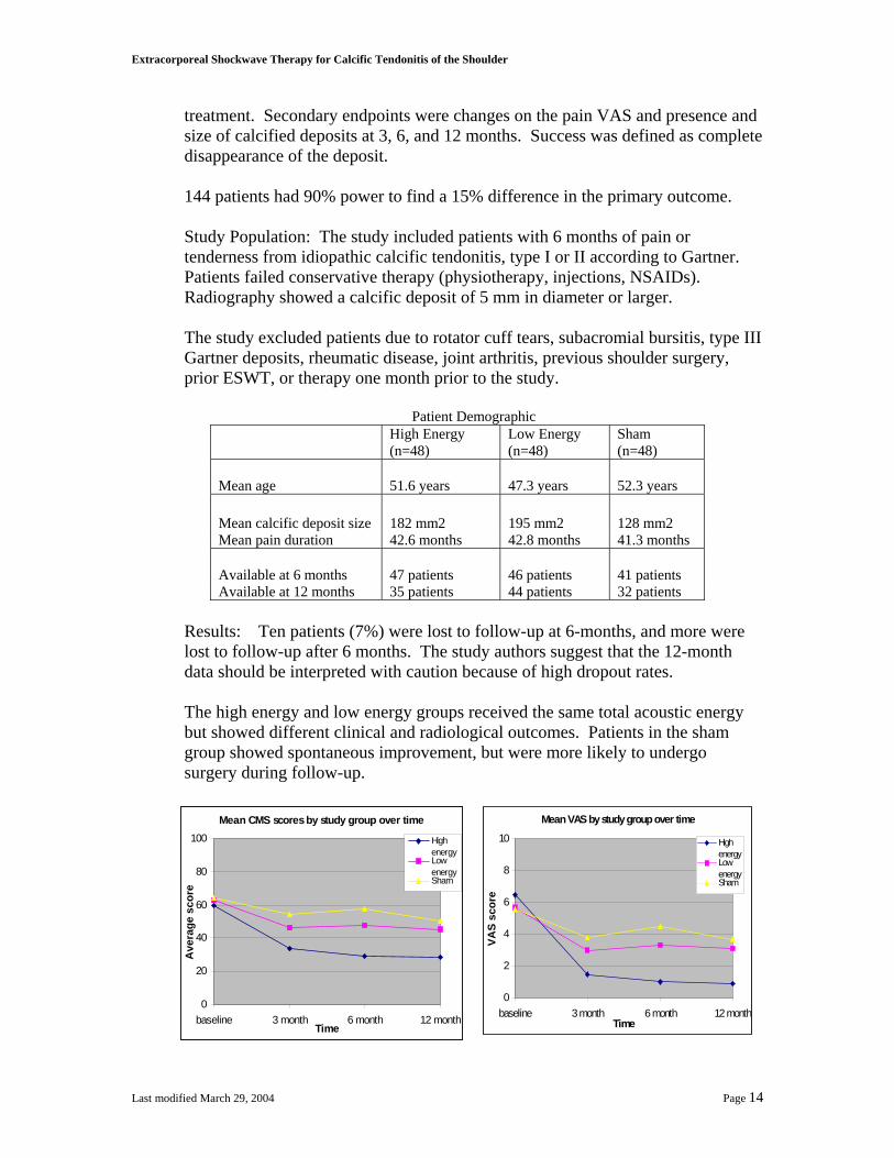
a. Gerdesmeyer compared high energy, low energy and placebo ESWT treatment in patients with chronic symptomatic calcific tendonitis of the supraspinatus tendon. The double-blind, placebo-controlled trial was conducted at 7 sites in Germany and Austria. Block randomization (48 per block) with a computer-generated algorithm allocated patients to treatment group. (Gerdesmeyer 2003)
Fluoroscopy identified the deposit. A sheet of polyethylene foil coupled the shockwave head to the patient’s shoulder. Coupling gel was used between the shockwave head and the foil and between the foil and the shoulder. ESWT was delivered after administering adequate analgesia without local anesthetics.
Both groups received 120 impulses per minute. The high-energy group received 1500 shockwaves at 0.32 mJ/mm2 per treatment. The low energy group received 6000 shockwaves at 0.08 mJ/mm2 per treatment. All patients received a second treatment after 12 to 16 days for a cumulative dose of 0.96 J/mm2.
The control group received sham treatment from an air chambered polyethylene foil with coupling gel placed against the skin. No coupling gel was applied to the shockwave head. A glass-fiber hydrophone demonstrated that no shockwaves passed through the foil. The device emitted 1500 shockwaves with an energy level of 0.32 mJ/mm2 for a total of 0.96 J/mm2.
All patients had 10 physiotherapy sessions following treatment and medication for unbearable pain. No other therapies were allowed until 6 months.
The primary endpoint was change in the Constant and Murley score (CMS) at 6 months. Patients who needed additional therapy were defined as failing
Last modified March 29, 2004 Page 13
Extracorporeal Shockwave Therapy for Calcific Tendonitis of the Shoulder
treatment. Secondary endpoints were changes on the pain VAS and presence and size of calcified deposits at 3, 6, and 12 months. Success was defined as complete disappearance of the deposit.
144 patients had 90% power to find a 15% difference in the primary outcome.
Study Population: The study included patients with 6 months of pain or tenderness from idiopathic calcific tendonitis, type I or II according to Gartner. Patients failed conservative therapy (physiotherapy, injections, NSAIDs). Radiography showed a calcific deposit of 5 mm in diameter or larger.
The study excluded patients due to rotator cuff tears, subacromial bursitis, type III Gartner deposits, rheumatic disease, joint arthritis, previous shoulder surgery, prior ESWT, or therapy one month prior to the study.
Patient Demographic
High Energy (n=48)
Low Energy (n=48)
Sham (n=48)
Mean age 51.6 years 47.3 years 52.3 years
Mean calcific deposit size 182 mm2 195 mm2 128 mm2 Mean pain duration 42.6 months 42.8 months 41.3 months
Available at 6 months 47 patients 46 patients 41 patients Available at 12 months 35 patients 44 patients 32 patients
Results: Ten patients (7%) were lost to follow-up at 6-months, and more were lost to follow-up after 6 months. The study authors suggest that the 12-month data should be interpreted with caution because of high dropout rates.
The high energy and low energy groups received the same total acoustic energy but showed different clinical and radiological outcomes. Patients in the sham group showed spontaneous improvement, but were more likely to undergo surgery during follow-up.
Mean VAS by study group over time
0
2
4
6
8
10
baseline 3 month 6 month 12 monthTime
VAS
scor
e
Highenergy LowenergySham
Mean CMS scores by study group over time
0
20
40
60
80
100
baseline 3 month 6 month 12 monthTime
Ave
rage
sco
re
Highenergy LowenergySham
Last modified March 29, 2004 Page 14
Extracorporeal Shockwave Therapy for Calcific Tendonitis of the Shoulder
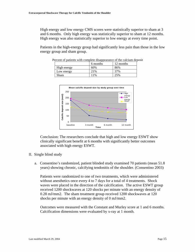
High energy and low energy CMS scores were statistically superior to sham at 3 and 6 months. Only high energy was statistically superior to sham at 12 months. High energy was also statistically superior to low energy at every time point.
Patients in the high-energy group had significantly less pain than those in the low energy group and sham group.
Percent of patients with complete disappearance of the calcium deposit
6 months 12 months High energy 60% 86% Low energy 21% 37% Sham 11% 25%
Mean calcific deposit size by study group over time
0
50
100
150
200
250
baseline 3 month 6 month 12 monthTime
Cal
cific
dep
osit
size
(mm
2)
Highenergy LowenergySham
Conclusion: The researchers conclude that high and low energy ESWT show clinically significant benefit at 6 months with significantly better outcomes associated with high energy ESWT.
II. Single blind study
a. Consentino’s randomized, patient blinded study examined 70 patients (mean 51.8 years) showing chronic, calcifying tendonitis of the shoulder. (Consentino 2003)
Patients were randomized to one of two treatments, which were administered without anesthetics once every 4 to 7 days for a total of 4 treatments. Shock waves were placed in the direction of the calcification. The active ESWT group received 1200 shockwaves at 120 shocks per minute with an energy density of 0.28 mJ/mm2. The sham treatment group received 1200 shockwaves at 120 shocks per minute with an energy density of 0 mJ/mm2.
Outcomes were measured with the Constant and Murley score at 1 and 6 months. Calcification dimensions were evaluated by x-ray at 1 month.
Last modified March 29, 2004 Page 15

Extracorporeal Shockwave Therapy for Calcific Tendonitis of the Shoulder
Study Population: The study included patients with shoulder pain for 10 months. Calcification in the supraspinatus tendon had a minimum diameter of 10mm on x-ray. Patients failed conservative treatment for 6 months.
The study excluded patients due to arthritis, osteoarthritis, neurological abnormalities, partial or complete ruptures of the rotator cuff, or treatment in the 4 weeks prior to ESWT.
Patient Demographics
ESWT Sham Number of patients 35 patients 35 patients Mean Duration of Symptoms 15 months 14.5 months
Results: At 6 months, 23 patients from the sham group withdrew from the study.
Mean Constant score by study group over time
0
20
40
60
80
100
baseline after treatment 1 month 6 monthTime
Con
stan
t sco
re
ESWT
sham
14 ESWT patients (40%) experienced partial resorption of the calcium deposits, and 11 ESWT patients (31%) experienced complete resorption. The deposits remained unmodified in the sham group.
The authors note that deposits may not always correlate with symptoms because ESWT reduced symptoms for patients with and without calcium deposits.
Conclusion: The researchers consider ESWT as an alternative treatment for chronic calcific tendonitis of the shoulder refractory to conventional treatments.
b. Pan’s assessor-blinded study randomly assigned patients with radiographically
and sonographically verified calcific tendonitis to ESWT or TENS. (Pan 2003) Sonography defined the painful area and directed ESWT delivery. 2000 shockwaves at 2 Hz with energy ranging from .26 mJ/mm2 to .32 mJ/mm2 were applied. TENS was applied 3 times a week for 4 weeks at 95 Hz for 20 minutes.
The Constant and Murley score, VAS, and radiography measured outcomes.
Last modified March 29, 2004 Page 16

Extracorporeal Shockwave Therapy for Calcific Tendonitis of the Shoulder
Study Population: The study included patients with calcific tendonitis who had a VAS of greater than 4 or minimum continuous pain for more than 6 months.
The study excluded patients due to rheumatic disease, rotator cuff tear, previous surgery for calcification, needle aspiration, or glucocorticosteroid injection within 3 months.
Patient Demographic
ESWT TENS Number of patients 32 28 Age 55.2 years 58 years Duration of symptoms 24.6 months 23.9 months Maximal calcification size 9.22 mm 9.17 mm
Results: 69% of shoulders in the ESWT group and 43% of shoulders in the TENS group had a Constant score of at least 85 at week 12. The percent of improved shoulders on the manual muscle testing increased to 69.7% for the ESWT group and 62.1% for the TENS group.
Mean Constant score by study group over time
0
20
40
60
80
100
Baseline Week 2 Week 4 Week 12
Time
Con
stan
t sco
re
ESWT
TENS
M e a n VAS by st udy gr oup ov e r t i me
0
2
4
6
8
10
Basel ine Week 2 Week 4 Week 12
T i me
ESWT
TENS
Ultrasonography indicated a significant decrease of plaque size after 12 weeks in both groups with the ESWT group showing a greater percentage change. Arc type calcification seemed to improve faster after ESWT as compared to TENS.
Mean calcification size by study group over time
0
2
4
6
8
10
Baseline Week 2 Week 4 Week 12
Time
Cal
cific
atio
n Si
ze (m
m)
ESWT
TENS
Last modified March 29, 2004 Page 17
Extracorporeal Shockwave Therapy for Calcific Tendonitis of the Shoulder
Conclusion: ESWT is more effective than TENS to achieve functional improvement and to alleviate pain in patients with chronic calcific tendonitis.
III. Case Series with a Comparison Group
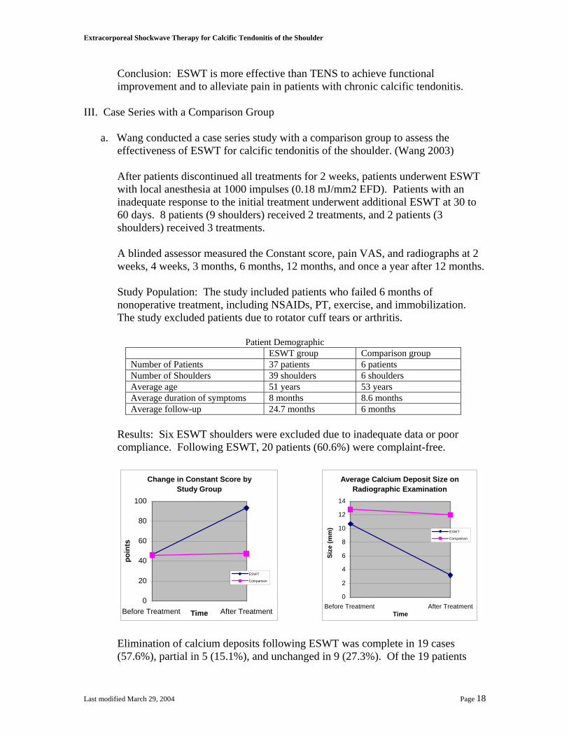
a. Wang conducted a case series study with a comparison group to assess the effectiveness of ESWT for calcific tendonitis of the shoulder. (Wang 2003)
After patients discontinued all treatments for 2 weeks, patients underwent ESWT with local anesthesia at 1000 impulses (0.18 mJ/mm2 EFD). Patients with an inadequate response to the initial treatment underwent additional ESWT at 30 to 60 days. 8 patients (9 shoulders) received 2 treatments, and 2 patients (3 shoulders) received 3 treatments. A blinded assessor measured the Constant score, pain VAS, and radiographs at 2 weeks, 4 weeks, 3 months, 6 months, 12 months, and once a year after 12 months.
Study Population: The study included patients who failed 6 months of nonoperative treatment, including NSAIDs, PT, exercise, and immobilization. The study excluded patients due to rotator cuff tears or arthritis.
Patient Demographic
ESWT group Comparison group Number of Patients 37 patients 6 patients Number of Shoulders 39 shoulders 6 shoulders Average age 51 years 53 years Average duration of symptoms 8 months 8.6 months Average follow-up 24.7 months 6 months
Results: Six ESWT shoulders were excluded due to inadequate data or poor compliance. Following ESWT, 20 patients (60.6%) were complaint-free.
Change in Constant Score by
Study Group
0
20
40
60
80
100
Before Treatment After TreatmentTime
poin
ts
ESWT
Comparison
Average Calcium Deposit Size on Radiographic Examination
0
2
4
6
8
10
12
14
Before Treatment After TreatmentTime
Size
(mm
)
ESWT
Comparison
Elimination of calcium deposits following ESWT was complete in 19 cases (57.6%), partial in 5 (15.1%), and unchanged in 9 (27.3%). Of the 19 patients
Last modified March 29, 2004 Page 18

Extracorporeal Shockwave Therapy for Calcific Tendonitis of the Shoulder
with elimination of calcium deposits, 17 were complaint-free with normal Constant scores.
Conclusion: The authors conclude that ESWT produced pain relief and functional restoration with negligible complications in the treatment of calcific tendonitis of the shoulder.
Last modified March 29, 2004 Page 19

Insurers in Washington
Insurers in Washington In September 2003, the Regence Group deemed ESWT as investigational for all indications, including but not limited to plantar fasciitis, lateral epicondylitis, tendinopathies including calcific tendonitis of the shoulder, stress fracture, delayed union, nonunion, and avascular necrosis of the femoral head. (Regence 2003) Although Medicare has not made a national determination concerning ESWT, Cigna Medicare in the Western region decided in July 2003 to cover ESWT. (Cigna 2003) Cigna considers all other conditions as investigational and not covered. ESWT is medically indicated for treatment of plantar fasciitis or lateral epicondylitis when all of the following criteria are met:
a. The patient has been symptomatic for at least six (6) months b. There has been a lack of response for at least the last two months to conservative
measures, including rest, physical therapy, anti-inflammatory medications, corticosteroid injections, orthotics, or forearm sleeve
c. The patient would otherwise be considered a candidate for surgical treatment. Cigna does not cover ESWT in the following situations:
a. There is active infection or an open wound at the treatment site. b. There is evidence of blood dyscrasia or bleeding disorder. c. There are plans to use a non-FDA approved devices or use an FDA approved
device for a condition other than a condition for which it has FDA approval.
Last modified March 29, 2004 Page 20
Conclusions
Conclusions Plantar fasciitis Two high quality, double-blind RCT have been conducted to examine the effect of ESWT on plantar fasciitis. Haake’s study showed no difference on Roles and Maudsley and pain outcomes following high energy ESWT with ultrasound guiding to the heel spur compared to placebo. Speed’s study showed that moderate energy ESWT with focusing on the site of maximal pain and placebo resulted in similar pain outcomes. Two single-blind RCT have also been conducted to evaluate ESWT for plantar fasciitis. Hammer’s control patients who crossed over to ESWT experienced comparable results on improved VAS and walking ability after undergoing ESWT. Rompe’s patient-blinded trial indicated that low energy ESWT benefited recreational runners over placebo on morning walking pain. Although the two studies suggest beneficial effect of ESWT on plantar fasciitis, the lack of double-blinding may introduce substantial bias of results. Lateral epicondylitis Two high quality double-blind trials have been conducted to examine the effect of ESWT on lateral epicondylitis. HealthTronics’ high energy ESWT with local anesthetic showed significantly better results than placebo on investigator assessment at 8 weeks. Differences on two other outcome measurements did not reach statistical significance. Melikyan’s study of ESWT with ultrasound guiding without local anesthetic did not show any difference from placebo on pain, grip strength, medication use, or eventual surgery for lateral epicondylitis. Calcific Tendonitis One high quality, double-blind RCT by Gerdesmeyer indicated that high and low energy ESWT showed significantly better results on Constant and Murley scores, pain, and rate of deposit resorption compared to sham therapy for calcific tendonitis. Two single-blinded RCT have also been conducted to examine the effect of ESWT on calcific tendonitis. Consentino’s patient-blinded study showed superior outcomes following ESWT compared to placebo on Constant scores and resorption of deposit. However, substantial dropout in the sham group may have affected results. Pan’s assessor-blinded study indicated that patients with calcific tendonitis showed greater improvement on Constant scores and pain after ESWT compared to TENS. Although the two studies suggest beneficial effect of ESWT on calcific tendonitis, the lack of double-blinding may introduce substantial bias of results.
Last modified March 29, 2004 Page 21

References
References Background and FDA Status Food and Drug Administration (FDA). “Approval Order.” 2002 January; Available at
http://www.fda.gov/cdrh/pdf/P000048a.pdf. Last accessed on March 22, 2004. Food and Drug Administration (FDA). “Summary of Safety and Effectiveness Data.”
2000 October; Available at http://www.fda.gov/cdrh/pdf/p990086b.pdf. Last accessed on March 22, 2004.
Food and Drug Administration (FDA). “Summary of Safety and Effectiveness Data.”
2002a October; Available at http://www.fda.gov/cdrh/pdf/P010039b.pdf. Last accessed on March 22, 2004.
Food and Drug Administration (FDA). “Summary of Safety and Effectiveness Data.”
2003 March; Available at http://www.fda.gov/cdrh/PDF/p990086s003b.pdf. Last accessed on March 22, 2004.
Department of Labor and Industries (LNI). “Health Technology Assessment:
Extracorporeal Shockwave Therapy for the Treatment of Musculoskeletal Disorders.” 2003 January 27; Available at http://www.lni.wa.gov/ClaimsInsurance/Files/OMD/EswtHta20030127.pdf. Last accessed on March 30, 2004.
Plantar Fasciitis Alvarez, RG, et al. "Symptom Duration of Plantar Fasciitis and the Effectiveness of
Orthotripsy." Foot and Ankle International. 2003; 24(12): 916-921. Haake, M, et al. "Extracorporeal shock wave therapy for plantar fasciitis: randomized
controlled multicentre trial." BMJ. 2003 July 12; 327(7406): 75.
Hammer, DS, et al. "Extracorporeal Shock Wave Therapy (ESWT) in Patients with Chronic Proximal Plantar Fasciitis: A 2-Year Follow-up." Foot and Ankle International. 2003; 24(11): 823-828.
Hammer, DS, et al. “Ultrasonographic evaluation at 6-month follow-up of plantar
fasciitis after extracorporeal shock wave therapy.” Archives of Orthopaedic and Trauma Surgery. 2003a October;
Lee, GP, et al. "Effect of Extracorporeal Shock Waves on Calcaneal Bone Spurs." Foot
and Ankle International. 2003; 24(12): 927-930. Perez, M, et al. "Extracorporeal shock wave therapy for plantar fasciitis." Clin Podiatr
Last modified March 29, 2004 Page 22

References
Med Surg. 2003; 20: 323-334. Rompe, JD, et al. “Shock Wave Application for Chronic Plantar Fasciitis in Running
Athletes.” American Journal of Sports Medicine. 2003; 31(2): 268-275. Speed, CA, et al. “Extracorporeal shock wave therapy for plantar fasciitis. A double
blind randomized controlled trial.” J of Orthopaedic Research. 2003; 21:937-940.
Lateral Epicondylitis Food and Drug Administration (FDA). “Summary of Safety and Effectiveness Data.”
2003 March; Available at http://www.fda.gov/cdrh/PDF/p990086s003b.pdf. Last accessed on March 22, 2004.
Melikyan, EY, et al. “Extracorporeal shock-wave treatment for tennis elbow: a
randomized double-blind study.” J Bone and Joint Surgery (Br). 2003; 85B: 852-855.
Calcific Tendonitis Consentino, R, et al. "Extracorporeal shock wave therapy for chronic calcific tendonitis
of the shoulder: single blind study." Annals of the Rheumatic Diseases. 2003 March; 62: 248-250.
Gerdesmeyer, L, et al. "Extracorporeal Shock Wave Therapy for the Treatment of
Chronic Calcifying Tendonitis of the Rotator Cuff: a Randomized Controlled Trial." JAMA. 2003 Nov 19; 290(19): 2573-2580.
Pan, PJ, et al. "Extracorporeal Shock Wave Therapy for Chronic Calcific Tendonitis of
the Shoulders: a Functional and Sonographic Study." Arch Phys Med Rehabil. 2003 July; 84: 988-993.
Wang, CJ, et al. "Shock Wave Therapy for Calcific Tendonitis of the Shoulder: a
Prospective Clinical Study with Two-Year Follow-up." American Journal of Sports Medicine. 2003; 31(3): 425-430.
Insurers Cigna. "Extracorporeal Shock Wave Therapy for Musculoskeletal Problems." 2003 July.
Available at www.cms.gov. Last accessed on March 26, 2004. The Regence Group. "Extracorporeal Shock Wave Treatment for Plantar Fasciitis and
Last modified March 29, 2004 Page 23
References
Other Musculoskeletal Conditions." Medical Policy. 2003 September; Available at www.regence.com. Last accessed on March 26, 2004.
Last modified March 29, 2004 Page 24