extensive protein hydrolyzation is indispensable to ... · extensive protein hydrolyzation is...
TRANSCRIPT
RESEARCH ARTICLE Open Access
Extensive protein hydrolyzation isindispensable to prevent IgE-mediatedpoultry allergen recognition in dogs and catsThierry Olivry1,2* , Jennifer Bexley3 and Isabelle Mougeot4
Abstract
Background: The central premise for the commercialization of diets with hydrolyzed ingredients is that the small-sized digested peptides would be unable to crosslink allergen-specific IgE at the surface of tissue mast cells andinduce their degranulation. Evidence for the validity of this concept to diagnose food allergies in dogs and cats islimited, however. Our objectives were to study the recognition of standard and variably hydrolyzed poultry extractsby sera from dogs and cats with elevated chicken-specific serum IgE.
Results: Forty sera from dogs and 40 from cats with undetectable, low, medium or high serum levels of chicken-specific IgE were tested by ELISA on plates coated with the positive controls chicken, duck and turkey meat extractsand the negative controls beef meat (dogs) or wheat (cats). Plates were also coated with a non-hydrolyzed chickenmeal, and mildly- or extensively-hydrolyzed poultry feather extracts. The frequencies of dogs with positive IgE againstthe various extracts were: chicken meat: 100%, duck and turkey meats: 97%, beef meat: 3%, non-hydrolyzed chickenmeal: 73%, mildly-hydrolyzed poultry feathers: 37% and extensively-hydrolyzed poultry feathers: 0%. For cats, theserespective percentages were (with wheat replacing beef as a negative control): 100, 84, 97, 7, 7, 0 and 0%. To detectany allergenic cross-reactivity between poultry meat-based and feather hydrolysate-derived extracts, an IgE ELISAinhibition was also done. Ten canine sera with the highest level of anti-poultry IgE in the previous experiment wereincubated overnight with a previously optimized 50 μg amount of each of the extracts used above. We performedELISA on plates coated with chicken, duck or turkey meats with or without inhibitors. The median inhibitionpercentages after incubation with the non-hydrolyzed chicken meal were ~22%, with the mildly-hydrolyzed poultryfeathers: 14–22%, and those with the extensively-hydrolyzed poultry feathers: 5 to 10%; the last inhibition level wassimilar to that of the beef meat negative control.
Conclusions: Altogether, these results suggest that an extensive—but not partial—hydrolyzation of the poultry featherextract is necessary to prevent the recognition of allergenic epitopes by poultry-specific IgE.
Keywords: Allergy, Canine, Food, Feline, Hydrolysate, Hypersensitivity, IgE
BackgroundIn dogs and cats, as in humans, the diagnosis of adversefood reactions (AFRs) relies on the recurrence of clinicalsigns after provocation with causative food ingredients.Such food re-challenges are typically done after a lengthyperiod of dietary restriction (i.e. “elimination”) to permit
the disappearance of existing clinical signs and thereturn to a state of normalcy that allows the detection ofpositive provocations [1]. In companion animals,restrictive dietary trials usually consist of feedingingredients not eaten previously [1] for 6 to 8 weeks [2].Finding a diet with “novel” ingredients, be it home-
cooked or commercially available, is becoming increas-ingly challenging because of the diverse—often unusualand sometimes even “exotic”—regimes eaten by today’spets and the rising complexity of currently availablecommercial foods. Furthermore, there is evidence thatsome commercial diets are contaminated by ingredients
* Correspondence: [email protected] of Clinical Sciences, College of Veterinary Medicine, NorthCarolina State University, Raleigh, NC, USA2Comparative Medicine Institute, College of Veterinary Medicine, NorthCarolina State University, Raleigh, NC, USAFull list of author information is available at the end of the article
© The Author(s). 2017 Open Access This article is distributed under the terms of the Creative Commons Attribution 4.0International License (http://creativecommons.org/licenses/by/4.0/), which permits unrestricted use, distribution, andreproduction in any medium, provided you give appropriate credit to the original author(s) and the source, provide a link tothe Creative Commons license, and indicate if changes were made. The Creative Commons Public Domain Dedication waiver(http://creativecommons.org/publicdomain/zero/1.0/) applies to the data made available in this article, unless otherwise stated.
Olivry et al. BMC Veterinary Research (2017) 13:251 DOI 10.1186/s12917-017-1183-4

not listed in their composition label [3–7]. Finally, thecharacterization of some food allergens for dogs [8, 9],as well as our recent serological study [10], revealed theexistence of extensive IgE cross-reactivity among aller-gens of taxonomically-related food groups in this spe-cies. Such studies suggest that some of these “novel”ingredients might cross-react with historically fed com-ponents, unless the protein source were evolutionarilydistant from all those eaten previously. Altogether, thepoints raised above should reduce our confidence in be-ing able to perform “accurately-restricted” dietary trialsusing so-called “novel” food items.To alleviate the need for a perpetual quest to identify
new ingredients and to reduce problems of allergeniccross-reactivity, pet food manufacturers created dietscontaining proteins hydrolyzed into peptides of a sizesupposedly insufficient to induce IgE-mediated mast cellactivation (reviewed in [1]). For many years, and due toproduction costs and technical aspects, only partiallyhydrolyzed diets were commercialized. While thesehydrolysate-based petfoods clearly exhibited a reducedallergenicity, there was evidence that some dogs withcutaneous AFRs still reacted clinically to their inges-tion, thereby suggesting that some proteins were in-sufficiently digested [11, 12]. The development of RoyalCanin Anallergenic (RCA, also known in North Americaas Ultamino, Royal Canin, Aimargues, France) was note-worthy, not only because of its high-grade of protein hy-drolyzation resulting in a diet almost entirely composed ofsingle amino acids and very short peptides, but also be-cause of its protein source (poultry feathers) being poten-tially quite different in protein composition from that ofother meat-based poultry-based diets. A recent studyestablished the relevance of extensive protein hydrolyza-tion to reduce clinical allergenicity, as ten chicken-allergicdogs did not react clinically after two weeks exclusivelyeating the poultry feather-containing RCA [12].To further study if protein hydrolyzation results in re-
duced immunological allergenicity in companion animals,as it does for food allergic humans, we set out to investi-gate if partially- or extensively-hydrolyzed poultry featherextracts were recognized by serum IgE from dogs and catssensitized to poultry meats. We will demonstrate hereinthat only the extensive hydrolyzation of the poultry featherextract results in a lack of IgE recognition by poultry-specific IgE, thereby confirming the validity of extensivehydrolyzation for hypoallergenic pet food development.
MethodsAnimal seraSurplus canine and feline sera submitted to AvactaAnimal Health (Wetherby, Leeds, UK) by veterinar-ians for food allergen-specific IgE serological testingwere used for this study. For dogs, the panel
consisted of beef, pork, lamb, duck, chicken, turkey,wheat, soybean, barley, rice, potato, corn, oat, cow’smilk, whole hen’s egg, white fish, venison, salmon andrabbit. For cats, the panel included beef, pork, lamb,duck, chicken, turkey, rabbit, salmon, tuna, white fish,wheat, soybean, rice, corn, cow’s milk and whole hen’segg. All dog and cat sera had been stored at −80 °Cfor a maximum of two and six years, respectively.Canine sera were selected according to their originallevels of serum chicken-specific IgE (IgE reactivitiesexpressed as optical density, OD, values at 405 nm,OD405 uncorrected for background) in the Sensitestcommercially available ELISA (Avacta).The D1 group (30 dogs) contained the following sera:
– D1-LCR: ten dogs with low chicken IgE reactivityand a previous OD405 between 0.3 and 0.4 in theSensitest assay.
– D1-MCR: ten dogs with moderate IgE chickenreactivity (OD405 between 0.5 and 0.9)
– D1-HCR: ten dogs with high chicken IgE reactivity(OD=405 > 1.0)
In their previous ELISAs done at the time of theoriginal submission, all of the dogs in this group haddetectable IgE to at least one food extract; one dog wasonly positive to chicken and none had any detectableIgE reactivity to beef.D2-NCR: This group included sera from ten dogs
with undetectable levels of IgE to chicken (i.e. “non-chicken reactive dogs” NCR; OD405 < 0.3). By previousELISAs, these dogs had no visible IgE reactivity to anyfood tested on the panel.Similarly, we selected feline sera based upon their
previously determined levels of serum chicken-specificIgE (OD405, uncorrected for background).The C1 group was composed of 31 cats divided into
the following subgroups:
– C1-LCR: eleven cats with low chicken IgE reactivity(OD405: ~0.2–0.4)
– C1-MCR: ten cats with moderate chicken IgEreactivity (OD405: ~0.5–1.0)
– C1-HCR: ten cats with high chicken IgE reactivity(OD=405: >1.0)
By previous ELISAs, all of the cats in this group had atdetectable IgE to at least three foods. All cats had meas-urable IgE against beef but none had IgE reactivity towheat.Finally, the C2-NCR group comprised sera from nine
cats with undetectable levels of IgE to chicken in theSensitest ELISA (OD405: <0.2). By previous ELISAs,there was no IgE reactivity to any food in these cats.
Olivry et al. BMC Veterinary Research (2017) 13:251 Page 2 of 9

Details from all selected canine and feline sera arefound in the Additional file 1.
ExtractsRaw chicken meat (CMT), turkey meat (TMT), duckmeat (DMT), beef meat (BMT) and wheat (WHT) ex-tracts were purchased from Greer Laboratories (Lenoir,NC, USA). Non-hydrolysed chicken meal (NHCM, theground, rendered clean parts of the carcasses ofslaughtered poultry), mildly-hydrolysed poultry feathers(MHPF) and extensively-hydrolysed poultry feathers(EHPF) were obtained from Royal Canin. The main dif-ference between the MHPF and EHPF was the presenceof residual proteins of molecular weight superior to10 kDa in the former, but not in the latter; the EHPFwere those used in the RCA. The commercial-grade ma-terial was defatted in acetone, filtered and then dried.Allergens were obtained after two series of successiveextractions in phosphate-buffered saline (PBS), followedby centrifugation and, ultimately, by dialyzing the super-natant against distilled water. The protein concentrationof each extract was determined at 280 nm by TrineanDropSense96 (Trinean, Gent, Belgium). The final proteinconcentrations of the NHCM, MHPF and EHPF extractswere 6.8, 3.1 and 2.4 mg/ml, respectively. Extracts werestored at 4 °C before use.
ELISAsCanine and feline food allergen-specific IgE serumlevels were determined by ELISA. Microtiter plates(Costar, Corning, NY, USA) were coated overnight at2–8 °C with 50 μL/well of each extract, diluted to aconcentration of 5 μg/mL of protein in 0.05 M carbon-ate/bicarbonate buffer, pH 9.6.Plates were washed three times with 150 μL/well Tris-
buffered saline (TBS) with 0.05% Tween-20 (TBST) andblocked with 150 μL/well TBS containing 0.5% sucroseand 0.5% PVP10 (block) for 2 h at room temperature.After removal of the block, plates were dried at 37 °Cfor 2 h. Canine and feline sera were diluted 1:10 inTBST, and incubated with coated extracts overnight at2–8 °C (50 μL/well. For standardization of the canineand feline ELISAs, serial three-fold dilutions of pools ofdog and cat reference sera with high levels of anti-beefIgE (for dog tests) or chicken (for cat tests) were in-cluded on each plate. Undiluted, these two standardpools were assigned a value of 500 arbitrary units (AU).Positive and negative serum controls, with moderateand negligible levels of beef- (dog tests) or wheat- (cattests) specific IgE, respectively, were also diluted at 1:10in TBST and included on each plate. Plates werewashed with TBST, as before, and incubated with0.5 μg/mL alkaline phosphatase-labelled anti-dog IgE(clone 5.91; Bruce Hammerberg, NC State University,
Raleigh, NC, USA) for 2 h at RT. This mouse monoclo-nal antibody has been described previously [13] andshown to recognize IgE from several mammalian spe-cies including cats [14]. The specificity of this antibodyfor dog IgE was confirmed in previous ELISAs, as astrong signal (OD405: 1.8) was found on a 0.5 μg/mLdog IgE coat without any concurrent detection (OD405:0.11) of dog IgG coated at up to 20 μg/mL. After the finalthree washes with TBST, plates were developed with50 μL/well of an alkaline phosphatase substrate (pNPP;BioFx Laboratories, Owings Mills, MD, USA) for 30 minat room temperature. The reactions were stopped by theaddition of 50 μL/well 1 M NaOH, and OD405 were deter-mined using a microplate reader (Tecan, Männedorf,Switzerland). For both ELISAs, standard curves were gen-erated by fitting the mean standard uncorrected OD405 toa sigmoidal 4PL curve (Prism, Graphpad Software, LaJolla, CA, USA) with log10 concentrations of the standarddog or cat serum pools.
Inhibition ELISAsInhibition ELISAs were performed to determine ifNHCM, MHPF and EHPF extracts could block serumIgE binding to poultry extracts, thereby demonstratingthe presence of shared IgE binding epitopes. Ten dogsera that showed the highest IgE reactivity to CMT,DMT and TMT in the ELISAs described above (i.e. theHCR sera) were selected for the inhibition assays. Dueto insufficient volumes available, inhibitions were notperformed with the feline sera. Inhibitor solutions ofNHCM, MHPF and EHPF were prepared in TBST, eachto a concentration of 500, 1000 and 2000 μg/mL (i.e.100×, 200×, and 400× coat level, respectively). To serveas the positive inhibitor, the CMT, DMT and TMT werecombined, as a three-meat extract in a 1:1:1 ratio ofequal protein levels, to the same concentrations as thatof the extracts above. As a negative inhibitor, we dilutedthe BMT extract to the same concentrations. Dog serawere pre-incubated overnight at 2–8 °C, at a dilution of1:10, with 50 μL each of the three increasing concentra-tions of the test and control inhibitor solutions.Additionally, each dog serum was diluted at 1:10 inTBST in the absence of inhibitor. All serum-inhibitormixes and no-inhibitor controls were then tested at50 μL/well by ELISA on the CMT, DMT and TMT ex-tracts, as described above. For any given concentrationof inhibitor, the percentage inhibition was calculated asfollows: 100 – ((OD405 of serum with inhibitor/OD405of serum without inhibitor) ×100) where OD405 repre-sents the background-corrected absorbance at 405 nm.
ImmunoblottingThe extracts above, thawed after two years of freezing,were heated at 70 °C for 10 min in the presence of 5%
Olivry et al. BMC Veterinary Research (2017) 13:251 Page 3 of 9
Criterion XT reducing agent (Bio-Rad Laboratories,Hercules, CA, USA) before sodium dodecyl sulfate poly-acrylamide gel electrophoresis (SDS-PAGE). Proteins(5 μg per lane) were separated on 4–12% Bis-TrisCriterion XT precast gels (Bio-Rad) at 180 V using MES(4-morpholineethane-sulfonic acid) running buffer on aCriterion Cell system (Bio-Rad). Molecular weightmarkers (10–190 kDa; PageRuler Plus, ThermoScientific) were run in parallel to the extracts. Followingelectrophoresis, the gels were either stained usingInstantBlue Coomassie stain (Sigma Aldrich, Dorset,UK) to verify protein loading and proteins were trans-ferred to a polyvinylidene difluoride (PVDF) membrane(BioRad). This was performed at 100 V for 1 h in Tris/glycine transfer buffer (Bio-Rad) containing 20% metha-nol using Criterion Blotter apparatus (Bio-Rad). Themembrane was blocked with TBS containing 0.5%PVP10 at room temperature for 2 h, and then incubatedwith a serum pool from three dogs with high chicken-specific IgE levels (>OD405: 1.0, by previous ELISAs), di-luted at 1/10 in TBST. The membrane was incubatedwith serum at RT overnight. After washing three timeswith TBST, the membrane was incubated with alkalinephosphatase-conjugated anti-dog IgE antibody (clone5.91, 0.5 μg/ml in TBST) at room temperature for 2 hwith shaking. The membrane was washed three timeswith TBST and rinsed twice prior to addition of nitro-blue tetrazolium/5-bromo-4-chloro-3-indolyl-phosphatesubstrate (Thermo Scientific). The reaction was stoppedwith deionised water and blots were then dried at roomtemperature. Images of gels and blots were capturedusing a G:BOX Chemi-XR5 gel imaging system(Syngene, Cambridge, UK).
StatisticsData were analyzed with SAS v9.3 (SAS, Cary, NC,USA). The frequencies of canine or feline positive ELISAtests with the various extracts were compared withpermutation tests-based on Fisher’s exact test with a
resampling size of 1000 times. Then, the alpha inflationrisk subsequent to multiple extract comparisons wascontrolled with false discovery rate adjustments. Thelevel of statistical significance was set atP < 0.05, for two-sided analyses.
ResultsELISA validationThe canine ELISA standard curve had an accuracy ofbetween 99 and 117% at concentrations varying between3.7 and 100 AU. The inter-assay coefficients of variation(made from 14 measurements over seven plates) of thenegative and positive controls were 12 and 7%, respect-ively. We established the positive threshold of 8.1 AU asthe mean OD405 plus three standard deviations of a largenumber (n = 95) of canine sera without detectable anti-poultry IgE.For the feline ELISA, the standard curve was 99–106%
accurate over the same concentration range as above.The inter-assay coefficient of variation of the positivecontrol was 6%. The positive threshold—determined asfor the canine ELISA, but with a similar number andtype of feline sera—was calculated to be 6.2 AU.
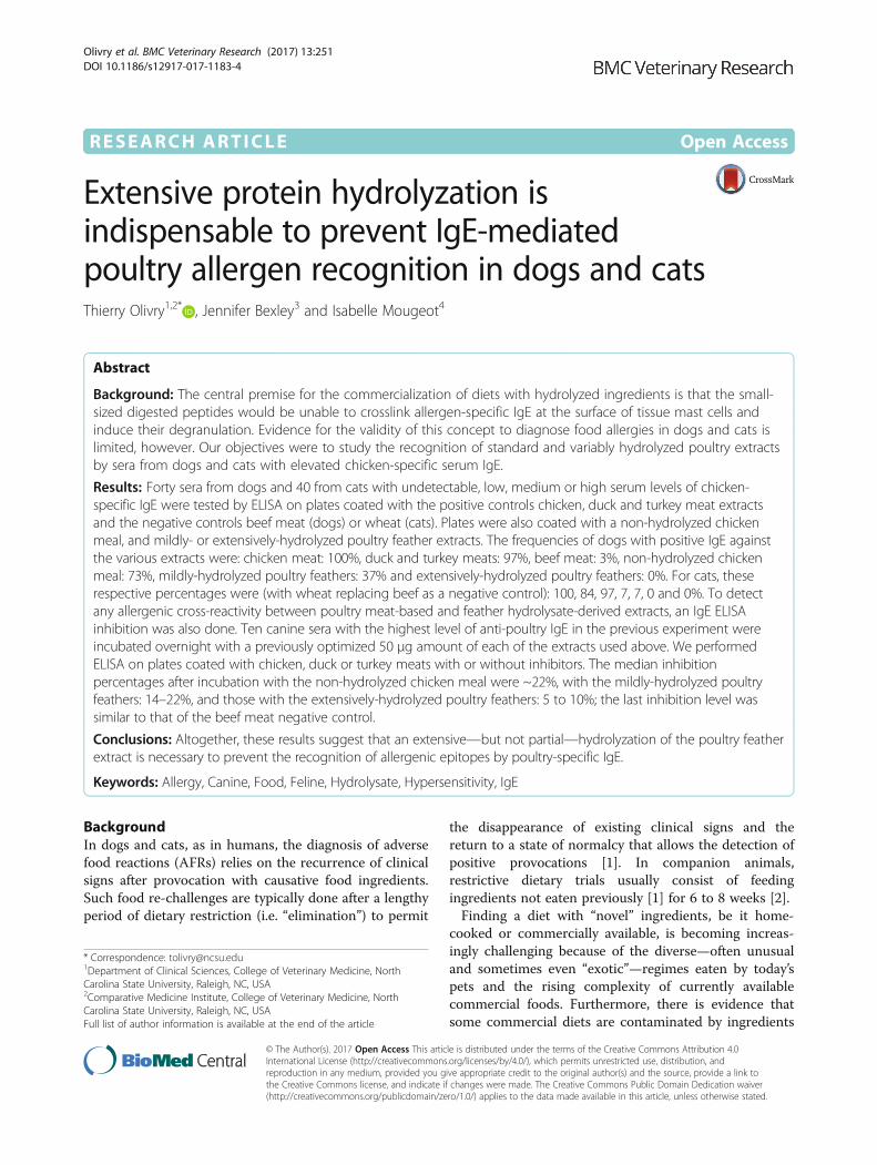
Canine ELISAsThe frequencies of group D1 and D2 sera testing posi-tive on the various extracts are shown in Fig. 1. Amongthe 30 dogs from the D1 group, the number (and fre-quencies) of dogs testing positive for the CMT, DMTand TMT extracts were 30 (100%), 29 (97%) and 29(97%) respectively. All dogs that had positive reactionsto TMT were also positive to DMT and CMT; all serapositive to DMT were also positive to TMT and CMT.Only one serum (3%) was positive to the BMT extract.Twenty-two sera (73%) were positive with the NHCM,
and these were also positive for the CMT. When testedon the two hydrolysates, 11 (37%) and no dogs (0%)reacted to the MHPF and EHPF extracts, respectively;sera positive on the MHPF were also positive when
Fig. 1 Frequencies of positive canine sera tested on the various extracts
Olivry et al. BMC Veterinary Research (2017) 13:251 Page 4 of 9

tested on the CMT and NHCM extracts. Details on theactual frequencies of reactivity of the various D1 serumsubgroups are provided in the Additional file 2. Asexpected, sera from the D1-LCR subgroup had the low-est rate of positive tests among those of the D1 group.The proportions of positive canine sera tested on the
various extracts were compared statistically and resultsare shown in Table 1. Of note is that the proportion ofreactivity to the EHPF extract was significantly lowerthan those to all other extracts, except for the BMTnegative control.In the group D2 (NCR), nine of ten sera (90%) had
negative results for all tested extracts; one (10%) was avery weak positive (8.8 AU; positive threshold: 8.1) whentested on the MHPF extract.
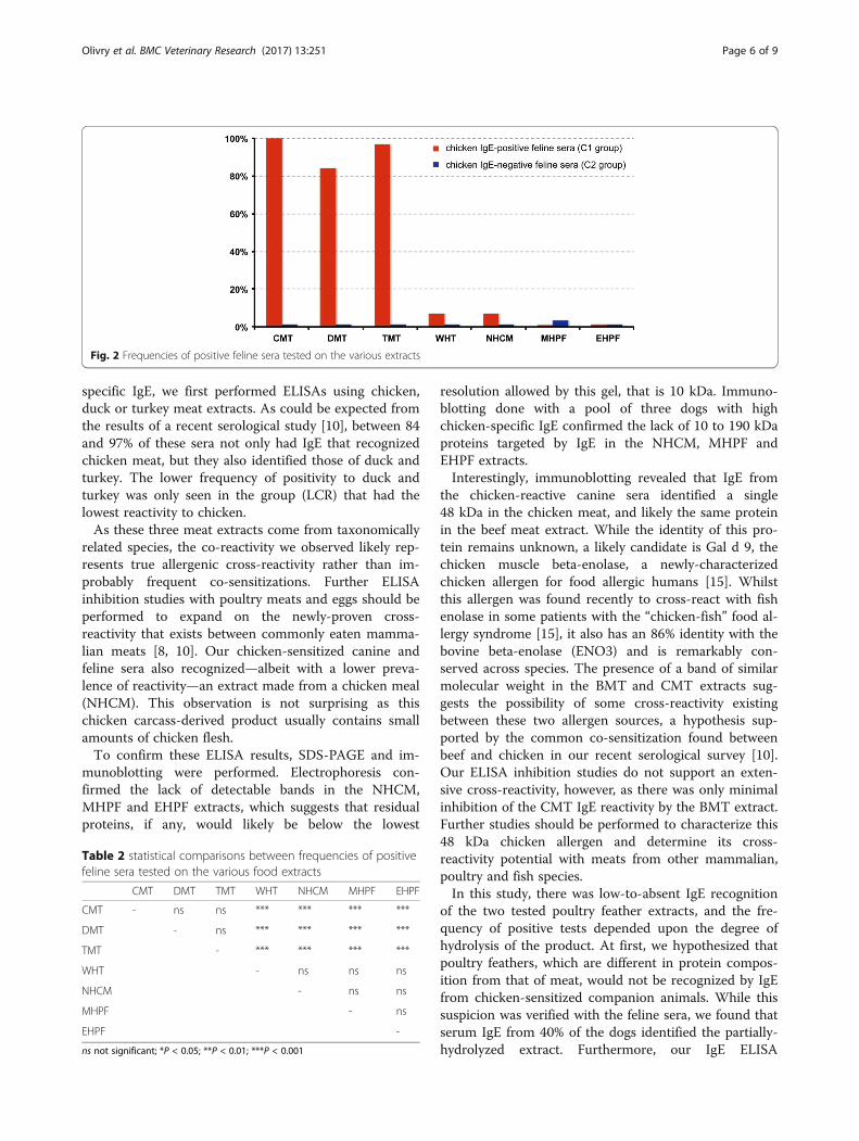
Feline ELISAsAll frequencies of positive tests using C1 and C2 seraand the various extracts are depicted in Fig. 2.All 31 feline sera from the C1 group tested positive on
the CMT; 30 (97%) reacted positively to TMT and 26(84%) to the DMT. Two sera (7%) had a positive test onthe NHCM extract, and these were also positive for theCMT, DMT and TMT; two reacted to the WHT extract(7%). In contrast, none of the sera reacted to either theMHPF or the EHPF extracts. The proportions of positivesera within the three C1 serum subgroups are availablein the Additional file 2.The proportions of C1 sera testing positive to CMT,
DMT, and TMT extracts were not significantly differentfrom each other (Table 2). Similarly, these percentageswere not significantly different between WHT, NHCM,MHPF and EHPF.Finally, none of the nine feline sera from the C2
(NCR) group reacted to any of the food extracts tested.
Canine inhibition ELISAThe percentage inhibition of the three meat positive in-hibitor on CMT, DMT and TMT plates correlated posi-tively with the increasing concentrations of inhibitorused; the inhibition with the negative control (BMT)also increased proportionally, due to nonspecific
binding. At this concentration, the inhibition of thenegative inhibitor background was minimal (<13%),whilst a strong inhibition (>50%) was present with thethree-meat positive inhibitor. Consequently, we presentherein the results of ELISAs obtained after incubation ofthe canine sera with 50 μg of the various inhibitors(50 μL of the serum + inhibitor mixtures), that is using a1000 μg/mL inhibitor solution.The percentage inhibition with the positive control
(three meats) varied between 86 and 91% dependingupon the extract on which it was tested (Fig. 3); thatwith the negative control was between 9 and 12%. Theinhibitions with the NHCM extract were ~22% of theoriginal (no-inhibitor) reactivities, those of the MHPFvaried between 14 and 22%. Finally, incubation with theEHPF led to inhibitions of only 5 to 10%, which werealways lower than those obtained with the negativeinhibitor BMT.
ImmunoblottingSodium dodecyl sulfate polyacrylamide gel electrophor-esis was used to separate the extract proteins under re-duced and denatured conditions. Six distinct bands ofapproximately 17, 42, 48, 51, 62 and 69 kDa molecularweight were visible in the BMT extract (Fig. 4a lane 2).Similarly, five bands of approximately 26, 40, 48, 52 and62 kDa molecular weight were observed in the CMT ex-tract (Fig. 4a lane 3). There were no detectable proteinbands in the 10 to 190 kDa gel separation range in theNHCM, MHPF and EHPF extracts.Western blot analysis was then performed to evaluate
the presence of IgE binding proteins in the same ex-tracts. Canine chicken-reacting sera contained IgE thatrecognized only one protein of approximately 48 kDamolecular weigth in the CMT extract (Fig. 4b, lane 3).Interestingly and surprisingly, a likely similar 48 kDaband, albeit weaker in intensity, was also found in theBMT control extract. In contrast and as expected, therewere no visible bands in the NHCM, MHPF and EHPFextracts.
DiscussionWith these experiments, we provide evidence that onlythe extensive, but not the partial, hydrolyzation ofpoultry feathers prevents their recognition by serum IgEfrom dogs and cats sensitized to poultry. Furthermore,we found the EHPF to be unable to inhibit any of theIgE reactivity to chicken, duck or turkey meats, therebysuggesting that such hydrolysate could be of value as arestrictive commercial diet, even in dogs or cats withIgE hypersensitivity to the meats of any of these threepoultry species.Using sera from AFR-suspected dogs and cats with un-
detectable, low, moderate or high levels of chicken-
Table 1 statistical comparisons between frequencies of positivecanine sera tested on the various food extracts
CMT DMT TMT BMT NHCM MHPF EHPF
CMT - ns ns *** ns *** ***
DMT - ns *** ns *** ***
TMT - *** ns *** ***
BMT - *** * ns
NHCM - ns ***
MHPF - **
ns not significant; *P < 0.05; **P < 0.01; ***P < 0.001
Olivry et al. BMC Veterinary Research (2017) 13:251 Page 5 of 9
specific IgE, we first performed ELISAs using chicken,duck or turkey meat extracts. As could be expected fromthe results of a recent serological study [10], between 84and 97% of these sera not only had IgE that recognizedchicken meat, but they also identified those of duck andturkey. The lower frequency of positivity to duck andturkey was only seen in the group (LCR) that had thelowest reactivity to chicken.As these three meat extracts come from taxonomically
related species, the co-reactivity we observed likely rep-resents true allergenic cross-reactivity rather than im-probably frequent co-sensitizations. Further ELISAinhibition studies with poultry meats and eggs should beperformed to expand on the newly-proven cross-reactivity that exists between commonly eaten mamma-lian meats [8, 10]. Our chicken-sensitized canine andfeline sera also recognized—albeit with a lower preva-lence of reactivity—an extract made from a chicken meal(NHCM). This observation is not surprising as thischicken carcass-derived product usually contains smallamounts of chicken flesh.To confirm these ELISA results, SDS-PAGE and im-
munoblotting were performed. Electrophoresis con-firmed the lack of detectable bands in the NHCM,MHPF and EHPF extracts, which suggests that residualproteins, if any, would likely be below the lowest
resolution allowed by this gel, that is 10 kDa. Immuno-blotting done with a pool of three dogs with highchicken-specific IgE confirmed the lack of 10 to 190 kDaproteins targeted by IgE in the NHCM, MHPF andEHPF extracts.Interestingly, immunoblotting revealed that IgE from
the chicken-reactive canine sera identified a single48 kDa in the chicken meat, and likely the same proteinin the beef meat extract. While the identity of this pro-tein remains unknown, a likely candidate is Gal d 9, thechicken muscle beta-enolase, a newly-characterizedchicken allergen for food allergic humans [15]. Whilstthis allergen was found recently to cross-react with fishenolase in some patients with the “chicken-fish” food al-lergy syndrome [15], it also has an 86% identity with thebovine beta-enolase (ENO3) and is remarkably con-served across species. The presence of a band of similarmolecular weight in the BMT and CMT extracts sug-gests the possibility of some cross-reactivity existingbetween these two allergen sources, a hypothesis sup-ported by the common co-sensitization found betweenbeef and chicken in our recent serological survey [10].Our ELISA inhibition studies do not support an exten-sive cross-reactivity, however, as there was only minimalinhibition of the CMT IgE reactivity by the BMT extract.Further studies should be performed to characterize this48 kDa chicken allergen and determine its cross-reactivity potential with meats from other mammalian,poultry and fish species.In this study, there was low-to-absent IgE recognition
of the two tested poultry feather extracts, and the fre-quency of positive tests depended upon the degree ofhydrolysis of the product. At first, we hypothesized thatpoultry feathers, which are different in protein compos-ition from that of meat, would not be recognized by IgEfrom chicken-sensitized companion animals. While thissuspicion was verified with the feline sera, we found thatserum IgE from 40% of the dogs identified the partially-hydrolyzed extract. Furthermore, our IgE ELISA
Fig. 2 Frequencies of positive feline sera tested on the various extracts
Table 2 statistical comparisons between frequencies of positivefeline sera tested on the various food extracts
CMT DMT TMT WHT NHCM MHPF EHPF
CMT - ns ns *** *** *** ***
DMT - ns *** *** *** ***
TMT - *** *** *** ***
WHT - ns ns ns
NHCM - ns ns
MHPF - ns
EHPF -
ns not significant; *P < 0.05; **P < 0.01; ***P < 0.001
Olivry et al. BMC Veterinary Research (2017) 13:251 Page 6 of 9

Fig. 3 Percentages of inhibition of the reactivities with the different extracts. The ELISA coats are represented as an animal icon, while the natureof inhibitors is indicated in the x-axis. The data presented on the figure correspond to the percentages of inhibition with a 1000 μg/mL solutionof the various inhibitors
Fig. 4 SDS-PAGE (a) and immunoblotting (b). a: Extracts (5 μg/lane) were separated in 4–12% gels by SDS-PAGE. b: Western immunoblotting ofthe same extracts. The blot was incubated with a chicken-reactive dog serum pool at a 1:10 dilution and revealed with anti-dog IgE-AP at 0.5 μg/mLfollowed by NBT/BCIP revelation. After incubation with this dog serum pool, a 48 kDa protein band was found to be the target of IgE in the CMT andBMT extracts; bands were not visible with the other extracts, however
Olivry et al. BMC Veterinary Research (2017) 13:251 Page 7 of 9
inhibition studies confirmed that the MHPF extract didblock between 14 to 22% of the original reactivity to thechicken, duck or turkey meats. These results imply theexistence of some residual allergen cross-reactivity be-tween poultry meats and feathers. In fact, the “bird-eggsyndrome” of humans is a well-known example of suchcross-reactivity [16]). In these patients, there is second-ary chicken meat and egg yolk allergy due to a primaryIgE sensitization to bird feathers with IgE recognition ofchicken serum albumin (also known as alpha-livetin orGal d 5) as an allergen [17]. Whether or not dogs withIgE recognizing the MHPF have IgE against Gal d 5 isunknown, but this is deserving of further study. Thepresence of residual albumin at the feather’s quill’s infer-ior umbilicus is anatomically plausible and quite prob-able, and it provides an argument against feathers beingentirely “novel” protein sources.In contrast to the observations above, the extensively-
digested poultry feather extract was not identified by IgEfrom any of our canine and feline sera, even by thosewith the highest levels of chicken-specific IgE. Further-more, the percentage of inhibition of the original reactiv-ity to chicken, duck or turkey meats was lower than thatof the negative beef meat inhibitor. These results suggestthat the extensive hydrolyzation of poultry feathers leadsto a complete or near-complete lack of IgE recognition,even in pets with high levels of serum poultry IgE. OurELISA inhibition data also revealed the apparent absenceof allergenic cross-reactivity of this EHPF with any ofthe three poultry types of meat tested. These serologicaltesting results suggests the “anallergenicity” of thisextract, and they are supported by the lack of clinicalreactivity seen in ten chicken-allergic dogs that ate theEHPF-containing RCA/Ultamino commercial diet fortwo weeks [12].
ConclusionsIn summary, these ELISA studies suggest that it is onlywith an extensive hydrolyzation that a protein source isnot recognized by serum IgE from sensitized animals,even in those with high levels of sensitization. As a re-sult, commercial foods containing extensive hydroly-sates, such as the RCA/Ultamino, would be valuable todiagnose AFRs in dogs and cats, as shown by our recenttrial of ten chicken-allergic dogs challenged negativelywith this diet [12]. Alternatively, they might be of inter-est as the sole food source for animals with high levelsof sensitization, even to the parent proteins of similarspecies. One should not forget, however, that AFR indogs might be also associated with non-IgE immunemechanisms, for example, those due to T-lymphocyteactivation [18–22]. Whether or not extensively-hydrolyzeddiets would be valuable in such animals has not yet beendetermined.
Additional files
Additional file 1: Details of selected animal sera. (DOCX 128 kb)
Additional file 2: Positive test frequencies of canine and feline serumgroups. (DOCX 74 kb)
AbbreviationsAFR: Adverse food reaction; BMT: Beef meat; CAFR: Cutaneous adverse foodreaction; CMT: Chicken meat; DMT: Duck meat; EHPF: Extensively-hydrolysedpoultry feathers; HCR: High chicken reactive sera; LCR: Low-chicken reactivesera; MCR: Medium chicken-reactive sera; MHPF: Mildly-hydrolysed poultryfeathers; NCR: Chicken non-reactive sera; NHCM: Non-hydrolysed chickenmeal; OD: Optical density; TMT: Turkey meat; WHT: Wheat
AcknowledgmentsThe authors thank Laure Boutigny and Alexandre Feugier for their help withstatistical analyses and their review of the manuscript.
FundingThis study was supported by a Royal Canin payment to Avacta Animal Health.
Availability of data and materialsThe data analyzed during the current study are available from thecorresponding author on reasonable request.
Authors’ contributionsTO and IM designed the original protocol, JB performed the analyses andwrote the study report. TO wrote the article, which was then reviewed by JBand IM. All authors approved the submitted version.
Ethics approval and consent to participateNot needed or relevant, as this study was done from archived sera.
Consent for publicationNot needed or relevant.
Competing interestsTO declares having received consulting and lecturing honoraria and researchfunding from Royal Canin; IM was an employee of this company at the timeof performance of this study; JB is an employee of Avacta Animal Health.
Publisher’s NoteSpringer Nature remains neutral with regard to jurisdictional claims inpublished maps and institutional affiliations.
Author details1Department of Clinical Sciences, College of Veterinary Medicine, NorthCarolina State University, Raleigh, NC, USA. 2Comparative Medicine Institute,College of Veterinary Medicine, North Carolina State University, Raleigh, NC,USA. 3Avacta Animal Health, Leeds, Wetherby, UK. 4Royal Canin, Aimargues,France.
Received: 31 January 2017 Accepted: 10 August 2017
References1. Verlinden A, Hesta M, Millet S, Janssens GPJ. Food allergy in dogs and cats:
a review. Crit Rev Food Sci Nutr. 2006;46:259–73.2. Olivry T, Mueller RS, Prélaud P. Critically appraised topic on adverse food
reactions of companion animals (1): duration of elimination diets. BMC VetRes. 2015;11:225.
3. Raditic DM, Remillard RL, Tater KC. ELISA testing for common food antigensin four dry dog foods used in dietary elimination trials. J Anim Physiol AnimNutr (Berl). 2011;95:90–7.
4. Ricci R, Granato A, Vascellari M, Boscarato M, Palagiano C, Andrighetto I,Diez M, Mutinelli F. Identification of undeclared sources of animal origin incanine dry foods used in dietary elimination trials. J Anim Physiol Anim Nutr(Berl). 2013;97(Suppl 1):32–8.
Olivry et al. BMC Veterinary Research (2017) 13:251 Page 8 of 9
5. Maine IR, Atterbury R, Chang KC. Investigation into the animal speciescontents of popular wet pet foods. Acta Vet Scand. 2015;57:7,015-0097-z
6. Okuma TA, Hellberg RS. Identification of meat species in pet foods using areal-time polymerase chain reaction (PCR) assay. Food Control. 2015;50:9–17.
7. Horvath-Ungerboeck C, Widmann K, Handl S. Detection of DNA fromundeclared animal species in commercial elimination diets for dogs usingPCR. Vet Dermatol. 2017;28:373–e86.
8. Martin A, Sierra MP, Gonzalez JL, Arevalo MA. Identification of allergensresponsible for canine cutaneous adverse food reactions to lamb, beef andcow's milk. Vet Dermatol. 2004;15:349–56.
9. Ohmori K, Masuda K, Kawarai S, Yasuda N, Sakaguchi M, Tsujimoto H.Identification of bovine serum albumin as an IgE-reactive beef component in adog with food hypersensitivity against beef. J Vet Med Sci. 2007;69:865–7.
10. Bexley J, Nuttall TJ, Hammerberg B, Halliwell RE. Co-sensitization andcross-reactivity between related and unrelated food allergens in dogs -a serological study. Vet Dermatol. 2017;28:31–e7.
11. Olivry T, Bizikova P. A systematic review of the evidence of reducedallergenicity and clinical benefit of food hydrolysates in dogs withcutaneous adverse food reactions. Vet Dermatol. 2010;21:31–40.
12. Bizikova P, Olivry T. A randomized, double-blinded crossover trial testing thebenefit of two hydrolysed poultry-based commercial diets for dogs withspontaneous pruritic chicken allergy. Vet Dermatol. 2016;27:289–e70.
13. Hammerberg B, Bevier D, DeBoer DJ, Olivry T, Orton SM, Gebhard D, VadenSL. Auto IgG anti-IgE and IgG x IgE immune complex presence and effectson ELISA-based quantitation of IgE in canine atopic dermatitis, demodecticacariasis and helminthiasis. Vet Immunol Immunopathol. 1997;60:33–46.
14. Bexley J, Hogg JE, Hammerberg B, Halliwell RE. Levels of house dustmite-specific serum immunoglobulin E (IgE) in different cat populationsusing a monoclonal based anti-IgE enzyme-linked immunosorbent assay.Vet Dermatol. 2009;20:562–8.
15. Kuehn A, Codreanu-Morel F, Lehners-Weber C, Doyen V, Gomez-Andre SA,Bienvenu F, Fischer J, Ballardini N, van Hage M, Perotin JM, Silcret-Grieu S,Chabane H, Hentges F, Ollert M, Hilger C, Morisset M. Cross-reactivity to fishand chicken meat - a new clinical syndrome. Allergy. 2016;71:1772–81.
16. Hemmer W, Klug C, Swoboda I. Update on the bird-egg syndrome andgenuine poultry meat allergy. Allergo J Int. 2016;25:68–75.
17. Szepfalusi Z, Ebner C, Pandjaitan R, Orlicek F, Scheiner O, Boltz-Nitulescu G,Kraft D, Ebner H. Egg yolk alpha-livetin (chicken serum albumin) is across-reactive allergen in the bird-egg syndrome. J Allergy Clin Immunol.1994;93:932–42.
18. Ishida R, Masuda K, Kurata K, Ohno K, Tsujimoto H. Lymphocyte blastogenicresponses to inciting food allergens in dogs with food hypersensitivity. JVet Intern Med. 2004;18:25–30.
19. Fujimura M, Masuda K, Hayashiya M, Okayama T. Flow cytometric analysis oflymphocyte proliferative responses to food allergens in dogs with foodallergy. J Vet Med Sci. 2011;73:1309–17.
20. Kawano K, Oumi K, Ashida Y, Horiuchi Y, Mizuno T. The prevalence of dogswith lymphocyte proliferative responses to food allergens in canine allergicdermatitis. Pol J Vet Sci. 2013;16:735–9.
21. Itoh N, Ito Y, Muraoka N, Masuda K, Kanai K, Chikazawa S, Hori Y, Hoshi F,Higuchi S. Food allergens detected by lymphocyte proliferative and serumIgE tests in 139 dogs with non-seasonal pruritic dermatitis. Jpn J VetDermatol. 2014;20:17–21.
22. Suto A, Suto Y, Onohara N, Tomizawa Y, Yamamoto-Sugawara Y, OkayamaT, Masuda K. Food allergens inducing a lymphocyte-mediatedimmunological reaction in canine atopic-like dermatitis. J Vet Med Sci.2015;77:251–4.
• We accept pre-submission inquiries
• Our selector tool helps you to find the most relevant journal
• We provide round the clock customer support
• Convenient online submission
• Thorough peer review
• Inclusion in PubMed and all major indexing services
• Maximum visibility for your research
Submit your manuscript atwww.biomedcentral.com/submit
Submit your next manuscript to BioMed Central and we will help you at every step:
Olivry et al. BMC Veterinary Research (2017) 13:251 Page 9 of 9