expression profile and regulation of spore and parasporal crystal
DESCRIPTION
expression profileTRANSCRIPT
Expression Profile and Regulation of Spore and Parasporal CrystalFormation-Associated Genes in Bacillus thuringiensisJieping Wang,† Han Mei,† Hongliang Qian, Qing Tang, Xiaocui Liu, Ziniu Yu, and Jin He*
State Key Laboratory of Agricultural Microbiology, College of Life Science and Technology, Huazhong Agricultural University,No. 1 Shizishan Street, Wuhan, Hubei 430070, China
*S Supporting Information
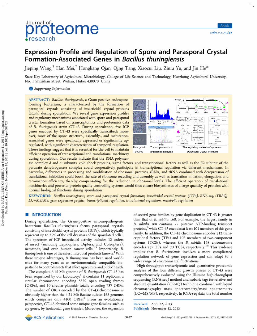
ABSTRACT: Bacillus thuringiensis, a Gram-positive endospore-forming bacterium, is characterized by the formation ofparasporal crystals consisting of insecticidal crystal proteins(ICPs) during sporulation. We reveal gene expression profilesand regulatory mechanisms associated with spore and parasporalcrystal formation based on transcriptomics and proteomics dataof B. thuringiensis strain CT-43. During sporulation, five ICPgenes encoded by CT-43 were specifically transcribed; more-over, most of the spore structure-, assembly-, and maturation-associated genes were specifically expressed or significantly up-regulated, with significant characteristics of temporal regulation.These findings suggest that it is essential for the cell to maintainefficient operation of transcriptional and translational machineryduring sporulation. Our results indicate that the RNA polymer-ase complex δ and ω subunits, cold shock proteins, sigma factors, and transcriptional factors as well as the E2 subunit of thepyruvate dehydrogenase complex could cooperatively participate in transcriptional regulation via different mechanisms. Inparticular, differences in processing and modification of ribosomal proteins, rRNA, and tRNA combined with derepression oftranslational inhibition could boost the rate of ribosome recycling and assembly as well as translation initiation, elongation, andtermination efficiency, thereby compensating for the reduction in ribosomal levels. The efficient operation of translationalmachineries and powerful protein-quality controlling systems would thus ensure biosyntheses of a large quantity of proteins withnormal biological functions during sporulation.
KEYWORDS: Bacillus thuringiensis, spore and parasporal crystal formation, insecticidal crystal proteins (ICPs), RNA-seq, iTRAQ,LC−MS/MS, gene expression profiles, transcriptional regulation, translational regulation, metabolic regulation
■ INTRODUCTION
During sporulation, the Gram-positive entomopathogenicbacterium Bacillus thuringiensis forms parasporal crystalsconsisting of insecticidal crystal proteins (ICPs), which typicallyrepresent up to 25% of the cell dry mass of the sporulated cells.1
The spectrum of ICP insecticidal activity includes 12 ordersof insect (including Lepidoptera, Diptera, and Coleoptera),nematode, and even human cancer cells.2,3 Importantly, B.thuringiensis is one of the safest microbial products known.4 Withthese unique advantages, B. thuringiensis has been used world-wide for many years as an environmentally compatible bio-pesticide to control insects that affect agriculture and public health.The complete 6.15 Mb genome of B. thuringiensis CT-43 has
been sequenced by our laboratory;5 it contains 11 replicons, acircular chromosome encoding 5529 open reading frames(ORFs), and 10 circular plasmids totally encoding 737 ORFs.The number of ORFs encoded by the CT-43 chromosome isobviously higher than the 4.21 Mb Bacillus subtilis 168 genome,which comprises only 4100 ORFs.6 From an evolutionaryperspective, CT-43 obtained some unique gene families, such ascry genes, by horizontal gene transfer. Moreover, the expansion
of several gene families by gene duplication in CT-43 is greaterthan that of B. subtilis 168. For example, the largest family inB. subtilis 168 contains 77 putative ATP-binding transportproteins,6 while CT-43 encodes at least 101members of this genefamily. In addition, the CT-43 chromosome encodes 312 trans-criptional factors (TFs) and 103 members of two-componentsystems (TCSs), whereas the B. subtilis 168 chromosomeencodes 237 TFs and 70 TCSs, respectively.7,8 This evidenceimplies that B. thuringiensis involves a more complicatedregulation network of gene expression and can adapt to awider range of environmental fluctuations.7
High-throughput transcriptomic and quantitative proteomicanalyses of the four different growth phases of CT-43 werecomprehensively evaluated using the Illumina high-throughputsequencing (RNA-seq) method and isobaric tags for relative andabsolute quantitation (iTRAQ) technique combined with liquidchromatography−mass spectrometry/mass spectrometry(LC−MS/MS), respectively. In RNA-seq data, the total number
Received: April 22, 2013Published: November 12, 2013
Article
pubs.acs.org/jpr
© 2013 American Chemical Society 5487 dx.doi.org/10.1021/pr4003728 | J. Proteome Res. 2013, 12, 5487−5501
Dow
nloa
ded
by N
AT
L L
BR
Y O
F SE
RB
IA o
n Se
ptem
ber
11, 2
015
| http
://pu
bs.a
cs.o
rg
Pub
licat
ion
Dat
e (W
eb):
Nov
embe
r 18
, 201
3 | d
oi: 1
0.10
21/p
r400
3728

of clean-reads for the four growth phases were 926 755, 1 096665, 577 810, and 1 493 721, respectively, with an average lengthof 110 nt. Therefore, the sequencing coverage of each samplereached 10- to 27-fold. In iTRAQ data, a total of 1756 proteinswere identified in eight samples (two biological replicates wereincluded for each growth phase), representing ∼28% of the total6266 proteins encoded by CT-43 genome.9 The results allowedelucidation of the regulatory mechanisms underlying the meta-bolic pathways and energy supply associated with sporulationand parasporal crystal formation in B. thuringiensis.9 Here wefocused on the gene expression profiles and regulatorymechanisms involved in spore and parasporal crystal formationin B. thuringiensis.
■ MATERIALS AND METHODS
RNA-seq and iTRAQ Analyses
Bacterial cultivation, cDNA library construction, RNA-seq,iTRAQ, LC−MS/MS, bioinformatics, and statistical analyseswere carried out as previously described.9 In brief, two biologicalreplicate cell samples of the B. thuringiensis strain CT-43 werecollected at 7, 9, 13, and 22 h. Each sample was divided into twoparts for whole-genome transcriptomics and proteomicsanalyses. For RNA-seq, total RNA was extracted from eachsample and treated with terminator 5′-phosphate-dependentexonuclease (Epicenter, Madison, WI) to deplete processedRNAs.10 Double-stranded cDNA libraries were constructed andsequenced with an Illumina Genome Analyzer IIx. The reads ofeach sample were mapped to the CT-43 genome using BlastNwith a threshold e value of 0.00001 and the “-F F” parameter.11
The expression level of each gene was reported as reads perkilobase of coding sequence per million mapped reads (RPKM),and differentially expressed genes were identified by the DEGseqpackage.12 We used FDR ≤ 0.001 and an absolute value of log2ratio≥1 as the threshold to judge the significance of gene expres-sion difference.For iTRAQ-LC−MS/MS, total proteins were extracted from
each sample and tryptically digested. The resulted peptides werelabeled individually with 8-plex iTRAQ reagents (AppliedBiosystems, Foster City, CA) at room temperature for 2 h asfollows: 7 h-1, 113; 7 h-2, 114; 9 h-1, 115; 9 h-2, 116; 13 h-1, 117;13 h-2, 118; 22 h-1, 119; and 22 h-2, 121. The labeled sampleswere pooled and then resolved into 12 fractions using anUltremex SCX column (Phenomenex, Torrance, CA). Afterdesalting using a Strata X C18 column (Phenomenex) and dryingunder vacuum, the labeled samples were subjected to LC−MS/MS analysis using a splitless nanoACQuity (Waters, Milford,MA) system coupled to a Triple TOF 5600 system (AB SCIEX,Concord, ON). Spectra from the 12 fractions were combinedinto one MGF (Mascot generic format) file and searched againstthe B. thuringiensis CT-43 protein database (6266 sequences,including 5529 proteins of the chromosome and 737 proteinsof the plasmids) combined with the reversed version of all pro-tein sequences using the Mascot search engine (2.3.02 version,Matrix Science). In the final search results, the false discovery rate(FDR) was <1.5%. The iTRAQ 8-plex was chosen for quantifica-tion during the search. A protein with ≥1.5-fold difference in theexpression level and a p value ≤0.05 was regarded as beingdifferentially expressed in our data.13,14
The iTRAQ labeling, SCX fractionation, LC−MS/MS analysisprocesses, and associated search parameters are also detailedlydescribed in the Supporting Information.
Determination of Expression of ICP Genes at the ProteinLevel
Strain CT-43 was grown under the same conditions as above. Ateach time point (7, 9, 11, 13, 15, 17, 19, 21, 22, and 23 h), 20 mLof cultures was separately collected and centrifuged for 15 min(6000g, 4 °C). Each cell pellet was then separately resuspendedin phosphate-buffered saline (137 mM NaCl, 2.7 mM KCl,10.1 mM Na2HPO4, 1.8 mM KH2PO4, pH 7.4); then, the sus-pension was sonicated on ice for 30 min (alternating 15 ssonication and 10 s pause at 130 W) using an ultrasonic pro-cessor model VC-130 (Sonics and Materials, Newtown, CT),and centrifuged for 10 min (12 000g, 4 °C). Each precipitate(cellular debris) was separately washed with 1 mol/L of NaCland sterile water. After that, the washed precipitate wasresuspended in 500 μL of 50 mmol/L Na2CO3−NaHCO3 (pH10.0) containing 5% β-mercaptoethanol and incubated at 37 °Cfor 1 h to dissolve the parasporal crystals. Thereafter, the sampleswere centrifuged for 10 min (12 000g, 4 °C), and the super-natants were analyzed by SDS-PAGE.
MS Identification of Proteins from ICP Bands
Two ICP bands corresponding to the 71 and 140 kDa wereexcised from SDS-PAGE gel (named as Cry_sample_1 andCry_sample_2, respectively; see Supplementary Figure S1 in theSupporting Information). They were subjected to in-gelreduction, alkylation, and digestion with trypsin as follows:samples were dehydrated with 500 μL of ACN, incubated for60 min at 56 °C with 200 μL of 10 mM DTT in 25 mMammonium bicarbonate buffer and then incubated for 45 minat room temperature in darkness with 200 μL of 55 mMiodoacetamide in 25 mM (NH4)HCO3 buffer. They were thenwashed twice with 25 mM (NH4)HCO3 buffer and dehydratedwith 500 μL of ACN, proteolyzed by trypsin overnight at 37 °C,and subsequently treated with 5% FA to stop proteolysis.The peptides were then eluted from the gel pieces once with
200 μL of 50% ACN and 0.1% FA and twice with 200 μL of 100%ACN and 0.1% FA. The eluted fractions were collected and driedunder vacuum. Each fraction was resuspended in certain volumeof buffer A (2% ACN, 0.1% FA) and centrifuged at 20 000g for10 min. The final concentration of the peptide in each fractionwas ∼0.5 μg/μL on average. Using an autosampler, 10 μL ofsupernatant was loaded on a 2 cm C18 trap column (innerdiameter 200 μm) on a Shimadzu LC-20AD nanoHPLC. Pep-tides were eluted onto a resolving 10 cm analytical C18 column(inner diameter 75 μm) that was assembled in-house. Thesamples were loaded at 15 μL/min for 4 min and eluted witha 44min gradient at 400 nL/min from 2 to 35%B (98%ACN, 0.1%FA), followed by a 2 min linear gradient to 80% B, maintenance at80% B for 4 min, and finally a return to 2% B in 1 min.The MS identification process and search parameters were the
same as the iTRAQ experiments, which are described in detail inthe Supporting Information.
Accession Number
The RNA-seq data from this article are available as raw shortreads data in the NCBI’s GEO database under accession numberGSE39479. Mass spectrometry data of the iTRAQ experimentfrom this article are deposited in the mzML format in theProteomeExchange database (http://proteomexchange.org/)under accession number PXD000020 through the PRIDE Website (http://www.ebi.ac.uk/pride/). Additionally, the MS data ofthe two ICP bands have been deposited to the ProteomeXchangewith identifier PXD000425.
Journal of Proteome Research Article
dx.doi.org/10.1021/pr4003728 | J. Proteome Res. 2013, 12, 5487−55015488
Dow
nloa
ded
by N
AT
L L
BR
Y O
F SE
RB
IA o
n Se
ptem
ber
11, 2
015
| http
://pu
bs.a
cs.o
rg
Pub
licat
ion
Dat
e (W
eb):
Nov
embe
r 18
, 201
3 | d
oi: 1
0.10
21/p
r400
3728

■ RESULTS AND DISCUSSION
Process of Spore Formation in B. thuringiensis
Sporulation in B. thuringiensis is characterized by seven stages:stages 0 to I, axial filament formation; stage II, forespore septumformation and asymmetric cell division; stage III, engulfment,forespore formation, and the first appearance of parasporalcrystals; stages IV to VI, cortex, coat, and exosporium assemblyand development of parasporal crystals; and stage VII, sporematuration and mother cell lysis and spore and parasporal crystalrelease9,15−17 (Figure 1A).Among the four time points selected,9 7 h (midlog growth
phase) represented vegetative growth stage, 9 h (early stationarygrowth phase) might cover stages 0 to II, 13 h (midstationarygrowth phase, the proportion of sporulating cells reached about30%) might include stages IV to VI, and 22 h (spore maturation
and mother cell lysis phase) mainly represented stages VI andVII, owing to sporulating population heterogeneity.18
ICP Gene Expression Profiles
In CT-43, the largest plasmid pCT281 (281.14 kb) encodes fourICP genes, cry1Aa3, cry1Ia14, cry2Aa9, and cry2Ab1, and thesecond largest plasmid pCT127 (127.86 kb) contains anotherICP gene, cry1Ba1. The results of RNA-seq indicated that thetranscripts of these genes were all specifically detected from 13 h(Table 1). Unfortunately, the ICPs were not detected at all byiTRAQ possibly because the ICPs are wrapped in compactparasporal crystals upon production and the protein samplepreparation procedure did not include an alkali dissolution step(e.g., treatment with Na2CO3−NaHCO3, pH 10.0 for 1 h at 37°C), which is necessary to dissolve ICPs out of the parasporalcrystals. However, the parasporal crystals composed of ICPs
Figure 1. Spore and parasporal crystal formation and the regulatory network of gene expression in B. thuringiensis. (A) Life cycle of B. thurigiensis and theformation processes of spore and parasporal crystal. The spore and parasporal crystal are emphasized by green and purple colors, respectively. (B)Temporal and spatial regulation network of σ factors and transcriptional factors. This part is drawn mainly according to figure 2 in ref 64. (C)Biosyntheses and transport of spore structural components.
Journal of Proteome Research Article
dx.doi.org/10.1021/pr4003728 | J. Proteome Res. 2013, 12, 5487−55015489
Dow
nloa
ded
by N
AT
L L
BR
Y O
F SE
RB
IA o
n Se
ptem
ber
11, 2
015
| http
://pu
bs.a
cs.o
rg
Pub
licat
ion
Dat
e (W
eb):
Nov
embe
r 18
, 201
3 | d
oi: 1
0.10
21/p
r400
3728

could be clearly observed in the cells during sporulation under aphase-contrast microscope (Figure 2A), and through the alkalidissolution step, two prominent ICP bands could be acquired, asshown in Figure 2B.According to the RPKM value of each gene, the transcriptional
levels of the genes cry1Ba1, cry2Aa9, and cry1Aa3 were higherthan others, suggesting that the proteins Cry1Ba1, Cry2Aa9, andCry1Aa3 might be the main components of parasporal crystalsin CT-43. By performing an additional MS identification(Supplementary Table S1 in the Supporting Information), weconfirmed that the 140 kDa band (the main band) shown inFigure 2B was composed of Cry1Ba1 and Cry1Aa3, while the
71 kDa band (the second band) was Cry2Aa9 and Cry2Ab1,together with the degraded parts of Cry1Ba1 and Cry1Aa3.According to the numbers of unique peptides identified,Cry1Ba1 (the main component of the main band), Cry2Aa9(the main component of the second band), and Cry1Aa3 (thesecond component of the main band) were high-abundantproteins. The result was in accordance with the transcriptomedata. The mode of parasporal crystal assembly likely varies withthe ICP type. The larger-sized ICPs (∼130 kDa) seem to formparasporal crystals by self-assembly, while the smaller-sized ICPs(71 kDa or less) that consist only of an insecticidal N-terminalregion require accessory proteins (such as P20 and ORF2) forparasporal crystal assembly.19,20 TheORF2 (29 kDa) in the sameoperon that contains the cry2Aa9 gene was also expressed at veryhigh levels (Table 1), so it would act as an accessory protein tofacilitate Cry2Aa9 assembling into parasporal crystals.20
Expression Profiles of the Spore Structure and AssemblyAssociated Genes
The spore structure and chemical composition play major rolesin spore resistance. From exterior to interior, the concentricallyarranged layers of B. thuringiensis spores include the exosporium,coat, outer membrane, cortex, germ cell wall, inner membrane,and central core.21,22 Next, the expression profiles of the sporestructure, assembly, and maturation-associated genes werepartially and briefly analyzed in the order from interior to exterior.
Core. The core is the innermost layer of the spore andcontains DNA, ribosomes, tRNA, and many enzymes. The mostsignificant differences between the growing cell cytoplasm andthe core are: (i) a large decrease in core water content (only25−50% of core wet weight), (ii) high levels of dipicolinic acid(DPA) (5−15% of core dry weight), and (iii) the saturation ofDNA with α/β-type small, acid-soluble spore proteins(SASPs).21,22
DPA was confirmed to be extremely important in sporeresistance and stability, which is a major driving force for thespore to accumulate such high levels of this small molecule.23
DPA is produced via a branch off of the lysine biosyntheticpathway. One step in this pathway is the condensation ofaspartate semialdehyde and pyruvate to produce dihydro-dipicolinic acid (DHDPA) catalyzed by DapA (dihydro-dipicolinate synthase).24 In the last step, DHDPA is oxidizedinto DPA by the enzymes SpoVFA and SpoVFB, the products ofthe spoVFAB operon25 (Figure 1C). Our RNA-seq resultsshowed that the dapA2 gene was highly expressed at 7 and 9 hand obviously down-regulated at 13 h, whereas the mRNA ofanother isozyme gene dapA1 was detected only at low levels at13 h (Supplementary Table S2 and Figure S2 in the SupportingInformation). At the protein level, DapA2 was identified, and itsabundance showed no increase at 13 h, which leads to the ques-tion of whether DHDPA was produced during the exponentialgrowth phase. The increase at both the mRNA and protein levels
Figure 2. Expression of ICP genes at the protein level. (A) Presence ofparasporal crystals determined by phase-contrast microscopy.B. thuringiensis strain CT-43 was grown in GYS medium for 17 h at28 °C and 200 rpm, which allowed sporulation process to occur. Then,the sample was observed under a phase-contrast microscopy (NikonECLIPSE E6000, Nikon, Tokyo, Japan). The scale bar represents10 μm. (B) Time course of the ICPs production in B. thuringiensis strainCT-43.
Table 1. Transcriptional Level of the Genes Encoding ICPs and an Accessory Protein
RPKM
gene name gene code molecular weight (kDa) 7 h 9 h 13 h 22 h
cry2Ab1 pCT281.272 71 0 0 96 8cry1Aa3 pCT281.277 133 0 0 6362 479cry1Ia14 pCT281.278 81 0 0 162 0cry2Aa9 pCT281.286 71 0 0 7553 43ORF2 pCT281.287 29 0 0 20231 17cry1Ba1 pCT127.021 140 0 0 26134 6853
Journal of Proteome Research Article
dx.doi.org/10.1021/pr4003728 | J. Proteome Res. 2013, 12, 5487−55015490
Dow
nloa
ded
by N
AT
L L
BR
Y O
F SE
RB
IA o
n Se
ptem
ber
11, 2
015
| http
://pu
bs.a
cs.o
rg
Pub
licat
ion
Dat
e (W
eb):
Nov
embe
r 18
, 201
3 | d
oi: 1
0.10
21/p
r400
3728

Table 2. Transcriptional Profiles of the Spore Peptidoglycan Synthesis-Associated Genes in B. thuringiensis CT-43
RPKM
gene gene code function 7 h 9 h 13 h 22 h
murA1 CH5327 UDP-N-acetylglucosamine 1-carboxyvinyltransferase 175 253 1153 918murA2 CH5374 UDP-N-acetylglucosamine 1-carboxyvinyltransferase 455 304 244 0murB CH3910 UDP-N-acetylenolpyruvoylglucosamine reductase 1579 2835 146 0murB2 CH5132 UDP-N-acetylenolpyruvoylglucosamine reductase 16 24 49 7murC CH4710 UDP-N-acetylmuramate-L-alanine ligase 102 162 187 58murD CH3913 UDP-N-acetylmuramoyl-L-alanyl-D-glutamate synthetase 102 63 162 74murE CH3915 UDP-N-acetylmuramoylalanyl-D-glutamate- 2,6-diaminopimelate ligase 70 172 130 0murF CH0220 UDP-N-acetylmuramoylalanyl-D-glutamyl 122 75 89 23murF CH2116 2,6-diaminopimelate-D-alanyl-D-alanyl ligase 4 7 33 14murG CH3690 N-acetylglucosaminyl transferase 0 0 107 0murG CH3911 N-acetylglucosaminyl transferase 0 0 158 0murG2 CH4267 N-acetylglucosaminyl transferase 91 152 97 56murG2 CH4269 N-acetylglucosaminyl transferase 74 235 23 23mraY CH3914 phospho-N-acetylmuramoyl-pentapeptide-transferase 54 29 118 58bacA1 CH0658 undecaprenyl pyrophosphate phosphatase 4 8 28 23bacA2 CH1313 undecaprenyl pyrophosphate phosphatase 371 145 269 0bacB CH2689 undecaprenyl pyrophosphate phosphatase 0 0 478 20
CH1513 oligosaccharide translocase (flippase) 0 0 261 0CH5470 oligosaccharide translocase (flippase) 40 5 67 0
dacA CH0009 D-alanyl-D-alanine carboxypeptidase (low MW PBP5) 90 45 232 0dacB CH1296 muramoyltetrapeptide carboxypeptidase (low MW PBP5*) 123 129 1314 14
CH1207 D-alanyl-D-alanine carboxypeptidase 21 10 26 0dacB CH1397 D-alanyl-D-alanine carboxypeptidase (low MW PBP5*) 0 0 333 0spmA CH1398 spore maturation protein A 0 0 218 0spmB CH1399 spore maturation protein B 0 0 833 0
CH1490 thermostable carboxypeptidase 1 669 251 98 20CH1620 D-alanyl-D-alanine carboxypeptidase 8 48 137 0CH1903 D-alanyl-D-alanine carboxypeptidase 9 36 18 0CH2011 D-alanyl-D-alanine carboxypeptidase 89 91 204 89CH2482 D-alanyl-D-alanine carboxypeptidase 0 0 50 0CH3010 D-alanyl-D-alanine carboxypeptidase 8 41 58 12CH3218 D-alanyl-D-alanine carboxypeptidase 74 296 141 0CH3463 D-alanyl-D-alanine carboxypeptidase 0 0 694 0CH3494 D-alanyl-D-alanine carboxypeptidase 29 213 174 0
dacF CH4083 D-alanyl-D-alanine carboxypeptidase (low MW PBP) 0 0 2135 0CH5069 D-alanyl-D-alanine carboxypeptidase 0 0 126 0CH5429 D-alanyl-D-alanine carboxypeptidase 7 5 36 4CH0425 penicillin-binding protein 0 0 112 0
pbpF CH1020 penicillin-binding protein (class A PBP) 0 0 123 0CH1383 penicillin-binding protein 0 0 256 0CH1476 penicillin-binding protein 0 28 45 0CH1616 penicillin-binding protein 0 42 84 43CH2155 penicillin-binding protein 62 132 174 0CH2245 penicillin-binding protein 51 47 166 0CH2619 penicillin-binding protein 0 53 0 0CH2644 penicillin-binding protein 47 0 0 0CH2668 penicillin-binding protein 0 0 234 0CH3054 penicillin-binding protein 60 106 77 7CH3426 penicillin-binding protein 0 88 101 13CH4049 penicillin-binding protein 137 200 386 0CH4289 penicillin-binding protein 151 215 81 0
ywhE CH4395 penicillin-binding protein (class A PBP) 0 0 3291 0CH5415 penicillin-binding protein 0 0 271 0
cwlD CH0144 spore-specific N-acetylmuramoyl-L-alanine amidase 0 0 262 0cwlD2 CH1421 N-acetylmuramoyl-L-alanine amidase 0 0 750 0pdaA? CH0306 polysaccharide deacetylase 14 3 31 14padA CH0411 N-acetylmuramic acid deacetylase 11 30 35 0cwlC CH2172 spore-specific N-acetylmuramoyl-L-alanine amidase 0 0 89 20135cwlB CH2234 N-acetylmuramoyl-L-alanine amidase 12 22 23 7494
Journal of Proteome Research Article
dx.doi.org/10.1021/pr4003728 | J. Proteome Res. 2013, 12, 5487−55015491
Dow
nloa
ded
by N
AT
L L
BR
Y O
F SE
RB
IA o
n Se
ptem
ber
11, 2
015
| http
://pu
bs.a
cs.o
rg
Pub
licat
ion
Dat
e (W
eb):
Nov
embe
r 18
, 201
3 | d
oi: 1
0.10
21/p
r400
3728

of the enzymes SpoVFA and SpoVFB during sporulationsuggests that DPA synthesis is temporally activated (Supple-mentary Table S2 in the Supporting Information). Meanwhile,Cax (calcium/proton antiporter) and a calcium-transportingATPase (CH3874) were also up-regulated at both the mRNAand protein levels during sporulation, which possibly satisfies theneed of DPA to form a 1:1 chelate with Ca2+. DPA is synthesizedonly in the mother cell compartment of the sporulating cell andis ultimately located only in the core of the spore (Figure 1C).The 1:1 chelate of DPA and Ca2+ is taken up into the foresporevia proteins encoded by the spoVAA∼F operon26 (Figure 1C),which was revealed by RNA-seq to be specifically induced(throughout the manuscript, “specially induced” at a time pointindicates that the gene/protein is not transcribed/expressedbefore this time point) at 13 h at relatively low levels (Supple-mentary Table S2 in the Supporting Information).The α/β-type SASPs are extremely abundant in the spore core,
comprising 3−6% of total spore protein.22 Moreover, they aresynthesized only in the developing forespore during sporula-tion.22 They can saturate the spore DNA and alter its structure,contributing to the resistance of the spores to heat, UV radiation,and many chemicals.21 The CT-43 chromosome contains 12genes encoding the SASPs, and the mRNAs of these genes couldbe detected only after 13 h (Supplementary Table S2 and FigureS2 in the Supporting Information). Among the eight SASPsidentified by iTRAQ, (i) CH0446, SspN and SspB seemed to beproduced starting at 9 h according to the extremely largedifference times at 7 h versus 9 h; (ii) the abundance of six SASPswas significantly increased at 13 h compared with 9 h, includingSspK (5.6-fold), SasP1 (10.7-fold), CH1242 (7.3-fold), CH1940(5.4-fold), SasP2 (4.8-fold), and SspB (11.3-fold); and (iii) theabundance of these SASPs all remained almost unchanged at 22 hcompared with 13 h (Supplementary Table S2 in the SupportingInformation). Thus, these results demonstrate the temporalnature of the SASP biosynthesis in the forespore.22
Cortex. After engulfment, the forespore becomes a double-membrane-bound cell within the mother cell. Two layers ofspore peptidoglycan (PG) are synthesized within the space be-tween the inner and outer membranes.27 The inner layer of sporePG, the germ cell wall, is synthesized first, followed by the outerlayer, the cortex.27 The PG of the germ cell wall synthesizedwithin the forespore has a structure that is probably identical tothat of vegetative cells, while the cortex PG synthesized within
the mother cell has several structural modifications relative tovegetative cell PG.21 In B. subtilis, the cortex PG differs from thevegetative cell PG most dramatically in: (i) the absence ofteichoic acids; (ii) the removal of many peptide side chains,∼1/4of the N-acetyl muramic acid (NAM) residues carrying a singleL-alanine residue; and (iii) conversion of ∼50% of the NAMresidues to muramic-δ-lactam, an important marker of the cortexPG.21,27,28
In bacteria, cytoplasmic PG precursors are synthesized as theUDP-linked N-acetyl glucosamine (NAG) and NAM. Aminoacids are sequentially added to UDP-NAM to produce thepentapeptide side chain. The NAM-pentapeptide is then trans-ferred to an undecaprenol lipid carrier to produce Lipid I, withsubsequent addition of NAG from UDP-NAG to produce LipidII. All of these cytoplasmic steps are catalyzed by the mur geneproducts (MurA∼G plus MraY).28 Subsequently, Lipid II isflipped across the membrane into the intermembrane space byflippases, which include two protein families: the COG2244family including SpoVB andMurJ29 as well as the SEDS family ofintegral membrane proteins, which includes FtsW, RodA, andSpoVE. The disaccharide−pentapeptide subunits are polymer-ized into glycan strands, and the peptide side chains are utilizedto cross-link the strands into a network via the glycosyl trans-ferase and transpeptidase activities carried jointly by penicillin-binding proteins (PBPs).28,29
Our results demonstrated that most enzymes requiredfor vegetative cell PG synthesis remained essentially constantat both the mRNA and protein levels during sporulation (Table 2,Supplementary Table S3 and Figure S3 in the SupportingInformation), suggesting that some enzymes required for sporePG synthesis are certainly shared with vegetative cell PGsynthesis machinery.28 Many enzymes were significantly up-regulated or specifically induced during sporulation (Table 2,Supplementary Table S3 and Figure S3 in the SupportingInformation), indicating that these enzymes could be specific forspore PG synthesis and modification.At the mRNA level, some mur genes were obviously up-
regulated at 13 h compared with 7 h (unless otherwise noted,“7 h” is hereafter used as the comparative object), includingmurA1 (more than six-fold), murB2 (about three-fold), murE(about two-fold), murF CH2116 (about eight-fold), and mraY(about two-fold). In addition, two murG genes, CH3690 andCH3911, were specifically induced at 13 h. At the protein level,
Table 2. continued
RPKM
gene gene code function 7 h 9 h 13 h 22 h
cwlH CH2815 N-acetylmuramoyl-L-alanine amidase 0 0 39 1246cwlH2 CH3622 N-acetylmuramoyl-L-alanine amidase 0 0 0 1652spoVB CH4429 stage V sporulation protein B 0 0 134 0spoVD CH3916 sporulation specific D,D-transpeptidase (class B PBP) 92 227 489 0spoVE CH3912 stage V sporulation protein E 87 313 539 0spoVR CH0741 stage V sporulation protein R 0 0 1947 0spoVK CH3647 cell division protein FtsH 0 0 1198 38spoVS CH2092 stage V sporulation protein S 803 2428 2147 7596spoVS2 CH3719 stage V sporulation protein S 4622 6601 4720 0spoVT CH0049 stage V sporulation protein T 524 363 674 116
CH1407 cell wall endopeptidase 0 0 599 0f tsW CH0984 cell division protein FtsW 38 20 18 0f tsW CH1140 cell division protein FtsW 0 0 23 0f tsW CH3954 cell division protein FtsW 283 163 80 0rodA CH4172 rod shape-determining protein RodA 121 169 119 0
Journal of Proteome Research Article
dx.doi.org/10.1021/pr4003728 | J. Proteome Res. 2013, 12, 5487−55015492
Dow
nloa
ded
by N
AT
L L
BR
Y O
F SE
RB
IA o
n Se
ptem
ber
11, 2
015
| http
://pu
bs.a
cs.o
rg
Pub
licat
ion
Dat
e (W
eb):
Nov
embe
r 18
, 201
3 | d
oi: 1
0.10
21/p
r400
3728

eight enzymes (includingMurA1, MurA2,MurB2, MurC,MurD,MurE, MurF CH0220, and MurG CH3911) were identified byiTRAQ, and the abundance of seven enzymes remained almostunchanged, except MurA1, which was down-regulated by 1.6-,2.5-, and 1.7-fold at 9, 13, and 22 h, respectively (SupplementaryTable S3 in the Supporting Information).Among the significant functionally redundant PBPs and
flippases, some enzymes have been confirmed to participate incortex PG synthesis, including: Class A PBPs, PbpF and YwhE;Class B PBPs, SpoVD and SpoVE; low-MW PBPs, DacA, DacB,and DacF, as well as SpoVB.28,29 Our RNA-seq data showed that17 genes of these functional groups were specifically inducedduring sporulation, which included the dacB-spmA-spmB operon,dacF, pbpF, spoVB, and ywhE. In addition, 14 genes were up-regulated at 13 h by 2- to 21-fold, including dacA (2-fold),dacB (10-fold), CH1620 (17-fold), CH3218 (21-fold), spoVD(5-fold), and spoVE (6-fold) (Table 2). In iTRAQ data, nine en-zymes of these functional groups were identified. DacF, CH2155,CH2245, and CH4289 were up-regulated by 1.6-, 3.6-, 1.2-, and1.6-fold at 9 h and by 2.1-, 2.2-, 1.5-, and 1.3-fold at 13 h,respectively; DacA, CH1476, and CH1490 remained unchanged,while two DacB proteins, CH1296 and CH1397, could not bequantified (Supplementary Table S3 in the SupportingInformation).During muramic-δ-lactam formation, the N-acetylmuramoyl-
L-alanine amidase encoded by cwlD, removes a peptide sidechain from the NAM residue, and the N-acetylmuramic aciddeacetylase, encoded by pdaA, sequentially catalyzes thedeacetylation of muramic acid and lactam ring formation.30,31
At the mRNA level, two cwlD genes, CH0144 and CH1421, werespecifically induced, and two pdaA genes, CH0306 and CH0411,were both up-regulated by about three-fold at 13 h (Table 2). IniTRAQ data, the abundance of CwlD2 (CH1421) was increasedby two-fold at 13 h and eight-fold at 22 h; the two PdaA enzymeswere identified but failed to be quantified (SupplementaryTable S3 in the Supporting Information). In addition, additionalfour N-acetylmuramoyl-L-alanine amidase genes (cwlC, cwlB,cwlH, and cwlH2) were highly expressed at the mRNA level onlyat 22 h, implying that these enzymes participate in mother celllysis or cortex hydrolysis during spore germination.21
Coat and Exosporium. In B. subtilis, the coat is theoutermost layer of the spore and is composed of three layers: theinner coat, the outer coat, and the crust.32,33 In the Bacillus cereusgroup, which includes Bacillus anthracis, B. cereus, and B.thuringiensis, the coat contains only two layers: the inner coatand outer coat, but the outermost layer consists of a different typecalled the exosporium.33,34 The function, structure, and assemblyof the coat and exosporium have been comprehensively andsystematically discussed in many excellent reviews.21,22,33,34 Onthe basis of our RNA-seq and iTRAQ data from CT-43, wefocused on the expression profiles of the coat and exosporiumstructure-, assembly-, and maturation-associated genes at both themRNA and protein levels.In RNA-seq data, the transcripts of the overwhelming majority
of the coat and exosporium-associated genes were specificallydetected at a very high abundance at 13 or 22 h (Table 3 andSupplementary Figure S4 in the Supporting Information).However, most of the 35 proteins identified by iTRAQ couldbe detected from 7 h, except that a few proteins (includingBxpB, CotJB, CotY, CwlJ, ExsY, SpoIVA, SpoVID, and YpzA)seemed to be specifically expressed during sporulation, ac-cording to the extremely large differences in protein abundance(Table 4). These results demonstrate that many coat and
exosporium-associated proteins are not synthesized de novoduring sporulation but rather are packaged from preexistingstocks.35,36 The highest mRNA levels of many genes appearedat 13 h, while the protein abundances of these genes reachedmaximum values at 22 h, obviously displaying a temporal delay inprotein expression relative to mRNA expression (Tables 3 and4). In addition, the mRNAs of many genes could not be detectedin both the 7 and 9 h samples, but the proteins encoded by thesegenes were detectable in the samples (Tables 3 and 4). Somestudies in other species also found that the mRNA and proteinchanges of many genes were not correlated,37−39 which could bedue to differential regulation of mRNA and protein expression,stability, and degradation.37
The total number of coat proteins is estimated to be more than70 in B. subtilis, and at least 50 coat proteins and morphogeneticfactors are encoded in the genomes of the B. cereus group34,40
(Table 3). The morphogenetic factors SpoIVA, SpoVID, ExsA/SafA, and CotE that are conserved in the genus Bacillus playmajor roles in coat morphogenesis.31,34 In B. subtilis, SpoIVAis located in close proximity to the forespore outer membraneand acts as the site of coat attachment, while SafA and CotE arenecessary for the assembly of the inner coat and outer coat,respectively.32−34 Our proteomics data showed that theabundances of SpoIVA and ExsA/SafA were markedly decreasedat 22 h compared with 13 h, while that of CotE remained almostunchanged (Table 4). These results might reflect the temporalityof coat assembly to a certain extent.Some germinant receptor proteins (GRPs), such as GerA and
GerB, localize in the inner membrane, while others, such as GerP,are located in the coat.41,42 The CT-43 chromosome contains atleast 38 GRPs belonging to different families. As revealed byRNA-seq, only gerD mRNA could be detected throughout thelife cycle, and three genes (CH2239, CH4520, and CH4750)were expressed only at 22 h. The mRNA transcripts of the other34 genes were all detected only starting at 13 h (SupplementaryTable S4 and Figure S5 in the Supporting Information). Un-fortunately, only four GRPs were identified by iTRAQ. Despitethis, GerD, GerE, GerM, and GerPC were up-regulated by 7.3-,1.3-, 7.1-, and 2.3-fold at 13 h compared with 9 h, respectively.Moreover, GerE and GerPC were up-regulated by 30.9- and 2.2-fold at 22 h compared with 13 h, respectively (SupplementaryTable S4 in the Supporting Information). In addition, the cortex-lytic enzyme CwlJ and its partner protein GerQ as well as thecortex-lytic enzyme YaaH are located in the coat.43,44 (Table 4).Recently, spore maturation was found to be necessary for
acquisition of full spore resistance.45 Among the 35 spore coatand exosporium-associated proteins identified by iTRAQ, theabundances of most proteins were markedly increased at22 h compared with 13 h. In addition, the mRNA of a sporeappendage protein (CH2337) was only detected at 22 h at veryhigh levels, and its protein abundance was increased by about34.4-fold at 22 h compared with 13 h. Furthermore, two types ofirreversible covalent cross-links, o,o′-dityrosine bond and ε-(γ-glutamyl)-lysil isopeptide bond, have been detected in coatproteins.33 A coat-specific transglutaminase, Tgl, catalyzes theformation of an ε-(γ-glutamyl) lysine isopeptide bond to cross-link and polymerize coat proteins.33,45,46 This transglutaminase-mediated cross-linking is believed to play an important role incoat protein assembly in the genus Bacillus.47,48 In our RNA-seqdata, the tgl gene (CH3969) encoding transglutaminase wasspecifically expressed at 22 h. Taken together, changes not onlyin the kinds of coat proteins but also (and particularly) in coatstructure likely accompany the process of spore maturation.45
Journal of Proteome Research Article
dx.doi.org/10.1021/pr4003728 | J. Proteome Res. 2013, 12, 5487−55015493
Dow
nloa
ded
by N
AT
L L
BR
Y O
F SE
RB
IA o
n Se
ptem
ber
11, 2
015
| http
://pu
bs.a
cs.o
rg
Pub
licat
ion
Dat
e (W
eb):
Nov
embe
r 18
, 201
3 | d
oi: 1
0.10
21/p
r400
3728

In our proteomics data, 10 exosporium-associated proteinswere identified, and the abundances of most proteins weresignificantly up-regulated at 22 h compared with 13 h, includingAlr (10-fold), BxpB (525-fold), CotB1(505-fold), CotB2 (52-fold), CotY (14-fold), ExsY/CotZ (3-fold), ExsK (381-fold), andInH (6-fold). The proteins BxpB, ExsY/CotZ, and CotY wereconfirmed to be required for exosporium assembly, and ExsA/SafA is required for both coat and exosporium assembly in theB. cereus group.49−51 Whereas BclA is the major component ofthe exosporium hair-like nap,52 this protein was not found in ourproteomics data, and this phenomenon also appeared in anothercase,53 possibly due to the unique structure of the exosporiumhair-like nap.The exosporium is a distinct glycoprotein layer with rhamnose
and methyl rhamnose representing the major carbohydrategroups of the exosporium glycoprotein in B. thuringiensis.54 In theB. cereus group genomes, the genes bclA, bxpB, cotY, and exsY/cotZ are clustered with the rhamnose biosynthesis operon(rfbACBD, also named as rmlACBD) and the genes encodingglycosyltransferases and O-methyltransferases.55 Our results indi-cated that all genes within the CT-43 gene cluster CH1141−1160seemed to be specifically expressed at 13 h at the mRNA level(Supplementary Table S4 and Figure S5 in the Supporting
Information). Moreover, 65% (13/20) of the proteins encoded bythis gene cluster were identified by iTRAQ, and the abundance ofmost proteins reached a peak during sporulation (SupplementaryTable S4 in the Supporting Information). Consequently, themajority of genes in this gene cluster likely participate inexosporium formation.55
Transcriptional Regulation
Transcriptional Machineries and Their RegulatoryRoles. Transcription is the first step in the process of geneexpression and is carried out by RNA polymerase (RNAP). InGram-positive bacteria like genus Bacillus, the RNAP coreenzyme possesses five subunits (α2ββ′ω) and an additionalsubunit (δ), RpoE, is required for the specificity of RNAP. Thecore enzyme could interact withmultiple interchangeable σ subunits(σ factor), which gives rise tomultiple forms of RNAPholoenzymes.According to the features of σ factors, different forms of RNAPholoenzymes could bind to their cognate promoters to initiatetranscription of specific genes (or operons).56
Interestingly, our RNA-seq results indicated that the gene rpoZencoding the ω subunit was induced only during the stationaryphase, being initiated at 9 h and up-regulated by more than two-fold at 13 h compared with 9 h (Supplementary Table S5 in the
Table 3. Transcriptional Profiles of the Spore Coat and Exosporium Structure- and Assemble-Associated Genes in B. thuringiensisCT-43
RPKM RPKM
gene gene code 7 h 9 h 13 h 22 h gene gene code 7 h 9 h 13 h 22 h
Spore Coat-Associated GenescotA CH4802 0 0 2466 16 yabG CH0037 0 111 53 10cotD CH1484 0 0 159 5424 ybaQ CH1157 0 0 1758 0cotE CH3713 0 0 11368 451 yckK CH0824 88 178 556 0cotH CH1987 0 0 147 87242 ycsK CH4131 11 16 194 1444cotI CH1996 0 0 5193 0 ydhD CH3361 0 0 35 5cotJA CH0777 0 0 2059 0 yhaX CH0840 0 0 4278 0cotJB CH0776 0 0 1311 0 yhbA CH0467 8 11 15 0cotJC CH0775 0 0 2309 0 yheD CH0829 0 0 964 0cotM CH3567 0 0 134 3498 yhjR CH3076 0 0 21 16cotN CH1209 33 314 58 0 yirY CH2259 39 50 13 0cotQ CH0356 0 0 0 292 yisY CH4803 0 0 745 53cotS CH5010 65 147 870 0 yisY1 CH2439 0 0 669 0cotSA CH0260 0 0 222 0 ykuD CH0725 0 0 0 99cwlJ CH5430 0 0 13940 0 ykvP CH3704 0 4 50 0exsA/safA CH4442 0 0 4220 9 ylbD CH3937 0 0 0 132gerQ CH5431 0 0 13838 7 yncD CH2023 0 0 75 0oxdD CH0973 0 5 23 0 yodI CH3527 0 0 0 30864sodA CH1396 0 0 1487 22 ypeP CH1526 32 43 475 0spoIVA CH1436 0 0 4230 0 yppG CH1483 0 0 744 42spoVID CH4474 0 0 5248 0 ypzA CH1528 748 273 1089 0tasA pCT8252.6 59 163 2086 581 ysxE CH4473 0 0 3005 0tgl CH3969 0 0 0 499 yusA CH5043 366 100 14 0yaaH CH3554 0 0 1360 0 yxeE CH3478 0 0 0 2401
Exosporium-Associated Genesalr CH0226 0 0 1680 2109 exsC CH2888 0 0 0 86920bclA CH1144 0 0 1672 3834 exsD CH2631 0 0 0 5048bclB CH1145 0 0 17492 17854 exsE CH1663 6 6 19 3betA CH3416 0 0 0 787 exsG CH2087 0 0 0 232bxpB CH1159 0 0 101 23994 exsJ CH2359 0 0 151 44219cotB1 CH0332 0 0 100 42004 exsK CH2477 0 0 0 37820cotB2 CH0333 0 0 0 11368 exsY/cotZ CH1160 0 0 18571 153cotY CH1156 0 0 2001 6693 inH CH2882 0 0 0 2100exsA/safA CH4442 0 0 4220 9 sod-Mn CH4291 5562 9915 572 8
Journal of Proteome Research Article
dx.doi.org/10.1021/pr4003728 | J. Proteome Res. 2013, 12, 5487−55015494
Dow
nloa
ded
by N
AT
L L
BR
Y O
F SE
RB
IA o
n Se
ptem
ber
11, 2
015
| http
://pu
bs.a
cs.o
rg
Pub
licat
ion
Dat
e (W
eb):
Nov
embe
r 18
, 201
3 | d
oi: 1
0.10
21/p
r400
3728

Supporting Information). At the same time, iTRAQ data showedthat the protein RpoZ was up-regulated by about 1.6-fold at 13 hcompared with 9 h (Supplementary Table S5 in the SupportingInformation). The β′ subunit was observed to be moreprone to proteolytic fragmentation in vitro when the RNAP ofMycobacterium smegmatis lacks the ω subunit.57 In another case,the purified Escherichia coli RNAP lacking the ω subunit did notrespond to the effector ppGpp of the stringent response in vitro.58
The ppGpp strongly and directly inhibits the promoters for rRNAand tRNA but directly or indirectly stimulates expression of a set ofgenes for amino acid biosynthesis and transport.58,59 Indeed, wefound that SpoT (a bifunctional enzyme that can both synthesizeand degrade ppGpp) and many proteases were markedly up-regulated during the stationary phase. Taken together, these resultssuggest that the increased expression of the ω subunit during thestationary phase would contribute to the prevention of the RNAP β′subunit from proteolytic fragmentation and couple to the ppGppsignaling system in transcriptional regulation of the stringentresponse in B. thuringiensis.Another RNAP subunit deserving particular attention is the
δ subunit. Because a strain containing a deletion of the rpoE gene
is viable and shows no major alterations in gene expression, it islikely to be a dispensable subunit of RNAP in B. subtilis.60,61 Inthe presence of δ, however, RNAP displays increased transcrip-tional specificity and RNA synthesis efficiency.61,62 Moreover,inactivation of the rpoE gene can alleviate suppression ofsporulation in B. subtilis at early stage III produced by disruptionof the pdhC gene encoding the E2 subunit of pyruvate de-hydrogenase, indicating that the δ subunit does have some director indirect role in sporulation.60 Because the protein RpoE wasup-regulated by about 1.5-fold at 13 h compared with 9 h(Supplementary Table S5 in the Supporting Information), wespeculate that this increase could also have some importantregulatory roles in spore and parasporal crystal formation inB. thuringiensis.Except for the above-mentionedω and δ subunits, the α, β, and
β′ subunits of core RNAP were all maintained at equivalent ex-pression levels during each growth phase. Furthermore, expres-sion of the genes greA (transcriptional elongation factor) and rho(transcriptional termination factor) were both up-regulated byabout three-fold at the mRNA level at 13 h. In iTRAQ data, theabundance of GreA was increased by 1.5-fold, and Rho remained
Table 4. Spore Coat and Exosporium-Associated Proteins Identified by iTRAQ
protein 7 h vs 9 ha 7 h vs 13 h 7 h vs 22 h 9 h vs 13 h 9 h vs 22 h 13 h vs 22 h
Alr 1.4b 1.5 15.0 1.0 10.0 10.0BxpB 2331.6 6876.2 72752.3 164.5 30.6 524.8CotA −c 154.7CotB1 1.7 1.5 152.9 6706.4 58.5 504.7CotB2 1.1 −1.1d 50.9 −1.4 37.2 51.9CotE 5.5 24.0 13.7 8.2 6.0 −1.3CotH 5.6 14.9 2.9CotJB 3992.9 3.1CotJC 1.0 6.9 20.3 7.7 19.5 2.6CotN 1.4 1.3 3.0 1.1 2.0 2.6CotQCotY 2300.1 16.7 3566.8 3.6 20.5 13.9CwlJ 2332.5 5338238.4 5572.5 1160.8 399.1 4.2ExsA/SafAe 2.5 26.3 82.6 26.3 91.5 −4.3ExsK 1.6 1.1 9.2 1.0 5.0 381.1ExsY/CotZ 3103.6 9135.6 15309.1 3.6 8.6 2.8GerQ 1.6 3.0 8.6 2.6 7.5 3.0InH 1.6 1.5 6.0 1.0 3.3 5.9Mn-SOD 1.0 1.1 1.1 1.1 1.1 1.0OxdD 5.5 4.0SodA 1.3 3.4 6.4 2.7 5.7 2.7SpoIVA 2299.6 3845.1 9.8 9.1 1.7 −4.2SpoVID 2597.2 15430.5 3793.8 9.6 7.6 2.9YaaH 1.2 4.2 13.2 3.8 11.8 3.7YbaG 27.7 16.4 203.4 1.1 12.9 10.9YbaQYckK −1.1 1.3 2.1 1.5 2.4 1.8YcsK 1.1 1.4 8.9 1.4 8.8 7.6YhaX 1.1 9.2 1.3 12.1 1.0 −3.0YheDYirY 1.1 1.0 −1.1 1.0 −1.2 −1.2YisY 31.8 3.1 7.3 3.1YpzA 2300.6 221.3 269.9 7.0 7.3 1.1YusA 1.0 1.4 2.4 1.4 2.3 2.0YxeE 1.1 1.3 4.2 −1.1 5.0 3.0
aProtein abundance of 9 h is compared with that of 7 h; the rest may be deduced by analogy. bNumerical value represents the up-regulated foldchange between two specific growth phases. cProtein abundance cannot be quantitatively compared between two specific growth phases. dNegativenumber means the down-regulated fold between two specific growth phases. eSame protein is assigned two different designations in two species.
Journal of Proteome Research Article
dx.doi.org/10.1021/pr4003728 | J. Proteome Res. 2013, 12, 5487−55015495
Dow
nloa
ded
by N
AT
L L
BR
Y O
F SE
RB
IA o
n Se
ptem
ber
11, 2
015
| http
://pu
bs.a
cs.o
rg
Pub
licat
ion
Dat
e (W
eb):
Nov
embe
r 18
, 201
3 | d
oi: 1
0.10
21/p
r400
3728

almost unchanged at 13 h. In addition, two genes (nusB andnusG) and their encoding proteins (the transcription antitermi-nation proteins) remained almost unchanged at 13 h at themRNA and protein levels (Supplementary Table S5 in theSupporting Information), respectively. These results implied thatthe transcriptional machineries were running efficiently duringsporulation.Regulatory Roles of Σ-Factors and Transcriptional
Factors. The σ-factors and TFs play vital roles in geneexpression regulation. The CT-43 chromosome encodes 27 σfactors and more than 50 σ-factor-associated genes (such as anti-σ-factor and anti-σ-factor antagonist) and at least 312 TFs thatbelong to 31 superfamilies and 59 families (Supplementary TableS5 and Figure S6 in the Supporting Information). Notably,∼80.5% (62/77) of σ factor and σ-factor-associated genes and55.8% (174/312) of TFs were expressed at the mRNA levelunder our experimental conditions (Supplementary Table S5and Figure S6 in the Supporting Information).We observed that the TF regulatory network were quite active
during sporulation. According to the RNA-seq data: (i) theexpression levels of 32 TF genes were increased and 46 TF geneswere specifically induced at 13 h; (ii) the expression levels of36 TF genes were decreased and 19 TF genes that wereexpressed both at 7 and 9 h were not transcribed at 13 h; (iii)eight TF genes were specifically expressed at 9 h; and (iv) 58 TFgenes, which included the six specifically induced genes, were stillexpressed at 22 h (Supplementary Table S5 in the SupportingInformation). In iTRAQ data, a total of 79 TFs were identified:11 TFs were up-regulated while 8 TFs were down-regulatedat 13 h; 41 TFs remained almost unchanged at 13 h; and 19 TFsfailed to be quantified (Supplementary Table S5 in the SupportingInformation). These results suggested that a complicated TFregulatory network was involved in gene expression duringsporulation.The sporulation-specific σ-factors, SigH, SigF, SigE, SigG, and
SigK, are spatially and temporally activated through a “criss-cross” mechanism63 (Figure 1B). In RNA-seq data, with theexception of sigH mRNA, which was detected from theexponential growth phase, the other sporulation-specificσ-factors were detected at high levels only during the stationarygrowth phase (Supplementary Table S5 and Figure S6 in theSupporting Information). At the protein level, within the 77σ-factor and σ-factor-associated genes, only seven σ-factors(including SigA, SigE, SigG, SigF, SigH, SigK, and SigL) andSpoIIAA (antisigma F factor antagonist) were identified byiTRAQ (Supplementary Table S5 and Figure S6 in theSupporting Information).Activation of Spo0A and SigH in predivisional cells leads to
asymmetric division, and these factors can be mutually promotedthrough a positive feed-forward loop (Figure 1B). Spo0A∼Pincreases transcription of the sigH gene by repressing tran-scription of the repressor abrB genes; activated SigH, in turn,elevates transcription of the spo0A gene using a SigH-dependentpromoter.64 In agreement with the feed-forward loop, themRNAlevel of spo0A peaked during the transition phase (9 h),while those of the three abrB genes encoded by CT-43 wereundetectable after 9 h (Supplementary Table S5 in theSupporting Information). In iTRAQ data, Spo0A was separatelyup-regulated by 2.3- and 2.6-fold at 9 and 13 h; SigH was the onlyone of the sporulation-specific σ-factors that failed to be identified.The two identified AbrB factors, CH0032 and CH1953, weredown-regulated by 3.2- and 1.6-fold at 13 h, respectively.
SigF is the first sporulation-specific σ-factor activated in theforespore, and its activity is necessary for processing of pro-SigEto active SigE in the mother cell.65 SigF and SigE regulate earlycompartmentalized gene expression, including the genes forengulfment. After engulfment, a second round of compartmen-talized gene expression occurs, with SigG and SigK becomingactive in the forespore and mother cell, respectively.63 Similarly,SigG activation in the forespore is necessary for processing ofpro-SigK to active SigK in the mother cell.65 SigG and SigKactivate transcription of genes that build the structural com-ponents of the spore, including the genes for producing andtransporting DPA, synthesis of spore PG, yielding the coatproteins and coat assembly as well as the DNA-protective SASPs(Figure 1B,C).21,66 SigE and SigK also promote transcription ofICP genes for parasporal crystal development (Figure 1A,B).16
As revealed by iTRAQ, the abundances of SigF, SigE, and SigGwere increased by about 4.4-, 4.0-, and 2.2-fold at 9 h, 1.7-,5.4-, and 3.9-fold at 13 h, and 1.4-, 4.7-, and 6.2-fold at 22 h,respectively, whereas SigK could not be quantified. In addition,the three identified RsfA factors (forespore-specific transcrip-tional activator), CH3923, CH4417, and CH5425, weretranslationally up-regulated by about 1.3-, 1.0-, and 2299.8-foldat 9 h, 16.3-, 1.5-, and 5.8-fold at 13 h, and 2.4-, 1.5-, and 47.8-foldat 22 h, respectively (Supplementary Table S5 in the SupportingInformation).
Regulatory Roles of the Pyruvate DehydrogenaseComplex E2 Subunit. The pdhABCD operon (CH3978−CH3975) of CT-43 encodes the pyruvate decarboxylase (E1αand E1β), dihydrolipoamide acetyltransferase (E2), anddihydrolipoamide dehydrogenase (E3) subunits of the pyruvatedehydrogenase complex, which transfers pyruvate into acetyl-CoA in the central carbohydrate metabolism pathways. In RNA-seq data, the mRNA levels of the four subunits were all decreasedby about 10-fold at 13 h (Supplementary Table S5 in theSupporting Information), while iTRAQ showed that theabundances of these four subunits were all decreased at 9 hand the E1α (PdhA) and E1β (PdhB) subunits were furtherdown-regulated at 13 and 22 h. However, the E2 (PdhC) and E3(PdhD) subunits remained almost unchanged at 13 h and wereboth up-regulated by 2.1-fold at 22 h (Supplementary Table S5 inthe Supporting Information).The B. subtilis E2 subunit was confirmed to be involved in
regulating gene expression at stage III.67 More importantly, theE2 subunit of B. thuringiensis can positively regulate transcriptionof the cry1 class genes by binding specifically to their promoterregions.68 These results imply that the E2 subunit could functionas a transcriptional factor independent of its enzymatic activity toregulate relative gene expression at the transcriptional level.It is a seemingly contradictory phenomenon that E2 boosts
parasporal crystal formation, which would consume a greatamount of material and energy, just when nutrients are deficient.However, this unique genetically regulated phenomenon wouldhave fundamental biological significance for the insect patho-genic bacterium B. thuringiensis: abundant parasporal crystalscould kill the host insects more effectively, which, in turn, couldprovide sufficient host nutrients for other members of thebacterial population or allow germination of dormant spores.16
Regulatory Roles of Cold Shock Proteins. Bacterial coldshock proteins (CSPs) can function as mRNA chaperones andtranscription antiterminators in response to temperature down-shifts and other various stresses.69,70 For the eight CSPs encodedby CT-43: (i) CH3483 was highly expressed at both the mRNAand protein levels and remained at similar levels during the four
Journal of Proteome Research Article
dx.doi.org/10.1021/pr4003728 | J. Proteome Res. 2013, 12, 5487−55015496
Dow
nloa
ded
by N
AT
L L
BR
Y O
F SE
RB
IA o
n Se
ptem
ber
11, 2
015
| http
://pu
bs.a
cs.o
rg
Pub
licat
ion
Dat
e (W
eb):
Nov
embe
r 18
, 201
3 | d
oi: 1
0.10
21/p
r400
3728
different growth phases, indicating its constitutive expression andhousekeeping function; (ii) CH2317 and CH2318 werespecifically induced at 13 h; and (iii) CH5219 was up-regulatedby more than 100-fold at 13 h at the mRNA level and up-regulated by 4- and 7.6-fold at 13 and 22 h, respectively, at theprotein level (Supplementary Table S5 and Figure S6 in theSupporting Information). Consequently, these CSPs could playcritical roles in transcriptional and posttranscriptional regulationduring sporulation.
Translational Regulation
Translational Machineries and Their Regulatory Roles.The CT-43 chromosome encodes at least 57 ribosomal structureproteins, 43 ribosomal biogenesis- and modification-associatedproteins, and 15 proteins that can directly regulate translation. Inaddition, the CT-43 chromosome contains 13 rRNA operons,85 tRNA genes, 21 tRNA processing- and modification-associated genes, and 37 aminoacyl-tRNA synthetase genes. IniTRAQ data, 74.8% (160/214) of the translation-associatedproteins were identified (Supplementary Table S6 and Figure S7in the Supporting Information), which was far superior to the28% (1756/6266) of the total proteins encoded by CT-43. Theseresults indicate the importance of maintaining efficient activityof the translational machinery. Because ribosomal biogenesisconsumes vast amounts of energy, the activity or intracellularlevels of the ribosome must be tightly controlled duringsporulation. However, very little is known about these regulatorymechanisms.71
Among the 57 genes encoding ribosomal structure proteins,53 were down-regulated at both the mRNA and protein levels(Supplementary Table S6 and Figure S7 in the SupportingInformation), suggesting that intracellular levels of ribosomeswere obviously decreased during sporulation. At themRNA level,however, the genes rplS, rpmB, and rpmH were up-regulated byabout two- to four-fold, and the rplI gene remained at similarlevels at 13 h. Unexpectedly, the protein RpmH was the onlyunidentified protein among the 57 ribosomal structural proteins(Supplementary Table S6 and Figure S7 in the SupportingInformation). The rpmB gene encoding L28 is reported to playan important role in ribosome assembly,72 and a singlenucleotide substitution in the rplS gene encoding L19 causesantibiotic resistance in Salmonella typhimurium.73 The functionof the gene rplI encoding L9 would be essential for viability ofB. subtilis because the rplI mutant could not be obtained.74
The CT-43 chromosome contains 25 RimI (ribosomal-protein-alanine acetyltransferase) and six RimL (ribosomal-protein-serine acetyltransferase) homologues that catalyzeacetylation of the N-terminal alanine and serine of ribosomalproteins, respectively.75,76 At the mRNA level, three rimI and tworimL homologues were specifically induced or up-regulated at13 h; five rimI and one rimL homologues reached their highestexpression levels at 9 h (Supplementary Table S6 in theSupporting Information). CH1972 (a rimL gene) was the onlyone that was up-regulated at both themRNA and protein levels at13 h. Within the nine rRNA processing- and modification-associated genes encoded by CT-43, the genes rimM, rlmCD,rluB, and rsuA were up-regulated by about two- to nine-fold at13 h at the mRNA level. At the protein level, the abundances ofRimM and RluB were slightly increased at 13 h, while RsuAseemed to be expressed only at 13 h (Supplementary Table S6 inthe Supporting Information). The protein RimM is associatedwith the maturation of the 30S ribosomal subunit.77 The enzymeRlmCD catalyzes 5-methyl uridine (m5U) formation in 23S
rRNAs.78 The proteins RluB and RsuA separately catalyzepseudouridylation of 23S and 16S rRNA, which promotespeptide release for translation termination.79 In RNA-seq data, 5(truA2, truA3, miaA, truB, and trmE) out of 21 tRNA processingand modification-associated genes were up-regulated, and 6genes (four copies of gidA, tRNA CCA-pyrophosphorylase, andtruA) remained almost unchanged at 13 h. In iTRAQ data, 16tRNA processing- and modification-associated proteins wereidentified; among them, TrmE (tRNA modification GTPase)was up-regulated by nearly two-fold at 22 h. Consequently, theseresults imply that the ribosomal structural proteins, rRNA, andtRNA would be specifically and differentially processed andmodified during sporulation.The aminoacyl-tRNA synthetases (including GatA/B/C,
HisS1, IleS, LysS_1, Fmt, PheS, and TrpS) for eight types ofaminoacyl-tRNA were up-regulated, and mRNA levels ofaminoacyl-tRNA synthetases (including AlaS, ArgS1, CysS,LysS, MetS, and ProS2) for six kinds of aminoacyl-tRNAremained at similar expression levels at 13 h. At the protein level,the aminoacyl-tRNA synthetases for 20 common amino acidswere all identified, and an overwhelming majority of aminoacyl-tRNA synthetases remained almost unchanged at 13 h, exceptthat one (CH3896) of the two isoenzymes of isoleucyl-tRNAsynthetase was down-regulated by more than 1.5-fold (Supple-mentary Table S6 in the Supporting Information).Furthermore, the genes for ribosome recycling ( f rr), trans-
lation initiation (infA, inf B, and inf C), elongation ( fusA, efp, tsf,and tuf), termination (prfA and prf B), and recycling tRNAmolecules from peptidyl-tRNA (spoVC) were down-regulated bytwo- to seven-fold at the mRNA level, but they all remainedalmost unchanged at the protein level during sporulation(Supplementary Table S6 in the Supporting Information).Cooperatively, the two translational repressor genes L-psp andyf iA80,81 were separately down-regulated by about 3-fold and55-fold at 13 h at the mRNA level, and the protein abundance ofYfiA was decreased by about 1.6-fold at 13 h and 4.7-fold at 22 h,respectively.Taking the above results together, we speculate that the dif-
ferences in processing and modification of ribosomal proteins,rRNA, and tRNA (combined with the derepression of transla-tional inhibition) could boost the rate of ribosome recycling andassembly as well as translation initiation, elongation, and ter-mination efficiency to compensate for the reduction in ribosomallevels. These changes could provide strong guarantees forsporulation and high-level ICP production in B. thuringiensis.
Protein Sorting and Quality-Controlling Systems. Ahighly efficient and robust protein quality controlling system isrequired for the protein synthesis process to accomplish theproper protein folding and ensure their normal biologicalfunctions. Two main strategies, namely, molecular chaperonesand general proteolysis, are employed to cope with proteinfolding stress in bacteria.82,83 Expression levels of some genes inthe important chaperone-protease systems of CT-43, includingTig (trigger factor), GroEL/ES, DnaK/DnaJ/GrpE, ClpC/X/P,HslV/U (also named as ClpY/Q), LonB (ATP-dependentprotease), HflB (a membrane-anchored protease), and ClpB(ClpP-independent disaggregase, not encoded by B. subtilis),82,83
remained high during sporulation, as revealed by both RNA-seqand iTRAQ (Supplementary Table S6 and Figure S8 in theSupporting Information). Moreover, an increase in protein levelswas observed for the proteins HslU and HslV (more than three-fold), GroEL and GroES (about three-fold), McsB and ClpC(nearly two-fold), and DnaK (1.6-fold) at 13 h. Additionally,
Journal of Proteome Research Article
dx.doi.org/10.1021/pr4003728 | J. Proteome Res. 2013, 12, 5487−55015497
Dow
nloa
ded
by N
AT
L L
BR
Y O
F SE
RB
IA o
n Se
ptem
ber
11, 2
015
| http
://pu
bs.a
cs.o
rg
Pub
licat
ion
Dat
e (W
eb):
Nov
embe
r 18
, 201
3 | d
oi: 1
0.10
21/p
r400
3728

a small heat shock protein (CH2168) was specifically inducedduring the stationary growth phase and up-regulated by aboutfive-fold at 13 h compared with 9 h, as revealed by iTRAQ.In B. subtilis, most exported proteins are synthesized as
preproteins with N-terminal signal peptides: some of theseproteins are secreted directly into the growth medium, whileothers that are involved in cell-wall turnover, substrate binding,or the folding andmodification of translocated secretory proteinsneed to be retained at the membrane/cell wall interface or bindwith the cell wall to fulfill their function.84,85 The largest numberof proteins was predicted to be transported through the majorsecretory (Sec) pathway. Besides, only a small amount ofexported proteins can be secreted through special-purposepathways such as the twin-arginine translocation (Tat) pathway,the pseudopilin export pathway for competence development,and the ATP-binding cassette (ABC) transporter pathway.84 Thechromosome of CT-43 encodes four type-I signal peptidase(SPase) genes: sipS, sipT, sipU, and sipW. Type I SPases cancleave both the twin-arginine and secretory signal peptides.86 Atthe transcriptional level, the house-keeping sipS and sipT genesremained almost unchanged, while sipU and sipW genes weredown-regulated at 13 h. SipS and SipU were identified byiTRAQ, whereby SipS remained unchanged, while SipU wasdown-regulated by ∼2.5-fold at the translational level (Supple-mentary Table S6 and Figure S8 in the Supporting Information).The SPases encoding genes comC (for pseudopilin-like signalpeptides) and lspA (for lipoprotein signal peptides) weredetected only at the mRNA levels: comC remained unchangedwhile lspA reached the highest transcriptional level at 9 h.Regarding the translocation machinery, the expression of thegenes f fh (for signal recognition particle protein), f tsY (for celldivision protein FtsY), csaA (for protein secretion chaperoninCsaA), secA2 (for preprotein translocase subunit SecA), secY (forpreprotein translocase subunit SecY), secE (for preproteintranslocase subunit SecE), secG (for preprotein translocasesubunit SecG), secDF (for bifunctional preprotein translocasesubunit SecD/SecF), yajC (for preprotein translocase subunitYajC), tatA, and tatC was detected at the mRNA levels, and theexpression remained high for most of them during sporulation.Taken together, these data indicate that these chaperone-
protease and protein sorting systems could be sufficient forcontrolling protein quality during sporulation.
■ CONCLUSIONSThe high-throughput transcriptomics and proteomics analyses ofB. thuringiensis strain CT-43 allowed us to comprehensively in-vestigate the gene expression profiles and regulatory mechanismsassociated with spore and parasporal crystal formation. Ourresults demonstrated that during sporulation ICP genes encodedby CT-43 were specifically expressed; moreover, most of thespore structure-, assembly-, and maturation-associated geneswere specifically expressed or significantly up-regulated. Thesefindings suggest that the expression of corresponding genesdisplayed significant characteristics of temporality.In transcriptional regulation, sigma factors and transcriptional
factors constitute a complicated regulatory network that could bethe central player. At the same time, the RNAP complex δ and ωsubunits, CSPs, and the E2 subunit of the pyruvate dehydrogen-ase complex could cooperatively contribute to this regulatorymachinery. Regarding the translation process, a high proportionof the translation-associated proteins (74.8% vs 28% of the totalproteins encoded by CT-43) were identified in our iTRAQ data,underscoring the importance of maintaining efficient activity
of the translational machinery. Among the 57 genes encodingribosomal structure proteins, 53 were down-regulated at both themRNA and protein levels, suggesting that intracellular levelsof ribosomes were obviously decreased during sporulation.According to the expression feature of corresponding genes, wespeculated that the differences in processing and modification ofribosomal proteins, rRNA, and tRNA combined with derepres-sion of translational inhibition could boost the rate of ribosomerecycling and assembly as well as translation initiation, elongationand termination efficiency, thereby compensating for thereduction in ribosomal levels. The efficient operation of transla-tional machineries and powerful protein sorting and qualitycontrolling systems would thus ensure the biosyntheses of a largequantity of proteins with normal biological functions duringsporulation.
■ ASSOCIATED CONTENT*S Supporting Information
Supplementary tables, supplementary methods, and supplemen-tary figures. This material is available free of charge via theInternet at http://pubs.acs.org.
■ AUTHOR INFORMATIONCorresponding Author
*Tel/Fax: +86-27-8728-0670. E-mail: [email protected] Contributions†J.W. and H.M. contributed equally to this work.Notes
The authors declare no competing financial interest.
■ ACKNOWLEDGMENTSWe thank Chinese National Human Genome Center at Shanghai(Shanghai, China) and BGI-Shenzhen (Shenzhen, China) for thetechnical support for the RNA-seq and iTRAQ-LC−MS/MSanalyses, respectively. This work was supported by the NationalNatural Science Foundation of China (grants 30930004,31070065, and 31270105), the National Basic Research Programof China (973 Program, grant 2010CB126105), and the NationalHigh-tech R&D Program of China (grant 2011AA10A205).
■ ABBREVIATIONSBLAST, basic local alignment search tool; cDNA, complemen-tary DNA; CSPs, cold shock proteins; DHDPA, dihydrodipico-linic acid; DPA, dipicolinic acid; FDR, false discovery rate; GRPs,germinant receptor proteins; ICPs, insecticidal crystal proteins;iTRAQ, isobaric tags for relative and absolute quantitation; LC-MS/MS, liquid chromatography−mass spectrometry/massspectrometry; MW, molecular weight; NAG, N-acetyl glucos-amine; NAM, N-acetyl muramic acid; ORFs, open readingframes; PBPs, penicillin-binding proteins; PG, peptidoglycan;RNAP, RNA polymerase; RNA-seq, RNA (cDNA) highthroughout sequencing; RPKM, reads per kilobase per millionmapped reads; SASPs, α/β-type small, acid-soluble sporeproteins; TFs, transcriptional factors
■ REFERENCES(1) Agaisse, H.; Lereclus, D. How does Bacillus thuringiensis produce somuch insecticidal crystal protein? J. Bacteriol. 1995, 177 (21), 6027−6032.(2) Schnepf, E.; Crickmore, N.; Van Rie, J.; Lereclus, D.; Baum, J.;Feitelson, J.; Zeigler, D. R.; Dean, D. H. Bacillus thuringiensis and its
Journal of Proteome Research Article
dx.doi.org/10.1021/pr4003728 | J. Proteome Res. 2013, 12, 5487−55015498
Dow
nloa
ded
by N
AT
L L
BR
Y O
F SE
RB
IA o
n Se
ptem
ber
11, 2
015
| http
://pu
bs.a
cs.o
rg
Pub
licat
ion
Dat
e (W
eb):
Nov
embe
r 18
, 201
3 | d
oi: 1
0.10
21/p
r400
3728

pesticidal crystal proteins. Microbiol. Mol. Biol. Rev. 1998, 62 (3), 775−806.(3) van Frankenhuyzen, K. Insecticidal activity of Bacillus thuringiensiscrystal proteins. J. Invertebr. Pathol. 2009, 101 (1), 1−16.(4) Sanahuja, G.; Banakar, R.; Twyman, R. M.; Capell, T.; Christou, P.Bacillus thuringiensis: a century of research, development andcommercial applications. Plant Biotechnol. J. 2011, 9 (3), 283−300.(5) He, J.; Wang, J. P.; Yin, W.; Shao, X. H.; Zheng, H. J.; Li, M. S.;Zhao, Y.W.; Sun,M.;Wang, S. Y.; Yu, Z. N. Complete genome sequenceof Bacillus thuringiensis subsp. chinensis strain CT-43. J. Bacteriol. 2011,193 (13), 3407−3408.(6) Kunst, F.; Ogasawara, N.; Moszer, I.; Albertini, A. M.; Alloni, G.;Azevedo, V.; Bertero, M. G.; Bessieres, P.; Bolotin, A.; Borchert, S.;Borriss, R.; Boursier, L.; Brans, A.; Braun, M.; Brignell, S. C.; Bron, S.;Brouillet, S.; Bruschi, C. V.; Caldwell, B.; Capuano, V.; Carter, N. M.;Choi, S. K.; Codani, J. J.; Connerton, I. F.; Danchin, A.; et al. Thecomplete genome sequence of the gram-positive bacterium Bacillussubtilis. Nature 1997, 390 (6657), 249−256.(7) de Been, M.; Francke, C.; Moezelaar, R.; Abee, T.; Siezen, R. J.Comparative analysis of two-component signal transduction systems ofBacillus cereus, Bacillus thuringiensis and Bacillus anthracis. Microbiology2006, 152 (Pt 10), 3035−3048.(8) Moreno-Campuzano, S.; Janga, S. C.; Perez-Rueda, E. Identi-fication and analysis of DNA-binding transcription factors in Bacillussubtilis and other Firmicutes–a genomic approach. BMCGenomics 2006,7, 147.(9) Wang, J. P.; Mei, H.; Zheng, C.; Qian, H. L.; Cui, C.; Fu, Y.; Su, J.M.; Liu, Z. D.; Yu, Z. N.; He, J. The metabolic regulation of sporulationand parasporal crystal formation in Bacillus thuringiensis revealed bytranscriptomics and proteomics. Mol. Cell. Proteomics 2013, 12 (5),1363−1376.(10) Sharma, C. M.; Hoffmann, S.; Darfeuille, F.; Reignier, J.; Findeiss,S.; Sittka, A.; Chabas, S.; Reiche, K.; Hackermuller, J.; Reinhardt, R.;Stadler, P. F.; Vogel, J. The primary transcriptome of the major humanpathogen Helicobacter pylori. Nature 2010, 464 (7286), 250−255.(11) Yoder-Himes, D. R.; Chain, P. S.; Zhu, Y.; Wurtzel, O.; Rubin, E.M.; Tiedje, J. M.; Sorek, R. Mapping the Burkholderia cenocepacia nicheresponse via high-throughput sequencing. Proc. Natl. Acad. Sci. U.S.A.2009, 106 (10), 3976−3981.(12) Wang, L.; Feng, Z.; Wang, X.; Wang, X.; Zhang, X. DEGseq: an Rpackage for identifying differentially expressed genes from RNA-seqdata. Bioinformatics 2010, 26 (1), 136−138.(13) Liu, J.; Chen, L.; Wang, J.; Qiao, J.; Zhang, W. Proteomic analysisreveals resistance mechanism against biofuel hexane in Synechocystis sp.PCC 6803. Biotechnol. Biofuels 2012, 5 (1), 68.(14) Marzinke, M. A.; Choi, C. H.; Chen, L.; Shih, I. M.; Chan, D. W.;Zhang, H. Proteomic analysis of temporally stimulated ovarian cancercells for biomarker discovery. Mol. Cell. Proteomics 2013, 12 (2), 356−368.(15) Bechtel, D. B.; Bulla, L. A., Jr. Electron microscope study ofsporulation and parasporal crystal formation in Bacillus thuringiensis. J.Bacteriol. 1976, 127 (3), 1472−1481.(16) Bechtel, D. B.; Bulla, L. A., Jr. Ultrastructural analysis ofmembrane development during Bacillus thuringiensis sporulation. J.Ultrastruct. Res. 1982, 79 (2), 121−132.(17) Ibrahim, M. A.; Griko, N.; Junker, M.; Lee, A. Bacillusthuringiensis: A genomics and proteomics perspective. BioengineeredBugs 2010, 1 (1), 31−50.(18) Gonzalez-Pastor, J. E. Cannibalism: a social behavior insporulating Bacillus subtilis. FEMS Microbiol. Rev. 2011, 35 (3), 415−424.(19) Hernandez-Soto, A.; Del Rincon-Castro, M. C.; Espinoza, A. M.;Ibarra, J. E. Parasporal body formation via overexpression of theCry10Aa toxin of Bacillus thuringiensis subsp. israelensis, and Cry10Aa-Cyt1Aa synergism. Appl. Environ. Microbiol. 2009, 75 (14), 4661−4667.(20) Park, H. W.; Bideshi, D. K.; Federici, B. A. Molecular geneticmanipulation of truncated Cry1C protein synthesis in Bacillusthuringiensis to improve stability and yield. Appl. Environ. Microbiol.2000, 66 (10), 4449−4455.
(21) Paredes-Sabja, D.; Setlow, P.; Sarker, M. R. Germination of sporesof Bacillales and Clostridiales species: mechanisms and proteins involved.Trends Microbiol. 2011, 19 (2), 85−94.(22) Setlow, P. Spores of Bacillus subtilis: their resistance to and killingby radiation, heat and chemicals. J. Appl. Microbiol. 2006, 101 (3), 514−525.(23) Magge, A.; Granger, A. C.; Wahome, P. G.; Setlow, B.;Vepachedu, V. R.; Loshon, C. A.; Peng, L.; Chen, D.; Li, Y. Q.;Setlow, P. Role of dipicolinic acid in the germination, stability, andviability of spores of Bacillus subtilis. J. Bacteriol. 2008, 190 (14), 4798−4807.(24) Boughton, B. A.; Dobson, R. C.; Gerrard, J. A.; Hutton, C. A.Conformationally constrained diketopimelic acid analogues as inhib-itors of dihydrodipicolinate synthase. Bioorg. Med. Chem. Lett. 2008, 18(2), 460−463.(25) Daniel, R. A.; Errington, J. Cloning, DNA sequence, functionalanalysis and transcriptional regulation of the genes encoding dipicolinicacid synthetase required for sporulation in Bacillus subtilis. J. Mol. Biol.1993, 232 (2), 468−483.(26) Li, Y.; Davis, A.; Korza, G.; Zhang, P.; Li, Y. Q.; Setlow, B.; Setlow,P.; Hao, B. Role of a SpoVA protein in dipicolinic acid uptake intodeveloping spores of Bacillus subtilis. J. Bacteriol. 2012, 194 (8), 1875−1884.(27) Vasudevan, P.; Weaver, A.; Reichert, E. D.; Linnstaedt, S. D.;Popham, D. L. Spore cortex formation in Bacillus subtilis is regulated byaccumulation of peptidoglycan precursors under the control of sigma K.Mol. Microbiol. 2007, 65 (6), 1582−1594.(28) Popham, D. L. Specialized peptidoglycan of the bacterialendospore: the inner wall of the lockbox. Cell. Mol. Life Sci. 2002, 59(3), 426−433.(29) Vasudevan, P.; McElligott, J.; Attkisson, C.; Betteken, M.;Popham, D. L. Homologues of the Bacillus subtilis SpoVB protein areinvolved in cell wall metabolism. J. Bacteriol. 2009, 191 (19), 6012−6019.(30) Gilmore, M. E.; Bandyopadhyay, D.; Dean, A. M.; Linnstaedt, S.D.; Popham, D. L. Production ofmuramic delta-lactam inBacillus subtilisspore peptidoglycan. J. Bacteriol. 2004, 186 (1), 80−89.(31) Popham, D. L.; Helin, J.; Costello, C. E.; Setlow, P. Muramiclactamin peptidoglycan of Bacillus subtilis spores is required for sporeoutgrowth but not for spore dehydration or heat resistance. Proc. Natl.Acad. Sci. U.S.A. 1996, 93 (26), 15405−15410.(32) McKenney, P. T.; Driks, A.; Eskandarian, H. A.; Grabowski, P.;Guberman, J.; Wang, K. H.; Gitai, Z.; Eichenberger, P. A distance-weighted interaction map reveals a previously uncharacterized layer ofthe Bacillus subtilis spore coat. Curr. Biol. 2010, 20 (10), 934−938.(33) McKenney, P. T.; Driks, A.; Eichenberger, P. The Bacillus subtilisendospore: assembly and functions of the multilayered coat. Nat. Rev.Microbiol. 2012, 11 (1), 33−44.(34) Henriques, A. O; Charles, P. M., Jr. Structure, assembly, andfunction of the spore surface layers. Annu. Rev. Microbiol. 2007, 61, 555−588.(35) McKenney, P. T.; Eichenberger, P. Dynamics of spore coatmorphogenesis in Bacillus subtilis. Mol. Microbiol. 2012, 83 (2), 245−260.(36) Bergman, N. H.; Anderson, E. C.; Swenson, E. E.; Niemeyer, M.M.; Miyoshi, A. D.; Hanna, P. C. Transcriptional profiling of the Bacillusanthracis life cycle in vitro and an implied model for regulation of sporeformation. J. Bacteriol. 2006, 188, 6092−6100.(37) Fournier, M. L.; Paulson, A.; Pavelka, N.; Mosley, A. L.; Gaudenz,K.; Bradford, W. D.; Glynn, E.; Li, H.; Sardiu, M. E.; Fleharty, B.; Seidel,C.; Florens, L.; Washburn, M. P. Delayed correlation of mRNA andprotein expression in rapamycin-treated cells and a role for Ggc1 incellular sensitivity to rapamycin.Mol. Cell. Proteomics 2010, 9 (2), 271−284.(38) Gedeon, T.; Bokes, P. Delayed protein synthesis reduces thecorrelation between mRNA and protein fluctuations. Biophys. J. 2012,103 (3), 377−385.(39) Griffin, T. J.; Gygi, S. P.; Ideker, T.; Rist, B.; Eng, J.; Hood, L.;Aebersold, R. Complementary profiling of gene expression at the
Journal of Proteome Research Article
dx.doi.org/10.1021/pr4003728 | J. Proteome Res. 2013, 12, 5487−55015499
Dow
nloa
ded
by N
AT
L L
BR
Y O
F SE
RB
IA o
n Se
ptem
ber
11, 2
015
| http
://pu
bs.a
cs.o
rg
Pub
licat
ion
Dat
e (W
eb):
Nov
embe
r 18
, 201
3 | d
oi: 1
0.10
21/p
r400
3728

transcriptome and proteome levels in Saccharomyces cerevisiae.Mol. Cell.Proteomics 2002, 1 (4), 323−333.(40) Lai, E. M.; Phadke, N. D.; Kachman, M. T.; Giorno, R.; Vazquez,S.; Vazquez, J. A.; Maddock, J. R.; Driks, A. Proteomic analysis of thespore coats of Bacillus subtilis and Bacillus anthracis. J. Bacteriol. 2003,185 (4), 1443−1454.(41) Hudson, K. D.; Corfe, B. M.; Kemp, E. H.; Feavers, I. M.; Coote,P. J.;Moir, A. Localization of GerAA andGerAC germination proteins inthe Bacillus subtilis spore. J. Bacteriol. 2001, 183 (14), 4317−4322.(42) Paidhungat, M.; Setlow, P. Localization of a germinant receptorprotein (GerBA) to the inner membrane of Bacillus subtilis spores. J.Bacteriol. 2001, 183 (13), 3982−3990.(43) Chirakkal, H.; O’Rourke, M.; Atrih, A.; Foster, S. J.; Moir, A.Analysis of spore cortex lytic enzymes and related proteins in Bacillussubtsilis endospore germination. Microbiology 2002, 148 (Pt 8), 2383−2392.(44) Heffron, J. D.; Lambert, E. A.; Sherry, N.; Popham, D. L.Contributions of four cortex lytic enzymes to germination of Bacillusanthracis spores. J. Bacteriol. 2010, 192 (3), 763−770.(45) Sanchez-Salas, J. L.; Setlow, B.; Zhang, P. F.; Li, Y. Q.; Setlow, P.Maturation of released spores is necessary for acquisition of full sporeheat resistance during Bacillus subtilis sporulation. Appl. Environ.Microbiol. 2011, 77 (19), 6746−6754.(46) Kobayashi, K.; Kumazawa, Y.; Miwa, K.; Yamanaka, S. ε-(γ-Glutamyl)lysine cross-links of spore coat proteins and transglutaminaseactivity in Bacillus subtilis. FEMSMicrobiol. Lett. 1996, 144 (2−3), 157−160.(47)Monroe, A.; Setlow, P. Localization of the transglutaminase cross-linking sites in the Bacillus subtilis spore coat protein GerQ. J. Bacteriol.2006, 188 (21), 7609−7616.(48) Zilhao, R.; Isticato, R.; Martins, L. O.; Steil, L.; Volker, U.; Ricca,E.; Moran, C. P., Jr.; Henriques, A. O. Assembly and function of a sporecoat-associated transglutaminase of Bacillus subtilis. J. Bacteriol. 2005,187 (22), 7753−7764.(49) Bailey-Smith, K.; Todd, S. J.; Southworth, T.W.; Proctor, J.; Moir,A. The ExsA protein of Bacillus cereus is required for assembly of coat andexosporium onto the spore surface. J. Bacteriol. 2005, 187 (11), 3800−3806.(50) Johnson, M. J.; Todd, S. J.; Ball, D. A.; Shepherd, A. M.; Sylvestre,P.; Moir, A. ExsY and CotY are required for the correct assembly of theexosporium and spore coat of Bacillus cereus. J. Bacteriol. 2006, 188 (22),7905−7913.(51) Thompson, B. M.; Hsieh, H. Y.; Spreng, K. A.; Stewart, G. C. Theco-dependency of BxpB/ExsFA and BclA for proper incorporation intothe exosporium of Bacillus anthracis.Mol. Microbiol. 2011, 79 (3), 799−813.(52) Sylvestre, P.; Couture-Tosi, E.; Mock, M. A collagen-like surfaceglycoprotein is a structural component of the Bacillus anthracisexosporium. Mol. Microbiol. 2002, 45 (1), 169−178.(53) Redmond, C.; Baillie, L. W.; Hibbs, S.; Moir, A. J.; Moir, A.Identification of proteins in the exosporium of Bacillus anthracis.Microbiology 2004, 150 (Pt 2), 355−363.(54) Garcia-Patrone, M.; Tandecarz, J. S. A glycoprotein multimerfrom Bacillus thuringiensis sporangia: dissociation into subunits andsugar composition. Mol. Cell. Biochem. 1995, 145 (1), 29−37.(55) Todd, S. J.; Moir, A. J.; Johnson, M. J.; Moir, A. Genes of Bacilluscereus and Bacillus anthracis encoding proteins of the exosporium. J.Bacteriol. 2003, 185 (11), 3373−3378.(56) Haugen, S. P.; Ross, W.; Gourse, R. L. Advances in bacterialpromoter recognition and its control by factors that do not bind DNA.Nat. Rev. Microbiol. 2008, 6 (7), 507−519.(57) Mathew, R.; Ramakanth, M.; Chatterji, D. Deletion of the generpoZ, encoding the omega subunit of RNA polymerase, inMycobacterium smegmatis results in fragmentation of the beta’ subunitin the enzyme assembly. J. Bacteriol. 2005, 187 (18), 6565−6570.(58) Vrentas, C. E.; Gaal, T.; Ross, W.; Ebright, R. H.; Gourse, R. L.Response of RNA polymerase to ppGpp: requirement for the omegasubunit and relief of this requirement by DksA. Genes Dev. 2005, 19(19), 2378−2387.
(59) Paul, B. J.; Barker, M. M.; Ross, W.; Schneider, D. A.; Webb, C.;Foster, J. W.; Gourse, R. L. DksA: A critical component of thetranscription initiation machinery that potentiates the regulation ofrRNA promoters by ppGpp and the initiating NTP. Cell 2004, 118 (3),311−322.(60) Gao, H.; Aronson, A. I. The delta subunit of RNA polymerasefunctions in sporulation. Curr. Microbiol. 2004, 48 (6), 401−404.(61) Lopez de Saro, F. J.; Yoshikawa, N.; Helmann, J. D. Expression,abundance, and RNA polymerase binding properties of the delta factorof Bacillus subtilis. J. Biol. Chem. 1999, 274 (22), 15953−15958.(62) Seepersaud, R.; Needham, R. H.; Kim, C. S.; Jones, A. L.Abundance of the delta subunit of RNA polymerase is linked to thevirulence of Streptococcus agalactiae. J. Bacteriol. 2006, 188 (6), 2096−2105.(63) Higgins, D.; Dworkin, J. Recent progress in Bacillus subtilissporulation. FEMS Microbiol. Rev. 2012, 36 (1), 131−148.(64) de Hoon, M. J.; Eichenberger, P.; Vitkup, D. Hierarchicalevolution of the bacterial sporulation network. Curr. Biol. 2010, 20 (17),R735−R745.(65) Eichenberger, P.; Fujita, M.; Jensen, S. T.; Conlon, E. M.; Rudner,D. Z.; Wang, S. T.; Ferguson, C.; Haga, K.; Sato, T.; Liu, J. S.; Losick, R.The program of gene transcription for a single differentiating cell typeduring sporulation in Bacillus subtilis. PLoS Biol. 2004, 2 (10), e328.(66) Abee, T.; Groot, M. N.; Tempelaars, M.; Zwietering, M.;Moezelaar, R.; van der Voort, M. Germination and outgrowth of sporesof Bacillus cereus group members: diversity and role of germinantreceptors. Food Microbiol. 2011, 28 (2), 199−208.(67) Gao, H. C.; Jiang, X.; Pogliano, K.; Aronson, A. I. The E1β and E2subunits of the Bacillus subtilis pyruvate dehydrogenase complex areinvolved in regulation of sporulation. J. Bacteriol. 2002, 184 (10), 2780−2788.(68) Walter, T.; Aronson, A. Specific binding of the E2 subunit ofpyruvate dehydrogenase to the upstream region of Bacillus thuringiensisprotoxin genes. J. Biol. Chem. 1999, 274 (12), 7901−7906.(69) Phadtare, S. Recent developments in bacterial cold-shockresponse. Curr. Issues Mol. Biol. 2004, 6 (2), 125−136.(70) Sachs, R.; Max, K. E.; Heinemann, U.; Balbach, J. RNA singlestrands bind to a conserved surface of the major cold shock protein incrystals and solution. RNA 2012, 18 (1), 65−76.(71) Nanamiya, H.; Kawamura, F. Towards an elucidation of the rolesof the ribosome during different growth phases in Bacillus subtilis. Biosci.Biotechnol. Biochem. 2010, 74 (3), 451−461.(72)Maguire, B. A.; Wild, D. G. The effects of mutations in the rpmB,Goperon of Escherichia coli on ribosome assembly and ribosomal proteinsynthesis. Biochim. Biophys. Acta 1997, 1353 (2), 137−147.(73) Maisnier-Patin, S.; Berg, O. G.; Liljas, L.; Andersson, D. I.Compensatory adaptation to the deleterious effect of antibioticresistance in Salmonella typhimurium. Mol. Microbiol. 2002, 46 (2),355−366.(74) Ohashi, Y.; Inaoka, T.; Kasai, K.; Ito, Y.; Okamoto, S.; Satsu, H.;Tozawa, Y.; Kawamura, F.; Ochi, K. Expression profiling of translation-associated genes in sporulating Bacillus subtilis and consequence ofsporulation by gene inactivation. Biosci. Biotechnol. Biochem. 2003, 67(10), 2245−2253.(75) Tanaka, S.; Matsushita, Y.; Yoshikawa, A.; Isono, K. Cloning andmolecular characterization of the gene rimL which encodes an enzymeacetylating ribosomal protein L12 of Escherichia coli K12. Mol. Gen.Genet. 1989, 217 (2−3), 289−293.(76) Vetting, M. W.; Bareich, D. C.; Yu, M.; Blanchard, J. S. Crystalstructure of RimI from Salmonella typhimurium LT2, the GNATresponsible for N(alpha)-acetylation of ribosomal protein S18. ProteinSci. 2008, 17 (10), 1781−1790.(77) Lovgren, J. M.; Bylund, G. O.; Srivastava, M. K.; Lundberg, L. A.;Persson, O. P.; Wingsle, G.; Wikstrom, P. M. The PRC-barrel domain ofthe ribosome maturation protein RimM mediates binding to ribosomalprotein S19 in the 30S ribosomal subunits. RNA 2004, 10 (11), 1798−1812.(78) Desmolaize, B.; Fabret, C.; Bregeon, D.; Rose, S.; Grosjean, H.;Douthwaite, S. A single methyltransferase YefA (RlmCD) catalyses both
Journal of Proteome Research Article
dx.doi.org/10.1021/pr4003728 | J. Proteome Res. 2013, 12, 5487−55015500
Dow
nloa
ded
by N
AT
L L
BR
Y O
F SE
RB
IA o
n Se
ptem
ber
11, 2
015
| http
://pu
bs.a
cs.o
rg
Pub
licat
ion
Dat
e (W
eb):
Nov
embe
r 18
, 201
3 | d
oi: 1
0.10
21/p
r400
3728

m5U747 and m5U1939 modifications in Bacillus subtilis 23S rRNA.Nucleic Acids Res. 2011, 39 (21), 9368−9375.(79) Ejby, M.; Sørensen, M. A.; Pedersen, S. Pseudouridylation of helix69 of 23S rRNA is necessary for an effective translation termination.Proc. Natl. Acad. Sci. U.S.A. 2007, 104 (49), 19410−19415.(80) Agafonov, D. E.; Kolb, V. A.; Spirin, A. S. Ribosome-associatedprotein that inhibits translation at the aminoacyl-tRNA binding stage.EMBO Rep. 2001, 2 (5), 399−402.(81)Morishita, R.; Kawagoshi, A.; Sawasaki, T.; Madin, K.; Ogasawara,T.; Oka, T.; Endo, Y. Ribonuclease activity of rat liver perchloric acid-soluble protein, a potent inhibitor of protein synthesis. J. Biol. Chem.1999, 274 (29), 20688−20692.(82) Frees, D.; Savijoki, K.; Varmanen, P.; Ingmer, H. Clp ATPases andClpP proteolytic complexes regulate vital biological processes in lowGC, Gram-positive bacteria. Mol. Microbiol. 2007, 63 (5), 1285−1295.(83)Moliere, N.; Turgay, K. Chaperone-protease systems in regulationand protein quality control in Bacillus subtilis. Res. Microbiol. 2009, 160(9), 637−644.(84) Tjalsma, H.; Antelmann, H.; Jongbloed, J. D.; Braun, P. G.;Darmon, E.; Dorenbos, R.; Dubois, J. Y.; Westers, H.; Zanen, G.; Quax,W. J.; Kuipers, O. P.; Bron, S.; Hecker, M.; van Dijl, J. M. Proteomics ofprotein secretion by Bacillus subtilis: separating the ″secrets″ of thesecretome. Microbiol. Mol. Biol. Rev. 2004, 68 (2), 207−233.(85) von Heijne, G. Protein transport: Life and death of a signalpeptide. Nature 1998, 396 (6707), 111−113.(86) Tjalsma, H.; Bolhuis, A.; van Roosmalen, M. L.; Wiegert, T.;Schumann, W.; Broekhuizen, C. P.; Quax, W. J.; Venema, G.; Bron, S.;van Dijl, J. M. Functional analysis of the secretory precursor processingmachinery of Bacillus subtilis: identification of a eubacterial homolog ofarchaeal and eukaryotic signal peptidases. Genes Dev. 1998, 12 (15),2318−2331.
Journal of Proteome Research Article
dx.doi.org/10.1021/pr4003728 | J. Proteome Res. 2013, 12, 5487−55015501
Dow
nloa
ded
by N
AT
L L
BR
Y O
F SE
RB
IA o
n Se
ptem
ber
11, 2
015
| http
://pu
bs.a
cs.o
rg
Pub
licat
ion
Dat
e (W
eb):
Nov
embe
r 18
, 201
3 | d
oi: 1
0.10
21/p
r400
3728