expression of calsequestrin in atrial and ventricular muscle of...
TRANSCRIPT

3403
INTRODUCTIONThe force of cardiac contraction is regulated at the level of individualmyocytes by modulating the free systolic Ca2+ concentration(Chapman, 1983). Transient rise of intracellular Ca2+ concentrationis produced by sarcolemmal (SL) entry of extracellular Ca2+ andintracellular release of Ca2+ from the sarcoplasmic reticulum (SR)via the Ca2+-induced Ca2+-release (CICR) mechanism (Fabiato andFabiato, 1978). According to the current paradigm of vertebratecardiac excitation–contraction (e–c) coupling, SL Ca2+ entry playsa much larger role in ectothermic vertebrates, e.g. frog and fish,than in endothermic hearts (Fabiato and Fabiato, 1978; Pizarro etal., 1985; Tibbits et al., 1992). Cardiac myocytes of ectothermicvertebrates are thinner and have more surface area for a unit volumethan mammalian cardiac myocytes, and their myofilaments aresuperficially located immediately beneath the SL, making SL Ca2+
entry an effective alternative to the SR Ca2+ release as a source ofactivating Ca2+ (Santer, 1985). Indeed, both L-type Ca2+ current andNa+-Ca2+ exchange (NCX) seem to make a significant contributionto intracellular Ca2+ in frog and fish hearts (Klizner and Morad,1983; Vornanen, 1997; Vornanen, 1999; Hove-Madsen et al.,2000). Consistent with the current paradigm, the force of cardiaccontraction in many ectothermic vertebrates is relatively resistantto ryanodine (Ry) and thapsigargin, specific inhibitors of SR Ca2+
release channel and SR Ca2+-ATPase, respectively (Penefsky, 1974;Driedzic and Gesser, 1988; Vornanen, 1989; Bonnet et al., 1994;Aho and Vornanen, 1999; Costa et al., 2000; Harwood et al., 2000).
However, some recent findings are seemingly inconsistent withthe hypothesis of the predominantly extracellular origin of activatorCa2+. In pacemaker cells of the frog heart sizeable SR Ca2+ storeshave been demonstrated (Ju and Allen, 1998), and in cardiacmyocytes of some fish species massive SR Ca2+ stores seem to exist.For example, in atrial and ventricular myocytes of the rainbow troutheart, caffeine releasable Ca2+ stores are almost an order ofmagnitude larger than in mammalian cardiac myocytes (Hove-Madsen et al., 1998; Hove-Madsen et al., 1999; Shiels et al., 2002).Thus, there is a large unresolved gap in our knowledge of fish cardiace–c coupling: fish cardiac myocytes can have massive SR Ca2+ storeswhereas SR seems to make only a relatively small contribution tosystolic Ca2+ (Moller-Nielsen and Gesser, 1992). Mechanisticexplanation of this dichotomy and its physiological significance areunexplained and difficult to solve in intact myocytes because bothSL Ca2+ influx and SR Ca2+ stores have enough capacity to causea large systolic Ca2+ transient and may compensate each other whenthe other is specifically blocked (Harwood et al., 2000). One possibleway of solving this problem is to approach it from bottom-up, i.e.to work out the capacities and kinetics of individual molecularentities of e–c coupling one by one and finally merge them togetherby computer simulation. Recently we showed that Ry receptors(RyRs) of the trout heart have low sensitivity to cytosolic Ca2+,which may be one factor that limits the release of SR Ca2+ stores(Vornanen, 2006). In this study we examine the presence of a SRCa2+ buffer, calsequestrin (CASQ), which is known to modulate
The Journal of Experimental Biology 212, 3403-3414Published by The Company of Biologists 2009doi:10.1242/jeb.031617
Expression of calsequestrin in atrial and ventricular muscle of thermally acclimatedrainbow trout
Hanna Korajoki* and Matti VornanenUniversity of Joensuu, Faculty of Biosciences, Joensuu, Finland
*Author for correspondence ([email protected])
Accepted 14 July 2009
SUMMARYCalsequestrin (CASQ) is the main Ca2+ binding protein within the sarcoplasmic reticulum (SR) of the vertebrate heart. Thecontribution of SR Ca2+ stores to contractile activation is larger in atrial than ventricular muscle, and in ectothermic fish heartsacclimation to low temperatures increases the use of SR Ca2+ in excitation–contraction coupling. The hypotheses that chamber-specific and temperature-induced differences in SR function are due to the increased SR CASQ content were tested in rainbowtrout (Oncorhynchus mykiss) acclimated at either 4°C (cold acclimation, CA) or 18°C (warm acclimation, WA). To this end, thetrout cardiac CASQ (omCASQ2) was cloned and sequenced. The omCASQ2 consists of 1275 nucleotides encoding a predictedprotein of 425 amino acids (54kDa in molecular mass, MM) with a high (75–87%) sequence similarity to other vertebrate cardiacCASQs. The transcript levels of the omCASQ2 were 1.5–2 times higher in CA than WA fish and about 2.5 times higher in the atriumthan ventricle (P<0.001). The omCASQ2 protein was measured from western blots using a polyclonal antibody against the aminoacid sequence 174–315 of the omCASQ2. Unlike the omCASQ2 transcripts, no differences were found in the abundance of theomCASQ2 protein between CA and WA fish, nor between the atrium and ventricle (P>0.05). However, a prominent qualitativedifference appeared between the acclimation groups: two CASQ isoforms with apparent MMs of 54 and 59kDa, respectively, werepresent in atrial and ventricular muscle of the WA trout whereas only the 54kDa protein was clearly expressed in the CA heart.The 59kDA isoform was a minor CASQ component representing 22% and 13% of the total CASQ proteins in the atrium andventricle of the WA fish, respectively. In CA hearts, the 59kDa protein was present in trace amounts (1.5–2.4%). Collectively, thesefindings indicate that temperature-related and chamber-specific differences in trout cardiac SR function are not related to theabundance of luminal Ca2+ buffering by cardiac CASQ.
Key words: sarcoplasmic reticulum, fish heart, excitation–contraction coupling, temperature.
THE JOURNAL OF EXPERIMENTAL BIOLOGY

3404
the SR Ca2+ load and regulate the activity of RyRs (Györke andTerentyev, 2008).
CICR occurs between L-type Ca2+ channels and RyRs, which areclosely apposed in the narrow junctional cleft between SL and SR.Ca2+ release from the SR is regulated by cytosolic Ca2+ level throughthe Ca2+-specific binding sites of the RyR (Meissner, 2004). A smallCa2+ influx through the L-type Ca2+ channels induces the openingof the release channel via a high affinity cytosolic Ca2+ binding siteof the RyR whereas a low affinity cytosolic Ca2+ binding site isimplicated in the inactivation of the release channel (Fabiato, 1985;Ashley and Williams, 1990; Laver et al., 1995; Xu and Meissner,1998). Opening of the cardiac RyR is also sensitive to the luminalCa2+ level, suggesting distinct Ca2+ regulatory sites on the luminalside of the channel or on associated proteins (Sitsapesan andWilliams, 1994; Györke and Györke, 1998; Xu and Meissner, 1998;Laver, 2007). Luminal Ca2+ dependence of RyR activity makes theintraluminal Ca2+ buffering a significant variable for CICR, eventhough the molecular mechanisms of this regulation are stillincompletely understood (Terentyev et al., 2003). CASQ2 is a majorCa2+ binding protein in the SR of cardiac muscle fibers and bindsCa2+ with a high capacity (about 60molof Ca2+ per mol of CASQ)and a moderate affinity (dissociation constant, Kd about 1mmoll–1)in a Ca2+-dependent polymerization process of CASQ (Beard et al.,2004; Park et al., 2004). CASQ buffers the free Ca2+ concentrationof the SR to a constant level, and the amount of CASQ within theSR plays a role in the Ca2+ load that SR is able to maintain.Furthermore, CASQ is suggested to modulate Ca2+ release from theSR via two regulatory proteins (Györke and Terentyev, 2008): in aCa2+-free mode CASQ inhibits RyR opening via triadin and junctinwhereas Ca2+ binding to CASQ may relieve this inhibitory effect(Terentyev et al., 2003). This mechanism is probably a part of theload-dependence of Ca2+ release where increased SR Ca2+ contentstimulates Ca2+ release whereas reduced SR Ca2+ content inhibitsCa2+ release from the SR of cardiac myocytes (Fabiato, 1985;Bassani et al., 1995; Lukyanenko et al., 1996). However, there issubstantial evidence indicating that RyRs can sense luminal Ca2+
in the absence of CASQ; thus, suggesting that CASQ is notabsolutely necessary for Ca2+-dependent modulation of RyR gating(Jiang et al., 2007; Kong et al., 2008; Knollmann et al., 2006).Considering the suggested role of CASQ in the regulation of SRCa2+ load and Ca2+ release, chamber-specific and temperature-related differences in SR function of the fish heart could be relatedto variation in the amount of CASQ in the SR. Therefore, theobjective of the present study is to examine the expression of CASQin the heart of thermally acclimated rainbow trout. As contractionof atrial muscle is more strongly inhibited by Ry than ventricularcontraction and as acclimation to low temperatures seems toincrease the participation of the SR in e–c coupling (Keen et al.,1994; Gesser, 1996; Aho and Vornanen, 1999), it was hypothesizedthat expression of CASQ is higher in CA than WA fish and higherin atrium than ventricle.
MATERIALS AND METHODSFish origin and care
Rainbow trout (Oncorhynchus mykiss Walbaum) (200–300g in bodymass) were obtained from a local fish farm (Kontiolahti, Finland).In the animal facilities of the University of Joensuu, the fish werekept in stainless steel tanks (500 or 1000l) with a constant circulationof aerated tap water. For temperature acclimation, fish wererandomly divided into two groups at 9°C and water temperaturewas gradually (3°C per day) cooled to 4°C (cold acclimation, CA)or warmed to 18°C (warm acclimation, WA). Fish were allowed to
remain at the constant temperature for at least four weeks under aregime of 15h:9h light:dark cycle and were fed to satiation fivetimes a week with nutrient pellets (Biomar, Brande, Denmark). Allexperiments were conducted with the consent of the nationalcommittee for animal experimentation (permission STH252A).
Cloning and sequencingFish were stunned with a blow to the head and killed by cutting thespine. The heart was cut into small pieces and homogenized in asterile mortar in a small amount of liquid nitrogen. Total RNA wasextracted from the pooled samples with TRIZOL Reagent(Invitrogen, Carslbad, CA, USA) and treated with RQ1 RNase-FreeDNase (Promega, Madison, WI, USA). RNA was extracted withphenol–chloroform–isoamyl alcohol (25:24:1) followed by a secondextraction step with chloroform–isoamyl alcohol (24:1). The RNAwas precipitated with 3moll–1 sodium acetate and absolute ethanol,washed with RNase-free 70% ethanol, dried and dissolved in RNase-free water.
The total RNA was reverse transcribed to cDNA with the M-MuLV RT enzyme (Finnzymes, Espoo, Finland) using 0.5–1gRNA with oligo(dT)18 random primers. Partial cDNAs for rainbowtrout cardiac CASQ were obtained by two PCR amplifications usingdegenerative primers designed to the corresponding vertebrategenes (Tables1 and 2, Fig.1). The DyNAzymeEXT (Finnzymes)polymerase was used in all reactions. The first amplificationconsisted of 3 cycles of denaturation at 94°C for 30s, annealing at40°C for 30s and extension at 72 for 1.5min, followed by 30 cyclesof denaturation at 94°C for 30s, annealing at 52–55°C for 30s,depending on the melting temperature of the primers, and extensionat 72°C for 1min. The second amplification consisted of 39 cyclesof denaturation at 95°C for 30s, annealing at 50–57°C for 30s andextension at 72°C for 0.5–1min, depending on the length of theproduct. The cDNA separated on a 0.8% agarose gel and purifiedusing the QIAEXII Gel Extraction Kit (Qiagen, Hilden, Germany)was ligated to the pGEM-T Easy vector (Promega) and transformedto Escherichia coli DH5 cells for cloning. Plasmids were extractedusing the E.Z.N.A. Plasmid Extraction Kit (Omega Bio-Tek,Norcross, GA, USA) and sequenced with an automatic sequencerusing M13 primers and the Terminator Sequencing Kit (AppliedBiosystems, Foster City, CA, USA).
Cloning of the 5� end of the cDNA was performed with 5� RACESystem Kit (Invitrogen). Only the CASQ2 mRNA was reversetranscribed to cDNA using a gene-specific primer GSP1 (Table1,Fig.1). A gene-specific primer GSP2 was used in the first PCRamplification and a gene-specific primer GSP3 in the second PCR.Cloning of the 3� end was performed with the 3� RACE System Kit(Invitrogen) using a gene-specific primer GSP5 (Table1, Fig.1). Inboth RACEs PCR amplifications consisted of a hot start at 95°Cfor 4min before adding the polymerase, 33 cycles of denaturationat 94°C for 45s, annealing at 55°C for 1min and extension at 72°Cfor 2min. The cloning and sequencing of the PCR products wereperformed as described above.
Primers specific to the beginning (F12) and the end (R10) of thecoding area (Table1, Fig.1) were used for cloning the trout cardiacomCASQ2 in one piece. Reagents and PCR program of theamplification were the same as in 5� RACE and 3� RACE. PCRproducts were separated on agarose gels and ligated to the pGEM-T Easy vector and cloned in E. coli DH5 cells for sequencing.
Sequence comparisonsStatistical analysis of the amino acid composition of cardiac CASQof seven vertebrate species was made using EMBOSS Pepstats
H. Korajoki and M. Vornanen
THE JOURNAL OF EXPERIMENTAL BIOLOGY

3405Fish cardiac calsequestrin
(https://hotpage.csc.fi/appl/molbio/Pise/5.a/pepstats.phtml, 10November 2008). The following sequences were obtained fromGenBank. Danio rerio: NM_001002682.1 and NP_001002682.1(CASQ2); Xenopus tropicalis: NM_203805.1 and NP_989136.1(CASQ2), NP_988894.1 (CASQ1); Mus musculus: NM_009814.1and NP_033944.1 (CASQ2), NP_033943.1 (CASQ1); Homosapiens: NM_001232.2 and NP_001223.2 (CASQ2), NP_001222(CASQ1); Oryctolagus cuniculus: NM_001101691.1 andNP_001095161.1 (CASQ2), NP_001075737.1 (CASQ1); Bostaurus: NM_001035374.1 and NP_001030451.1 (CASQ2),NP_001071345.1 (CASQ1). Identities of cDNAs and similaritiesof amino acid sequences were calculated in BioEdit SequenceAlignment Editor 7.0.5.3 (Tom Hall, Ibis Biosciences, Carlsbad,CA, USA) using the matrix files ‘IDENTIFY’ and ‘BLOSUM62’for cDNA and amino acid sequences, respectively. Sequencealignments were made with ClustalX 2.0.10 (Conway Institute,UCD, Dublin, Ireland).
A phylogenetic tree of the teleost CASQs was constructed withClustalX 2.0.10 using the Neighbor Joining method and viewed withTreeView 1.6.6 (University of Glasgow, UK). Amino acid sequencesof skeletal and cardiac CASQs of five teleost species, obtained via
Ensembl Genome Browser, were included in the analysis (Table3).CASQ1 of the nematode Caenorhabditis elegans was used as anout-group member of the phylogenetic tree.
Real-time quantitative PCRExpression of the omCASQ2 gene was measured by quantitativeRT-PCR (qRT-PCR) using the DyNAmo SYBR Green 2-Step qRT-PCR Kit (Finnzymes). RNA isolation was performed using theTRIZOL Reagent (Invitrogen), and DNase treatment with 0.1UlRQ1 RNase-Free DNase using 2g of RNA in a total reactionvolume of 20l. Expression levels were measured using three primerpairs: QF1/QR1, QF2/QR2 and QF3/QR1 (Table1; Fig.2A). In theSYBR Green qPCR, 1l of the cDNA was used as a template and10pmol of the primers were applied. DnaJ (Hsp40) homolog,subfamily A, member 2 (DnaJA2) was used as a reference gene towhich the expression levels of the omCASQ2 were normalized(Hassinen et al., 2008). The SYBR Green fluorescence was red bythe real-time PCR instrument (Peltier Thermal Cycler 200 withChromo 4 Continuous Fluorescence Detector, MJ Research Inc.,Waltham, MA, USA) after every 40 cycles of denaturing at 94°Cfor 10s, annealing at 56°C for 20s and extension at 72°C for 30s.
Table 1. Description of the primers used in this study, their melting temperatures (Tm) and suppliers
Primer name Primer Tm Description Supplier
F1 5�-TTCCCCACRTAYGAYGGGAARGAC-3� 64.4 Cloning TAG Copenhagen, DenmarkF2 5�-GATGTCYTRGTGGARTTYCTCTTGGA-3� 63.2 Cloning TAG Copenhagen, DenmarkF4 5�-TATGARCCMTTYATGGAKGARCC-3� 59.8 Cloning TAG Copenhagen, DenmarkF6 5�-CCGACAAAGACACCAAGGTT-3� 60.2 Cloning/antigen TAG Copenhagen, Denmark
preparationF12 5�-ATGCTTGCCCTGTGGCTGCT-3� 61.4 Cloning TAG Copenhagen, DenmarkR2 5�-ATCTCCATCCAGTCYTCCAGCTCCTC-3� 64.8 Cloning TAG Copenhagen, DenmarkR3 5�-CTTGAARGTCWTCTCCCARTARG-3� 59.8 Cloning TAG Copenhagen, DenmarkR5 5�-AGCAGTGGGAAGTCATCAGG-3� 60.3 Cloning/antigen TAG Copenhagen, Denmark
preparationR7 5�-YTAKTCATCRTCMTCRTCRYYRTC-3� 60.2 Cloning TAG Copenhagen, DenmarkR10 5�-TTATTCATCATCGTCATCCTCGTCT-3� 59.7 Cloning TAG Copenhagen, DenmarkAAP 5�-GGCCACGCGTCGACTAGTACGGGIIGGGIIGGGIIG-3� 77.4 5� RACE Invitrogen, UKAUAP 5�-GGCCACGCGTCGACTAGTAC-3� 63.4 3� RACE Invitrogen, UKGSP1 5�-CTCCTTTTCCTCCATAAC-3� 51.4 5� RACE TAG Copenhagen, DenmarkGSP2 5�-CTGAGCAGCCAGCTCCAGCACCAT-3� 67.8 5� RACE TAG Copenhagen, DenmarkGSP3 5�-GTTGTTTCTGCAGCTCCTTCCCATCAG-3� 66.5 5� RACE TAG Copenhagen, DenmarkGSP5 5�-TACTGACGCTGACAGCATCTGGCTGGA-3� 68.0 3� RACE TAG Copenhagen, DenmarkQF1 5�-GATAGGAAATGCTCTGGAGCTG-3� 60.3 RT-qPCR TAG Copenhagen, DenmarkQF2 5�-GAGAATCACTGAAATGGTTCATAGC-3� 59.7 RT-qPCR TAG Copenhagen, DenmarkQF3 5�-GGATCAGCTTACTGTTATGTTATGCTAT-3� 60.7 RT-qPCR TAG Copenhagen, DenmarkQR1 5�-CACTGTAGTGTTCTGAGTCCTCGT-3� 59.9 RT-qPCR TAG Copenhagen, DenmarkQR2 5�-TGTTGTCTCTAGCCACCTCCTT-3� 60.3 RT-qPCR TAG Copenhagen, DenmarkM13 universe 5�-GTAAAACGACGGCCAGT-3� 52.8 pGEM-T Easy TAG Copenhagen, DenmarkM13 reverse 5�-CAGGAAACAGCTATGAC-3� 50.4 pGEM-T Easy TAG Copenhagen, DenmarkT7 promoter 5�-TAATACGACTCACTATAGGG-3� 53.2 pET-15b TAG Copenhagen, DenmarkT7 terminator 5�-GCTAGTTATTGCTCAGCGG-3� 56.7 pET-15b TAG Copenhagen, Denmark
Table 2. Primers used in cloning of the omCASQ2 cDNA from the heart of cold acclimated and warm acclimated rainbow trout
Area of the product Length of theFirst amplification Second amplification
(nucleotide positions) product (bp) Forward primer Reverse primer Forward primer Reverse primer
1 –78–211 289 AAP GSP2 AUAP GSP32 76–972 897 F1 R2 F1 R33 397–1040 644 F1 R2 F2 R24 651–1263 613 F4 R7 F4 R75 1011–1881 871 GSP5 AUAP – –
CASQ, casquestrin.
THE JOURNAL OF EXPERIMENTAL BIOLOGY

3406
The melting curve was measured from 72°C to 95°C in every 0.5°Cfor 1s. The results were analyzed using Opticon Monitor version2.03.5 (MJ Research) and Microsoft Excel 2000 (MicrosoftCorporation, Redmond, WA, USA). Each PCR amplification wasrepeated 3–4 times and the final results are the mean of the repeatsfrom 3–5 samples.
Production of polyclonal antibody to the omCASQ2EMBOSS Antigenic software (Kolaskar and Tongaonkar, 1990) andAntigen Design Tool (http://www.genscript.com, 28 December2005) were used in designing the region for the omCASQ2 antigen.The cDNA in the area of 516–944, cloned in E. coli DH5 cells,was isolated and purified from the plasmid using restrictionendonucleases Tru9 I and Mae I (Roche Diagnostics, Basel,Switzerland), 0.8% agarose gel electrophoresis and QIAEXII GelExtraction Kit. The antigen-coding region was ligated to the pET-15b vector (Novagen Merck KGaA, Darmstadt, Germany) cut withthe restriction endonuclease Nde I (Finnzymes). After ensuring theaccuracy of the product by cloning in E. coli JM109 cells (Promega)and sequencing using T7 promoter and terminator primers (Table1),the vector with the proper ligand was transformed to an expressionhost BL21(DE3) cells of E. coli for antigen production. A total of2.4mg of 30kDa antigenic product was purified and lyophilized
from the BL21(DE3) cells and used for polyclonalantibody production in rabbits (Inbio Ltd, Tallinn,Estonia). The protein-A purified and ELISA-testedrabbit anti-omCASQ2 antibody (IgG) was used fordetection of the trout cardiac CASQs in westernblots.
Western blottingHearts were frozen in a liquid nitrogen, cut intosmall pieces and homogenized with a glass–teflonhomogenizer in 5 volumes of 10mmoll–1 Tris-HClbuffer containing 1mmoll–1 EDTA and EGTA,0.5mmoll–1 phenylmethylsulphonyl fluoride and1% Protease Inhibitor Cocktail (Sigma ChemicalCo., St Louis, MO, USA). Protein concentration wasmeasured according to Lowry et al. (Lowry et al.,1951), and sodium dodecyl sulphate polyacrylamidegel electrophoresis (SDS-PAGE) was performedaccording to Laemmli (Laemmli, 1970). Thesamples were diluted in Laemmli sample buffer toa protein concentration of 2.5mgml–1 and heatedat 95°C for 5min. Fortyg of proteins was separatedon a 12% gel and stained with Coomassie BrilliantBlue R-250 or transferred to a nitrocellulosemembrane for the omCASQ2 detection withpolyclonal antibodies (Towbin et al., 1979). Themembrane was balanced in Tris-buffered saline(TBS) for 15min before blocking the non-specificbinding sites in 5% non-fat milk powder (Valio,Helsinki, Finland) in TBS for one hour. Next it wasrinsed three times in TBS including 0.05% Tween20 (TBST) and incubated with gentle shake inprimary antibody for 2h. The antibodies werediluted in TBST at 1:3000 and 1:1000 for the anti-omCASQ2 antibody and the ab3516 anti-caninecardiac CASQ antibody (Abcam, Cambridge, UK),respectively. Unbound antibody was washed awayby shaking the membrane three times for 5min inTBST, and thereafter it was incubated in secondary
antibody (Goat anti-Rabbit IgG-h&l alk phos, Novus BiologicalsInc., Littleton, CO, USA) diluted at 1:5000 in TBST for 2h, washedtwo times in TBST for 5min and rinsed with deionized water. Themembrane was dried and incubated in 0.1moll–l Tris, 0.5mmoll–1
MgCl2, 0.03% nitroblue tetrazolium and 0.015% 5-bromo-4-chloro-3-indolylphosphate for 4min, after which the reaction was stoppedby incubation in 20mmoll–1 Tris and 5mmoll–1 EDTA for 15min.All incubations were done at room temperature. Acrylamide wasfrom Merck (Darmstadt, Germany), glycerol from JT Baker(Deventer, Holland), TEMED, N,N�-methylene-bis-acrylamide andSDS from Bio-Rad (Hercules, CA, USA) and other reagents werefrom Sigma. The intensity of the bands was measured using Kodak1D 3.5 software (Eastman Kodak Company, NY, USA). BroadRange Molecular Weight Standards or Kaleidoscope PrestainedStandards (Bio-Rad) were run on the gels for molecular mass (MM)determination.
StatisticsStatistical comparisons between mean values were made with theStudent’s non-paired t-test using the SPSS software package (SPSSInc., Chicago, IL, USA). Kolmogorov–Smirnov test was used forstudying the normality of the samples and Levene’s test for theequality of variances. Data are presented as means ± s.e.m.
H. Korajoki and M. Vornanen
Table 3. Protein sequences from five fish species used in construction of thephylogenetic tree for teleost calsequestrins (CASQs)
Species Gene and its paralogs Protein ID of the transcript
Caenorhabditis elegans CASQ1 F40E10.3.1
Danio rerio CASQ2 ENSDARP00000020399zgc:154027 ENSDARP00000011545
ENSDARP00000070377zgc:100957 ENSDARP00000056539
Gasterosteus aculeatus CASQ2, 1 of 2 ENSGACP00000004905ENSGACP00000004907
CASQ2, 2 of 2 ENSGACP00000018543CASQ1, 1 of 2 ENSGACP00000000240CASQ1, 2 of 2 ENSGACP00000018651
Oryzias latipes CASQ2 ENSORLP00000022393CASQ1, 1 of 2 ENSORLP00000010862CASQ1, 2 of 2 ENSORLP00000015085
Takifugu rubripes CASQ2, 1 of 2 ENSTRUP00000027964ENSTRUP00000027965ENSTRUP00000027966ENSTRUP00000027967ENSTRUP00000027968ENSTRUP00000027969ENSTRUP00000027970
CASQ2, 2 of 2 ENSTRUP00000009515ENSTRUP00000009516ENSTRUP00000009517ENSTRUP00000009518ENSTRUP00000009519
CASQ1, 1 of 2 ENSTRUP00000013081ENSTRUP00000013082
CASQ1, 2 of 2 ENSTRUP00000018278
Tetraodon nigroviridis CASQ2, 1 of 2 ENSTNIP00000016949CASQ2, 2 of 2 ENSTNIP00000013238CASQ1, 1 of 2 ENSTNIP00000010220CASQ1, 2 of 2 ENSTNIP00000010750
The last 2–5 numbers of the protein ID (marked in bold) appear in the tree after the gene nameand identify the various transcripts of the gene. CASQ1 of the nematode C. elegans was usedas an out-group member of the phylogenetic tree.
THE JOURNAL OF EXPERIMENTAL BIOLOGY
3407Fish cardiac calsequestrin
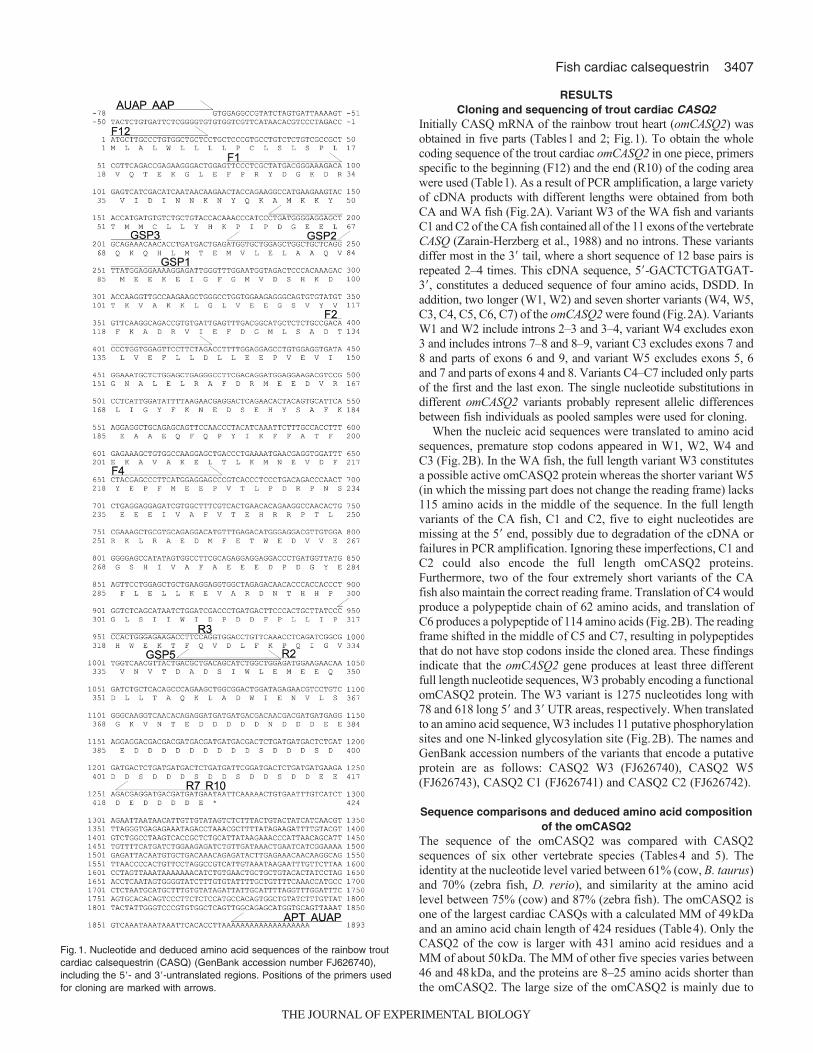
RESULTSCloning and sequencing of trout cardiac CASQ2
Initially CASQ mRNA of the rainbow trout heart (omCASQ2) wasobtained in five parts (Tables1 and 2; Fig.1). To obtain the wholecoding sequence of the trout cardiac omCASQ2 in one piece, primersspecific to the beginning (F12) and the end (R10) of the coding areawere used (Table1). As a result of PCR amplification, a large varietyof cDNA products with different lengths were obtained from bothCA and WA fish (Fig.2A). Variant W3 of the WA fish and variantsC1 and C2 of the CA fish contained all of the 11 exons of the vertebrateCASQ (Zarain-Herzberg et al., 1988) and no introns. These variantsdiffer most in the 3� tail, where a short sequence of 12 base pairs isrepeated 2–4 times. This cDNA sequence, 5�-GACTCTGATGAT-3�, constitutes a deduced sequence of four amino acids, DSDD. Inaddition, two longer (W1, W2) and seven shorter variants (W4, W5,C3, C4, C5, C6, C7) of the omCASQ2 were found (Fig.2A). VariantsW1 and W2 include introns 2–3 and 3–4, variant W4 excludes exon3 and includes introns 7–8 and 8–9, variant C3 excludes exons 7 and8 and parts of exons 6 and 9, and variant W5 excludes exons 5, 6and 7 and parts of exons 4 and 8. Variants C4–C7 included only partsof the first and the last exon. The single nucleotide substitutions indifferent omCASQ2 variants probably represent allelic differencesbetween fish individuals as pooled samples were used for cloning.
When the nucleic acid sequences were translated to amino acidsequences, premature stop codons appeared in W1, W2, W4 andC3 (Fig.2B). In the WA fish, the full length variant W3 constitutesa possible active omCASQ2 protein whereas the shorter variant W5(in which the missing part does not change the reading frame) lacks115 amino acids in the middle of the sequence. In the full lengthvariants of the CA fish, C1 and C2, five to eight nucleotides aremissing at the 5� end, possibly due to degradation of the cDNA orfailures in PCR amplification. Ignoring these imperfections, C1 andC2 could also encode the full length omCASQ2 proteins.Furthermore, two of the four extremely short variants of the CAfish also maintain the correct reading frame. Translation of C4 wouldproduce a polypeptide chain of 62 amino acids, and translation ofC6 produces a polypeptide of 114 amino acids (Fig.2B). The readingframe shifted in the middle of C5 and C7, resulting in polypeptidesthat do not have stop codons inside the cloned area. These findingsindicate that the omCASQ2 gene produces at least three differentfull length nucleotide sequences, W3 probably encoding a functionalomCASQ2 protein. The W3 variant is 1275 nucleotides long with78 and 618 long 5� and 3� UTR areas, respectively. When translatedto an amino acid sequence, W3 includes 11 putative phosphorylationsites and one N-linked glycosylation site (Fig.2B). The names andGenBank accession numbers of the variants that encode a putativeprotein are as follows: CASQ2 W3 (FJ626740), CASQ2 W5(FJ626743), CASQ2 C1 (FJ626741) and CASQ2 C2 (FJ626742).
Sequence comparisons and deduced amino acid compositionof the omCASQ2
The sequence of the omCASQ2 was compared with CASQ2sequences of six other vertebrate species (Tables4 and 5). Theidentity at the nucleotide level varied between 61% (cow, B. taurus)and 70% (zebra fish, D. rerio), and similarity at the amino acidlevel between 75% (cow) and 87% (zebra fish). The omCASQ2 isone of the largest cardiac CASQs with a calculated MM of 49kDaand an amino acid chain length of 424 residues (Table4). Only theCASQ2 of the cow is larger with 431 amino acid residues and aMM of about 50kDa. The MM of other five species varies between46 and 48kDa, and the proteins are 8–25 amino acids shorter thanthe omCASQ2. The large size of the omCASQ2 is mainly due to
Fig.1. Nucleotide and deduced amino acid sequences of the rainbow troutcardiac calsequestrin (CASQ) (GenBank accession number FJ626740),including the 5�- and 3�-untranslated regions. Positions of the primers usedfor cloning are marked with arrows.
THE JOURNAL OF EXPERIMENTAL BIOLOGY

3408
the long unstructured C-terminal tail consisting of acidic (Asp andGlu) and uncharged (Ser) polar amino acids (Fig.2C). Thepercentage of acidic Asp and Glu residues was 34% in W3 and W5(Table4), despite differences in their sequence lengths. Thepercentage of acidic residues in the omCASQ2 is similar as in zebrafish (34%) and frog (35%) CASQ but higher than in CASQs ofendothermic vertebrates (29–31%).
A phylogenetic tree was constructed using amino acid sequencesof skeletal and cardiac CASQs of five teleost species and rooted
with the CASQ1 of the nematode Caenorhabditis elegans(F40E10.3.1) (Table3; Fig.2D). In the tree, cardiac CASQs formeda separate group from skeletal CASQs. The four sequences of theomCASQ2 (W3, W5, C1 and C2) were placed next to each other inthe group of cardiac CASQ2; thus, indicating that they are productsof the same gene and not sequences of different paralogs. Rainbowtrout, zebra fish and medaka have only one CASQ2 gene whereasfugu, tetraodon and stickleback have two CASQ2 paralogs. The threetranscript products of the zebra fish paralogs, zgc:154027 and
H. Korajoki and M. Vornanen
Fig. 2. See next page for legend.
THE JOURNAL OF EXPERIMENTAL BIOLOGY

3409Fish cardiac calsequestrin
Fig.2. Transcript variability of the trout cardiac calsequestrin (CASQ). (A)Nucleotide sequence alignment of the omCASQ2 variants from warm-acclimated(WA) (W1–W5) and cold-acclimated (CA) (C1–C7) fish. Putative exon/intron borders are marked with vertical lines and the exon number is shown next tothe line. Positions of the primers used in quantitative RT-PCR analyses are marked with arrows. (B)Deduced amino acid sequence alignment of theomCASQ2 variants. W3, W5, C1 and C2 avoided premature stop codons and produced full length sequences. From the shorter variants only C4 and C6avoided premature stop codons. There are 11 phosphorylation sites (marked with blue quadrangle) and one N-linked glycosylation site (marked with redquadrangle) in the omCASQ2. The asterisks indicate the stop codons. (C)Alignment of cardiac CASQ amino acid sequences from seven different species,including the W3 variant of the rainbow trout (Oncorhynchus mykiss). Other species are zebra fish (Danio rerio), pipid frog (Xenopus tropicalis), rabbit(Oryctolagus cuniculus), cow (Bos taurus), mouse (Mus musculus) and human (Homo sapiens). In addition, the sequence of nematode Caenorhabditiselegans CASQ1 is shown at the bottom of the alignment. (D)The phylogenetic tree constructed using amino acid sequences of skeletal and cardiac CASQparalogs of five teleost species: zebra fish, medaka (Oryzias latipes), fugu (Takifugu rubripes), tetraodon (Tetraodon nigroviridis) and stickleback(Gasterosteus aculeatus). The products of paralog genes lie in their own groups and the products of the various transcripts are settled side by side. The treewas constructed using Neighbor Joining method and rooted with CASQ1 sequence of the nematode Caenorhabditis elegans as an out-group.
Fig. 2. Continued on next page.
THE JOURNAL OF EXPERIMENTAL BIOLOGY

3410
zgc:100957, settled in the group of skeletal CASQs and thereforerepresent CASQ1 genes. Taken together, the phylogenetic sequenceanalyses indicate that the omCASQ2 belongs to the family of teleostcardiac CASQs and is clearly different from the skeletal CASQs.
Real-time quantitative PCRConsistent with the cloning results three different PCR productswere present in mRNA analyses. The primer pair QF1/QR1 canpick up four cloned variants of the WA fish (W1–W4) and threecloned variants of the CA fish (C1–C3) but excludes variants W5and C4–C7 (Fig.2A). When the expression level of the CASQ2QF1/QR1 transcript of the CA atrium (N5) is set as 1, the relative
expression levels of the omCASQ2 in WA atrium (N5), CAventricle and WA ventricle are 0.504, 0.330 and 0.228, respectively(Fig.3A). Thus, the expression level of the omCASQ2 is about 2(P<0.001) and 1.5 times (P<0.005) as high in CA fish than in WAfish for atrial and ventricular muscle, respectively. Furthermore, theexpression level of the omCASQ2 is 2–3 times higher in atrium thanin ventricle (P<0.001).
H. Korajoki and M. Vornanen
Fig. 2. See previous page for legend.
Fig.3. The transcript expressions of cardiac calsequestrin (CASQ) in atriumand ventricle of warm-acclimated (WA) and cold-acclimated (CA) rainbowtrout using primers QF1/QR1 (all the full-length variants, values shownunder the category axis), QF2/QR2 (W4 and other variants that include theintron 8–9, values shown on the right side of the columns) and QF3/QR1(W1, W2 and other variants that included the intron 3–4, values shown onthe right side of the columns). The relative expressions of variants W3,C1–C3 (value shown inside the columns) were calculated by subtractingthe expression of variants W1, W2 and W4 from the expression of all fulllength variants. The expression levels are relative to the expression levelsof the mRNA of the DnaJA2 gene. (A)All results are normalized to theexpression of obtained with the primer pair QF1/QR1 in CA atrium.**P<0.005; ***P<0.001. (B)The QF1/QR1 relative expression in each organand acclimation group is set as 100% and the QF2/QR2 and QF3/QR1results are normalized to this value. Values are means ± s.e.m. (N5).
Table 4. Identities of cardiac calsequestrin (CASQ) cDNA sequences of seven vertebrate species
Oncorhynchus Xenopus Mus Oryctolagus cDNA identity % (IDENTIFY) mykiss Danio rerio tropicalis musculus Homo sapiens cuniculus Bos taurus
Oncorhynchus mykiss ID 70 67 66 62 66 61Danio rerio 70 ID 69 67 67 68 65Xenopus tropicalis 67 69 ID 68 66 68 64Mus musculus 66 67 68 ID 82 83 78Homo sapiens 62 67 66 82 ID 87 84Oryctolagus cuniculus 66 68 68 83 87 ID 82Bos taurus 61 65 64 78 84 82 ID
The sequence of the W3 variant of the rainbow trout omCASQ2 was compared with the CASQ2 sequences of other vertebrate species obtained fromGenBank.
Table 5. Similarities of cardiac calsequestrin (CASQ) amino acid sequences of seven vertebrate species
Oncorhynchus Xenopus Mus Oryctolagus Amino acid similarity % (BLOSUM62) mykiss Danio rerio tropicalis musculus Homo sapiens cuniculus Bos taurus
Oncorhynchus mykiss ID 87 81 80 78 80 75Danio rerio 87 ID 85 85 84 86 80Xenopus tropicalis 81 85 ID 84 81 83 78Mus musculus 80 85 84 ID 91 93 88Homo sapiens 78 84 81 91 ID 95 91Oryctolagus cuniculus 80 86 83 93 95 ID 92Bos taurus 75 80 78 88 91 92 ID
The W3 variant of the trout omCASQ2 was compared with the CASQ2 sequences of other vertebrate species obtained from GenBank.
THE JOURNAL OF EXPERIMENTAL BIOLOGY

3411Fish cardiac calsequestrin
The difference observed in the amount of the CASQ2 QF1/QR1transcripts between CA and WA fish did not exist in the CASQ2QF2/QR2 or CASQ2 QF3/QR1 transcripts (N5) (Fig.3B).Furthermore, the expression levels of the CASQ2 QF2/QR2 andCASQ2 QF3/QR1 transcripts were only 3–13% of the expressionlevels of CASQ2 QF1/QR1 transcripts in the atrium and 1–6% inthe ventricle of CA and WA fish, respectively. In PCR amplificationof the omCASQ2 QF3/QR1, the measured expression level consistsof two cloned variants in WA fish (W1, W2) and putative variantsof the CA fish that were not found in cloning. In PCR amplificationof the CASQ2 QF2/QR2 the measured expression levels representonly the W4 and putative uncloned variants of the CA fish.Collectively, these results indicate that transcript abundance of theomCASQ2 is higher in the atrium than ventricle and upregulated inboth cardiac chambers during acclimation to the cold.
Expression of CASQ protein in the trout heartA polyclonal antibody raised to the omCASQ2 sequence betweenamino acids 174 and 315 was used for identifying and quantifyingthe omCASQ2 protein in western blots. Surprisingly, in the atriumand ventricle of WA and CA trout hearts, the antibody clearlyrecognized two proteins with an apparent MM of 54 and 59kDa,respectively (Fig.4). The 59kDa protein was a minor componentrepresenting 22% and 13% of the total anti-omCASQ2 sensitiveproteins in the atrium and ventricle of WA fish, respectively (P>0.05)(Fig.4A). In contrast to WA trout, the 59kDa protein was practicallynon-extant in the atrium and ventricle of CA fish (Fig.4B).
The staining intensity of the 54kDa protein was similar for thesamples of CA and WA fish (P>0.05) in both cardiac chambers(Fig.4C), indicating stability of the omCASQ2 expression inthermally acclimated trout. When compared with the amount ofCASQ (MM about 70kDa) in trout skeletal muscle (MM about70kDa), the intensity of the cardiac 54kDa protein was 60–80% inthe atrium and ventricle of WA fish (N3) and CA fish (N3) (datanot shown) in comparison with the CASQ of trout myotomal muscle.
We compared the results of our fish-specific antibody western blotswith those probed with a commercially available polyclonal caninecardiac CASQ antibody. The anti-canine CASQ, ab3516, detectedboth the 54kDa and the 59kDa protein of the CA trout, but contraryto the anti-omCASQ2 antibody, ab3516 seemed to recognize the59kDa protein better than the 54kDa protein (Fig.5A,B,D,E). Ingeneral agreement with the results of the fish specific antibody, theintensity of the 59kDa protein was 46 and 19 times stronger in WA
than CA trout for atrium and ventricle, respectively (Fig.5B,E).Similar differences in the expression of 59kDa protein are also evidentin Coomassie stained SDS-PAGE gels (Fig.5C,F): larger amounts ofthe 59kDa protein are present in WA than CA fish, and the 59kDaprotein is slightly more strongly expressed in the atrium than ventricle.
Taken together, these results indicate that CA trout expresses mainlyone CASQ isoform (54kDa) whereas WA trout expresses two CASQisoforms. The omCASQ2 content of atrial and ventricular muscle ofthe trout heart is similar and there are no temperature-relateddifferences in omCASQ2 abundance in either the atrium or ventricle.
DISCUSSIONAtrial and ventricular myocytes of the rainbow trout heart havemassive SR Ca2+ stores, exceeding those of the mammalian heart6–10-fold (Hove-Madsen et al., 1998; Hove-Madsen et al., 1999;Shiels et al., 2002). Furthermore, acclimation to low temperatureincreases contribution of SR Ca2+ stores to cardiac e–c coupling asevidenced by the increased sensitivity of contraction force to Ry inCA trout (Keen et al., 1994; Aho and Vornanen, 1999). Mechanisticbasis of the large SR Ca2+ storing capacity and molecular mechanismsresponsible for the increased SR Ca2+ release in CA hearts are,
Fig.4. Identification and quantification of the trout cardiaccalsequestrins (CASQs) with a polyclonal antibody to theomCASQ2. Representative western plots comparing CASQcontent in atrium and ventricle of warm-acclimated (WA) (A) andcold-acclimated (CA) (B) trout. Numbers on the bottom and onthe top of the western plots show staining intensities of the54kDa and 59kDa proteins, respectively, relative to the strongestband of the 54kDa protein in each run. (C)A bar graph showingthe mean expression (± s.e.m.) of 54kDa and 59kDa proteins(N5). Fortyg of atrial and ventricular homogenate was appliedto each lane. WA–At, atrium of WA fish; CA–At, atrium of CAfish; WA–Ve, ventricle of WA fish; CA–Ve, ventricle of CA fish.
Fig.5. Identification of trout cardiac calsequestrins (CASQs) with a polyclonalantibody to canine cardiac CASQ (ab3516). Comparison of CASQ staining inatrial and ventricular homogenates with the anti-omCASQ2 antibody (A,D)and the ab3516 (B,E). Note the strong staining of the 59kDa protein withab3516. C and F show corresponding SDS-PAGE gels stained withCoomassie Brilliant Blue R-250. For labeling see the caption of Fig.4.
THE JOURNAL OF EXPERIMENTAL BIOLOGY

3412
however, largely unknown. Although CASQ is not absolutelynecessary for SR Ca2+ storage and release (Knollmann et al., 2006),it is generally expressed in vertebrate heart and regulates cardiac e–ccoupling. The present study examines, for the first time, the expressionof CASQ2 protein in the fish heart. A 54kDa isoform of cardiac CASQ(omCASQ2) was the main CASQ component in the atrium andventricle of the trout heart in both CA and WA fish. Notably, thermalacclimation did not affect the abundance of the 54kDa CASQ2proteins, suggesting that the effect of chronic thermal changes on SRCa2+ release are not related to the amount of this omCASQ2 isoform.However, it is shown that thermal acclimation is associated with aqualitative change in CASQ2 protein expression: acclimation of troutto 18°C induces the expression of 59kDa CASQ2 in addition to the54kDa omCASQ2. However, the identity of this gene product couldnot be resolved and its physiological function remains to be shown.
Comparison of the omCASQ2 with other vertebrate CASQIn mammals, two CASQ isoforms exist, one specific for the skeletalmuscle (CASQ1) and another for the cardiac muscle (CASQ2), thelatter being the sole CASQ isoform in the heart (MacLennan andWong, 1971; Cala and Jones, 1983; Campbell et al., 1983). Theapparent MM of the trout omCASQ2 in the alkaline SDS gels(Laemmli, 1970) was 54kDa, which corresponds well the MM of themammalian CASQ2 (53kDa) (Cala and Jones, 1983; Campbell etal., 1983). The sequence comparisons indicate that the omCASQ2belongs to the family of the vertebrate cardiac CASQs and is clearlydistinct from the skeletal CASQs. Furthermore, it is clear that thedifferent variants of the omCASQ2 are products of the same gene.Multiple transcript variants are unlikely to ensue from cloningartifacts, because several of the products are outcomes of the splicingprocess on exon/intron borders and because they constitute significantamounts (0.6–21.0%) of the total transcripts. For example W1 andW2 result from incomplete splicing and include two introns, whichintroduce premature stop codons in the transcripts. Similarly, W4 isa splicing product missing one exon and including two introns butwould have a premature stop codon even after removal of the introns.Due to the premature stop codons these transcripts will not producetrue splice variants but they may still have regulative effects on Ca2+
binding and polymerization of the omCASQ2 or interaction of theomCASQ2 with other proteins. The C-terminal tail is important informing Ca2+ binding pockets in the polymerized chains of CASQand includes binding motifs for junctin and triadin (Beard et al., 2009).Therefore, the products of apparently incomplete splicing, and eventhe shortest omCASQ2 variants having the C-terminal tail, couldinterfere with the function of the full length omCASQ2. It is wellknown that incomplete protein from alternative splicing can interferewith the function of catalytically active isoforms by competing forsubstrate molecules or by direct inhibition/interaction of the full-lengthprotein (Ross et al., 1997; Nagano et al., 1999).
The trout omCASQ2 is, in several respects, similar to cardiacCASQs of other vertebrates but also displays some differences. Thetrout omCASQ2 is 8–25 residues longer than the cardiac CASQs ofendothermic vertebrates (with the exception of the cow CASQ2 whichis 7 residues longer than the omCASQ2) and 15 resides longer thanthe zebra fish CASQ2. CASQ2 is a highly acidic protein with aboutone third of the amino acids being either Glu or Asp. The number ofnegatively charged Glu and Asp residues of the trout omCASQ2 issimilar (34%) to CASQ of zebra fish (D. rerio) (34%) and clawedfrog (Xenopus laevis) (35%) but higher than those of mammalian andavian CASQs (29–31%), mainly due to the longer C-terminal tail.Although the long C-terminal tail increases the number of negativecharges of the omCASQ2 molecule, it probably does not have anymajor effect on Ca2+ binding, which is mainly dependent onpolymerization of CASQ into linear needle-shaped crystals (Park etal., 2004). The negative charges of the C-terminal tail are needed toform back-to-back (C-terminal) dimers of CASQ, which starts thepolymerization process at relatively low Ca2+ concentrations (Wanget al., 1998). The long C-terminal tail of ectothermic CASQ2s mightregulate the Ca2+-sensitivity of the polymerization.
Identity of the 59 kDa protein of the trout heart is presentlyunresolved. The 59kDa protein was recognized by two polyclonalCASQ antibodies, the fish-specific anti-omCASQ2 antibody and theanti-canine cardiac CASQ antibody, strongly suggesting that the59kDa protein is CASQ or a CASQ-like protein. Although, its MMis similar to mammalian skeletal CASQ (60kDa), it is probably nota skeletal CASQ, because the MM of the rainbow trout skeletal CASQ(70kDa), like that of the common carp (68kDa) (Cyprinus carpio)(Watabe et al., 1991), is much higher than the MM of mammalianskeletal CASQ. Even though the 59kDa protein was regularlyexpressed in atrial and ventricular muscle of the WA fish, omCASQ2transcripts of the corresponding length were not present in theomCASQ2 clones of the WA trout heart. CASQ is a glycoprotein,which contains mannose and N-acetylglucosamine sugars (Jorgensenet al., 1977), and therefore the 59kDa protein could be a glycolysatedform of the 54kDa protein. Similar to canine CASQ (Scott et al.,1988; Cala and Jones, 1991), trout CASQ2 variant W3 amino acidsequence includes several putative casein kinase II phosphorylationsites and one N-linked glycosylation site. However, enzymaticremoval of the carbohydrate moiety of the cardiac CASQ usuallyreduces its MM only by 1–2kDa (Milner et al., 1991), which is toolittle to explain the 5kDa difference in MM between the trout cardiacCASQs. Further studies are needed to solve the identity andphysiological function of the 59kDa protein whose expression isinduced by exposure of the trout to high environmental temperatures.
Expression of the omCASQ2Expression of both mRNA and protein levels of the omCASQ2 weremeasured. Although transcript expression of the full length
H. Korajoki and M. Vornanen
Table 6. Amino acid composition of W3 and W5 variants of the omCASQ2 gene and CASQ2 sequences of six other vertebrate species fromGenBank
MM Residues Aliphatic Aromatic Non-polar Polar Charged Basic Acidic
Oncorhynchus mykiss W3 49033 424 113 46 199 225 168 47 121Oncorhynchus mykiss W5 34606 301 84 27 145 156 116 30 86Danio rerio 47330 409 89 50 199 210 161 45 116Xenopus tropicalis 47951 414 106 50 199 215 170 49 121Mus musculus 48354 416 92 49 196 220 158 48 110Homo sapiens 46436 399 90 52 196 203 151 49 102Bos taurus 49915 431 118 56 209 222 165 56 109Oryctolagus cuniculus 47356 409 113 51 199 210 157 50 107
MM, molecular mass.
THE JOURNAL OF EXPERIMENTAL BIOLOGY

3413Fish cardiac calsequestrin
omCASQ2 was elevated 1.5–2-fold in atrial and ventricular muscleof the CA trout heart, the amount of the omCASQ2 protein did notdiffer between the acclimation groups. The divergence betweentranscript and protein levels may be partly due to the 59 kDa isoform,which was almost exclusively expressed in the WA heart. It ispossible that transcripts of the 59kDa protein were not picked upby the primers designed to the omCASQ2. Considering that the59kDa isoform formed only 13–22% of the total CASQs in the WAheart, the 1.5–2-fold difference in omCASQ2 transcript levelsbetween WA and CA fish, does not however match with the proteinexpression. Deviation between transcript and protein levels suggestthat the abundance of the omCASQ2 protein is regulated by thetranslation process, and therefore transcript levels cannot be usedas a measure for the amount of the omCASQ2 protein.
In mammalian heart, SR Ca2+ release is enhanced at high SR Ca2+
loads when Ca2+ binds to CASQ and increases the opening probabilityof the RyRs (Györke and Terentyev, 2008). Because SR Ca2+ loadof trout atrial myocytes is higher than in ventricular myocytes(Haverinen and Vornanen, 2009), it was anticipated that this wouldbe expressed as a higher omCASQ2 content in atrial tissue andtherefore might also explain the higher Ry sensitivity of atrialcontraction in comparison with ventricle (Gesser, 1996; Aho andVornanen, 1999). Contrary to this hypothesis, the amount ofomCASQ2 was similar in both cardiac chambers. Similar to the troutheart, atrial and ventricular muscle of the rat heart have similar CASQcontents, even though SR Ca2+ content is about 3 times larger in atrialthan ventricular myocytes (Walden et al., 2009). With regards tochamber-related differences in SR Ca2+ load and CASQ content, fishand mammalian hearts seem to be qualitatively similar. It can beconcluded that atrio-ventricular differences in SR Ca2+ content of thevertebrate heart are not dependent on CASQ concentration andtherefore must be associated with other properties or molecular entitiesof the SR, like SR volume or additional SR Ca2+ buffers.
In rainbow trout, acclimation to cold increases Ry sensitivity ofcardiac contraction, indicating thermal plasticity of the SR function(Keen et al., 1994; Aho and Vornanen, 1999). However, theomCASQ2 content of the trout heart was resistant to thermalacclimation in both cardiac chambers, suggesting that changes inthe amount of CASQ are not responsible for the altered SR function.In different pathophysiological states of the mammalian heart, CASQcontent remains quite constant unlike many other molecules of thee–c coupling machinery (Lehnart et al., 1998). Evidently, cardiacCASQ is not a particularly plastic entity in vertebrate heart and tendsto be conserved at the characteristic level of the tissue. It should benoted however that the trout heart differs from mammalian heartsin that the WA trout heart expresses two CASQ isoforms, i.e. thereis a qualitative change in cardiac CASQ composition.
Quantitative aspectsBecause SR Ca2+ stores of the trout cardiac myocytes are much largerthan those of the mammalian cardiac myocytes, it would be interestingto know whether this is associated with the higher omCASQ2 content.Our objective was to compare CASQ content in mouse and trout heartsusing CASQ antibodies but those studies were not feasible, becauseantibodies to fish and mammalian cardiac CASQs differed in theiraffinity to heterologous CASQs. It was, however, shown that CASQcontent is 20–40% less in the heart than in the myotomal muscle ofthe trout. We could not find similar tissue comparisons for othervertebrates, and overall surprisingly little quantitative data exist forthe amount of CASQ in vertebrate muscles (Volpe and Simon, 1991;Murphy et al., 2009). Data on absolute amounts of CASQ in cardiacmuscle are totally lacking but it was recently shown that rat skeletal
muscles composed primarily of fast type II or slow type I fibers contain32 and 11mol CASQ per liter of fiber volume, respectively (Murphyet al., 2009). Considering that the trout heart contains 60–80% of theCASQ content of the myotomal muscle and using the value of11moll–1 measured for slow fibers of the rat skeletal muscle, CASQcontent of the trout heart would be 6.6–8.8moll–1 myocyte volume.Further, assuming that fish SR volume is 4% of the myocyte volume(Bowler and Tirri, 1990), concentration of the omCASQ2 in troutheart SR would be 0.165–0.220mmoll–1. Cardiac CASQ can bindabout 60 Ca2+ ions per molecule (Park et al., 2004), which gives atotal Ca2+ buffering capacity of 15.8–21.12mmoll–1 within the SR(396–528moll–1 myocyte volume) for the trout heart. This isslightly more than in mammalian heart (Shannon and Bers, 1997)and in reasonable agreement with the maximum SR Ca2+ content of18.4 and 26.88mmoll–1 (462 and 672moll–1 myocyte volume) inthe trout ventricular and atrial cells, respectively (Haverinen andVornanen, 2009). It should be noted, however, that this calculationis based on lager SR volume (4%) in fish cardiac myocytes incomparison with mammalian cardiac myocytes (1.5%) (Page, 1978),and assumption of a similar SR volume in the two vertebrate groupswould mean substantially larger SR Ca2+ buffering by CASQ in fishhearts.
ConclusionsThe present results suggest that adequate amounts of the omCASQ2are present in trout atrial and ventricular muscle to buffer the largecaffeine releasable Ca2+ stores of trout atrial and ventricularmyocytes. We did not manage to make direct comparisons on CASQcontent between trout and mouse hearts, and therefore the questionon larger Ca2+ buffering capacity of trout cardiac SR in comparisonwith mammalian heart still remains unanswered. Purified cardiacCASQs as standards in Western blots and species-specific antibodiesare needed to make quantitative comparisons of CASQ contentbetween vertebrate groups and between different fish species andto better understand the physiological significance of CASQ contentin cardiac e–c coupling. Ca2+ binding capacity of CASQ might betemperature-dependent (Watabe et al., 1991), and it is important toexamine the temperature-dependent properties of Ca2+ binding offish cardiac CASQs in order to elucidate its role in SR Ca2+ bufferingand regulation of RyR function in ectothermic vertebrates.
LIST OF ABBREVIATIONSCA cold acclimationCASQ calsequestrinCICR Ca2+-induced Ca2+-releasee–c excitation–contraction couplingMM molecular massRy ryanodineRyRs ryanodine receptorsSL sarcolemmalSR sarcoplasmic reticulumTBS tris-buffered salineWA warm acclimation
Anita Kervinen and Riitta Pietarinen are appreciated for technical assistance, andJuha Lemmetyinen for assisting in construction of the phylogenetic tree. Thisresearch was supported by grants from The Academy of Finland (210400 and119583) to M.V.
REFERENCESAho, E. and Vornanen, M. (1999). Contractile properties of atrial and ventricular
myocardium of the heart of rainbow trout Oncorhynchus mykiss: effects of thermalacclimation. J. Exp. Biol. 202, 2663-2677.
Ashley, R. H. and Williams, A. J. (1990). Divalent cation activation and inhibition ofsingle calcium release channels from sheep cardiac sarcoplasmic reticulum. J. Gen.Physiol. 95, 981-1005.
THE JOURNAL OF EXPERIMENTAL BIOLOGY

3414
Bassani, J. W. M., Yuan, W. and Bers, D. M. (1995). Fractional SR Ca release isregulated by trigger Ca and SR Ca content in cardiac myocytes. Am. J. Physiol. 268,C1313-C1319.
Beard, N. A., Laver, D. R. and Dulhunty, A. F. (2004). Calsequestrin and the calciumrelease channel of skeletal and cardiac muscle. Prog. Biophys. Mol. Biol. 85, 33-69.
Beard, N. A., Wei, L. and Dulhunty, A. F. (2009). Control of muscle ryanodine receptorcalcium release channels by proteins in the sarcoplasmic reticulum lumen. Clin. Exp.Pharmacol. Physiol. 36, 340-345.
Bonnet, V., Badaoui, A., Huchet-cadiou, C. and Leoty, C. (1994). Potentiation of thetwitch responses by inhibitors of sarcoplasmic reticulum Ca2+-ATPase in frog atrialfibres. Eur. J. Pharmacol. 264, 69-76.
Bowler, K. and Tirri, R. (1990). Temperature dependence of the heart isolated from thecold or warm acclimated perch (Perca fluviatilis). Comp. Biochem. Physiol. 96A, 177-180.
Cala, S. E. and Jones, L. R. (1983). Rapid purification of calsequestrin from cardiac andskeletal muscle sarcoplasmic reticulum vesicles by Ca2+-dependent elution fromphenyl-sepharose. J. Biol. Chem. 258, 11932-11936.
Cala, S. E. and Jones, L. R. (1991). Phosphorylation of cardiac and skeletal musclecalsequestrin isoforms by casein kinase. II. Demonstration of a cluster of uniquerapidly phosphorylated sites in cardiac calsequestrin. J. Biol. Chem. 266, 391-398.
Campbell, K. P., MacLennan, D. H., Jorgensen, A. O. and Mintzer, M. C. (1983).Purification and characterization of calsequestrin from canine cardiac sarcoplasmicreticulum and identification of the 53,000 dalton glycoprotein. J. Biol. Chem. 258, 1197-1204.
Chapman, R. A. (1983). Control of cardiac contractility at the cellular level. Am. J.Physiol. 245, H535-H552.
Costa, M. J., Rivaroli, L., Rantin, F. T. and Kalinin, A. L. (2000). Cardiac tissuefunction of the teleost fish Oreochromis niloticus under different thermal conditions. J.Therm. Biol. 25, 373-379.
Driedzic, W. R. and Gesser, H. (1988). Differences in force–frequency relationships andcalcium dependency between elasmobranch and teleost hearts. J. Exp. Biol. 140, 227-241.
Fabiato, A. (1985). Time and calcium dependence of activation and inactivation ofcalcium-induced release of calcium from the sarcoplasmic reticulum of a skinnedcanine cardiac Purkinje cell. J. Gen. Physiol. 85, 247-289.
Fabiato, A. and Fabiato, F. (1978). Calcium-induced release of calcium from thesarcoplasmic reticulum of skinned cells from adult human, dog, cat, rabbit, rat and froghearts and from fetal and new-born rat ventricles. Ann. NY Acad. Sci. 307, 491-522.
Gesser, H. (1996). Cardiac force-interval relationship, adrenaline and sarcoplasmicreticulum in rainbow trout. J. Comp. Physiol. B 166, 278-285.
Györke, I. and Györke, S. (1998). Regulation of the cardiac ryanodine receptor channelby luminal Ca2+ involves luminal Ca2+ sensing sites. Biophys. J. 75, 2801-2810.
Györke, S. and Terentyev, D. (2008). Modulation of ryanodine receptor by luminalcalcium and accessory proteins in health and cardiac disease. Cardiovasc. Res. 77,245-255.
Harwood, C. L., Howarth, F. C., Altringham, J. D. and White, E. (2000). Rate-dependent changes in cell shortening, intracellular Ca(2+) levels and membranepotential in single, isolated rainbow trout (Oncorhynchus mykiss) ventricular myocytes.J. Exp. Biol. 203, 493-504.
Hassinen, M., Haverinen, J. and Vornanen, M. (2008). Electrophysiological propertiesand expression of the delayed rectifier potassium (ERG) channels in the heart ofthermally acclimated rainbow trout. Am. J. Physiol. 295, R297-R308.
Haverinen, J. and Vornanen, M. (2009). Comparison of sarcoplasmic reticulum calciumcontent in atrial and ventricular myocytes of three fish species. Am. J. Physiol. Regul.Integr. Comp. Physiol. doi:10.1152/ajpregu.00022.2009/.
Hove-Madsen, L., Llach, A. and Tort, L. (1998). Quantification of Ca2+ uptake in thesarcoplasmic reticulum of trout ventricular myocytes. Am. J. Physiol. 275, R2070-R2080.
Hove-Madsen, L., Llach, A. and Tort, L. (1999). Quantification of calcium release fromthe sarcoplasmic reticulum in rainbow trout atrial myocytes. Pflugers Arch. 438, 545-552.
Hove-Madsen, L., Llach, A. and Tort, L. (2000). Na+/Ca2+-exchange activity regulatescontraction and SR Ca2+ content in rainbow trout atrial myocytes. Am. J. Physiol. 279,R1856-R1864.
Jiang, D., Chen, W., Wang, R., Zhang, L. and Chen, S. R. W. (2007). Loss of luminalCa2+ activation in the cardiac ryanodine receptor is associated with ventricularfibrillation and sudden death. Proc. Natl. Acad. Sci. USA 13, 8309-18314.
Jorgensen, A. O., Kalnins, V. I., Zubrzycka, E. and MacLennan, D. H. (1977).Assembly of the sarcoplasmic reticulum. Localization by immunofluorescence ofsarcoplasmic reticulum proteins in differentiating rat skeletal muscle cell cultures. J.Cell Biol. 74, 287-298.
Ju, Y. K. and Allen, D. G. (1998). Intracellular calcium and Na+-Ca2+ exchange currentin isolated toad pacemaker cells. J. Physiol. 508, 153-166.
Keen, J. E., Vianzon, D. M., Farrell, A. P. and Tibbits, G. F. (1994). Effect oftemperature and temperature acclimation on the ryanodine sensitivity of the troutmyocardium. J. Comp. Physiol. B 164, 438-443.
Klizner, T. and Morad, M. (1983). Excitation-contraction coupling in frog heart ventricle.Possible Ca2+ transport mechanisms. Pflugers Arch. 398, 274-283.
Knollmann, B. C., Chopra, N., Hlaing, T., Akin, B., Yang, B., Ettensohn, K.,Knollmann, B. E. C., Horton, K. D., Weissman, N. J., Holinstat, I. et al. (2006).Casq2 deletion causes sarcoplasmic reticulum volume increase, premature Ca2+
release, and catecholaminergic polymorphic ventricular tachycardia. J. Clin. Invest.116, 2510-2520.
Kolaskar, A. S. and Tongaonkar, P. C. (1990). A semi-empirical method for predictionof antigenic determinants on protein antigens. FEBS Lett. 276, 172-174.
Kong, H., Jones, P. P., Koop, A., Zhang, L., Duff, H. J. and Chen, S. R. W. (2008).Caffeine induces Ca2+ release by reducing the threshold for luminal Ca2+ activation ofthe ryanodine receptor. Biochem. J. 414, 441-452.
Laemmli, U. K. (1970). Cleavage of structural proteins during the assembly of the headof bacteriophage T4. Nature 227, 680-685.
Laver, D. R. (2007). Ca2+ stores regulate ryanodine receptor Ca2+ release channels vialuminal and cytosolic Ca2+ sites. Clin. Exp. Pharmacol. Physiol. 34, 889-896.
Laver, D. R., Roden, L. D., Ahern, G. P., Eagar, K. R., Junankar, P. R. and Dulhunty,A. F. (1995). Cytoplasmic Ca2+ inhibits the ryanodine receptor from cardiac muscle. J.Membr. Biol. 147, 7-22.
Lehnart, S. E., Schillinger, W., Pieske, B., Prestle, J., Just, H. and Hasenfuss, G.(1998). Sarcoplasmic reticulum proteins in heart failure. Ann. NY Acad. Sci. 16, 220-230.
Lowry, O. H., Rosenbrough, N. J., Farr, A. L. and Randall, R. J. (1951). Proteinmeasurement with the Folin phenol reagent. J. Biol. Chem. 193, 265-275.
Lukyanenko, V., Györke, I. and Györke, S. (1996). Regulation of calcium release bycalcium inside the sarcoplasmic reticulum in ventricular myocytes. Pflugers Arch. 432,1047-1054.
MacLennan, D. H. and Wong, P. T. S. (1971). Isolation of a calcium-sequestring proteinfrom sarcoplasmic reticulum. Proc Natl. Acad. Sci. USA 68, 1231-1235.
Meissner, G. (2004). Molecular regulation of cardiac ryanodine receptor ion channel. CellCalcium 35, 621-628.
Milner, R. E., Michalak, M. and Wang, L. C. H. (1991). Altered properties ofcalsequestrin and the ryanodine receptor in the cardiac sarcoplasmic reticulum ofhibernating mammals. Biochim. Biophys. Acta 1063, 120-128.
Moller-Nielsen, T. and Gesser, H. (1992). Sarcoplasmic reticulum andexcitation–contraction coupling at 20 and 10°C in rainbow trout myocardium. J. Comp.Physiol. B 162, 526-534.
Murphy, R. M., Larkins, N. T., Mollica, J. P., Beard, N. A. and Lamb, G. D. (2009).Calsequestrin content and SERCA determine normal and maximal Ca2+ storage levelsin sarcoplasmic reticulum of fast- and slow-twitch fibers of rat. J. Physiol. 587, 443-460.
Nagano, K., Fukami, K., Minagawa, T., Watanabe, Y., Ozaki, C. and Takenawa, T.(1999). A novel phospholipase C delta 4 (PLCdelta 4) splice variant as a negativeregulator of PLC. J. Biol. Chem. 274, 2872-2879.
Page, E. (1978). Quantitative ultrastructural analysis in cardiac membrane physiology.Am. J. Physiol. 235, C147-C158.
Park, H., Park, I. Y., Kim, E., Youn, B., Fields, K., Dunker, A. K. and Kang, C. (2004).Comparing skeletal and cardiac calsequestrin structures and their calcium binding: aproposed mechanism for coupled calcium binding and protein polymerization. J. Biol.Chem. 279, 18026-18033.
Penefsky, Z. J. (1974). Studies on mechanism of inhibition of cardiac muscle contractiletension by ryanodine. Pflugers Arch. 347, 173-184.
Pizarro, G., Cleeman, L. and Morad, M. (1985). Optical measurement of voltage-dependent Ca2+ influx in frog heart. Proc. Natl. Acad. Sci. USA 82, 1864-1868.
Ross, R. J. M., Esposito, N., Shen, X. Y., Von Laue, S., Chew, S. L., Dobson, P. R.M., Postel-Vinay, M. C. and Finidori, J. (1997). A short isoform of the human growthhormone receptor functions as a dominant negative inhibitor of the full-length receptorand generates large amounts of binding protein. Mol. Endocrinol. 11, 265-273.
Santer, R. M. (1985). Morphology and innervation of the fish heart. Adv. Anat. Embryol.Cell Biol. 89, 1-102.
Scott, B. T., Simmerman, H. K., Collins, J. H., Nadal-Ginard, B. and Jones, L. R.(1988). Complete amino acid sequence of canine cardiac calsequestrin deduced bycDNA cloning. J. Biol. Chem. 263, 8958-8964.
Shannon, T. R. and Bers, D. M. (1997). Assessment of intra-SR free [Ca] and bufferingin rat heart. Biophys. J. 73, 1524-1531.
Shiels, H. A., Vornanen, M. and Farrell, A. P. (2002). Temperature dependence ofcardiac sarcoplasmic reticulum in rainbow trout. J. Exp. Biol. 205, 3631-3639.
Sitsapesan, R. and Williams, A. J. (1994). Regulation of the gating of the sheepcardiac sarcoplasmic reticulum Ca2+-release channel by luminal Ca2+. J. Membr. Biol.137, 215-226.
Terentyev, D., Viatchenko-Karpinski, S., Györke, I., Volpe, P., Williams, S. C. andGyörke, S. (2003). Calsequestrin determines the functional size and stability of cardiacintracellular calcium stores: mechanism for hereditary arrhythmia. Proc. Natl. Acad. Sci.USA 30, 11759-11764.
Tibbits, G. F., Moyes, C. D., Hove-Madsen, L., Hoar, W. S., Randall, D. J. andFarrell, A. P. (1992). Excitation–contraction coupling in the teleost heart. In FishPhysiology, Volume XII, Part A: The Cardiovascular System, pp. 267-304. San Diego,CA: Academic Press.
Towbin, H., Staehelin, T. and Gordon, J. (1979). Electrophoretic transfer of proteinsfrom polyacrylamide to nitrocellulose sheets: procedure and some applications. Proc.Natl. Acad. Sci. USA 76, 4350-4354.
Volpe, P. and Simon, B. J. (1991). The bulk of Ca2+ released to the myoplasm is free inthe sarcoplasmic reticulum and does not unbind from calsequestrin. FEBS Lett. 278,274-278.
Vornanen, M. (1989). Regulation of contractility of the fish (Carassius carassius L.) heartventricle. Comp. Biochem. Physiol. 94C, 477-483.
Vornanen, M. (1997). Sarcolemmal Ca influx through L-type Ca channels in ventricularmyocytes of a teleost fish. Am. J. Physiol. 272, R1432-R1440.
Vornanen, M. (1999). Na+-Ca2+ exchange current in ventricular myocytes of fish heart:contribution to sarcolemmal Ca2+ influx. J. Exp. Biol. 202, 1763-1775.
Vornanen, M. (2006). Temperature and Ca2+ dependence of [3H]ryanodine binding inthe burbot (Lota lota L.) heart. Am. J. Physiol. 290, R345-R351.
Walden, A. P., Dibb, K. M. and Trafford, A. W. (2009). Differences in intracellularcalcium homeostasis between atrial and ventricular myocytes. J. Mol. Cell. Cardiol. 46,463-473.
Wang, S., Trumble, W. R., Liao, H., Wesson, C. R., Dunker, A. K. and Kang, C. H.(1998). Crystal structure of calsequestrin from rabbit skeletal muscle sarcoplasmicreticulum. Nat. Struct. Biol. 5, 476-483.
Watabe, S., Ushio, H. and Hashimoto, K. (1991). Purification and characterization of acalsequestrin-like calcium-binding protein from carp (Cyprinus carpio) sarcoplasmicreticulum. Comp. Biochem. Physiol. 99B, 545-552.
Xu, L. and Meissner, G. (1998). Regulation of cardiac muscle Ca2+ release channels bysarcoplasmic reticulum lumenal Ca2+. Biophys. J. 75, 2302-2312.
Zarain-Herzberg, A., Fliegel, L. and MacLennan, D. H. (1988). Structure of the rabbitfast-twitch skeletal muscle calsequestrin gene. J. Biol. Chem. 263, 4807-4812.
H. Korajoki and M. Vornanen
THE JOURNAL OF EXPERIMENTAL BIOLOGY