expression changes and bioinformatic analysis of wallerian ... · pdf fileexpression changes...
TRANSCRIPT

Neurosci Bull June 1, 2013, 29(3): 321–332. http://www.neurosci.cnDOI: 10.1007/s12264-013-1340-0 321
·Original Article·
Expression changes and bioinformatic analysis of Wallerian degeneration after sciatic nerve injury in ratDengbing Yao1, 2, #, Meiyuan Li1, #, Dingding Shen1, Fei Ding1, Shibi Lu3, Qing Zhao3, Xiaosong Gu1
1Jiangsu Key Laboratory of Neuroregeneration, 2School of Life Sciences, Nantong University, Nantong 226019, China3Key Laboratory of the People’s Liberation Army, Institute of Orthopaedics, Chinese PLA General Hospital, Beijing 100853, China#These authors contributed equally to this work.Corresponding authors: Qing Zhao and Xiaosong Gu. E-mail: [email protected], [email protected]
© Shanghai Institutes for Biological Sciences, CAS and Springer-Verlag Berlin Heidelberg 2013
ABSTRACT
Wallerian degeneration (WD) remains an important research topic. Many genes are differentially expressed during the process of WD, but the precise mechanisms responsible for these differentiations are not completely understood. In this study, we used microarrays to analyze the expression changes of the distal nerve stump at 0, 1, 4, 7, 14, 21 and 28 days after sciatic nerve injury in rats. The data revealed 6 076 differentially-expressed genes, with 23 types of expression, specifically enriched in genes associated with nerve development and axonogenesis, cytokine biosynthesis, cell differentiation, cytokine/chemokine production, neuron differentiation, cytokinesis, phosphorylation and axon regeneration. Kyoto Encyclopedia of Genes and Genomes pathway analysis gave findings related mainly to the MAPK signaling pathway, the Jak-STAT signaling pathway, the cell cycle, cytokine-cytokine receptor interaction, the p53 signaling pathway and the Wnt signaling pathway. Some key factors were NGF, MAG, CNTF, CTNNA2, p53, JAK2, PLCB1, STAT3, BDNF, PRKC, collagen II, FGF, THBS4, TNC and c-Src, which were further validated by real-time quantitative PCR, Western blot, and immunohistochemistry. Our findings contribute to a better understanding of the functional analysis of differentially-expressed genes in WD and may shed light on the molecular mechanisms of nerve degeneration and regeneration.
Keywords: Wallerian degeneration; rat; sciatic nerve; expression change; microarrays
INTRODUCTION
Wallerian degeneration (WD) in the peripheral nervous system involves processes following nerve fiber cut or crush and is also known as orthograde, anterograde or secondary degeneration. Axonal injury induces responses in the distal nerve segment termed WD[1,2]. The process of WD can be induced by autoimmune or inflammatory injuries to the axon, as well as by laceration and crush. The term WD now refers to the following events in the distal stump: injury is followed by Ca2+ influx and axonal protease activation[3-10]; an important process is the activation and recruitment of macrophages that respond with phagocytosis, leading to the clearance of axonal debris and myelin that is required for axonal repair or regeneration[3-10].
Peripheral nerve injuries are common and studies of the underlying molecular mechanisms have been extensive. Many genes and re lated prote ins are differentially expressed during WD, the latter including cytokines, neurotrophic factors, axonal myelin and cell adhesion molecules[11-18]. The development of tissue biochemistry and molecular biology, together with improvements in microscopic resolution, has largely facilitated the understanding of the significance of WD in the processes of nerve degeneration and subsequent repair/regeneration[11-18]. Understanding the molecular mechanisms of axonal debris and myelin clearance after

Neurosci Bull June 1, 2013, 29(3): 321–332322
peripheral nerve injury may, therefore, provide insights into how to more rapidly promote myelin clearance and nerve repair/regeneration. Although the molecular mechanisms regulating WD have been studied extensively, the precise mechanisms underlying the events in the distal stump are not completely understood.
In this study, we used microarrays to screen the gene-expression changes of WD at 0, 1, 4, 7, 14, 21 and 28 days in the distal stump after sciatic nerve injury in rats. The data were further validated by real-time quantitative PCR, Western blot, and immunohistochemistry.
MATERIALS AND METHODS
Animals and ModelsMale Sprague-Dawley rats (180–200 g) were provided by the Experimental Animal Center of Nantong University. The animals were randomly divided into 7 groups (6 rats/group) to undergo sciatic neurectomy. All animal experiments were in accordance with the NIH Guidelines for the Care and Use of Laboratory Animals and approved by the Institutional Animal Care Committee. Animals were anesthetized by an injection of complex narcotics (95 mg/kg ketamine, 10 mg/kg xylazine, and 0.7 mg/kg acepromazine). The sciatic nerve was lifted from the right hindlimb on the lateral aspect of the mid-thigh and a 1-cm segment was excised. The distal and proximal nerve stumps were prevented from regenerating. After surgery, the rats were allowed free access to food and water. One group was euthanized immediately and the other groups at days 1, 4, 7, 14, 21 and 28 after surgery. Distal nerve stumps were obtained after euthanasia.
RNA Isolation and Affymetrix Microarray ScanTotal RNA was isolated from the distal nerve stumps using TRIzol reagent (Invitrogen, Grand Island, NY) according to the protocol. An Affymetrix GeneChip Hybridization Oven 640 and Gene Array Scanner 3000 were used for GeneChip analysis. The labeling and hybridization were performed at the Shanghai Biochip Co., Ltd., according to the protocols of the Affymetrix GeneChip System. Affymetrix GeneChip scan control software was used for scanning the microarray slides and the Affymetrix Fluidics Station 450 was used for image analysis. The microarray data were processed using GeneSpring GCOS1.2 software. Statistical
analysis was performed using the two-sample (independent groups) t-test, and differences were considered statistically significant at P <0.05.
Bioinformatics AnalysisGene screening and bioinformatics analyses were performed at the Bioinformatics Center at the Key Laboratory of Systems Biology, Shanghai Institute for Biological Sciences. We selected differentially-expressed genes in a logical sequence according to RVM (random variance model)-corrected ANOVA. Differentially-expressed genes were analyzed by Affymetrix scan at each time point with ratios set for different genes. We used a strategy for clustering the gene-expression data and defined unique profiles. The gene-expression model profiles were related to the actual number of genes assigned to each model profile. The significant profiles demonstrated a higher probability than expected by Fisher’s Exact Test and multiple comparison tests[19,20]. Statistical Gene Ontology (GO) analysis was applied to determine the main functions of the differentially-expressed genes according to Gene Ontology, the key functional classification of the NCBI. Signal pathway analysis was used to determine the significant pathways of the differentially-expressed genes according to the Kyoto Encyclopedia of Genes and Genomes (KEGG)[21-23]. Gene regulatory networks were analyzed using a Continuous Time Recurrent Neural Network. Key regulatory factors and networks as well as signal flow were calculated according to gene fold-change expression and gene interactions in pathways[24]. The networks were constructed ex tempore and were unique for the uploaded data, using an analytical network algorithm with default settings for biological networks.
Real-Time Quantitative PCR The distal nerve stumps were dissected out, suspended in RNA stabilization reagent, and stored at −80°C. The total RNA was isolated from the stored specimens using an RNeasy Mini Kit according to the manufacturer ’s protocol. cDNA was synthesized using a cDNA Reverse Transcription Kit and real-time quantitative PCR was performed using a 7300 Real-Time PCR System, according to the manufacturers’ protocols. The primers are provided in Table S1. The relative expression value of each mRNA was calculated using comparative Ct and normalized to mature

Dengbing Yao, et al. Expression changes in rat Wallerian degeneration 323
GAPDH mRNA for each data point. All data are expressed as mean ± SD. PCR reactions were performed in triplicate.
Western Blot AnalysisThe distal nerve stumps were lysed with lysis buffer (100 mmol/L dithiothreitol, 50 mmol/L Tris-HCl, pH 6.8, 2% SDS, and 10% glycerol) containing protease inhibitors. The total protein concentration was determined using the BCA method. Lysates with equal amounts of protein were resolved on SDS-PAGE, and then transferred to a PVDF membrane. The membrane was blocked with 5% non-fat dry milk in TBST buffer (100 mmol/L NaCl, 50 mmol/L Tris-HCl, pH 7.4 and 0.1% Tween-20) at 4°C overnight. The membranes were washed with TBST buffer and then incubated with monoclonal antibodies against phospholipase C beta 1 (PLCB1), thrombospondin 4 (THBS4), secreted phosphoprotein 1 (SPP1) and protein kinase C, alpha (PRKCA) at room temperature for 1.5 h. After washing, the samples were incubated for 2 h at room temperature with HRP-conjugated goat anti-mouse IgG (1:1 000). The membrane was then washed with buffer and the image scanned with a GS800 Densitometer Scanner. Optical density data were analyzed using PD Quest 7.2.0 software. Beta-actin was used as control.
ImmunohistochemistryThe distal nerve stumps were fixed in 4% paraformaldehyde for 30 min at room temperature. The distal nerve stumps were incubated for 1 h in 3% bovine serum albumin, 10% goat serum and 0.1% Triton-X 100 at 37°C to block nonspecific binding. Sections were then incubated with mouse monoclonal anti-S100 antibody, and antibody against PRKCA, SPP1 or PRKCQ (protein kinase C, theta) for 48 h at 4°C. After washing, the sections were incubated with FITC-labeled goat anti-mouse IgG (1:250) for 2 h. The sections were then mounted and observed with a confocal laser scanning microscope. Control sections were incubated with non-labeled secondary antibodies. The sections were also incubated with 5 mg/mL Hoechst 33342 and anti-PLCB1 antibody at 37°C for 1 h and then fixed and blocked as above. After washing, the sections were further incubated with goat anti-mouse IgG (1:250 dilution) or anti-rabbit IgG (1:100). The sections were viewed under the confocal laser microscope. Control sections were treated similarly.
Statistical AnalysisData are presented as mean ± SD, and were analyzed by one-way ANOVA and Scheffe’s post-hoc test with the SPSS software package. P <0.05 was considered as statistically significant.
RESULTS
Gene Expression Profiling and Regulation Patterns during WD after Sciatic Nerve InjuryGO analysis of genes differentially expressed during WD To screen for genes differentially expressed during WD, we took the union of sets of differentially-expressed genes at different time points, compared to 0 h in each group, and thereby obtained 6 076 differentially-expressed genes and 23 types of significant differentially-expressed tendencies (Table S2). The data summarized all possible expression trends and statistical judgments, in accordance with P <0.05/N as the standard[22,23,25].
GO is an internationally-standardized functional classification analysis system for genes. It offers an updated, dynamic and controlled vocabulary for strictly defining concepts in describing the properties of genes and their related proteins. GO has three ontologies: biological process, cellular component, and molecular function[22,23,25].
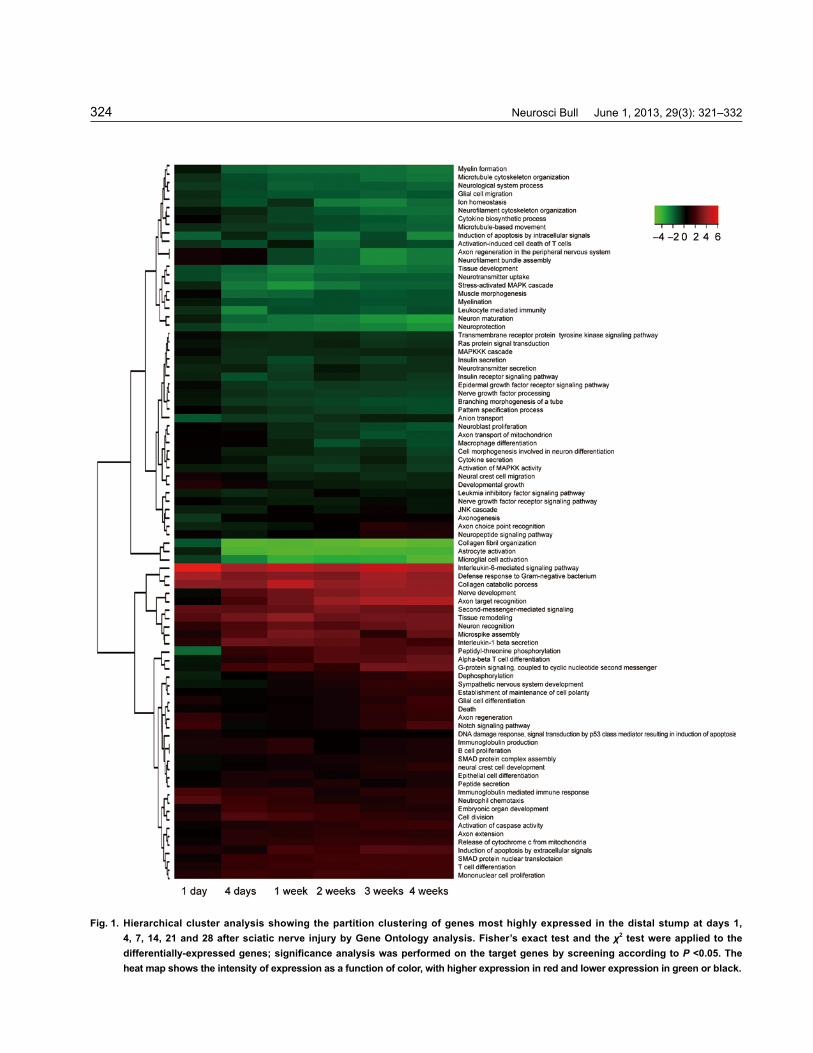
GO analysis was conducted using gene expression trends in a series of experiments, followed by significant and separate analyses of the differential gene expression changes during WD. Based on the GO database, the differentially-expressed genes were distributed into functional categories, which included genes with putative functions in the regulation of gene expression, nerve-nerve synaptic transmission, neurotransmitter uptake, positive regulation of cell adhesion, axon guidance, cytokinesis, nervous system development, regulation of phosphorylation, negative regulation of neuron apoptosis, axon target recognition, positive regulation of cell proliferation, neurogenesis, positive regulation of protein amino-acid dephosphorylation, positive regulation of phosphoinositide 3-kinase activity, cell differentiation, regulation of cell differentiation, cytokine production, and axon regeneration (Fig. 1). KEGG analysis of genes differentially expressed during WD Based on the GO database, χ2 tests and Fisher’s exact test were applied to the differentially-
Neurosci Bull June 1, 2013, 29(3): 321–332324
Fig. 1. Hierarchical cluster analysis showing the partition clustering of genes most highly expressed in the distal stump at days 1, 4, 7, 14, 21 and 28 after sciatic nerve injury by Gene Ontology analysis. Fisher’s exact test and the χ2 test were applied to the differentially-expressed genes; significance analysis was performed on the target genes by screening according to P <0.05. The heat map shows the intensity of expression as a function of color, with higher expression in red and lower expression in green or black.

Dengbing Yao, et al. Expression changes in rat Wallerian degeneration 325
expressed genes, significance analysis was performed on the pathways involving target genes, and the significant pathways were obtained by screening according to P <0.05.
Functional classification by KEGG analysis indicates the signal networks of molecular interactions in the cells or tissues specific to particular organisms, helping to understand the functions and molecular interactions of differentially-expressed genes. Based on the GO database using BLAST with an E value cut-off <10-5, in the process of WD 6 076 single genes had significant matches with the database and were assigned to 108 KEGG signal pathways (Table S3). Among these assignments, the pathways enriched during WD were axon guidance, neuroactive ligand-receptor interaction and the MAPK signaling pathway, oxidative phosphorylation, the p53 signaling pathway, cytokine-cytokine receptor interaction, the cell cycle, the tight junction, the insulin signaling pathway, ECM-receptor interaction, the calcium signaling pathway, the adherens junction, the TGF-β signaling pathway, the Wnt signaling pathway, cell adhesion molecules, regulation of the actin cytoskeleton, the gap junction, the Jak-STAT signaling pathway, the ErbB signaling pathway, the VEGF signaling pathway, apoptosis, the B-cell receptor signaling pathway, the Toll-like receptor signaling pathway, natural killer cell-mediated cytotoxicity, and the T-cell receptor signaling pathway (Fig. 2). In the KEGG map, the genes assigned to the pathways were involved in axon guidance, the cell cycle, oxidative phosphorylation, and regulation of the actin cytoskeleton, as well as degeneration and regeneration/repair. The results involved high-throughput screening to identify novel genes expressed during WD after nerve injury. The many genes expressed differentially in the present study provide a basis for understanding the biological processes that regulate nerve degeneration and repair/regeneration.Key network analysis of genes differentially expressed during WD A dynamic regulatory network may exist in protein-protein interactions and expression kinetics that are reflected in stimulus-induced cellular behavior. To analyze this network, the relationships of the differential gene expression data were calculated using a Continuous Time Recurrent Neural Network. Using a genetic algorithm, we estimated the model parameters. A heat map showed the partition clustering of genes highly-expressed in the distal nerve stump after sciatic nerve injury (Fig. 3).
Key networks were constructed from experiments. The gene content of the uploaded files was used as the list for the networks, which were analyzed with the default settings. This is the shortest-paths variant algorithm with the main parameters of differential gene relative enrichment with uploaded data and relative saturation of canonical pathways networks. These networks were built ex tempore, are unique to the uploaded data and are based on the number of fragments of networks with canonical pathways.
Genes encoding signal transducers or growth factors involved in WD, such as the immune response, cell death, transport and transcriptional regulation, showed injury-specific expression. In fact, many of them are important for repair or regeneration[26-28]. Here, we demonstrated that the key factors in WD included NGF, MAG, CNTF, CTNNA2, p53, JAK2, PLCB1, STAT3, BDNF, PRKC, collagen II, FGF, THBS4, TNC and c-Src (Fig. 3). These key networks cover the most important pathways that play important roles in regulating this differential gene expression through mainly biological GO processes during WD after sciatic nerve injury. The data shown here are consistent with published reports[15-17] and supported the conclusion that developing and regenerating peripheral nerves exhibit differences in genetic programs. Further, the data revealed that signal transducers were significantly regulated in the lesioned distal stump after sciatic nerve injury. The molecules that were significantly up- or down-regulated after such injury are known to modulate the signal pathways in key networks[15,29-32]. It is supposed that the key networks play roles in Schwann cell activation and proliferation as well as WD and subsequent regeneration[33-35]. The data also suggested that the functional group of transcriptional regulators indicated in the changes of expression of these genes may be an essential prerequisite for peripheral nerve degeneration and/or regeneration.
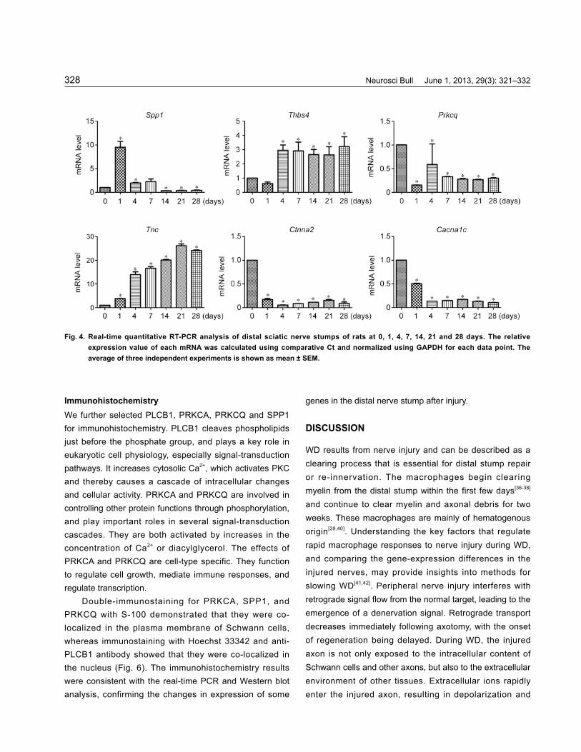
Real-Time Quantitative PCR To validate our data, real-time quantitative PCR was performed to analyze the expression changes of the selected genes, Spp1, Thbs4, Tnc (tenascin C), Ctnna2 [catenin (cadherin -associated protein), alpha 2], Cacna1c (calcium channel, voltage-dependent, L type, alpha 1C subunit) and Prkcq in WD. TNC is produced by migrating cells like neural crest cells, is abundant in developing tendon, bone and cartilage, and appears around healing

Neurosci Bull June 1, 2013, 29(3): 321–332326
wounds. THBS4 belongs to the thrombospondin protein family that mediates cell-to-matrix or cell-to-cell interactions. It has been reported that THBS4 may be involved in local signaling in the developing and adult nervous system[20-22].
Real-time quantitative PCR demonstrated that Spp1, Thbs4, Tnc, Ctnna2, Cacna1c and Prkcq were specifically expressed in the distal nerve stump of rats after injury (Fig. 4). The results are consistent with the gene-expression data from microarray analysis.
Western Blot Analysis The distal nerve stumps of rats at days 0, 1, 4, 7, 14, 21 and 28 after injury were directly lysed with a buffer (100 mmol/L dithiothreitol, 50 mmol/L Tris-HCl, pH 6.8, 10% glycerol, and 2% SDS). The results were analyzed as the
ratio of sample signal intensity to positive control signal intensity. Each sample signal and positive-control signal was normalized by background subtraction. The data indicated that the expression was similar to the results of microarray analysis and real-time quantitative PCR (Fig. 5). SPP1, also known as osteopontin, is reported to be an immune modulator and is an adhesion protein involved in wound-healing and cell attachment. It mediates cytokine production and cell activation, possibly promoting cell survival by regulating apoptosis, although the regulatory pathways have not been identified. PRKC plays important roles in signal transduction cascades, and through phosphorylating other proteins, it mediates immune responses and regulates transcription and cell growth.
Fig. 2. Hierarchical cluster analysis showing partition clustering of the genes most highly expressed in the distal nerve stump after sciatic nerve injury. KEGG analysis of distal sciatic nerve stumps from rats at 1, 4, 7, 14, 21, and 28 days. Based on the KEGG database, Fisher’s exact test and the χ2 test were applied to the differentially-expressed genes, significance analysis was performed with the pathways involving the target genes, and the significant pathways were obtained by screening according to P <0.05. The heat map shows the intensity of expression as a function of color, with higher expression in red and lower expression in green or black.

Dengbing Yao, et al. Expression changes in rat Wallerian degeneration 327
Fig. 3. Key network analysis of distal sciatic nerve stump of rats at 1, 4, 7, 14, 21, and 28 days. The genes listed in the uploaded files were used as the input for the generation of biological networks using the “analyze network” algorithm with default settings. The networks are prioritized based on the number of fragments of canonical pathways. Thick cyan lines indicate the fragments of canonical pathways. Upregulated genes are marked with red circles; downregulated genes with blue circles. The 'checker-board' indicates mixed expression of the gene between files or between multiple tags for the same gene.
Neurosci Bull June 1, 2013, 29(3): 321–332328
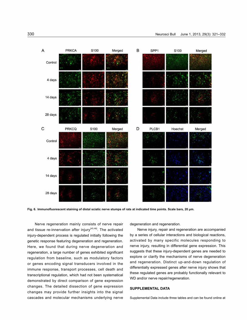
ImmunohistochemistryWe further selected PLCB1, PRKCA, PRKCQ and SPP1 for immunohistochemistry. PLCB1 cleaves phospholipids just before the phosphate group, and plays a key role in eukaryotic cell physiology, especially signal-transduction pathways. It increases cytosolic Ca2+, which activates PKC and thereby causes a cascade of intracellular changes and cellular activity. PRKCA and PRKCQ are involved in controlling other protein functions through phosphorylation, and play important roles in several signal-transduction cascades. They are both activated by increases in the concentration of Ca2+ or diacylglycerol. The effects of PRKCA and PRKCQ are cell-type specific. They function to regulate cell growth, mediate immune responses, and regulate transcription.
Double-immunostaining for PRKCA, SPP1, and PRKCQ with S-100 demonstrated that they were co-localized in the plasma membrane of Schwann cells, whereas immunostaining with Hoechst 33342 and anti-PLCB1 antibody showed that they were co-localized in the nucleus (Fig. 6). The immunohistochemistry results were consistent with the real-time PCR and Western blot analysis, confirming the changes in expression of some
genes in the distal nerve stump after injury.
DISCUSSION
WD results from nerve injury and can be described as a clearing process that is essential for distal stump repair or re-innervation. The macrophages begin clearing myelin from the distal stump within the first few days[36-38] and continue to clear myelin and axonal debris for two weeks. These macrophages are mainly of hematogenous origin[39,40]. Understanding the key factors that regulate rapid macrophage responses to nerve injury during WD, and comparing the gene-expression differences in the injured nerves, may provide insights into methods for slowing WD[41,42]. Peripheral nerve injury interferes with retrograde signal flow from the normal target, leading to the emergence of a denervation signal. Retrograde transport decreases immediately following axotomy, with the onset of regeneration being delayed. During WD, the injured axon is not only exposed to the intracellular content of Schwann cells and other axons, but also to the extracellular environment of other tissues. Extracellular ions rapidly enter the injured axon, resulting in depolarization and
Fig. 4. Real-time quantitative RT-PCR analysis of distal sciatic nerve stumps of rats at 0, 1, 4, 7, 14, 21 and 28 days. The relative expression value of each mRNA was calculated using comparative Ct and normalized using GAPDH for each data point. The average of three independent experiments is shown as mean ± SEM.

Dengbing Yao, et al. Expression changes in rat Wallerian degeneration 329
injury-induced action potentials[12,43,44]. In this study, we found that from day 1 to 4 weeks after
injury, ~3 500 regulated genes (1 800 up, 1 600 down) were detected in the distal stump of the injured sciatic nerve. Axons begin to grow into the distal endoneurial tubes 1 week after nerve injury[6,39,45]; Schwann cell/axon contacts are re-established and remyelination occurs
at about 2 weeks; and the proper target tissues are re-innervated at about 4 weeks after sciatic nerve injury. In this process, a large number of genes with marked up- or down-regulation were differentially expressed, indicating sustained regulation at consecutive time points, meaning regeneration/repair by means of a continuously progressive program[4,10,35].
Fig. 5. Western blot analysis of total protein lysates of distal sciatic nerve stumps of rats at days 0, 1, 4, 7, 14, 21, and 28 (upper panel) and the quantification (lower panels). Data are from three independent experiments and shown as mean ± SEM.
Neurosci Bull June 1, 2013, 29(3): 321–332330
Nerve regeneration mainly consists of nerve repair and tissue re-innervation after injury[45,46]. The activated injury-dependent process is regulated initially following the genetic response featuring degeneration and regeneration. Here, we found that during nerve degeneration and regeneration, a large number of genes exhibited significant regulation from baseline, such as modulatory factors or genes encoding signal transducers involved in the immune response, transport processes, cell death and transcriptional regulation, which had not been systematical demonstrated by direct comparison of gene expression changes. The detailed dissection of gene expression changes may provide further insights into the signal cascades and molecular mechanisms underlying nerve
degeneration and regeneration.Nerve injury, repair and regeneration are accompanied
by a series of cellular interactions and biological reactions, activated by many specific molecules responding to nerve injury, resulting in differential gene expression. This suggests that these injury-dependent genes are needed to explore or clarify the mechanisms of nerve degeneration and regeneration. Distinct up-and-down regulation of differentially expressed genes after nerve injury shows that these regulated genes are probably functionally relevant to WD and/or nerve repair/regeneration.
SUPPLEMENTAL DATA
Supplemental Data include three tables and can be found online at
Fig. 6. Immunofluorescent staining of distal sciatic nerve stumps of rats at indicated time points. Scale bars, 20 μm.

Dengbing Yao, et al. Expression changes in rat Wallerian degeneration 331
www.neurosci.cn/epData.asp?id=95.
ACKNOWLEDGEMENTS
This work was supported by grants from the National Natural Science Foundation of China (81130080, 30870811), the Scientific Research Foundation for Returned Overseas Chinese Scholars, Ministry of Education of China, the Natural Science Foundation of Jiangsu Province, China (BK2010282) and the Priority Academic Program Development of Higher Education Institutions of Jiangsu Province, China.
Received date: 2012-07-13; Accepted date: 2012-09-21
REFERENCES
[1] Stoll G, Jander S, Myers RR. Degeneration and regeneration of the peripheral nervous system: from Augustus Waller's observations to neuroinflammation. J Peripher Nerv Syst 2002, 7: 13–27.
[2] Waller A. Experiments on the section of the glossopharyngeal and hypoglossal nerves of the frog, and observations of the alterations produced thereby in the structure of their primitive fibres. Phil Transact Royal Soc London 1850, 140: 423–429.
[3] Ambron RT, Walters ET. Priming events and retrograde injury signals. A new perspective on the cellular and molecular biology of nerve regeneration. Mol Neurobiol 1996, 13: 61–79.
[4] Boivin A, Pineau I, Barrette B, Filali M, Vallières N, Rivest S, Lacroix S. Toll-like receptor signaling is critical for Wallerian degeneration and functional recovery after peripheral nerve injury. J Neurosci 2007, 27: 12565–12576.
[5] De S, Trigueros MA, Kalyvas A, David S. Phospholipase A2 plays an important role in myelin breakdown and phagocytosis during Wallerian degeneration. Mol Cell Neurosci 2003, 24: 753–765.
[6] Girolami EI, Bouhy D, Haber M, Johnson H, David S. Differential expression and potential role of SOCS1 and SOCS3 in Wallerian degeneration in injured peripheral nerve. Exp Neurol 2010, 223: 173–182.
[7] Guertin AD, Zhang DP, Mak KS, Alberta JA, Kim HA. Microanatomy of axon/glial signaling during Wallerian degeneration. J Neurosci 2005, 25: 3478–3487.
[8] Martini R, Fischer S, Lopez-Vales R, David S. Interactions between Schwann cells and macrophages in injury and inherited demyelinating disease. Glia 2008, 56: 1566–1577.
[9] Tofaris GK, Patterson PH, Jessen KR, Mirsky R. Denervated Schwann cells attract macrophages by secretion of leukemia inhibitory factor (LIF) and monocyte chemoattractant protein-1 in a process regulated by interleukin-6 and LIF. J
Neurosci 2002, 22: 6696–6703.[10] Navarro X, Vivo M, Valero-Cabre A. Neural plasticity after
peripheral nerve injury and regeneration. Prog Neurobiol 2007, 82: 163–201.
[11] Parkinson DB, Bhaskaran A, Arthur-Farraj P, Noon LA, Woodhoo A, Lloyd AC, et al. c-Jun is a negative regu-lator of myelination. J Cell Biol 2008, 181(4): 625–637.
[12] Kirsch M, Terheggen U, Hofmann HD. Ciliary neurotrophic factor is an early lesion-induced retrograde signal for axotomized facial motoneurons. Mol Cell Neurosci 2003, 24: 130–138.
[13] Lindholm D, Heumann R, Meyer M, Thoenen H. Interleukin-1 regulates synthesis of nerve growth factor in non-neuronal cells of rat sciatic nerve. Nature 1987, 330: 658–659.
[14] Perrin FE, Lacroix S, Avi les-Trigueros M, David S. Involvement of monocyte chemoattractant protein-1, macrophage inflammatory protein-1a and interleukin-1b in Wallerian degeneration. Brain 2005, 4: 854–866.
[15] Raivich G, Bohatschek M, Da Costa C, Iwata O, Galiano M, Hristova M, et al. The AP-1 transcription factor c-Jun is required for efficient axonal regeneration. Neuron 2004, 43: 57–67.
[16] Sendtner M, Gotz R, Holtmann B, Thoenen H. Endogenous ciliary neurotrophic factor is a lesion factor for axotomized motoneurons in adult mice. J Neurosci 1997, 17: 6999–7006.
[17] Wiklund P, Ekstrom PA, Edstrom A. Mitogen-activated protein kinase inhibition reveals differences in signalling pathways activated by neurotrophin-3 and other growth-stimulating conditions of adult mouse dorsal root ganglia neurons. J Neurosci Res 2002, 67: 62–68.
[18] Zochodne DW, Levy D, Zwiers H, Sun H, Rubin I, Cheng C, et al. Evidence for nitric oxide and nitric oxide synthase activity in proximal stumps of transected peripheral nerves. Neuroscience 1999, 91: 1515–1527.
[19] Ramoni MF, Sebastiani P, Kohane IS. Cluster analysis of gene expression dynamics. Proc Natl Acad Sci U S A 2002, 99: 9121–9126.
[20] Miller LD, Long PM, Wong L, Mukherjee S, McShane LM, Liu ET. Optimal gene expression analysis by microarrays. Cancer Cell 2002, 2: 353–361.
[21] Kanehisa M, Goto S, Kawashima S, Okuno Y, Hattori M. The KEGG resource for deciphering the genome. Nucleic Acids Res 2004, 32: D277–280.
[22] Yi M, Horton JD, Cohen JC, Hobbs HH, Stephens RM. WholePathwayScope: a comprehensive pathway-based analysis tool for high-throughput data. BMC Bioinformatics 2006, 7: 30.
[23] Draghici S, Khatri P, Tarca AL, Amin K, Done A, Voichita C, et al. A systems biology approach for pathway level analysis. Genome Res 2007, 17: 1537–1545.

Neurosci Bull June 1, 2013, 29(3): 321–332332
[24] Busch H, Camacho-Trullio D, Rogon Z, Breuhahn K, Angel P, Eils R, et al. Gene network dynamics controlling keratinocyte migration. Mol Syst Biol 2008, 4: 199.
[25] Zhou S, Yu B, Qian T, Yao D, Wang Y, Ding F, et al. Early changes of microRNAs expression in the dorsal root ganglia following rat sciatic nerve transection. Neurosci Lett 2011, 494: 89–93.
[26] Ghosh A, Greenberg ME. Calcium signaling in neurons: molecular mechanisms and cellular consequences. Science 1995, 268: 239–247.
[27] Hanz S, Perlson E, Willis D, Zheng JQ, Massarwa R, Huerta JJ, et al. Axoplasmic importins enable retrograde injury signaling in lesioned nerve. Neuron 2003, 40: 1095–1104.
[28] Kim D, Lee S, Lee SJ. Toll-like receptors in peripheral nerve injury and neuropathic pain. Curr Top Microbiol Immunol 2009, 336: 169–186.
[29] Berti-Mattera LN, Harwalkar S, Hughes B, Wilkins PL, Almhanna K. Proliferative and morphological effects of endothelins in Schwann cells: roles of p38 mitogen-activated protein kinase and Ca(2+)-independent phospholipase A2. J Neurochem 2001, 79: 1136–1148.
[30] Koehler JA, Moran MF. Regulation of extracellular signal-regulated kinase activity by p120 RasGAP does not involve its pleckstrin homology or calcium-dependent lipid binding domains but does require these domains to regulate cell proliferation. Cell Growth Differ 2001, 12: 551–561.
[31] Lindwall C, Kanje M. Retrograde axonal transport of JNK signaling molecules influence injury induced nuclear changes in p-c-Jun and ATF3 in adult rat sensory neurons. Mol Cell Neurosci 2005, 29: 269–282.
[32] Schwaiger FW, Harger G, Schmitt AB, Horvat A, Hager G, Streif R, et al. Peripheral but not central axotomy induces changes in Janus kinases (JAK) and signal transducers and activators of transcription (STAT). Eur J Neurosci 2000, 12: 1165–1176.
[33] Lee HK, Seo IA, Park HK, Park YM, Ahn KJ, Yoo YH, et al. Nidogen is a prosurvival and promigratory factor for adult Schwann cells. J Neurochem 2007, 102: 686–698.
[34] Lee N, Neitzel KL, Devlin BK, MacLennan AJ. STAT3 phosphorylation in injured axons before sensory and motor neuron nuclei: potential role for STAT3 as a retrograde signaling transcription factor. J Comp Neurol 2004, 474:
535–545.[35] Sheu JY, Kulhanek DJ, Eckenstein FP. Differential patterns
of ERK and STAT3 phosphorylation after sciatic nerve transection in the rat. Exp Neurol 2000, 166: 392–402.
[36] de Bilbao F, Giannakopoulos P, Srinivasan A, Dubois-Dauphin M. In vivo study of motoneuron death induced by nerve injury in mice deficient in the caspase 1/ interleukin-1beta-converting enzyme. Neuroscience 2000, 98(3): 573–583.
[37] Herdegen T, Waetzig V. The JNK and p38 signal transduction following axotomy. Restor Neurol Neurosci 2001, 19: 29–39.
[38] Kuhn G, Lie A, Wilms S, Muller HW. Coexpression of PMP22 gene with MBP and P0 during de novo myelination and nerve repair. Glia 1993, 8: 256–264.
[39] Shubayev VI, Angert M, Dolkas J, Campana WM, Palenscar K, Myers RR. TNFalpha induced MMP-9 promotes macrophage recruitment into injured peripheral nerve. Mol Cell Neurosci 2006, 31: 407–415.
[40] Sun W, Oppenheim RW. Response of motoneurons to neonatal sciatic nerve axotomy in Baxknockout mice. Mol Cell Neurosci 2003, 24: 875–886.
[41] Ugolini G, Raoul C, Ferri A, Haenggeli C, Yamamoto Y, Salaün D, et al. Fas/tumor necrosis factor receptor death signaling is required for axotomy-induced death of motoneurons in vivo. J Neurosci 2003, 23: 8526–8531.
[42] Waetzig V, Herdegen T. MEKK1 controls neurite regrowth after experimental injury by balancing ERK1/2 and JNK2 signaling. Mol Cell Neurosci 2005, 30: 67–78.
[43] Shadiack AM, Sun Y, Zigmond RE. Nerve growth factor antiserum induces axotomy-like changes in neuropeptide expression in intact sympathetic and sensory neurons. J Neurosci 2001, 21: 363–371.
[44] Yoo S NM, Fukuda M, Bittner GD, Fishman HM. Plasmalemmal sealing of transected mammalian neurites is a gradual process mediated by Ca(2+)-regulated proteins. J Neurosci Res 2003, 74: 541–551.
[45] Bosse F, Hasenpusch-Thei l K, Küry P, Mül ler HW. Gene expression profiling reveals that peripheral nerve regeneration is a consequence of both novel injury-dependent and reactivated developmental processes. J Neurochem 2006, 96: 1441–1457.
[46] Makwana M, Raivich G. Molecular mechanisms in successful peripheral regeneration. FEBS J 2005, 272: 2628–2638.