expression and clinical significance of hedgehog signaling
TRANSCRIPT
Asian Pacific Journal of Cancer Prevention, Vol 13, 2012 2319
DOI:http://dx.doi.org/10.7314/APJCP.2012.13.5.2319 Hedgehog Signaling Pathway Related Components in Colorectal Cancer
Asian Pacific J Cancer Prev, 13, 2319-2324
Introduction
Colorectal cancer (CRC) is a common cancer ranking as the fourth most frequent causes of cancer death in the world (Jemal et al., 2008; Kim et al., 2012; Kimman et al., 2012; Park et al., 2012). In order to predict the prognosis and improve the treatment of CRC, there is a great need to understand the molecular mechanisms leading to tumor formation and progression. The Hedgehog (Hh) signaling pathway, which regulates proliferation, angiogenesis, matrix remodeling and stem-cell renewal, plays an important role during embryogenesis and in adult life. It is involved in the patterning in a wide variety of the organs including gastrointestinal tract (McMahon et al., 2003; Dessaud et al., 2008). By contrast, deregulation of the Hh pathway is involved in tumor development, as the mutations of several components of this pathway were found in patients with many types of cancers including CRC (Evangelista et al., 2006; Scales et al., 2009). In addition, recent researches have also indicated that tumorigenesis of Hh signaling might be through the molecular pathways of
Department of Gastroenterology, Guangzhou First Municipal People’s Hospital Affiliated to Guangzhou Medical College, Guangzhou China *For correspondence: [email protected]
Abstract
Aim: To investigate the expression of three components of the Hedgehog (Hh) signaling pathway (SHH, SMO and GLI1) in human colorectal cancer (CRC) tissues and evaluate their association with clinicopathologic characteristics of the patients. Methods: Fresh tumor tissues and matched tissues adjacent to the tumor were collected from 43 CRC patients undergoing surgery. Normal colorectal tissues from 20 non-CRC cases were also sampled as normal controls. The expression of SHH, SMO, GLI1 mRNAs was assessed by RT-PCR and proteins were detected by immunohistochemical staining. Associations with clinicopathological characteristics of patients were analyzed. Results: SHH mRNA was expressed more frequently in tumor tissues than in normal tissues, but the difference did not reach significance in comparison to that in the adjacent tissues. SMO and GLI1 mRNAs were expressed more frequently in tumor tissues than in both adjacent andnormal tissues. The expression intensities of SHH, SMO, GLI1 mRNA in tumor tissues were significantly higher than those in adjacent tissues and normal tissues. Proteins were also detected more frequently in tumors than other tissues. No significant links were apparent with gender, age, location, degree of infiltration or Dukes stage. Conclusion: Positive rates and intensities of mRNA and protein expression of Hh signaling pathway related genes SHH, SMO, GLI1 were found to be significantly increased in CRC tissues. However, over-expression did not appear to be associated with particular clinicopathological characteristics. Keywords: Gene - hedgehog signaling pathway - colorectal cancer - clinicopathological variables
RESEARCH COMMUNICATION
Expression and Clinical Significance of Hedgehog Signaling Pathway Related Components in Colorectal Cancer
Hong Wang, Yu-Yuan Li*, Ying-Ying Wu, Yu-Qiang Nie
cancer stem cell (CSC) (Varnat et al., 2009). There are three members in human Hh family i.e. Sonic HH (SHH), Indian HH (IHH), and Desert HH (DHH) (Echelard et al., 1993; Marigo et al., 1995). All these ligands bind to a 12-transmembrane receptor called Patched (PTCH) that restricts Hh signaling by inhibiting a 7-transmembrane receptor called smoothened (SMO) (Bijlsma et al., 2006). SMO releasing leads to stimulation of the downstream Hh signaling cascade, which culminates in the activation of the glioma-associated oncogene homolog (GLI) family of transcription factors. The forms of GLI family, which included GLI1, GlI2 and GlI3, migrate to the cells to bind HH target genes (McMahon et al., 2003; Evangelista et al., 2006; Varjosalo et al., 2008). OnthefieldofHhsignalingpathwayintumorigenesis,SHH, SMO and GLI1 have been studied frequently. However, most studies focused on the cancers of other organs (Steg et al., 2012; Yang et al., 2012). The data about the roles they play on CRC have not been well documented. Some of them even remained controversial (Berman et al., 2003; Douard et al., 2006; Monzo et al., 2006; Chatel et al., 2007; Varnat et al., 2009;
Hong Wang et al
Asian Pacific Journal of Cancer Prevention, Vol 13, 20122320
Yoshikawa et al., 2009; Fu et al., 2010; Saqui-Salces and Merchant, 2010; You et al., 2010; Tapati et al., 2011). There were disagreements in the roles of downstream Hh signaling components i.e. SMO and GLI1 play in tumorigenesis of CRC and the possible association of Hh pathway activation with patient’s clinicopathological characteristics, i.e. tumor stage and differentiation. In the present study, we simultaneously investigate the mRNA and protein expressions of SHH, SMO, GLI1 related genes in human CRC tissues and evaluated their association with the clinicopathologic characteristics of the patients. Materials and Methods
Patients and specimens This study was conducted in accordance with the declaration of Helsinki. The study protocols were approved by the ethics committee of Guangzhou First Municipal People’sHospitalAffiliated toGuangzhouMedical College. Written consent was obtained from each patient. Consecutive patients (43 cases) with pathologically confirmed colorectal adenocarcinomas,who underwent surgical operations at Department of Gastroenterological Surgery of this hospital between December 1, 2008 and April 30, 2009, were included. None of them received chemotherapy or radiation therapy before surgery. Matched non-CRC patients (20 cases) undergoing surgical operations during the same period of time were also collected as controls. Fresh tumor tissues and matched mucosa adjacent to the cancer (10 cm beyond the cancer margin) from 43 patients were collected. Normal colorectal tissues from 20 non-CRC patients undergoing surgical operations were also collected as normal controls. Two specimens from each site obtained at surgery resections were immediately frozen in liquid nitrogen and stored at -80 °C until total RNA extraction. An additional sample was fixed in formadehyde saline and embedded in paraffinfor pathological examination. The Dukes pathologic staging system was applied to classify the patients. Dukes’ A: lesions contiguous with the bowel wall but not penetrating the muscularis. Dukes’ B: lesions penetrating the muscularis into the surrounding fat or adventitia, Dukes’ C: lesions with positive lymph node involvement. Total RNA extraction and quantitative real-time RT-PCR Total RNA was extracted by chloroform-isopropanol-ethanol method using Trizol (Invitrogen, USA) according to the manufacturer’s instructions. The A260/A280 ratio of RNA determined with colorimetry was between
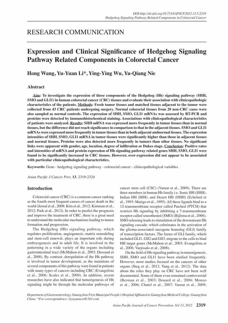
1.80~2.00.RNAintegritywasqualifiedwithformaldehydedegeneration gel electrophoresis at 80V for 15 min. Three electrophoresis bands (5S, 18S, 28S) were visualized. Complementary DNA (cDNA) was synthesized with the instructionsonM-MLVreversetranscriptasekit.Briefly,2μgoftotalRNA,0.5µgoligo(dT)15primerand12µlDEPC-treated H2Oinafinal15µlreactionvolumewereincubated at 70 °C for 5 min and subsequently on ice for 5 min to synthesize RNA- primer compound. Then, 5 µlM-MLVbuffer,1.25µldNTP,0.625µlRNasin,1µlM-MLVreversetranscriptaseand2.125µlDEPC-treatedH2OconstitutedRT-reactionmixture.Afterward,15µlRNA-primercompoundand10µlRT-reactionmixturewere blended, and incubated at 37 °C for 60 min and then at 70 °C for 15 min to synthesize cDNA. Three components of the Hedgehog signaling pathway, SHH, SMO and GLI1, were selected for quantitative real-time RT-PCR analysis. The reference sequences for each gene were obtained from Genebank on the NCBI Entrez web site (http://www.ncbi.nlm.nih.gov/genome/guide/human). Forward and reverse primer pairs were designed using Primer Premier 5.0 for Windows (PREMIER Biosoft International, Palo Alto, CA, USA) and synthesized by Invitrogen Inc., Shanghai, China, which were listed in Table 1. Real-time PCR was carried out on a Bio-Radgradient PCR with electrophresis apparatus. The reactions included 1μL cDNA (1:10 dilution), 1ul each primer, 2×PCRmaster mix 10ul and ddH2O7ul.Thefinalreactionvolumewas 20 μL. PCRwas performed under the followingcycling conditions: an initial denaturation step at 95°C for 4min,followedby35cyclesofamplification(forSHHand SMO cDNA, 95 °C for 30 s, 56 °C for 30 s, and 72 °C for 30 s; for GLI1 cDNA, 95 °C for 30 s, 66 °C for 30 s, and 72 °C for 30 s),then elongation at 72 °C for 5 min. Every PCR reaction of each gene and each sample was done in duplicate. PCR products were detected with 2% agarose gel electrophoresis and quantitated using an agarose gel formatter (Figure 1). The mRNA positive rates were the percentage of the numbers appearing electrophoretic straps
Table 1. Sequences of Primer PairsGenes Primers (5’g3’) Size (bp) GeneBank
SHH F GCTGATGACTCAGAGGTGTAAGGA 280 NM_000193 R CCACCGAGTTCTCTGCTTTCA SMO F CTGGTGTGGTTTGGTTTGTG 366 NM_005631 R TGGTCTCGTTGATCTTGCTG GLI1 F CGGGGTCTCAAACTGCCCAGCTT 387 NM_001160045 R GGCTGGGTCACTGGCCCTC GAPDH F TGGTCTCCTCTGACTTCAAC 222 NM_002046 R GTGAGGGTCTCTCTCTTCCT
Figure 1. Gel Electrophoresis of SHH, SMO and GLI1 mRNA Expressions. A: M: marker; 1-2: bland control; 3-7: SHH (280bp). B: M: marker; 1, 3: SMO (366bp); 2, 4: GAPDH (222bp); 5: bland control. C: M: marker; 1, 3: GLI1 (387bp); 2, 4: GAPDH (222bp)

Asian Pacific Journal of Cancer Prevention, Vol 13, 2012 2321
DOI:http://dx.doi.org/10.7314/APJCP.2012.13.5.2319 Hedgehog Signaling Pathway Related Components in Colorectal Cancer
0
25.0
50.0
75.0
100.0
New
ly d
iagn
osed
with
out
trea
tmen
t
New
ly d
iagn
osed
with
tre
atm
ent
Pers
iste
nce
or r
ecur
renc
e
Rem
issi
on
Non
e
Chem
othe
rapy
Radi
othe
rapy
Conc
urre
nt c
hem
orad
iatio
n
10.3
0
12.8
30.025.0
20.310.16.3
51.7
75.051.1
30.031.354.2
46.856.3
27.625.033.130.031.3
23.738.0
31.3
Table 2. Expressions of SHH mRNA and Protein in CRC, Adjacent Tissues and Normal Colorectal Tissues n Protein positive mRNA positive Rate (%) - ± + ++ Rate (%) Intensity
CRC 43 11(25.6) 12 20 6 5 12(27.9) 0.72±0.19adjacent 43 6(15.0) 20 17 5 1 8(18.6) 0.56±0.11normal 20 0(0) 17 3 0 0 0(0) 0P 0.03 0.01 0.03 0.03
Table 3. Expressions of SMO mRNA and Protein in CRC, Adjacent Tissues and Normal Colorectal Tissues n Protein positive mRNA positive Rate (%) - ± + ++ Rate (%) Intensity
CRC 43 26(60.5) 5 12 11 15 36(83.7) 0.88±0.11adjacent 43 12(27.9) 22 9 6 6 25(58.1) 0.72±0.22normal 20 0(0) 18 2 0 0 0(0) 0P 0.00 0.02 0.00 0.00
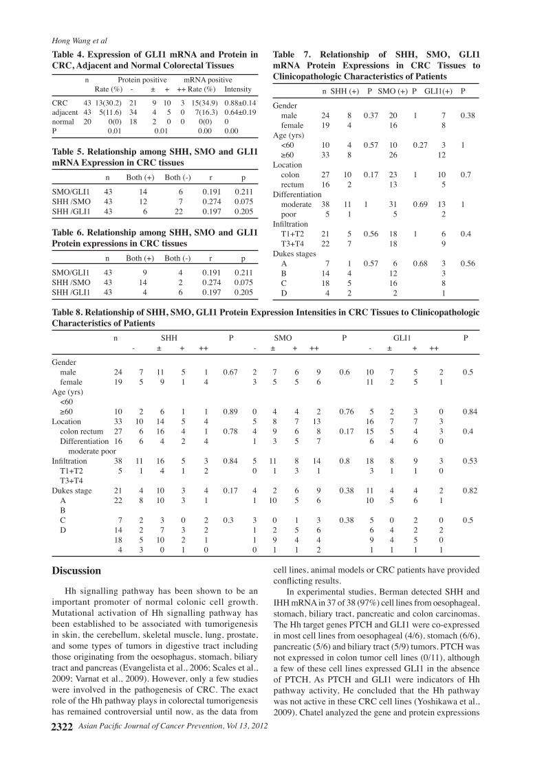
Figure 2. SHH, SMO, GLI1 Protein Expressions (Imunostaining × 200). A SHH (+) in cancer, B SHH (-) in normal control; C SMO (+) in cancer, D SMO (-) in normal control; E GLI1 (+) in cancer, F GLI1 (-) in normal control
divided by the total numbers of cases.
Immunohistochemistry Formalin-fixed,paraffin-embeddedsampleswereusedinthestudy.Sectionswereseriallycutat4μm.Envisiontechniquewasusedforimmunostaining.Briefly,Sectionswere deparaffinized in xylene and rehydrated throughgraded ethanol, followed by incubation at 60 °C for 50 min in 3% hydrogen peroxide to block endogenous peroxidase.Acaseinserum-freeblockingsolutionspecificfor formalin-fixed tissueswas applied for 20min andsections were incubated overnight at 4 °C with primary antibodies. Primary antibodies were used at a dilution ratio of 1:50 for SHH, 1:50 for SMO, 1:100 for GLI1 (Anti-SHH and Anti- GLI1 s were purchased from Santa Cruz Corp. USA, Anti- SMO antibody was purchased from Abcam Corp. UK). Slides were given three 5-min washes in PBS and incubated with secondary antibodies at 37 °C for 30 min in PV-6001 Complex (SMO,GLI1, purchased from Jinqiao Corp. Beijing, China) or in PV-9003 Complex
(SHH, purchased from Jinqiao Corp. Beijing, China). The antibody complex was visualized with diaminobenzidine. Sections were counterstained with HE staining. Negative controls (absence of primary antibody) were included for each antibody and analyzed for each case (Figure 2). The intensity of staining in the cytoplasm was scored arbitrarily to reflect negative (-), light (±),moderate (+), and strong, (++) respectively. The scores were determined by a pathologist blinded to the study program. The positive rates were calculated as the numbers of (+) plus (++) cases divided in each by the total numbers of cases.
Statistical analysis The data were examined with the SPSS 13.0 for Windows statistical package (Chicago, IL, USA). Continuous data were expressed as mean ±SD and examined by Student’s t-test. Categorical variables were expressed as percentage and examined by x2 test. Pearson test (two-tailed) was used to compare the variables among groups. ANOVA analysis was applied for multiple comparisons. Apvalue≤0.05wasconsideredstatisticallysignificant.
Results
Overall 43 patients with CRC (males 24, females 19) with the mean age of 66.4±10.5 years (range, 36-82 years) were enrolled in study group. In non-CRC control group, there were 20 patients (males 15, females 5, mean age 61.5±16.4 years). Histopathologically, all of them were adenocarcinomas with 38 being moderately differentiated and 5 poor differentiated. For Dukes staging, Dukes’ A included 7 cases, Dukes’ B 14 cases, Dukes’ C 18 cases and Dukes’ C 4 cases. The positive rate of SHH mRNA expressions in tumortissueswassignificantlyhigherthanthatinnormalcolorectaltissues(P<0.05),butdidnotreachsignificancein comparison to that in the adjacent tissues (P > 0.05) (Table 2). The positive rates of SMO and GLI1 mRNA expressions in tumor tissueswere significantly higherthan those in the adjacent tissues (P < 0.05) and those in normal colorectal tissues (P < 0.05). The expression levels of SHH, SMO, GLI1 mRNA in tumor tissues were significantlyhigherthanthoseintheadjacenttissues(P< 0.05) and normal colorectal tissues (P < 0.05) (Table 2-4). Immunohistochemical staining demonstrated that SHH, SMO, GLI1 proteins were mainly expressed in the cytoplasm of tumor cells and normal gland cells. The positive rates and expression intensities of SHH, SMO, GLI1proteinsintumortissuesweresignificantlyhigherthan those in the adjacent tissues and those in normal colorectal tissues (P < 0.05) (Table 2). Therewere no significant relationships among theSHH, SMO and GLI1 mRNA and protein expressions in tumor tissues (r = 0.191-0.274, P > 0.05) (Tables 5, 6). Furthermore, there were no apparent links found between SHH/SMO/GLI1 expression and any of the clinicopathological characteristics of the cancer patients (P > 0.05) (Tables 7, 8)..
Hong Wang et al
Asian Pacific Journal of Cancer Prevention, Vol 13, 20122322
Discussion
Hh signalling pathway has been shown to be an important promoter of normal colonic cell growth. Mutational activation of Hh signalling pathway has been established to be associated with tumorigenesis in skin, the cerebellum, skeletal muscle, lung, prostate, and some types of tumors in digestive tract including those originating from the oesophagus, stomach, biliary tract and pancreas (Evangelista et al., 2006; Scales et al., 2009; Varnat et al., 2009). However, only a few studies were involved in the pathogenesis of CRC. The exact role of the Hh pathway plays in colorectal tumorigenesis has remained controversial until now, as the data from
cell lines, animal models or CRC patients have provided conflictingresults.
In experimental studies, Berman detected SHH and IHH mRNA in 37 of 38 (97%) cell lines from oesophageal, stomach, biliary tract, pancreatic and colon carcinomas. The Hh target genes PTCH and GLI1 were co-expressed in most cell lines from oesophageal (4/6), stomach (6/6), pancreatic (5/6) and biliary tract (5/9) tumors. PTCH was not expressed in colon tumor cell lines (0/11), although a few of these cell lines expressed GLI1 in the absence of PTCH. As PTCH and GLI1 were indicators of Hh pathway activity, He concluded that the Hh pathway was not active in these CRC cell lines (Yoshikawa et al., 2009). Chatel analyzed the gene and protein expressions
Table 8. Relationship of SHH, SMO, GLI1 Protein Expression Intensities in CRC Tissues to Clinicopathologic Characteristics of Patients n SHH P SMO P GLI1 P - ± + ++ - ± + ++ - ± + ++
Gender male 24 7 11 5 1 0.67 2 7 6 9 0.6 10 7 5 2 0.5 female 19 5 9 1 4 3 5 5 6 11 2 5 1 Age (yrs) <60 ≥60 10 2 6 1 1 0.89 0 4 4 2 0.76 5 2 3 0 0.84Location 33 10 14 5 4 5 8 7 13 16 7 7 3 colon rectum 27 6 16 4 1 0.78 4 9 6 8 0.17 15 5 4 3 0.4 Differentiation 16 6 4 2 4 1 3 5 7 6 4 6 0 moderate poor Infiltration 38 11 16 5 3 0.84 5 11 8 14 0.8 18 8 9 3 0.53 T1+T2 5 1 4 1 2 0 1 3 1 3 1 1 0 T3+T4 Dukes stage 21 4 10 3 4 0.17 4 2 6 9 0.38 11 4 4 2 0.82 A 22 8 10 3 1 1 10 5 6 10 5 6 1 B C 7 2 3 0 2 0.3 3 0 1 3 0.38 5 0 2 0 0.5 D 14 2 7 3 2 1 2 5 6 6 4 2 2 18 5 10 2 1 1 9 4 4 9 4 5 0 4 3 0 1 0 0 1 1 2 1 1 1 1
Table 5. Relationship among SHH, SMO and GLI1 mRNA Expression in CRC tissues n Both (+) Both (-) r p
SMO/GLI1 43 14 6 0.191 0.211SHH /SMO 43 12 7 0.274 0.075SHH /GLI1 43 6 22 0.197 0.205
Table 6. Relationship among SHH, SMO and GLI1 Protein expressions in CRC tissues n Both (+) Both (-) r p
SMO/GLI1 43 9 4 0.191 0.211SHH /SMO 43 14 2 0.274 0.075SHH /GLI1 43 4 6 0.197 0.205
Table 7. Relationship of SHH, SMO, GLI1 mRNA Protein Expressions in CRC Tissues to Clinicopathologic Characteristics of Patients n SHH (+) P SMO (+) P GLI1(+) P
Gender male 24 8 0.37 20 1 7 0.38 female 19 4 16 8 Age (yrs) <60 10 4 0.57 10 0.27 3 1 ≥60 33 8 26 12Location colon 27 10 0.17 23 1 10 0.7 rectum 16 2 13 5 Differentiation moderate 38 11 1 31 0.69 13 1 poor 5 1 5 2 Infiltration T1+T2 21 5 0.56 18 1 6 0.4 T3+T4 22 7 18 9 Dukes stages A 7 1 0.57 6 0.68 3 0.56 B 14 4 12 3 C 18 5 16 8 D 4 2 2 1
Table 4. Expression of GLI1 mRNA and Protein in CRC, Adjacent and Normal Colorectal Tissues n Protein positive mRNA positive Rate (%) - ± + ++ Rate (%) Intensity
CRC 43 13(30.2) 21 9 10 3 15(34.9) 0.88±0.14adjacent 43 5(11.6) 34 4 5 0 7(16.3) 0.64±0.19normal 20 0(0) 18 2 0 0 0(0) 0P 0.01 0.01 0.00 0.00
Asian Pacific Journal of Cancer Prevention, Vol 13, 2012 2323
DOI:http://dx.doi.org/10.7314/APJCP.2012.13.5.2319 Hedgehog Signaling Pathway Related Components in Colorectal Cancer
of the main key members of the Hh pathway (SHH, IHH, PTCH, SMO, GLI1, GLI2, GLI3, SUFU and HHIP) in 7 colon cancer cell lines and found none of them expressed the complete set of Hh pathway members (Chatel et al., 2007). However, by analyzing CRC cell lines as well as human tissues from 40 patients with local or metastatic CRCs, Varnat found consistent expression of PTCH, GLI1 and SHH as well as other Hh-GLI1 pathway components. Expression was detected in epithelial tumor cells in situ in the colon. He did not observe expression of GLI1 protein in the stroma, indicating that the stroma did not have an active Hh-GLI signaling (Varnat et al., 2009). Yoshikawa found that colorectal adenomas expressed SHH more frequently compared with normal mucosa and adenocarcinoma, which suggested that SHH expression might increase during carcinogenesis and then decrease during tumor progression in the adenoma-carcinoma sequence (Yoshikawa et al., 2009). By contrast, in a clinical study for SHH, IHH and GLI1 genes in 36 CRC patients, Fu found the expressions of SHHandGLI1proteinswereincreasedsignificantlyintumor tissues compared with those in hyperplastic polyps and colorectal adenomas. IHH was almost lost in both colorectal adenomas and CRC tissues (Fu et al., 2010). In Monzo study, SHH mRNA expression levels were higher in tumor tissue than matched normal tissue from 57 CRC patients, and SHH expression was correlated with patient’s clinicopathological characteristics. Higher levels of SHH expression were associated with early stage of the tumor (Monzo et al., 2006). However, the previous study showed that CRC tissues in the high-risk patients for metastasis expressed lower levels of PTCH1 mRNA than those in the low-risk patients. The similar result was found in his experimental study. The mRNA and protein levels of PTCH1 were inversely correlated with the metastatic potential of CRC (LoVo) cell lines. But the expression levels of SHH genes did not differ between the two clinical groups (You et al., 2010). Douard found that SHH mRNA was overexpressed in CRCs in 38 of 44 (86%) patients. Expression levels of GLI1 correlated with SHH expression. SHH overexpression did not appear to correlate with the patient characteristics. Similarly, when he studied HT-29 colorectal cell line, exogenous SHH promoted cell proliferation, while inhibition of SHH expression decreased proliferation. Expression of GLI1 mRNA increased with exogenous exposure to SHH (Douard et al., 2006).
The controversies in the association of Hh pathway activation with tumor stage, differentiation and tumor subtype were also reported in gastric carcinoma. SHH was overexpressed 12.8-fold of tumor tissues as compared with that in the surrounding non-cancerous gastric tissue. There was no correlation between the levels of SHH mRNA expression and the pathologic or clinical features of the tumor, including tumor type, size, location, and stag (Ohta et al., 2005). The elevated expression of Hh target genes PTCH1 or GLI1 occurred in 63 of the 99 primary gastric cancers. However, the activity of the Hh pathway was associated with poorly differentiated and more aggressive tumors (Ma et al., 2005).
To our knowledge, the present study is the first
study to simultaneously investigate the mRNA and protein expressions of SHH, SMO, GLI1 related genes in human CRC tissues and evaluated their correlation with the clinicopathologic characteristics of the patients. OurfindingsthattheexpressionsofthoseHhsignallingpathway related genes in CRC tissues were consistent with the results from most reports. Our findings of no association of those gene expressions with the clinicopathological characteristics of the patients providedadditionalinformationtotheconflictingresultsmentioned above.This study proved an efficientwayfor the diagnosis of CRC and a new strategy for CRC treatment by inhibiting Hh signaling and its downstream components. Further prospective studies to determine the potential value of Hh signaling are needed.
There are limitations in this study. Firstly, the comparativelysmallsamplenumbermaybeinefficientfor a strong conclusion. Secondly, we have not followed-up the patients to evaluate their prognosis. On the whole, this study still added new information to the knowledge already documented.
References
Berman DM, Karhadkar SS, Maitra A, et al (2003). Widespread requirement for Hedgehog ligand stimulation in growth of digestive tract tumours. Nature, 425, 846-51.
Bijlsma MF, Spek CA, Zivkovic D, et al (2006). Repression of smoothened by patched-dependent (pro-) vitamin D3 secretion. PLoS Biol, 4, e232.
Chatel G, Ganeff C, Boussif N, et al (2007). Hedgehog signaling pathway is inactive in colorectal cancer cell lines. Int J Cancer, 121, 2622-7.
Dessaud E, McMahon AP, Briscoe J (2008). Pattern formation in the vertebrate neural tube: a sonic hedgehog morphogen-regulated transcriptional network. Development, 135, 2489-503.
Douard R, Moutereau S, Pernet P, et al (2006). Sonic Hedgehog-dependent proliferation in a series of patients with colorectal cancer. Surgery, 139, 665-70.
Echelard Y, Epstein DJ, St-Jacques B, et al (1993). Sonic hedgehog, a member of a family of putative signaling molecules, is implicated in the regulation of CNS polarity. Cell, 75, 1417-30.
Evangelista M, Tian H, de Sauvage FJ (2006). The hedgehog signaling pathway in cancer. Clin Cancer Res, 12, 5924-8.
Fu X, Yang X, Li J, et al (2010). Opposite expression patterns of Sonic hedgehog and Indian hedgehog are associated with aberrant methylation status of their promoters in colorectal cancers. Pathology, 42, 553-9.
Jemal A, Siegel R, Ward E, et al (2008). Cancer statistics, 2008. CA Cancer J Clin, 58, 71-96.
Kim J, Park S, Nam BH (2011). The risk of colorectal cancer is associated with the frequency of meat consumption in a population-based cohort in Korea. Asian Pac J Cancer Prev, 12, 2371-6.
Kimman M, Norman R, Jan S, Kingston D, Woodward M (2012). The burden of cancer in member countries of the Association of Southeast Asian Nations (ASEAN). Asian Pac J Cancer Prev, 13, 411-20.
Marigo V, Roberts DJ, Lee SM, et al (1995). Cloning, expression, and chromosomal location of SHH and IHH: two human homologues of the Drosophila segment polarity gene hedgehog. Genomics, 28, 44-51.
Ma X, Chen K, Huang S, et al (2005). Frequent activation of the

Hong Wang et al
Asian Pacific Journal of Cancer Prevention, Vol 13, 20122324
hedgehog pathway in advanced gastric adenocarcinomas. Carcinogenesis, 26, 1698-705.
McMahon AP, Ingham PW, Tabin CJ (2003). Developmental rolesandclinicalsignificanceofhedgehogsignaling.Curr Top Dev Biol, 53, 1-114.
Monzo M, Moreno I, Artells R, et al (2006). Sonic hedgehog mRNA expression by real-time quantitative PCR in normal and tumor tissues from colorectal cancer patients. Cancer Letters, 233, 117-23.
Ohta M, Tateishi K, Kanai F, et al (2005). p53-Independent negative regulation of p21/cyclin-dependent kinase-interacting protein 1 by the sonic hedgehog- glioma-associated oncogene 1 pathway in gastric carcinoma cells. Cancer Res, 65, 10822-9.
Park B, Lee HY, Choi KS, et al (2011). Cancer screening in Korea, 2010: results from the Korean National Cancer Screening Survey. Asian Pac J Cancer Prev, 12, 2123-8.
Saqui-Salces M, Merchant JL (2010). Hedgehog signaling and gastrointestinal cancer. Biochim Biophys Acta, 1803, 786-95.
Scales SJ, de Sauvage FJ (2009). Mechanisms of Hedgehog pathway activation in cancer and implications for therapy. Trends Pharmacol Sci, 30, 303-12.
Steg AD, Katre AA, Bevis KS et al (2012). Smoothened antagonists reverse taxane resistance in ovarian cancer. Mol Cancer Ther. [Epub ahead of print]
Tapati M, Jennifer D, Ting S, et al (2011). Hedgehog signaling drives cellular survival in human colon carcinoma cells. Cancer Res, 71, 1092-102.
Varjosalo M, Taipale J (2008). Hedgehog: functions and mechanisms. Genes Dev, 22, 2454-72.
Varnat F, Duquet A, Malerba M, et al (2009). Human colon cancer epithelial cells harbour active HEDGEHOG-GLI signalling that is essential for tumour growth, recurrence, metastasis and stem cell survival and expansion. EMBO Mol Med, 1, 338-51.
Yang Q, Shen SS, Zhou S et al (2012). STAT3 activation and aberrant ligand-dependent sonic hedgehog signaling in human pulmonary adenocarcinoma. Exp Mol Pathol. [Epub ahead of print]
Yoshikawa K, Shimada M, Miyamoto H, et al (2009) Sonic hedgehog relates to colorectal carcinogenesis. J Gastroenterol, 44, 1113-7.
You S, Zhou J, Chen S, et al (2010). PTCH1, a receptor of Hedgehog signaling pathway, is correlated with metastatic potential of colorectal cancer. Ups J Med Sci, 115, 169-75.