expression and characterization of vegf receptors their ... · expression and characterization of...
TRANSCRIPT
EEXXPPRREESSSSIIOONN AANNDD
CCHHAARRAACCTTEERRIIZZAATTIIOONN OOFF VVEEGGFF
RREECCEEPPTTOORRSS:: TTHHEEIIRR UUSSEE IINN DDRRUUGG
DDIISSCCOOVVEERRYY
Rossella Di Stasi
Dottorato in Scienze Biotecnologiche –XXI ciclo Indirizzo Biotecnologie Molecolari
Università di Napoli Federico II

Dottorato in Scienze Biotecnologiche –XXI ciclo Indirizzo Biotecnologie Molecolari
Università di Napoli Federico II
EEXXPPRREESSSSIIOONN AANNDD
CCHHAARRAACCTTEERRIIZZAATTIIOONN OOFF VVEEGGFF
RREECCEEPPTTOORRSS:: TTHHEEIIRR UUSSEE IINN DDRRUUGG
DDIISSCCOOVVEERRYY
Rossella Di Stasi
Dottoranda: Rossella Di Stasi
Relatore: Prof. Ettore Benedetti Correlatore: Dr. Luca D. D’Andrea
Coordinatore: Prof. Giovanni Sannia


Index
ABBREVIATIONS pag. 1
SUMMARY pag. 4
RIASSUNTO pag. 6
INTRODUCTION pag. 11
How does a blood vessel form? Vasculogenesis and angiogenesis pag. 12
Promoters and inhibitors of angiogenesis pag. 14
Promoters of angiogenesis pag. 15
Inhibitors of angiogenesis pag. 19
The vascular endothelial growth factor (VEGF) family pag. 20
VEGFs pag. 21
Regulation of VEGF-A expression pag. 23
VEGF receptors pag. 24
VEGFR-1 pag. 24
VEGFR-2 pag. 25
VEGFR-3 pag. 26
Neuropilins pag. 26
Structure and functional analysis of VEGF receptors Flt-1 and KDR pag. 27
Structures of VEGF-A and PlGF pag. 28
Structure of VEGF-A in the free form and bound to D2 of VEGFR-1 pag. 28
Structure of PlGF in the free form and bound to D2 of VEGFR-1 pag. 29
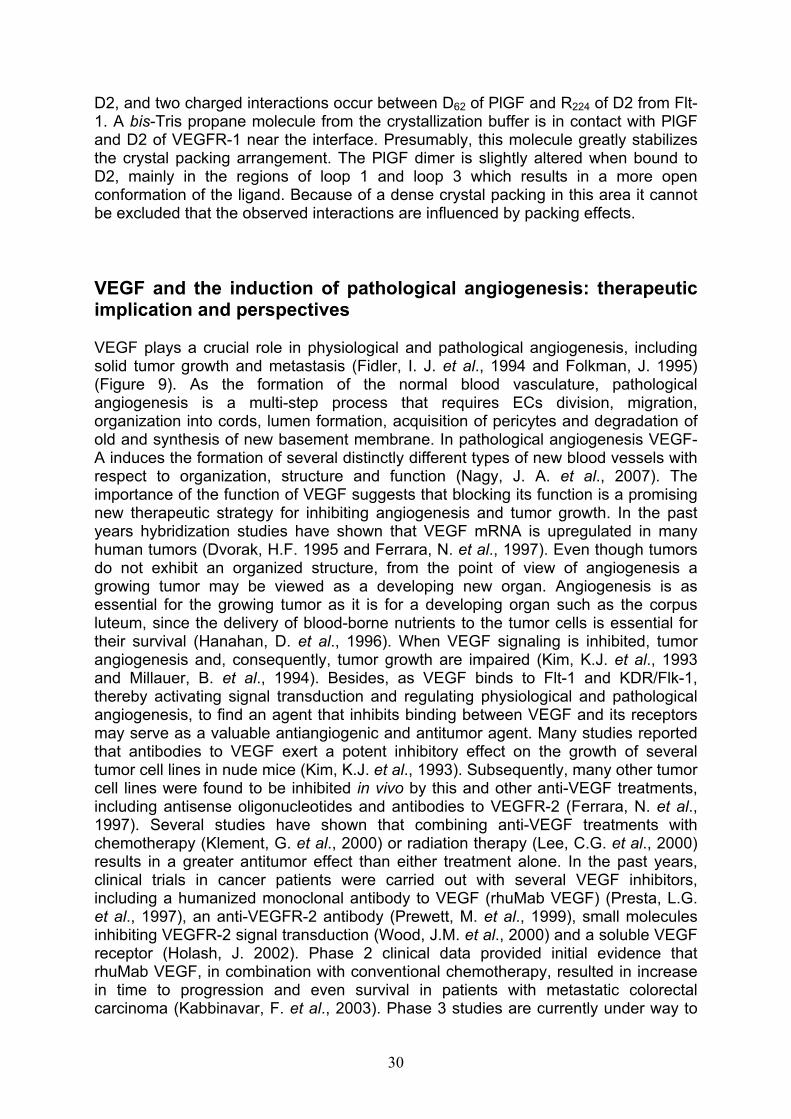
VEGF and the induction of pathological angiogenesis: therapeutic implications and perspectives
pag. 30
De novo engineered VEGF mimicking peptides pag. 31
The aim of the work pag. 32
MATERIALS AND METHODS pag. 34
Strains, enzymes and reagents pag. 34
Antibiotics pag. 34
E. coli cells transformation techniques pag. 34
Preparation of E. coli TOPF’10 cells and transformation by electroporation pag. 34
Preparation of E. coli BL21 (DE3) competent cells and transformation by heat shock
pag. 35
Proteins analyses pag. 35
Determination of the protein concentration pag. 35
Electophoretic analysis of proteins (SDS-PAGE) pag. 35
Bioinformatic tools pag. 36
Domains cloning of human KDR and Flt-1 receptors pag. 36
Cloning of 1-3, 2-3, 1-2 and 2 KDR receptor domains pag. 36
kdrD2 gene mutagenesis pag. 38
Cloning of 1-4, 2-4, 2-3 and 2 Flt-1 receptor domains pag. 40
Expression and purification of recombinant KDR and Flt-1 extracellular domains
pag. 41
Expression pag. 41
Batch purification of 6xHis-tagged proteins under denaturing conditions pag. 41
Expression and refolding of 15N-labeled recombinant Flt-1D2 for NMR studies
pag. 42
Western blot analysis pag. 42
TEV digestion of 6xHis-tagged proteins pag. 43
Flt-1D2 alkylation by iodoacetamide and trypsin digestion pag. 43
Flt-1D2 CD analysis pag. 44
Flt-1D2-VEGF binding competition assay on ECs by Fluorescence activated cell sorting (FACS)
pag. 44
NMR Spectroscopy pag. 45
Preparation of NMR samples pag. 45
Peptide-protein interactions pag. 45
Chemical Shift Mapping pag. 45
Saturation Transfer Difference pag. 45
RESULTS pag. 46
Domains cloning of human KDR and Flt-1 receptors pag. 46
Cloning of 1-3, 2-3, 1-2 and 2 KDR receptor domains pag. 46
Cloning of 1-4, 2-4, 2-3 and 2 Flt-1 receptor domains pag. 48
Expression and purification of recombinant human KDR and Flt-1 extracellular domains
pag. 50
Expression of 1-3, 2-3, 1-2 and 2 KDR receptor domains pag. 50
kdrD2 gene mutagenesis pag. 54
Expression of 1-4, 2-4 and 2-3 Flt-1 receptor domains pag. 55
Expression, refolding and purification of 2 Ig-like Flt-1 receptor domain cloned into the pETM11 expression vector
pag. 57
TEV digestion and native-PAGE analysis of Flt-1D2 pag. 58
LC-MS analyses of Flt-1D2 after TEV cleavage, alkylation and trypsin digestion
pag. 60
Flt-1D2 VEGF-binding competition assay by Fluorescence Activated Cell Sorting (FACS)
pag. 64
Expression, refolding and purification of 15N-Flt-1D2 pag. 65
CD spectroscopic analysis pag. 65
Proteins secondary structure content pag. 66
NMR Characterization of Flt-1D2 pag. 69
NMR analysis of peptide-protein interaction pag. 71
Identification of the binding site on Flt-1D2 via Chemical Shift Mapping pag. 71
Epitope mapping of QK and MA peptides bound to Flt-1D2 via SaturationTranfer Difference
pag. 74
DISCUSSION pag. 76
REFERENCES pag. 80
PUBLICATIONS and COMUNICATIONS pag. 90

Abbreviations
AMBIC ammonium bicarbonate
Ang angiopoietins
APS ammonium persulfate
BM basement membrane
bp base pair
BSA albumin from bovine serum
CD circular dichroism
dam DNA adenine methylase
D2, D1-2, D2-3, D1-3, D1-4, D2-4
domain 2, domain 1-2, domain 2-3, domain 1-3, domain 1-4, domain 2-4 of VEGFRs
DNA deoxyribonucleic acid
dNTP deoxy nucleotide tri-phosphate
DTT dithiothreitol
EBM endothelial basal medium
E. coli Escherichia coli
EC endothelial cell
ECL enhanced chemi-luminescence
ECM extracellular matrix
EDTA ethylene-diamino-tetraacetic acid
EGF epithelial growth factor
EGM endothelial growth medium
ESI electron spry ionization source
FACS fluorescence activated cell sorting
FCS fetal calf serum
FDA Food and Drug Administration
FGF fibroblast growth factor
FGFR fibroblast growth factor receptor
FID free induction delay
FITC fluorescein isothiocyanate
Flt-1 fms-like tyrosine kinase-1
F.T. flow-through
gor glutathione reductase gene
1

GTE Glucose-Tris-EDTA
h hour
HF hypotensive factor
HIF-1 hypoxia-inducible factor-1
HPLC high performance liquid chromatography
HSC haematopoietic stem cells
HSPG heparan sulfate proteoglycan
HSQC heteronuclear single quantum coherence
HUVEC Human umbilical vein endothelial cell
Ig immunoglobulin
Kd dissociation constant
IPTG isopropyl-beta-D-thiogalactopyranoside
KDa Kilo Dalton
KDR kinase domain region
LB Luria-Bertani Broth (10 g/L bacto-triptone, 5 g/L yeast exstract, 10 g/L NaCl)
LC-MS liquid chromatography mass spectrometry
MCS multi cloning site
MAPK mitogen-activated protein kinase
min minute
MMP metalloproteinase
ms millisecond
MWCO molecular weight cut off
NIH-3T3 Mouse embryonic fibroblast cell line
nm nanometer
NMR Nuclear Magnetic Resonance
NOESY Nuclear Overhauser Effect SpectroscopY
NRP neuropilin
NRTK non receptor tyrosine kinase
Ni-NTA nickel-nitrilotriacetic acid
O.D. optical density
orf open reading frame
PAI-1 plasminogen activator inhibitor 1
PBS phosphate buffer saline
2

PC pericyte
PCR polymerase chain reaction
PDGF platelet derived growth factor
PDGFR platelet derived growth factor receptor
pI isoelectric point
PlGF placenta growth factor
PVDF polyvinylidene fluoride
RT room temperature
RTK receptor tyrosine kinase
SDS-PAGE sodium dodecyl sulfate polyacrylamide gel electrophoresis
sec second
SMC smooth muscle cells
STD saturation transfer difference
svVEGF snake venom vascular endothelial growth factor
TAE Tris acetate EDTA
TE 10 mM Tris/HCl pH 8, 1 mM EDTA pH 8
TEMED N,N,N’,N’-tetramethyl ethylene diamine
TEV tobacco etch virus
TFA trifluoroacetic acid
TIC total ion current
TK tyrosine kinase
TM trans-membrane domain
TnI troponin I
Tris Tris (hidroxy methyl) amino methane
trxB thioredoxin reductase gene
TrxA thioredoxin A
TSP-1 thrombospondin-1
U units
uPA urokinase-type plasminogen activator
UV ultra violet
VEGF vascular endothelial growth factor
VEGFR VEGF receptor
VPF vascular permeability factor
The one letter code is used for amino acids.
3
Summary
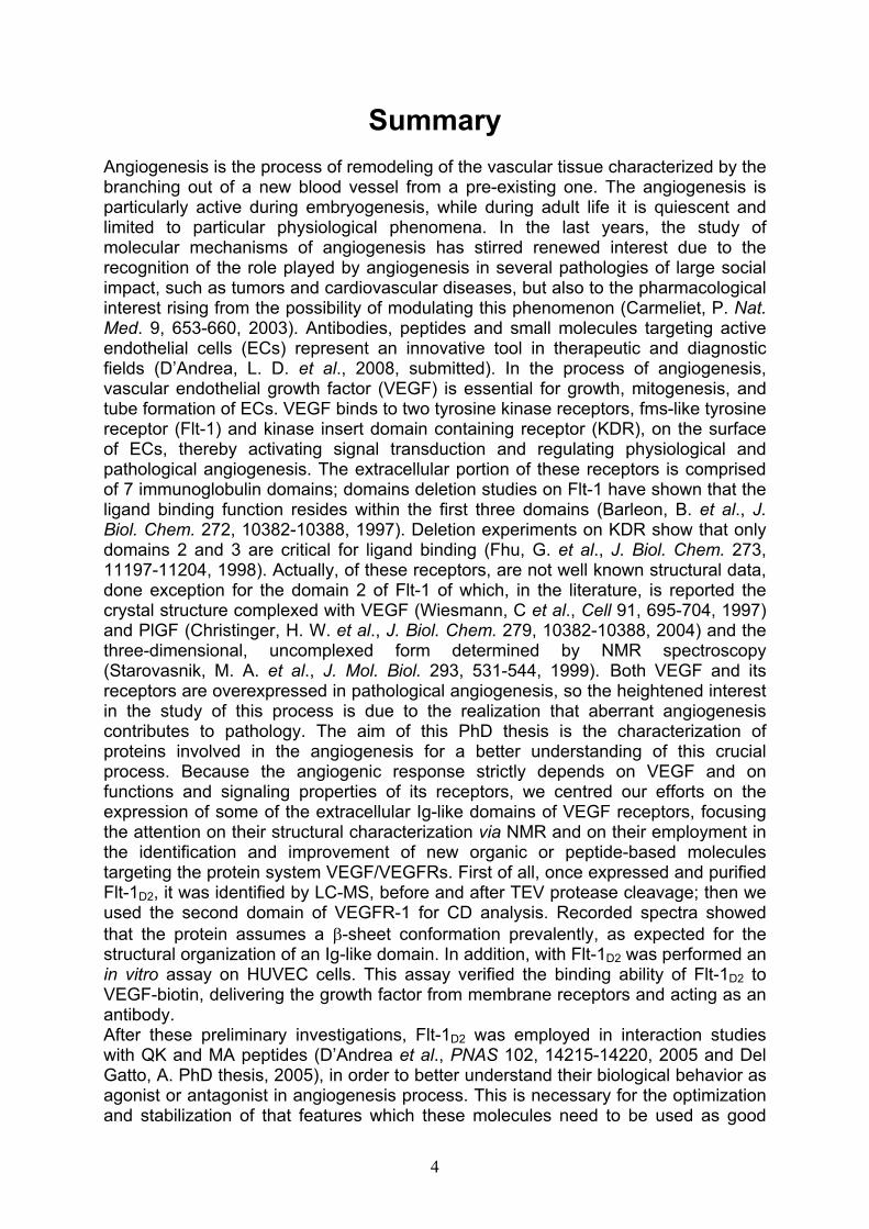
Angiogenesis is the process of remodeling of the vascular tissue characterized by the branching out of a new blood vessel from a pre-existing one. The angiogenesis is particularly active during embryogenesis, while during adult life it is quiescent and limited to particular physiological phenomena. In the last years, the study of molecular mechanisms of angiogenesis has stirred renewed interest due to the recognition of the role played by angiogenesis in several pathologies of large social impact, such as tumors and cardiovascular diseases, but also to the pharmacological interest rising from the possibility of modulating this phenomenon (Carmeliet, P. Nat.Med. 9, 653-660, 2003). Antibodies, peptides and small molecules targeting active endothelial cells (ECs) represent an innovative tool in therapeutic and diagnostic fields (D’Andrea, L. D. et al., 2008, submitted). In the process of angiogenesis, vascular endothelial growth factor (VEGF) is essential for growth, mitogenesis, and tube formation of ECs. VEGF binds to two tyrosine kinase receptors, fms-like tyrosine receptor (Flt-1) and kinase insert domain containing receptor (KDR), on the surface of ECs, thereby activating signal transduction and regulating physiological and pathological angiogenesis. The extracellular portion of these receptors is comprised of 7 immunoglobulin domains; domains deletion studies on Flt-1 have shown that the ligand binding function resides within the first three domains (Barleon, B. et al., J.Biol. Chem. 272, 10382-10388, 1997). Deletion experiments on KDR show that only domains 2 and 3 are critical for ligand binding (Fhu, G. et al., J. Biol. Chem. 273, 11197-11204, 1998). Actually, of these receptors, are not well known structural data, done exception for the domain 2 of Flt-1 of which, in the literature, is reported the crystal structure complexed with VEGF (Wiesmann, C et al., Cell 91, 695-704, 1997) and PlGF (Christinger, H. W. et al., J. Biol. Chem. 279, 10382-10388, 2004) and the three-dimensional, uncomplexed form determined by NMR spectroscopy (Starovasnik, M. A. et al., J. Mol. Biol. 293, 531-544, 1999). Both VEGF and its receptors are overexpressed in pathological angiogenesis, so the heightened interest in the study of this process is due to the realization that aberrant angiogenesis contributes to pathology. The aim of this PhD thesis is the characterization of proteins involved in the angiogenesis for a better understanding of this crucial process. Because the angiogenic response strictly depends on VEGF and on functions and signaling properties of its receptors, we centred our efforts on the expression of some of the extracellular Ig-like domains of VEGF receptors, focusing the attention on their structural characterization via NMR and on their employment in the identification and improvement of new organic or peptide-based molecules targeting the protein system VEGF/VEGFRs. First of all, once expressed and purified Flt-1D2, it was identified by LC-MS, before and after TEV protease cleavage; then we used the second domain of VEGFR-1 for CD analysis. Recorded spectra showed that the protein assumes a -sheet conformation prevalently, as expected for the structural organization of an Ig-like domain. In addition, with Flt-1D2 was performed an in vitro assay on HUVEC cells. This assay verified the binding ability of Flt-1D2 to VEGF-biotin, delivering the growth factor from membrane receptors and acting as an antibody.After these preliminary investigations, Flt-1D2 was employed in interaction studies with QK and MA peptides (D’Andrea et al., PNAS 102, 14215-14220, 2005 and Del Gatto, A. PhD thesis, 2005), in order to better understand their biological behavior as agonist or antagonist in angiogenesis process. This is necessary for the optimization and stabilization of that features which these molecules need to be used as good
4

modulators in angiogenesis. The interaction between peptides and Flt-1D2 has been studied through NMR techniques founded on the observation of ligand (Saturation Tranfer Difference) and protein (Chemical Shift Mapping). These experiments showed that QK is able to bind on Flt-1 receptor the same region tied by VEGF. Therefore, the recombinant Flt-1D2 was employed for a NMR screening of a library of small molecules, in order to find other VEGF agonist/antagonist and design new molecules more specific and selective in their targeting VEGFRs system. These are preliminary data.
5

6
Riassunto
Scopo di questo progetto di dottorato è stata l’espressione in forma ricombinante e la caratterizzazione di proteine coinvolte nel processo di angiogenesi, al fine di impiegarle nello screening di molecole in grado di fungere da modulatori della crescita di nuovi vasi sanguigni. L’angiogenesi, infatti, è un processo di rimodellamento del tessuto vascolare caratterizzato dalla formazione di nuovi vasi sanguigni a partire da strutture preesistenti. La formazione di un nuovo vaso sanguigno, così come quella di ogni tessuto, è garantita dall’ interazione dinamica tra le cellule che lo costituiscono ed il loro microambiente che risulta essere sostanzialmente costituito dalla matrice extracellulare e da altri citotipi con i quali la popolazione cellulare interagisce. La matrice extracellulare è composta da proteine fibrose e da proteoglicani, ma in essa sono presenti anche citochine e fattori di crescita, generalmente associati a componenti della matrice stessa. L’angiogenesi è regolata prevalentemente da uno di questi fattori di crescita, il VEGF (VascularEndothelial Growth Factor), potente fattore angiogenico e mitogeno specifico per le cellule endoteliali che, insieme alla matrice extracellulare dell’endotelio vascolare, alla membrana basale, alle cellule muscolari lisce e a quelle di supporto, concorrono alla formazione dei canali che dirigono e contengono il flusso sanguigno. La funzione biologica del VEGF si esplica attraverso il suo legame a due recettori di membrana con attività tirosin-chinasica: il Kinase Domain Receptor (KDR) e l’Fms-like tyrosin kinase (Flt-1), entrambi presenti sulla superficie di diversi tipi di cellule endoteliali, ma in grado di attivare vie di trasduzione distinte, nonostante condividano un elevato grado di omologia (Figura R1) (Ferrara, N et al., Endocr. Rev. 18, 4-25, 1997 and Ferrara, N. Curr. Opin. Biotechnol. 11, 617-624, 2000).
Figura R1_ Rappresentazione delle interazioni del VEGF e dei suoianaloghi con alcuni recettori espressi sulla superficie delle celluleendoteliali.

Per quanto riguarda la porzione extracellulare di queste proteine, attualmente, non sono noti dati strutturali, fatta eccezione per il dominio 2 di Flt-1 di cui, in letteratura, è riportata la struttura cristallografica complessata al VEGF (Figura R2) e al PlGF (Wiesmann, C. et al., Cell 91, 695-704, 1997 and Christinger, H. W. et al., J. Biol. Chem. 279, 10382-10388, 2004) e quella libera in soluzione (Starovasnik, M. A. etal., J. Mol. Biol. 293, 531-544, 1999).
Figura R2_ Dominio 2 del recettore Flt-1 (in arancione) complessato alVEGF8-109 (in rosso e verde). In blu sono rappresentati i residui del VEGF acontatto con il recettore.
Studi di delezione hanno dimostrato che i maggiori responsabili del riconoscimento specifico per il VEGF sono i primi 3 domini extracellulari per il recettore Flt-1 (Barleon, B. et al., J. Biol. Chem. 272, 10382-10388, 1997) e i domini 2 e 3 per KDR (Fhu, G. et al., J. Biol. Chem. 273, 11197-11204, 1998). Affinché l’angiogenesi possa svolgersi correttamente, le interazioni che hanno luogo tra le cellule endoteliali e la matrice extracellulare debbono svolgersi secondo una sequenza precisa ed ordinata. In più, l’angiogenesi, come la maggior parte dei processi biologici, è il risultato di un equilibrio tra fattori “pro-angiogenici” ed “anti-angiogenici”. Lo spostamento da questa situazione di equilibrio (noto come switch angiogenico) è alla base di manifestazioni patologiche di varia natura quali tumori ed ischemie (Carmeliet, P. Nat. Med. 9, 653-660, 2003). Alla luce di queste conoscenze, lo studio del sistema costituito dal VEGF e dai suoi recettori risulta, pertanto, di notevole interesse. La possibilità di disporre di nuovi composti capaci di modulare la risposta angiogenica del VEGF avrebbe così numerose applicazioni in campo terapeutico e diagnostico. Difatti, negli ultimi anni lo studio dei meccanismi dell’angiogenesi è stato uno tra i campi maggiormente investigati e finanziati nell’ambito della ricerca medica. Al momento, tra gli inibitori del processo approvati come agenti terapeutici si annoverano l’Avastin, un anticorpo monoclonale contro il VEGF, usato per il trattamento del tumore del colon-retto, ed un suo derivato, il Lucentis, in uso per patologie oculari. Sono invece pochi i farmaci disponibili che stimolano l’angiogenesi VEGF-dipendente. Attualmente un solo composto pro-angiogenico è stato approvato, il Regranex (PDGF umano
7

ricombinante), per il trattamento delle ulcere del piede diabetico. L’utilizzo farmacologico di fattori di crescita come il VEGF è stato finora precluso a causa di numerosi effetti collaterali (Carmeliet, P. Nat. Med. 6, 1102-1103, 2000b; Lee, C. G. et al., Nat. Med. 10, 1095-1103, 2004; Weis, S. M. et al., Nature 437, 497-504, 2005). Recentemente è stata riportata in letteratura la caratterizzazione strutturale e le proprietà biologiche di un peptide sintetico che mima il VEGF. Il peptide, QK, modellato sulla regione 17-25 dell’elica del VEGF, lega i recettori del VEGF, attivando poi il processo di proliferazione cellulare VEGF-dipendente. Tale peptide potenzia la risposta biologica del VEGF, e promuove la formazione di capillari in vitroed in vivo (D’Andrea, L. D. et al., PNAS 102, 14215-14220, 2005). Dunque, si rende necessario lo sviluppo di inibitori di basso peso molecolare e di nuovi agonisti in modo da modulare la crescita di nuovi vasi sanguigni e, in tal modo, la progressione di patologie derivanti da un alterato funzionamento del processo angiogenico. Nel caso dello sviluppo di un tumore, occorre considerare che l’angiogenesi e la formazione di una metastasi costituiscono due esempi paradigmatici del ruolo cruciale svolto dal microambiente all’interno del quale questi due processi si svolgono, nella dinamica spazio-temporale del loro accadere. Il microambiente diventa allora anche un potenziale bersaglio terapeutico per mantenere l’angiogenesi in un ambito fisiologico o per deregolare il potenziale metastatico di una popolazione cellulare. In tal senso sono tuttora allo studio molecole in grado di bloccare l’interazione tra i recettori e i fattori di crescita angiogenici, impedendo così a questi ultimi di interagire con le cellule endoteliali che esprimono sulla loro superficie i recettori. Tale progetto di dottorato si inserisce dunque in questo ampio discorso ed ha come obiettivo la caratterizzazione di proteine coinvolte nell’angiogenesi, in particolare, dei domini extracellulari dei recettori KDR e Flt-1. La loro espressione in forma ricombinante ed il loro impiego in studi condotti mediante analisi NMR, offrirebbe, infatti, la possibilità di uno screening di piccoli peptidi o di collezioni di molecole organiche al fine di trovare nuovi ligandi per i recettori e quindi probabili modulatori dell’angiogenesi, in qualità di antagonisti e/o agonisti del processo. La mia attività scientifica nell’ambito di questo progetto si è focalizzata, pertanto, sul clonaggio e l’espressione di alcuni dei domini extracellulari dei recettori del VEGF Flt-1 e KDR. A tal fine sono stati utilizzati diversi vettori per il clonaggio delle sequenze geniche di interesse e diversi ceppi batterici per l’ottimizzazione dei livelli di espressione delle corrispondenti proteine ricombinanti. l sistemi di espressione scelti, sfruttando la presenza di un promotore forte ed inducibile qual è quello regolato dalla RNA polimerasi T7, sono stati in grado di fornire alti livelli di espressione delle proteine di interesse; essi recano marcatori di selezione costituiti da geni che conferiscono resistenza ad un antibiotico (Canamicina o Ampicillina), presentano un’origine di replicazione riconosciuta dall’ospite batterico ed un sito di policlonaggio. La maggior parte dei suddetti sistemi consente, inoltre, di esprimere le proteine di interesse con una coda di poli-istidine all’N-terminale della catena polipeptidica, permettendone una più efficace ed immediata purificazione dai contaminanti di E.coli mediante cromatografia di affinità. Le sequenze geniche corrispondenti ai domini extracellulari di Flt-1 e KDR (flt-1D1-4, flt-1D2-4, flt-1D2-3 e flt-1D2; kdrD1-3, kdrD2-3, kdrD1-2
e kdrD2) sono state amplificate tramite PCR, utilizzando come stampo il cDNA degli interi recettori e come iniziatori della reazione di amplificazione a catena della polimerasi coppie di oligonucleotidi disegnate sulla base delle sequenze geniche in esame. Il DNA dei costrutti ricombinanti è stato opportunamente purificato e verificato tramite sequenziamento. I clonaggi di tutte le sequenze geniche amplificate sono riusciti con successo ed i costrutti ottenuti nei diversi sistemi di espressione
8

sono stati utilizzati per l’espressione nei ceppi di E. coli scelti per tale scopo. Tutti i costrutti espressi nei diversi ceppi batterici hanno prodotto proteine insolubili, il cui processo di rinaturazione, dopo estrazione in presenza di agenti denaturanti quali urea 8M o Gu-HCl 6M, non sempre è stato portato a termine con successo o ha permesso di ottenere quantità di proteina non sufficienti per essere impiegate in studi di caratterizzazione. Il nostro interesse si è pertanto focalizzato maggiormente sul costrutto pETM11-flt-1D2, che siamo riusciti ad esprimere a buoni livelli nel ceppo BL21 Codon Plus (DE3) RIL di E. coli (60 mg/L) e a rinaturare con rese soddisfacenti. In seguito a trasformazione chimica nel suddetto ceppo batterico, i cloni selezionati su piastra sono stati propagati in terreno liquido. La proteina di interesse è stata espressa in forma insolubile, sia a 22°C che a 37°C, pertanto è stata purificata in condizioni denaturanti mediante cromatografia di affinità su resina funzionalizzata con ioni Nickel (Ni2+-NTA resin) e sottoposta, successivamente, ad un protocollo di refolding su colonna, in modo tale da sottrarle gradualmente l’agente denaturante (urea) utilizzato per la sua estrazione dai corpi di inclusione. Terminato il refolding, la proteina Flt-1D2 è stata eluita con alte concentrazioni di imidazolo, dializzata e, mediante taglio con la proteasi TEV, è stata allontanata dalla sua estremità N-terminale la coda di poli-istidine con cui è stata espressa. La proteina è stata purificata all’omogeneità, mediante un ulteriore step su resina Ni2+-NTA per allontanare da essa sia la TEV che l’HIS-TAG e sottoposta ad un’ampia caratterizzazione. Flt-1D2 è stata infatti identificata mediante spettometria di massa (LC-MS) prima e dopo il taglio con la proteasi; è stata trattata con iodoacetammide (prima e dopo riduzione con DTT) per stabilire l’eventuale alchilazione a carico delle due cisteine presenti nella sequenza amminoacidica del dominio proteico; è stata infine sottoposta ad una proteolisi limitata con tripsina. I dati ottenuti attraverso queste prove ci hanno fornito informazioni utili, seppure indirette, riguardo lo stato assunto dalla proteina in seguito al refolding su colonna. Infatti, il trattamento con iodoacetammide ha lasciato invariata la massa di Flt-1D2 non ridotta, mentre ha alchilato la proteina precedentemente trattata con DTT. Ciò ci ha dato conferma del fatto che le due cisteine presenti nella sequenza di Flt-1D2 sono impegnate in un ponte disolfurico, così come confermato da dati strutturali (Wiesmann, C. et al., Cell91, 695-704, 1997). In più, la reazione di proteolisi limitata, esaminata alla massa a tempi diversi (dopo 45 e dopo 90 minuti) ha mostrato una struttura abbastanza compatta, non intaccata drasticamente dalla tripsina, così come atteso per un dominio immunoglobulinico quale è il dominio 2 del recettore Flt-1. Di Flt-1D2 sono state effettuate analisi mediante dicroismo circolare e gli spettri acquisiti hanno mostrato che la proteina assume una conformazione prevalentemente di tipo -sheet, così come atteso per l’organizzazione strutturale di un dominio di tipo immunoglobulinico. La temperatura di denaturazione di Flt-1D2 è stata stimata intorno ai 60°C ed il processo è risultato reversibile. Parallelamente, il dominio proteico è stato utilizzato in un saggio funzionale di fluorescenza condotto in vitro su cellule HUVEC (Human umbelical vein endothelial cells). Tale saggio ha dimostrato la capacità di legame del VEGF-biotina ai recettori di membrana presenti sulla superficie delle HUVEC e allo stesso tempo la capacità di Flt-1D2 di spiazzare il VEGF da tale interazione. In presenza di un anticorpo anti-VEGF, l’intensità della fluorescenza registrata inviando sulle HUVEC il solo VEGF-biotina è diminuita significativamente. Un decremento analogo si è osservato anche quando le cellule sono state incubate con VEGF-biotina pretrattato con Flt-1D2, indicando che il recettore è in grado di legare il VEGF e di competere per tale legame con i recettori di membrana, comportandosi alla stregua di un anticorpo. In seguito all’ottenimento
9

di questi dati preliminari, si è passati alla caratterizzazione vera e propria della proteina che, per tale scopo, è stata marcata con 15N, mediante espressione in terreno minimo contenente, come unica fonte di azoto, il cloruro di ammonio marcato (15NH4Cl). Flt-1D2 è stata espressa e purificata secondo lo stesso protocollo utilizzato per la proteina non marcata, quindi è stata concentrata (0.3 mM) e sottoposta a studi di caratterizzazione mediante NMR. L’acquisizione degli spettri protonici mono- e bidimensionali di Flt-1D2 ha messo in evidenza la presenza di numerosi picchi relativi ai protoni ammidici, con una buona dispersione dei chemical shifts, così come atteso per la proteina nativa (Starovasnik, M. A. et al., J. Mol. Biol. 293, 531-544, 1999). E’ stato pertanto possibile asserire che il dominio proteico ottenuto in forma ricombinante e purificato all’omogeneità dopo refolding ha una conformazione correttamente ripiegata. Successivamente, la proteina è stata impiegata in studi di interazione con due piccoli peptidi di quindici amminoacidi sintetizzati e caratterizzati nel laboratorio in cui è stato svolto questo lavoro di tesi; i peptidi in esame sono QK (Ac-KLTWQELYQLKYKGI-NH2) ed MA (Ac-KLTWMELYQLAYKGI-NH2) (D’Andrea, L. D. et al., PNAS 102, 14215-14220, 2005 and Del Gatto, A. PhD thesis 2005). Entrambi rappresentano un esempio di quello che viene definito approccio di sintesi mediante rational-design e riproducono l’elica 17-25 del VEGF; infatti, l’analisi accurata dei dati strutturali, così come gli studi di mutagenesi, hanno permesso di identificare i residui amminoacidici coinvolti nel legame del VEGF ai suoi recettori. Essi sono distribuiti su una superficie discontinua costituita appunto dall’elica 17-25 presente all’estremità N-terminale di ciascun monomero del VEGF (Keyt, B. A. et al.,J. Biol. Chem. 271, 5638-5646, 1996; Muller, Y. A. et al., Proc. Natl. Acad. Sci. USA94, 7192-7197, 1997; Wiesmann, C. et al., Cell 91, 695-704, 1997). Attualmente, la stragrande maggioranza delle molecole peptidiche in grado di modulare l’interazione tra il VEGF ed i suoi recettori deriva da screening di collezioni fagiche, mentre sono pochi gli esempi derivanti da un approccio di tipo razionale. L’interazione dei due peptidi con il secondo dominio del recettore Flt-1 è stata studiata attraverso tecniche NMR basate sull’osservazione del ligando (Saturation Transfer Difference) e della proteina (Chemical Shift Mapping). Tale interazione ha permesso di identificare, sia per QK che per MA, le regioni impegnate nel binding con la proteina, dimostrando che il peptide QK lega sul recettore la stessa regione di legame del VEGF. Flt1D2 è stata infine impiegata in uno studio di drug discovery, attraverso lo screening di una collezione di piccole molecole organiche con l’intento di selezionare nuovi ligandi a basso peso molecolare capaci di bloccare l’interazione tra il VEGF ed i suoi recettori. I dati ottenuti sono del tutto preliminari.
10

Introduction
Angiogenesis is a phenomenon intimately associated with endothelial cells (ECs) migration and proliferation. During embryonic development, ECs rapidly proliferate, thereby forming new blood vessels. In adult life, however, ECs turnover is very low, except for physiological wound healing and female reproductive functions. Angiogenesis is also implicated in pathological conditions associated with tumors, intraocular neovascular disorders, chronic ischemia and other diseases (Carmeliet, P. 2003). The mediators of this process have been identified in a series of vascular growth factors such as the Vascular Endothelial Growth Factor (VEGF). VEGF represents a key regulator of angiogenesis; its biological effects are prevalently mediated by two receptor tyrosine kinases (RTK): VEGFR-1 and VEGFR-2 which are localized on the cell surface of various endothelial cell types. VEGF and its receptors are over-expressed in pathological angiogenesis, making this protein system a target for therapeutic and diagnostic applications (Ferrara, N et al., 1997 and Ferrara, N. 2000). Therapeutic angiogenesis is sought as the ultimate intervention to solve chronic ischemia in those conditions that cannot be treated alternatively; its converse, the anti-angiogenic treatment, with the blockage of the VEGF/VEGFRs pathway, is a promising therapy in oncology. To date, a number of different strategies to inhibit VEGF signal transduction are available and they include the use of humanized neutralizing anti-VEGF monoclonal antibodies, receptor antagonists, soluble receptors and inhibitors of VEGF receptors function (Moreira, I. S. et al.,2007). Moreover, new molecular entities as peptides have been reported to bind to the extracellular region of the VEGF receptors. A large number of them show an antagonist activity and only few behave as agonists (D’Andrea, L. D. et al., 2008, submitted).The introduction is not meant to cover the VEGF/VEGFR field completely, but rather to emphasize selected aspects of these molecules which are important for the aims described in this PhD thesis.
11
How does a blood vessel form? Vasculogenesis and Angiogenesis
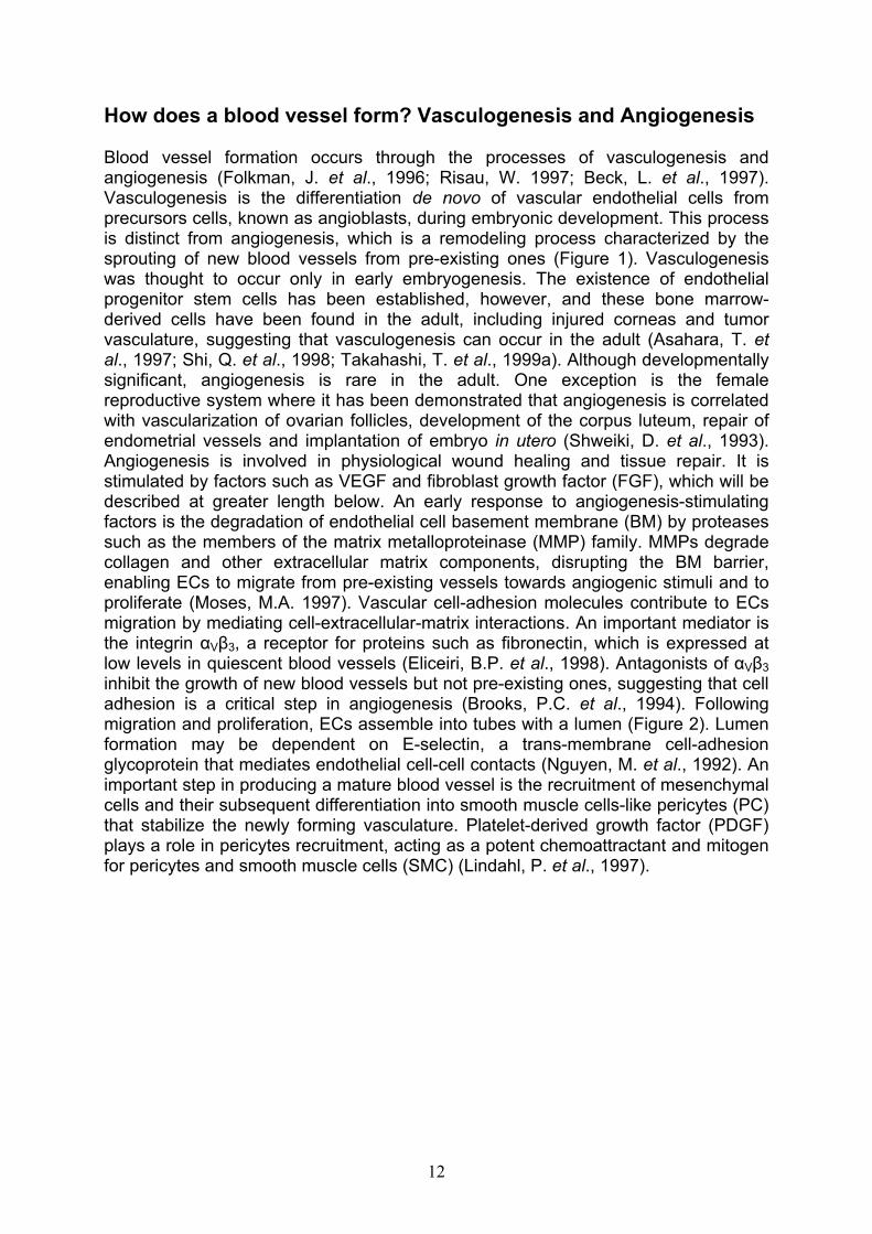
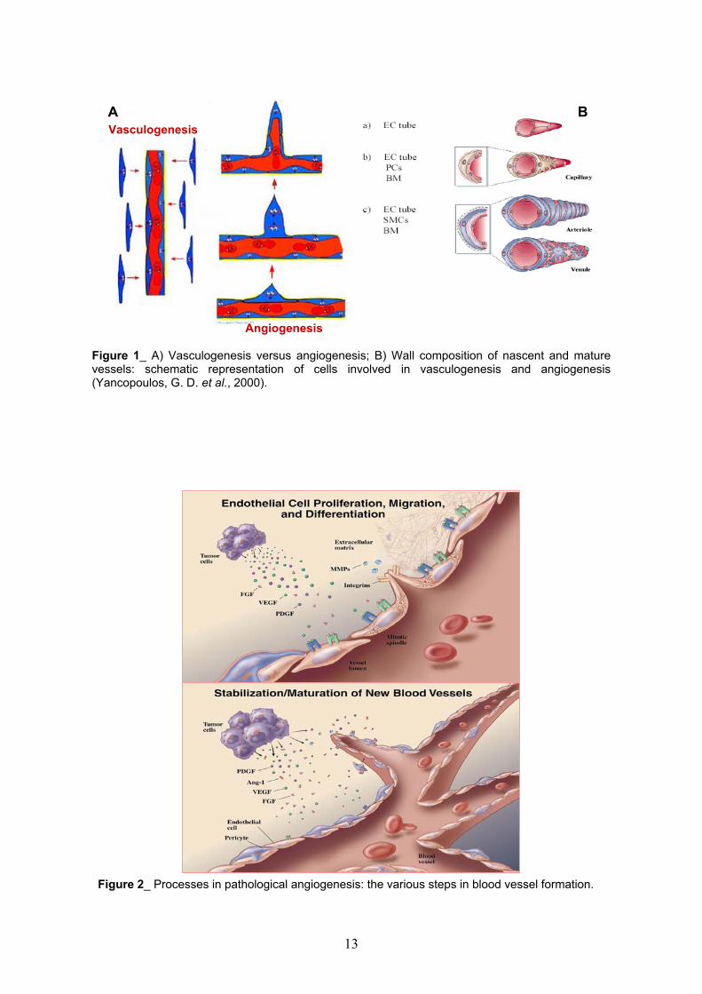
Blood vessel formation occurs through the processes of vasculogenesis and angiogenesis (Folkman, J. et al., 1996; Risau, W. 1997; Beck, L. et al., 1997). Vasculogenesis is the differentiation de novo of vascular endothelial cells from precursors cells, known as angioblasts, during embryonic development. This process is distinct from angiogenesis, which is a remodeling process characterized by the sprouting of new blood vessels from pre-existing ones (Figure 1). Vasculogenesis was thought to occur only in early embryogenesis. The existence of endothelial progenitor stem cells has been established, however, and these bone marrow-derived cells have been found in the adult, including injured corneas and tumor vasculature, suggesting that vasculogenesis can occur in the adult (Asahara, T. etal., 1997; Shi, Q. et al., 1998; Takahashi, T. et al., 1999a). Although developmentally significant, angiogenesis is rare in the adult. One exception is the female reproductive system where it has been demonstrated that angiogenesis is correlated with vascularization of ovarian follicles, development of the corpus luteum, repair of endometrial vessels and implantation of embryo in utero (Shweiki, D. et al., 1993). Angiogenesis is involved in physiological wound healing and tissue repair. It is stimulated by factors such as VEGF and fibroblast growth factor (FGF), which will be described at greater length below. An early response to angiogenesis-stimulating factors is the degradation of endothelial cell basement membrane (BM) by proteases such as the members of the matrix metalloproteinase (MMP) family. MMPs degrade collagen and other extracellular matrix components, disrupting the BM barrier, enabling ECs to migrate from pre-existing vessels towards angiogenic stimuli and to proliferate (Moses, M.A. 1997). Vascular cell-adhesion molecules contribute to ECs migration by mediating cell-extracellular-matrix interactions. An important mediator is the integrin V 3, a receptor for proteins such as fibronectin, which is expressed at low levels in quiescent blood vessels (Eliceiri, B.P. et al., 1998). Antagonists of V 3
inhibit the growth of new blood vessels but not pre-existing ones, suggesting that cell adhesion is a critical step in angiogenesis (Brooks, P.C. et al., 1994). Following migration and proliferation, ECs assemble into tubes with a lumen (Figure 2). Lumen formation may be dependent on E-selectin, a trans-membrane cell-adhesion glycoprotein that mediates endothelial cell-cell contacts (Nguyen, M. et al., 1992). An important step in producing a mature blood vessel is the recruitment of mesenchymal cells and their subsequent differentiation into smooth muscle cells-like pericytes (PC) that stabilize the newly forming vasculature. Platelet-derived growth factor (PDGF) plays a role in pericytes recruitment, acting as a potent chemoattractant and mitogen for pericytes and smooth muscle cells (SMC) (Lindahl, P. et al., 1997).
12
A B
Vasculogenesis
Angiogenesis
Figure 1_ A) Vasculogenesis versus angiogenesis; B) Wall composition of nascent and maturevessels: schematic representation of cells involved in vasculogenesis and angiogenesis(Yancopoulos, G. D. et al., 2000).
Figure 2_ Processes in pathological angiogenesis: the various steps in blood vessel formation.
13
Promoters and inhibitors of angiogenesis
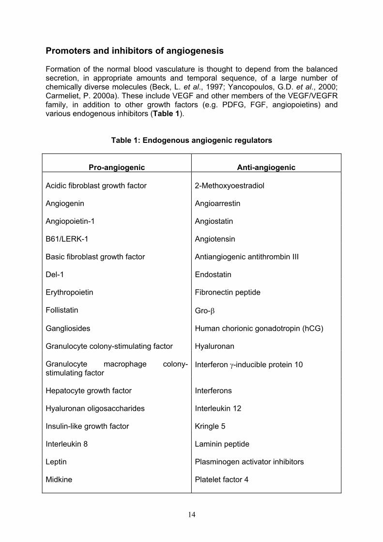
Formation of the normal blood vasculature is thought to depend from the balanced secretion, in appropriate amounts and temporal sequence, of a large number of chemically diverse molecules (Beck, L. et al., 1997; Yancopoulos, G.D. et al., 2000; Carmeliet, P. 2000a). These include VEGF and other members of the VEGF/VEGFR family, in addition to other growth factors (e.g. PDFG, FGF, angiopoietins) and various endogenous inhibitors (Table 1).
Table 1: Endogenous angiogenic regulators
Pro-angiogenic Anti-angiogenic
Acidic fibroblast growth factor 2-Methoxyoestradiol
Angiogenin Angioarrestin
Angiopoietin-1 Angiostatin
B61/LERK-1 Angiotensin
Basic fibroblast growth factor Antiangiogenic antithrombin III
Del-1 Endostatin
Erythropoietin Fibronectin peptide
Follistatin Gro-
Gangliosides Human chorionic gonadotropin (hCG)
Granulocyte colony-stimulating factor Hyaluronan
Granulocyte macrophage colony-stimulating factor
Interferon -inducible protein 10
Hepatocyte growth factor Interferons
Hyaluronan oligosaccharides Interleukin 12
Insulin-like growth factor Kringle 5
Interleukin 8 Laminin peptide
Leptin Plasminogen activator inhibitors
Midkine Platelet factor 4
14
Monocyte chemoattractant protein 1 Prolactin 16kD fragment
Oestrogens Proliferin-related protein
Placenta growth factor Thrombospondin-1
Platelet-derived growth factor Tissue inhibitors of metalloproteinase
Pleiotrophin Transforming growth factor-beta (TGF-b)
Progranulin Tumour necrosis factor (high dose)
Proliferin Tumstatin
Prostaglandin E1, E2 Vasculostatin
Transforming growth factor and Vasostatin (calreticulin fragment)
Tumour necrosis factor (low dose)
Vascular endothelial growth factor
Promoters of angiogenesis
Angiopoietins and Tie receptors
Tie receptors and their ligands, the angiopoietins, play a critical role in embryonic angiogenesis (Partanen, J. et al., 1999 and Davis, S. et al., 1999). Tie receptors, Tie1 and Tie2, are tyrosine kinases expressed by ECs. Mice deficient in Tie2 are embryonic lethal with defects in the proper development of the endothelial lining of the heart. Tie1 is required for the structural integrity of ECs and mice deficient in Tie1 die by birth (Dumont, D.J. et al., 1994 and Sato, T.N. et al., 1995). Angiopoietin-1 (Ang1) is a ligand for Tie2 (Davis, S. et al., 1996). Ang1 induces phosphorylation of Tie2 in ECs, but does not induce cell proliferation. Over-expression of Ang1 results in more numerous and more highly branched vessels. Targeted disruption of Ang1 is embryonic lethal and the defects in these mice resemble those in the Tie2 deficient mice. In addiction to heart defects, there are a reduced number of vessels and remodeling of the initial primary capillary network does not occur. Importantly, there is a lack of recruitment of periendothelial supporting cells to the ECs in Ang1-deficient mice, which leads to lack of blood vessels stabilization and maturation (Suri, C. et al.,1998). Ang1 is expressed embryonically, most prominently in heart myocardium. In adult tissue Ang1 is constitutively and widely expressed especially in platelets, megakaryocytes as well as in highly vascularized tissues, but not in tissues with low vascularization (Thurston, G. et al., 2000). It has been suggested that mesenchymal cells produce Ang1, which activates Tie2 which in turn leads to the production and release of factors (e.g. PDGF-BB) that recruit pericytes and smooth muscle cells
15
(Folkman, J. et al., 1996). Angiopoietin 2 (Ang2) is 60% identical to Ang1, but does not stimulate Tie2 tyrosine phosphorylation. Ang2 might be a natural antagonist of Ang1 that blocks Ang1 activation of Tie2 receptor (Maisonpierre, P.C. et al., 1997). Neither Ang1 nor Ang2 alone could trigger an angiogenic response, but could enhance VEGF-induced angiogenesis, leading to the notion of an angiogenic balance: Ang1 signaling via Tie2 leads to vessel maturation, whereas Ang2 blocks the Ang1/Tie2 signal and leads to angiogenesis or vessel regression and apoptosis, depending on the presence of VEGF (Hanahan, D. 1997 and Asahara, T. et al.,1998).
Platelet derived growth factor (PDGF)
PDGF has been shown to be one of the most potent angiogenesis inducers and is licensed for the treatment of neuropathic diabetic foot ulcers (Bennett, S.P. et al.,2003). The Platelet-derived Growth Factor belongs to a family of structurally and functionally related growth factors containing cysteine knots, which includes also the VEGF (Fredriksson, L. et al., 2004a). The currently known PDGFs include the PDGF-A, PDGF-B, PDGF-C, PDGF-D chains. All members harbour a growth factor core domain, containing a conserved set of cysteine residues, which is necessary and sufficient for receptor binding activation. PDGF-A and PDGF-B contain N-terminal pro-domains, which are removed intracellularly by furin or other convertases; cleavage of these domains result in protein activation and receptor binding (Heldin, C. H. et al., 1999 and Fredriksson, L. et al., 2004a). PDGF-C and PDGF-D instead are processed in the extracellular space (Fredriksson, L. et al., 2004b). PDGFs act through two tyrosine kinase receptors, PDGFR and PDGFR . The receptors are transmembrane proteins, composed of an extracellular ligand-binding domain, a transmembrane domain and a cytoplasmic tyrosine kinase domain (Fantl, W. J. et al.,1993). The extracellular region contains five immunoglobulin-like domains, while the intracellular domain carries a tyrosine kinase domain and a unique sequence with no homology to kinases (Matsui, T. et al., 1989). Upon ligand binding, the PDGF receptors dimerize and phosphorylate each other in trans on specific tyrosine residues (Kelly, J. D. et al., 1991). Although in theory there are several possible combinations of ligand and receptors, the functional in vivo interaction has been proved only for PDGF-AA and PDGF-CC with PDGFR and for PDGF-BB with PDGFR (Andrae, J. et al., 2008) (Figure 3). PDGFR interacts with several other proteins involved in multiple cellular and developmental responses including integrins (Hynes, R.O. 2002) and transcription factors. PDGFs play key roles in embryonal development, being involved in the formation of blood vessels, kidney, lungs, connective tissues and central nervous system. Furthermore, together with other growth factors, PDGF regulate the wound healing process; it has been shown that wounds treated with PDGF exhibit an increase in the re-epithelization and neovascularization rates (Heldin, C. H. et al., 1999 and Sundberg, C. et al., 1997). Overexpression of the PDGFs has been associated with several diseases associated with increased cell proliferation. Experimental evidences show that PDGF is involved in pulmonary fibrosis (Yi, E. S. et al., 1996) and in the formation of cholesterol induced atherosclerosis (Rutherford, C. et al., 1997). PDGF overexpression was also observed in human solid tumors, as gastrointestinal stromal and central nervous system tumors (Heldin, C. H. et al., 1999 and Alvarez, R. H. et al., 2006).
16

Fibroblast growth factor (FGF)
In the 1980s purification of pro-angiogenic proteins led to the identification, and sequencing of the two prototypic heparin-binding angiogenic growth factors FGF1 (acidic FGF) and FGF2 (basic FGF) (Abraham, J. A. et al., 1986a-b). Since then the FGF family has increased in number and complexity as many more members of the family (23 in total) with a variety of biological activities, have been discovered (Powers, C. J. et al., 2000). Acidic and basic fibroblast growth factors (FGF-1 and FGF-2, respectively) are very potent inducers of EC migration, proliferation and tube formation in vitro and are highly angiogenic in vivo (Klagsbrun, M. et al., 1991a). All FGFs share 30–70% homology in their amino acid sequences and consist of two highly conserved core-domains that fold in 12 antiparallel -strands, leading to the formation of a cylindrical barrel (Ago, H. et al., 1991; Eriksson, A. E. et al., 1991; Zhu, X. et al., 1991; Zhang, J. D. et al., 1991). These domains are separated by a central spacer region of variable length as well as variable are the C- and N-terminal regions of the different FGFs (Reuss, B. et al., 2003). Many studies have shown that FGFs can act on different cell types inducing cell proliferation, differentiation, migration and survival (Ornitz, D. M. et al., 2001). All these actions are made possible through the dual interaction of FGFs with high-affinity tyrosine kinase receptors (FGFRs) and with a large number of low affinity sites on the cell surface and within the surrounding extracellular matrix (ECM) (Klagsbrun, T. et al., 1991b). These sites were identified as heparan sulfate proteoglycans (HSPGs) obligatory partners of the FGF interaction with FGFR (Ornitz, D. M. et al., 1992 and Mansukhani, A. et al., 1992). To date four FGF transmembrane receptors (FGFRs1-4) and one soluble receptor (FGFR5) are known (Chaffer, C. L. et al., 2007). Isolation and structural characterization in 1989 of a protein with high affinity for FGF1 (Lee, P. L. et al., 1989) led to the identification of the prototypic of all FGF receptors, which are transmembrane proteins with three extracellular immunoglobulin-like domains (IgI, IgII, and IgIII), an acidic box and heparin-binding domain between IgI and IgII, a hydrophobic transmembrane domain, and an intracellular region containing a split tyrosine kinase domain (Figure 4). In the structure of the FGF receptor gene four possible splice sites exist, generating an FGFR subtype that expresses only two Ig-like domains, and an FGFR subtype that expresses all the three Ig-like domains. Those splice events alter FGF/FGFR specificity by exposing or obscuring the acidic box that lies between the IgI and IgII domains.
17

Figure 3_ Schematic representation of the PDGFs/PDGFRs interaction. Three differentPDGFR isoforms are showed: the homodimers aa and bb and the heterodimer ab. Theextracellular domains of all PDGFRs contain five immunoglobulin-like (Ig) domains whichinteract with the four different homodimeric PDGF isoforms AA, BB, CC, DD, and theheterodimeric isoform AB. PDGFRs also have a transmembrane (TM) region and anintracellular region containing a tyrosine kinase (TK) domain.
Figure 4_ Schematic representation of the FGFs/FGFRs interaction. The extracellular domain ofFGFRs contains three immunoglobulin-like (IgI–III) domains. FGFs interact with the second andthird of these domains, and heparan sulfate proteoglycans (HSPG) are integral to the FGF–FGFRsignaling complex. FGFRs also have a transmembrane (TM) region and an intracellular regioncontaining a split tyrosine kinase (TK) domain.
18
Inhibitors of angiogenesis
A large number of angiogenesis inhibitors have been described. For example, there are natural molecules that apparently act directly on ECs to block their migration, proliferation and/or their ability to form capillary-like tubes. These include proteins such as angiostatin, endostatin, thrombospondin-1, troponin-1 and others (Hanahan, D. et al., 1996).
Angiostatin
Angiostatin is a 38 KDa internal fragment of plasminogen (O’ Reilly, M.S. et al.,1994). Angiostatin enables micrometastasis to remain dormant by increasing the tumor cell apoptotic rate. Tumor cells do not synthesize angiostatin directly, but may instead produce and secrete an as yet an unidentified enzyme that enters the circulation and interacts with plasminogen to release the angiostatin portion (Holmgren, L. et al., 1995).
Thrombospondin-1
A 140 KDa fragment of Thrombospondin-1 (TSP-1) was one of the first natural angiogenesis inhibitors to be described. TSP-1 is an inhibitor of tumor growth and metastasis in a number of animal models. Its expression is inversely correlated with angiogenic activity (Rastinejad, F. et al., 1989). In studies of fibroblasts cultured from patients with Li-Fraumeni disease, it was shown that TSP-1 is regulated by the p53tumor suppressor gene. Loss of p53 results in suppression of TSP-1 and in a concomitant increase in angiogenic activity (Dameron, K.M. et al., 1994).
Endostatin
Endostatin is a 20 KDa carboxy-terminal fragment of collagen XVIII. The identity of the enzyme that releases endostatin from collagen XVIII in unknown. Endostatin specifically inhibits capillary ECs proliferation in vitro and is a potent inhibitor of the growth of primary and metastatic tumors (O’ Reilly, M.S. et al., 1997; Dnanabal, M. etal., 1999; Bergers, G. et al., 1999). Endostatin has several properties that suggest it might be of potential clinical use as an anti-angiogenic agent. One is that endostatin does not induce drug resistance as do conventional chemotherapy and radiation. In addition, in mouse tumor models, repeated cycles of administering systemic endostatin results in prolonged tumor dormancy without further treatment, suggesting that endostatin could completely suppress a tumor rather than just inhibit it transiently (Boehm, T. et al., 1997).
Troponin
Troponin I (TnI) is a 22 KDa angiogenesis inhibitor isolated from cartilage (Moses, M.A. et al., 1999); it is an endothelial cell-specific inhibitor of FGF- and VEGF-driven capillary ECs proliferation in vitro and of neovascularization in cornea models. Interestingly, TnI is used clinically as a sensitive serum marker to asses the degree of myocardial damage during infarction. It is speculated that the anti-angiogenic properties of TnI might responsible for the difficulty in revascularizing ischemic myocardium following injury.
19

The vascular endothelial growth factor (VEGF) family
The VEGF family and its receptors play a central, specific role in angiogenesis, mediating vascular permeability, endothelial cells proliferation, migration and survival (Ferrara, N. 2002). New vessel growth and maturation are highly complex and coordinated processes, requiring the sequential activation of a series of receptors by numerous ligands (Ferrara, N. 1999; Yancopoulos, G.D. et al., 2000; Carmeliet, P. 2000a). VEGF (also referred to as VEGF-A) belongs to a gene family that includes placental growth factor (PlGF), VEGF-B, VEGF-C and VEGF-D. Homologous of VEGF have also been identified in the genome of the sheep parapoxvirus and shows to have VEGF-like activities (Ferrara, N. et al., 1997 and Neufeld, G. et al., 1999)(Figure 5).
MMP cleavage (a.a. 113)
VEGFR-2binding site
Plasmin cleavage (a.a. 110)
Heparin-binding site
Neuropilin-binding site
VEGFR-1binding site
Signalsequence
1 2 3 4 5 6a 6b 7 8EXON
2 41 3 5 6a 7 8VEGF-A189
1 2 3 4 5 7 8VEGF-A165
1 2 3 4 5 7 9VEGF-A165b
1 2 3 4 5 6a 8VEGF-A146
1 2 3 4 5 8VEGF-A121
4’1 2 3 4 5 76PlGF-4
4’1 2 3 4 5 7PlGF-3
1 2 3 4 5 76PlGF-2
PlGF-1 1 2 3 4 5 7
VEGF-ENZ-7
Figure 5_ Structure of some VEGF family members. Alternative splicing results in the generation ofseveral VEGF-A and PlGF isoforms. VEGF-A gene possesses eight exons, the first of whichencodes a hydrophobic leader sequence typical of secreted proteins. VEGF-A189 lacks a portion ofexon 6, whereas VEGF-A165, generally the most commonly expressed isoform, lacks all of exon 6.VEGF-A145 lacks both exons 6b and 7, and VEGF-A121 lacks all of exons 6 and 7. VEGF-A165b is aninhibitory form of VEGF-A that lacks exons 6 and 8 and terminates with a portion of the supposed 3’untraslated region, here designated as exon 9 (Nagy et al. 2007). VEGF receptor and heparin-binding sites, as well as protease cleavage sites, are shown. Viral VEGF-E and snake venomVEGFs are encoded by one transcript.
Tfsv-VEGF
20
VEGFs
A well documented in vitro activity of VEGF is the ability to promote growth of vascular ECs derived from arteries and veins. VEGF induces a potent angiogenic response in a variety of in vivo models (Leung, D.W. et al., and Plouet, J. et al.,1989) and its delivery also induces lymphangiogenesis in mice (Nagy, J.A. et al.,2002). VEGF is a survival factor for ECs, both in vitro and in vivo. In vitro, it prevents apoptosis induced by serum starvation, in vivo induces expression of the anti-apoptotic proteins Bcl-2 and A1 in ECs (Gerber, H.P. et al., 1998 a and b). VEGF is known also as vascular permeability factor (VPF), based on its ability to induce vascular leakage (Senger, D.R. et al., 1983 and Dvorak, H.F. et al., 1995). It is now well established that such permeability-enhancing activity underlies significant roles of this molecule in inflammation and other pathological circumstances (Bates, D.O. etal., 1997).
VEGF-A
VEGF-A is the most comprehensively studied member of the VEGF family and has singular importance in vascular development (Brown, L.F. et al., 1997; Dvorak, H.F. 2003; Ferrara, N. et al., 2003; Dvorak, H.F. 2006). Encoded by a single gene of eight exons separated by seven introns (Figure 5), VEGF-A is a highly conserved, disulfide-bonded dimeric glycoprotein. It shares low, but significant, sequence homology with PDGF, and, like PDGF, has cysteines that form inter- and intra-chain bonds. Crystal structure reveals that the two chains, that comprise VEGF-A, are arranged antiparallel with receptor binding sites at either end (Muller, Y.A. et al.,1997). Upon reduction, VEGF-A separates into its individual chains and loses all biological activity (Dvorak, H.F. 2003). The importance of VEGF-A as a central regulator of angiogenesis and vasculogenesis was demonstrated in mice targeted gene disruption studies. Even animals that lack one of the two VEGF alleles die before birth because of defects in the development of the cardiovascular system (Ferrara, N. et al., 1996 and Carmeliet, P. et al., 1996). These observations indicate that the development of the cardiovascular system depends on the generation of precise VEGF concentration gradients and that a decrease in the amounts of the VEGF produced during the development of the embryo may lead to decreased angiogenesis with fatal consequences. The human VEGF-A gene is located on the short arm of chromosome 6 and is differentially spliced to yield predominant isoforms that encode polypeptides of 206, 189, 165, 145 and 121 amino acids in human cells; corresponding murine proteins are one amino acid shorter. An additional splice variant, 165b, has been found in normal kidneys; it is an endogenous inhibitor of VEGF-A165 and is downregulated in renal tumors and therefore may be antiangiogenic (Woolard, J. et al., 2004). All VEGF-A isoforms differ significantly in their binding to cells and matrices (Park, J.E. et al., 1993; Grunstein, J. et al., 2000; Maes, C. et al., 2002; Yu, J.L. et al., 2002; Ruhrberg, C. 2003). VEGF-A165, the most prominently expressed isoform in most normal tissues and in pathological angiogenesis, is a positively charged molecule, with an iso-electric point (pI) of 8.5, that binds to proteoglycans and to other negatively charged matrices (Ferrara, N. etal., 1992); in fact, VEGF-A was originally purified on heparin affinity columns (Senger, D.R. et al., 1983). VEGF-A189 contains the peptides encoded by exon 6 and binds to heparin more strongly than VEGF-A165, whereas VEGF-A121 lacks amino acids encoded by exons 6 and 7 of the VEGF gene and is acidic, so does not bind to heparin and diffuses freely in tissues. These different properties have consequences
21
for VEGF-A function in vivo. Developing blood vessels require both long- and short-range guidance cues for directional migration (Ruhrberg, C. 2003 and Eichmann, A. et al., 2005). VEGF-A165, which is both soluble and matrix bound, can supply both types of cues, but when only VEGF-A121 is present, ECs lack guidance over short distances. However, when only proteoglycan-bound VEGF-A189 is present, there is a deficit in long-range guidance, and migrating ECs fail to branch appropriately. It is not surprising, therefore, that mice engineered to express only VEGF-A164 isoform develop normally, whereas those engineered to express only VEGF-A120 or VEGF-A188 develop severe anomalies (Carmeliet, P. 1997 and Yu, J.L. et al., 2002). MatrixVEGF-A may be inactive or ineffectual and in such circumstances proteases such as plasmin or metalloproteinases, which cleave the C-terminal portion of VEGF-A, are required to generate a biologically active peptide (Park, J.E. et al., 1993).
PlGF (placental growth factor)
PlGF is another member of the VEGF family mainly expressed in the placenta and tumors. It forms disulphide-linked homodimers, but can also form heterodimers with VEGF. PlGF exists in four splice variants that all bind to VEGFR-1, but only PlGF-2 can also bind to heparin and to the co-receptors neuropilin-1 and 2, due to the insertion of a highly basic 21 amino acid sequence encoded by exon 6 of PlGF gene (Persico, M. G. et al., 1999). PlGF knock out studies in mice indicate that PlGF is not crucial in embryonic vasculogenesis but affects angiogenesis and plasma extravasation under pathological conditions (Carmeliet, P. et al., 2001). The bioavailability of VEGF and FGF-2 and therefore their capacity to induce angiogenesis might be controlled by PlGF-2. PlGF can induce angiogenesis in vivo to an extent comparable to VEGF (Bacillari et al., 1998 and Luttun, A. et al., 2002).
VEGF-B, C and D
VEGF-B is approximately 44% identical to VEGF-A; it exists in the two isoforms VEGF-B167 and VEGF-B186 and can form heterodimers with VEGF. VEGF-B binds only to VEGFR-1, inducing expression and increased activity of urokinase-type plasminogen activator (uPA) and plasminogen activator inhibitor 1 (PAI-1), suggesting a role in extracellular matrix (ECM) degradation and ECs migration. VEGF-C and VEGF-D form a subgroup within the VEGF family as they consist of a central VEGF homology domain with N- and C-terminal extensions that are cleaved during protein maturation and are not seen in the other VEGFs or PlGF. Both, VEGF-C and VEGF-D consist of non-covalent dimers and bind to VEGFR-2 and VEGFR-3. VEGF-C is mainly expressed during embryogenesis, whereas VEGF-D is also expressed in adult heart and skeletal muscle (Jussila, L. et al., 2002).
VEGF-E
VEGF-E is a novel VEGF variant that was found in different strains of Orf viruses (Lyttle, D. J. et al., 1994). Orf viruses belong to the parapox virus family and produce a dermatitis in sheep and humans. The lesion produced after infection show extensive proliferation of vascular ECs and dilatation of blood vessels. All this could be attributed to the presence of a gene encoding for a VEGF family member which was designated VEGF-E (Savory, L. J. et al., 2000). The VEGF-E genes of more than 20 independent parapox virus variants show an extraordinary degree of sequence variation (Mercer, A. A. et al., 2002). It was shown that VEGF-E variants exclusively bind to VEGFR-2 stimulating proliferation of human ECs in vitro and
22
vascularization of sheep skin in vivo, with potencies equivalent to VEGF-A165 (Wise,L. M. et al., 2003). VEGF-E family members show only ~25-35% amino acid identity with VEGF-A; interestingly, VEGF-E variants are most closely related to VEGF-A121,in fact there is no heparin-binding site comparable to exon 7 of VEGF-A165.
Snake venom VEGFs (vammin and VR-1)
Vammin and VR-1 are two anti-HF antiserum reactive proteins isolated from the venoms of Vipera ammodytes ammodytes and Daboia russelli russelli, respectively. Each chain of vammin contains a total of 110 amino acid residues, while the number of residues in each chain of VR-1 is 109 (Yamazaki, Y. et al., 2003). The primary structure of vammin and VR-1 shares identity with members of VEGF family, and the cysteine knot motif, a characteristic of the VEGF family proteins is completely conserved. Snake venom VEGFs (svVEGFs) specifically bind only to VEGFR-2 with essentially an equal affinity to VEGF-A165. In fact, most of the important residues for KDR binding are highly conserved in vammin and VR-1, but some critical residues involved in Flt-1 binding are altered to other amino acid residues (Muller, Y.A. et al.,1997 and Pan, B. et al., 2002).
Regulation of VEGF-A expression
A number of factors regulate low-level expression of VEGF-A in normal tissues and induce its over-expression in pathological angiogenesis. One of these, hypoxia, acts stimulating both VEGF-A transcription and mRNA stabilization (Claffey, K.P. et al.,1996 and Levy, A.P. et al., 1997). VEGF-A transcription is under the control of hypoxia-inducible factor-1 (HIF-1), a heterodimeric protein transcription factor. One HIF-1 subunit, HIF-1 , is constitutively expressed, while the other, HIF-1 , is rapidly degraded under normoxic conditions through the ubiquitin pathway. In fact, in thepresence of oxygen, proline 564 in the HIF-1 subunit is hydroxylated by a prolyl hydroxylase (Ivan, M. et al., 2001). Then, the product of von Hippel-Lindau tumor-suppressor gene, pVHL, which is the main regulator of HIF-1, attaches to hydroxylated proline within HIF-1. Once bound, pVHL mediates the ubiquitination of HIF-1 , which is designated for destruction by the proteasome. Instead, in the absence of oxygen, stabilized by hypoxia, HIF-1 translocates into the nucleus, dimerizes with HIF-1 and the complex binds to and activates a hypoxia-responsive element in the VEGF-A promoter (Fukumura, D. et al., 2001). HIF-1 also activates the transcription of other hypoxia-inducible genes such as PDGF, TGF- and erythropoietin (George, D. J. et al., 2003). Hypoxic regulation of VEGF-A expression has been demonstrated in many tumors; however, many other tumors express VEGF-A constitutively at high levels even under normoxic conditions. In these istances, regulation must be achieved by other means. One important mechanism is that mediated by oncogenes (i.e. ras) and tumor suppressor genes (i.e. p53). It is now clear that these genes, once thought to exert their effects solely on the growth properties of tumor cells, have an additional function, inducing VEGF-A expression and thereby angiogenesis (Rak, J. et al., 2004 and Mukhopadhyay, D. et al., 2004).
23
24
VEGF receptors
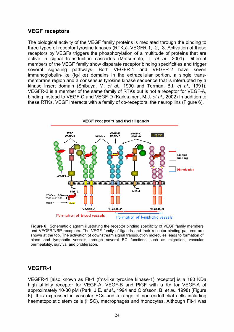
The biological activity of the VEGF family proteins is mediated through the binding to three types of receptor tyrosine kinases (RTKs), VEGFR-1, -2, -3. Activation of these receptors by VEGFs triggers the phosphorylation of a multitude of proteins that are active in signal transduction cascades (Matsumoto, T. et al., 2001). Different members of the VEGF family show disparate receptor binding specificities and trigger several signaling pathways. Both VEGFR-1 and VEGFR-2 have seven immunoglobulin-like (Ig-like) domains in the extracellular portion, a single trans-membrane region and a consensus tyrosine kinase sequence that is interrupted by a kinase insert domain (Shibuya, M. et al., 1990 and Terman, B.I. et al., 1991). VEGFR-3 is a member of the same family of RTKs but is not a receptor for VEGF-A, binding instead to VEGF-C and VEGF-D (Karkkainen, M.J. et al., 2002) In addition to these RTKs, VEGF interacts with a family of co-receptors, the neuropilins (Figure 6).
VEGFR-1
VEGFR-1 [also known as Flt-1 (fms-like tyrosine kinase-1) receptor] is a 180 KDa high affinity receptor for VEGF-A, VEGF-B and PlGF with a Kd for VEGF-A of approximately 10-30 pM (Park, J.E. et al., 1994 and Olofsson, B. et al., 1998) (Figure 6). It is expressed in vascular ECs and a range of non-endothelial cells including haematopoietic stem cells (HSC), macrophages and monocytes. Although Flt-1 was
Figure 6_ Schematic diagram illustrating the receptor binding specificity of VEGF family members and VEGFR/NRP receptors. The VEGF family of ligands and their receptor-binding patterns are shown at the top. The activation of downstream signal transduction molecules leads to formation ofblood and lymphatic vessels through several EC functions such as migration, vascularpermeability, survival and proliferation.

the first RTK to be identified as a VEGF receptor more than a decade ago (de Vries, C. et al., 1992), the precise function of this molecule is still under debate. Recent evidences indicate that the functions and signaling properties of Flt-1can be different depending on the developmental stage of the animal and the cell type. The mechanisms responsible for VEGFR-1 over-expression are not yet fully understood, but hypoxia, which stimulates the expression of VEGF may also upregulate Flt-1 by the HIF-1 dependent mechanism (Gerber, H.P. et al., 1997). A natural soluble Flt-1 receptor (sFlt-1), produced by endothelial and tumor cells has been identified as an alternative splice product that contains the first six of the seven amino-terminal immunoglobulin-like domains of Flt-1 (Kendall, R. L. et al., 1996). Soluble Flt-1 binds to VEGF with high affinity and forms a heterodimeric complex with VEGFR-2. sFlt-1 protein inhibits VEGF-induced endothelial cell proliferation, in fact, transfection of tumor cells with sFlt-1 resulted in reduced tumor growth and metastasis (Goldman, C.K. et al., 1998). These results suggest that sFlt-1 acts as an angiogenesis inhibitor by sequestering VEGF and/or by acting as a dominant negative effector by forming heterodimers with trans-membrane VEGFR tyrosine kinases. Therefore, sFlt-1 and Flt-1 receptors have been demonstrated to suppress retinal neovascularization in a murine model of ischemic retinopathy (Aiello, L.P. et al., 1995). Besides, gene targeting studies have demonstrated that Flt-1-/- mice die in utero between days 8.5 and 9.5 due to obstruction of vessels by an overgrowth of ECs. In fact, ECs develop, but do not organize into vascular channels, indicating that, at least during early development, Flt-1 is a negative regulator of VEGF action (Fong, G.H. et al., 1995 and Fong, G.H. et al., 1999). It was determined that the VEGF (and PlGF) binding site is located primarily to the first three immunoglobulin-like domains of Flt-1 and that two Flt-1 receptors can be linked by a VEGF bridge. It was also determined that the fourth immunoglobulin-like loop contains a receptor dimerization domain (Wiesmann, C. et al., 1997). Although Flt-1 receptor binds to VEGF with high affinity, it is a kinase-impaired receptor; Flt-1 is poorly tyrosine phosphorylated following ligand binding and expresses only weak kinase activity thereafter (Rahimi, N. 2006). The activation of Flt-1 receptor by VEGF in cells lacking VEGFR-2 does not induce cell proliferation, while activation of the VEGFR-2 receptor by VEGF in cells devoid of Flt-1 results in a mitogenic response (Waltenberger, J. et al., 1994). However, activation of Flt-1 by VEGF does induce cell migration, a response that is also induced as a result of VEGFR-2 activation by VEGF (Yoshida, A et al., 1996). These results indicate that the signal transduction cascades induced by VEGFR-1 and VEGFR-2 are somewhat different. It is not yet completely clear why VEGFR-1 does not induce cell proliferation in response to VEGF and VEGFR-2 does. MAP kinase was not activated by VEGF in cell expressing recombinant VEGFR-1 in two separate studies (Seetharam, L. et al., 1995). So, it is possible that VEGFR-1 does not induce cell proliferation, because it does not activate MAP kinase.
VEGFR-2
VEGFR-2 [in human: KDR (kinase domain region) receptor, in mouse: Flk-1 (fetal liver kinase-1) receptor] is a 200-230 KDa high affinity receptor for VEGF-A, the processed forms of VEGF-C and –D, and VEGF-E (Figure 6). It is expressed in both vascular endothelial and lymphatic endothelial cells; its expression has also been demonstrated in several other cell types such as HSCs (Katoh, O. et al., 1995). KDR-
/- embryos die in utero between days 8.5 and 9.5, exhibiting defects in the
25

development of endothelial and haematopoietic precursors, indicating that the receptor is crucial for vascular development (Shalaby, F. et al., 1995). VEGF-A binds to the second and third extracellular immunoglobulin-like domains of VEGFR-2 with a Kd of 75-125 pM; this affinity is lower than the affinity of VEGF-A for VEGFR-1, but VEGFR-2 is expressed in higher copy numbers than VEGFR-1 (Waltenberger, J. etal., 1994). VEGFR-2 is considered to be the major mediator of several physiological and pathological effects of VEGF-A on ECs. These include proliferation and survival, migration and permeability. VEGFR-2, like many other receptors, induces proliferation through activation of the classical extracellular regulated kinase pathway, leading to gene transcription (Takahashi, T. et al., 1999b). VEGFR-2 function is modulated through co-receptors such as heparan sulfated proteoglycans, which interact with certain VEGF isoforms and with VEGFR-2 (Gitay-Goren, H. et al.,1992). The functional VEGF-VEGFR-2 complex includes neuropilins (Soker, S. et al.,1998), which are ubiquitous membrane-bound molecules also implicated in axon guidance by binding to the semaphorin family members (Neufeld, G. et al., 2002). Neuropilins might act by stabilizing the VEGF-VEGFR-2 complex. While hypoxia stimulates VEGF expression and upregulates Flt-1, it downregulates VEGFR-2 (Detmar, M. et al., 1997).
VEGFR-3
VEGFR-3 (also known as Flt-4) is a 195 KDa high affinity receptor for VEGF-C and –D. Distinct features of VEGFR-3 includes cleavage during synthesis within the fifth extracellular immunoglobulin loop (Figure 6); the two regulating polypeptides are kept together by a disulfide bridge (Pajusola, K. et al., 1994). There are two VEGFR-3 splice variants in humans, one short and one long. Mouse embryos lacking expression of VEGFR-3 die at embryonal day 9.5 due to deficient vessel remodeling. Larger vessels are disorganized, leading to fluid accumulation and cardiovascular failure (Dumont, D. J. et al., 1998). These effects might be owing to a direct loss of VEGFR-3 function or because of an indirect effect caused by an increased availability of VEGF-C and –D for activation of VEGFR-2 (Hamada, K. et al., 2000). In the adult, VEGFR-3 expression is detected primarily in lymphatic endothelial cells, where its activation induces proliferation, migration and survival.
Neuropilins
Neuropilins (NRP-1 and NRP-2) are two non-kinase VEGF receptors possessing a lower mass than either VEGFR-1 or VEGFR-2 (Gitay-Goren, H. et al., 1992). These receptors are expressed not only on vascular endothelium, but also on many types of normal and tumor cells. They have long been known as receptors for the semaphorins/collapsin family of neuronal guidance mediators (Neufeld, G. et al.,2002 and Eichmann, A. et al., 2005). The neuropilins have a short intracellular domain; gene disruption studies indicate that neuropilin-1 is probably an important regulator of blood vessel development as mouse embryos lacking a functional neuropilin-1 gene die because their cardiovascular system fails to develop properly (Kitsukawa, T. et al., 1997). Neuropilin-1 is a VEGF co-receptor (Figure 6); this assumption is supported by experiments showing that VEGFR-2 binds to VEGF more
26

efficiently in cells expressing NRP-1 (Soker, S. et al., 1998). Neuropilin-1 is not able to function as a VEGFR-1 co-receptor (Migdal, M. et al., 1998).
Structure and functional analysis of VEGF receptors Flt-1 and KDR
VEGF induces proliferation of ECs through the binding to two receptor tyrosine kinases, Flt-1 and KDR/Flk-1. Both receptors belong to the type III tyrosine kinases and are characterized by seven Ig-like loops within their extracellular domain and a split kinase domain within the cytoplasmatic moiety (Fantl, W. et al., 1993). Both VEGF receptors contain several N-glycosylation sites and the apparent molecular weights of the mature proteins suggest that both receptors are extensively glycosylated (de Vries, C. et al., 1992 and Millauer, B. et al., 1993). Since the dimeric structure of VEGF is a prerequisite of receptor activation, it can be speculated that one VEGF molecule bridges two receptors via two similar recognition sites (Herren, B. et al., 1993). Characterization of VEGF binding to its receptors by mutational analysis of the ligand supports the assumption that VEGF has two contacts sites for its receptors (Keyt, B. A. et al., 1996). Therefore, domain deletion studies on Flt-1 (Barleon, B. et al., 1997) generating several soluble mutants of the extracellular domains of Flt-1, each consisting of a different stretch of Ig-like loops, have suggested that the recognition site for VEGF is located on the first three Ig-like loops, whereas dimerization is stabilized due to an additional domain located on Ig-like loop four. These studies have also demonstrated that glycosylation is not a prerequisite of high affinity binding of VEGF to Flt-1. Besides, deletion experiments on KDR have shown that only domains two and three are critical for ligand binding. KDR fourth Ig-like loop may also function as a dimerization domain, although there is no experimental data as yet to prove that assumption (Fuh, G. et al., 1998). The receptor binding face of VEGF has been identified by mutagenesis studies (Muller, Y.A. et al., 1997), revealing that the binding epitope for KDR contains two “hot spots”, each of which extends across the dimer interface. Charge-reversal mutagenesis has indicated that some of these same residues are also important for VEGF binding to Flt-1 (Keyt, B. A. et al., 1996). The crystal structure to 1.7Å resolution of the complex between the receptor-binding domain of VEGF (VEGF 8-109) and Flt-1 domain 2 (Flt-1D2) (Wiesmann, C. et al., 1997) shows that an Flt-1 construct consisting of domain 2 binds to VEGF with affinity only about 60 fold weaker than the entire extracellular portion. The crystal structure of the complex is the first example of a cystine-knot growth factor bound to a domain of its receptor. So, the second domain of Flt-1 (Flt-1D2) is necessary and sufficient for high affinity VEGF binding. The 1.7Å resolution crystal structure of Flt-1D2 bound to VEGF revealed that this domain is a member of the immunoglobulin superfamily, but has several unusual features, including a region near the N-terminus that bulges away from the domain rather than pairing with the neighboring -strands (Wiesmann, C. et al., 1997). Some of the residues in this region make contact with VEGF, raising the possibility that this bulge could be a consequence of VEGF binding and might not be present in the absence of ligand. But, in 1999, Starovasnik and co-workers reported the three-dimensional structure of Flt-1D2 in its uncomplexed form determined by NMR spectroscopy (Starovasnik, M. A. et al., 1999). The solution structure is very similar to the previously reported VEGF-bound crystal structure and the N-terminal bulge is still present. 1H-15N heteronuclear NOEs indicate that this region is flexible in solution. Thus, VEGF-binding is not
27

accompanied by significant structural change in Flt-1D2, and the unusual structural features of Flt-1D2 are an intrinsic property of this domain (Starovasnik, M. A. et al.,1999).
Structures of VEGF-A and PlGF
Structure of VEGF-A in the free form and bound to D2 of VEGFR-1
VEGF-A in the free form
The crystal structure of VEGF-A (residues 8-109, VEGF-A8-109) shows a homodimer, organized in an antiparallel arrangement with a 2-fold axis perpendicular to the plane of the -sheets (Figure 7A). The homodimer is covalently linked by 2 intermolecular disulfide bonds between C51 and C60. Each monomer consists of a central irregular antiparallel four-stranded -sheet with the characteristic cysteine-knot motif. The knot is formed by two intra-molecular disulfide bonds C57-C102 and C61-C104 which form a ring structure through which a third disulfide bond C26-C68 passes and connects 1and the end of loop 2. The cysteine-knot motif is similar to that of the related growth factor PDGF. Each monomer contains three solvent-accessible loop regions connecting the individual strands (Figure 7B). In addition, an -helix is present at each N-terminus. While the monomer-monomer interface is predominantly formed by a hydrophobic core, the -sheets are solvent exposed on both sides (Muller, Y.A. etal., 1997 and Starovasnik, M. A. et al., 1999).
PlGF). While loop 2 adopts a very similar conformation in both proteins, loop 1 and 3 show
Figure 7_ Representation of VEGF-A (A) and superposition of VEGF-A and PlGF in thefree form (B). Crystal structures form VEGF-A (green) and PlGF (blue) were superimposedaccording to the C atoms. (A) Disulfide bonds forming the cysteine-knot in monomer A arehighlighted in yellow. In addition the 2 intermolecular S-S bridges are shown. (B) Ribbonrepresentation: the individual -strands are connected by three loops (shown for monomer A of
slight deviations.
28
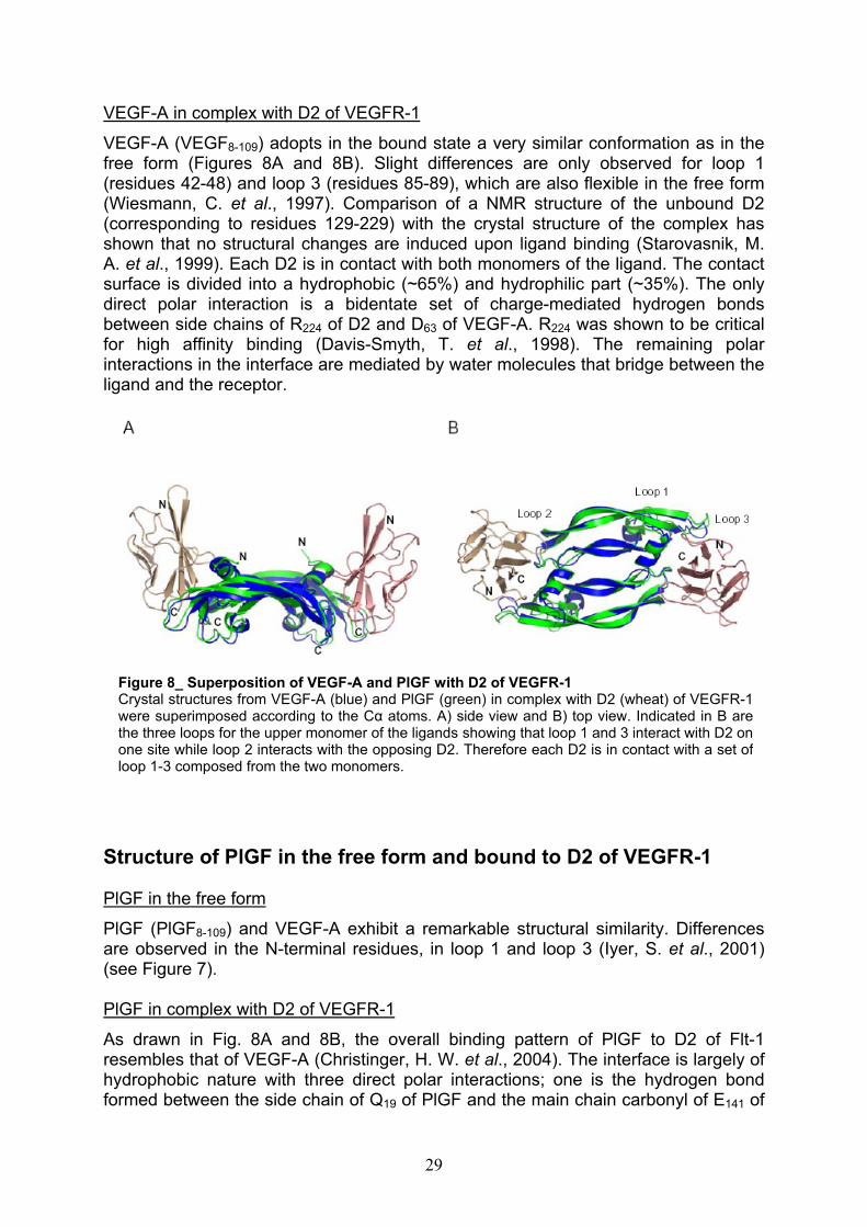
VEGF-A in complex with D2 of VEGFR-1
VEGF-A (VEGF8-109) adopts in the bound state a very similar conformation as in the free form (Figures 8A and 8B). Slight differences are only observed for loop 1 (residues 42-48) and loop 3 (residues 85-89), which are also flexible in the free form (Wiesmann, C. et al., 1997). Comparison of a NMR structure of the unbound D2 (corresponding to residues 129-229) with the crystal structure of the complex has shown that no structural changes are induced upon ligand binding (Starovasnik, M. A. et al., 1999). Each D2 is in contact with both monomers of the ligand. The contact surface is divided into a hydrophobic (~65%) and hydrophilic part (~35%). The only direct polar interaction is a bidentate set of charge-mediated hydrogen bonds between side chains of R224 of D2 and D63 of VEGF-A. R224 was shown to be critical for high affinity binding (Davis-Smyth, T. et al., 1998). The remaining polar interactions in the interface are mediated by water molecules that bridge between the ligand and the receptor.
loop 1-3 composed from the two monomers.
Figure 8_ Superposition of VEGF-A and PlGF with D2 of VEGFR-1Crystal structures from VEGF-A (blue) and PlGF (green) in complex with D2 (wheat) of VEGFR-1were superimposed according to the C atoms. A) side view and B) top view. Indicated in B arethe three loops for the upper monomer of the ligands showing that loop 1 and 3 interact with D2 onone site while loop 2 interacts with the opposing D2. Therefore each D2 is in contact with a set of
Structure of PlGF in the free form and bound to D2 of VEGFR-1
PlGF in the free form
PlGF (PlGF8-109) and VEGF-A exhibit a remarkable structural similarity. Differences are observed in the N-terminal residues, in loop 1 and loop 3 (Iyer, S. et al., 2001) (see Figure 7).
PlGF in complex with D2 of VEGFR-1
As drawn in Fig. 8A and 8B, the overall binding pattern of PlGF to D2 of Flt-1 resembles that of VEGF-A (Christinger, H. W. et al., 2004). The interface is largely of hydrophobic nature with three direct polar interactions; one is the hydrogen bond formed between the side chain of Q19 of PlGF and the main chain carbonyl of E141 of
29
D2, and two charged interactions occur between D62 of PlGF and R224 of D2 from Flt-1. A bis-Tris propane molecule from the crystallization buffer is in contact with PlGF and D2 of VEGFR-1 near the interface. Presumably, this molecule greatly stabilizes the crystal packing arrangement. The PlGF dimer is slightly altered when bound to D2, mainly in the regions of loop 1 and loop 3 which results in a more open conformation of the ligand. Because of a dense crystal packing in this area it cannot be excluded that the observed interactions are influenced by packing effects.
VEGF and the induction of pathological angiogenesis: therapeutic implication and perspectives
VEGF plays a crucial role in physiological and pathological angiogenesis, including solid tumor growth and metastasis (Fidler, I. J. et al., 1994 and Folkman, J. 1995)(Figure 9). As the formation of the normal blood vasculature, pathological angiogenesis is a multi-step process that requires ECs division, migration, organization into cords, lumen formation, acquisition of pericytes and degradation of old and synthesis of new basement membrane. In pathological angiogenesis VEGF-A induces the formation of several distinctly different types of new blood vessels with respect to organization, structure and function (Nagy, J. A. et al., 2007). The importance of the function of VEGF suggests that blocking its function is a promising new therapeutic strategy for inhibiting angiogenesis and tumor growth. In the past years hybridization studies have shown that VEGF mRNA is upregulated in many human tumors (Dvorak, H.F. 1995 and Ferrara, N. et al., 1997). Even though tumors do not exhibit an organized structure, from the point of view of angiogenesis a growing tumor may be viewed as a developing new organ. Angiogenesis is as essential for the growing tumor as it is for a developing organ such as the corpus luteum, since the delivery of blood-borne nutrients to the tumor cells is essential for their survival (Hanahan, D. et al., 1996). When VEGF signaling is inhibited, tumor angiogenesis and, consequently, tumor growth are impaired (Kim, K.J. et al., 1993 and Millauer, B. et al., 1994). Besides, as VEGF binds to Flt-1 and KDR/Flk-1, thereby activating signal transduction and regulating physiological and pathological angiogenesis, to find an agent that inhibits binding between VEGF and its receptors may serve as a valuable antiangiogenic and antitumor agent. Many studies reported that antibodies to VEGF exert a potent inhibitory effect on the growth of several tumor cell lines in nude mice (Kim, K.J. et al., 1993). Subsequently, many other tumor cell lines were found to be inhibited in vivo by this and other anti-VEGF treatments, including antisense oligonucleotides and antibodies to VEGFR-2 (Ferrara, N. et al.,1997). Several studies have shown that combining anti-VEGF treatments with chemotherapy (Klement, G. et al., 2000) or radiation therapy (Lee, C.G. et al., 2000) results in a greater antitumor effect than either treatment alone. In the past years, clinical trials in cancer patients were carried out with several VEGF inhibitors, including a humanized monoclonal antibody to VEGF (rhuMab VEGF) (Presta, L.G. et al., 1997), an anti-VEGFR-2 antibody (Prewett, M. et al., 1999), small molecules inhibiting VEGFR-2 signal transduction (Wood, J.M. et al., 2000) and a soluble VEGF receptor (Holash, J. 2002). Phase 2 clinical data provided initial evidence that rhuMab VEGF, in combination with conventional chemotherapy, resulted in increase in time to progression and even survival in patients with metastatic colorectal carcinoma (Kabbinavar, F. et al., 2003). Phase 3 studies are currently under way to
30
confirm and fully assess the benefit of these anti-VEGF treatments in patients with advanced cancer. Another humanized anti-VEGF-A monoclonal antibody is Bevacizumab (trade name Avastin), approved, in February 2004, by the US Food and Drug Administration (FDA) in the United States for the treatment of metastatic colorectal cancer, in combination with chemotherapy regimens. This followed from a phase 3 study showing survival benefits (Hurwitz, H. et al., 2004). The first VEGF receptor tyrosine kinase inhibitor, SU5416, have already been reported; it has produced certain clinical benefits in clinical trials involving patients suffering from advanced malignancies (Stopeck, A. et al., 2002). Besides, Yasuij Ueda and co-workers, using a high throughput screening method, have found a small molecular compound, 5-[N-methyl-N-(4-octadecyloxyphenyl) acetil] amino-2-methylthiobenzoic acid (VGA1155), that was selected for its potent inhibition of binding of VEGF to its receptors through association with KDR/Flk-1 (Ueda, Y. et al., 2003).
Figure 9_ Endothelial cell survival in pathological angiogenesis: tumor growth.
De novo engineered VEGF mimicking peptides
The analysis of structural and mutagenesis data allowed to identify the residues involved in the binding of VEGF to its receptors. They are distributed over a discontinuous surface that includes residues from the N-terminal helix 17-25. KDR and Flt-1 share the VEGF binding region, in fact, five of the seven most important VEGF binding residues are present in both interfaces (Keyt, B. A. et al., 1996; Muller, Y.A. et al., 1997; Wiesmann, C. et al., 1997). Many approaches have been pursued to modulate the VEGF-receptors interaction, and new molecular entities as peptides have been reported to bind to the extracellular region of the VEGF receptors. A large number of them show an antagonist activity and only few behave as agonists (D’Andrea, L. D. et al., 2008, submitted). Remarkably, the peptides modulating the VEGF-receptors interaction are mainly derived by phage display libraries screening,
31
and only few examples of rational design approaches have been reported so far (Zilberberg, L. et al., 2003). In our laboratory was designed, by a structure-based approach, a peptide of 15 amino acid residues, QK (Ac-KLTWQELYQLKYKGI-NH2),reproducing the VEGF 17-25 helix region (D’Andrea, L. D. et al., 2005). It is well known that peptide fragments spanning helices, turns and -hairpins of natural proteins show little propensity, with very few exceptions, to reproduce their natural secondary structure under physiological conditions (De Grado, W. F. et al., 1999). Nevertheless, the stabilization of suitable conformation in aqueous solutions is a condition to gain the binding of designed peptides to their targets. All data collected on the structural preferences of the QK peptide in aqueous solution strongly indicated that it mainly folds in helical conformation. In particular, the first indication derives from the CD spectrum, which is well confirmed by the H chemical shift analysis and the NMR structure determination (D’Andrea, L. D. et al., 2005). The basis of the QK helical fold seems to reside on the presence of amino acids with intrinsic helix preference and on the amphipathic nature of the helix. The biological properties of QK were tested performing an in vitro angiogenesis assay. In this assay QK induced the formation of newly formed connections in a dose-dependent manner and enhanced the VEGF response. These results demonstrate that QK binds to VEGF receptors in vitro and show that it is a potential agonist for angiogenesis. QK shows an unusual thermal stability (D’Andrea, L. D. et al., 2005) and very recently it was showed to be active in gastric ulcer healing in rodents when it is administered either orally or systemically (Dudar G.K. et al., 2008). Although QK was designed to bind to KDR and Flt-1, its agonist-like activity is surprising because receptor dimerization is necessary for receptor activation. QK is a small peptide and probably is unable to induce receptor dimerization by itself. To explain the activity of QK and its additive effects in presence of VEGF, we can speculate that the QK binding possibly induces a conformational change of the receptor that either triggers, in an unknown way, the dimerization or induces the receptor activation through a different uncharacterized mechanism that may not require dimerization. Further structural and biochemical studies are needed to address this issue. Anyway, these data are suggestive that either QK or improved analogues might fulfil the request for a safer pro-angiogenic drug. As therapeutic angiogenesis in cardiovascular conditions such as chronic ischemia or heart failure is sought as a promise of modern biotechnology, potential applications for QK are in the diagnostic field and in therapy of cardiovascular disease. An analogue of QK is MA, a peptide of 15 amino acid residues (Ac-KLTWMELYQLAYKGI-NH2) synthesized and characterized by NMR in our laboratory. MA-VEGF competition binding assays were carried out by FACS indicating that the peptide is able to bind with specificity to VEGF receptors on ECs surface, inhibiting VEGF anti-apoptotic activity of over 80%, thus showing an antagonist activity with respect to VEGF (Del Gatto, A. PhD thesis, 2005).
The aim of the work
In the last years, much of the basic research in angiogenesis has been focused on detailing the steps involved in blood vessel growth and in identifying molecules that play a key role in regulating this process. This heightened analysis is due in part to the realization that aberrant angiogenesis is involved in relevant pathological conditions. Over the past decade, intensive efforts have been undertaken to inhibit
32
angiogenesis in cancer, ocular, joint or skin disorders or to develop therapeutic strategies to promote revascularization of ischemic tissues (Carmeliet, P. 2005). Despite some initial setbacks and negative clinical trial results, new angiogenic modulators have been developed in the last years. They mainly fall in three classes: antibodies, peptides and small molecules. Some of these molecules have been approved for treatment of cancer (Avastin), or are in phase 3 of clinical trials (Ferrara, N. 2005). Because of VEGF’s predominant role in angiogenesis, inhibition of VEGF seems to be necessary but is probably insufficient to permanently halt this process in many disorders. Considering the anti-tumor therapy, the angiogenic inhibitors are, generally, more safe and less toxic than chemotherapeutic substances, but their exclusive administration as drugs in the treatment of tumors is rare. In fact, a wide range of different growth factors are involved in the stimulation of angiogenesis and the blockage of one factor alone might not be sufficient to inhibit the angiogenesis for a prolonged time period, as tumors could compensate by overproducing other growth factors (Carmeliet, P. 2005). Despite the problems involved, the anti-angiogenic therapy continues to arouse notable interest. In fact, angiogenic inhibitors, if used in conjunction with traditional therapies, have the ability to enhance therapeutic efficacy without significantly raising the risk of toxic side effects. In the process of angiogenesis, vascular endothelial growth factor binds to Flt-1 and KDR, thereby activating signal transduction and regulating physiological and pathological angiogenesis (Neufeld, G. 1999). Actually, of these receptors, are not well known structural data, done exception for the domain 2 of Flt-1 of which, in the literature, is reported the crystal structure complexed with VEGF and PlGF (Wiesmann, C. et al.,1997 and Christinger, H. W. et al., 2004) and the three-dimensional, uncomplexed form determined by NMR spectroscopy (Starovasnik, M. A. et al., 1999). The aim of this PhD thesis is the expression and characterization of the extracellular domains of VEGF receptors, in order to find new compounds of biotechnology interest for modulating the angiogenesis process. Because the angiogenic response strictly depends on VEGF and on functions and signaling properties of its receptors, we centered our efforts on the characterization of the extracellular portion of Flt-1 (VEGFR-1) and KDR (VEGFR-2) receptors. In particular, we expressed some of their extracellular Ig-like domains, focusing the attention on their structural characterization via NMR and on their employment in the identification and improvement of new organic or peptide-based molecules targeting the VEGF receptors. In fact, once expressed and purified Flt-1D2, we used the second domain of VEGFR-1 for interaction studies with QK (VEGF agonist) and MA (VEGF antagonist) peptides (D’Andrea, L. D. et al., 2005 and Del Gatto, A. PhD thesis, 2005), in order to better understand their biological behavior in angiogenesis process. Therefore, the recombinant Flt-1D2 was employed for the screening of a library of organic molecules by drug discovery analysis, in order to find other new class of VEGF receptor binders.
33
Materials and Methods
Strains, enzymes and reagents
Reagents used for preparation of buffers and growth media of Escherichia coli andthe reagents for polyacrylamide gels electrophoresis (Acrylamide, APS, TEMED, SDS, Tris, Glycine) were supplied by Sigma Aldrich, Euroclone, Applichem and ICN Biomedicals. The molecular weight markers for proteins were from Sigma Aldrich. The restriction enzymes and the “modification enzymes” (calf intestine phosphatase, T4 DNA ligase and T4 DNA polynucleotide kinase) were supplied by New England Biolabs (NEB) and the molecular weight markers for nucleic acids were supplied by NEB and Roche. The Pfu Turbo polymerase (2.5 U/µL) was supplied by Stratagene, while Taq DNA polymerase (5 U/µL) was from NEB. The synthesis of theoligonucleotides was committed at Sigma-Genosys; pETM11 and pETM20 E. coli expression plasmids were supplied by Novagen, while the expression vector pPROEXHTa was supplied by Invitrogen. E. coli TOP F’10 strain, used for cloning, was supplied by Invitrogen; E. coli BL21 Codon Plus (DE3) RIL cells, used for overexpression, were supplied by Stratagene. BL21(DE3), BL21(DE3) STAR and JM101 strains, also used for expression, were supplied by Invitrogen while Rosetta GAMI (DE3) were from Novagen. Complete Protease Inhibitor Cocktail Tablets were supplied by Roche and used as a mixture of protease inhibitors, according to manufacturer’s instruction. Ethanol, isopropilic alcohol and acetic acid were supplied by J.T. Baker. TFA was from Fluka. Purity and identity of protein samples were assessed by liquid chromatography mass spectrometry (LC-MS) performed on a Thermo Electron Corporation mass spectrometer equipped with an ESI source. The analysis of proteins was carried out on a C4 column, using a method (referred to as “method A”) developed at 0.2 mL/min with a linear gradient of 0.1% aqueous TFA-0.1%TFA in CH3CN, from 5% to 70% for 30 min.
AntibioticsAmpicillin (supplied by Sigma Aldrich as ampicillin sodium salt) and kanamycin (supplied by Sigma Aldrich as kanamycin sulphate) were solubilized in deionized water at a concentration of 1000X, filter sterilized and stored at –20°C until use. Ampicillin was used at a concentration of 100 µg/mL and kanamicin was used at a concentration of 50 µg/mL in both solid and liquid media. Chloramphenicol and tetracycline were solubilized in ethanol 96% and used at a concentration of 33 µg/mLand 12.5 µg/mL, respectively.
E. coli cells transformation techniques
Preparation of E. coli TOP F’10 cells and transformation by electroporation 2.5 mL of an overnight culture of E. coli TOP F’10 cells were inoculated into 250 mL of LB medium. The cells were grown up to mid-log phase (0.6 OD600) at 37°C, stored on ice for 20-30 min and then harvested by centrifugation (6000 rpm, 10 min, 4°C). The pellet was washed in 250 mL of sterile water. After the second centrifugation the cells were washed in 125 mL of sterile water. The third washing was performed in 5 mL 10% glycerol; the cells were then harvested by centrifugation and the pellet was
34

resuspended in 750 µL of 10% glycerol. Aliquots of 1010 cells/mL (40 µL) were mixed with 1 µL of the DNA ligase reaction, incubated for 1 min on ice and transferred into chilled plastic cuvettes with an electrode gap of 0.2 cm (M-Medical). High voltage electroporation (25 µF) was performed with a Bio-Rad Gene Pulser XcellTM at a field strength of 2.5 kV/cm and 200 Ohm. A shock pulse was applied to competent cells producing pulse length of ~5.0-5.5 ms. Immediately after electroporation cell mixtures were diluted to 1 mL with LB medium and incubated for ~1 h at 37°C under shacking. The cells were then plated onto selective solid medium supplemented with 100 µg/mL ampicillin or 50 µg/mL kanamicin to isolate the recombinant clones. Single clones were inoculated in 5 mL LB medium with the same antibiotics and grown over night at 37°C under shaking. Finally, were harvested by centrifugation (13.000 rpm, 2 min, 4°C) and processed to extract plasmidic DNA by using the QIAprep Spin Miniprep Kit or the QIAGEN plasmid Maxi Kit (both supplied by Qiagen).
Preparation of E. coli BL21 (DE3) competent cells and transformation by heat shockSingle clones of BL21 (DE3) E. coli strains, grown at 37°C in LB agar, were inoculated into 2.5 mL of LB medium and incubated overnight at 37°C on a shaker. The cells were inoculated into 250 mL of LB medium and the culture was grown up to mid-log phase (0.6 OD600) at 37°C, stored on ice for 20-30 min and then harvested by centrifugation (6000 rpm, 10 min, 4°C). The pellet was washed in 125 mL of cold 50 mM CaCl2 and stored on ice for 30 min. Successively, the cells were harvested by centrifugation and the pellet resuspended in 16 mL of cold 50 mM CaCl2. Aliquots of 200 µL of competent cells were mixed with 50 ng of plasmidic DNA and stored on ice for ~30 min. The cells mixtures were transferred at 42°C for 90 sec, on ice for 2 min (heat shock) and then diluted to 1 mL with LB medium. An incubation for ~1 h at 37°C under shacking was performed before plating the cells onto selective solid medium supplemented with the opportune antibiotics, depending on the strain.
Proteins analyses
Determination of the protein concentration The concentration of the proteins in solution was determined according to the Bradford’s method (Bradford, 1976). The Coomassie Brilliant (Bio-Rad) reagent was added to the samples and the absorbance at 595 nm was monitored. A solution of 1 µg/µL of bovine serum albumin (BSA) was used as standard. Protein concentration was also measured by UV spectroscopy, reading the tryptophan absorbance at 280 nm, using a Jasco V-550 UV-VIS spectrophotometer, in an 1 cm quartz cell.
Electrophoretic analysis of proteins (SDS-PAGE) The electrophoresis on polyacrylamide gel in the presence of SDS (SDS-PAGE) was performed according to Laemmli’s protocol (Laemmli, 1970). The samples were denaturized at 100°C for 10 min in 1% SDS (Applichem), 5% -mercaptoethanol(Sigma), 0.001% bromophenol blue (ICN Biomedicals) and 10% glycerol (Applichem). The samples were then loaded on a 15% or 18% polyacrylamide gel and electrophoresed in 0.025 M Tris-HCl, 0.2 M glycine pH 8.3 and 0.1% SDS. The electrophoresis was performed at 25 mA for ~1.5 hours. The proteins were then revealed by Coomassie Brilliant-Blue (Applichem) staining; the gel was submerged in the staining solution (0.1% Coomassie Brilliant-Blue R250, 25% isopropilic alcohol
35

and 10% acetic acid) for ~30 min with gentle agitation. The gel was washed in a solution containing 30% ethanol and 10% acetic acid to remove the excess of Coomassie and then stored in 10% acetic acid. Alternatively, the proteins were transferred by electro-blotting from gel to a polyvinylidene fluoride (PVDF) membrane as described in the paragraph “Western blot analysis”. For native PAGE the samples were not denaturized at 100°C but loaded on a 10% polyacrylamide gel in 0.001% bromophenol blue and 10% glycerol and electrophoresed in Tris-glycine buffer without SDS. The electrophoresis was performed at 15 mA for ~2 hours, after a gel pre-run of 30 min. The proteins were then revealed by Coomassie Brilliant-Blue staining, as described above.
Bioinformatic tools Protein sequences were processed using the pI/Mw tool available on the ExPASy Server (http://www.expasy.ch), in proteomics and sequence analysis tools section. Compute pI/Mw is a tool which allows the computation of the theoretical pI (isoelectric point) and Mw (molecular weight) for a list of UniProt Knowledgebase(Swiss-Prot or TrEMBL) entries or for user entered sequences.
Domains cloning of human KDR and Flt-1 receptors
The genes corresponding to the extracellular domains of KDR and Flt-1 receptors were amplified by PCR from NIH-3T3 cDNA of the entire receptors (kindly provided by Dr. Nicola Normanno).
Cloning of 1-3, 2-3, 1-2 and 2 KDR receptor domains The amplification of domains 1, 2, 3 of KDR was performed by using the following couples of primers:
kdrD1-3 gene (residues 1-335, Fuh et al., 1998)
Forward: 5’- CGGGATCCGAAAGACATACTTACAATTAAGGCTAA -3’
Reverse: 5’- CCGCTCGAGCTATCATCCAAAAGCAACAAAAGGTTTTTCATG-3’
kdrD2-3 gene (residues 120-335, Fuh et al., 1998)
Forward: 5’- CGGGATCCGTACAGATCTCCATTTATTGCTTCTGTT -3’
Reverse: 5’- CCGCTCGAGCTATCATCCAAAAGCAACAAAAGGTTTTTCATG -3’
In this case, the reverse primer was the same used for the amplification of kdrD1-3
gene.
kdrD1-2 gene (residues1-222, Fuh et al., 1998)
Forward: 5’- CGGGATCCGAAAGACATACTTACAATTAAGGCTAA -3’
Reverse: 5’- CCGCTCGAGCTATCACCTATACCCTACAACGACAACTATGTA -3’
In this case, the forward primer was the same used for the amplification of kdrD1-3
gene. Finally, the PCR reaction necessary to obtain domain 2 of KDR was performed with the following primers:
36
kdrD2 gene (residues 120-222, Fuh et al., 1998)
Forward: 5’- CGGGATCCGTACAGATCTCCATTTATTGCTTCTGTT -3’
Reverse: 5’- CCGCTCGAGCTATCACCTATACCCTACAACGACAACTATGTA -3’.
The forward primers indicated above contained the BamHI restriction site, while the reverse primers were designed with the XhoI restriction site (underlined in the sequences) positioned downstream to two stop-translation codons (in bold in the sequences). This cloning strategy was used to obtain the gene products fused (at the N-terminal) with the 6xHistidine-tag of the pPROEXHTa expression vector (see below, Figure 10). The pPROEXHTa plasmid is designed to allow inducible, high level intracellular expression of genes; it contains a tac promoter for chemical induction, a multiple cloning site (MCS), an internal lac Iq gene compatible to any E. coli host and a TEV protease recognition site for cleaving the fusion protein.All amplification reactions were performed in a final volume of 100 µL, using 50 ng of template DNA. The reaction mixture contained the 2 primers (0.25 µM each), dNTPs (0.25 mM each) and the Pfu turbo polymerase (5U) with its buffer 1x. PCR was performed using an Eppendorf Mastercycler personal apparatus, following the procedure indicated below:
Initial denaturation step 3 min at 95°C Denaturation (step 2) 1 min at 95°C Annealing (step 3) 1 min at the convenient temperature for each
gene amplifiedElongation (step 4) 1 min at 72°C
for 30 cycles, from step 2.
All amplification products were analyzed by 1% agarose (Euroclone) gel electrophoresis performed in TAE buffer (18.6 g/L EDTA, 242 g/L Tris base. Add acetic acid until pH 7.8). PCR products were purified by using the QIAquick PCR
Purification Kit (Qiagen), and digested with BamHI (20 U/ L) and XhoI (20 U/ L)restriction enzymes. Each amplified fragment (1 g) was digested with 3U of restriction enzymes for 2 hours at 37ºC in a buffer containing 50 mM NaCl, 10 mM Tris-HCl, 10 mM MgCl2, 1 mM DTT pH 7.9 supplemented with BSA 100 µg/mL. Following the digestion, each fragment was cloned into the corresponding sites of the pPROEXHTa expression vector, downstream to the His-tag sequence. To this purpose, the expression vector was previously digested with the same restriction enzymes (3 U/ g), and treated with calf intestine phosphatase (CIP, 10U) (NEB) for 30 min at 37ºC. CIP enzyme (10 U/ L) was then inactivated at 75°C for 10 min. After digestion, PCR amplifications were purified by QIAquick PCR Purification Kit (Qiagen), while the BamHI/XhoI pPROEXHTa was purified by QIAquick Gel Extraction Kit (Qiagen). For ligation reactions was used a 1:3 molar ratio (vector/insert DNA) for shorter fragments and a 1:6 molar ratio for the longer ones. The reactions were performed using 20 U/ g DNA of the T4 DNA Ligase (400 U/ L),in a final volume of 10 L, for 3 hours at RT. The identity of the inserts in the resulting recombinant plasmids was confirmed by DNA sequencing (MWG-Biotech). E. coli TOP F’10 strain was used for cloning. kdrD2 gene was cloned also in pETM11 and pETM20 expression vectors (see Figures 11, 12). The primers used for amplification of the gene in these systems were:
37
Forward: 5’- GCCATGGCGTACAGATCTCCATTTATTGCT -3’
Reverse: 5’- CCGCTCGAGCTATCACCTATACCCTACAACGACAACTATGTA -3’
where the reverse primer was the same used for the amplification of kdrD1-2 gene,while the forward primer contained the NcoI restriction site. pETM11 and pETM20 expression vectors contain a T7 promoter for chemical induction and overexpression of recombinant proteins, a multiple cloning site (MCS) and a TEV protease recognition site for cleaving the fusion protein. pETM11 contains a 6xHistidine-tag, while the pETM20 system contains a 6xHistidine-TrxA fusion tag for the increase of the solubility of gene products.
kdrD2 gene mutagenesis The C161 residue of domain 2 was mutated in Alanine by using the QuikChange Site Directed Mutagenesis Kit (Stratagene). The primers employed for mutagenesis PCR reaction are reported below:
Forward: 5’- AACGTGTCACTTGCTGCAAGATACCCA -3’
Reverse: 5’- TGGGTATCTTGCAGCAAGTGACACGTT -3’
Codons corresponding to the mutated amino acid are underlined in primers sequences. The recombinant construct pETM11-kdrD2 was used as template. The PCR reaction was performed, according to the Kit instructions, using an EppendorfMastercycler personal apparatus, following the procedure indicated below:
Initial denaturation step 1 min at 95°C Denaturation (step 2) 50 sec at 95°C Annealing (step 3) 50 sec at 58°CElongation (step 4) 12 min at 68°C
for 16 cycles from step 2.
XL1-Blue supercompetent cells (supplied by the mutagenesis Kit) were used for the transformation of the circular, nicked dsDNA (the methylated, non-mutated, parental DNA from mutagenesis PCR was first digested, for 2 hours at 37°C, with the restriction enzyme DpnI-10 U/ L). The identity of the mutagenic insert was confirmed by DNA sequencing.
38
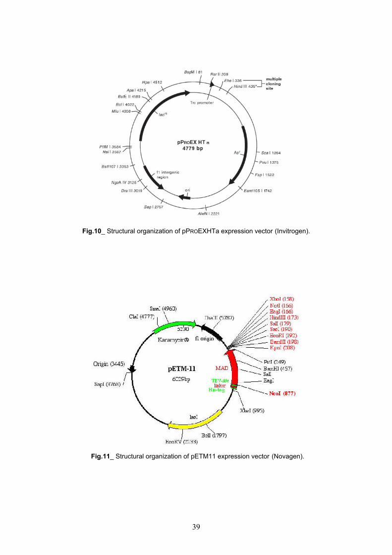
Fig.10_ Structural organization of pPROEXHTa expression vector (Invitrogen).
Fig.11_ Structural organization of pETM11 expression vector (Novagen).
39
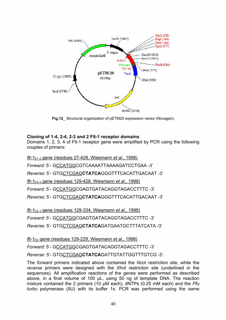
Fig.12_ Structural organization of pETM20 expression vector (Novagen).
Cloning of 1-4, 2-4, 2-3 and 2 Flt-1 receptor domainsDomains 1, 2, 3, 4 of Flt-1 receptor gene were amplified by PCR using the following couples of primers:
flt-1D1-4 gene (residues 27-428, Wiesmann et al., 1998)
Forward: 5’- GCCATGGCGTCAAAATTAAAAGATCCTGAA -3’
Reverse: 5’- GTGCTCGAGCTATCAGGGTTTCACATTGACAAT -3’
flt-1D2-4 gene (residues 129-428, Wiesmann et al., 1998)
Forward: 5’- GCCATGGCGAGTGATACAGGTAGACCTTTC -3’
Reverse: 5’- GTGCTCGAGCTATCAGGGTTTCACATTGACAAT -3’
flt-1D2-3 gene (residues 129-334, Wiesmann et al., 1998)
Forward: 5’- GCCATGGCGAGTGATACAGGTAGACCTTTC -3’
Reverse: 5’- GTGCTCGAGCTATCAGATGAATGCTTTATCATA -3’
flt-1D2 gene (residues 129-229, Wiesmann et al., 1998)
Forward: 5’- GCCATGGCGAGTGATACAGGTAGACCTTTC -3’
Reverse: 5’- GTGCTCGAGCTATCAGATTGTATTGGTTTGTCG -3’.
The forward primers indicated above contained the NcoI restriction site, while the reverse primers were designed with the XhoI restriction site (underlined in the sequences). All amplification reactions of the genes were performed as described above, in a final volume of 100 µL, using 50 ng of template DNA. The reaction mixture contained the 2 primers (10 µM each), dNTPs (0.25 mM each) and the Pfuturbo polymerase (5U) with its buffer 1x. PCR was performed using the same
40

apparatus, following the procedure indicated above for KDR receptor domains. flt-1D2
gene was also cloned in the pETM20 expression vector using the same primers employed in the cloning of the gene in pETM11. PCR products were purified and digested following the same procedure described above for kdr genes.
Expression and purification of recombinant KDR and Flt-1 extracellular domains
ExpressionAfter a first screening of small expression cultures, performed using different strains, temperatures and IPTG concentration, all the recombinant constructs obtained with cloning of Ig-like KDR and Flt-1 receptors domains in pPROEXHTa, pETM11 and pETM20 were transferred into E. coli BL21 Codon Plus (DE3) RIL cells. In fact, in all used conditions and strains, the proteins of interest were expressed in inclusion bodies, so we selected E. coli BL21 Codon Plus (DE3) RIL cells that assured a better expression of all proteins. Single clones of RIL strain, previously transformed with each recombinant expression vector and grown at 37ºC on LB agar containing antibiotics, were inoculated in 10 mL of LB medium, containing the same antibiotics. The overnight cultures were inoculated into 1L of prewarmed LB medium supplemented with antibiotics: 100 µg/mL ampicillin (for pPROEXHTa and pETM20 recombinant vectors) or 50 µg/mL kanamycin (for pETM11 recombinant vectors) and 33 µg/mL chloramphenicol. Cultures were grown at 37°C under shaking until they reached the mid-log phase (0.7/0.8 OD600) then, they were induced with 0.7 mM IPTG (Inalco). After 3-4 hours the cells were harvested by centrifugation (6000 rpm, 15 min, 4°C). Cell pellets were resuspended in 50 mM Tris–HCl, pH 8 containing protease inhibitors, to avoid proteins degradation, and the suspension was sonicated for 6 min, by using a Misonix Sonicator 3000 apparatus with a micro tip probe and an impulse output of 1.5/2 (=9/12 Watt). Bacterial lysates were then centrifuged (15000 rpm, 30 min, 4°C) and the supernatant (soluble fraction) and the pellet, obtained from each sample, were loaded on a 15% or 18% polyacrylamide gel.
Batch purification of 6xHis-tagged proteins under denaturing conditions The expression of each recombinant plasmid produced insoluble proteins, so they were solubilized from inclusion bodies using a lysis buffer containing 50 mM Tris–HCl, pH 8, 10 mM imidazole, 8 M urea. The cleared lysate containing the His-tagged proteins in 8 M urea was added to 50% Ni2+-NTA agarose slurry resin (Qiagen) for the affinity chromatography purification. The binding capacity of Ni2+-NTA resins is between 5-10 mg/mL for 6xHis-tagged proteins. Before loading the lysate, the resin was extensively washed with water and finally equilibrated in the lysis buffer. The lysate was then loaded in presence of 10 mM imidazole and 100 mM NaCl to avoid non-specific binding of E. coli contaminants and the column was gently shaken for 45 min-1h. Then, the flow-through (F.T.), containing all unbound proteins, was collected and the resin was washed three times with the lysis buffer containing 100 mM NaCl, and finally the His-tagged proteins were eluted with high concentrations of imidazole (250/300 mM). All the proteins expressing KDR extracellular domains and Flt-1D1-4,Flt-1D2-4, Flt-1D2-3 were eluted and then refolded by step dialysis to remove urea. Only Flt-1D2 was subjected to an “on column” refolding protocol. Once loaded the lysate, collected the flow-through and wash fractions, the bound protein was refolded by equilibrating the column in buffers with decreasing concentrations of urea. All
41
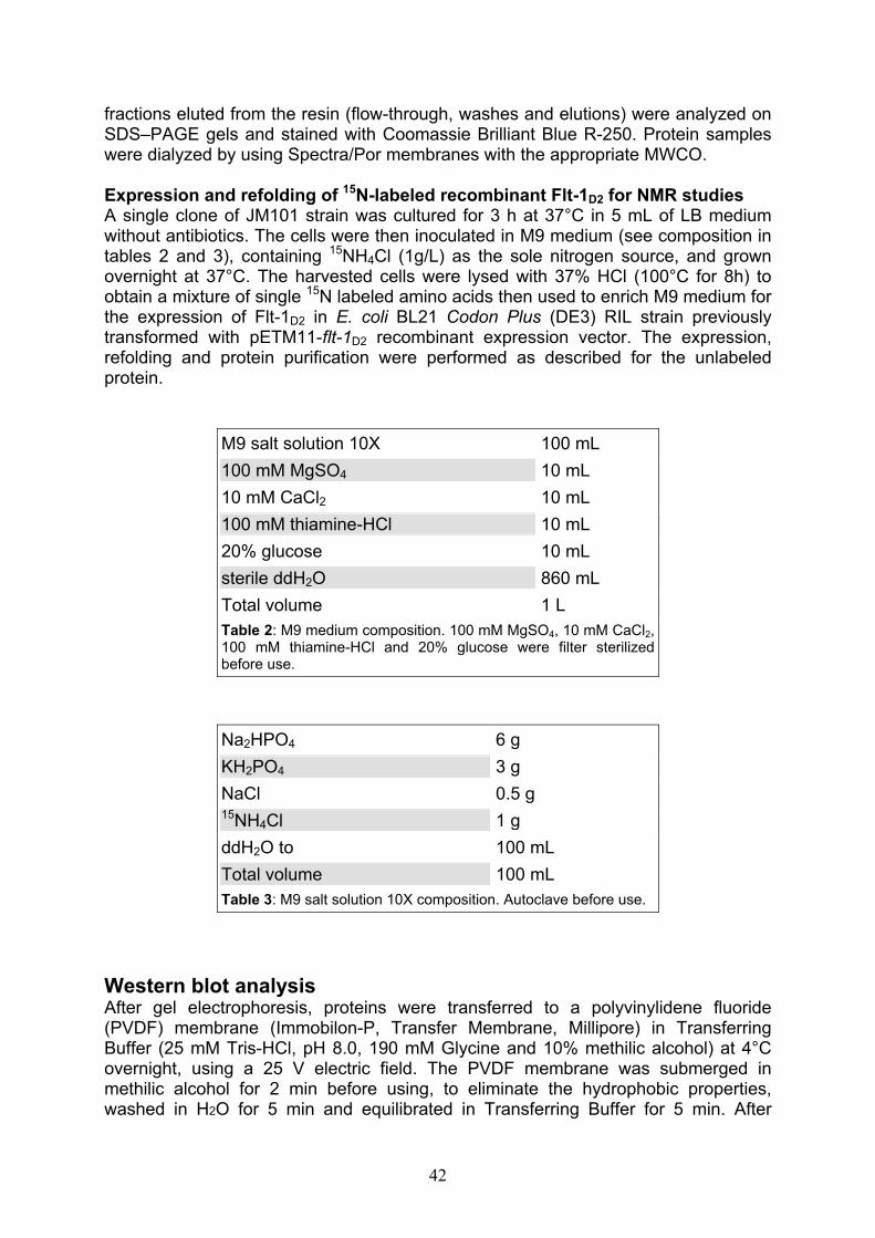
fractions eluted from the resin (flow-through, washes and elutions) were analyzed on SDS–PAGE gels and stained with Coomassie Brilliant Blue R-250. Protein samples were dialyzed by using Spectra/Por membranes with the appropriate MWCO.
Expression and refolding of 15N-labeled recombinant Flt-1D2 for NMR studies A single clone of JM101 strain was cultured for 3 h at 37°C in 5 mL of LB medium without antibiotics. The cells were then inoculated in M9 medium (see composition in tables 2 and 3), containing 15NH4Cl (1g/L) as the sole nitrogen source, and grown overnight at 37°C. The harvested cells were lysed with 37% HCl (100°C for 8h) to obtain a mixture of single 15N labeled amino acids then used to enrich M9 medium for the expression of Flt-1D2 in E. coli BL21 Codon Plus (DE3) RIL strain previously transformed with pETM11-flt-1D2 recombinant expression vector. The expression, refolding and protein purification were performed as described for the unlabeled protein.
M9 salt solution 10X 100 mL
100 mM MgSO4 10 mL
10 mM CaCl2 10 mL
100 mM thiamine-HCl 10 mL
20% glucose 10 mL
sterile ddH2O 860 mL
Total volume 1 L
Table 2: M9 medium composition. 100 mM MgSO4, 10 mM CaCl2,100 mM thiamine-HCl and 20% glucose were filter sterilized before use.
Na2HPO4 6 g
KH2PO4 3 g
NaCl 0.5 g 15NH4Cl 1 g
ddH2O to 100 mL
Total volume 100 mL
Table 3: M9 salt solution 10X composition. Autoclave before use.
Western blot analysisAfter gel electrophoresis, proteins were transferred to a polyvinylidene fluoride (PVDF) membrane (Immobilon-P, Transfer Membrane, Millipore) in Transferring Buffer (25 mM Tris-HCl, pH 8.0, 190 mM Glycine and 10% methilic alcohol) at 4°C overnight, using a 25 V electric field. The PVDF membrane was submerged in methilic alcohol for 2 min before using, to eliminate the hydrophobic properties, washed in H2O for 5 min and equilibrated in Transferring Buffer for 5 min. After
42

overnight electro-blotting, membrane was stained with Ponceau Red to verify proteins transferring and incubated in the blocking solution (5% BSA in PBS buffer, containing 0.05% Tween 20) at room temperature for 1 hour. It was washed with PBS buffer (137 mM NaCl, 2.7 mM KCl, 10 mM Na2HPO4, 2 mM KH2PO4 pH 7.4), containing 0.05% Tween 20, and incubated with mouse anti-His monoclonal antibody (supplied by Sigma Aldrich) dilution (1:6000) at room temperature for 1 hour.Successively, membrane was washed with PBS buffer and incubated on a shaker, for 1 hour at room temperature, with horseradish peroxidase-conjugated anti-mouse antibody (supplied by Pierce) dilution (1:10000) as the secondary antibody. Membrane was finally washed with PBS buffer. The detection of immunopositive species by enzyme-linked chemiluminescence (enhanced chemiluminescence: ECL) was performed according to the manufacturer’s instructions (Super Signal®West Pico Chemiluminescent Substrate, Pierce), using a Chemi doc XRS apparatus (Bio-Rad).
TEV digestion of 6xHis-tagged proteins All 6xHis-tagged proteins, after purification by affinity chromatography and on column/step dialysis refolding, were dialyzed overnight against TEV buffer (50 mM Tris-HCl, pH 7.0/8.0 in redox buffer containing 3 mM glutathione/0.3 mM oxidized glutathione) at 20°C. To protein substrates was added TEV protease, using a molar ratio (protease:substrate) of 1:35 and the cleavage was allowed to proceed overnight at 20°C. Cleaved products were analyzed by 18% polyacrylamide gel electrophoresis performed in Laemmli buffer, then, mixture was loaded onto a Ni2+-NTA affinity column equilibrated in binding buffer (the same employed for digestion reaction) containing 10 mM imidazole; the proteins without His-tag were collected in the flow-through, while His-tag and TEV protease remained bound to the column. TEV protease was expressed and purified in our laboratory, after transforming the BL21 pLysisS cells (Invitrogen) with the pET24a-TEV recombinant vector kindly provided by Dr. Nina Dathan. Purity and identity of Flt-1D2 before and after the cleavage by TEV protease were assessed by liquid chromatography mass spectrometry (LC-MS), using a Phenomenex Jupiter C4 column (250x2.00 mm, 5 m, 300 Å) and the method A described in “stains, enzymes and reagents” paragraph.
Flt-1D2 alkylation by Iodoacetamide and trypsin digestion Cleaved and purified Flt-1D2 was dialyzed overnight, at 20°C, against 50 mM Tris-HCl, pH 7.0 containing NaCl 100 mM and concentrated until 0.3 mM by using the Amicon Ultra system (5000 MWCO, Millipore). Then, the protein was alkylated by iodoacetamide to estimate the oxidation state of the cysteines present in its sequence. In fact, Flt-1D2, as attested by structural data, has two cysteine residues involved in the formation of a disulfide bridge (Wiesmann, C. et al., 1997). Flt-1D2 was diluted until 10 M in AMBIC buffer 100 mM, pH 8.0; then, iodoacetamide, dissolved in AMBIC buffer 100 mM, pH 8.0 to a final concentration of 55 mM, was added for the alkylation reaction. Sample was allowed to mix by vortexing and incubated for 30 min at RT, in the dark. As positive control, Flt-1D2 was first reduced with DTT 10 mM and then alkylated by iodoacetamide. The reduction reaction was allowed to proceed 45 min at 60°C in a heating block. Purity and identity of both alkylated and reduced-alkylated proteins were assessed by LC-MS, using a Phenomenex Jupiter C4 column (250x2.00 mm, 5 m, 300 Å) and the method A described in “stains, enzymes and reagents” paragraph. To estimate the globular state of Flt-1D2, was performed a
43

trypsin digestion in 50 mM Tris-HCl, pH 7.5 containing 20 mM CaCl2, adding trypsin with a molar ratio trypsin:Flt-1D2 of 1:100. The reaction was allowed to proceed at 37°C and was analyzed at different times (45, 90 min) by LC-MS, using the same column and method described above.
Flt-1D2 CD analysisFlt-1D2 far-UV circular dichroism spectra were obtained using a Jasco J-715spectropolarimeter, equipped with a PTC-423S/15 Peltier temperature controller, using a 0.1mm quartz cell in the far UV range 190-260 nm. Spectra were acquired using a band width of 1 nm, a response of 8 sec, a data pitch of 0.2 nm and a scanning speed of 10 nm/min. Each spectrum was the average of three scans with the background of the buffer solution subtracted. Spectra were performed at 20°C at a protein concentration of 15 M, in 20 mM phosphate buffer at pH 7. CD data were expressed as mean residue ellipticity ( ). Spectra processing was obtained by using the Spectra Manager software, while the analysis of the secondary structure content of the proteins was performed following the programs Continll (Provencher, S. W. etal., 1981), Selcon3 (Sreerama, N. et al., 2000) and CDSSTR (Manavalan, P. et al.,1987) of the CDPro program pack (http://lamar.colostate.edu/~sreeram/CDPro/) andone of the most widely used neural network program: CDNN (Bohm, G. et al., 1992). An additional method named Cluster was employed for determination of tertiary structure class of examined proteins (Venyaminov, S. et al., 1994). Flt-1D2 thermal unfolding was performed at 15 M protein concentration in 20 mM phosphate buffer, pH 7.0, using the same Jasco apparatus and the temperature/wavelength scan measurement program of the Spectra Manager software. The temperature was increased from 10°C to 100°C at a rate of 30°C/h.The CD signals were acquired at 1°C intervals at the indicated wavelength (214 nm).
Flt-1D2-VEGF binding competition assay on ECs by Fluorescence Activated Cell Sorting (FACS) The assay was performed on HUVEC cells by using the Fluorokine Biotinylated Human VEGF Kit supplied by R&D System. HUVEC cells were cultured in EBM (Endothelial basal medium) enriched with 2% FCS (Fetal Calf Serum), 0.5 ng/mL VEGF, 22.5 µg/mL heparin, 0.1 ng/mL EGF, 1 ng/mL bFGF, 1 µg/mL hydrocortisone, 50 ng/mL amphotericin B, 50 µg/mL gentamicyn to obtain the EGM (Endothelial Growth Medium). Both HUVEC cells and the components added to obtain the EGM were supplied by Cambrex. The cells were kept with 5% CO2 at a temperature of 37°C. All the experiments (two) were conduced using sub-confluent cells cultures (between 5°-6° passages). Then, the cells were treated with Cell Dissociation Solution Non-enzymatic (Sigma) to remove them from flasks (25 cm2) without damaging the membrane receptors. According to the kit instructions, one cells sample was incubated with VEGF-biotin (positive control), another cells sample was treated with an aspecific biotinylated protein supplied by the kit (negative control); one other sample was pre-incubated with human IgG, then treated with VEGF-biotin in presence of anti-human VEGF IgG to block the binding of VEGF to cell surface membrane (specificity control). The last cells sample was treated with VEGF-biotin pre-incubated with Flt-1D2 (using a VEGF-biotin:Flt-1D2 molar ratio of 1:20). After one hour incubation at 4°C all the samples were incubated with provided FITC-avidin for 30 min, at 4°C, in the dark; then were washed with the supplied buffer and analyzed
44
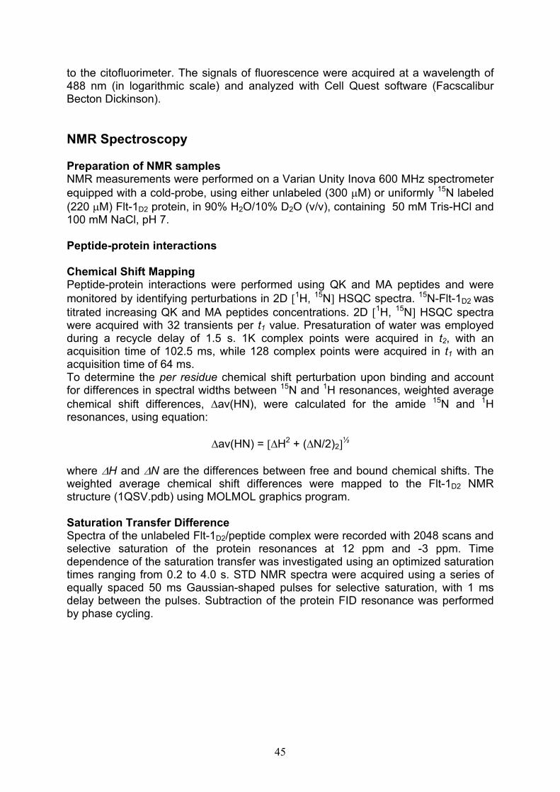
to the citofluorimeter. The signals of fluorescence were acquired at a wavelength of 488 nm (in logarithmic scale) and analyzed with Cell Quest software (Facscalibur Becton Dickinson).
NMR Spectroscopy
Preparation of NMR samplesNMR measurements were performed on a Varian Unity Inova 600 MHz spectrometer equipped with a cold-probe, using either unlabeled (300 M) or uniformly 15N labeled (220 M) Flt-1D2 protein, in 90% H2O/10% D2O (v/v), containing 50 mM Tris-HCl and 100 mM NaCl, pH 7.
Peptide-protein interactions
Chemical Shift MappingPeptide-protein interactions were performed using QK and MA peptides and were monitored by identifying perturbations in 2D 1H, 15N HSQC spectra. 15N-Flt-1D2 wastitrated increasing QK and MA peptides concentrations. 2D 1H, 15N HSQC spectra were acquired with 32 transients per t1 value. Presaturation of water was employed during a recycle delay of 1.5 s. 1K complex points were acquired in t2, with an acquisition time of 102.5 ms, while 128 complex points were acquired in t1 with an acquisition time of 64 ms. To determine the per residue chemical shift perturbation upon binding and account for differences in spectral widths between 15N and 1H resonances, weighted average chemical shift differences, av(HN), were calculated for the amide 15N and 1Hresonances, using equation:
av(HN) = H2 + ( N/2)2½
where H and N are the differences between free and bound chemical shifts. The weighted average chemical shift differences were mapped to the Flt-1D2 NMR structure (1QSV.pdb) using MOLMOL graphics program.
Saturation Transfer DifferenceSpectra of the unlabeled Flt-1D2/peptide complex were recorded with 2048 scans and selective saturation of the protein resonances at 12 ppm and -3 ppm. Time dependence of the saturation transfer was investigated using an optimized saturation times ranging from 0.2 to 4.0 s. STD NMR spectra were acquired using a series of equally spaced 50 ms Gaussian-shaped pulses for selective saturation, with 1 ms delay between the pulses. Subtraction of the protein FID resonance was performed by phase cycling.
45

Results
Domains cloning of human KDR and Flt-1 receptors
Cloning of 1-3, 2-3, 1-2 and 2 KDR receptor domains One step PCR subcloning technique was used to generate bacterial expression vectors containing the sequences encoding an N-terminal His-Tag, a TEV cleavage site and the extracellular domains 1-3, 2-3, 1-2 and 2 of VEGFR-2 (KDR). First, sequences D1-3 (residues 1-335, Fuh et al., 1998), D2-3 (residues 120-335, Fuh etal., 1998), D1-2 (residues 1-222, Fuh et al., 1998) and D2 (residues 120-222, Fuh etal., 1998) were amplified by PCR from NIH-3T3 cDNA of the entire receptor using the primers designed on the basis of the sequence of the same domains (see Materials and Methods section). This cloning strategy was used to obtain the gene products fused (at the N-terminal) with the 6xHistidine-tag of pPROEXHTa expression vector. Single products were amplified by PCR, purified and digested with BamHI-XhoIrestriction enzymes. In Figure 13 are showed the products purified after digestion.
a b c d
900/1000 bp
300 bp 200 bp
500/517 bp 400 bp
100 bp
Figure 13_ 1% agarose-gel analysis of KDR domains PCR products. In a, b, c and d arereported, respectively, the BamHI/XhoI digested kdrD1-2-3 (911 bp), kdrD1-2 (570 bp), kdrD2-3
(647 bp), kdrD2 (330 bp) genes amplified from NIH-3T3 KDR receptor cDNA. DNA molecularweight marker is the Ladder 100 bp.
pPROEXHTa expression vector was digested with the same enzymes, purified using the QIAquick Gel Extraction Kit and analyzed by electrophoresis on a 1% agarose gel (Figure 14a). kdrD2 gene was cloned also in pETM11 and pETM20 expression vectors. In this case, both vectors (Figure 14 b-c) and PCR fragment corresponding to the second domain of VEGFR-2 (Figure 15) were digested with NcoI/XhoIrestriction enzymes.
46

b
300 bp
100 bp
400 bp
200 bp
1517 bp
900/1000 bp
a c
500/517 bp
M D UD U MD M U
Figure 14 a-b-c_ 1% agarose-gel analysis of the digested vectors. In b lane labelled with “D” indicates the pPROEXHTa expression vector digested with BamH1/Xho1 restriction enzymes, while in a and c the NcoI/XhoI digested pETM20 and pETM11, respectively. Lanes marked with “U” indicate undigested vectors. M is the DNA molecular weight marker (Ladder 100 bp).
900/1000 bp
300 bp
500/517 bp 400 bp
100 bp 200 bp
M
Figure 15_ 1% agarose-gel analysis of the kdrD2
gene PCR product amplified from NIH-3T3 receptorcDNA and digested with NcoI/XhoI endonucleases.M, Ladder 100 bp.
All amplificates obtained from PCR reactions were ligated into the corresponding expression vectors; then, each recombinant construct resulted from cloning, was transformed in TOP F’10 cells by electroporation. Plasmidic DNA was extracted from overnight cultures and purified using the QIAprep Spin Miniprep Kit. The identity of the inserts was tested with a PCR screening (Figures 16, 17) and confirmed by DNA sequencing.
47
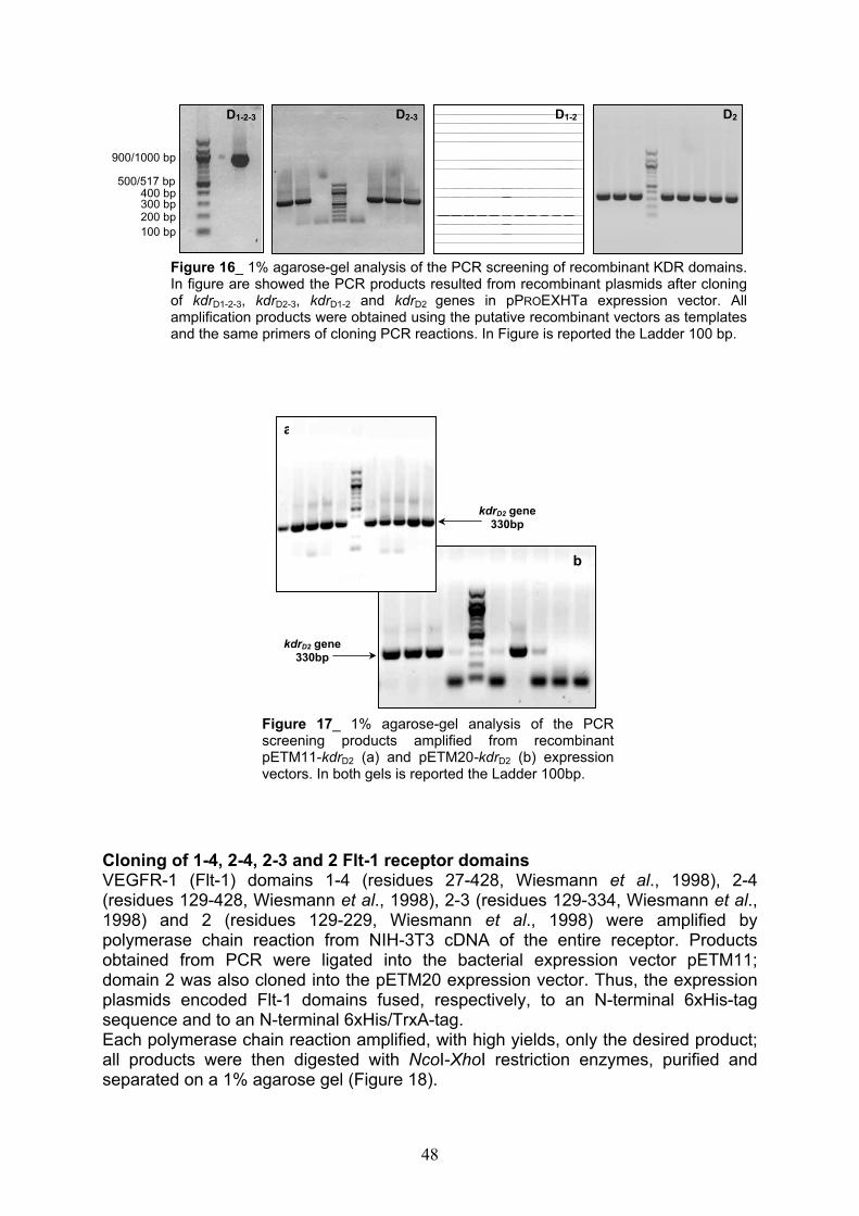
D2
300 bp
100 bp
500/517 bp
200 bp
900/1000 bp
D2-3 D1-2D1-2-3
400 bp
Figure 16_ 1% agarose-gel analysis of the PCR screening of recombinant KDR domains.In figure are showed the PCR products resulted from recombinant plasmids after cloningof kdrD1-2-3, kdrD2-3, kdrD1-2 and kdrD2 genes in pPROEXHTa expression vector. Allamplification products were obtained using the putative recombinant vectors as templatesand the same primers of cloning PCR reactions. In Figure is reported the Ladder 100 bp.
a
kdrD2 gene 330bp
b
kdrD2 gene 330bp
Figure 17_ 1% agarose-gel analysis of the PCRscreening products amplified from recombinantpETM11-kdrD2 (a) and pETM20-kdrD2 (b) expressionvectors. In both gels is reported the Ladder 100bp.
Cloning of 1-4, 2-4, 2-3 and 2 Flt-1 receptor domains VEGFR-1 (Flt-1) domains 1-4 (residues 27-428, Wiesmann et al., 1998), 2-4 (residues 129-428, Wiesmann et al., 1998), 2-3 (residues 129-334, Wiesmann et al.,1998) and 2 (residues 129-229, Wiesmann et al., 1998) were amplified by polymerase chain reaction from NIH-3T3 cDNA of the entire receptor. Products obtained from PCR were ligated into the bacterial expression vector pETM11; domain 2 was also cloned into the pETM20 expression vector. Thus, the expression plasmids encoded Flt-1 domains fused, respectively, to an N-terminal 6xHis-tag sequence and to an N-terminal 6xHis/TrxA-tag. Each polymerase chain reaction amplified, with high yields, only the desired product; all products were then digested with NcoI-XhoI restriction enzymes, purified and separated on a 1% agarose gel (Figure 18).
48
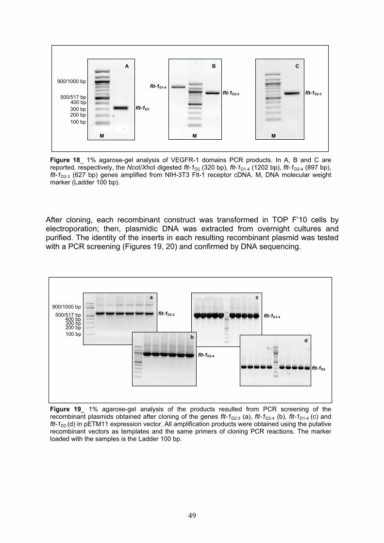
A B C
900/1000 bp flt-1D1-4
flt-1D2-4 flt-1D2-3500/517 bp
400 bp 300 bp 200 bp
flt-1D2
100 bp
M M M
Figure 18_ 1% agarose-gel analysis of VEGFR-1 domains PCR products. In A, B and C arereported, respectively, the NcoI/XhoI digested flt-1D2 (320 bp), flt-1D1-4 (1202 bp), flt-1D2-4 (897 bp),flt-1D2-3 (627 bp) genes amplified from NIH-3T3 Flt-1 receptor cDNA. M, DNA molecular weightmarker (Ladder 100 bp).
After cloning, each recombinant construct was transformed in TOP F’10 cells by electroporation; then, plasmidic DNA was extracted from overnight cultures and purified. The identity of the inserts in each resulting recombinant plasmid was tested with a PCR screening (Figures 19, 20) and confirmed by DNA sequencing.
a c
900/1000 bp flt-1D2-3500/517 bp
100 bp
400 bp 300 bp 200 bp
flt-1D1-4
bd
flt-1D2-4
flt-1D2
Figure 19_ 1% agarose-gel analysis of the products resulted from PCR screening of therecombinant plasmids obtained after cloning of the genes flt-1D2-3 (a), flt-1D2-4 (b), flt-1D1-4 (c) andflt-1D2 (d) in pETM11 expression vector. All amplification products were obtained using the putativerecombinant vectors as templates and the same primers of cloning PCR reactions. The markerloaded with the samples is the Ladder 100 bp.
49
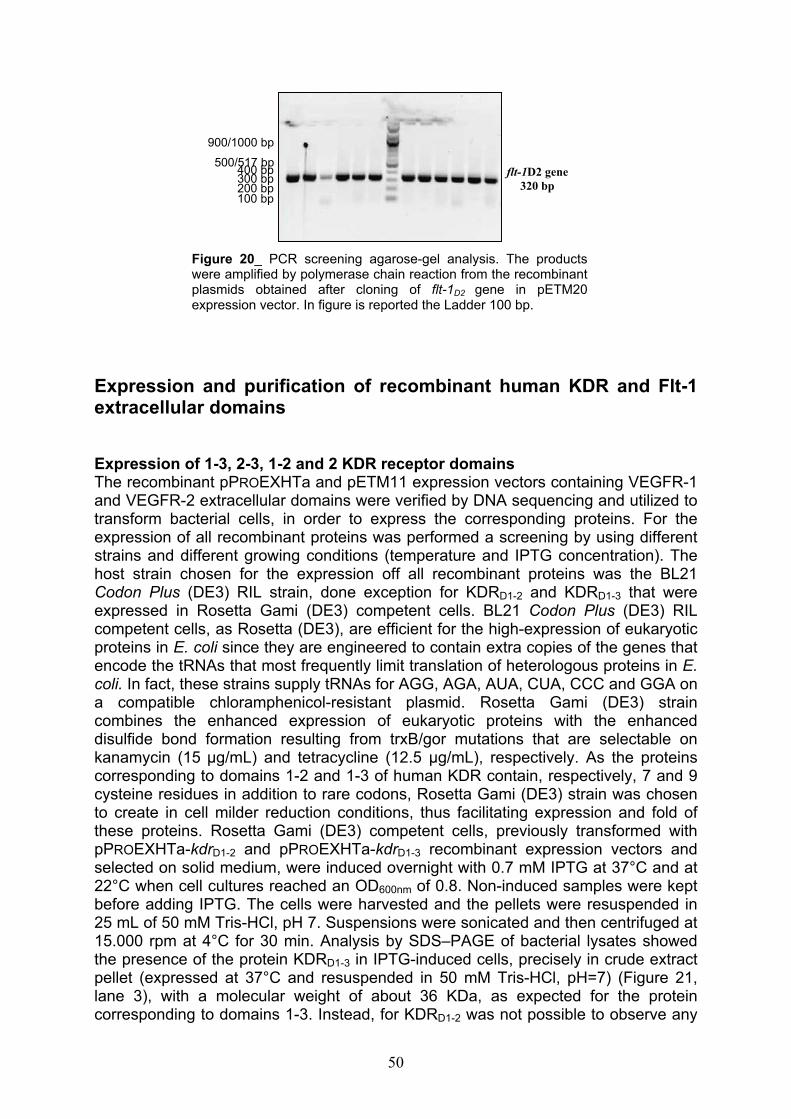
900/1000 bp
500/517 bp
100 bp
400 bp 300 bp 200 bp
flt-1D2 gene
320 bp
Figure 20_ PCR screening agarose-gel analysis. The productswere amplified by polymerase chain reaction from the recombinant plasmids obtained after cloning of flt-1D2 gene in pETM20 expression vector. In figure is reported the Ladder 100 bp.
Expression and purification of recombinant human KDR and Flt-1 extracellular domains
Expression of 1-3, 2-3, 1-2 and 2 KDR receptor domainsThe recombinant pPROEXHTa and pETM11 expression vectors containing VEGFR-1 and VEGFR-2 extracellular domains were verified by DNA sequencing and utilized to transform bacterial cells, in order to express the corresponding proteins. For the expression of all recombinant proteins was performed a screening by using different strains and different growing conditions (temperature and IPTG concentration). The host strain chosen for the expression off all recombinant proteins was the BL21 Codon Plus (DE3) RIL strain, done exception for KDRD1-2 and KDRD1-3 that were expressed in Rosetta Gami (DE3) competent cells. BL21 Codon Plus (DE3) RIL competent cells, as Rosetta (DE3), are efficient for the high-expression of eukaryotic proteins in E. coli since they are engineered to contain extra copies of the genes that encode the tRNAs that most frequently limit translation of heterologous proteins in E.coli. In fact, these strains supply tRNAs for AGG, AGA, AUA, CUA, CCC and GGA on a compatible chloramphenicol-resistant plasmid. Rosetta Gami (DE3) strain combines the enhanced expression of eukaryotic proteins with the enhanced disulfide bond formation resulting from trxB/gor mutations that are selectable on kanamycin (15 µg/mL) and tetracycline (12.5 µg/mL), respectively. As the proteins corresponding to domains 1-2 and 1-3 of human KDR contain, respectively, 7 and 9 cysteine residues in addition to rare codons, Rosetta Gami (DE3) strain was chosen to create in cell milder reduction conditions, thus facilitating expression and fold of these proteins. Rosetta Gami (DE3) competent cells, previously transformed with pPROEXHTa-kdrD1-2 and pPROEXHTa-kdrD1-3 recombinant expression vectors and selected on solid medium, were induced overnight with 0.7 mM IPTG at 37°C and at 22°C when cell cultures reached an OD600nm of 0.8. Non-induced samples were kept before adding IPTG. The cells were harvested and the pellets were resuspended in 25 mL of 50 mM Tris-HCl, pH 7. Suspensions were sonicated and then centrifuged at 15.000 rpm at 4°C for 30 min. Analysis by SDS–PAGE of bacterial lysates showed the presence of the protein KDRD1-3 in IPTG-induced cells, precisely in crude extract pellet (expressed at 37°C and resuspended in 50 mM Tris-HCl, pH=7) (Figure 21, lane 3), with a molecular weight of about 36 KDa, as expected for the protein corresponding to domains 1-3. Instead, for KDRD1-2 was not possible to observe any
50
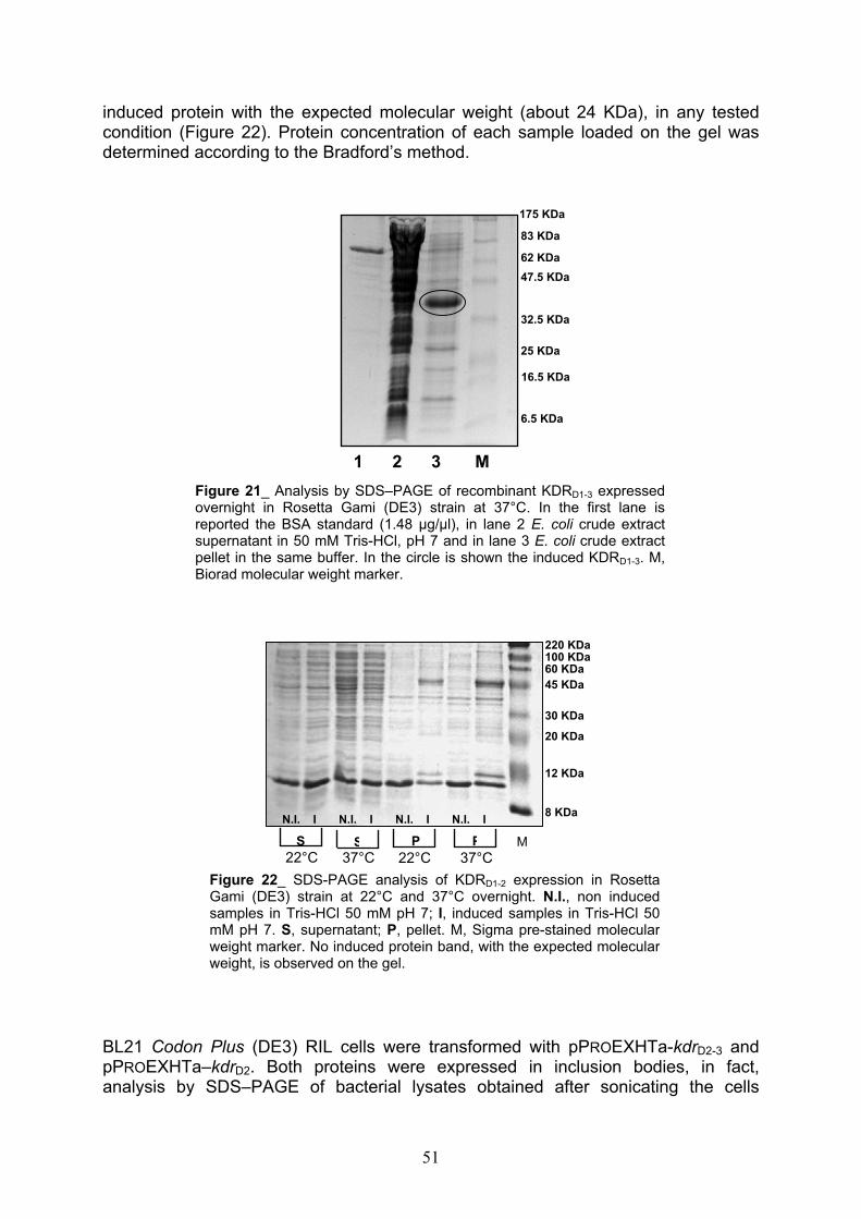
induced protein with the expected molecular weight (about 24 KDa), in any tested condition (Figure 22). Protein concentration of each sample loaded on the gel was determined according to the Bradford’s method.
175 KDa
83 KDa
62 KDa
47.5 KDa
32.5 KDa
25 KDa
16.5 KDa
6.5 KDa
1 2 3 M
Figure 21_ Analysis by SDS–PAGE of recombinant KDRD1-3 expressedovernight in Rosetta Gami (DE3) strain at 37°C. In the first lane isreported the BSA standard (1.48 µg/µl), in lane 2 E. coli crude extractsupernatant in 50 mM Tris-HCl, pH 7 and in lane 3 E. coli crude extractpellet in the same buffer. In the circle is shown the induced KDRD1-3. M,Biorad molecular weight marker.
220 KDa 100 KDa 60 KDa
45 KDa
30 KDa
20 KDa
12 KDa
8 KDa N.I. I N.I. I N.I. I N.I. I
22°CP
37°CP
22°CS M
37°CS
Figure 22_ SDS-PAGE analysis of KDRD1-2 expression in Rosetta Gami (DE3) strain at 22°C and 37°C overnight. N.I., non induced samples in Tris-HCl 50 mM pH 7; I, induced samples in Tris-HCl 50 mM pH 7. S, supernatant; P, pellet. M, Sigma pre-stained molecularweight marker. No induced protein band, with the expected molecularweight, is observed on the gel.
BL21 Codon Plus (DE3) RIL cells were transformed with pPROEXHTa-kdrD2-3 and pPROEXHTa–kdrD2. Both proteins were expressed in inclusion bodies, in fact, analysis by SDS–PAGE of bacterial lysates obtained after sonicating the cells
51
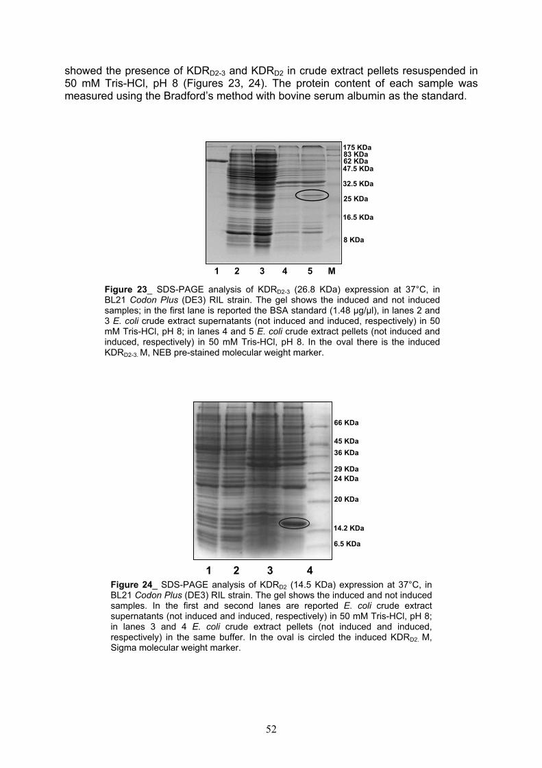
showed the presence of KDRD2-3 and KDRD2 in crude extract pellets resuspended in 50 mM Tris-HCl, pH 8 (Figures 23, 24). The protein content of each sample was measured using the Bradford’s method with bovine serum albumin as the standard.
175 KDa83 KDa 62 KDa47.5 KDa
32.5 KDa
25 KDa
16.5 KDa
8 KDa
1 2 3 4 5 M
Figure 23_ SDS-PAGE analysis of KDRD2-3 (26.8 KDa) expression at 37°C, inBL21 Codon Plus (DE3) RIL strain. The gel shows the induced and not inducedsamples; in the first lane is reported the BSA standard (1.48 µg/µl), in lanes 2 and3 E. coli crude extract supernatants (not induced and induced, respectively) in 50mM Tris-HCl, pH 8; in lanes 4 and 5 E. coli crude extract pellets (not induced andinduced, respectively) in 50 mM Tris-HCl, pH 8. In the oval there is the inducedKDRD2-3. M, NEB pre-stained molecular weight marker.
66 KDa
45 KDa
36 KDa
29 KDa
24 KDa
20 KDa
14.2 KDa
6.5 KDa
1 2 3 4
Figure 24_ SDS-PAGE analysis of KDRD2 (14.5 KDa) expression at 37°C, in BL21 Codon Plus (DE3) RIL strain. The gel shows the induced and not inducedsamples. In the first and second lanes are reported E. coli crude extract supernatants (not induced and induced, respectively) in 50 mM Tris-HCl, pH 8;in lanes 3 and 4 E. coli crude extract pellets (not induced and induced, respectively) in the same buffer. In the oval is circled the induced KDRD2. M,Sigma molecular weight marker.
52
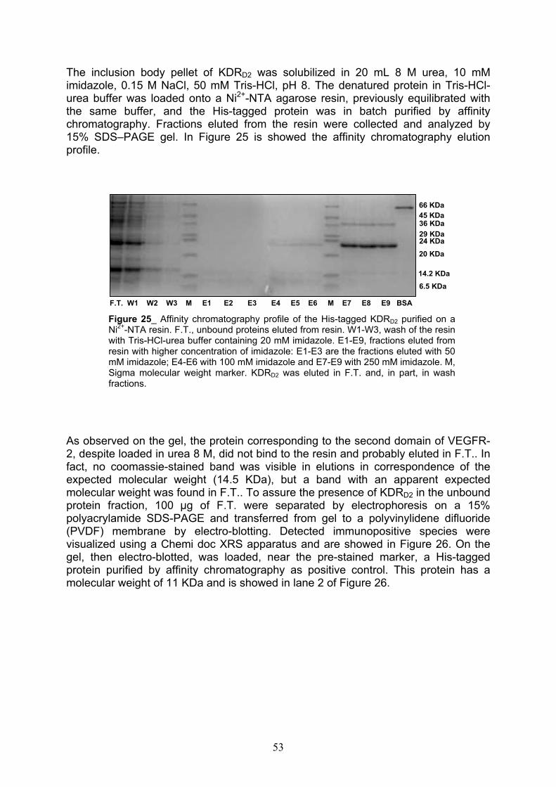
The inclusion body pellet of KDRD2 was solubilized in 20 mL 8 M urea, 10 mM imidazole, 0.15 M NaCl, 50 mM Tris-HCl, pH 8. The denatured protein in Tris-HCl-urea buffer was loaded onto a Ni2+-NTA agarose resin, previously equilibrated with the same buffer, and the His-tagged protein was in batch purified by affinity chromatography. Fractions eluted from the resin were collected and analyzed by 15% SDS–PAGE gel. In Figure 25 is showed the affinity chromatography elution profile.
66 KDa
45 KDa 36 KDa
29 KDa 24 KDa
20 KDa
14.2 KDa
6.5 KDa
F.T. W1 W2 W3 M E1 E2 E3 E4 E5 E6 M E7 E8 E9 BSA
Figure 25_ Affinity chromatography profile of the His-tagged KDRD2 purified on aNi2+-NTA resin. F.T., unbound proteins eluted from resin. W1-W3, wash of the resinwith Tris-HCl-urea buffer containing 20 mM imidazole. E1-E9, fractions eluted fromresin with higher concentration of imidazole: E1-E3 are the fractions eluted with 50mM imidazole; E4-E6 with 100 mM imidazole and E7-E9 with 250 mM imidazole. M,Sigma molecular weight marker. KDRD2 was eluted in F.T. and, in part, in washfractions.
As observed on the gel, the protein corresponding to the second domain of VEGFR-2, despite loaded in urea 8 M, did not bind to the resin and probably eluted in F.T.. In fact, no coomassie-stained band was visible in elutions in correspondence of the expected molecular weight (14.5 KDa), but a band with an apparent expected molecular weight was found in F.T.. To assure the presence of KDRD2 in the unbound protein fraction, 100 µg of F.T. were separated by electrophoresis on a 15% polyacrylamide SDS-PAGE and transferred from gel to a polyvinylidene difluoride (PVDF) membrane by electro-blotting. Detected immunopositive species were visualized using a Chemi doc XRS apparatus and are showed in Figure 26. On the gel, then electro-blotted, was loaded, near the pre-stained marker, a His-tagged protein purified by affinity chromatography as positive control. This protein has a molecular weight of 11 KDa and is showed in lane 2 of Figure 26.
53

positive control
KDRD2
1 M 2
Figure 26_ Western analysis of KDRD2. The first lane of theblot was found to contain a protein that migrates with theexpected molecular weight (in the oval). In lane 2 there isthe positive control constituted by a His-tagged protein witha molecular weight of 11 KDa. Both species wererecognized by an anti-His antibody. M, NEB pre-stainedmarker.
Western blotting analysis showed the presence of the His-tagged KDRD2 in F.T., but did not explain the unusual behavior of the protein. The second domain of VEGFR-2 was successively cloned in pETM11 and pETM20 expression vectors, but the recombinant protein was expressed in inclusion bodies and did not bind to the resin again (data not shown). So, its sequence was mutated and cloned again in pETM11.
kdrD2 gene mutagenesis As in vitro site-directed mutagenesis is an available technique for studying protein structure-function relationship and gene expression, we used this approach to change one of the three residues of cysteine (C161) of the second domain of KDR receptor in Alanine. Our aim was to obtain a protein capable to bind to the Ni2+-NTAresin and to be purified by affinity chromatography as the second domain of VEGFR-1 that has only two cysteine residues in its sequence (see below). According to the procedure suggested by the Quik Change Site Directed Mutagenesis Kit, we utilized a supercoiled double-stranded DNA (dsDNA) vector with the insert of interest and two synthetic oligonucleotide primers contained the desired mutation. In our case, was used the pETM11-kdrD2 plasmid and the oligonucleotide primers, each complementary to opposite strands of the vector, contained the mutation for an alanine in the middle. The primers were extended during temperature cycling; their incorporation generated a mutated plasmid containing staggered nicks. In fact, as we can see in Figure 27, PCR mutagenesis product (mut) is shifted on the agarose gel with respect to the control represented by the non mutated construct pETM11-kdrD2
(wt), as attended for a nicked dsDNA plasmid. Following PCR reaction, the product was treated with DpnI, an endonuclease specific for methylated and hemimethylated DNA, used to digest the parental DNA template and to select for mutation-containing
54
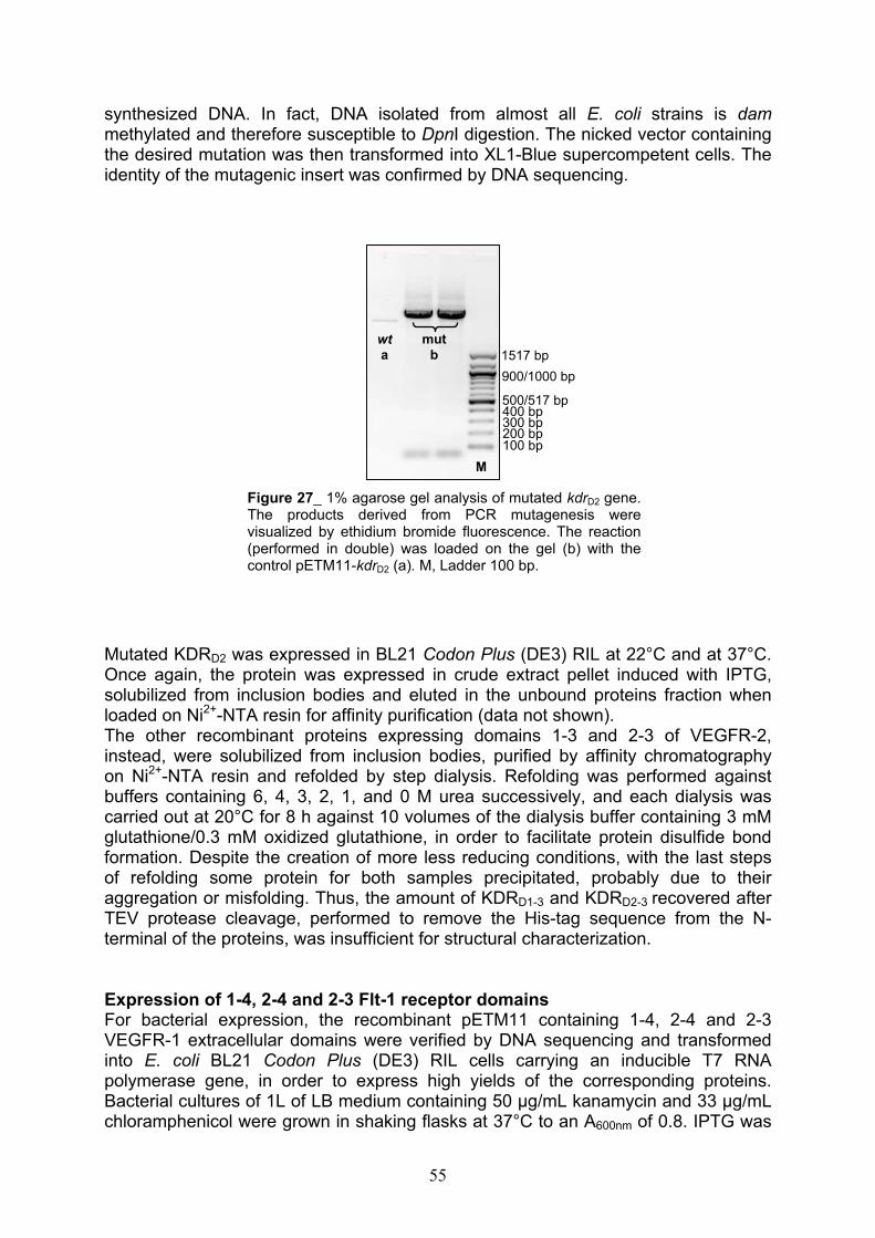
synthesized DNA. In fact, DNA isolated from almost all E. coli strains is dammethylated and therefore susceptible to DpnI digestion. The nicked vector containing the desired mutation was then transformed into XL1-Blue supercompetent cells. The identity of the mutagenic insert was confirmed by DNA sequencing.
mut
b
wt
a 1517 bp
900/1000 bp
300 bp
500/517 bp 400 bp
100 bp 200 bp
M
Figure 27_ 1% agarose gel analysis of mutated kdrD2 gene.The products derived from PCR mutagenesis werevisualized by ethidium bromide fluorescence. The reaction(performed in double) was loaded on the gel (b) with thecontrol pETM11-kdrD2 (a). M, Ladder 100 bp.
Mutated KDRD2 was expressed in BL21 Codon Plus (DE3) RIL at 22°C and at 37°C. Once again, the protein was expressed in crude extract pellet induced with IPTG, solubilized from inclusion bodies and eluted in the unbound proteins fraction when loaded on Ni2+-NTA resin for affinity purification (data not shown).The other recombinant proteins expressing domains 1-3 and 2-3 of VEGFR-2, instead, were solubilized from inclusion bodies, purified by affinity chromatography on Ni2+-NTA resin and refolded by step dialysis. Refolding was performed against buffers containing 6, 4, 3, 2, 1, and 0 M urea successively, and each dialysis was carried out at 20°C for 8 h against 10 volumes of the dialysis buffer containing 3 mM glutathione/0.3 mM oxidized glutathione, in order to facilitate protein disulfide bond formation. Despite the creation of more less reducing conditions, with the last steps of refolding some protein for both samples precipitated, probably due to their aggregation or misfolding. Thus, the amount of KDRD1-3 and KDRD2-3 recovered after TEV protease cleavage, performed to remove the His-tag sequence from the N-terminal of the proteins, was insufficient for structural characterization.
Expression of 1-4, 2-4 and 2-3 Flt-1 receptor domainsFor bacterial expression, the recombinant pETM11 containing 1-4, 2-4 and 2-3 VEGFR-1 extracellular domains were verified by DNA sequencing and transformed into E. coli BL21 Codon Plus (DE3) RIL cells carrying an inducible T7 RNA polymerase gene, in order to express high yields of the corresponding proteins. Bacterial cultures of 1L of LB medium containing 50 µg/mL kanamycin and 33 µg/mLchloramphenicol were grown in shaking flasks at 37°C to an A600nm of 0.8. IPTG was
55

added to a final concentration of 0.7 mM and the cultures were grown for another 3 h. From each culture was kept a non induced sample, before adding IPTG. Analyses by SDS–PAGE of bacterial lysates obtained after sonicating the cells showed the presence of all interest proteins in IPTG-induced crude extract pellet fractions (Figures 28, 29, 30).
66KDa
45KDa
36KDa
29KDa24KDa
20KDa
14.2KDa
Figure 28_ SDS-PAGE analysis of Flt-1D1-4 (48.7 KDa) expressed in E. coli BL21Codon Plus (DE3) RIL strain at 37°C. Gel shows the induced and not inducedsamples. In the first and second lanes are reported E. coli crude extractsupernatants (not induced and induced, respectively) in 50 mM Tris-HCl, pH 8; inlanes 3 and 4 E. coli crude extract pellets (not induced and induced, respectively)in the same buffer. Black arrow indicates the induced protein. M, Sigma molecularweight marker.
1 2 M 3 4 BSA
Flt-1D2-3
45KDa
66KDa
29KDa24KDa
36KDa
20KDa
14.2KDa
6.5KDa1 2 3 4 M 1 2 3 4 M
Flt-1D2-4
Figure 29_ SDS-PAGE analysis of expressed Flt-1D2-4 and Flt-1D2-3 (respectively 37.4and 26.8 KDa). Gels show the induced and not induced samples. In the first and secondlanes of both gels are reported E. coli crude extract pellets (not induced and induced,respectively) in Tris-HCl 50 mM, pH 8; in lanes 3 and 4 E. coli crude extract supernatants(not induced and induced, respectively) in the same buffer. In the ovals are reported theinduced proteins. M, Sigma molecular weight marker.
56
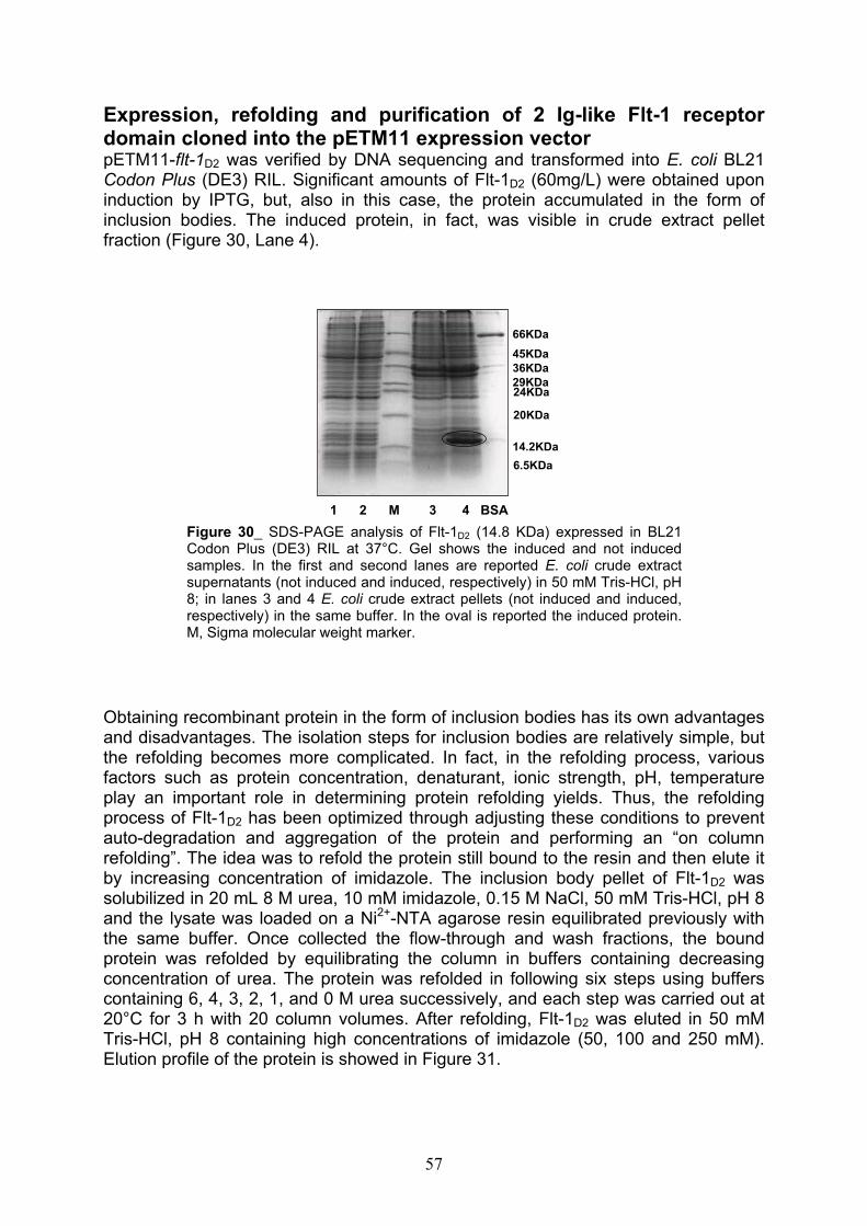
Expression, refolding and purification of 2 Ig-like Flt-1 receptor domain cloned into the pETM11 expression vector pETM11-flt-1D2 was verified by DNA sequencing and transformed into E. coli BL21 Codon Plus (DE3) RIL. Significant amounts of Flt-1D2 (60mg/L) were obtained upon induction by IPTG, but, also in this case, the protein accumulated in the form of inclusion bodies. The induced protein, in fact, was visible in crude extract pellet fraction (Figure 30, Lane 4).
1 2 M 3 4 BSA
66KDa
45KDa
29KDa24KDa
36KDa
20KDa
14.2KDa
6.5KDa
Figure 30_ SDS-PAGE analysis of Flt-1D2 (14.8 KDa) expressed in BL21Codon Plus (DE3) RIL at 37°C. Gel shows the induced and not inducedsamples. In the first and second lanes are reported E. coli crude extractsupernatants (not induced and induced, respectively) in 50 mM Tris-HCl, pH8; in lanes 3 and 4 E. coli crude extract pellets (not induced and induced,respectively) in the same buffer. In the oval is reported the induced protein.M, Sigma molecular weight marker.
Obtaining recombinant protein in the form of inclusion bodies has its own advantages and disadvantages. The isolation steps for inclusion bodies are relatively simple, but the refolding becomes more complicated. In fact, in the refolding process, various factors such as protein concentration, denaturant, ionic strength, pH, temperature play an important role in determining protein refolding yields. Thus, the refolding process of Flt-1D2 has been optimized through adjusting these conditions to prevent auto-degradation and aggregation of the protein and performing an “on column refolding”. The idea was to refold the protein still bound to the resin and then elute it by increasing concentration of imidazole. The inclusion body pellet of Flt-1D2 was solubilized in 20 mL 8 M urea, 10 mM imidazole, 0.15 M NaCl, 50 mM Tris-HCl, pH 8 and the lysate was loaded on a Ni2+-NTA agarose resin equilibrated previously with the same buffer. Once collected the flow-through and wash fractions, the bound protein was refolded by equilibrating the column in buffers containing decreasing concentration of urea. The protein was refolded in following six steps using buffers containing 6, 4, 3, 2, 1, and 0 M urea successively, and each step was carried out at 20°C for 3 h with 20 column volumes. After refolding, Flt-1D2 was eluted in 50 mM Tris-HCl, pH 8 containing high concentrations of imidazole (50, 100 and 250 mM). Elution profile of the protein is showed in Figure 31.
57
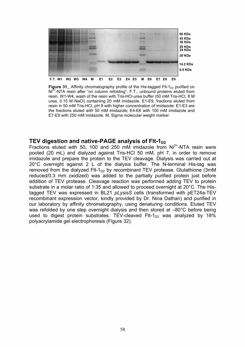
66 KDa
45 KDa 36 KDa
29 KDa 24 KDa
20 KDa
14.2 KDa
6.5 KDa
F.T. W1 W2 W3 W4 M E1 E2 E3 E4 E5 M E6 E7 E8 E9
Figure 31_ Affinity chromatography profile of the His-tagged Flt-1D2 purified onNi2+-NTA resin after “on column refolding”. F.T., unbound proteins eluted fromresin. W1-W4, wash of the resin with Tris-HCl-urea buffer (50 mM Tris-HCl, 8 Murea, 0.15 M NaCl) containing 20 mM imidazole. E1-E9, fractions eluted fromresin in 50 mM Tris-HCl, pH 8 with higher concentration of imidazole: E1-E3 arethe fractions eluted with 50 mM imidazole; E4-E6 with 100 mM imidazole andE7-E9 with 250 mM imidazole. M, Sigma molecular weight marker.
TEV digestion and native-PAGE analysis of Flt-1D2
Fractions eluted with 50, 100 and 250 mM imidazole from Ni2+-NTA resin were pooled (20 mL) and dialyzed against Tris-HCl 50 mM, pH 7, in order to remove imidazole and prepare the protein to the TEV cleavage. Dialysis was carried out at 20°C overnight against 2 L of the dialysis buffer. The N-terminal His-tag was removed from the dialyzed Flt-1D2 by recombinant TEV protease. Glutathione (3mM reduced/0.3 mm oxidized) was added to the partially purified protein just before addition of TEV protease. Cleavage reaction was performed adding TEV to protein substrate in a molar ratio of 1:35 and allowed to proceed overnight at 20°C. The His-tagged TEV was expressed in BL21 pLysisS cells (transformed with pET24a-TEV recombinant expression vector, kindly provided by Dr. Nina Dathan) and purified in our laboratory by affinity chromatography, using denaturing conditions. Eluted TEV was refolded by one step overnight dialysis and then stored at –80°C before being used to digest protein substrates. TEV-cleaved Flt-1D2 was analyzed by 18% polyacrylamide gel electrophoresis (Figure 32).
58

66 KDa
45 KDa36 KDa
29 KDa 24 KDa
20 KDa
14.2 KDa
6.5 KDa
BSA TEV 1 2 M
Figure 32_ SDS-PAGE analysis of thecleaved Flt-1D2. Lane 1, negativecontrol constituted by the uncut protein;lane 2, cleaved Flt-1D2. M, Sigmamolecular weight marker.
Then, mixture obtained after TEV cleavage was subjected to a second step of affinity purification on Ni2+-NTA resin and finally concentrated until 0.3 mM by using the Amicon Ultra system with a molecular weight cut off of 5000. Flt-1D2 concentration was estimated according to the Bradford’s method and by UV spectroscopy, reading the tryptophan absorbance at 280 nm, and verified by SDS-PAGE analysis. As observed on the gel, the protein of interest resulted as a homogeneous product (Figure 33).
66KDa
45KDa
36KDa
29KDa24KDa
20KDa
14.2KDa
6.5KDa
Figure 33_ SDS-PAGE analysis of Flt-1D2
after concentration in Amicon Ultra. Asvisualized by comassie-staining, theprotein was purified homogeneously. NearFlt-1D2 was loaded Sigma molecularweight marker.
59

An aliquot of Flt-1D2 was loaded on native-PAGE for an analysis of its conformation in absence of denaturing detergents and reducing agents. For native PAGE the sample was not denaturized at 100°C but loaded on a 10% polyacrylamide gel in 0.001% bromophenol blue and 10% glycerol and electrophoresed in Tris-glycine buffer without SDS. As shown in Figure 34, the protein was able to migrate into the gel showing a single coomassie-stained band and suggesting its monomeric conformation.
Figure 34_ Native-PAGEanalysis of Flt-1D2 cleaved byTEV and purified. As shown,coomassie-stained protein bandentered the gel, withoutremaining into the well.
In order to obtain the second domain of VEGFR-1 in a soluble form, flt-1D2 gene was cloned in pETM20 expression vector. This cloning strategy allows the expression of the protein of interest with an N-terminal tag constituted by TrxA, a very soluble protein in most of cases able to solubilize its fusion partner.In spite of this, the recombinant Flt-1D2 was again expressed in inclusion bodies (data not shown).
LC-MS analyses of Flt-1D2 after TEV cleavage, alkylation and trypsindigestionPurity and identity of Flt-1D2 before and after TEV protease cleavage were assessed by liquid chromatography mass spectrometry. The analysis of proteins was carried out on a C4 column, using the method A described in “Strains, enzymes and reagents” section of Materials and Methods. In Figure 35 are reported the chromatogram and mass spectrum of the His-tagged protein purified by affinity chromatography after “on column refolding”, while in Figure 36 are showed the chromatogram and mass spectrum of the untagged protein. The molecular weights calculated from the experimental data were in accordance with theoretical ones, for both proteins. Successively, the protein was employed in experiments of alkylation by iodoacetamide, in order to determine the oxidation state of its two cysteine residues. In fact, as previously described, these two cysteine residues are involved in the
60
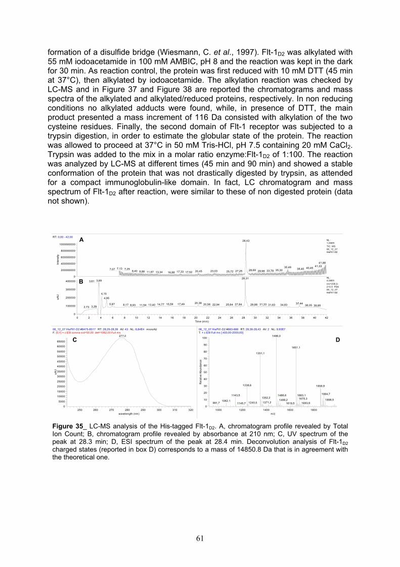
formation of a disulfide bridge (Wiesmann, C. et al., 1997). Flt-1D2 was alkylated with 55 mM iodoacetamide in 100 mM AMBIC, pH 8 and the reaction was kept in the dark for 30 min. As reaction control, the protein was first reduced with 10 mM DTT (45 min at 37°C), then alkylated by iodoacetamide. The alkylation reaction was checked by LC-MS and in Figure 37 and Figure 38 are reported the chromatograms and mass spectra of the alkylated and alkylated/reduced proteins, respectively. In non reducing conditions no alkylated adducts were found, while, in presence of DTT, the main product presented a mass increment of 116 Da consisted with alkylation of the two cysteine residues. Finally, the second domain of Flt-1 receptor was subjected to a trypsin digestion, in order to estimate the globular state of the protein. The reaction was allowed to proceed at 37°C in 50 mM Tris-HCl, pH 7.5 containing 20 mM CaCl2.Trypsin was added to the mix in a molar ratio enzyme:Flt-1D2 of 1:100. The reaction was analyzed by LC-MS at different times (45 min and 90 min) and showed a stable conformation of the protein that was not drastically digested by trypsin, as attended for a compact immunoglobulin-like domain. In fact, LC chromatogram and mass spectrum of Flt-1D2 after reaction, were similar to these of non digested protein (data not shown).
RT: 0,00 - 42,00
0 2 4 6 8 10 12 14 16 18 20 22 24 26 28 30 32 34 36 38 40 42
Time (min)
0
100000
200000
300000
400000
uA
U
0
200000000
400000000
600000000
800000000
1000000000
Inte
nsi
ty
28,43
41,8841,3335,49 40,457,13 38,457,257,07 35,3028,69 33,7829,9823,038,40 27,2520,459,88 25,7217,3311,97 17,5213,34 16,88
28,313,693,61
4,15
4,95
20,38 37,4417,495,87 15,59 31,3320,58 27,8414,77 25,6422,94 29,8813,40 31,638,17 34,8311,548,93 38,05 39,853,292,73
NL:1,05E9
TIC MS06_12_07HisFlt1-D2
NL:4,08E5
nm=209,5-210,5 PDA06_12_07HisFlt1-D2
06_12_07 HisFlt1-D2 #8475-8517 RT: 28,25-28,39 AV: 43 NL: 6,84E4 microAUF:
250 260 270 280 290 300 310 320
wavelength (nm)
0
5000
10000
15000
20000
25000
30000
35000
40000
45000
50000
55000
60000
65000
uA
U
277,0
06_12_07 HisFlt1-D2 #663-666 RT: 28,38-28,43 AV: 2 NL: 9,93E7T: + c ESI Full ms [ 400,00-2000,00]
1000 1200 1400 1600 1800
m/z
0
10
20
30
40
50
60
70
80
90
100
Re
lativ
e A
bu
nd
an
ce
1486,0
1651,1
1351,1
1238,6 1856,9
1884,71663,11143,5 1486,61352,2 1675,3 1898,51499,21062,11371,21240,5991,7 1693,91619,51145,7
B
C D
A
{0;1} + c ESI corona sid=50,00 det=1082,00 Full ms
Figure 35_ LC-MS analysis of the His-tagged Flt-1D2. A, chromatogram profile revealed by TotalIon Count; B, chromatogram profile revealed by absorbance at 210 nm; C, UV spectrum of thepeak at 28.3 min; D, ESI spectrum of the peak at 28.4 min. Deconvolution analysis of Flt-1D2
charged states (reported in box D) corresponds to a mass of 14850.8 Da that is in agreement withthe theoretical one.
61
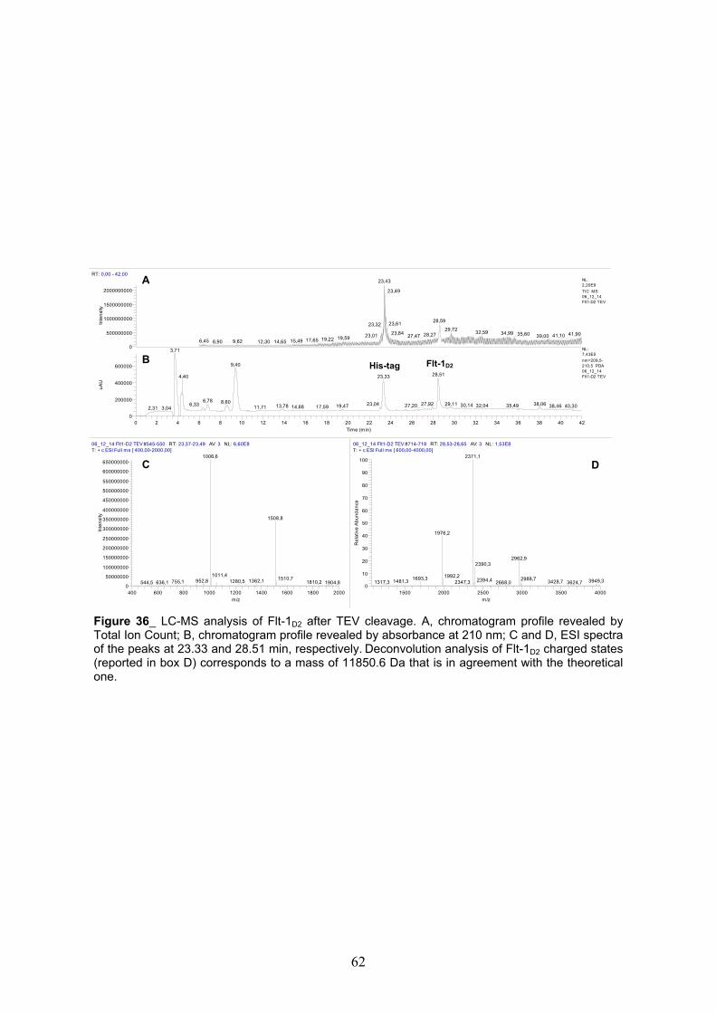
RT: 0,00 - 42,00
0 2 4 6 8 10 12 14 16 18 20 22 24 26 28 30 32 34 36 38 40 42
Time (min)
0
200000
400000
600000
uA
U
0
500000000
1000000000
1500000000
2000000000
Inte
nsi
ty
23,43
23,49
28,5923,6123,32
29,7232,59 34,9923,84 35,60 41,9028,2723,01 41,1027,47 39,0019,5919,2217,6515,496,45 9,626,90 14,6512,30
3,71
9,40
28,514,40 23,33
6,78 8,60 38,0629,1127,9223,046,33 30,1427,20 32,04 35,4913,78 38,4619,47 40,3017,5914,6811,713,042,31
NL:2,20E9
TIC MS06_12_14Flt1-D2 TEV
NL:7,43E5
nm=209,5-210,5 PDA06_12_14Flt1-D2 TEV
06_12_14 Flt1-D2 TEV #545-550 RT: 23,37-23,49 AV: 3 NL: 6,60E8T: + c ESI Full ms [ 400,00-2000,00]
400 600 800 1000 1200 1400 1600 1800 2000
m/z
0
50000000
100000000
150000000
200000000
250000000
300000000
350000000
400000000
450000000
500000000
550000000
600000000
650000000
Inte
nsi
ty
1006,6
1508,8
1011,41510,71362,1952,8 1280,5755,1 1810,2 1904,8636,1544,5
06_12_14 Flt1-D2 TEV #714-718 RT: 28,53-28,65 AV: 3 NL: 1,53E8T: + c ESI Full ms [ 600,00-4000,00]
1500 2000 2500 3000 3500 4000
m/z
0
10
20
30
40
50
60
70
80
90
100
Re
lativ
e A
bu
nd
an
ce
2371,1
1976,2
2962,92390,3
1992,21693,3 2988,72394,4 3949,31481,3 3428,71317,3 2347,3 2668,0 3624,7
A
B Flt-1D2His-tag
C D
Figure 36_ LC-MS analysis of Flt-1D2 after TEV cleavage. A, chromatogram profile revealed byTotal Ion Count; B, chromatogram profile revealed by absorbance at 210 nm; C and D, ESI spectraof the peaks at 23.33 and 28.51 min, respectively. Deconvolution analysis of Flt-1D2 charged states(reported in box D) corresponds to a mass of 11850.6 Da that is in agreement with the theoreticalone.
62
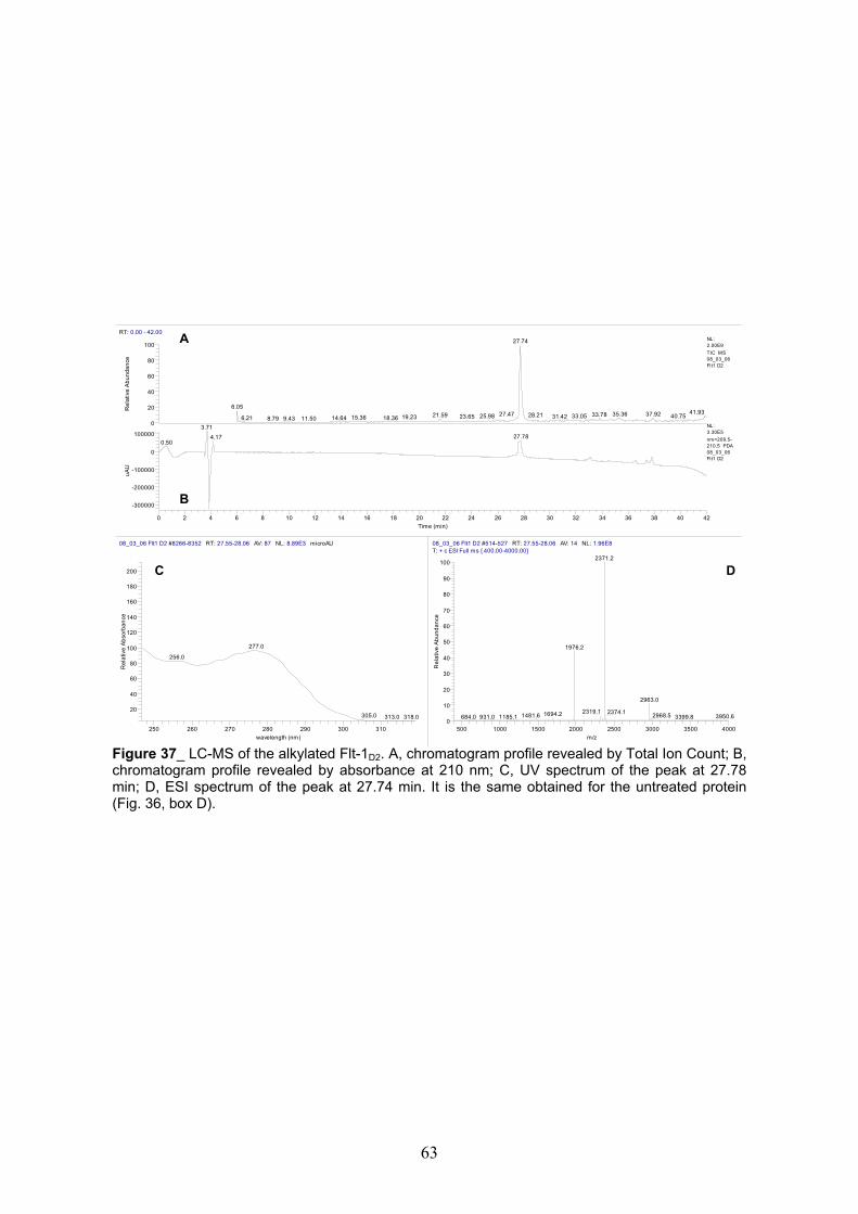
RT: 0.00 - 42.00
0 2 4 6 8 10 12 14 16 18 20 22 24 26 28 30 32 34 36 38 40 42
Time (min)
-300000
-200000
-100000
0
100000
uA
U
0
20
40
60
80
100
Re
lativ
e A
bu
nd
an
ce
27.74
6.0541.9327.47 37.9235.3633.7821.59 28.21 33.0525.98 40.7531.4223.6519.2315.36 18.366.21 14.6411.508.79 9.43
3.71
27.784.170.50
NL:2.00E9
TIC MS08_03_06Flt1 D2
NL:3.30E5
nm=209.5-210.5 PDA08_03_06Flt1 D2
08_03_06 Flt1 D2 #8266-8352 RT: 27.55-28.06 AV: 87 NL: 8.89E3 microAU
250 260 270 280 290 300 310
wavelength (nm)
20
40
60
80
100
120
140
160
180
200
Re
lativ
e A
bso
rba
nce
277.0
256.0
305.0 313.0 318.0
08_03_06 Flt1 D2 #514-527 RT: 27.55-28.06 AV: 14 NL: 1.96E8T: + c ESI Full ms [ 400.00-4000.00]
500 1000 1500 2000 2500 3000 3500 4000
m/z
0
10
20
30
40
50
60
70
80
90
100
Re
lativ
e A
bu
nd
an
ce
2371.2
1976.2
2963.0
2319.1 2374.11694.2 2968.51481.6 3950.63399.81185.1931.0684.0
B
C
A
D
Figure 37_ LC-MS of the alkylated Flt-1D2. A, chromatogram profile revealed by Total Ion Count; B,chromatogram profile revealed by absorbance at 210 nm; C, UV spectrum of the peak at 27.78min; D, ESI spectrum of the peak at 27.74 min. It is the same obtained for the untreated protein(Fig. 36, box D).
63
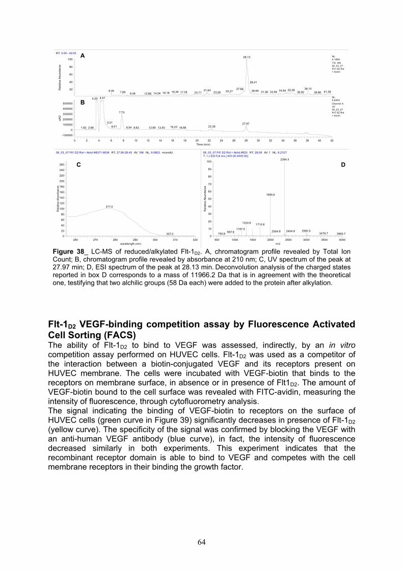
RT: 0.00 - 42.00
0 2 4 6 8 10 12 14 16 18 20 22 24 26 28 30 32 34 36 38 40 42
Time (min)
-100000
0
100000
200000
300000
400000
500000
uA
U
20
40
60
80
100R
ela
tive
Ab
un
da
nce
28.13
28.41
38.1027.66 35.3821.64 34.8428.696.05 25.27 41.3816.39 33.5931.367.89 36.9217.28 39.6623.2820.7116.1814.0412.669.49
4.574.25
7.73
5.21 27.976.01 22.3916.228.54 9.63 12.69 13.50 16.982.681.93
NL:5.14E8
TIC MS08_03_07Flt1 D2 Rid+ Alchil
NL:5.64E5
Channel AUV08_03_07Flt1 D2 Rid+ Alchil
08_03_07 Flt1 D2 Rid + Alchil #8371-8536 RT: 27.90-28.45 AV: 166 NL: 9.08E3 microAU
260 270 280 290 300 310 320
wavelength (nm)
0
20
40
60
80
100
120
140
160
180
200
220
240
260
Re
lativ
e A
bso
rba
nce
277.0
307.0
08_03_07 Flt1 D2 Rid + Alchil #520 RT: 28.09 AV: 1 NL: 6.21E7T: + c ESI Full ms [ 400.00-4000.00]
500 1000 1500 2000 2500 3000 3500 4000
m/z
0
10
20
30
40
50
60
70
80
90
100
Re
lativ
e A
bu
nd
an
ce
2394.4
1995.6
1330.61710.8
1197.52992.52404.92004.8997.9 3479.7 3990.7750.9
B
C D
A
Figure 38_ LC-MS of reduced/alkylated Flt-1D2. A, chromatogram profile revealed by Total IonCount; B, chromatogram profile revealed by absorbance at 210 nm; C, UV spectrum of the peak at27.97 min; D, ESI spectrum of the peak at 28.13 min. Deconvolution analysis of the charged statesreported in box D corresponds to a mass of 11966.2 Da that is in agreement with the theoreticalone, testifying that two alchilic groups (58 Da each) were added to the protein after alkylation.
Flt-1D2 VEGF-binding competition assay by Fluorescence Activated Cell Sorting (FACS) The ability of Flt-1D2 to bind to VEGF was assessed, indirectly, by an in vitrocompetition assay performed on HUVEC cells. Flt-1D2 was used as a competitor of the interaction between a biotin-conjugated VEGF and its receptors present on HUVEC membrane. The cells were incubated with VEGF-biotin that binds to the receptors on membrane surface, in absence or in presence of Flt1D2. The amount of VEGF-biotin bound to the cell surface was revealed with FITC-avidin, measuring the intensity of fluorescence, through cytofluorometry analysis.The signal indicating the binding of VEGF-biotin to receptors on the surface of HUVEC cells (green curve in Figure 39) significantly decreases in presence of Flt-1D2
(yellow curve). The specificity of the signal was confirmed by blocking the VEGF with an anti-human VEGF antibody (blue curve), in fact, the intensity of fluorescence decreased similarly in both experiments. This experiment indicates that the recombinant receptor domain is able to bind to VEGF and competes with the cell membrane receptors in their binding the growth factor.
64

Num
ber
of ce
lls
Fluorescence (expressed in log10)
Figure 39_ Analysis of the binding of VEGF-biotin to receptors on HUVEC cell membrane. The redcurve represents the cells incubated with a not specific biotinylated protein + FITC-avidin (negativecontrol); the green curve shows the cells treated with VEGF-biotin + FITC-avidin (positive control);the blue curve represents the cells incubated with VEGF-biotin + FITC-avidin in the presence ofanti-human VEGF IgG that blocks VEGF, and the yellow curve indicates the cells incubated withVEGF-biotin + FITC-avidin in the presence of Flt-1D2.
Expression, refolding and purification of 15N-Flt-1D2
E. coli JM101 cells were inoculated in M9 medium, containing 1g/L 15NH4Cl as the sole nitrogen source, and grown overnight at 37°C. The harvested cells were lysed with 37% HCl and the mixture of single 15N labeled amino acids was used to enrich M9 medium for the expression of Flt-1D2 in BL21 Codon Plus (DE3) strain previously transformed with pETM11-flt-1D2 recombinant expression vector. Refolding and purification of the labeled Flt-1D2 (referred to as 15N-Flt-1D2) were successfully performed using the same conditions described for the unlabeled protein. The yield of 15N-Flt-1D2 was comparable to that obtained for the unlabeled protein. The homogeneous product was concentrated until 0.22 mM and employed, together with the unlabeled protein, in protein-peptide NMR interaction studies.
CD spectroscopic analysisCD spectroscopic analyses were performed on both labeled and unlabeled proteins. Proteins were tested at a concentration of 15 µM in 20 mM phosphate buffer, pH 7.0 and spectra were recorded at 20°C in the range 190-260 nm. The overall far-UV CD spectrum of Flt-1D2 at neutral pH is shown in Figure 40. It indicates that the protein has high sheet content, with two negative bands at about 198 nm and 214 nm, suggesting that the protein assumes the correct fold expected for an immunoglobulin-like domain and in agreement with data reported in the literature (Wiesmann, C. etal., 1997 and Starovasnik, M. A. et al., 1999). To perform heat-denaturation experiments, the proteins under test were dissolved in 20 mM phosphate buffer, pH 7.0 at a concentration of 15 µM. Thermal unfolding curve of Flt-1D2 is shown in Figure 41. The curve shows a sigmoidal behavior, with a single transition and a Tm of about 60°C, indicating that a two-state unfolding
65
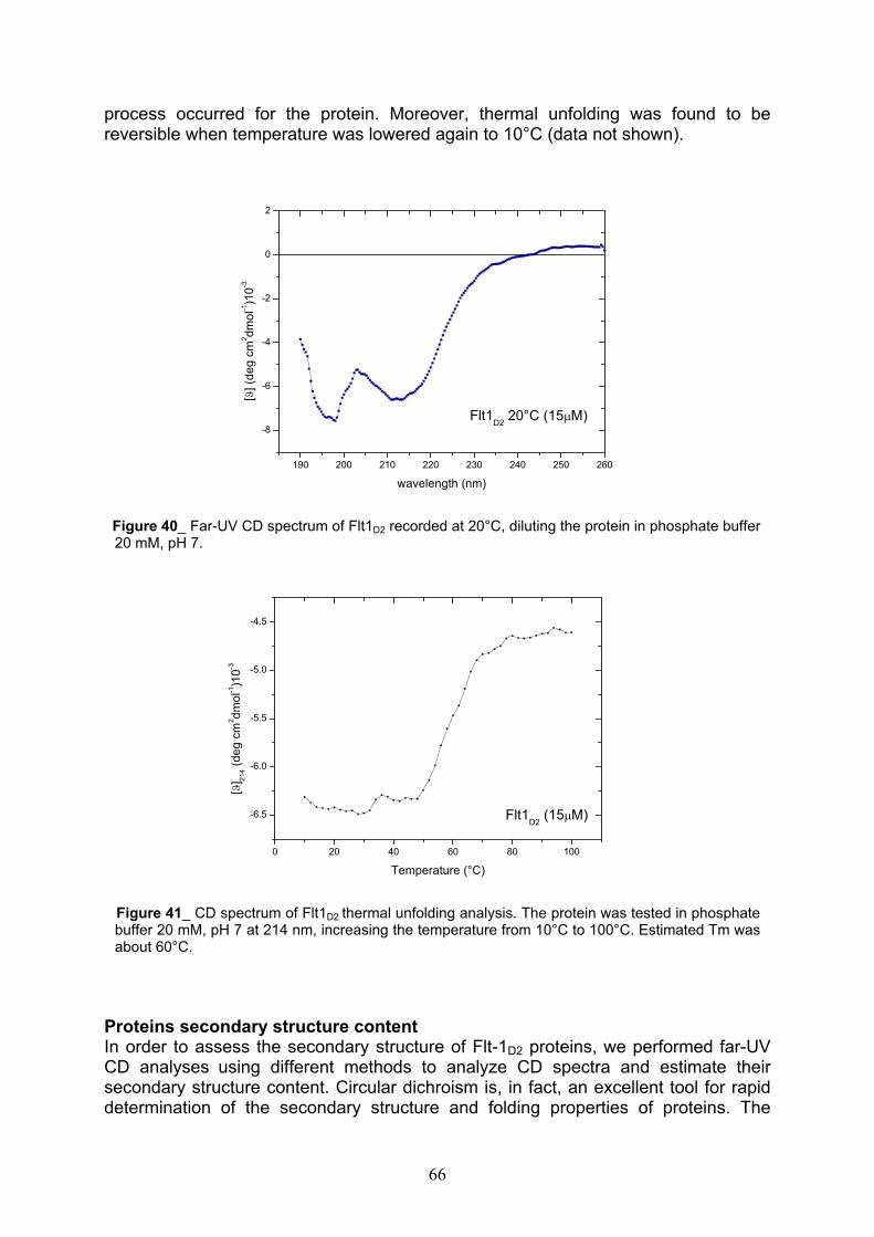
process occurred for the protein. Moreover, thermal unfolding was found to be reversible when temperature was lowered again to 10°C (data not shown).
190 200 210 220 230 240 250 260
-8
-6
-4
-2
0
2
[]
(de
g c
m2 d
mo
l-1)1
0-3
wavelength (nm)
Flt1D2
20°C (15 M)
Figure 40_ Far-UV CD spectrum of Flt1D2 recorded at 20°C, diluting the protein in phosphate buffer 20 mM, pH 7.
0 20 40 60 80 100
-6.5
-6.0
-5.5
-5.0
-4.5
[] 2
14 (
de
g c
m2 d
mo
l-1)1
0-3
Temperature (°C)
Flt1D2
(15 M)
Figure 41_ CD spectrum of Flt1D2 thermal unfolding analysis. The protein was tested in phosphatebuffer 20 mM, pH 7 at 214 nm, increasing the temperature from 10°C to 100°C. Estimated Tm was about 60°C.
Proteins secondary structure content In order to assess the secondary structure of Flt-1D2 proteins, we performed far-UV CD analyses using different methods to analyze CD spectra and estimate their secondary structure content. Circular dichroism is, in fact, an excellent tool for rapid determination of the secondary structure and folding properties of proteins. The
66
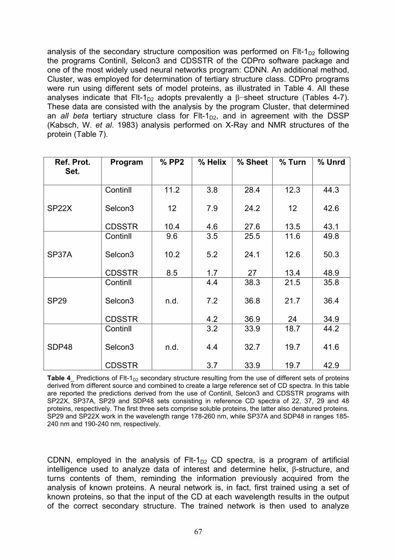
analysis of the secondary structure composition was performed on Flt-1D2 following the programs Continll, Selcon3 and CDSSTR of the CDPro software package and one of the most widely used neural networks program: CDNN. An additional method, Cluster, was employed for determination of tertiary structure class. CDPro programs were run using different sets of model proteins, as illustrated in Table 4. All these analyses indicate that Flt-1D2 adopts prevalently a sheet structure (Tables 4-7). These data are consisted with the analysis by the program Cluster, that determined an all beta tertiary structure class for Flt-1D2, and in agreement with the DSSP (Kabsch, W. et al. 1983) analysis performed on X-Ray and NMR structures of the protein (Table 7).
Ref. Prot. Set.
Program % PP2 % Helix % Sheet % Turn % Unrd
SP22X
Continll
Selcon3
CDSSTR
11.2
12
10.4
3.8
7.9
4.6
28.4
24.2
27.6
12.3
12
13.5
44.3
42.6
43.1
SP37A
Continll
Selcon3
CDSSTR
9.6
10.2
8.5
3.5
5.2
1.7
25.5
24.1
27
11.6
12.6
13.4
49.8
50.3
48.9
SP29
Continll
Selcon3
CDSSTR
n.d.
4.4
7.2
4.2
38.3
36.8
36.9
21.5
21.7
24
35.8
36.4
34.9
SDP48
Continll
Selcon3
CDSSTR
n.d.
3.2
4.4
3.7
33.9
32.7
33.9
18.7
19.7
19.7
44.2
41.6
42.9
Table 4_ Predictions of Flt-1D2 secondary structure resulting from the use of different sets of proteins derived from different source and combined to create a large reference set of CD spectra. In this tableare reported the predictions derived from the use of Continll, Selcon3 and CDSSTR programs with SP22X, SP37A, SP29 and SDP48 sets consisting in reference CD spectra of 22, 37, 29 and 48 proteins, respectively. The first three sets comprise soluble proteins, the latter also denatured proteins. SP29 and SP22X work in the wavelength range 178-260 nm, while SP37A and SDP48 in ranges 185-240 nm and 190-240 nm, respectively.
CDNN, employed in the analysis of Flt-1D2 CD spectra, is a program of artificial intelligence used to analyze data of interest and determine helix, -structure, and turns contents of them, reminding the information previously acquired from the analysis of known proteins. A neural network is, in fact, first trained using a set of known proteins, so that the input of the CD at each wavelength results in the output of the correct secondary structure. The trained network is then used to analyze
67

unknown proteins. Below, in Tables 5 and 6, are reported the results obtaining from Flt-1D2 CDNN analysis, starting from two sets of globular reference proteins consisting in 23 and 33 elements, respectively.
Table 5_ CDNN 2.1 (Spectra basis 23)
180-260 nm
185-260 nm
190-260nm
195-260nm
200-260nm
205-260nm
210-260nm
Helix n.d. n.d. 13,1% 12,5% 12,1% 12,3% 11,0% Antiparallel n.d. n.d. 39,8% 40,6% 42,7% 41,9% 44,7% Parallel n.d. n.d. 4,4% 5,2% 5,5% 5,5% 5,3% Beta-Turn n.d. n.d. 20,9% 21,2% 21,7% 21,8% 22,1% Rndm. Coil n.d. n.d. 32,8% 33,8% 36,3% 37,1% 35,5% Total Sum - - 111,0% 113,4% 118,3% 118,6% 118,6%
Table 6_ CDNN 2.1 (Spectra basis 33)
180-260 nm
185-260 nm
190-260nm
195-260nm
200-260nm
205-260nm
210-260nm
Helix n.d. n.d. 6,8% 8,0% 8,2% 6,5% 7,0% Antiparallel n.d. n.d. 35,0% 33,3% 34,5% 34,5% 35,6% Parallel n.d. n.d. 3,4% 4,7% 5,3% 5,4% 5,4% Beta-Turn n.d. n.d. 19,7% 20,6% 21,2% 20,2% 19,4% Rndm. Coil n.d. n.d. 35,2% 35,9% 36,9% 35,2% 35,3% Total Sum - - 100,1% 102,6% 106,2% 101,7% 102,7%
Starting from X-Ray and NMR structures of the second domain of Flt-1D2 (Wiesmann, C. et al., 1997 and Starovasnik, M. A. et al., 1999), the program DSSP calculated another set of values to describe the secondary structure content of the protein. The DSSP program was designed to standardize secondary structure assignments. DSSP is a database of secondary structure assignment for all proteins entries in the Protein Data Bank (PDB). In this case, we obtained a similar percentage of sheetcontent for both crystal and in solution structures (Table 7), in agreement with data collected on the refolded Flt-1D2 by running CDPro and CDNN programs.
Table 7
Program % Alpha % Beta % Random
DSSP-NMRFlt-1D2 5 41 54DSSP-XRayFlt-1D2 3 47 50
68
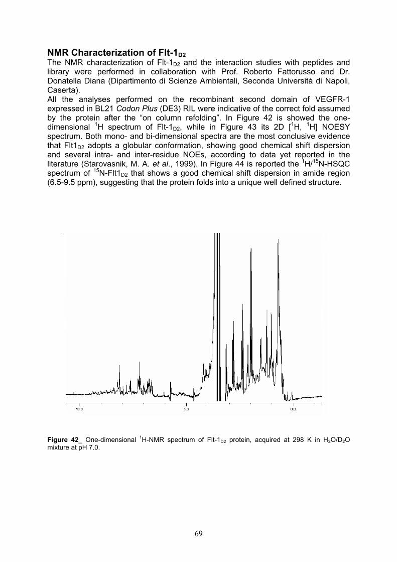
NMR Characterization of Flt-1D2
The NMR characterization of Flt-1D2 and the interaction studies with peptides and library were performed in collaboration with Prof. Roberto Fattorusso and Dr. Donatella Diana (Dipartimento di Scienze Ambientali, Seconda Università di Napoli, Caserta).All the analyses performed on the recombinant second domain of VEGFR-1 expressed in BL21 Codon Plus (DE3) RIL were indicative of the correct fold assumed by the protein after the “on column refolding”. In Figure 42 is showed the one-dimensional 1H spectrum of Flt-1D2, while in Figure 43 its 2D [1H, 1H] NOESY spectrum. Both mono- and bi-dimensional spectra are the most conclusive evidence that Flt1D2 adopts a globular conformation, showing good chemical shift dispersion and several intra- and inter-residue NOEs, according to data yet reported in the literature (Starovasnik, M. A. et al., 1999). In Figure 44 is reported the 1H/15N-HSQCspectrum of 15N-Flt1D2 that shows a good chemical shift dispersion in amide region (6.5-9.5 ppm), suggesting that the protein folds into a unique well defined structure.
Figure 42_ One-dimensional 1H-NMR spectrum of Flt-1D2 protein, acquired at 298 K in H2O/D2Omixture at pH 7.0.
69
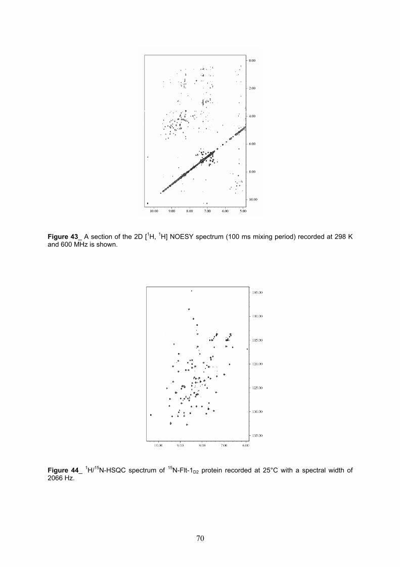
Figure 43_ A section of the 2D [1H, 1H] NOESY spectrum (100 ms mixing period) recorded at 298 Kand 600 MHz is shown.
Figure 44_ 1H/15N-HSQC spectrum of 15N-Flt-1D2 protein recorded at 25°C with a spectral width of2066 Hz.
70

NMR analysis of peptide-protein interaction Despite their conformational similarity, QK and MA peptides show a different biological activity, since the first has an agonist-like activity (D’Andrea, L. D. et al.,2005), while the second shows an interesting antagonist behavior compared with VEGF (Del Gatto, A. PhD thesis, 2005). QK and MA were employed in binding studies with Flt-1D2 aimed to investigate the nature of this interaction and to better understand their behavior from a molecular point of view.
Identification of the binding site on Flt-1D2 via Chemical Shift Mapping To map the binding site of Flt-1D2, were used NMR titration experiments, monitoring the interaction between QK peptide and the protein. Upon progressive additions of QK to the free 15N-Flt-1D2 (Table 8), were observed continuous changes in 1H and 15N chemical shifts for several signals of 15N-Flt-1D2 in 2D [1H, 15N] HSQC spectra. These chemical shifts changes indicated the formation of the complex 15N-Flt-1D2/QKin intermediate and in fast exchange on the NMR scale. Flt-1D2 residues with the most significant changes in 1H and 15N chemical shifts ( >0.04 ppm) upon formation of the complex were mapped onto the NMR solution structure of Flt-1D2 and are located in the strand f (E201, I202, G203, L204, T205), the strand g (L215, Y216, K217, R219,Y 220, L221), the loop between the strand a’ and b (K170, F172, P173, L174) and the region spanning the amino acid sequence Y139, S140, E141, I142, E144, as showed in Figure 45.
[15N-Flt-1D2](µM)
[QK](µM)
15N-Flt-1D2:QKRatio
220 0 1:0
220 55 1:0.25
220 100 1:0.50
220 165 1:0.75
220 220 1:1
220 440 1:2
220 660 1:3
Table 8_ Concentrations of 15N-Flt-1D2 protein and QK peptide used in the titration experiments. In the table is also reported the molar ratio protein:peptide determined for each concentration of both species.
71
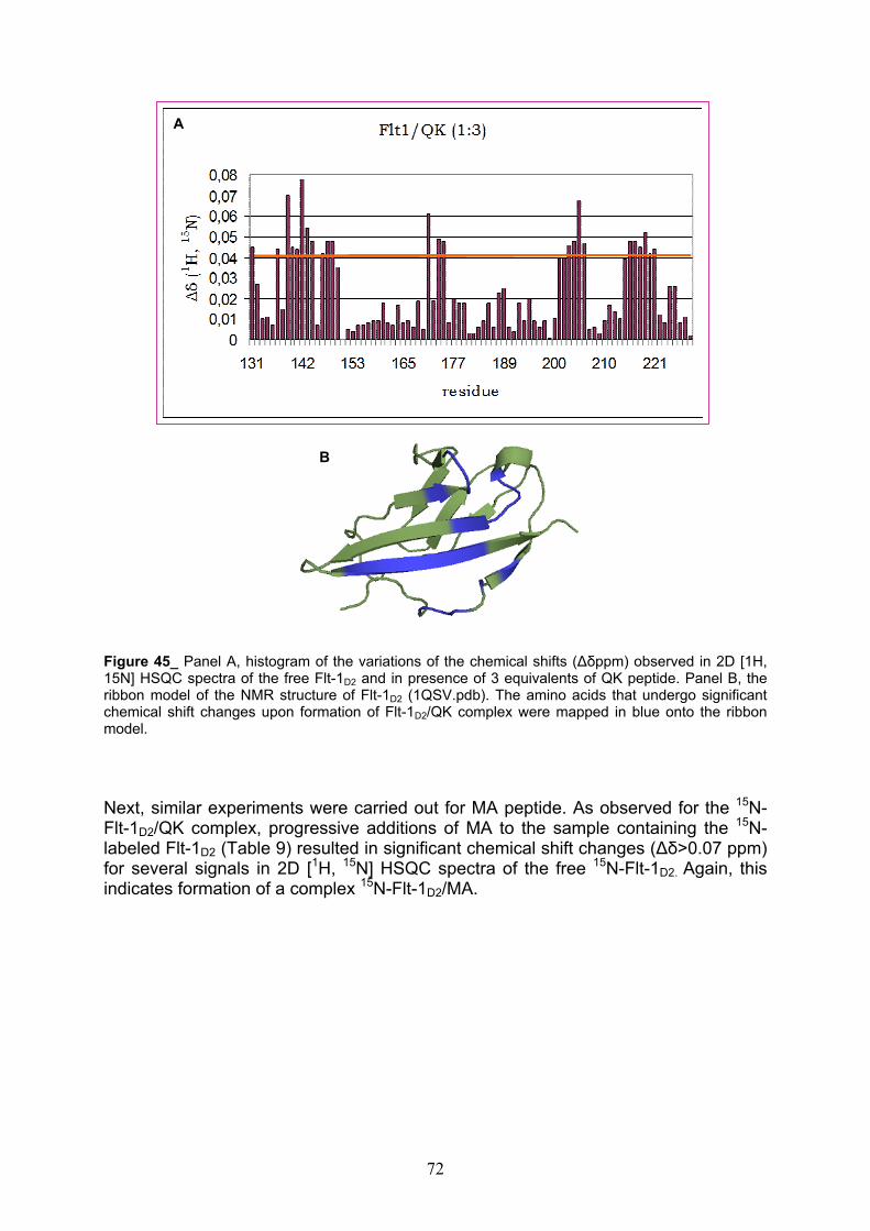
A
B
Figure 45_ Panel A, histogram of the variations of the chemical shifts ( ppm) observed in 2D [1H, 15N] HSQC spectra of the free Flt-1D2 and in presence of 3 equivalents of QK peptide. Panel B, theribbon model of the NMR structure of Flt-1D2 (1QSV.pdb). The amino acids that undergo significant chemical shift changes upon formation of Flt-1D2/QK complex were mapped in blue onto the ribbonmodel.
Next, similar experiments were carried out for MA peptide. As observed for the 15N-Flt-1D2/QK complex, progressive additions of MA to the sample containing the 15N-labeled Flt-1D2 (Table 9) resulted in significant chemical shift changes ( >0.07 ppm) for several signals in 2D [1H, 15N] HSQC spectra of the free 15N-Flt-1D2. Again, this indicates formation of a complex 15N-Flt-1D2/MA.
72

[15N-Flt-1D2](µM)
[MA](µM)
15N-Flt-1D2:MARatio
220 0 1:0
220 55 1:0.25
220 100 1:0.50
220 165 1:0.75
220 220 1:1
220 440 1:2
Table 9_ Concentrations of 15N-Flt-1D2 protein and MA peptide used in the titration experiments. In the table is also reported the molar ratio protein:peptide determined for each concentration of both species.
When mapped onto the solution structure of the free Flt-1D2, the residues of the refolded protein, upon formation of the complex with MA peptide, displayed significant chemical shift changes. They are located on a discontinuous surface defined by strands b (L169, K170, P172), f (E208, A209), g (V211, L215, K217, N219), and on a flexible region localized at the N-terminus (R133, P135, Y139, S140), as shown in Figure 46.
changes upon formation of Flt-1D2/MA complex weremapped in blue onto the ribbon model.
A
Figure 46_ Panel A, histogram of the variations inchemical shifts ( ppm) observed in 2D [1H, 15N]HSQC spectra of the free Flt-1D2 and in presence of 2equivalent of MA peptide. Panel B, the ribbon modelof the NMR structure of Flt-1D2 (1QSV.pdb). Theamino acids that undergo significant chemical shift
B
73

Epitope Mapping of QK and MA peptides bound to Flt-1D2 via Saturation Transfer Difference To provide additional information regarding the interaction between peptides and Flt-1D2, at atomic resolution, STD (Saturation Transfer Difference) NMR experimentswere performed. Without the unlabeled protein, STD spectra did not contain ligand signals because saturation transfer does not occur. The sample containing Flt-1D2
protein was the only one to show saturation transfer from the protein to their ligands in STD spectra. STD NMR studies provided peptide’s binding epitope for both QK and MA and were used to classify the residues relevant for the interaction with Flt-1D2. STD spectra were recorded studying the unlabeled Flt-1D2/peptide complex and, in the case of QK, clearly demonstrate the involvement of the aromatic region protons of the residues such as W4, Y8, Y12. For the side chain of Q5 and Q9 the signal of N Hprotons has similar large intensities (Figure 47).
W4
(H 1)
E6
(HN) Y8/Y12
(QD)
Y8/Y12
(QE)
Q5
(H 21)
W4
(HN) Q9
(H 22)
W4
(HH2/
QZ2)
Q5
(H 22)
W4
(H 1)Q9
(H 21)
b
a
Figure 47_ a) expansion of the amide and aromatic region of 1H NMR spectrum of QK alone. b)expansion of the region containing resonances of the amide and aromatic protons of STDspectrum for a QK:Flt-1D2 ratio of 1:20. In red is showed the backbone of the QK representativestructure; side chain of the interacting residues with the Flt1D2 are shown.
74
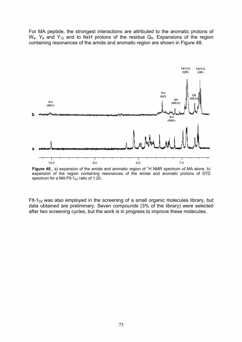
For MA peptide, the strongest interactions are attributed to the aromatic protons of W4, Y8 and Y12 and to N H protons of the residue Q9. Expansions of the region containing resonances of the amide and aromatic region are shown in Figure 48.
expansion of the region containing resonances of the amide and aromatic protons of STDspectrum for a MA:Flt-1D2 ratio of 1:20.
b
Y8/Y12
(QD)Y8/Y12
(QE)
W4
(QZ) Q9
(HE22)Q9
(HE21)W4
(HE1)
W4
(HD1)
a
Figure 48_ a) expansion of the amide and aromatic region of 1H NMR spectrum of MA alone. b)
Flt-1D2 was also employed in the screening of a small organic molecules library, but data obtained are preliminary. Seven compounds (3% of the library) were selected after two screening cycles, but the work is in progress to improve these molecules.
75
Discussion
In recent years the study of the mechanisms of angiogenesis and the research of new molecules able to modulate this process have been among the most well funded fields of the medical research. A strong clinical interest has emerged in developing anti-angiogenesis agents as therapeutic strategies for inhibiting tumor growth and metastasis, as well as a variety of other pathologies. Progresses towards a therapeutic treatment culminated, in 2004, in the approval of the first antiangiogenic agents for gastrointestinal cancer (Hurwitz, H. et al., 2004). On the other hand, therapeutic angiogenesis, consisting in promoting new vessel growth to treat disorders, such as ischemia, is an exciting frontier of cardiovascular medicine.The aim of this work is the expression of the extracellular domains of VEGF receptors KDR and Flt-1, in order to characterize them via NMR studies. Structural data on the extracellular portion of the receptors are, in fact, still very limited. The only structural data about Flt-1 receptor regards the domain 2 of the extracellular region that has been described in the free form (Starovanik, M. A. et al., 1999), bound to VEGF (Wiesmann, C. et al., 1997) or PlGF (Christinger, H. W. et al., 2004). So far, no structural data have been reported for Flt-4 and the extracellular domains of KDR. Instead, KDR intracellular kinase domain has been described (McTigue, M. A. et al.,1999). A partial characterization of NRP-1 and NRP-2 receptors has been performed (Pellet-Many, C. et al., 2008) and, very recently, the complex between VEGF and the extracellular Flt-1 domain has been observed by electron microscopy (Ruch, C. et al.2007). Hence, to extend the structural information about VEGF receptors is a very attractive goal. Besides, our intention is to test the binding capacity of the recombinant domains in interaction experiments with small synthetic peptides to understand the molecular bases of the interaction between VEGF receptors and its selected agonists/antagonists. This represents, in fact, an important step towards the designing of therapeutic approaches aimed at the treatment or prevention of angiogenic diseases. Finally, our aim is to employ the recombinant domains in drug discovery studies by targeting phage or small organic molecules libraries since, to date, no small organic molecules have been identified to target VEGF receptors in their extracellular portion. Flt-1 and KDR extracellular domains have been cloned successfully in several expression systems and expressed in Escherichia coli. E. coli is a very suitable host for the over-expression of heterologous proteins whose activity does not request post-translational modifications and allows achieving greater efficiency, reproducibility and convenience than mammalian cells. VEGF receptors contain several putative N-glycosylation sites and the mature proteins are extensively glycosylated, but this post-translational modification is not a prerequisite of high affinity binding of receptors to VEGF (Barleon, B. et al., 1997). Besides, in spite of mammalian cells, it is possible to grow E. coli strains in minimum medium, in order to express labeled proteins for characterization studies, without affecting physiology of bacterial cells. In addition, most of the commercially available receptors are Fc-IgG chimeras, resulting impossible to study via NMR because of their dimension and costs. Instead, the bacterial expression systems designed for the expression in E.coli enable to produce the recombinant proteins fused to a convenient tag which can be removed. Thus, the different constructs introduced in bacterial host cells allowed, for most of them, achieving high yields of recombinant receptors domains. In some cases, however, the limiting step was just the expression, as, for example, for KDRD1-2.
76

Despite different cloning strategies and the optimization of their expression conditions, the first two domains of KDR receptor have never been expressed. The domain 2, instead, was expressed, but was not possible to purify it, because, either under native or denaturing conditions and, in spite of its N-terminal 6xHis-tag, it did not bind to the Ni2+-NTA resin. In addition, even if for each recombinant construct have been tested several conditions to optimize the expression of proteins of interest and try to get them in soluble form, no domain of both receptors was soluble expressed, but all proteins were extracted with denaturing agents from inclusion bodies and refolded. On the other hand, refolding was not always successfully and the difficulty to obtain a refolded protein from inclusion bodies in sufficient amount made so far impossible further structural and functional studies for their characterization. These negative results make clear how difficult is to express these proteins and reflect the absence, in the literature, of extensive structural information on KDR and Flt-1 extracellular domains. Instead, it was developed an experimental protocol for refolding and purification of the second domain of VEGF Flt-1 receptor. The structure of Flt-1D2 has been solved either by X-Ray (complexed with VEGF) (Wiesmann. C. et al. 1997) or by NMR studies (Starovasnik, MA et al. 1999). In both cases, the protein was cloned and expressed in E. coli as inclusion bodies and refolded. Despite these available protocols, we were not able to purify the protein as a homogeneous product. In fact, according to Wiesmann et al., the construct comprising residues 129-229 was expressed as an insoluble protein in E. coli and inclusion bodies were treated with 6 M urea in 20 mM Tris-HCl, pH 7.5. Flt-1D2 was then refolded in two step dialysis by diluting the above solution in 6 M urea to 0.05 mg/mL. But in the last step Flt-1D2
precipitated. On the other hand, Starovasnik et al. proposed first the purification of the protein by affinity chromatography under denaturing conditions, then by reversed-phase HPLC, in order to separate the folded Flt-1D2 from the unfolded form (Starovasnik, M. A. et al., 1999). Consequently, the case would have requested an additional refolding step, but, also this method failed. Our protocol consists of the expression of the recombinant 6xHis-tagged Flt-1D2 in E. coli BL21 Codon Plus (DE3) strain. The protein, expressed as inclusion bodies, is refolded by an “on column refolding” protocol which resulted determinant for succeeding in the production to homogeneity of the second domain of VEGF Flt-1 receptor. The protein was, in fact, first refolded, still bound to the resin Ni -NTA, and then eluted in Tris buffer containing high concentration of imidazole. This strategy allowed making refolding in relatively shorter time than step dialysis and with less amount of urea. Besides, only two purification steps of affinity chromatography occurred, after which the homogeneous Flt-1D2 was concentrated to 3.5 mg/mL. Hence, the final product was analyzed to investigate its conformational and functional properties using a multidisciplinary approach.
2+
Protein Alkylation was performed to analyze the oxidation state of its two cysteine residues on both reduced and not reduced samples and showed that, in non reducing conditions, no alkylated adducts were found, while, in presence of DTT, the main product presented the two cysteine residues alkylated with an increment of 58 Da for each thiol group derived from reduction reaction. This experiment shows that in the refolded Flt-1D2 the two cysteine residues are involved in the formation of a disulfide bridge, as expected from literature data (Wiesmann, C. et al., 1997). Besides, the partial digestion with trypsin demonstrated the compact structure of the Ig-like domain, suggesting again the correct fold of the refolded protein.
77
Conformation preference of the recombinant Flt-1D2 was inspected by circular dichroism analyses. The content in secondary structure derived from the CD spectra analysis was in agreement with X-Ray data reported for Flt-1D2 (Wiesmann, C. et al.,1997), prevalently folded in a sheet structure. Thermal unfolding analysis showed a sigmoidal curve, with a single transition (Tm of about 60°C), indicating that a two-state unfolding process occurred. Moreover, thermal unfolding was reversible. In addition, the functionality of Flt-1D2 was assessed with a competition assay performed on HUVEC cells. The experiment verified the capability of the Flt-1 second domain to bind to VEGF, interfering with the interaction VEGF/receptors on HUVEC membrane.The results were fully confirmed by NMR studies conduced on both 15N-labeled and unlabeled proteins. Protonic mono- and bi-dimensional spectra of the unlabeled protein show the same strong signal dispersion of chemical shifts reported in the literature (Starovasnik, MA et al. 1999). The 1H/15N-HSQC spectrum of the uniformly 15N-labeled sample indicates that the global fold of the refolded Flt-1D2 is very similar in that reported by Starovasnik and in the crystalline state complexed to VEGF (Wiesmann, C. et al., 1997). We used the protein in order to understand the molecular bases of the interaction with two rationally designed peptides: QK and MA (D’Andrea, L. D. et al., 2005 and Del Gatto, A. PhD thesis, 2005). This would be the start point for the optimization and stabilization of that features which these molecules need to be used as good modulators in angiogenesis. QK and MA differ in their sequence for only two amino acid residues, but the first acts as an agonist of VEGF (D’Andrea, L. D. et al., 2005), while MA is a VEGF antagonist (Del Gatto, A. PhD thesis, 2005). In the literature are reported evidences that QK competes with VEGF for a binding site on endothelial cells expressing both KDR and Flt-1 receptors (D’Andrea, L. D. et al., 2005). The peptide binds to and activates both receptors similarly to VEGF and its agonist-like behavior is confirmed by in vitro and in vivo cell proliferation experiments. Although QK was designed to bind to KDR and Flt-1, its agonist-like activity is surprising because receptor dimerization is necessary for receptor activation and QK is a small peptide, probably unable to induce receptor dimerization by itself. Maybe, QK binding induces a conformational change of the receptor that either triggers, in an unknown way, the dimerization or induces the receptor activation through a different, previously uncharacterized mechanism that may not require dimerization. To address this issue, further structural and biochemical studies are needed.The interaction with peptides occurred following two approaches: one based on ligand observation, through Saturation Transfer Difference experiments, the other on observation of the protein, with Chemical Shift Mapping analysis. The interaction studies with QK peptide identified the binding site on protein surface through analysis of chemical shift perturbations of 15N-Flt-1D2. For this purpose were recorded 2D [15N, 1H] HSQC spectra of the protein, adding increasing amounts of peptide.From variations in chemical shifts induced by QK on 15N-Flt-1D2, significant changes in the fast exchange regime, in respect to the time scale of NMR, are identified for the strand f (E201, I202, G203, L204, T205), the strand g (L215, Y216, K217, R219, Y220,L221), the loop between strand a' and b (K170, F172, P173, L174) and the region comprising residues Y139, S140, E141, I142, E144.On the other hand, STD experiments have identified peptide’s binding epitope, confirming the involvement of side chains of amino acids W4, Q5, Y8, Q9, Y12 in interacting with the receptor.
78
Similar experiments were carried out for MA peptide. Progressive additions of MA to the sample containing the 15N-labeled Flt-1D2 result in significant chemical shift changes for several signals in the [1H, 15N] HSQC spectra of the free 15N-Flt-1D2,indicating, again, the formation of a complex 15N-Flt-1D2/MA. When mapped onto the solution structure of the free Flt-1D2, the residues displaying significant chemical shift changes are mapped on a discontinuous surface defined by strands b (L169, K170,P172), f (E208, A209), g (V211, L215, K217, N219), and on a flexible region localized at the N-terminus (R133, P135, Y139, S140). For MA peptide, the strongest interactions are attributed to the aromatic protons of W4, Y8 and Y12 and to N H protons of the residue Q9.Although peptides binding epitopes are clearly overlapping, NMR data indicate that QK and MA bind Flt-1D2 in distinct ways. In fact, on protein surface, QK induces chemical shift changes that are not observed in the case of the interaction with MA. On the basis of the chemical shift analysis, QK interacts with Flt-1D2 through -strands g and a’ and c- c’ loop region, whereas MA interacts more extensively with the residues located on the strands c and f. However, it is evident that both peptides bind to a region of Flt-1D2 protein known to contain the contact surface for VEGF. This region comprises highly positively charged residues, suggesting that ionic interactions play an important role in the binding, as well as hydrophobic interactions.On the other hand, STD NMR studies of QK and MA peptides in the presence of Flt-1D2 show that the aromatic protons of the residues Trp4, Tyr8 and Tyr12 interact with the receptor. In addition, the HN side chain of the residues Gln5 (only for QK) and Gln9 are recognized, whereas the HN backbone amide protons are less implicated.The second domain of Flt-1 receptor was also used in the screening of a library of small organic molecules, in order to find new compounds of biotechnology interest for modulating the angiogenesis process. Seven compounds (3% of the library) were selected after two screening cycles, but the work is in progress to improve these molecules.In conclusion, in this work we cloned and expressed different portions of the extracellular region of KDR and Flt-1 receptors. Only the second domain of Flt-1 receptor was refolded and purified using effective and reliable conditions to produce the protein at NMR concentration, developing an experimental protocol for the “on column refolding” resulted fast, cheap and efficient for our purpose. The protein, even when 15N-labelled expressed, was obtained as a homogeneous product (3.5 mg/mL), without adding VEGF or other cofactors that in the literature are reported to facilitate its concentration (Wiesmann, C. et al. 1999). Its proper folding was verified by nuclear magnetic resonance, biochemical and functional analyses. The protein was used in NMR interaction studies with two bioactive peptides and in a drug discovery analysis with the screening of a library of organic compounds. Our next perspective is to deepen the interaction studies with peptides, in order to obtain the dissociation constants of their binding to the second domain of Flt-1 receptor and try to explain their agonist or antagonist activity from a molecular point of view. Regarding the screening of the library, the obtainment of seven compounds to be improved is a very satisfactory result. To date, there are no small organic molecules able to target VEGF receptors in their extracellular portion and to identify new low molecular weight compounds able to act in this way, modulating the interaction between VEGF and its receptors, could be very exciting. Finally, our intention is to express and refold the other domains of VEGF receptors, in order to characterize them by NMR studies.
79

References
Abraham, J. A. et al. Human basic fibroblast growth factor: nucleotide sequence, genomic organization and expression in mammalian cells. Cold Spring Harb. Symp.Quant. Biol. 51 Pt1, 657-668 (1986a). Abraham, J. A. et al. Nucleotide sequence of a bovine clone encoding the angiogenic protein, basic fibroblast growth factor. Science 233, 545-548 (1986b). Ago, H. et al. Crystal structure of basic fibroblast growth factor at 1,6 Å resolution. J.Biochem. 110, 360-363 (1991). Aiello, L. P. et al. Suppression of retinal neovascularization in vivo by inhibition of vascular endothelial growth factor (VEGF) using soluble VEGF-receptor chimeric proteins. Proc. Natl. Acad. Sci. USA 92, 10457-10461 (1995). Alvarez, R. H. et al. Biology of platelet-derived growth factor and its involvement in disease. Mayo. Clin. Proc. 81, 1241-1257 (2006). Andrae, J. et al. Role of platelet-derived growth factors in physiology and medicine. Genes Dev. 22, 1276-1312 (2008). Asahara, T. et al., and Isner, J.M. Isolation of putative progenitor endothelial cells for angiogenesis. Science 275, 964-967 (1997). Asahara, T. et al. Tie-2 receptor ligands , angiopoietin-1 and angiopoietin-2, modulate VEGF-induced postnatal neovascularization. Circ. Res. 83, 233-240 (1998).Barillari, G. et al. The basic residues of placenta growth factor type 2 retrieve sequestered angiogenic factors into a soluble form. Implications for tumor angiogenesis. Am. J. Pathol. 152, 1161-1166 (1998).Barleon, B. et al. Mapping of the sites for ligand binding and receptor dimerization at the extracellular domain of the vascular endothelial growth factor receptor Flt-1. J.Biol. Chem. 272, 10382-10388 (1997). Bates, D. O. & Curry, F.E. Vascular endothelial growth factor increases microvascular permeability via a Ca(2+)-dependent pathway. Am. J. Physiol. 273,H687-H694 (1997). Beck, L., Jr and D’Amore, P. Vascular development: cellular and molecularregulation. FASEB J. 11, 365-373 (1997). Bennett, S. P. et al. Growth factors in the treatment of diabetic foot ulcers. Br. J. Surg. 90, 133-146 (2003). Bergers, G. et al. Effects of angiogenesis inhibitors on multistage carcinogenesis in mice. Science 284, 808-812 (1999). Boehm, T. et al. Antiangiogenic therapy of experimental cancer does not induce acquired drug resistance. Nature 390, 404-407 (1997). Bohm, G. et al. Quantitative analysis of protein far-UV CD spectra by neural networks. Protein Eng. 5, 191-195 (1992). Bradford, M. A rapid and sensitive method for the quantitation of microgram quantities of protein utilizing the principle of protein dye-binding. Anal. Biochem. 72,248-254 (1976). Brooks, P. C. et al., and Cheresh D.A. Integrin V 3 antagonists promote tumor regression by inducing apoptosis of angiogenic blood vessels. Cell 79, 1157-1164 (1994).Brown, L. F. et al., Vascular permeability factor/ vascular endothelial growth factor: a multifunctional angiogenic cytokine. EXS 79, 233-269 (1997).
80
Carmeliet, P. et al. Abnormal blood vessel development and lethality in embryos lacking a single VEGF allele. Nature 380, 435-439 (1996).Carmeliet, P. Mechanisms of angiogenesis and arteriogenesis. Nat. Med. 6, 389-395 (2000a).Carmeliet, P. VEGF gene therapy: stimulating angiogenesis or angioma-genesis? Nat. Med. 6, 1102-1103 (2000b). Carmeliet, P. et al. Synergism between vascular endothelial growth factor contributes to angiogenesis and plasma extravasation in pathological conditions. Nat.Med. 7, 575-583 (2001). Carmeliet, P. Angiogenesis in health and disease. Nat. Med. 9, 653-660 (2003). Carmeliet, P. Angiogenesis in life, disease and medicine. Nature 438, 932-936 (2005).Chaffer, C. L. et al. Aberrant fibroblast growth factor receptor signaling in bladder and other cancers. Differentiation 75, 831-842 (2007). Christinger, H. W. et al. The crystal structure of PlGF in complex with domain 2 of VEGFR-1. J. Biol. Chem. 279, 10382-10388 (2004). Claffey, K. P. and Robinson G.S. Regulation of VEGF/VPF expression in tumor cells: consequences for tumor growth and metastasis. Canc. Metast. Rev. 15, 165-176 (1996).D’Andrea, L. D. et al. Targeting angiogenesis: structural characterization and biological properties of a de novo engineered VEGF mimicking peptide. PNAS 102,14215-14220 (2005). Dameron, K. M. et al. Control of angiogenesis in fibroblasts by p53 regulation of thrombospondin-1. Science 265, 1582-1584 (1994). Davis, S. et al., and Yancopoulos, G.D. Isolation of angiopoietin-1, a ligand for the Tie-2 receptor, by secretion-trap expression cloning. Cell 87, 1161-1169 (1996). Davis, S. and Yancopoulos, G.D. The angiopoietins : Yin and Yang in angiogenesis. Curr.Top. Microbiol. Immunol. 237, 173-185 (1999). Davis-Smyth, T., Presta, L. G. & Ferrara, N. Mapping the charged residues in the second immunoglobulin-like domain of the vascular endothelial growth factor/placenta growth factor receptor Flt-1 required for binding and structural stability. J. Biol. Chem. 273, 3216-3222 (1998). DeGrado, W. F. et al. De novo design and structural characterization of proteins and metalloproteins. Annu. Rev. Biochem. 68, 779-819 (1999). Detmar, M. et al. Hypoxia regulates the expression of vascular permeability factor/vascular endothelial growth factor (VPF/VEGF) and its receptors in human skin. J. Invest. Dermatol. 108, 263-268 (1997). de Vries, C. et al. The fms-like tyrosine kinase, a receptor for vascular endothelial growth factor. Science 255, 989-991 (1992).Dnanabal, M. et al. Endostatin induces endothelial cell apoptosis. J. Biol. Chem.274, 11721-11726 (1999). Dudar, G.K. et al. A Vascular Endothelial Growth Factor Mimetic Accelerates Gastric Ulcer Healing in an iNOS-Dependent Manner. Am. J. Physiol. Gastrointest. Liver Physiol. 295, 374-381 (2008). Dvorak, H. F., Brown, L.F., Detmar, M. & Dvorak, A.M. Vascular permeability factor/vascular endothelial growth factor, microvascular hyperpermeability, and angiogenesis. Am. J. Pathol. 146, 1029-1039 (1995).Dvorak, H. F. How tumors make bad blood vessels and stroma. Am. J. Pathol. 162,1747-1757 (2003).
81

Dvorak, H. F. Vascular permeability factor/ vascular endothelial growth factor (VPF/ VEGF, VEGF-A). Microvascular research 1, 97-103 (2006). Dumont, D. J. et al. Dominant-negative and targeted null mutations in the endothelial receptor tyrosine kinase, tek, reveal a critical role in vasculogenesis of the embryo. Genes Dev. 8, 1897-1909 (1994). Dumont, D. J. et al. Cardiovascular failure in mouse embryos deficient in VEGF receptor 3. Science 282, 946-949 (1998). Eichmann, A. et al. Guidance of vascular and neural network formation. Curr. Opin. Neurobiol. 15, 108-115 (2005). Eliceiri, B.P. and Cheresh, D.A. The role of V 3 integrins during angiogenesis. Mol.Med. 4, 741-750 (1998). Eriksson, A. E. et al. Three-dimensional structure of human basic fibroblast growth factor. Proc. Natl. Acad. Sci. USA 88, 3441-3445 (1991).Fantl, W. et al. Signaling by receptor tyrosine kinases. Annu. Rev. Biochem. 62,453-481 (1993). Ferrara, N. et al. Molecular and biological properties of the vascular endothelial growth factor family of proteins. Endocr, Rev. 13, 18-32 (1992). Ferrara, N. et al. Heterozygous embryonic lethality induced by targeted inactivation of the VEGF gene. Nature 380, 439-442 (1996).Ferrara, N. & Davis-Smyth, T. The biology of vascular endothelial growth factor. Endocr. Rev. 18, 4-25 (1997). Ferrara, N. & Alitalo, K. Clinical applications of angiogenic growth factors and their inhibitors. Nat. Med. 5, 1359-1364 (1999).Ferrara, N. VEGF: an update on biological and therapeutic effects. Curr. Opin. Biotechnol. 11, 617-624 (2000). Ferrara, N. Role of Vascular endothelial growth factor in physiologic and pathologic angiogenesis: therapeutic implications. Semin. Oncol. 29, 10-14 (2002). Ferrara, N. et al. The biology of VEGF and its receptors. Nat. Med. 9, 669-676 (2003).Ferrara, N. et al. Angiogenesis as a therapeutic target. Nature 438, 967-974 (2005). Fidler, I. J. et al. The implications of angiogenesis for the biology and therapy of cancer metastasis. Cell 79, 185-188 (1994).Folkman, J. and D’Amore, P. Blood vessel formation: what is its molecular basis? Cell 87, 1153-1155 (1996). Folkman, J. Angiogenesis in cancer, reumathoid and other diseases. Nat. Med. 1,27-31 (1995). Fong, G.H., Rossant, J., Gertsenstein, M. & Breitman, M.L. Role of the Flt-1 receptor tyrosine kinase in regulating the assembly of vascular endothelium. Nature 376, 66-70 (1995).Fong, G. H., Zhang, L., Bryce, D.M. & Peng, J. Increased hemangioblast commitment, not vascular disorganization, is the primary defect in flt-1 knock-out mice. Development 126, 3015-3025 (1999).Fredriksson, L. et al. The PDGF family: four gene products form five dimeric isoforms. Cytokine Growth Factor Rev. 15, 197-204 (2004a). Fredriksson, L. et al. Tissue plasminogen activator is a potent activator of PDGF-CC. EMBO J. 23, 3793-3802 (2004). Fuh, G. et al. Requirements for binding and signaling of the kinase domain receptor for vascular endothelial growth factor. J. Biol. Chem. 273, 11197-11204 (1998).
82
Fukumura, D. et al. Hypoxia and acidosis independently up-regulate vascular endothelial growth factor transcription in brain tumors in vivo. Canc. Res. 61, 6020-6024 (2001). George DJ, Kaelin WG Jr. The von Hippel-Lindau protein, vascular endothelial growth factor, and kidney cancer. N. Engl. J. Med. 349, 419-421(2003).Gerber, H.P., Condorelli, F., Park, J. & Ferrara, N. Differential transcriptional regulation of the two VEGF receptor genes. Flt-1, but not Flk-1/KDR, is up-regulated by hypoxia. J. Biol. Chem. 272, 23659-23667 (1997).Gerber, H.P., Dixit, V. & Ferrara, N. Vascular endothelial growth factor induces expression of the antiapoptotic proteins Bcl-2 and A1 in vascular endothelial cells. J.Biol. Chem. 273, 13313-13316 (1998a).Gerber, H. P. et al. VEGF regulates endothelial cell survival by the PI3-kinase/Akt signal transduction pathway. Requirement for Flk-1/KDR activation. J. Biol. Chem.273, 30366-30343 (1998b).Gitay-Goren, H. et al. The binding of vascular endothelial growth factor to its receptors is dependent on cell surface-associated heparin-like molecules. J. Biol. Chem. 267, 6093-6098 (1992).Goldman, C. K. et al. Paracrine expression of a native soluble form of the vascular endothelial growth factor receptor inhibits tumor growth, metastasis and mortality rate. Proc. Natl. Acad. Sci. USA 95, 8795-8800 (1998). Grunstein, J. et al. Isoforms of vascular endothelial growth factor act in a coordinate fashion to recruit and expand tumor vasculature. Mol. Cell Biol. 20, 7282-7291 (2000).Hamada, K. et al. VEGF-C signaling patways through VEGFR-2 and VEGFR-3 in vasculogenesis and hematopoiesis. Blood 96, 3793-3800 (2000).Hanahan, D. and Folkman, J. Patterns and emerging mechanisms of the angiogenic switch during tumorigenesis. Cell 86, 353-364 (1996).Hanahan, D. Signaling vascular morphogenesis and maintenance. Science 277, 48-50 (1997). Heldin, C. H. et al. Mechanism of action and in vivo role of platelet-derived growth factor. Physiol. Rev. 79, 1283-1316 (1999). Herren, B. et al. Dimerization of extracellular domains of platelet-derived growth factor receptors. A revised model of receptor-ligand interactions. J. Biol. Chem. 268,15088-15095 (1993). Holash, J. VEGF-Trap: a VEGF blocker with potent antitumor effects. Proc. Natl. Acad. Sci. USA 99, 11393-11398 (2002). Holmgren, L., O’ Reilly, M.S. and Folkman, J. Dormancy of micrometastasis: balanced proliferation and apoptosis in the presence of angiogenesis suppression. Nat. Med. 1, 149-153 (1995). Hurwitz, H. et al. Bevacizumab plus irinotecan, fluorouracil and leucovorin for metastatic colorectal cancer. N. Engl. J. Med. 350, 2335-2342 (2004). Hynes, R. O. A reevaluation of integrins as regulators of angiogenesis. Nat. Med. 8,918-921 (2002).Ivan, M. et al. HIF targeted for VHL-mediated destruction by proline hydroxylation: implications for O2 sensing. Science 292, 464-468 (2001). Iyer, S. et al. The crystal structure of human placenta growth factor-1 (PlGF-1), an angiogenic protein, at 2.0 ANG. Resolution. J. Biol. Chem. 276, 12153-12161 (2001). Jussila, L. and Alitalo, K. Vascular growth factors and lymphangiogenesis. Physiol.Rev. 82, 673-700 (2002).
83
Kabbinavar, F. et al. Phase II, randomized trial comparing bevacizumab plus fluorouracil (FU)/leucovorin (LV) with FU/LV alone in patients with metastatic colorectal cancer. J. Clin. Oncol. 21, 60-65 (2003). Kabsch, W. et al. Dictionary of protein secondary structure: pattern recognition of hydrogen-bonded and geometrical features. Biopolymers 22, 2577-2637 (1983). Karkkainen, M. J., Makinen, T. & Alitalo, K. Lymphatic endothelium: a new frontier of metastasis research. Nat. Cell Biol. 4, E2-E5 (2002). Katoh, O. et al. Expression of the vascular endothelial growth factor (VEGF) gene receptor , KDR, in hematopoietic cells and inhibitory effect of VEGF on apoptotic cell death caused by ionizing radiation. Cancer Res. 55, 5687-5692 (1995). Kelly, J. D. et al. Platelet-derived growth factor stimulates PDGF receptor subunit dimerization and intersubunit trans-phosphorylation. J. Biol. Chem. 266, 8987-8992 (1991)..Kendall, R. L. et al. Identification of a natural soluble form of the vascular endothelial growth factor receptor , Flt-1 and its heterodimerization with KDR. Biochem. Biophys. Res. Commun. 226, 324-328 (1996). Keyt, B. A. et al. Identification of vascular endothelial growth factor determinants for binding KDR and Flt-1 receptors. J. Biol. Chem. 271, 5638-5646 (1996).Kim, K.J. et al. Inhibition of vascular endothelial growth factor-induced angiogenesis suppresses tumor growth in vivo. Nature 362, 841-844 (1993).Kitsukawa, T. et al. Neuropilin-semaphorin III/D-mediated chemorepulsive signals play a crucial role in peripheral nerve projection in mice. Neuron 19, 995-1005 (1997).Klagsbrun, M. and D’Amore, P. Regulators of angiogenesis. Annu. Rev. Physiol. 53,217-239 (1991a). Klagsbrun, M. et al. A dual receptor system is required for basic fibroblast growth factor activity. Cell 67, 229-231 (1991b). Klement, G. et al. Continuous low-dose therapy with vinblastine and VEGF receptor-2 antibody induces sustained tumor regression without overt toxicity. J. Clin. Invest.105, R15-R24 (2000).Laemmli, U. Cleavage of structural proteins during the assembly of the head of bacteriophage T4. Nature 227, 680-685 (1970). Lee, C. G. et al. Anti-vascular endothelial growth factor treatment augments tumor radiation response under normoxic or hypoxic conditions. Cancer Res. 60, 5565-5570 (2000).Lee, C.G. et al. Vascular endothelial growth factor (VEGF) induces remodeling and enhances TH2-mediated sensitization and inflammation in the lung. Nat. Med. 10,1095-1103 (2004) . Lee, P. N. et al. Purification and complementary DNA cloning of a receptor for basic fibroblast growth factor. Science 245, 57-60 (1989). Leung, D. W., Cachianes, G., Kuang, W.J., Goeddel, D.V. & Ferrara, N. Vascular endothelial growth factor is a secreted angiogenic mitogen. Science 246, 1306-1309 (1989).Levy, A. P. et al. Regulation of vascular endothelial growth factor by hypoxia and its modulation by the von Hippel-Lindau tumor suppressor gene. Kidney Int. 51, 575-578 (1997).Lindahl, P. et al. Pericyte loss and microaneurysm formation in PDGF-B –deficient mice. Science, 277, 242-245 (1997).
84

Luttun, A. et al. Revascularization of ischemic tissues by PlGF treatment and inhibition of tumor angiogenesis, arthritis and atherosclerosis by anti-Flt-1. Nat, Med.8, 831-840 (2002). Lyttle, D. J. et al. Homologs of vascular endothelial growth factor are encoded by the poxvirus orf virus. Virol. 68, 84-92 (1994). Maes, C. et al. Impaired angiogenesis and endochondral bone formation in mice lacking the vascular endothelial growth factor isoforms VEGF164 and VEGF188. Mech. Dev. 111, 61.73 (2002). Maisonpierre, P. C. et al., and Yancopoulos, G.D. Angiopoietin-2, a natural antagonist for Tie-2 that disrupts in vivo angiogenesis. Science 277, 55-60 (1997). Manavalan, P. et al. Variable selection method improves the prediction of protein secondary structure from CD spectra. Anal. Biochem. 167, 76-85 (1987). Mansukhani, A. et al. Characterization of the murine BEK fibroblast growth factor (FGF) receptor: activation by three members of the FGF family and requirement for heparin. Proc. Natl. Acad. Sci. USA 89, 3305-3309 (1992). Matsui T. et al. Isolation of a novel receptor cDNA establishes the existence of two PDGF receptor genes. Science 243, 800-804 (1989). Matsumoto, T. & Claesson-Welsh, L. VEGF receptor signal transduction. Sci. STKE 2001, RE21(2001). McTigue, M. A. et al. Crystal structure of the kinase domain of human vascular endothelial growth factor receptor 2: a key enzyme in angiogenesis. Structure 7, 319-330 (1999). Mercer, A. A. et al. Vascular endothelial growth factors encoded by Orf virus show surprising sequence variation but have a conserved, functionally relevant structure. J.Gen. Virol. 83, 2845-2855 (2002). Merelo, J. J. et al. Proteinotopic Feature Maps. Neurocomputing 6, 443-454 (1994). Migdal, M. et al. Neuropilin-1 is a placenta growth factor-2 receptor. J. Biol. Chem.273, 22272-22278 (1998).Millauer, B. et al. High affinity VEGF binding and developmental expression suggest Flk-1 as a major regulator of vasculogenesis and angiogenesis. Cell 72, 835-846 (1993).Millauer, B. et al. Glioblastoma growth inhibited in vivo by a dominant-negative Flk-1 mutant. Nature 367, 576-579 (1994). Moreira, I. S. et al. VEGF inhibition-a critical review. Anti-Cancer agents in medicinalchemistry. 7, 223-245 (2007). Moses, M. A. The regulation of neovascularization by matrix metalloproteinases and their inhibitors. Stem Cells 15, 180-189 (1997). Moses, M. A. et al., and Langer, R. Troponin I is present human cartilage and inhibits angiogenesis. Proc. Natl. Acad. Sci. USA 96, 2645-2650 (1999). Mukhopadhyay, D. and Datta, K. Multiple regulatory pathways of vascular permeability factor/ vascular endothelial growth factor (VPF/VEGF) expression in tumors. Semin. Canc. Biol. 14, 123-130 (2004). Muller, Y. A. et al. Vascular endothelial growth factor: crystal structure and functional mapping of the kinase domain receptor binding site. Proc. Natl. Acad. Sci. USA 94,7192-7197 (1997). Nagy, J. A. et al. Vascular permeability factor/vascular endothelial growth factor induces lymphangiogenesis as well as angiogenesis. J. Exp. Med. 196, 1497-1506 (2002).Nagy, J. A. et al. VEGF-A and the induction of pathological angiogenesis. Annu.Rev. Pathol. Mech. Dis. 2, 251-275 (2007).
85
Neufeld, G., Cohen, T., Gengrinovitch, S. & Poltorak, Z. Vascular endothelial growth factor (VEGF) and its receptors. FASEB J. 13, 9-22 (1999).Neufeld, G. et al. The neuropilins: multifunctional semaphorin and VEGF receptors that modulate axon guidance and angiogenesis. Trends Cardiovasc. Med. 12, 13-19 (2002).Nguyen, M., Folkman, J. and Biscoff, J. 1-Deoxymannojirimycin inhibits capillary tube formation in vitro. Analysis of N-linked oligosaccharides in bovine capillary endothelial cells. J. Biol. Chem. 267, 26157-25165 (1992). O’ Reilly, M. S. et al., and Folkman, J. Angiostatin: a novel angiogenesis inhibitor that mediates the suppression of metastasis by a Lewis lung carcinoma. Cell 79,315-328 (1994). O’ Reilly, M.S. et al., and Folkman, J. Endostatin: an endogenous inhibitor of angiogenesis and tumor growth. Cell 88, 277-285 (1997). Olofsson, B. et al. Vascular endothelial growth factor B (VEGFB) binds to VEGF receptor-1 and regulates plasminogen activator activity in endothelial cells. Proc.Natl. Acad. Sci. USA 95, 11709-11714 (1998).Ornitz, D. M. et al. Ligand specificity and heparin dependence of fibroblast growth factor receptors 1 and 3. J. Biol. Chem. 267, 16305-16311 (1992). Ornitz, D. M. et al. Fibroblast growth factors. Genome Biol. 2, Review S3005 (2001). Pajusola, K. et al. Signaling properties of Flt-4, a proteolytically processed receptor tyrosine kinase related to two VEGF receptors. Oncogene 9, 3545-3555 (1994). Pan, B. et al. Solution structure of a phage-derived peptide antagonist in complex with vascular endothelial growth factor. J. Mol. Biol. 316, 769-787 (2002). Park, J. E., Keller, H.-A. & Ferrara, N. The vascular endothelial growth factor (VEGF) isoforms: differential deposition into the subepithelial extracellular matrix and bioactivity of extracellular matrix-bound VEGF. Mol. Biol. Cell 4, 1317-1326 (1993). Park, J. E., Chen, H.H., Winer, J., Houck, K.A. & Ferrara, N. Placenta growth factor. Potentiation of vascular endothelial growth factor bioactivity, in vitro and in vivo, and high affinity binding to Flt-1 but not to Flk-1/KDR. J. Biol. Chem. 269, 25646-25654 (1994).Partanen, J. and Dumont, D.J. Functions of Tie1 and Tie2 receptor tyrosine kinases in vascular development. Curr.Top. Microbiol. Immunol. 237, 160-172 (1999). Pellet-Many, C. et al. Neuropilins: structure, function and role in disease. Biochem J.411, 211-226 (2008). Persico, M. G. et al. Structure, expression and receptor-binding properties of placenta growth factor (PlGF) Curr. Top. Microbiol. Immunol. 237, 31-40 (1999). Plouet, J., Schilling, J. & Gospodarowicz, D. Isolation and characterization of a newly identified endothelial cell mitogen produced by AtT20 cells. EMBO J. 8, 3801-3808 (1989).Powers, C. J. et al. Fibroblast growth factors, their receptors and signaling. Endocr.Relat. Cancer 7, 165-197 (2000). Presta, L.G. et al. Humanization of an anti-VEGF monoclonal antibody for the therapy of solid tumors and other disorders. Cancer Res. 57, 4593-4599 (1997). Prewett, M. et al. Antivascular endothelial growth factor receptor (fetal liver kinase 1) monoclonal antibody inhibits tumor angiogenesis. Cancer Res. 59, 5209-5218 (1999).Provencher, S. W. et al. Estimation of globular protein secondary structure from CD. Biochemistry 20, 33-37 (1981). Rahimi, N. VEGFR-1 and VEGFR-2: two nonidentical twins with a unique physiognomy. Front Biosci. 11, 819-829 (2006).
86

Rak, J. and Yu, J.L. Oncogenes and tumor angiogenesis: the question of vascular “supply” and vascular “demand”. Semin. Canc. Biol. 14, 93-104 (2004). Rastinejad, F. et al. Regulation of the activity of a new inhibitor of angiogenesis by a cancer suppressor gene. Cell 56, 345-355 (1989). Reuss, B. et al. Fibroblast growth factors and their receptors in the central nervous system. Cell Tissue Res. 313, 139-157 (2003). Risau, W. Mechanisms of angiogenesis. Nature 386, 671–674 (1997). Ruch, C. et al. Structure of a VEGF-VEGF receptor complex determined by electron microscopy. Nat. Struct. Mol. Biol. 14, 249-250 (2007).Ruhrberg, C. Growing and shaping the vascular tree: multiple roles for VEGF. Bioessays 25, 1052-1060 (2003). Rutherford, C. et al. Endogenously elicited antibodies to platelet-derived growth factor.-BB and platelet cytosolic protein inhibit aortic lesion development in the cholesterol-fed rabbit. Int. J. Exp. Phatol. 78, 21-32 (1997).Sato, T. N. et al. Distinct roles of the receptor tyrosine kinases Tie-1 and Tie-2 in blood vessel formation. Nature 376, 70-74 (1995). Savory, L. J. et al. Viral vascular endothelial growth factor plays a critical role in orf virus infection. Virol. 74, 10699-10706 (2000). Seetharam, L. et al. A unique signal transduction from FLR tyrosine kinase, a receptor for vascular endothelial growth factor (VEGF), Oncogene 10, 135-147 (1995).Senger, D. R. et al. Tumor cells secrete a vascular permeability factor that promotes accumulation of ascites fluids. Science 219, 983-985 (1983). Shalaby, F. et al. Failure of blood-island formation and vasculogenesis in Flk-1 deficient mice. Nature 376, 62-66 (1995). Shi, Q. et al., and Hammond W.P. Evidence for circulating bone marrow-derived endothelial cells. Blood 92, 362-367 (1998). Shibuya, M. et al. Nucleotide sequence and expression of a novel human receptor-type tyrosine kinase (flt) closely related to the fms family. Oncogene 8, 519-527 (1990).Shibuya, M. Role of VEGF-FLT receptor system in normal and tumor angiogenesis. Cancer Res., 67: 281-316 (1995). Shweiki, D. et al. Patterns of expression of vascular endothelial growth factor (VEGF) and VEGF receptors in mice suggest a role in hormonally regulated angiogenesis. J. Clin. Invest. 91, 2235-2243 (1993). Soker, S. et al. Neuropilin-1 is expressed by endothelial and tumor cells as an isoform-specific receptor for vascular endothelial growth factor. Cell 92, 735-745 (1998).Sreerama, N. et al. Estimation of protein secondary structure from CD spectra: comparison of CONTIN, SELCON and CDSSTR methods with an expanded reference set. Anal. Biochem. 287, 252-260 (2000). Starovasnik, M. A. et al. Solution structure of the VEGF-binding domain of Flt-1 :comparison of its free and bound states. J. Mol. Biol. 293, 531-544 (1999). Stopeck, A. et al. Results of a phase I dose-escalating study of the antiangiogenic agent, SU5416, in patients with advanced malignancies. Clin. Cancer Res. 8, 2798-2805 (2002). Sundberg, C. et al. Tumor cell and connective tissue cell interactions in human colorectal adenocarcinima. Transfer of platelet-derived growth factor-AB/BB to stromal cells. Am. J. Phatol. 151, 479-492 (1997).
87

Suri, C. et al., and Yancopoulos, G.D. Increased vascularization in mice overexpressing angiopoietin-1. Science 282, 468-471 (1998). Takahashi, T. et al., and Asahara , T. Ischemia- and cytokine-induced mobilization of bone marrow-derived endothelial progenitor cells for neovascularization. Nat. Med. 5,434-438 (1999a). Takahashi, T. et al. VEGF activates protein kinase C-dependent, but Ras independent Raf-MEK-MAP kinase pathway for DNA synthesis in primary endothelial cells. Oncogene 18, 2221-2230 (1999b). Terman, B. I. et al. Identification of a new endothelial cell growth factor receptor tyrosine kinase. Oncogene 6, 1677-1683 (1991). Thurston, G. et al. Angiopoietin-1 protects the adult vasculature against plasma leakage. Nat. Med. 6, 460-463 (2000). Ueda, Y. et al. A novel low molecular weight antagonist of vascular endothelial growth factor receptor binding: VGA1155. Mol. Cancer Ther. 2, 1105-1111 (2003). Venyaminov, S. et al. Determination of protein tertiary structure class from CD spectra. Anal. Biochem. 222, 176-184 (1994). Waltenberger, J. et al. Different signal transduction properties of KDR and Flt-1, two receptors for vascular endothelial growth factor. J. Biol. Chem. 269, 26988-26995 (1994).Weis, S.M. et al. Pathophysiological consequences of VEGF-induced vascular permeability. Nature 437, 497-504 (2005). Wiesmann, C. et al. Crystal structure at 1.7 Angstrom resolution of VEGF in complex with domain 2 of the Flt-1 receptor. Cell 91, 695-704 (1997).Wiesmann, C. et al. Crystal structure of the complex between VEGF and a receptor –blocking peptide. Biochemistry 37, 17765-17772 (1998). Wise, L. M. et al. Viral vascular endothelial growth factors vary extensively in amino acid sequence, receptor-binding specificities, and the ability to induce vascular permeability yet are uniformly active mitogens. J. Biol. Chem. 278, 38004-38014 (2003).Wood, J. M. et al. PTK787/ZK 222584, a novel and potent inhibitor of vascular endothelial growth factor receptor tyrosine kinases, impairs vascular endothelial growth factor-induced responses and tumor growth after oral administration. CancerRes. 60, 2178-2189 (2000).Woolard, J. et al. VEGF165b, an inhibitory vascular endothelial growth factor splice variant: mechanism of action, in vivo effect on angiogenesis and endogenous protein expression. Canc. Res. 64, 7822-7835 (2004). Yamazaki, Y. et al. Snake venom Vascular endothelial factors (VEGFs) exhibit potent activity through their specific recognition of KDR (VEGF receptor 2). J. Biol. Chem. 278, 51985-51988 (2003). Yancopoulos, G. D. et al. Vascular-specific growth factors and blood vessel formation. Nature 407, 242-248 (2000).Yi, E. S. et al. Platelet-derived growth factor causes pulmonary cell proliferation and collagen deposition in vivo. Am. J. Phatol. 149, 539-548 (1996). Yoshida, A. et al. Differential endothelial migration and proliferation to basic fibroblast growth factor and vascular endothelial growth factor. Growth Factors, 13,57-64 (1996).Yu, J. L. et al. Vascular endothelial growth factor isoforms expression as a determinant of blood vessel patterning in human melanoma xenografts. Canc. Res.62, 1838-1846 (2002).
88

Zhang, J. D. et al. Three-dimensional structure of human basic fibroblast growth factor, a structural homolog of interleukin 1 beta. Proc. Natl. Acad. Sci. USA 88,3446-3450 (1991). Zhu, X. et al. Three-dimensional structures of acidic and basic fibroblast growth factors. Science 251, 90-93 (1991). Zilberberg, L. et al. Structure and inhibitory effects on angiogenesis and tumor development of a new vascular endothelial growth inhibitor. J. Biol. Chem.278,35564-35573 (2003).
89

Publications
“In Vivo and In Vitro Characterization of CCK8 Bearing a Histidine-Based Chelator Labeled With 99mTc-Tricarbonyl.”Luca D. D’Andrea, Irma Testa, MariaRosaria Panico, Rossella Di Stasi, Corradina Caraco`, Laura Tarallo, Claudio Arra, Antonio Barbieri, Alessandra Romanelli, Luigi Aloj.
“A vascular endothelial growth factor mimetic accelerates gastric ulcer healing in an iNOS-dependent manner.” Genevieve K. Dudar, Luca D. D’Andrea, Rossella Di Stasi, Carlo Pedone, and John L. Wallace.
Comunications
R. Di Stasi, B. Ziaco, D. Capasso, R. Palumbo, M. Serino, E. Benedetti & L. D. D’andrea.“Design of small protein scaffold to target VEGF receptors”. 10th Naples Workshop on bioactive peptides – Naples June 11-14, 2006.
R. Di Stasi, D. Capasso, C. Pedone & L. D. D’Andrea. “Extracellular domains of VEGF receptors: expression and characterisation”. National Congress 2007 of Chemistry Division of Biological Systems – Montagnana, Padova November 8-9, 2007.
R. Di Stasi, D. Capasso, D. Diana, R. Fattorusso, C. Pedone & L. D. D’Andrea. “Vascular Endothelial Growth Factor (VEGF) and its receptors: key regulators of angiogenesis”. 11th Naples Workshop on bioactive peptides – Naples May 24-27, 2008.
R. Di Stasi, D. Capasso, D. Diana, R. Fattorusso, C. Pedone & L. D. D’Andrea. “VEGF receptors-peptide interaction: an NMR analysis”.30th European Peptide Symposium-Helsinki, Finland 31 August-5 September 2008.
R. Di Stasi, D. Capasso, D. Diana, R. Fattorusso, C. Pedone & L. D. D’Andrea. “Expression of extracellular domains of VEGF receptors and their use in NMR Drug discovery studies”. 53rd National Meeting of the Italian Society of Biochemistry and Molecular Biology and National Meeting of Chemistry of Biological Systems (Italian Chemical Society)-Riccione September 23-26 2008.
90
In Vivo and In Vitro Characterization of CCK8 Bearing a Histidine-BasedChelator Labeled With 99mTc-Tricarbonyl
Luca D. D’Andrea,1 Irma Testa,1 MariaRosaria Panico,1 Rossella Di Stasi,1 Corradina Caraco,2
Laura Tarallo,2 Claudio Arra,3 Antonio Barbieri,3 Alessandra Romanelli,4 Luigi Aloj2
1Istituto di Biostrutture e Bioimmagini, CNR, 80134 Napoli, Italy
2Area Funzionale di Medicina Nucleare, Istituto Nazionale Tumori, Fondazione ‘‘G. Pascale’’, 80131 Napoli, Italy
3UOS Sperimentazione Animale, Istituto Nazionale Tumori, Fondazione ‘‘G. Pascale’’, 80131 Napoli, Italy
4Dipartimento delle Scienze Biologiche, Universita di Napoli ‘‘Federico II, 80134 Napoli, Italy
Received 2 May 2008; revised 11 June 2008; accepted 16 June 2008
Published online 9 July 2008 in Wiley InterScience (www.interscience.wiley.com). DOI 10.1002/bip.21041
This article was originally published online as an accepted
preprint. The ‘‘PublishedOnline’’date corresponds to the preprint
version. You can request a copy of the preprint by emailing the
Biopolymers editorial office at [email protected]
INTRODUCTION
Radiopharmaceuticals are widely employed in nuclear
medicine for the diagnosis and therapy of different
diseases. In the last years, many new radiopharma-
ceuticals have been developed using a target specific
approach. In fact, in some diseases, such as cancer,
In Vivo and In Vitro Characterization of CCK8 Bearing a Histidine-BasedChelator Labeled With 99mTc-Tricarbonyl
Correspondence to: Luigi Aloj, Area Funzionale di Medicina Nucleare, Istituto
Nazionale Tumori, Fondazione ‘‘G. Pascale’’, Via M. Semmola, 80131, Napoli, Italy;
e-mail: [email protected]
ABSTRACT:
The development of receptor targeting radiolabeled
ligands has gained much interest in recent years for
diagnostic and therapeutic applications in nuclear
medicine. Cholecystokinin (CCK) receptors have been
shown to be overexpressed in a subset of neuroendocrine
and other tumors. We are evaluating binding and
biodistribution properties of a CCK8 peptide derivative
labeled with 99mTc(I)-tricarbonyl. The CCK8 peptide was
modified at its N-terminus by adding to its N-terminus
two lysine–histidine modules (KH), where histidine is
coupled to the side chain of the lysine ((KH)2-CCK8).99mTc(I)-tricarbonyl was generated with the IsoLinkTM
kit. A431 cells stably transfected with a cDNA encoding
for the human CCK2 receptor were utilized to determine
binding affinity, internalization, and retention of the
labeled peptide, in comparison with wild-type A431 cells.
A nude mouse tumor model was obtained by generating
A431-CCK2R and A431-control tumors in opposite
flanks of the animals. High specific activity labeling with99mTc was achieved. In A431-CCK2R cells, specific
saturable binding was observed as well as evident
internalization of the radiolabeled peptide after binding.
Biodistribution experiments showed rapid, specific
localization of (KH)2-CCK8 on A431-CCK2R xenografts
compared with control tumors, although absolute uptake
values were not markedly higher compared with
background activity. Clearance of unbound radioactivity
was both urinary and hepatobiliary. In imaging
experiments, while targeting to CCK2R positive tumors
could be appreciated, there was poor contrast between
target and nontarget areas. (KH)2-CCK8 shows adequate
in vitro and in vivo properties for CCK2R targeting
although improvement of biodistribution warrant further
development. # 2008 Wiley Periodicals, Inc. Biopolymers
(Pept Sci) 90: 707–712, 2008.
Keywords: technetium; radiopeptide; imaging; organome-
tallic complexes; carbonyl; cholecystokinin receptors
VVC 2008 Wiley Periodicals, Inc.
PeptideScience Volume 90 / Number 5 707

cells may express on their surface, specific receptors; mole-
cules capable of targeting these receptors can discriminate
between normal and cancer cells lowering the toxicity and
the side effects of the drug. These radiopharmaceuticals are
mainly designed by the following two approaches: integrated
and bifunctional. The latter approach is generally accom-
plished by assembling a targeting molecule, a linker, a chela-
tor, and the radionuclide.1
Antibodies and peptides are the two classes of compounds
widely used as targeting molecules in receptor imaging. In
particular, peptides, which recognize the cognate receptor
with high binding affinity and specificity, present some
advantages over antibodies such as: they are easily synthe-
sized and modified, are relatively inexpensive, have rapid
blood clearance, and high tumor to background ratios.1–3 A
successful example is represented by the radiolabled somato-
statin analog 111In-octreotide widely used in cancer imaging
(octreoscan).4 After the FDA approval of octreoscan, many
other peptides are being investigated.5
A well-characterized molecular target for imaging is repre-
sented by the cholecystokin (CCK) receptors.6 CCK recep-
tors, CCK1R and CCK2R, are membrane bound receptors
belonging to the superfamily of G-protein-coupled receptors
(GPCRs) and are predominantly located in the gastrointesti-
nal tract and the central nervous system.7 They differ in the
affinity for gastrin; CCK1R has low affinity, whereas CCK2R
binds both gastrin and cholecystokinin (CCK) with high
affinity. CCK2R is overexpressed in a variety of tumors such
as medullary thyroid carcinoma, small cell lung cancer, astro-
cytomas, stromal ovarian tumors, and some gastroentero-
patic tumors.8 Several CCK and gastrin derivatives have been
characterized over the past years for the purpose of in vivo
receptor targeting, and the feasibility of using CCK8-based
peptides to target CCK2R in vivo has been shown.6,9,10 The
C-terminal sequence of cholecystokinin, CCK8, is capable of
binding both CCK receptors, but shows higher affinity and
selectivity for CCK2R9 and has been extensively used for
imaging with different chelators.9–17
Several radionuclides are being used in nuclear medicine,
and 99mTc(I) is rapidly gaining in popularity since the intro-
duction by Alberto and coworkers18,19 of a one-pot proce-
dure to synthesize stable organometallic Tc(I)-complexes
[99mTc(OH2)3(CO)3]+ (TcCO). 99mTc(I) presents very favor-
able physical properties for application as radiopharmaceuti-
cals. Its half-life (t1/2 ¼ 6 h) is long enough to carry out
chemical synthesis and get useful images after administration
to the patient. At the same time, it is short enough to permit
administration of relatively high amounts of 99mTc radioac-
tivity without exposing the patient to excessive radiation
doses. The 140 keV photons are ideal for nuclear imaging
applications, and 99mTc isotope is readily available from
commercial 99Mo-99mTc generetors.1,20
In the TcCO complex, the water molecules can be
replaced by ligands to obtain very stable d6 low-spin com-
plexes. Preferred ligands are molecules possessing, as coordi-
nating atoms, sp2 nitrogens such as present in the side chain
of the amino acid histidine.19 Ligands replacing all the three
water molecules can form very stable complexes, avoiding
trans-chelation reactions, which may occur in vivo, and the
generation of free Tc. Recently, Alberto et al. reviewed the
ligands (mono-, bi-, and tridentate) available for peptide
labeling with TcCO.21 Histidine is a convenient ligand to
functionalize peptides because of its small dimension, and it
can be easily introduced during chain assembly on the resin.
When His is inserted in a peptide chain, it can act as mono-
dentate ligand for TcCO, but when positioned at the N-ter-
minus, it acts as bidentate ligand. The a-NH2 group and the
d-N aromatic amine are efficient chelators for the fac-
[99mTc(CO)3]+ core under formation of a six-member ring.
Several articles have been reported the utility of histidine and
histidine-based ligand labeled with TcCO for imaging appli-
cations.22–28
Aim of this work was to characterize the in vitro and in
vivo properties of CCK8 tagged with a chelator composed of
two N-terminal His-like residues and labeled with 99mTc(I)-
tricarbonyl. The presence of multiple His units could favor
the metal complexation and allow to achieve higher specific
activities. This in turn would allow targeting of higher
amounts of radioactivity per unit of peptide molecule, a fea-
ture that may prove advantageous for therapeutic applica-
tions using 186/188Re(I)-tricarbonyl.
EXPERIMENTAL SECTIONAll amino acids and coupling reagents (HOBT and HBTU) were
purchased from Novabiochem (Laufelfingen, Switzerland), N,N-
dimethylformamide (DMF) from Lab-Scan (Dublin, Ireland),
DIPEA and trifluoroacetic acid (TFA) from Romil (Cambridge,
UK), acetic anhydride from Applied Biosystem Dichloromethane,
Piperidine, 1,2-ethandithiol (EDT) from Fluka (Steinheim,
Germany), and triisopropylsilane from Aldrich (Steinheim,
Germany). Acetonitrile HPLC grade was from Riedel-de Haen
(Schnelldorf, Germany).
Peptide SynthesisPeptide amide was synthesized using ABI 433 peptide synthesizer
(Applied Biosystem) on solid phase using Rink Amide MBHA resin
(Novabiochem) and standard Fmoc (N-(9-fluorenyl)methoxycar-
bonyl) chemistry. All the amino acids presented standard protection
group on the side chain except for the lysines, which were protected
with methyltrytil group (Mtt) to allow selective deprotection.
Lysines were selectively deprotected with a solution of 1% TFA, 5%
708 D’Andrea et al.
Biopolymers (Peptide Science)
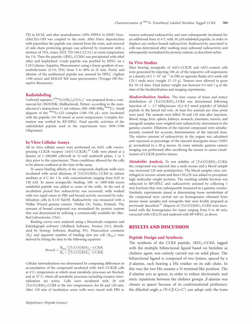
TIS in DCM, and after neutralization (20% DIPEA in DMF) Fmo-
cHis(Trt)-OH was coupled to the resin. After Fmoc deprotection
with piperidine the peptide was cleaved from the resin, and removal
of side-chain protecting groups was achieved by treatment with a
mixture of TFA, water, EDT, TIS (94:2.5:2.5:1) at room temperature
for 3 h. Then the peptide ((KH)2-CCK8) was precipitated with ethyl
ether and liophylized. Crude peptide was purified by HPLC on a
C18 Column (Juppiter, Phenomenex) using a linear gradient of ace-
tonitrile/water (0.1% TFA) from 5 to 40% in 25 min. Purity and
identity of the synthesized peptide was assessed by HPLC (Agilent
1100 series) and MALDI-ToF mass spectrometry (Voyager DE-Per-
septive Biosystem).
RadiolabelingCarbonyl aquaion [99mTc(OH2)3(CO)3]
+ was prepared from a com-
mercial kit (ISOLINK, Mallinckrodt, Petten) according to the man-
ufacturer’s instructions (1 ml volume; 200–1000 MBq 99mTc). Small
aliquots of the 99mTc-CO complex (100–150 lL) were incubated
with the peptide (10–50 nmol) at room temperature. Complex for-
mation was verified by RP-HPLC. Final specific activities of the
radiolabeled peptide used in the experiments were 1850–3700
GBq/mmol.
In Vitro Cellular AssaysAll in vitro cellular assays were performed on A431 cells overex-
pressing CCK2R receptor (A431-CCK2R).10 Cells were plated at a
density of 1–200,000 cells/well in 12-well multiwell plates, 2 or 3
days prior to the experiments. These conditions allowed for the cells
to be almost confluent at the time of the assay.
To assess binding affinity of the compound, duplicate wells were
incubated with serial dilutions of [TcCO](KH)2-CCK8 in culture
medium at 48C for 1 h, with concentrations ranging from 0.05 to
120 nM. To assess nonspecific binding, 100- to 1000-fold excess
unlabeled peptide was added to some of the wells. At the end of
incubation period free radioactivity was recovered, wells washed
with two rapid rinses in PBS and bound activity recovered by solu-
bilization cells in 0.1N NaOH. Radioactivity was measured with a
Wallac Wizard gamma counter (Wallac Oy, Turku, Finland). The
amount of bound compound was normalized for protein content
that was determined by utilizing a commercially available kit (Bio-
Rad Laboratories, USA).
Binding curves were analyzed using a Macintosh computer and
Kaleidagraph software (Abelbeck Software, Version 3.0.5, distrib-
uted by Synergy Software, Reading, PA). Dissociation constants
(Kd) and apparent number of binding sites per cell (Bmax) were
derived by fitting the data to the following equation:
Bound ¼ Bmax�½½TcCO� KHð Þ2�CCK8�
Kd þ ½½TcCO� KHð Þ2�CCK8� :
Cellular internalization was determined by comparing differences in
accumulation of the compound incubated with A431-CCK2R cells
at 48C, temperature at which most metabolic processes are blocked,
and at 378C, where all metabolic processes including receptor inter-
nalization are active. Cells were incubated with 20 nM
[TcCO](KH)2-CCK8 at the two temperatures, for 60 and 120 min.
After 120 min of incubation some wells were rinsed with PBS to
remove unbound radioactivity and were subsequently incubated for
an additional hour at 48C with 30 lM unlabeled peptide, in order to
displace any surface bound radioactivity. Radioactivity associated to
cells was determined after washing away unbound radioactivity and
subsequently normalized for protein content, as described earlier.
In Vivo StudiesMice bearing xenografts of A431-CCK2R and A431-control cells
were generated by injecting 100 lL of the respective cell suspensions
at a density of 2 3 107 ml�1 in PBS in opposite flanks of 6-week-old
CD-1 nude mice (weight 17–23 g). Tumors were allowed to grow
for 10–14 days. Final tumor weight was between 0.5 and 1 g at the
time of the biodistribution and imaging experiments.
Biodistribuition Studies. The time course of tissue and tumor
distribution of [TcCO](KH)2-CCK8 was determined following
injection of * 3.7 MBq/mouse (0.2–0.5 nmol peptide) of labeled
peptide in the lateral tail vein. At least five animals per time point
were used. The animals were killed 30 and 120 min after injection.
Blood, lungs, liver, spleen, kidneys, stomach, intestines, muscle, and
xenograft samples were weighed and radioactivity determined in the
gamma counter. Dilutions of the injected compound were simulta-
neously counted for accurate determination of the injected dose.
The relative amount of radioactivity in the organs was calculated
and expressed as percentage of the injected dose/gram tissue (%ID/
g) normalized to a 20 g mouse. In some animals, gamma camera
imaging was performed after sacrificing the mouse to assess visual-
ization of CCK2R positive tissues.
Metabolite Analysis. To test stability of [TcCO](KH)2-CCK8,
the compound was injected into a nude mouse and a blood sample
was recovered 120 min postinjection. The blood samples were cen-
trifuged to recover serum and then CH3CN was added to precipitate
high molecular weight proteins. The resulting soluble fraction was
subjected to RP-HPLC and radioactivity assessed by collecting 1
min fractions that were subsequently measured in a gamma counter.
Similarly, experiments aimed at determining tissue metabolism of
the compound were carried out on homogenates obtained from
mouse tissue samples and xenografts that were freshly prepared as
previously described.10 Aliquots of [TcCO](KH)2-CCK8 were incu-
bated with the homogenates for times ranging from 0 to 60 min,
extracted with CH3CN and analyzed with RP-HPLC as above.
RESULTS AND DISCUSSION
Peptide Design and Synthesis
The synthesis of the CCK8 peptide, (KH)2-CCK8, tagged
with the multiple bifunctional ligand based on histidine as
chelator agent, was entirely carried out on solid phase. The
bifunctional ligand is composed of two lysines, spaced by a
b-alanine, each bearing a His residue on its side chain. In
this way the two His assume a N-terminal-like position. The
b-alanine acts as spacer, in order to reduce electrostatic and
steric repulsions between the chelator groups. b-alanine waschosen as spacer because of its conformational preference:
the dihedral angle l (N-Cb-Ca-C0) can adopt only the trans
Characterization of 99mTc Tricarbonyl Labeled Histidine Tagged CCK8 709
Biopolymers (Peptide Science)

(1808) and/or gauche (+608) conformations29 keeping the
two His residues far away from each other. The peptide
sequence is reported in Figure 1.
The peptide and the ligand are entirely synthesized in
solid phase, this is an advantage over peptide-chelate conju-
gates that need to be prepared coupling in solution the che-
lating units. The product is obtained with high yields and
can be purified by HPLC yielding a high homogeneous prod-
uct. Moreover, the modular composition of the chelating
unit allows the design of a class of ligands in which the physi-
cochemical properties of the whole molecule, such as hydro-
philicity, could be easily modulated.
Radiolabeling and Quality Control
The purified peptide was efficiently labeled using the isolink
kit at high specific activity starting from the carbonyl aqua-
ion [99mTc(OH2)3(CO)3]. The reaction was followed by
reverse phase HPLC and a mixture, most presumably, of
mono and double labeled species, were obtained. There were
no purification steps for the radiolabeled material prior to
experimental use.
Receptor Binding and Internalization
Initially, we tested whether [TcCO](KH)2-CCK8 was capable
of binding A431 cells overexpressing the CCK2R in vitro.
Figure 2 shows data from a saturation binding experiment
with increasing concentration of peptide [TcCO](KH)2-
CCK8. The experiment shows rapid initial binding of the
compound to the CCK2R expressing cells and a slight increase
in cell associated activity over time suggesting that part of the
radioactivity is internalized. Nonspecific binding, as assessed
by incubation of the compound with nonreceptor expressing
cells or in receptor expressing cells in the presence of 100-fold
excess unlabeled peptide, was very low. These experiments
were performed at 48C, in order to block receptor internaliza-
tion and thus measure only ligand-receptor interaction. The
peptide binds to the CCK2 receptor with a dissociation con-
stants (Kd) of * 30 nM and with an apparent receptor num-
ber (Bmax) of 5.8 pmol/mg protein. The binding was saturable.
All parameters were in agreement with previously character-
ized CCK8-based molecules10 and are in the range useful for
receptor-binding of radiopharmaceuticals.30 This data show
that the introduction of the bis-His chelating unit does not
affect the binding of the targeting molecule to the receptor.
The CCK receptors, such as GPCRs, internalize after the
ligand binding event. A radiopharmaceutical targeting the
CCK2R can take advantage of this biological mechanism
because the receptor turn over always recycle free receptor on
the cell surface, and the segregation of the peptide in the
cytosol shift the ligand-binding equilibrium toward the
receptor-ligand complex formation. To evaluate if the pep-
tide was being internalized by the cells, we performed parallel
experiments in which A431-CCK2R cells were incubated
with 20 nM [TcCO](KH)2-CCK8 at 48C (internalization
blocked) or 378C (internalization active). The results are
reported in Figure 3. The radioactivity is progressively accu-
mulated in cells incubated at 378C with labeled peptide alone
(60 min vs. 120 min), whereas the levels of cell-associated
radioactivity are constant in cells incubated at 48C at 60 and
120 min, suggesting that internalization is occurring to some
extent at 378C. Again, the presence of the multifunctional
ligand does not appear to alter the biological activity of the
CCK8 peptide portion.
In Vivo Characterization
The in vivo characterization was carried out in nude mice
bearing xenografts of A431-CCK2R and A431-control cells.
FIGURE 1 Schematic representation of the labeled peptide
(KH)2-CCK8. The amino acid sequence of CCK8 is: Asp-Tyr-Met-
Gly-Trp-Met-Asp-Phe-amide.
FIGURE 2 Receptor binding. A431 cells overexpressing CCK2R
were incubated with increasing concentration of TcCo labeled pep-
tide. The radioactivity of bound compound was normalized for pro-
tein content.
710 D’Andrea et al.
Biopolymers (Peptide Science)
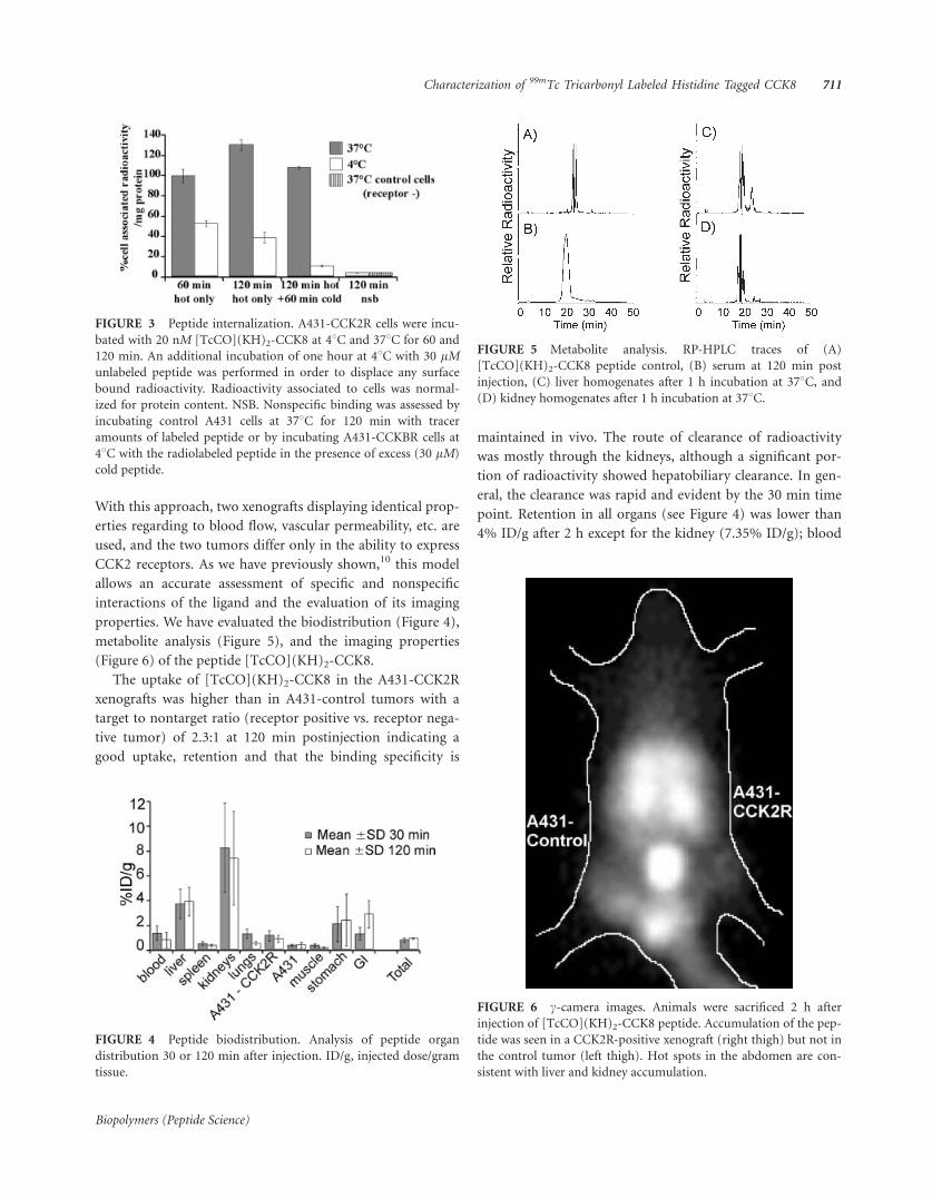
With this approach, two xenografts displaying identical prop-
erties regarding to blood flow, vascular permeability, etc. are
used, and the two tumors differ only in the ability to express
CCK2 receptors. As we have previously shown,10 this model
allows an accurate assessment of specific and nonspecific
interactions of the ligand and the evaluation of its imaging
properties. We have evaluated the biodistribution (Figure 4),
metabolite analysis (Figure 5), and the imaging properties
(Figure 6) of the peptide [TcCO](KH)2-CCK8.
The uptake of [TcCO](KH)2-CCK8 in the A431-CCK2R
xenografts was higher than in A431-control tumors with a
target to nontarget ratio (receptor positive vs. receptor nega-
tive tumor) of 2.3:1 at 120 min postinjection indicating a
good uptake, retention and that the binding specificity is
maintained in vivo. The route of clearance of radioactivity
was mostly through the kidneys, although a significant por-
tion of radioactivity showed hepatobiliary clearance. In gen-
eral, the clearance was rapid and evident by the 30 min time
point. Retention in all organs (see Figure 4) was lower than
4% ID/g after 2 h except for the kidney (7.35% ID/g); blood
FIGURE 3 Peptide internalization. A431-CCK2R cells were incu-
bated with 20 nM [TcCO](KH)2-CCK8 at 48C and 378C for 60 and
120 min. An additional incubation of one hour at 48C with 30 lMunlabeled peptide was performed in order to displace any surface
bound radioactivity. Radioactivity associated to cells was normal-
ized for protein content. NSB. Nonspecific binding was assessed by
incubating control A431 cells at 378C for 120 min with tracer
amounts of labeled peptide or by incubating A431-CCKBR cells at
48C with the radiolabeled peptide in the presence of excess (30 lM)
cold peptide.
FIGURE 4 Peptide biodistribution. Analysis of peptide organ
distribution 30 or 120 min after injection. ID/g, injected dose/gram
tissue.
FIGURE 5 Metabolite analysis. RP-HPLC traces of (A)
[TcCO](KH)2-CCK8 peptide control, (B) serum at 120 min post
injection, (C) liver homogenates after 1 h incubation at 378C, and(D) kidney homogenates after 1 h incubation at 378C.
FIGURE 6 c-camera images. Animals were sacrificed 2 h after
injection of [TcCO](KH)2-CCK8 peptide. Accumulation of the pep-
tide was seen in a CCK2R-positive xenograft (right thigh) but not in
the control tumor (left thigh). Hot spots in the abdomen are con-
sistent with liver and kidney accumulation.
Characterization of 99mTc Tricarbonyl Labeled Histidine Tagged CCK8 711
Biopolymers (Peptide Science)

levels of 0.75% ID/g at 2 h indicate fairly rapid plasma clear-
ance. We also evaluated the stability of the peptide conjugate
under different conditions (see Figure 5). The compound
was injected into a nude mouse, and a blood sample was
recovered 120 min postinjection. HPLC analysis showed deg-
radation of the starting compound to hydrophilic byproducts
and the starting material was no longer detectable (Figures
5A and 5B). As previous experience with CCK8-based
ligands10 indicate that metabolism in the organs rather than
in serum plays a major role, the compound was incubated
with tissue homogenates obtained from liver and kidney
(Figures 5C and 5D). In both cases, rapid degradation of the
compound to very hydrophilic byproducts was observed;
however, in kidney extracts the degradation was more com-
plete, whereas in the liver we could still observe a significant
amount of the starting material.
Imaging properties of [TcCO](KH)2-CCK8 were assessed
by performing gamma camera images 2 h after injection (see
Figure 6). Concentration of the compound in the CCK2R pos-
itive tumor can be appreciated; however, the image also shows
high-level background activity in kidney, liver, and gastroin-
testinal tract as expected from the biodistribution data.
CONCLUSIONSWe have reported the chemical synthesis, in vitro and in vivo
biological properties of a new molecule, [TcCO](KH)2-
CCK8, based on CCK8 peptide as targeting agent bearing an
amino acid-based chelator and labeled with the techne-
tium(I) carbonyl. The compound shows specific binding to
CCK2R receptors with high affinity, low nonspecific binding,
and cellular internalization. The specificity of compound-
receptor interaction is maintained in vivo. However, imaging
applications of this compound could benefit from reduction
of the relatively high-background radioactivity levels, likely
linked to significant hepatobiliary clearance. The ease of syn-
thesis and the modular design of this class of ligands allow
their modification and further development with the aim of
favoring renal clearance through increased hydrophilicity.
REFERENCES1. Liu, S.; Edwards, D. S. Chem Rev 1999, 99, 2235–2268.
2. Aloj, L.; Morelli, G. Curr Pharm Des 2004, 10, 3009–3031.
3. Benedetti, E.; Morelli, G.; Accardo, A.; Mansi, R.; Tesauro, D.;
Aloj, L. BioDrugs 2004, 18, 279–295.
4. Kwekkeboom, D.; Krenning, E. P.; de Jong, M. J Nucl Med 2000,
41, 1704–1713.
5. Heppeler, A.; Froidevaux, S.; Eberle, A. N.; Maecke, H. R. Curr
Med Chem 2000, 7, 971–994.
6. Aloj, L.; Panico, M. R.; Caraco, C.; Zannetti, A.; Del Vecchio, S.;
Di Nuzzo, C.; Arra, C.; Morelli, G.; Tesauro, D.; De Luca, S.;
Pedone, C.; Salvatore, M. Biopolymers 2002, 66, 370–380.
7. Wank, S. A. Am J Physiol 1998, 274, G607–G613.
8. Reubi, J. C.; Schaer, J. C.; Waser, B. Cancer Res 1997, 57, 1377–
1386.
9. Aloj, L.; Panico, M.; Caraco, C.; Del Vecchio, S.; Arra, C.;
Affuso, A.; Accardo, A.; Mansi, R.; Tesauro, D.; De Luca, S.;
Pedone, C.; Visentin, R.; Mazzi, U.; Morelli, G.; Salvatore, M.
Cancer Biother Radiopharm 2004, 19, 93–98.
10. Aloj, L.; Caraco, C.; Panico, M.; Zannetti, A.; Del Vecchio, S.;
Tesauro, D.; De Luca, S.; Arra, C.; Pedone, C.; Morelli, G.; Salva-
tore, M. J Nucl Med 2004, 45, 485–494.
11. Ertay, T.; Unak, P.; Bekis, R.; Yurt, F.; Biber, F. Z.; Durak, H.
Nucl Med Biol 2001, 28, 667–678.
12. Morelli, G.; De Luca, S.; Tesauro, D.; Saviano, M.; Pedone, C.;
Dolmella, A.; Visentin, R.; Mazzi, U. J Pept Sci 2002, 8, 373–381.
13. Reubi, J. C.; Waser, B.; Schaer, J. C.; Laederach, U.; Erion, J.; Sri-
nivasan, A.; Schmidt, M. A.; Bugaj, J. E. Eur J Nucl Med 1998,
25, 481–490.
14. Laverman, P.; Roosenburg, S.; Gotthardt, M.; Park, J.; Oyen, W.
J.; de Jong, M.; Hellmich, M. R.; Rutjes, F. P.; van Delft, F. L.;
Boerman, O. C. Eur J Nucl Med Mol Imaging 2008, 35, 386–
392.
15. Melis, M.; Krenning, E. P.; Bernard, B. F.; de Visser, M.; Rolle-
man, E.; de Jong, M. Nucl Med Biol 2007, 34, 633–641.
16. Agostini, S.; Bolzati, C.; Didone, E.; Cavazza-Ceccato, M.; Refo-
sco, F.; Aloj, L.; Arra, C.; Aurilio, M.; Tornesello, A. L.; Tesauro,
D.; Morelli, G. J Pept Sci 2007, 13, 211–219.
17. Laverman, P.; Behe, M.; Oyen, W. J.; Willems, P. H.; Corstens, F.
H.; Behr, T. M.; Boerman, O. C. Bioconjug Chem 2004, 15,
561–568.
18. Alberto, R.; Schibli, R.; Egli, A.; Schubiger, P. A.; Abram, U.;
Kaden, T. A. J Am Chem Soc 1998, 120, 7987–7988.
19. Waibel, R.; Alberto, R.; Willuda, J.; Finnern, R.; Schibli, R.;
Stichelberger, A.; Egli, A.; Abram, U.; Mach, J. P.; Pluckthun, A.;
Schubiger, P. A. Nat Biotechnol 1999, 17, 897–901.
20. Metzler-Nolte, N. Angew Chem Int Ed Engl 2001, 40, 1040–
1043.
21. Alberto, R.; Pak, J. K.; van Staveren, D.; Mundwiler, S.; Benny, P.
Biopolymers 2004, 76, 324–333.
22. von Guggenberg, E.; Behe, M.; Behr, T. M.; Saurer, M.; Seppi,
T.; Decristoforo, C. Bioconjug Chem 2004, 15, 864–871.
23. Zhang, K.; An, R.; Gao, Z.; Zhang, Y.; Aruva, M. R. Nucl Med
Biol 2006, 33, 505–512.
24. Maes, V.; Garcia-Garayoa, E.; Blauenstein, P.; Tourwe, D. J Med
Chem 2006, 49, 1833–1836.
25. Egli, A.; Alberto, R.; Tannahill, L.; Schibli, R.; Abram, U.; Schaff-
land, A.; Waibel, R.; Tourwe, D.; Jeannin, L.; Iterbeke, K.; Schu-
biger, P. A. J Nucl Med 1999, 40, 1913–1917.
26. La Bella, R.; Garcia-Garayoa, E.; Langer, M.; Blauenstein, P.;
Beck-Sickinger, A. G.; Schubiger, P. A. Nucl Med Biol 2002, 29,
553–560.
27. La Bella, R.; Garcia-Garayoa, E.; Bahler, M.; Blauenstein, P.;
Schibli, R.; Conrath, P.; Tourwe, D.; Schubiger, P. A. Bioconjug
Chem 2002, 13, 599–604.
28. Bullok, K. E.; Dyszlewski, M.; Prior, J. L.; Pica, C. M.; Sharma,
V.; Piwnica-Worms, D. Bioconjug Chem 2002, 13, 1226–1237.
29. Di Blasio, B.; Pavone, V.; Lombardi, A.; Pedone, C.; Benedetti,
E. Biopolymers 1993, 33, 1037–1049.
30. Eckelman, W. C. Nucl Med Biol 1994, 21, 759–769.
712 D’Andrea et al.
Biopolymers (Peptide Science)

A vascular endothelial growth factor mimetic accelerates gastric ulcer healingin an iNOS-dependent manner
Genevieve K. Dudar,1 Luca D. D’Andrea,2 Rossella Di Stasi,2 Carlo Pedone,2 and John L. Wallace1
1Inflammation Research Network, University of Calgary, Calgary, Alberta, Canada; and 2Istituto di Biostrutture eBioimmagini, Consiglio Nazionale delle Ricerche, Napoli, Italy
Submitted 5 May 2008; accepted in final form 20 June 2008
Dudar GK, D’Andrea LD, Di Stasi R, Pedone C, Wallace JL. Avascular endothelial growth factor mimetic accelerates gastric ulcerhealing in an iNOS-dependent manner. Am J Physiol GastrointestLiver Physiol 295: 374–381, 2008. First published June 26, 2008;doi:10.1152/ajpgi.90325.2008.—Angiogenesis is crucial to all typesof wound healing, including gastric ulcer healing. The most potentpromoter of angiogenesis is vascular endothelial growth factor(VEGF). We hypothesized that a 15-amino acid peptide designed tomimic the angiogenic action of VEGF would accelerate gastric ulcerhealing. Gastric ulcers were induced in mice by serosal application ofacetic acid. Treatment with the VEGF mimetic accelerated gastriculcer healing when administered orally or intraperitoneally, at a doseof 50 ng/kg or greater. Such healing was not observed when thereverse sequence pentadecapeptide or the full-length VEGF proteinwas administered. Contrary to our hypothesis, the VEGF mimetic didnot significantly increase angiogenesis in the ulcerated stomach. Theenhancement of ulcer healing by the VEGF mimetic occurred inde-pendently of cyclooxygenase-2 (COX-2) activity but was blocked byinhibitors of inducible nitric oxide synthase (iNOS). These resultsdemonstrate that a VEGF mimetic is a potent stimulus for gastric ulcerhealing, even when given orally. The effects of the mimetic wereindependent of stimulatory effects on angiogenesis and COX-2 activ-ity but were dependent on iNOS-derived NO production.
stomach; nitric oxide; cyclooxygenase; angiogenesis; prostaglandin;gastric mucosa
CHRONIC GASTRIC ULCER HEALING involves epithelial cell migrationand proliferation, matrix remodeling, and angiogenesis. Theseprocesses can be modulated by a plethora of transcriptionfactors, growth factors, and cytokines (36). Growth factors thatinfluence angiogenesis have been of particular interest to manyinvestigators (13, 23, 31). Vascular endothelial growth factor(VEGF), a 46-kDa homodimeric glycoprotein, is the mostpotent stimulator of angiogenesis (31). It is produced by avariety of cell types including macrophages, smooth musclecells, fibroblasts, megakaryocytes, and neoplastic cells (2, 17).
Several studies have provided evidence for a role of VEGFin gastric ulcer healing. For example, expression of VEGF wasdetected in the margins of human gastric ulcers (32). The samegroup observed that rat gastric fibroblasts could express VEGF,and this expression was enhanced when the fibroblasts wereexposed to growth factors that stimulated ulcer healing (e.g.,epidermal growth factor and hepatocyte growth factor) (33).Expression of VEGF was observed to be elevated followinginduction of gastric ulcers in rats, in parallel with increases inangiogenesis (29).
There is also a link among prostaglandin synthesis by thegastric mucosa, VEGF expression, and angiogenesis. Althoughindomethacin suppressed ulcer healing and angiogenesis inrats, it did not affect gastric VEGF expression (29). It has beenknown for several years that cyclooxygenase-2 (COX-2) makesan important contribution to the healing of ulcers throughout thegastrointestinal tract (15, 20, 25, 28). Again, this may be linked tothe role of VEGF in ulcer healing. COX-2 and VEGF have beencolocalized in fibroblasts in the ulcer bed (19). A selective COX-2inhibitor suppressed VEGF release from human gastric fibro-blasts, and this could be reversed by addition of prostaglandin E2
(PGE2) to the culture medium (19).The possibility that VEGF can be exploited therapeutically
to improve gastric ulcer healing has been investigated. Joneset al. (10) observed enhanced ulcer healing in rats following asingle injection of naked DNA encoding VEGF. This effectwas significantly reduced by cotreatment with an antibodydirected against VEGF. We exploited the platelet as a richsource of VEGF to examine its effects on gastric ulcer healing.Ulcer healing could be substantially accelerated by oral treat-ment with a suspension of human platelets, and this effect wasreversed by immunoneutralization of VEGF (40).
In the present study, we have examined the effects of a15-amino acid peptide that adopts a helical conformation inaqueous solution that resembles the �-helical region (17–25) ofthe VEGF protein (4). This peptide has been shown to bind toboth the VEGF-1 and VEGF-2 receptors and to induce endo-thelial cell proliferation, activate the same signaling cascadesas VEGF, and stimulate capillary formation in vitro (4). Wehave examined the ability of this VEGF mimetic to influ-ence experimental ulcer healing and characterized the de-pendence of such effects on angiogenesis and on COX-2 andnitric oxide synthase (NOS) activity.
MATERIALS AND METHODS
Animals. All experiments were approved by the University ofCalgary Animal Care Committee. Male Wistar rats weighing 150–175 g and male C57BL/6 mice weighing 20–25 g were obtained fromCharles River Laboratories (Montreal, Quebec, Canada). The animalshad free access to a standard laboratory chow and were housed in aroom with a 12-h:12-h light-dark cycle.
Peptides. The VEGF mimetic used in these studies is a 15-aminoacid peptide (KLTWQELYQLKYKGI), synthesized as described byD’Andrea et al. (4). The NH2 terminus of the peptide is acetylated,whereas the COOH terminus is capped with an amide group. Theeffects of a peptide with the reverse sequence of the VEGF mimetic(IGKYKLQYLEQWTLK) were also assessed. As with the VEGF
Address for reprint requests and other correspondence: J. Wallace, Dept. ofPharmacology & Therapeutics, Univ. of Calgary, 3330 Hospital Dr. NW,Calgary, Alberta, T2N 4N1, Canada (e-mail: [email protected]).
The costs of publication of this article were defrayed in part by the paymentof page charges. The article must therefore be hereby marked “advertisement”in accordance with 18 U.S.C. Section 1734 solely to indicate this fact.
Am J Physiol Gastrointest Liver Physiol 295: 374–381, 2008.First published June 26, 2008; doi:10.1152/ajpgi.90325.2008.
0193-1857/08 $8.00 Copyright © 2008 the American Physiological Society http://www.ajpgi.orgG374
on Septem
ber 22, 2008 ajpgi.physiology.org
Dow
nloaded from

mimetic, the NH2 terminus of the reverse peptide is capped with anacetyl group and the COOH terminus ends in an amide group.
Gastric ulcer induction. Gastric ulcers were induced in mice accord-ing to the method outlined by Wang et al. (45), with slight modifica-tions (41). In brief, a laparotomy was performed under halothaneanesthesia, and the stomach was externalized. A 1-ml syringe, with itsbarrel cut and filed smooth, was placed firmly against the serosal sideof the stomach. Acetic acid [200 �l of 20% (vol/vol)] was added to thesyringe such that it was in contact with the serosal surface for 1 min.The acetic acid was removed from the syringe by aspiration andreplaced with several rinses of sterile saline. The stomach was thenplaced back to its original anatomical position, and the midlineincision was closed with sutures.
The procedure used to induce gastric ulcers in rats was similar tothat employed for mice, with a few differences. The rats were fastedovernight before the surgery. Acetic acid (80% vol/vol) was applied tothe serosal wall of the stomach for 1 min with the barrel of a 3-mlsyringe.
Measurement of gastric ulcer area. In each experiment, a group ofmice or rats was euthanized on day 3 after ulcer induction. Thisprovided data on the size of ulcers before treatment. Groups ofanimals were similarly euthanized at various times after beginningtreated with test drugs. In all cases, the stomach was excised andpinned out (mucosal surface up) onto a wax block. A 25-mm2 papergrid was placed adjacent to the ulcer, and a photograph was taken. Thephotograph was then enlarged and used for planimetric measurementof the ulcer area (performed by an observer unaware of the treatmentsthe animals had received). The area of ulceration was then convertedinto units of mm2 using the paper grid as a reference. In someexperiments, samples of gastric tissue were processed for furtherevaluation (e.g., immunohistochemistry).
Effects of the VEGF mimetic on ulcer healing. The ability of theVEGF mimetic to accelerate ulcer healing was first examined in themouse. Beginning 3 days after ulcer induction, mice were treatedtwice-daily intraperitoneally with the VEGF mimetic (100 ng/kg) orvehicle (sterile 0.9% saline). Groups of 5–7 mice were euthanized ondays 5, 7, and 10 after ulcer induction, and gastric ulcer areas weremeasured.
Next, the ability of different doses of the VEGF mimetic to enhancegastric ulcer healing was assessed. Beginning on day 3 after ulcerinduction, mice were treated intraperitoneally with vehicle (n � 16) orthe VEGF mimetic at 25 ng/kg (n � 4), 50 ng/kg (n � 13), or 100ng/kg (n � 4). The mice were euthanized on day 7 after ulcerinduction for measurement of ulcer area.
Whether or not the VEGF mimetic would be effective whenadministered orally was then examined. Groups of mice were treatedtwice daily with vehicle or the VEGF mimetic (50 ng/kg), each beinggiven either intraperitoneally or orally (n � 5–9 per group). The micewere euthanized on day 7 for measurement of gastric ulcer area.
A series of experiments was performed to determine whether thebeneficial effects of the VEGF mimetic on ulcer healing could beobserved with the full-length VEGF protein. Groups of 6–9 rats weretreated twice daily with VEGF (1 �g/kg) or vehicle, given eitherorally or intraperitoneally, beginning on day 3 after ulcer inductionand continuing until day 7. The mice were then euthanized, and ulcerareas were measured. The dose of VEGF used in this experimentrepresents an equimolar dose to 50 ng/kg of the VEGF mimetic.
The effects of the VEGF mimetic on gastric ulcer healing were thencompared with the effects of a peptide with the same length as themimetic but with the reverse sequence. Twice daily intraperitonealtreatment with vehicle (n � 6), the VEGF mimetic (50 ng/kg, n � 6),or the reverse sequence peptide (50 ng/kg ip, n � 7) was conductedover a 3-day period beginning on the third day after ulcer induction.The mice were then euthanized, and ulcer areas were determined.
The experiments described above all involved the use of mice. Thefollowing experiment was performed to determine whether the VEGFmimetic could influence ulcer healing in rats. Beginning on day 3 after
ulcer induction, groups of 5 rats each were treated twice dailyintraperitoneally with the VEGF mimetic (50 ng/kg) or vehicle. Therats were euthanized on day 7 after ulcer induction for measurementof gastric ulcer area.
Gastric acid secretion. Since agents that suppress gastric acidsecretion can accelerate ulcer healing in this model (9), we examined thepossibility that the VEGF mimetic might suppress gastric acid secre-tion in the mouse. Mice were fasted overnight and anesthetized withhalothane. The pyloric sphincter was ligated, and the mice wereallowed to recover consciousness. The mice were then treated intra-peritoneally with vehicle (n � 4) or the VEGF mimetic (100 ng/kg,n � 5). Three hours later, the mice were anesthetized with halothane,the lower gastro-esophageal junction was clamped, and the stomachwas carefully excised. The contents of the stomach were collected intoa tube and the volume of the fluid determined gravimetrically. The pHand the titratable acidity of the gastric juice were determined, asdescribed previously (1).
Angiogenesis. Gastric tissues harvested at the end of some of theabove-described experiments were assessed for the extent of angio-genesis occurring in the ulcer bed and at the ulcer margin. Microves-sels were visualized via immunohistochemical staining of CD31, anendothelial cell-specific marker (18). Tissue samples were immersedin formalin-free zinc fixative and embedded into paraffin blocks.Sections (7 �m) of the tissues were then cut and placed ontopositively charged glass slides, heated overnight at 60°C, and storeduntil further processing.
The sections were deparaffinized and rehydrated using an ethanolgradient and then boiled in citrate buffer for 10 min to achieve epitoperetrieval. The tissue was blocked with normal rabbit serum blockingsolution for 1 h before overnight incubation at 4°C with rat anti-mouseCD31 antibody (Pharmingen, San Diego, CA). Afterward, an endog-enous peroxidase-blocking step was performed with the use of 3%hydrogen peroxide in 0.01 M PBS (pH 7.4) solution for 10 min atroom temperature. The primary antibody that bound to the tissuesections was detected using the horseradish peroxidase-Streptavidinmethod (Vector Laboratories, Burlington, ON, Canada). The mi-crovessels were detectable once the tissue underwent a reaction withdiaminobenzidine. The stained microvessels in the granulation tissuewere counted in a blind manner under a microscope at a magnificationof 400�. The results were reported as the number of microvessels permm2 of tissue.
Role of COX-2. COX-2 plays an important role in the healing ofulcers in mice and rats (15, 19) and has also been shown to regulaterelease of VEGF from gastric fibroblasts (19). We therefore investi-gated the possibility that COX-2 activity may be necessary for theenhancement of ulcer healing by the VEGF mimetic. Groups of micewith gastric ulcers were treated twice daily, from day 3 to day 7, witha selective COX-2 inhibitor (lumiracoxib, 10 mg/kg po, n � 8), withthe VEGF mimetic (50 ng/kg ip, n � 14), or with both the VEGFmimetic and lumiracoxib (n � 9). An additional group of mice wastreated with the vehicles for lumiracoxib and the VEGF mimetic (1%carboxymethylcellulose and 0.9% saline, respectively). On day 7, themice were euthanized and ulcer areas were measured. Lumiracoxib isa selective inhibitor of COX-2 (8). We confirmed that the 10-mg/kgdose of lumiracoxib produced a selective inhibition of COX-2 in mice.As described in detail elsewhere (38, 46), injection of carrageenan orzymosan into an air pouch on the rat or mouse results in a markedstimulation of PGE2 synthesis, with this synthesis occurring almostexclusively via COX-2. Oral pretreatment with lumiracoxib (10 mg/kg) reduced zymosan-induced PGE2 synthesis by 90% (P � 0.01)while not significantly affecting whole blood thromboxane synthesis[the latter occurring exclusively via COX-1 (37)].
Role of NOS. NO contributes significantly to gastric mucosaldefense (42, 44) and has been shown to contribute significantly toexperimental ulcer healing (12, 14). Expression of inducible NOS(iNOS) has been noted in inflammatory cells at the base of gastriculcers in rodents (14). NO has also been reported to be a crucial
G375VEGF MIMETIC AND ULCER HEALING
AJP-Gastrointest Liver Physiol • VOL 295 • AUGUST 2008 • www.ajpgi.org
on Septem
ber 22, 2008 ajpgi.physiology.org
Dow
nloaded from
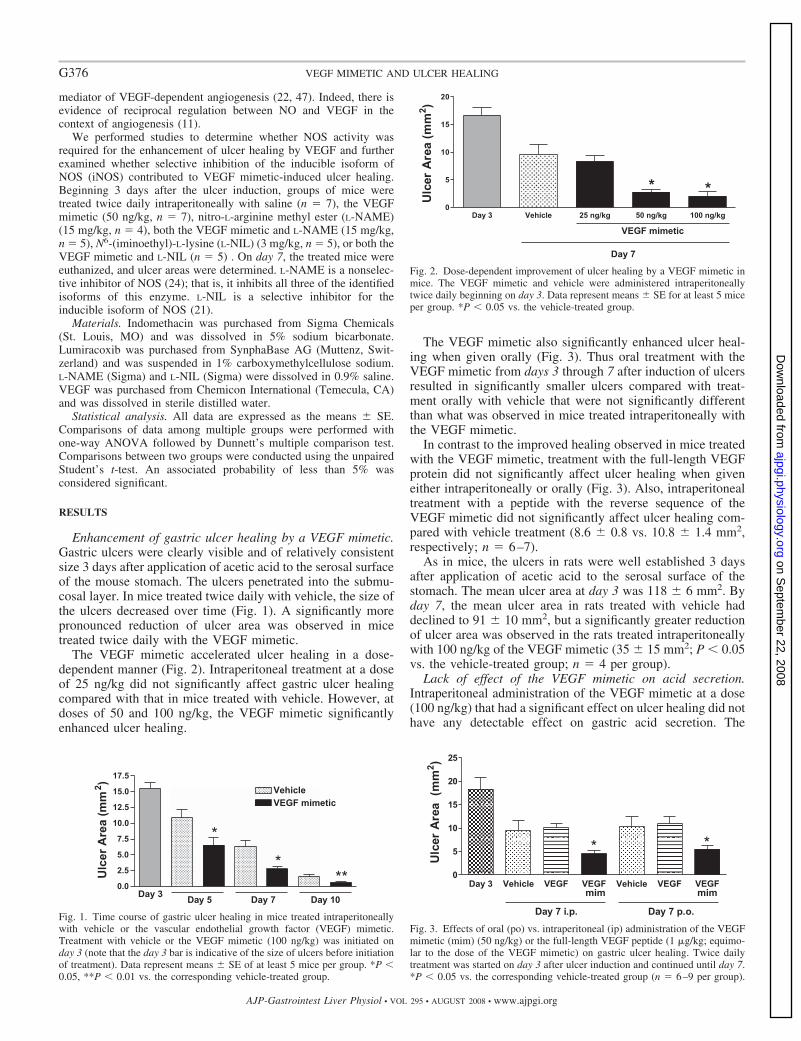
mediator of VEGF-dependent angiogenesis (22, 47). Indeed, there isevidence of reciprocal regulation between NO and VEGF in thecontext of angiogenesis (11).
We performed studies to determine whether NOS activity wasrequired for the enhancement of ulcer healing by VEGF and furtherexamined whether selective inhibition of the inducible isoform ofNOS (iNOS) contributed to VEGF mimetic-induced ulcer healing.Beginning 3 days after the ulcer induction, groups of mice weretreated twice daily intraperitoneally with saline (n � 7), the VEGFmimetic (50 ng/kg, n � 7), nitro-L-arginine methyl ester (L-NAME)(15 mg/kg, n � 4), both the VEGF mimetic and L-NAME (15 mg/kg,n � 5), N6-(iminoethyl)-L-lysine (L-NIL) (3 mg/kg, n � 5), or both theVEGF mimetic and L-NIL (n � 5) . On day 7, the treated mice wereeuthanized, and ulcer areas were determined. L-NAME is a nonselec-tive inhibitor of NOS (24); that is, it inhibits all three of the identifiedisoforms of this enzyme. L-NIL is a selective inhibitor for theinducible isoform of NOS (21).
Materials. Indomethacin was purchased from Sigma Chemicals(St. Louis, MO) and was dissolved in 5% sodium bicarbonate.Lumiracoxib was purchased from SynphaBase AG (Muttenz, Swit-zerland) and was suspended in 1% carboxymethylcellulose sodium.L-NAME (Sigma) and L-NIL (Sigma) were dissolved in 0.9% saline.VEGF was purchased from Chemicon International (Temecula, CA)and was dissolved in sterile distilled water.
Statistical analysis. All data are expressed as the means � SE.Comparisons of data among multiple groups were performed withone-way ANOVA followed by Dunnett’s multiple comparison test.Comparisons between two groups were conducted using the unpairedStudent’s t-test. An associated probability of less than 5% wasconsidered significant.
RESULTS
Enhancement of gastric ulcer healing by a VEGF mimetic.Gastric ulcers were clearly visible and of relatively consistentsize 3 days after application of acetic acid to the serosal surfaceof the mouse stomach. The ulcers penetrated into the submu-cosal layer. In mice treated twice daily with vehicle, the size ofthe ulcers decreased over time (Fig. 1). A significantly morepronounced reduction of ulcer area was observed in micetreated twice daily with the VEGF mimetic.
The VEGF mimetic accelerated ulcer healing in a dose-dependent manner (Fig. 2). Intraperitoneal treatment at a doseof 25 ng/kg did not significantly affect gastric ulcer healingcompared with that in mice treated with vehicle. However, atdoses of 50 and 100 ng/kg, the VEGF mimetic significantlyenhanced ulcer healing.
The VEGF mimetic also significantly enhanced ulcer heal-ing when given orally (Fig. 3). Thus oral treatment with theVEGF mimetic from days 3 through 7 after induction of ulcersresulted in significantly smaller ulcers compared with treat-ment orally with vehicle that were not significantly differentthan what was observed in mice treated intraperitoneally withthe VEGF mimetic.
In contrast to the improved healing observed in mice treatedwith the VEGF mimetic, treatment with the full-length VEGFprotein did not significantly affect ulcer healing when giveneither intraperitoneally or orally (Fig. 3). Also, intraperitonealtreatment with a peptide with the reverse sequence of theVEGF mimetic did not significantly affect ulcer healing com-pared with vehicle treatment (8.6 � 0.8 vs. 10.8 � 1.4 mm2,respectively; n � 6–7).
As in mice, the ulcers in rats were well established 3 daysafter application of acetic acid to the serosal surface of thestomach. The mean ulcer area at day 3 was 118 � 6 mm2. Byday 7, the mean ulcer area in rats treated with vehicle haddeclined to 91 � 10 mm2, but a significantly greater reductionof ulcer area was observed in the rats treated intraperitoneallywith 100 ng/kg of the VEGF mimetic (35 � 15 mm2; P � 0.05vs. the vehicle-treated group; n � 4 per group).
Lack of effect of the VEGF mimetic on acid secretion.Intraperitoneal administration of the VEGF mimetic at a dose(100 ng/kg) that had a significant effect on ulcer healing did nothave any detectable effect on gastric acid secretion. The
Fig. 1. Time course of gastric ulcer healing in mice treated intraperitoneallywith vehicle or the vascular endothelial growth factor (VEGF) mimetic.Treatment with vehicle or the VEGF mimetic (100 ng/kg) was initiated onday 3 (note that the day 3 bar is indicative of the size of ulcers before initiationof treatment). Data represent means � SE of at least 5 mice per group. *P �0.05, **P � 0.01 vs. the corresponding vehicle-treated group.
Fig. 2. Dose-dependent improvement of ulcer healing by a VEGF mimetic inmice. The VEGF mimetic and vehicle were administered intraperitoneallytwice daily beginning on day 3. Data represent means � SE for at least 5 miceper group. *P � 0.05 vs. the vehicle-treated group.
Fig. 3. Effects of oral (po) vs. intraperitoneal (ip) administration of the VEGFmimetic (mim) (50 ng/kg) or the full-length VEGF peptide (1 �g/kg; equimo-lar to the dose of the VEGF mimetic) on gastric ulcer healing. Twice dailytreatment was started on day 3 after ulcer induction and continued until day 7.*P � 0.05 vs. the corresponding vehicle-treated group (n � 6–9 per group).
G376 VEGF MIMETIC AND ULCER HEALING
AJP-Gastrointest Liver Physiol • VOL 295 • AUGUST 2008 • www.ajpgi.org
on Septem
ber 22, 2008 ajpgi.physiology.org
Dow
nloaded from
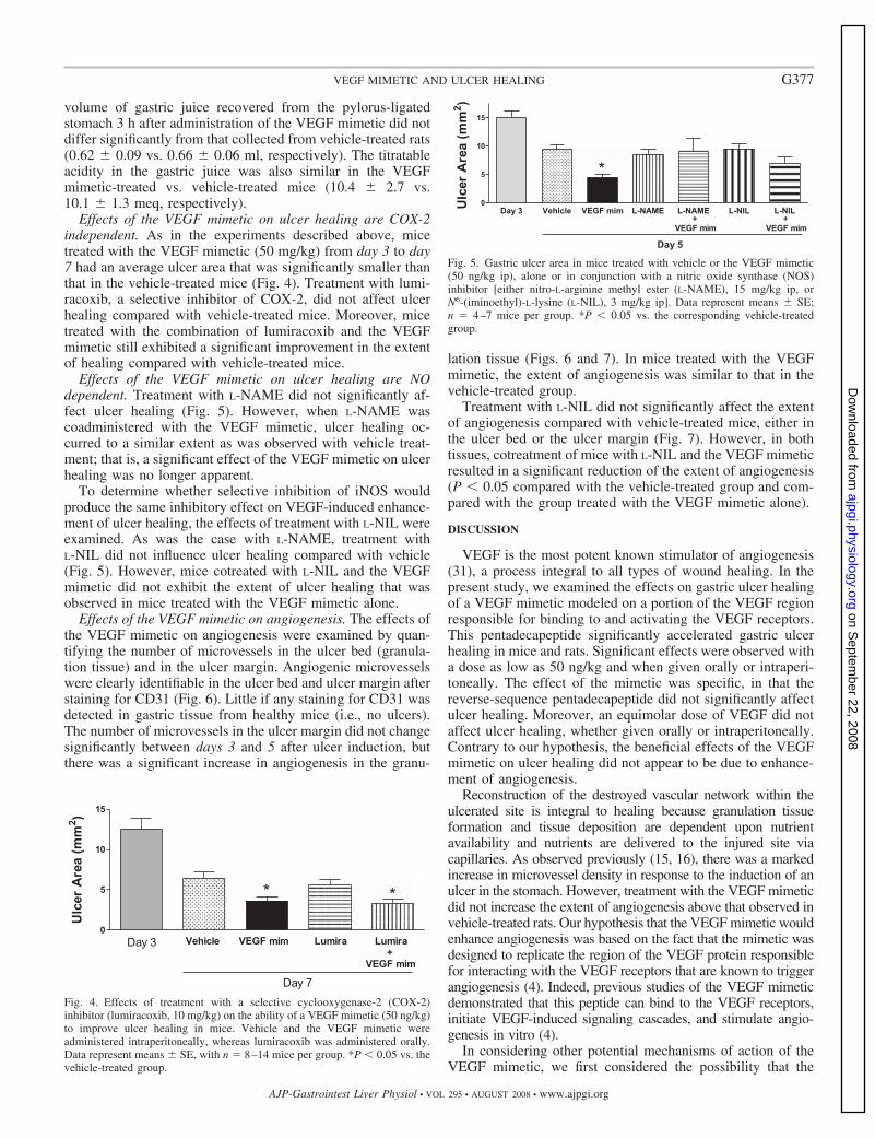
volume of gastric juice recovered from the pylorus-ligatedstomach 3 h after administration of the VEGF mimetic did notdiffer significantly from that collected from vehicle-treated rats(0.62 � 0.09 vs. 0.66 � 0.06 ml, respectively). The titratableacidity in the gastric juice was also similar in the VEGFmimetic-treated vs. vehicle-treated mice (10.4 � 2.7 vs.10.1 � 1.3 meq, respectively).
Effects of the VEGF mimetic on ulcer healing are COX-2independent. As in the experiments described above, micetreated with the VEGF mimetic (50 mg/kg) from day 3 to day7 had an average ulcer area that was significantly smaller thanthat in the vehicle-treated mice (Fig. 4). Treatment with lumi-racoxib, a selective inhibitor of COX-2, did not affect ulcerhealing compared with vehicle-treated mice. Moreover, micetreated with the combination of lumiracoxib and the VEGFmimetic still exhibited a significant improvement in the extentof healing compared with vehicle-treated mice.
Effects of the VEGF mimetic on ulcer healing are NOdependent. Treatment with L-NAME did not significantly af-fect ulcer healing (Fig. 5). However, when L-NAME wascoadministered with the VEGF mimetic, ulcer healing oc-curred to a similar extent as was observed with vehicle treat-ment; that is, a significant effect of the VEGF mimetic on ulcerhealing was no longer apparent.
To determine whether selective inhibition of iNOS wouldproduce the same inhibitory effect on VEGF-induced enhance-ment of ulcer healing, the effects of treatment with L-NIL wereexamined. As was the case with L-NAME, treatment withL-NIL did not influence ulcer healing compared with vehicle(Fig. 5). However, mice cotreated with L-NIL and the VEGFmimetic did not exhibit the extent of ulcer healing that wasobserved in mice treated with the VEGF mimetic alone.
Effects of the VEGF mimetic on angiogenesis. The effects ofthe VEGF mimetic on angiogenesis were examined by quan-tifying the number of microvessels in the ulcer bed (granula-tion tissue) and in the ulcer margin. Angiogenic microvesselswere clearly identifiable in the ulcer bed and ulcer margin afterstaining for CD31 (Fig. 6). Little if any staining for CD31 wasdetected in gastric tissue from healthy mice (i.e., no ulcers).The number of microvessels in the ulcer margin did not changesignificantly between days 3 and 5 after ulcer induction, butthere was a significant increase in angiogenesis in the granu-
lation tissue (Figs. 6 and 7). In mice treated with the VEGFmimetic, the extent of angiogenesis was similar to that in thevehicle-treated group.
Treatment with L-NIL did not significantly affect the extentof angiogenesis compared with vehicle-treated mice, either inthe ulcer bed or the ulcer margin (Fig. 7). However, in bothtissues, cotreatment of mice with L-NIL and the VEGF mimeticresulted in a significant reduction of the extent of angiogenesis(P � 0.05 compared with the vehicle-treated group and com-pared with the group treated with the VEGF mimetic alone).
DISCUSSION
VEGF is the most potent known stimulator of angiogenesis(31), a process integral to all types of wound healing. In thepresent study, we examined the effects on gastric ulcer healingof a VEGF mimetic modeled on a portion of the VEGF regionresponsible for binding to and activating the VEGF receptors.This pentadecapeptide significantly accelerated gastric ulcerhealing in mice and rats. Significant effects were observed witha dose as low as 50 ng/kg and when given orally or intraperi-toneally. The effect of the mimetic was specific, in that thereverse-sequence pentadecapeptide did not significantly affectulcer healing. Moreover, an equimolar dose of VEGF did notaffect ulcer healing, whether given orally or intraperitoneally.Contrary to our hypothesis, the beneficial effects of the VEGFmimetic on ulcer healing did not appear to be due to enhance-ment of angiogenesis.
Reconstruction of the destroyed vascular network within theulcerated site is integral to healing because granulation tissueformation and tissue deposition are dependent upon nutrientavailability and nutrients are delivered to the injured site viacapillaries. As observed previously (15, 16), there was a markedincrease in microvessel density in response to the induction of anulcer in the stomach. However, treatment with the VEGF mimeticdid not increase the extent of angiogenesis above that observed invehicle-treated rats. Our hypothesis that the VEGF mimetic wouldenhance angiogenesis was based on the fact that the mimetic wasdesigned to replicate the region of the VEGF protein responsiblefor interacting with the VEGF receptors that are known to triggerangiogenesis (4). Indeed, previous studies of the VEGF mimeticdemonstrated that this peptide can bind to the VEGF receptors,initiate VEGF-induced signaling cascades, and stimulate angio-genesis in vitro (4).
In considering other potential mechanisms of action of theVEGF mimetic, we first considered the possibility that the
Fig. 4. Effects of treatment with a selective cyclooxygenase-2 (COX-2)inhibitor (lumiracoxib, 10 mg/kg) on the ability of a VEGF mimetic (50 ng/kg)to improve ulcer healing in mice. Vehicle and the VEGF mimetic wereadministered intraperitoneally, whereas lumiracoxib was administered orally.Data represent means � SE, with n � 8–14 mice per group. *P � 0.05 vs. thevehicle-treated group.
Fig. 5. Gastric ulcer area in mice treated with vehicle or the VEGF mimetic(50 ng/kg ip), alone or in conjunction with a nitric oxide synthase (NOS)inhibitor [either nitro-L-arginine methyl ester (L-NAME), 15 mg/kg ip, orN6-(iminoethyl)-L-lysine (L-NIL), 3 mg/kg ip]. Data represent means � SE;n � 4–7 mice per group. *P � 0.05 vs. the corresponding vehicle-treatedgroup.
G377VEGF MIMETIC AND ULCER HEALING
AJP-Gastrointest Liver Physiol • VOL 295 • AUGUST 2008 • www.ajpgi.org
on Septem
ber 22, 2008 ajpgi.physiology.org
Dow
nloaded from
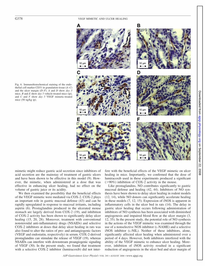
mimetic might reduce gastric acid secretion since inhibitors ofacid secretion are the mainstay of treatment of gastric ulcersand have been shown to be effective in this model (9). How-ever, the mimetic, when administered at a dose that waseffective in enhancing ulcer healing, had no effect on thevolume of gastric juice or its acidity.
We then examined the possibility that the beneficial effectsof the VEGF mimetic were mediated via COX-2. COX-2 playsan important role in gastric mucosal defense (43) and can berapidly upregulated in response to mucosal irritants, includingaspirin (6). Prostaglandins produced in the ulcerated mousestomach are largely derived from COX-2 (19), and inhibitionof COX-2 activity has been shown to significantly delay ulcerhealing (15, 20, 28). Moreover, treatment with conventionalnonsteroidal anti-inflammatory drugs (NSAIDs) and selectiveCOX-2 inhibitors at doses that delay ulcer healing in rats wasalso found to alter the ratios of pro- and antiangiogenic factors(VEGF and endostatin, respectively) in serum. COX-2-derivedprostaglandins can stimulate the release of VEGF (19), whereasNSAIDs can interfere with downstream proangiogenic signalingof VEGF (30). In the present study, we found that treatmentwith a selective COX-2 inhibitor (lumiracoxib) did not inter-
fere with the beneficial effects of the VEGF mimetic on ulcerhealing in mice. Importantly, we confirmed that the dose oflumiracoxib used in these experiments produced a significant(�90%) inhibition of COX-2 activity in the mouse.
Like prostaglandins, NO contributes significantly to gastricmucosal defense and healing (42, 44). Inhibitors of NO syn-thesis have been shown to delay ulcer healing in rodent models(12, 14), while NO donors can significantly accelerate healingin these models (7, 12, 15). Expression of iNOS is apparent ininflammatory cells in the ulcer bed in rats (14). The delay ingastric ulcer healing that occurs following administration ofinhibitors of NO synthesis has been associated with diminishedangiogenesis and impaired blood flow at the ulcer margin (3,12, 35). In the present study, the potential role of NO synthesisin the actions of the VEGF mimetic was examined through theuse of a nonselective NOS inhibitor (L-NAME) and a selectiveiNOS inhibitor (L-NIL). Neither of these inhibitors, alone,significantly affected ulcer healing when administered over aperiod of 4 days. However, both inhibitors interfered with theability of the VEGF mimetic to enhance ulcer healing. More-over, inhibition of iNOS activity resulted in a significantreduction of angiogenesis in the ulcer bed and ulcer margin of
Fig. 6. Immunohistochemical staining of the endo-thelial cell marker CD31 in granulation tissue (A–C)and the ulcer margin (D–F). A and D show day 3mice, B and E show day 5 vehicle-treated mice (ip),and C and F show day 5 VEGF mimetic-treatedmice (50 ng/kg ip).
G378 VEGF MIMETIC AND ULCER HEALING
AJP-Gastrointest Liver Physiol • VOL 295 • AUGUST 2008 • www.ajpgi.org
on Septem
ber 22, 2008 ajpgi.physiology.org
Dow
nloaded from
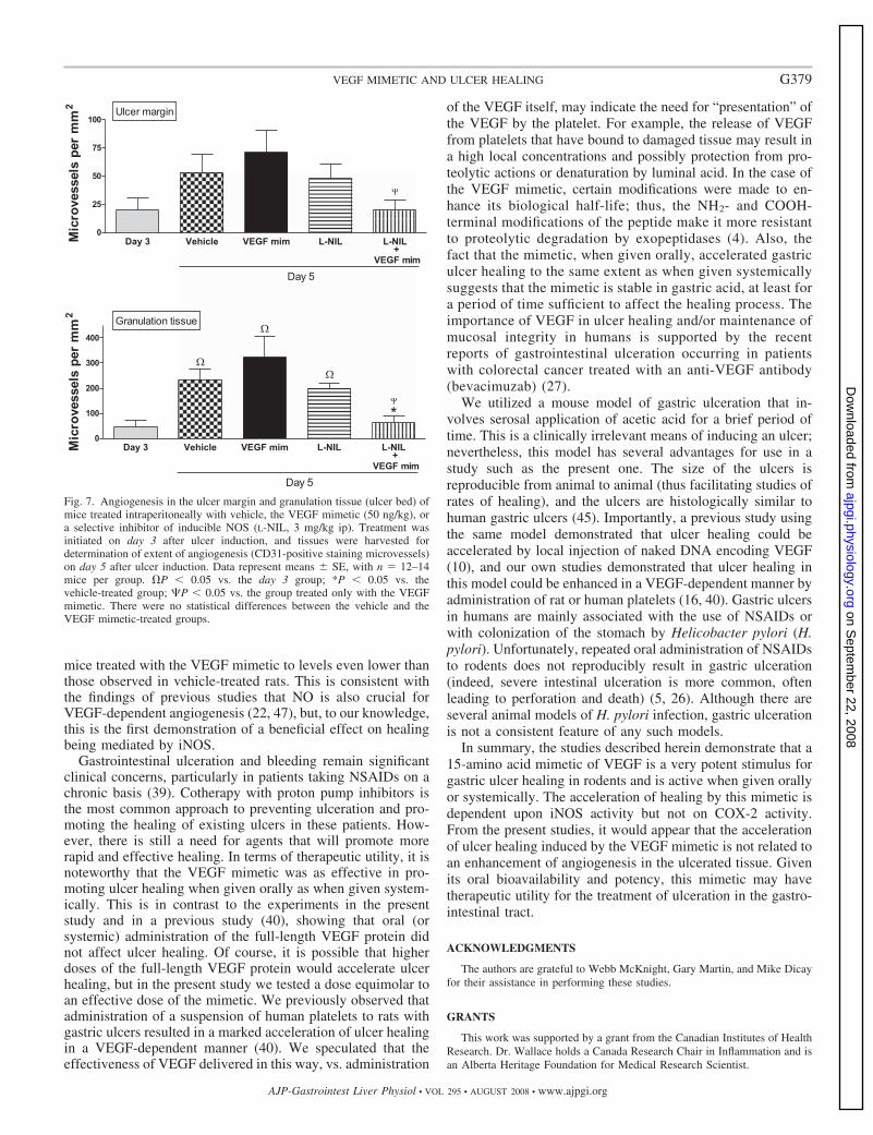
mice treated with the VEGF mimetic to levels even lower thanthose observed in vehicle-treated rats. This is consistent withthe findings of previous studies that NO is also crucial forVEGF-dependent angiogenesis (22, 47), but, to our knowledge,this is the first demonstration of a beneficial effect on healingbeing mediated by iNOS.
Gastrointestinal ulceration and bleeding remain significantclinical concerns, particularly in patients taking NSAIDs on achronic basis (39). Cotherapy with proton pump inhibitors isthe most common approach to preventing ulceration and pro-moting the healing of existing ulcers in these patients. How-ever, there is still a need for agents that will promote morerapid and effective healing. In terms of therapeutic utility, it isnoteworthy that the VEGF mimetic was as effective in pro-moting ulcer healing when given orally as when given system-ically. This is in contrast to the experiments in the presentstudy and in a previous study (40), showing that oral (orsystemic) administration of the full-length VEGF protein didnot affect ulcer healing. Of course, it is possible that higherdoses of the full-length VEGF protein would accelerate ulcerhealing, but in the present study we tested a dose equimolar toan effective dose of the mimetic. We previously observed thatadministration of a suspension of human platelets to rats withgastric ulcers resulted in a marked acceleration of ulcer healingin a VEGF-dependent manner (40). We speculated that theeffectiveness of VEGF delivered in this way, vs. administration
of the VEGF itself, may indicate the need for “presentation” ofthe VEGF by the platelet. For example, the release of VEGFfrom platelets that have bound to damaged tissue may result ina high local concentrations and possibly protection from pro-teolytic actions or denaturation by luminal acid. In the case ofthe VEGF mimetic, certain modifications were made to en-hance its biological half-life; thus, the NH2- and COOH-terminal modifications of the peptide make it more resistantto proteolytic degradation by exopeptidases (4). Also, thefact that the mimetic, when given orally, accelerated gastriculcer healing to the same extent as when given systemicallysuggests that the mimetic is stable in gastric acid, at least fora period of time sufficient to affect the healing process. Theimportance of VEGF in ulcer healing and/or maintenance ofmucosal integrity in humans is supported by the recentreports of gastrointestinal ulceration occurring in patientswith colorectal cancer treated with an anti-VEGF antibody(bevacimuzab) (27).
We utilized a mouse model of gastric ulceration that in-volves serosal application of acetic acid for a brief period oftime. This is a clinically irrelevant means of inducing an ulcer;nevertheless, this model has several advantages for use in astudy such as the present one. The size of the ulcers isreproducible from animal to animal (thus facilitating studies ofrates of healing), and the ulcers are histologically similar tohuman gastric ulcers (45). Importantly, a previous study usingthe same model demonstrated that ulcer healing could beaccelerated by local injection of naked DNA encoding VEGF(10), and our own studies demonstrated that ulcer healing inthis model could be enhanced in a VEGF-dependent manner byadministration of rat or human platelets (16, 40). Gastric ulcersin humans are mainly associated with the use of NSAIDs orwith colonization of the stomach by Helicobacter pylori (H.pylori). Unfortunately, repeated oral administration of NSAIDsto rodents does not reproducibly result in gastric ulceration(indeed, severe intestinal ulceration is more common, oftenleading to perforation and death) (5, 26). Although there areseveral animal models of H. pylori infection, gastric ulcerationis not a consistent feature of any such models.
In summary, the studies described herein demonstrate that a15-amino acid mimetic of VEGF is a very potent stimulus forgastric ulcer healing in rodents and is active when given orallyor systemically. The acceleration of healing by this mimetic isdependent upon iNOS activity but not on COX-2 activity.From the present studies, it would appear that the accelerationof ulcer healing induced by the VEGF mimetic is not related toan enhancement of angiogenesis in the ulcerated tissue. Givenits oral bioavailability and potency, this mimetic may havetherapeutic utility for the treatment of ulceration in the gastro-intestinal tract.
ACKNOWLEDGMENTS
The authors are grateful to Webb McKnight, Gary Martin, and Mike Dicayfor their assistance in performing these studies.
GRANTS
This work was supported by a grant from the Canadian Institutes of HealthResearch. Dr. Wallace holds a Canada Research Chair in Inflammation and isan Alberta Heritage Foundation for Medical Research Scientist.
Fig. 7. Angiogenesis in the ulcer margin and granulation tissue (ulcer bed) ofmice treated intraperitoneally with vehicle, the VEGF mimetic (50 ng/kg), ora selective inhibitor of inducible NOS (L-NIL, 3 mg/kg ip). Treatment wasinitiated on day 3 after ulcer induction, and tissues were harvested fordetermination of extent of angiogenesis (CD31-positive staining microvessels)on day 5 after ulcer induction. Data represent means � SE, with n � 12–14mice per group. �P � 0.05 vs. the day 3 group; *P � 0.05 vs. thevehicle-treated group; P � 0.05 vs. the group treated only with the VEGFmimetic. There were no statistical differences between the vehicle and theVEGF mimetic-treated groups.
G379VEGF MIMETIC AND ULCER HEALING
AJP-Gastrointest Liver Physiol • VOL 295 • AUGUST 2008 • www.ajpgi.org
on Septem
ber 22, 2008 ajpgi.physiology.org
Dow
nloaded from

REFERENCES
1. Barnett K, Bell CJ, McKnight W, Dicay M, Sharkey KA, Wallace JL.Role of cyclooxygenase-2 in modulating gastric acid secretion in thenormal and inflamed rat stomach. Am J Physiol Gastrointest Liver Physiol279: G1292–G1297, 2000.
2. Berse B, Brown LF, Van de Water L, Dvorak HF, Senger DR.Vascular permeability factor (vascular endothelial growth factor) gene isexpressed differentially in normal tissues, macrophages, and tumors. MolBiol Cell 3: 211–220, 1992.
3. Brzozowski T, Konturek PC, Sliwowski Z, Drozdowicz D, Hahn EG,Konturek SJ. Importance of nitric oxide and capsaicin-sensitive afferentnerves in healing of stress lesions induced by epidermal growth factor.J Clin Gastroenterol 25: S28–S38, 1997.
4. D’Andrea LD, Iaccarino G, Fattorusso R, Sorriento D, CarannanteC, Capasso D, Trimarco B, Pedone C. Targeting angiogenesis:structural characterization and biological properties of a de novoengineered VEGF mimicking peptide. Proc Natl Acad Sci USA 102:14215–14220, 2005.
5. Davies NM, Røseth AG, Appleyard CB, McKnight W, Del Soldato P,Calignano A, Cirino G, Wallace JL. NO-naproxen vs. naproxen: ulcero-genic, analgesic and anti-inflammatory effects. Aliment Pharmacol Ther11: 69–79, 1997.
6. Davies NM, Sharkey KA, Asfaha S, MacNaughton WK, Wallace JL.Aspirin causes rapid up-regulation of cyclo-oxygenase-2 expression in thestomach of rats. Aliment Pharmacol Ther 11: 1101–1108, 1997.
7. Elliott SN, McKnight W, Cirino G, Wallace JL. A nitric oxide releasingnonsteroidal anti-inflammatory drug accelerates gastric ulcer healing inrats. Gastroenterology 109: 524–530, 1995.
8. Esser R, Berry C, Du Z, Dawson J, Fox A, Fujimoto RA, Haston W,Kimble EF, Koehler J, Peppard J, Quadros E, Quintavalla J, ToscanoK, Urban L, van Duzer J, Zhang X, Zhou S, Marshall PJ. Preclinicalpharmacology of lumiracoxib: a novel selective inhibitor of cyclooxygen-ase-2. Br J Pharmacol 144: 538–550, 2005.
9. Ito M, Segami T, Tsukahara T, Kojima R, Suzuki Y. Effect ofcimetidine and omeprazole on gastric ulcer healing of rats with limitedfood intake time. Eur J Pharmacol 263: 245–251, 1994.
10. Jones MK, Kawanaka H, Baatar D, Szabo IL, Tsugawa Pai R, KohGY, Kim I, Sarfeh J, Tarnawski AS. Gene therapy for gastric ulcers withsingle local injection of naked DNA encoding VEGF and angiopoietin-1.Gastroenterology 121: 1040–1047, 2001.
11. Kimura H, Esumi H. Reciprocal regulation between nitric oxide andvascular endothelial growth factor in angiogenesis. Acta Biochim Pol 50:49–59, 2003.
12. Konturek SJ, Brzozowski T, Majka J, Pytko-Polonczyk J, Stachura J.Inhibition of nitric oxide synthase delays healing of chronic gastric ulcers.Eur J Pharmacol 239: 215–217, 1993.
13. Konturek SJ. Role of growth factors in gastroduodenal protection andhealing of peptic ulcers. Gastroenterol Clin North Am 19: 41–65, 1990.
14. Ma L, Wallace JL. Endothelial nitric oxide synthase modulates gastriculcer healing in rats. Am J Physiol Gastrointest Liver Physiol 279:G341–G346, 2000.
15. Ma L, del Soldato P, Wallace JL. Divergent effects of new cyclooxy-genase inhibitors on gastric ulcer healing: Shifting the angiogenic balance.Proc Natl Acad Sci USA 99: 13243–13247, 2002.
16. Ma L, Elliott SN, Cirino G, Buret A, Ignarro LJ, Wallace JL.Platelets modulate gastric ulcer healing: role of endostatin and vascularendothelial growth factor release. Proc Natl Acad Sci USA 98: 6470 –6475, 2001.
17. Maloney JP, Silliman CC, Ambruso DR, Wan J, Tuder RM,Voelkel NF. In vitro release of vascular endothelial growth factorduring platelet aggregation. Am J Physiol Heart Circ Physiol 275:H1054 –H1061, 1998.
18. Mesri M, Morales-Ruiz M, Ackermann EJ, Bennett CF, Pober JS,Sessa WC, Altieri DC. Suppression of vascular endothelial growthfactor-mediated endothelial cell protection by surviving targeting. Am JPathol 158: 1757–1765, 2001.
19. Miura S, Tatsuguchi A, Wada K, Takeyama H, Shinji Y, Hiratsuka T,Futagami S, Miyake K, Gudis K, Mizokami Y, Matsuoka T, SakamotoC. Cyclooxygenase-2-regulated vascular endothelial growth factor releasein gastric fibroblasts. Am J Physiol Gastrointest Liver Physiol 287:G444–G451, 2004.
20. Mizuno H, Sakamoto C, Matsuda K, Wada K, Uchida T, Noguchi H,Akamatsu T, Kasuga M. Induction of cyclooxygenase 2 in gastric
mucosal lesions and its inhibition by the specific antagonist delays healingin mice. Gastroenterology 112: 387–397, 1997.
21. Moore WM, Webber RK, Jerome GM, Tjoeng FS, Misko TP, CurrieMG. L-N6-(1-iminoethyl)lysine: a selective inhibitor of inducible nitricoxide synthase. J Med Chem 37: 3886–3888, 1994.
22. Morbidelli L, Chang CH, Douglas JG, Granger HJ, Ledda F, ZicheM. Nitric oxide mediates mitogenic effect of VEGF on coronaryvenular endothelium. Am J Physiol Heart Circ Physiol 270: H411–H415, 1996.
23. Podolsky DK. Review article: healing after inflammatory injury—coor-dination of a regulatory peptide network. Aliment Pharmacol Ther, 14Suppl 1: 87–93, 2000.
24. Rees DD, Palmer RM, Schulz R, Hodson HF, Moncada S. Character-ization of three inhibitors of endothelial nitric oxide synthase in vitro andin vivo. Br J Pharmacol 101: 746–52, 1990.
25. Reuter BK, Asfaha S, Buret A, Sharkey KA, Wallace JL. Exacerbationof inflammation-associated colonic injury in rat through inhibition ofcyclooxygenase-2. J Clin Invest 98: 2076–2085, 1996.
26. Reuter BK, Davies NM, Wallace JL. Nonsteroidal anti-inflammatorydrug enteropathy in rats: role of permeability, bacteria, and enterohepaticcirculation. Gastroenterology 112: 109–17, 1997.
27. Saif MW, Elfiky A, Salem RR. Gastrointestinal perforation due tobevacizumab in colorectal cancer. Ann Surg Oncol 14: 1860 –1869,2007.
28. Schmassmann A, Peskar BM, Stettler C, Netzer P, Stroff T, FlogerziB, Halter F. Effects of inhibition of prostaglandin endoperoxide syn-thase-2 in chronic gastro-intestinal ulcer models in rats. Br J Pharmacol123: 795–804, 1998.
29. Suzuki N, Takahashi S, Okabe S. Relationship between vascular endo-thelial growth factor and angiogenesis in spontaneous and indomethacin-delayed healing of acetic acid-induced gastric ulcers in rats. J PhysiolPharmacol 49: 515–527, 1998.
30. Szabo IL, Pai R, Soreghan B, Jones MK, Baatar D, Kawanaka H,Tarnawski A. NSAIDs inhibit the activation of egr-1 gene in microvas-cular endothelial cells. A key to inhibition of angiogenesis? J PhysiolParis 95: 379–383, 2001.
31. Szabo S, Vincze A. Growth factors in ulcer healing: lessons from recentstudies. J Physiol Paris 94: 77–81, 2000.
32. Takahashi M, Kawabe T, Ogura K, Maeda S, Mikami Y, Kaneko N,Terano A, Omata M. Expression of vascular endothelial growth factor atthe human gastric ulcer margin and in cultured gastric fibroblasts: a newangiogenic factor for gastric ulcer healing. Biochem Biophys Res Commun234: 493–498, 1997.
33. Takahashi M, Maeda S, Ogura K, Terano A, Omata M. The possiblerole of vascular endothelial growth factor (VEGF) in gastric ulcer healing:effect of sofalcone on VEGF release in vitro. J Clin Gastroenterol 27:S178–S182, 1998.
34. Takahashi M, Ogura K, Maeda S, Mori K, Mafune K, Mikami Y,Terano A, Omata M. Promoters of epithelialization induce expression ofvascular endothelial growth factor in human gastric epithelial cells inprimary culture. FEBS Lett 418: 115–118, 1997.
35. Takeuchi K, Kato S, Takehara K, Asada Y, Yasuiro T. Role of nitricoxide in mucosal blood flow response and the healing of HCl-inducedlesions in the rat stomach. Digestion 58: 19–27, 1997.
36. Tarnawski AS. Cellular and molecular mechanisms of gastrointestinalulcer healing. Dig Dis Sci 50: S24–S33, 2005.
37. Wallace JL, Bak A, McKnight W, Asfaha S, Sharkey KA, MacNaugh-ton WK. Cyclooxygenase 1 contributes to inflammatory responses in ratsand mice: implications for gastrointestinal toxicity. Gastroenterology 115:101–109, 1998.
38. Wallace JL, Chapman K, McKnight W. Limited anti-inflammatoryefficacy of cyclo-oxygenase-2 inhibition in carrageenan-airpouch inflam-mation. Br J Pharmacol 126: 1200–1204, 1999.
39. Wallace JL, del Soldato P. The therapeutic potential of NO-NSAIDs.Fundam Clin Pharmacol 17: 11–20, 2003.
40. Wallace JL, Dicay M, McKnight W, Dudar GK. Platelets accelerategastric ulcer healing through presentation of vascular endothelial growthfactor. Br J Pharmacol 148: 274–278, 2006.
41. Wallace JL, Dicay M, McKnight W, Martin GR. Hydrogen sulfideenhances ulcer healing in rats. FASEB J 21: 4070–4076, 2007.
42. Wallace JL, Miller MJ. Nitric oxide in mucosal defense: a little goes along way. Gastroenterology 119: 512–520, 2000.
G380 VEGF MIMETIC AND ULCER HEALING
AJP-Gastrointest Liver Physiol • VOL 295 • AUGUST 2008 • www.ajpgi.org
on Septem
ber 22, 2008 ajpgi.physiology.org
Dow
nloaded from

43. Wallace JL, McKnight W, Reuter BK, Vergnolle N. NSAID-inducedgastric damage in rats: requirement for inhibition of both cyclooxygenase1 and 2. Gastroenterology 119: 706–714, 2000.
44. Wallace JL, Tigley AW. Review article: new insights into prostaglandinsand mucosal defence. Aliment Pharmacol Ther 9: 227–235, 1995.
45. Wang JY, Yamasaki S, Takeuchi K, Okabe S. Delayed healing of aceticacid-induced gastric ulcers in rats by indomethacin. Gastroenterology 96: 393–402, 1989.
46. Zanardo RC, Brancaleone V, Distrutti E, Fiorucci S, Cirino G,Wallace JL. Hydrogen sulfide is an endogenous modulator of leukocyte-mediated inflammation. FASEB J 20: 2118–2120, 2006.
47. Ziche M, Morbidelli L, Choudhuri R, Zhang HT, Donnini S,Granger HJ, Bicknell R. Nitric oxide synthase lies downstream fromvascular endothelial growth factor-induced but not basic fibroblastgrowth factor-induced angiogenesis. J Clin Invest 99: 2625–2634,1997.
G381VEGF MIMETIC AND ULCER HEALING
AJP-Gastrointest Liver Physiol • VOL 295 • AUGUST 2008 • www.ajpgi.org
on Septem
ber 22, 2008 ajpgi.physiology.org
Dow
nloaded from