exposing hidden dimensions of embryonic stem cell cycle control
TRANSCRIPT
Cell Stem Cell
Previews
Exposing Hidden Dimensionsof Embryonic Stem Cell Cycle Control
Stephen Dalton1,*1Department of Biochemistry and Molecular Biology, Paul D. Coverdell Center for Biomedical and Health Sciences, The University of Georgia,500 D.W. Brooks Drive, Athens, GA 30602, USA*Correspondence: [email protected] 10.1016/j.stem.2008.12.003
In a recent issue of Nature Genetics, Wang and colleagues (2008) describe a mechanism for how the mir-290microRNA cluster regulates the cell cycle of murine embryonic stem cells. A focal point of this regulation is thecyclin-dependent kinase inhibitor p21Cip1.
Recent work from Robert Blelloch’s labo-
ratoryhassoughttoestablishamechanism
to explain the observed role of microRNAs
(miRNAs) in murine embryonic stem cell
(mESC) biology (Wang et al., 2007). In their
most recent report, Wang and colleagues
(2008) describe a role for mESC-specific
miRNAs in establishing rapid cell division
cycles. This topic has been on the radar
screen of stem cell biologists for many
years, but little is known about mecha-
nisms underpinning the rapid division rates
observed in mESCs, or what significance
this short cell-cycle time may have in rela-
tion to their biology. The Blelloch study
makes some important progress toward
addressing some of these issues.
mESCs exhibit extraordinary rates of
proliferation, with generation times typi-
cally in the order of �10 hr (Burdon
et al., 2002). The growth kinetics of
mESCs are in close concordance with
the short generation times observed in
pluripotent epiblast cells in the peri-
implantation mouse embryo (Snow,
1977). Rapid cell division therefore
appears to be a general trait of embryo-
derived pluripotent cells in the rodent.
Rapid cell division is associated with an
unusual cell-cycle structure, most notably
the short length of G1 (�2 hr) and the high
percentage of cells in S phase (Burdon
et al., 2002; Stead et al., 2002). As mESCs
differentiate, generation times increase
(>18 hr) and the cell cycle is remodeled
so that the length of G1 increases (Stead
et al., 2002; White et al., 2005). Interest-
ingly, this adjustment coincides with loss
of tumorigenic potential and the activation
of pathways that couple mitogenic
signaling to the cell-cycle machinery
(White et al., 2005). Although human ESCs
cycle more slowly (�30–36 hr) than their
rodent counterparts, ESCs of both
species undergo similar cell-cycle remod-
eling during differentiation, in that the
proportion of cells in G1 increases with
the loss of pluripotency (Ohtsuka and
Dalton, 2008). The biological significance
of these observations has yet to be fully
elucidated, but several major questions
emerge from these early studies. First,
what is the molecular mechanism under-
pinning rapid cell division in pluripotent
cells? Second, what is the biological
significance of rapid embryonic division,
and does it have a role in the establish-
ment and/or maintenance of pluripo-
tency? Finally, is rapid cell division by
ESCs required for the pluripotent cells to
eventually enter a normal differentiation
program?
The first question was addressed some
time ago by Stead and coworkers, who
established that many of the basic rules
of somatic cell division cycle control
do not apply in mESCs. To appreciate
this issue, it is important to remember
that transition through the cell cycle is
controlled by the activity of phase-
specific cyclin-dependent protein kinases
(Cdks). These kinases are activated and
inactivated at precise points of the cell
cycle and phosphorylate substrates
required for the different cell-cycle transi-
tions. The periodicity of Cdk activity is
critical for the normal sequence of events
that occur during a typical somatic cell
cycle. In mESCs, Cdk2, which controls
G1 progression into S phase, displays
elevated activity throughout the cell cycle
and shows no obvious periodicity (Stead
et al., 2002; White et al., 2005). As plurip-
otent cells begin to differentiate, Cdk2
activity declines and becomes cell cycle
regulated, explaining the G1 phase expan-
Cell Stem C
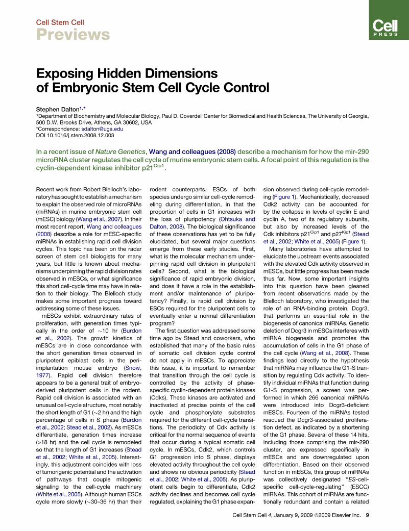
sion observed during cell-cycle remodel-
ing (Figure 1). Mechanistically, decreased
Cdk2 activity can be accounted for
by the collapse in levels of cyclin E and
cyclin A, two of its regulatory subunits,
but also by increased levels of the
Cdk inhibitors p21Cip1 and p27Kip1 (Stead
et al., 2002; White et al., 2005) (Figure 1).
Many laboratories have attempted to
elucidate the upstream events associated
with the elevated Cdk activity observed in
mESCs, but little progress has been made
thus far. Now, some important insights
into this question have been gleaned
from recent observations made by the
Blelloch laboratory, who investigated the
role of an RNA-binding protein, Dcgr3,
that performs an essential role in the
biogenesis of canonical miRNAs. Genetic
deletion of Dcgr3 in mESCs interferes with
miRNA biogenesis and promotes the
accumulation of cells in the G1 phase of
the cell cycle (Wang et al., 2008). These
findings lead directly to the hypothesis
that miRNAs may influence the G1-S tran-
sition by regulating Cdk activity. To iden-
tify individual miRNAs that function during
G1-S progression, a screen was per-
formed in which 266 canonical miRNAs
were introduced into Dcgr3-deficient
mESCs. Fourteen of the miRNAs tested
rescued the Dcgr3-associated prolifera-
tion defect, as indicated by a shortening
of the G1 phase. Several of these 14 hits,
including those comprising the mir-290
cluster, are expressed specifically in
mESCs and are downregulated upon
differentiation. Based on their observed
function in mESCs, this group of miRNAs
was collectively designated ‘‘ES-cell-
specific cell-cycle-regulating’’ (ESCC)
miRNAs. This cohort of miRNAs are func-
tionally redundant and contain a related
ell 4, January 9, 2009 ª2009 Elsevier Inc. 9
Cell Stem Cell
Previews
‘‘seed’’ sequence, indicating that they
target overlapping downstream RNA
targets for translational repression. A
computational survey of potential ESCC
miRNA targets identified the Cdk2 inhibitor
p21Cip1 (Cdkn1a) as being of special
interest because of its link to proliferative
control and Cdk regulation. A link between
A
B
Figure 1. Proposed Mechanism for mir-290miRNA Regulation of G1 Progression inmESCs(A) mESCs exhibit high mir-290 miRNA levels andelevated Cdk2-cyclin E activity, leading to acceler-ated G1-S progression and a correspondingshort G1 phase. mir-290 inhibits the accumulationof p21Cip1, allowing unrestrained Cdk2-cyclin Eactivity.(B) As mir-290 miRNAs are downregulated duringdifferentiation, or in cells deficient for Dcgr3,p21Cip1 accumulates and assembles intocomplexes that inactivate Cdk2-cyclin E. Thediminished cdk activity is thought to delay progres-sion from G1 into S phase, thus lengthening overallgeneration times.
10 Cell Stem Cell 4, January 9, 2009 ª2009
ESCC miRNAs and p21Cip1 was estab-
lished by demonstrating that ESCC
miRNAs suppress p21Cip1 protein levels
and regulate the Cdkn1a transcript through
its 30 untranslated region, consistent with
classic miRNA-mediated translational in-
hibition. Moreover, overexpression of
p21Cip1 in mESCs was sufficient to repro-
duce the G1-S delay previously described
in a Dcgr3-deficient background. Addi-
tional cell-cycle target genes such as
Rb1, Rbl1, Rbl2, and Lats2 were also
shown to be targeted by this regulatory
pathway. These results suggest that
miRNAs can modulate the G1-S transition
by restraining the ability of p21Cip1 to inhibit
Cdk2-cyclin E complexes (Figure 1).
Since the mir-290 cluster is expressed at
high levels in mESCs and downregulated
early during differentiation, members of
this ESCC subset are likely to be the key
regulatorsofp21Cip1 in thepluripotent state.
Other miRNAs identified in the original
screen by Wang et al. may play roles in
othercell types that exhibit rapid cell cycles.
Cells of the intestinal crypt and activated
T cells, for example, also have short gener-
ation timesandcouldbesubject toacontrol
mechanism similar to that described by
Wang et al. in early development. It will
also be important to establish if tumor cells
express any of the 14 miRNAs identified by
the Blelloch studies and to then establish if
the ESCCs participate in the deregulation of
cancer cell growth. The ability to generate
rapidly dividing subpopulations of tumor
cells would provide a powerful selective
advantage and could thus lead to rapid
amplification of the disregulated cells. In
the context of pluripotent cell biology, the
role of ESCC miRNAs remains only partially
Elsevier Inc.
defined. That is, although Dcgr3 is required
for normal proliferation and differentiation,
ESCC miRNAs themselves have only been
investigated in the context of cell-cycle
control, and not for effects on long-term
self-renewal or differentiation potential.
Observations made in Dcgr3-deficient
mESCs raise the possibility that rapid
cell division is functionally linked to the
molecular program responsible for initia-
tion of differentiation. However, Wang
et al. comment that ESCC miRNAs do
not rescue the differentiation defect in
Dcgr3 cells. This observation suggests
that additional miRNAs outside the mir-
290 cluster lie downstream of Dcgr3. In
summary, the Blelloch studies have
exposed a previously hidden dimension
of cell-cycle control in mESCs that should
ultimately point the way toward a better
understanding of basic pluripotent cell
biology and differentiation mechanics.
REFERENCES
Burdon, T., Smith, A., and Savatier, P. (2002).Trends Cell Biol. 12, 432–438.
Ohtsuka, S., and Dalton, S. (2008). Gene Ther. 15,74–81.
Snow, M.H.L. (1977). J. Embryol. Exp. Morphol. 42,293–303.
Stead, E., White, J., Faast, R., Conn, S., Goldstone,S., Rathjen, J., Dhingra, U., Rathjen, P., Walker, D.,and Dalton, S. (2002). Oncogene 21, 8320–8333.
Wang, Y., Medvid, R., Melton, C., Jaenisch, R., andBlelloch, R. (2007). Nat. Genet. 39, 380–385.
Wang, Y., Baskerville, S., Shenoy, A., Babiarz, J.E.,Baehner, L., and Blelloch, R. (2008). Nat. Genet.40, 1478–1483.
White, J., Stead, E., Faast, R., Conn, S., Cart-wright, P., and Dalton, S. (2005). Mol. Biol. Cell16, 2018–2027.