exploring early designs for teaching anatomy and ...jonf/publications/norooz_exploringearly... ·...
TRANSCRIPT

Exploring Early Designs for Teaching Anatomy and Physiology to Children Using Wearable E-Textiles
Leyla Norooz1 and Jon Froehlich2
Makeability Lab | HCIL 1 College of Information Studies
University of Maryland College Park, MD [email protected]
Makeability Lab | HCIL 2 Dept. of Computer Science
University of Maryland College Park, MD [email protected]
ABSTRACT Unlike external body parts, organs are invisible and untouchable, making it difficult for children to learn their size, position, and function. Traditionally, human anatomy (body form) and physiology (body function) are taught using a mixture of techniques from worksheets to three-dimensional models. With the advent of low-cost sensing, ubiquitous computation, and emerging e-textiles, new teaching approaches are developing that link the physical and virtual worlds. In this demo, we explore and illustrate the use of a custom wearable e-textile shirt to teach anatomy and physiology to children. Our current implementation uses dyed fabric to show the size and position of body organs and a mixture of electronic sensors and visualizations to dynamically exhibit the wearer’s physiology (e.g., heart beat). Though we are still in an early design stage, we discuss our design process, our progress thus far, and plans for future iterations.
Categories and Subject Descriptors H.5.2 [Information Interfaces and Presentation]: User Interfaces—user-centered design
General Terms Design, Human Factors
Keywords Tangible interaction; e-Textiles; active learning; teaching; anatomy; physiology; sensing and visualization; body knowledge
1 INTRODUCTION Learning about the position, structure, and function of internal body parts is challenging for children [12, 14, 17]. Although by age four most children have a fairly well-defined concept of their external body and the relationship between its parts, this is not the case with their inner body [17]. Unlike fingers, arms, toes, and other external parts, internal organs remain hidden beneath layers of skin, muscle, and tissue and operate without conscious thought, making it difficult for children—and even adults (e.g., [1])—to understand the internal workings of their bodies.
Traditionally, human anatomy (body form) and physiology (body function) are taught in pre-school and primary school education using a mixture of techniques including three-dimensional models and dolls, coloring and activity books, stories, audio-visuals, and
video games [17]. Most schools include anatomy and physiology as part of their K-8 science curriculum, which is then extended through high school biology [10]. Teaching pre-school and primary school children about their anatomy and physiology can help with self-care and self-understanding and generally leads to greater compliance with health care regimens [14, 15]. For example, young children with asthma are more likely to take inhaled medications if they understand how their lungs function [14]. Other researchers emphasize the critical role of anatomy and physiology in teaching basic science (e.g., biology) [4].
In this short demo paper, we introduce and explore a new approach to teaching anatomy and physiology: utilizing a custom wearable e-textile shirt that combines embedded sensing and visualization to reveal otherwise “invisible” parts and functions of the human body (Figure 1). Though we are still in the early prototyping phase of our design process, our overarching goal is to develop e-textiles that automatically sense the wearer’s physiology and visualize that function in an anatomically correct form for teaching purposes. While other efforts have explored the use of three-dimensional models and even fabric representations of anatomy (e.g., iheartguts.com), to our knowledge we are the first to explore a digitalized manifestation that actively visualizes and responds to the anatomy and physiology of the wearer.
For our demonstration at IDC, we will show an early prototype of our shirt that focuses primarily on the circulatory, respiratory, and gastrointestinal systems as tangibly actualized in a wearable e-textile shirt (Figure 1b). The organs themselves are made of colorful fabric stuffed with cotton to create and maintain anatomical form. Light-emitting diodes (LEDs) and electroluminescent (EL) wire are hooked up to a pulse sensor controlled by an Arduino Uno that reacts to the wearer’s heartbeat. We are also currently exploring respiratory monitoring using flex-sensors and digestive sensing using microphones along with new types of output that include audio and haptics; however,
Copyright is held by the owner/author(s). IDC '13, Jun 24-27 2013, New York, NY, USA ACM 978-1-4503-1918-8/13/06.
Figure 1: (a) An early design sketch of the e-textile shirt. (b) Our current prototype.
(a) (b)
such functionality has not yet been implemented. We hope to complete these iterations by the IDC conference in June.
2 RELATED WORK As noted in the introduction, by age four most children have well-defined understandings of their external body and the relationships between body parts (e.g., fingers to hand to arm); however, their conception of the inner-body is comparatively weaker [17]. Children between the ages of four and eight can recall approximately three to six internal body parts with those most commonly identified being the brain, heart, bones, and blood (ibid). However, children often misconceive of their size, shape, position, and function. For example, the heart is typically drawn as a playing card “valentine” heart (e.g., [2, 5]) and the stomach is considered a respiratory mechanism because it moves in and out with breath (e.g., [6]). In addition, few children have a clear idea of how food passes through their body and waste is eliminated [11]. Interestingly, though a review of 25 studies exploring children’s conceptions of human anatomy and physiology found that knowledge generally increases with age [14], even studies of college-educated adults have found that some misconceptions can persist into adulthood [1].
Most researchers emphasize that because internal organs are not visible or touchable, they are difficult for children to understand, observe, and experiment with in daily life (e.g., [12]). Thus, pre-school and primary school methodologies often take a multi-sensory approach, which utilizes a student’s multiple senses to receive, interpret, and respond to material about the human body. Vessey et al. [17] suggest using three-dimensional teaching aids such as anatomic dolls or models to accompany worksheets, stories, audio-visuals, and games (both board games and video games). Although few experimental studies exist on testing the effects of different teaching methods on children’s body knowledge, two studies point to the benefits of using three-dimensional teaching aids specifically [13, 16]. From these studies, researchers recommend that teaching artifacts be engaging (e.g., comprised of bright colors and different textures), realistic but approachable (i.e., not “scary”), and interactive (e.g., Schmidt [13] discovered that children learn more from interactive lungs than stationary ones).
In addition to the above, our work also takes inspiration from research in tangible interactive computing and education that emphasizes the important role of physical action and manipulation in learning, which can be enhanced through computation (e.g., [8, 9, 20]. Additionally, the use of wearable sensing and visualization for anatomy as well as our overall design aesthetic was influenced by Lam and Ngo’s Warning Signs project, which sensed and visualized ambient air pollution (carbon monoxide) via an e-
textile heart or lung sewn into the wearer’s t-shirt [18].
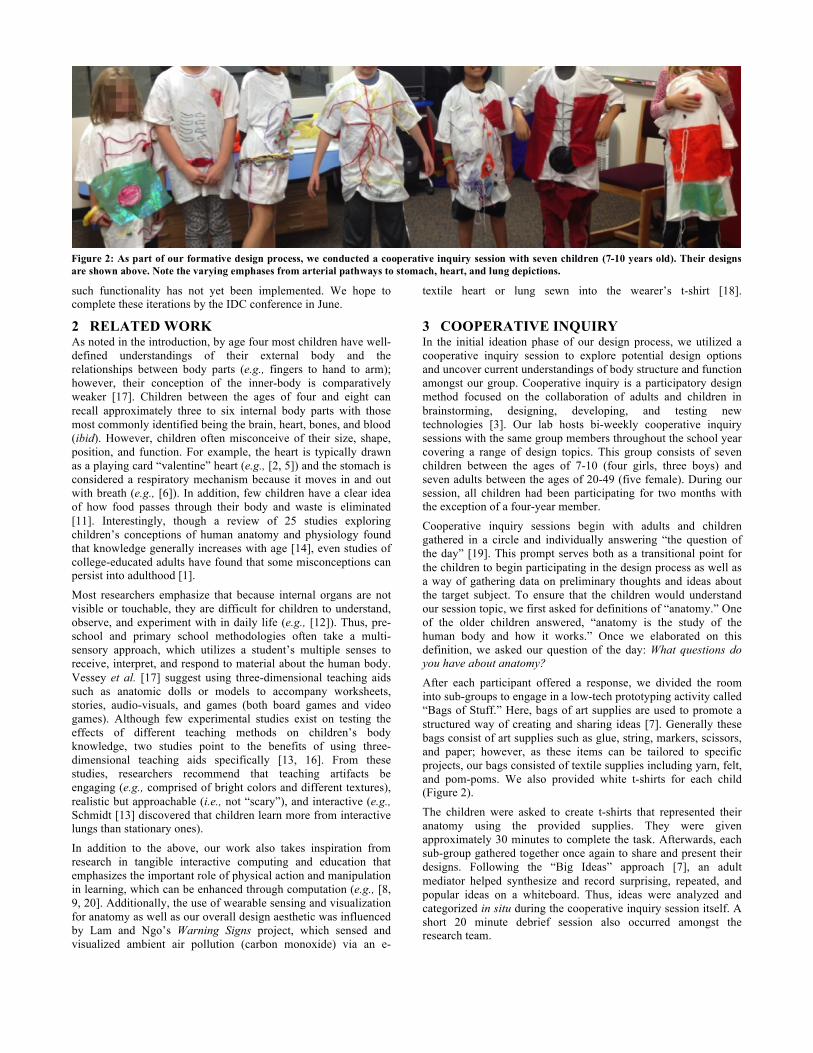
3 COOPERATIVE INQUIRY In the initial ideation phase of our design process, we utilized a cooperative inquiry session to explore potential design options and uncover current understandings of body structure and function amongst our group. Cooperative inquiry is a participatory design method focused on the collaboration of adults and children in brainstorming, designing, developing, and testing new technologies [3]. Our lab hosts bi-weekly cooperative inquiry sessions with the same group members throughout the school year covering a range of design topics. This group consists of seven children between the ages of 7-10 (four girls, three boys) and seven adults between the ages of 20-49 (five female). During our session, all children had been participating for two months with the exception of a four-year member.
Cooperative inquiry sessions begin with adults and children gathered in a circle and individually answering “the question of the day” [19]. This prompt serves both as a transitional point for the children to begin participating in the design process as well as a way of gathering data on preliminary thoughts and ideas about the target subject. To ensure that the children would understand our session topic, we first asked for definitions of “anatomy.” One of the older children answered, “anatomy is the study of the human body and how it works.” Once we elaborated on this definition, we asked our question of the day: What questions do you have about anatomy?
After each participant offered a response, we divided the room into sub-groups to engage in a low-tech prototyping activity called “Bags of Stuff.” Here, bags of art supplies are used to promote a structured way of creating and sharing ideas [7]. Generally these bags consist of art supplies such as glue, string, markers, scissors, and paper; however, as these items can be tailored to specific projects, our bags consisted of textile supplies including yarn, felt, and pom-poms. We also provided white t-shirts for each child (Figure 2).
The children were asked to create t-shirts that represented their anatomy using the provided supplies. They were given approximately 30 minutes to complete the task. Afterwards, each sub-group gathered together once again to share and present their designs. Following the “Big Ideas” approach [7], an adult mediator helped synthesize and record surprising, repeated, and popular ideas on a whiteboard. Thus, ideas were analyzed and categorized in situ during the cooperative inquiry session itself. A short 20 minute debrief session also occurred amongst the research team.
Figure 2: As part of our formative design process, we conducted a cooperative inquiry session with seven children (7-10 years old). Their designs are shown above. Note the varying emphases from arterial pathways to stomach, heart, and lung depictions.

3.1 Cooperative Inquiry Results Though each sub-group in our cooperative inquiry had unique design ideas, a set of overarching themes emerged around the use of color, sound, lights, and movement. Color was used to distinguish between organs and their function (e.g., red for veins and the heart). For sound, we found that children used audio to increase the playfulness and reactivity of their shirts. For example, children designed breakable ribs and spines with “cracking” sound effects, “talking” organs, and using the spine as a musical instrument. Lights were used mainly to indicate an action such as a pumping heart, hunger, or blood moving through veins. Finally, the most popular design theme was the use of movement. For example, children illustrated food traveling through the digestive system and dissolving in the stomach, physically pumping hearts, and “breathing” lungs. In summary, while the design ideas ranged in feasibility, there was a clear emphasis on dynamics, interactivity, and reactivity to the human form and function.
4 OUR E-TEXTILE PROTOTYPE As we are using a t-shirt as our visual teaching medium, the areas covered include the thoracic region above the diaphragm and the abdominal region below it. For the thoracic cavity, we included the lungs, heart, and esophagus but not, currently, the trachea and the thymus gland. The abdominal cavity extends down to the pelvic cavity and includes most of the digestive organs including the esophagus, stomach, liver, gallbladder, pancreas, small intestine, and large intestine. Currently, our prototype shirt does not depict reproductive organs or human waste orifices. Vessey [17] warns that anatomical teaching aids can abstract complexity (e.g., to simplify and capture interest) but should not create or reinforce children’s false perceptions. Thus, we attempted to correctly shape and position each organ on the shirt though we did attempt to minimize overlap to avoid occlusion (Figure 1). Organs were created using two pieces of felt, cut into anatomically correct shapes, sewn together, and stuffed with pillow stuffing. This creates a plush, tangible aesthetic aimed at attracting a child’s attention and touch.
Our shirt includes multiple bright colors, which was influenced by our cooperative inquiry session as well as related work (e.g., [15, 17]). Because children in our cooperative inquiry group are often sensitive to perceived gender delineations, we selected a gender-neutral green color for the t-shirt itself (the base). The remaining colors balance the functional representation for each organ and overall visual aesthetic. Although some of the organ’s colors are not anatomically correct, we believe that specific colors may help children remember the functionality and purpose of each individual organ. For example, one child in the cooperative inquiry session had a brown colored large intestine in her design,
as it represented the final stage of the digestive system. She said, “that’s where the poop comes out.”
4.1 Heart and Lungs The heart is comprised of red and blue felt to represent blood entering and leaving the heart (Figure 3). Embedded in the heart are six LEDs. These are placed on an inflexible perfboard and surrounded by soft stuffing and an outer layer of felt. Their colors correspond to the surrounding felt (blue or red). The LEDs are connected to a pulse sensor (pulsesensor.com) controlled by an Arduino Uno, which uses infrared to detect the wearer’s heart rate. The LEDs light up with the user’s heartbeat (Figure 3b). Through experimentation, we found that the pulse sensor functioned best when attached to the finger. Future designs will attempt to incorporate multiple pulse sensors embedded into the shirt itself thereby eliminating the finger attachment.
For the lungs, each side is attached to opposite ends of the heart. To illustrate the veins running through the lungs, we covered the lung surfaces with blue and red EL wire. These colors maintain the theme of blood entering and leaving the heart. Similar to the heart LEDs, the EL wire is connected to the Arduino and “pulse” in accordance with the user’s heart rate (Figure 3b).
4.2 Digestive System The digestive system consists of the esophagus, stomach, liver, gallbladder, pancreas, small intestine, and large intestine. The esophagus was created using a grooved portion of a suction pump. This design was chosen due to its similar visual appearance of the human esophagus, which uses surrounding muscles to pinch inward and send food to the stomach. The esophagus is also visibly attached to the beginning of the stomach, which is made of stuffed fabric. Similarly, other digestive organs such as the liver, gallbladder, and the pancreas are built from colored fabric. We are currently experimenting with different sensing and visualization techniques to illustrate the digestive process including the use of sound and an LED matrix for animation.
The fabric stomach is visibly attached to the small intestine, represented here in blue. In our cooperative inquiry session, one child used strings of yarn to represent her small intestine and to highlight its surprising length. Consequently, we designed our small intestine using Velcro to detach and unravel from the shirt allowing children to fully investigate its length. Finally, the small intestine is visibly attached to the large intestine represented in brown.
5 CONCLUSION AND FUTURE WORK In this demo paper, we introduced and discussed the design and design process for an early prototype e-textile shirt for teaching
Figure 3: A close-up of our fabric heart (red and blue) with embedded LEDs and lungs (orange) with vein accents made of EL wire.
(a) (b)

children about anatomy and physiology. However, our current prototype is limited to the pulse sensor as a form of input and LEDs and EL wire as a form of output. In the future, we plan to explore the use of additional input including breathing sensors, a method of detecting swallowing, and temperature sensors. These new input devices will allow us to display real-time breathing, digestion, and body temperature on the shirt. Some organs are more difficult to sense and visualize than others (e.g., the heart vs. the pancreas). Thus, for all organs we plan to add physical labels attached via Velcro, and a touch sensor that plays back a short description of the specific organ.
Our ultimate vision is to, whenever possible, automatically sense and visualize the wearer’s physiology promptly when the shirt is placed on the body. We are currently exploring a flexible fabric band enclosure on the inside of the shirt that would allow embedded pulse sensors direct access to the skin. In addition, we are investigating the use of conductive stretch fabric to monitor breathing rate.
We also plan aesthetic and comfort improvements. Over time, our current prototype shirt became stretched due to the weight of the organs and the amount of pillow stuffing used. To avoid this and to allow the user to feel more comfortable, we are currently designing a flat, 2D prototype similar to everyday graphic t-shirts but equipped with our embedded sensing and output modalities. Finally, we plan to create a Do-It-Yourself (DIY) tutorial for school teachers to create their own versions of our shirt to share with their community and to help improve the education of anatomy and physiology for primary school children. We have interviewed two 2nd grade teachers about our prototype so far. Both responded positively and enthusiastically to our prototype and were open to collaboration and evaluation in the classroom.
6 REFERENCES [1] Blum, L.H. 1977. Health information via mass media: Study of
the individual’s concepts of the body and its parts. Psychological Reports. 40, (1977), 991–999.
[2] Cuthbert, A. 2000. Do Children Have a Holistic View Of Their Internal Body Maps. The School Science Review. 82, 299 (2000), 25–32.
[3] Druin, A. 1999. Cooperative inquiry: developing new technologies for children with children. Proceedings of the SIGCHI Conference on Human Factors in Computing Systems (New York, New York, USA, May. 1999), 592–599.
[4] Garcia-Barros, S., Martínez-Losada, C. and Garrido, M. 2011. What do Children Aged Four to Seven Know about the Digestive System and the Respiratory System of the Human Being and of Other Animals? International Journal of Science Education. 33, 15 (Oct. 2011), 2095–2122.
[5] Gatt, S. and Saliba, M. 2006. Young Children’s Ideas About the Heart. 3rd International Conference on Hands-On Science (2006), 17–23.
[6] Gellert, E. 1962. Children’s conceptions about the content and functions of the human body. Genetic Psychology Monograph. 62, (1962), 293–411.
[7] Guha, M.L., Druin, A. and Fails, J.A. 2013. Cooperative Inquiry revisited: Reflections of the past and guidelines for the future of intergenerational co-design. International Journal of Child-Computer Interaction. 1, 1 (Jan. 2013), 14–23.
[8] Manches, A. and Price, S. 2011. Designing learning representations around physical manipulation: hands and objects. Proceedings of the 10th International Conference on Interaction Design and Children (New York, NY, USA, 2011), 81–89.
[9] Marshall, P. 2007. Do tangible interfaces enhance learning? Proceedings of the 1st international conference on Tangible and embedded interaction (New York, NY, USA, 2007), 163–170.
[10] National Committee on Science Education Standards and Assessment 1996. National Science Education Standards. The National Academies Press.
[11] Osborne, J., Wadsworth, P. and Black, P. 1992. Primary SPACE Research Research Report: Processes of Life. Liverpool University Press.
[12] Óskarsdóttir, G. 2006. The development of children’s ideas about the body: How these ideas change in a teaching environment. PhD Dissertation, University of Iceland, Faculty of Social Sciences.
[13] Schmidt, C.K. 2001. Comparison of three teaching methods on 4-7 year old children’s understanding of the lungs in relation to a peak flow meter in the management of asthma: A pilot study. Journal of Asthma. (2001).
[14] Schmidt, C.K. 2001. Development of Children’s Body Knowledge, Using Knowledge of the Lungs as An Exemplar. Issues in Comprehensive Pediatric Nursing. 24, (2001), 177–191.
[15] Tonkin, A. 2007. Inside Story. Nursery World.
[16] Vessey, J.A. 1988. Comparison of two teaching methods on children’s knowledge of their internal bodies. Nursing Research. 37, 5 (1988), 262–267.
[17] Vessey, J.A., Braithwaite, K.B. and Widemann, M. 1990. Teaching Children About Their Internal Bodies. Pediatric Nursing. 16, 1 (1990), 29–33.
[18] Warning Signs: 2010. http://blog.nienlam.com/2010/12/19/warning-signs/. Accessed: 2013-03-25.
[19] Yip, J.C., Foss, E., Bonsignore, E., Guha, M.L., Norooz, L., Rhodes, E., McNally, B., Papadatos, P., Golub, E. and Druin, A. 2013. Children Initiating and Leading Cooperative Inquiry. Proceedings of IDC 2013, To Appear.
[20] Zuckerman, O., Arida, S. and Resnick, M. 2005. Extending tangible interfaces for education: digital montessori-inspired manipulatives. Proceedings of the SIGCHI Conference on Human Factors in Computing Systems (New York, NY, USA, 2005), 859–868.
Figure 4: The small intestine (in light blue) unravels to help illustrate its extensive length.