exploring bioflavonoids as novel antivirals against …
TRANSCRIPT

EXPLORING BIOFLAVONOIDS AS NOVEL ANTIVIRALS AGAINST
CHIKUNGUNYA VIRUS
RAFIDAH BINTI LANI
DISSERTATION SUBMITTED IN FULFILMENT OF THE
REQUIREMENTS FOR THE DEGREE OF
MASTER OF MEDICAL SCIENCE
FACULTY OF MEDICINE
UNIVERSITY OF MALAYA
KUALA LUMPUR
2016
Univers
ity of
Mala
ya

ii
UNIVERSITI MALAYA
ORIGINAL LITERARY WORK DECLARATION
Name of Candidate: RAFIDAH BINTI LANI
Registration/Matric No: MGN 120062
Name of Degree: MASTER OF MEDICAL SCIENCE
Title of Project Paper/Research Report/Dissertation/Thesis (“this Work”): EXPLORING BIOFLAVONOIDS
AS NOVEL ANTIVIRALS AGAINST CHIKUNGUNYA VIRUS
Field of Study: MEDICAL MICROBIOLOGY (VIROLOGY)
I do solemnly and sincerely declare that:
(1) I am the sole author/writer of this Work;
(2) This Work is original;
(3) Any use of any work in which copyright exists was done by way of fair dealing and for permitted
purposes and any excerpt or extract from, or reference to or reproduction of any copyright work has been
disclosed expressly and sufficiently and the title of the Work and its authorship have been acknowledged in
this Work;
(4) I do not have any actual knowledge nor ought I reasonably to know that the making of this work
constitutes an infringement of any copyright work;
(5) I hereby assign all and every rights in the copyright to this Work to the University of Malaya (“UM”), who
henceforth shall be owner of the copyright in this Work and that any reproduction or use in any form or by
any means whatsoever is prohibited without the written consent of UM having been first had and obtained;
(6) I am fully aware that if in the course of making this Work I have infringed any copyright whether
intentionally or otherwise, I may be subject to legal action or any other action as may be determined by UM.
Candidate’s Signature Date
Subscribed and solemnly declared before,
Witness’s Signature Date
Name:
Designation:
Univers
ity of
Mala
ya

iii
ABSTRACT
Chikungunya virus (CHIKV) is a mosquito-borne virus that recently has been
classified as a Category C pathogen by National Institute of Allergy and Infectious
Diseases (NIAID). This alphavirus causes several clinical features similar to dengue virus
infection, except polyarthritis and tenosynovitis, where the similarities would usually cause
misdiagnosis. CHIKV has caused many large outbreaks all over sub-Sahara Africa and
tropical Asia including India and the Western Pacific. This could possibly turn into an
emerging global pandemic if no effective preventive measures are taken. Since CHIKV
spreads by increased global travels, immunologically naive populations such as in the
United States (US) is at risk, since it is one of the non-endemic regions. Besides the US
recently, there have been travel-associated CHIKV cases in Australia, Asia and European
countries as well.
The challenge posed by CHIKV is there is no vaccine and antiviral treatment
currently available for CHIKV infection. CHIKV infection is treated symptomatically by
the administration of non-steroidal anti-inflammatory drugs or steroids, bed rest and fluids.
In worst case scenarios, such as debilitating chronic CHIKV infection, corticosteroids is the
only option. Available treatments such as chloroquine can only inhibit CHIKV cell-to-cell
spread but not the replication of the infected cells. Research on vaccines and antivirals are
still actively pursued to produce safer vaccines with longer protective effects and persistent
antibodies. Live vaccines were produced, but with side effects including risks of producing
chronic rheumatism. Most antiviral drugs that have been suggested are nucleoside
analogues where they are potentially teratogenic, embryotoxic, carcinogenic and possess
anti-proliferative activities.
Univers
ityof
Malaya

iv
Turning to organic sources may prove to be more beneficial in the search for anti-
CHIKV compounds, such as natural bioflavonoids which can be derived from most herbal
medicines and ordinary fruits. Bioflavonoids are phenolic compounds that possess anti-
oxidant, anti-tumor, anti-proliferative, anti-inflammatory, antibacterial and antiviral
activities. Thus, in this study, the main objective is to find non-toxic bioflavonoid
compounds that could inhibit the CHIKV infection or at least reduce the CHIKV
replication at in vitro level.
In order to meet the objectives of this study, various antiviral assays were performed
including the CHIKV replicon cell line-based assay, immunofluorescence assay and
western blotting analyses. The replication efficiency of CHIKV at each antiviral assay was
determined by using the qRT-PCR assay with RNA copy number as the parameter.
Statistical analysis was performed by using the Graph Pad Prism 5 software with suitable
statistical analysis for each assay. Through this study, 4 out of 14 bioflavonoid compounds
were identified to exhibit intracellular antiviral activity against CHIKV at different stages
of CHIKV life cycle.
These compounds are baicalein, fisetin, quercetagetin and silymarin. These
compounds were also able to suppress the accumulation of important CHIKV proteins such
as pE2, E2, nsP1 and nsP3 proteins in addition to the ability to interfere with CHIKV
replication cycle. This study is the first step towards finding a potent anti-CHIKV
compound.
Univers
ity of
Mala
ya
v
ABSTRAK
Virus Chikungunya (CHIKV) ialah virus bawaan nyamuk yang mutakhir ini
dikelaskan sebagai patogen kategori C oleh National Institute of Allergy and Infectious
Diseases (NIAID). Alphavirus ini menyebabkan beberapa ciri klinikal yang sama seperti
virus denggi, kecuali polyarthritis and tenosynovitis, dan persamaan tersebut sering
menyebabkan ketidaktepatan dalam kajian diagnostik. CHIKV telah menyebabkan banyak
kes penularan sekitar Sahara Afrika dan Asia tropika termasuk India dan Pasifik barat. Hal
ini boleh menyebabkan pandemik global yang baru jika tiada sebarang tindakan yang
efektif diambil. Populasi yang naif immunologi seperti di Amerika Syarikat adalah berisiko
untuk penularan CHIKV dengan meningkatnya pelancongan global memandangkan ianya
bukan kawasan endemik. Malangnya, sudah terdapat beberapa kes CHIKV berkaitan
pelancongan di Eropah, Australia, Asia dan baru-baru ini Amerika Syarikat.
Masalah yang membimbangkan sekarang ialah tidak ada sebarang vaksin dan
rawatan antivirus yang terdapat bagi CHIKV. Jangkitan CHIKV dirawat berdasarkan gejala
dengan ubatan anti-radang bukan steroid atau steroid, rehat dan cecair. Kortikosteroid
digunakan bagi gejala yang lebih teruk seperti jangkitan CHIKV kronik yang melemahkan.
Ubatan yang boleh didapati seperti chloroquine cuma boleh menyekat penyebaran dari sel
ke sel dan bukan replikasi di dalam sel yang telah dijangkiti. Penyelidikan mengenai vaksin
dan antivirus masih lagi diteruskan bagi mencari vaksin yang selamat dengan kesan
perlindungan yang berpanjangan dan antibodi yang berterusan. Vaksin hidup merupakan
salah satu calon akan tetapi risiko sakit sendi yang kronik tidak boleh diambil ringan.
Kebanyakan antivirus yang dicadangkan adalah analog nucleoside dan ia mempunyai
potensi teratogenic, embriotoksik, karsinogenik dan mempunyai aktiviti anti-proliferatif.
Univers
ity of
Mala
ya

vi
Adalah tidak salah meningkatkan peluang bagi mendapatkan antivirus yang sesuai
dengan meluaskan bidang penyelidikan apatah lagi bila beralih kepada kompaun
bioflavonoid semula jadi yang boleh didapati menerusi kebanyakan ubatan herba dan buah-
buahan. Bioflavonoid merupakan kompaun fenolik yang mempunyai aktiviti anti-oksida,
anti-tumor, anti-proliferatif, anti-radang, anti-bakteria dan antivirus. Maka, pada kajian ini,
matlamat utama ialah bagi mencari kompaun bioflavonoid yang tidak toksik pada
kepekatan tertentu boleh menyekat jangkitan CHIKV atau sekurang-kurangnya boleh
mengurangkan replikasi CHIKV pada tahap in vitro.
Bagi mencapai matlamat tersebut, pelbagai jenis kajian antivirus telah dijalankan
termasuk kajian berdasarkan sel replikon CHIKV, immunofluorescence dan analisis
western blotting. Keberkesanan replikasi CHIKV pada setiap kajian antivirus ditentukan
melalui qRT-PCR dengan menggunakan bilangan salinan RNA sebagai parameter. Analisis
statistical telah dijalankan dengan menggunakan perisian Graph Pad Prism 5 dengan
analisis yang sesuai bagi setiap kajian antivirus. Melalui kajian ini, 4 daripada 14 kompaun
bioflavonoid telah dikenalpasti mempunyai aktiviti antivirus intraselular terhadap CHIKV
pada tahap yang berbeza dalam kitar hidup CHIKV.
Kompaun tersebut merupakan baicalein, fisetin, quercetagetin dan silymarin.
Kompaun ini juga boleh menyekat akumulasi protein-protein penting CHIKV seperti pE2,
E2, nsP1 and nsP3 selain berkebolehan mengganggu kitar replikasi CHIKV. Semoga kajian
ini menjadi langkah pertama ke arah mencari poten anti-CHIKV.
Univers
ity of
Mala
ya

vii
ACKNOWLEDGEMENT
“And pursue not that of which you have no knowledge; for surely the hearing, the sight, the
heart, all of those shall be questioned of” (Quran, 17:36).
First and above all, I praise Allah, nothing but Allah the Almighty, for providing me
this opportunity and granting me the capability to proceed successfully. Peace and blessings
be upon the best of mankind, our beloved prophet Muhammad s.a.w. I would never have
been able to finish my dissertation without the guidance of my research team members,
help from friends, and support from my family, husband and lovely daughter.
I would like to express my deepest gratitude to my supervisor, Assoc. Prof. Dr.
Keivan Zandi, for his excellent guidance, caring, patience, and providing me with an
excellent atmosphere to conduct research. I would like to thank Prof. Dr. Sazaly Abu
Bakar, who let me experience the research of virology and for patiently corrected my
writing.
I would also like to thank my labmates for guiding my research for the past several
years and helping me to develop my background in microbiology, virology, and cell
culture-related work. Special thanks to Dr. Mohammad Reza Amiri for guiding me on
statistical analysis. I would also like to recognize University Malaya (Postgraduate
Research Fund Grant No. PG037-2013B) and the HIR-MOHE (Grant No. H-20001-E-
000087) for the research grants provided and funding of this research. My sincere thanks to
all the staff of the Department of Medical Microbiology, Tropical Infectious Disease
Research Centre (TIDREC), suppliers and collaborators for all the supports and
cooperation.
Univers
ity of
Mala
ya

viii
Finally, I would like to thank my husband, Abdul Halim Poh Yuen Wu, my family,
in-laws and best friends for inspiring me with their endless love, moral support and
encouragement throughout these years.
Univers
ity of
Mala
ya

ix
TABLE OF CONTENTS
TITLE PAGE
ORIGINAL LITERARY WORK DECLARATION FORM ii
ABSTRACT iii
ABSTRAK v
ACKNOWLEDGEMENTS vii
TABLE OF CONTENTS ix
LIST OF FIGURES xiv
LIST OF TABLES xvii
LIST OF SYMBOLS AND ABBREVIATIONS xviii
LIST OF APPENDICES xxii
CHAPTER 1 INTRODUCTION 1
1.1 Chikungunya virus 1
1.1.1 Epidemiology of chikungunya virus 3
1.1.2 Chikungunya virus and genome organization 5
1.1.3 The pathogenesis of chikungunya virus 6
1.1.4 The symptoms and diagnosis of chikungunya virus infection 7
1.1.5 Research and development on vaccine and antiviral drugs 8
1.2 Bioflavonoids 11
Univers
ity of
Mala
ya
x
1.3 Research objectives 16
CHAPTER 2 LITERATURE REVIEW 17
2.1 Chikungunya virus life cycle and replication 17
2.1.1 Entry (attachment, penetration and uncoating) 17
2.1.2 Virus assembly and budding 17
2.2 Difficulties in developing antiviral agents 18
2.3 Bioflavonoids and its potential 18
2.3.1 Baicalein (5,6,7-trihydroxyflavone) 19
2.3.2 Isoflavones (2-phenyl-4H-1-benzopyr-4-one) 20
2.3.3 Glycitein (4',7-Dihydroxy-6-methoxyisoflavone) 22
2.3.4 Quercetagetin (2-(3,4-dihydroxyphenyl)-3,5,6,7-
tetrahydroxychromen-4-one) 22
2.3.5 Fisetin (2-(3,4-dihydroxyphenyl)-3,7-dihydroxychromen-4-one) 23
2.3.6 Genistein (5,7-dihydroxy-3-(4-hydroxyphenyl)chromen-4-one) 23
2.3.7 Luteolin (2-(3,4-dihydroxyphenyl)-5,7-dihydroxy-4-chromenone) 24
2.3.8 Flavanone (2-Phenyl-2,3-dihydro-4H-chromen-4-one) 24
2.3.9 Kaempferol (3,5,7-Trihydroxy-2-(4-hydroxyphenyl)-4H-chromen-4-
one) 25
Univers
ity of
Mala
ya

xi
2.3.10 Orientin (2-(3,4-dihydroxyphenyl)-5,7-dihydroxy-8-
[(2S,3R,4R,5S,6R)-3,4, 5-trihydroxy-6-(hydroxymethyl)oxan-2-
yl]chromen-4-one) 25
2.3.11 Apigenin (5,7-Dihydroxy-2-(4-hydroxyphenyl)-4H-1-benzopyran-4-
one) 26
2.3.12 Diosmin (5-Hydroxy-2-(3-hydroxy-4-methoxyphenyl)- 7-
[(2S,3R,4S,5S,6R)-3,4,5-trihydroxy -6-[[(2R,3R,4R,5R,6S) -3,4,5-
trihydroxy-6-methyloxan-2-yl]oxymethyl]oxan-2-yl]oxychromen-4-
one) 26
2.3.13 Silymarin ((2R,3S)-3,5,7-Trihydroxy-2-[(2R)-2-(4-hydroxy-3-
methoxyphenyl)-3-(hydroxymethyl)-2,3-dihydro-1,4-benzodioxin-6-
yl]-2,3-dihydro-4H-chromen-4-one) 27
2.3.14 Quercetin (2-(3,4-dihydroxyphenyl)-3,5,7-trihydroxy-4H-chromen-
4-one) 28
2.4 Recent news regarding the spread of CHIKV in America 28
CHAPTER 3 MATERIALS AND METHODS 30
3.1 Cell lines, virus and antibodies 30
3.1.1 CHIKV propagation in cell culture 31
3.1.2 CHIKV titration assay 32
3.1.3 Anti-CHIKV antibodies 33
3.2 Bioflavonoids and nucleoside analogue 33
Univers
ity of
Mala
ya

xii
3.2.1 Cytotoxicity assay 34
3.3 In vitro antiviral assays 35
3.3.1 Antiviral screening assay using CHIKV replicon cell line 35
3.3.2 Continuous treatment assay 35
3.3.3 Time-of-addition assay 36
3.3.4 Virucidal assay 37
3.3.5 Anti-adsorption assay 38
3.3.6 Anti-entry assay 39
3.3.7 Post-adsorption assay 39
3.4 Virus yield assay using quantitative reverse transcription PCR (qRT-PCR)40
3.4.1 CHIKV RNA extraction and the generation of cDNA 40
3.4.2 qRT-PCR 41
3.5 Immunofluorescence assay 41
3.6 Immunoblot assay 42
3.7 Statistical analysis 44
CHAPTER 4 RESULTS 45
4.1 Cell viability on flavonoids treatment 45
4.2 Four flavonoids show inhibitory activity against CHIKV on primary
screening 46
Univers
ity of
Mala
ya

xiii
4.3 The inhibitory effect of selected flavonoids on CHIKV replicon cell line 49
4.4 The window in the CHIKV replication cycle when the flavonoids exert its
antiviral effect 53
4.5 Selected flavonoids inactivate CHIKV particles 59
4.6 Selected flavonoids interfere the adsorption of CHIKV on the cells 61
4.7 Selected flavonoids interfere the entry of CHIKV into the cells 63
4.8 Selected flavonoids interfere the intracellular CHIKV replication 65
4.9 Selectivity index value 67
4.10 Selected flavonoids reduced the success rate of CHIKV infection 67
4.11 Selected flavonoids reduced the efficiency of CHIKV proteins
accumulation 73
CHAPTER 5 DISCUSSION 79
CHAPTER 6 CONCLUSION 88
REFERENCES 89
SUPPLEMENTARY 108
LIST OF PUBLICATIONS AND PAPER PRESENTED 108
APPENDIX 116
Univers
ity of
Mala
ya

xiv
LIST OF FIGURES
Figure 1.1 Chikungunya virus genome 5
Figure 1.2 The basic structure of the bioflavonoids 13
Figure 1.3 The chemical structure of bioflavonoid compounds used in this study 15
Figure 4.1 The bioflavonoids inhibited CHIKV-induced cytotoxic effect in continuous
treatment 47
Figure 4.2 The cell viability assay results obtained through the MTS assay from the
continuous treatment 48
Figure 4.3 The Rluc activity has been reduced by baicalein treatment on the CHIKV
replicon cell line 50
Figure 4.4 The Rluc activity has been reduced significantly at all concentrations of
fisetin treatment in the CHIKV replicon cell line 51
Figure 4.5 The Rluc activity reduced by quercetagetin treatment on the CHIKV
replicon cell line 52
Figure 4.6 The Rluc activity has reduced insignificantly by silymarin treatment on the
CHIKV replicon cell line 53
Figure 4.7(a) The RNA copy numbers of CHIKV reduced significantly at the early hour of
baicalein treatment in the time-of-addition assay 55
Figure 4.7(b) The RNA copy numbers of CHIKV reduced significantly at all time of
treatment with fisetin 56
Univers
ity of
Mala
ya

xv
Figure 4.7(c) The RNA copy numbers of CHIKV reduced significantly up to 3 hpi of
quercetagetin treatment in the time-of-addition assay 57
Figure 4.7(d) The RNA copy numbers of CHIKV reduced significantly at all time of
silymarin treatment in the time-of-addition assay 58
Figure 4.8 The reduction of the RNA copy number by the treatment of baicalein,
fisetin, quercetagetin and silymarin in the virucidal assay 60
Figure 4.9 The reduction of the RNA copy number by the treatment of baicalein,
fisetin, silymarin and quercetagetin in the anti-adsorption assay 62
Figure 4.10 The reduction of the RNA copy number by the treatment of quercetagetin,
baicalein, silymarin and fisetin in the anti-entry assay 64
Figure 4.11 The reduction of the RNA copy number by the treatment of quercetagetin,
baicalein, fisetin, silymarin and ribavirin at the post-adsorption assay 66
Figure 4.12 Dose-dependent inhibition of baicalein on CHIKV infectivity shown using
immunofluorescence assay 68
Figure 4.13 Dose-dependent inhibition of fisetin on CHIKV infectivity shown using
immunofluorescence assay 69
Figure 4.14 Dose-dependent inhibition of quercetagetin on CHIKV infectivity shown
using immunofluorescence assay 70
Figure 4.15 Dose-dependent inhibition of silymarin on CHIKV infectivity shown using
immunofluorescence assay 71
Figure 4.16 Dose-dependent inhibition of ribavirin on CHIKV infectivity shown using
immunofluorescence assay 72
Univers
ity of
Mala
ya

xvi
Figure 4.17 Baicalein suppressed the accumulation of CHIKV-encoded proteins 74
Figure 4.18 Fisetin suppressed the accumulation of CHIKV-encoded proteins 75
Figure 4.19 Quercetagetin suppressed the accumulation of CHIKV-encoded proteins 77
Figure 4.20 Silymarin suppressed the accumulation of CHIKV-encoded proteins 78
Univers
ity of
Mala
ya

xvii
LIST OF TABLES
Table 1.1 The roles of the non-structural proteins 6
Table 1.2 The compounds/drugs with their anti-CHIKV activities 11
Table 1.3 Bioflavonoids and their chemical formula and molecular weight 14
Table 2.1 Isoflavone glycosides and its aglycones 20
Table 4.1 The CC50 and MNTD values of the flavonoid compounds and nucleoside
analogue on Vero cells 45
Table 4.2 The CC50 and MNTD values of the flavonoid compounds on BHK-21
cells 49
Table 4.3 Selectivity index of compounds for each antiviral assays 67
Table 5.1 Summary of the result obtained from various assays performed in the current
study 82
Univers
ity of
Mala
ya

xviii
LIST OF SYMBOLS AND ABBREVIATIONS
- Minus
% Percentage
˚C Degree Celsius
> More than
÷ Divide
≥ More than or equal to
µg/ml Microgram per milliliter
µM Micro molar
Ae. Aedes species
Akt Protein kinase B
bp Base pair
CA16 Coxsackie virus 16
CC50 Half-maximal concentration exhibit cytotoxicity
CD4 Cluster of differentiation 4
CHIKV Chikungunya virus
CPE Cytopathic effect
DENV Dengue virus
DMSO Dimethyl sulfoxide
Univers
ity of
Mala
ya

xix
E1 Envelope protein 1
EC50 Half-maximal showing effective respons
EGF Epidermal growth factor
EGFP Enhanced green fluorescent protein
EtOAc Ethyl acetate
EV71 Enterovirus 71
H1N1 A strain of influenza virus
HCMV Human cytomegalovirus
HCV Hepatitis C virus
HCVpp Hepatitis C virus pseudoparticles
HIV Human immunodeficiency virus
hpi Hour post-infection
IC50 Half-maximal treatment concentration showing inhibitory effect/untreated
infected control
ID50 Half-maximal dose causing infection
IE-1 Immediate-early protein 1
IE-2 Immediate-early protein 2
IFN-α Interferon alpha
IgG Immunoglobulin G
Univers
ity of
Mala
ya

xx
IgM Immunoglobulin M
IL-8 Interleukin 8
IRES Internal ribosome entry site
IV Intravenous
JEV Japanese encephalitis virus
kb Kilo base
kDa Kilo Dalton
mg Milligram
mL Milliliter
MNTD Maximum non-toxic dose
MOI Multiplicity of infection
mRNA Messenger ribonucleic acid
NC Nucleocapsid
ng Nano gram
nm Nanometer
NS5B Non-structural protein 5B
nsps Non-structural proteins
ORF Open reading frame
PBS Phosphate buffer saline
Univers
ity of
Mala
ya

xxi
pH A figure expressing the acidity or alkalinity of a solution on a logarithmic
scale on which 7 is neutral, lower values are more acid, and higher values
more alkaline
qRT-PCR Quantitative reverse transcription- polymerase chain reaction
RdRp RNA-dependent RNA polymerase
RNA Ribonucleic acid
rpm Revolutions per minute
SDF-1 Stromal-derived factor 1
SDS-PAGE Sodium dodecyl sulphate polyacrylamide gel electrophoresis
SEM Standard error mean
SI Selectivity index
ssRNA Single-stranded ribonucleic acid
TCID50 Half-maximal tissue culture inhibition dose
UV Ultraviolet
μg/ml Microgram per milliliter
Univers
ity of
Mala
ya

xxii
LIST OF APPENDICES
APPENDIX A The phylogenetic analysis of 837bp partial E1 CHIKV sequences116
APPENDIX B Schematic representation of the used CHIKV replicon 117
Univ
ersity
of M
alaya

CHAPTER 1: INTRODUCTION
1.1 Chikungunya virus
The word ‘chikungunya’ was derived from Makonde language which means ‘that
which bends up’, refers to the contorted posture of the infected person who is suffering
from the severe joint pain (Mavalankar et al., 2008). Chikungunya virus (CHIKV) is
classified under the Togaviridae family, in Alphavirus genus (Johnston & Peters, 1996).
The transmission of the virus is through infected Aedes aegypti, the classical vector, and
Aedes albopictus (Tsetsarkin et al., 2007) as well as the maternal-fetal transmission
(Gerardin et al., 2008) which occurs in some cases of a recent epidemic (Schuffenecker et
al., 2006).
Since the outbreak in La Réunion, Ae. albopictus has become one of the vectors for
CHIKV (de Lamballerie et al., 2008). The CHIKV has proven to adapt to geographic
changes and temporal distribution of the vectors (Watts et al., 1987), thus increasing
replication efficiency and viral incubation period inside the vectors (Alto & Juliano, 2001).
Thus, the changes in envelope protein (E1) helped it to achieve the adaptation (Gould &
Higgs, 2009). The changes in E1 protein (class II viral fusion protein) assists the adaptation
of CHIKV into a new vector (Gibbons et al., 2003), Ae. albopictus in a way that it mediates
the entry of the virus at a low pH and at once (Gibbons et al., 2004), affect the viral fusion,
assembly as well as the cell tropism (Kielian & Rey, 2006). Also, Ala226Val mutation in
E1 protein was observed to be absent in the previous strains but exists in >90% of the
CHIKV strains after the La Réunion outbreak (Schuffenecker et al., 2006). The ability of
CHIKV to infect insect cells improved due to the cholesterol dependency which is affected
Univers
ity of
Mala
ya

2
through the mutation by providing a preferable adaptation to the lipid composition of the
cells and thus, ease the spread of CHIKV (Schwartz & Albert, 2010).
CHIKV virions exhibit the typical icosahedral shape of the alphavirus structure and
they are made up of multiple arranged shells of molecules (Powers & Logue, 2007; Jose et
al., 2009). The icosahedral nucleocapsid (NC) is formed by the capsid proteins which
encapsidates the genomic RNA. Its envelope is a host-derived lipid bilayer from the host
cell membrane in which the viral glycoproteins are arranged in icosahedral lattice and 240
copies of E1 and E2 are embedded (Strauss & Strauss, 1994).
The CHIKV genome consists of linear, positive-sense, single-stranded RNA
molecule of approximately 11.8 kb in size (Powers & Logue, 2007). The 5’ two thirds of
the CHIKV genome encoded the non-structural (NS) proteins (nsP1, nsP2, nsP3 and nsP4)
required for virus replication and the 3’ one third of the genome encoded the structural
genes (Simizu et al., 1984). The genome is capped with 7-methylguanosine at its 5’ end
while the 3’ end is polyadenylated (Powers & Logue, 2007).
Phylogenetic analysis of the 837bp partial E1 CHIKV sequences (APPENDIX A)
differentiated the CHIKV into 3 genotypes; West African, Central/East African and Asian
genotypes, which were initially named based on the regions from the strains were first
reported. The strains isolated from outbreak in Malaysia were the Central/East African
genotype and Asian genotype (Sam et al., 2009). The strain used in the present study is the
Central/East African genotype (accession number: Malaysia08/MY/065/FN295485).
Univers
ity of
Mala
ya

3
1.1.1 Epidemiology of chikungunya virus
CHIKV was first isolated in Tanzania from human and mosquitoes (Ae. aegypti)
during the 1953’s outbreak (Robinson, 1955). After 1954, occasional outbreaks occurred all
over sub-Sahara Africa and tropical Asia (Hammon et al., 1960) including India and
Western Pacific (Rao, 1971), and from Africa to Asia the outbreaks flourished in
Philippines, Thailand, Indonesia, India, Sri Lanka, Vietnam, Cambodia as well as Myanmar
(Thuang et al., 1975; Adesina & Odelola, 1991).
The studies done on differences between the transmission of CHIKV in Africa and
Asia proposed that the transmission of CHIKV in Asia is based on mosquito-to-human
transmission by the vector Aedes aegypti (Turrel et al., 1992), whereas, the transmission of
CHIKV in Africa is only among human who lives in villages and rural areas (Diallo et al.,
1999) and the main cycle maintained by non-human primates such as baboons and
Cercopithecus monkeys (Adesina & Odelola, 1991). Massive CHIKV outbreaks befell the
islands in Indian Ocean (the Comoros, Mauritius, Seychelles, Madagascar, Mayotte and
Réunion) in 2006. About 265 000 clinical cases and 237 deaths were reported in Réunion
island which has a population of only 770 000 (Charrel et al., 2007).
Among the first published studies in Malaysia on CHIKV was the serosurvey on
anti-CHIKV antibody in Peninsular Malaysia by Marchete et al., 1978. Anti-CHIKV
antibody was reported to be found in subjects older than 20 years and mainly live in the
northern part of Malaysia including Perlis, Kedah and Kelantan (on the border with
Thailand). In 1980, Marchete and colleagues again have reported that the presence of
specific neutralizing antibody against CHIKV in a chicken in Kelantan and a pig in Kedah,
and these further supported the CHIKV activity throughout the Malaysia-Thailand border.
Univers
ity of
Mala
ya

4
The aforementioned studies showed that Malays who were mainly rural and
aborigines, with the way of life as forest-dwellers, had high frequency of CHIKV antibody
as their habitation was also inhabited by monkeys which are vertebrate hosts (Marchete et
al., 1980). During the time the studies were performed, there were no recorded CHIKV
outbreaks in Malaysia. The first CHIKV outbreak affected the residents in the suburb
Klang, Selangor in 1998. Fever, rashes and joint pains were reported from the Klang
outbreak and poor sanitation as well as unsatisfactory refuse disposal were cited as the
cause (Lam et al., 2001).
In the early of 2006, second outbreak occurred in Bagan Panchor in Perak and the
causal genotype was the Asian genotype, which is similar to the first outbreak
(Kumarasamy et al., 2006). In another part of Perak state, Batu Gajah had an isolated case
caused by Central/East African genotype. It was contracted from India in late 2006.
Between April and September of 2008, southern part of Johor in Malaysia had the outbreak
of more than a thousand suspected cases reported (Chew et al., 2009). From April 2008 to
March 2010, over 10 000 cases without fatalities were reported nationwide (MOH, 2010).
The amplification of viral pools in wild rodents or birds has affected the ecology of
arboviral species, and the zoonotic cycles which have been maintained in nearby forests
and wetland are said to be associated with the large outbreaks (Weaver & Reisen, 2010;
Pohjala et al., 2011). The rise of Ae. albopictus which has adapted to urban environment
has changed the pattern of transmission through increased human-to-human transmission
by feeding mosquitoes (Power & Logue, 2007).
Univers
ity of
Mala
ya

5
1.1.2 Chikungunya virus genome organization
Once the approximately 11.8kb genome (Figure 1.1) is free and ready for
translation, the non-structural proteins will be initially translated. This positive single-
stranded RNA (ssRNA) genome encodes for two open reading frames (ORF) which are
flanked by the 5’ and 3’ untranslated regions (Schwartz & Albert, 2010). Two precursors of
non-structural proteins (nsPs) are translated from the 5’ ORF of the genomic RNA by the
cap-dependent mechanism. These precursors will then cleave into nsP1, nsP2, nsP3 and
nsP4 which are responsible for the cytoplasmic RNA replication as well as the modulation
of cellular antiviral responses (Tsetsarkin et al., 2011). The roles of each non-structural
protein are shown in the Table 1.1.
Figure 1.1: Chikungunya virus genome. Adapted from Spurgers and Glass, 2011.
Univers
ity of
Mala
ya

6
Table 1.1: The roles of the non-structural proteins
Non-structural protein (nsP) Roles
nsP1 Involved in the synthesis of the negative strand of
viral RNA and and has RNA capping properties
nsP2 Displays RNA helicase, RNA triphospatase and
proteinase activities and involved in the shut-off of
host cell transcription
nsP3 Part of replicase unit
nsP4 Viral RNA polymerase
A full-length negative-strand RNA intermediate is synthesized by the viral
replication complex made up of those proteins, and serves as the template for the synthesis
of both subgenomic (26S) and genomic (49S) RNAs. The subgenomic RNA is translated
from the 3’ ORF, which is also capped, to yield three major structural virus proteins which
are capsid, E2 and E1 glycoproteins (Schwartz & Albert, 2010). These three proteins are
derived from the precursor polyprotein, C-pE2-6K-E1, through the processing by an
autoproteolytic serine protease. The positive-strand and subgenomic RNA are synthesized
exclusively later after infections and all the viral components synthesis are ready to
assemble and leave the infected cells to infect others (Tsetsarkin et al., 2011).
1.1.3 The pathogenesis of chikungunya virus
Alphaviruses are classified into two subgroups based on their clinical pathogenesis
which are; the New World viruses (those associated with encephalitis) (Weaver & Reisen,
2009) and the Old World viruses (those associated with polyarthritis and a rash) (Griffin,
2007). CHIKV is a member of arthritogenic alphaviruses, although both different
manifestations can be seen due to its infection especially after the recent outbreak.
Meningoencephalities (primarily in neonates) and haemorrhagic cases were documented
from the recent outbreak (Gerardin et al., 2008). Despite infecting neurons like other
Univers
ity of
Mala
ya

7
typical encephalogenic alphaviruses, CHIKV appears to infect the lining of the choroid
plexus, specifically the stromal cells of the central nervous system.
Following a bite of infected Ae. aegypti and Ae. albopictus, CHIKV replicates in the
skin (Talarmin et al., 2007) and spreads to the liver and joints (Robin et al., 2009), most
likely through the blood (Couderc et al., 2008) and subsequently followed by a sudden
onset of clinical manifestation after 2-4 days. Years of research on determining the
pathogenesis of CHIKV has shown that CHIKV grows well and has tropism towards
various human adherent cells including epithelial and endothelial primary cells and cell
lines, fibroblasts and even towards the monocyte-derived macrophages (Sourisseau et al.,
2007). Through this finding, Vero cells and BHK21 have been used in this study as CHIKV
is highly cytopathic in mammal cell cultures and rapidly induces apoptotic cell death
(Griffin, 2007).
1.1.4 The symptoms and diagnosis of chikungunya virus infection
During the acute phase of CHIKV infection, which typically lasts from a few days
to a couple of weeks, the infected person might suffer from one or all of the symptoms such
as; high fever, rigors, headache, photophobia and petechial rash or maculopapular rash
(Mourya & Mishra, 2006). Unfortunately, the most unpleasant symptom of all is the
incapacitating severe joint pain which recurs in 30-40% of those infected and last for even
years after infection (Morrison, 1979). Routine chemistry and haematology profile of acute
infection shows the viral load as high as 108 viral particles/ml of blood (Ng et al., 2009),
0.5-2 ng/ml of type 1 interferons (IFN) (Chirathaworn et al., 2010) as well as other pro-
inflammatory cytokines and chemokines (Eckels et al., 1970). The laboratory diagnosis of
CHIKV infection is done based on the detection of virus on early samples and /or anti-
Univers
ity of
Mala
ya

8
CHIKV antibodies (after 5 days for IgM and few days later for IgG) on blood samples.
Commercial diagnostic kits are available for both detection and occasionally come with
excellent specificity and sensitivity (Presti et al., 2014).
1.1.5 Research and development on vaccine and antiviral drugs
As the CHIKV infection is symptomatic, the treatment offered for it also typically
soothe the symptoms. The antipyretics and anti-inflammatory agents are used to combat the
principal signs such as fever and joint pain. Various drugs have been tested and used
including aspirin (Tesh., 1982), acetaminophen, ibuprofen (Brehin et al., 2009), steroid and
non-steroidal drugs such as indomethacin and corticosteroids (Queyriaux et al., 2008).
However, major drawbacks have been observed including hemorrhagic manifestation
caused by the administration of aspirin and serious side effects after prolonged exposure to
the later drugs (Powers & Logue, 2007). Fortunately, there are many natural or synthesized
compounds and entities to be explored for their potency as antiviral agents. Chikungunya
virus has all the features entitling it to be one of the targeted virus in the field of vaccine
and antiviral drug development as it has high infection rates, broad geographical
distribution and severe impact of morbidity. The availability of vaccine and antiviral drugs
for CHIKV would open a whole new chapter in CHIKV treatment especially for the benefit
of travelers to endemic areas and laboratory or medical workers with occupational risks.
The CHIKV vaccine development has started as early as in 1970s, which was
initiated by Walter Reed (USA) with two modes of activation; formalin fixation and ether
extraction. Both ways succeeded in inactivating the CHIKV and at the same time
stimulating the neutralization antibodies, complements and haemagglutination (Eckels et
al., 1970; Harrison et al., 1971). The results of the potency tests varied depending on the
Univers
ity of
Mala
ya

9
dose, route of administration and the concentration. The formalin-fixed CHIKV vaccine
harvested from green monkey kidney cells (GMKC) was also evaluated in human trials
consisting of 16 US army recruits, and high levels of neutralizing antibodies were detected
and no detectable viraemia was observed in the monkeys (Harrison et al., 1967).
The study went through a relatively slow progress. 30 years later, it was continued
with Phase II for safety and immunogenicity and adverse events trials by using attenuated
CHIKV strain from Thailand’s 1962 outbreak (designated as 15561 strain). Both mice and
human trials were performed and there were no adverse effects except mild to moderate
joint pains in five subjects, and neutralizing antibodies developed with significant levels
after 42 days (Edelman et al., 2000). The final form of the vaccine named CHIK 181/clone
25, gave promising results and was proposed for new drug application. However, the
relevance of proceeding with the development of this vaccine was being questioned since
no outbreaks occurred yet at that time. With the lack of commercial possibility and
inadequate resources, the vaccine development could go nowhere but a Material Transfer
Agreement signed by the United States Army Medical Research Institue for Infectious
Diseases (USAMRIID) and The French National Institute of Health and Medical Research
(Inserm) Transfert, Inserm’s technology-transfer organization on 2006. There was no
progress reported later but further clinical trials are still being held in the affected areas
(Powers & Logue, 2007).
Despite unresolved vaccine development, investigations continued with another
available option by studying antivirals. Most of the probable antivirals are still in
preliminary stages and some of them have shown in vivo efficacy. The well-known broad
spectrum antivirals such as chloroquine, ribavirin and IFN-α have shown potency as an
anti-CHIKV. Unfortunately, the pathogenesis and biology of CHIKV as well as the life
Univers
ity of
Mala
ya

10
cycle at molecular levels were poorly understood and remain unknown. These questions
posed the greatest challenge towards finding an effective CHIKV antiviral. However, more
studies and more antiviral candidates would broaden the probability of finding the solution.
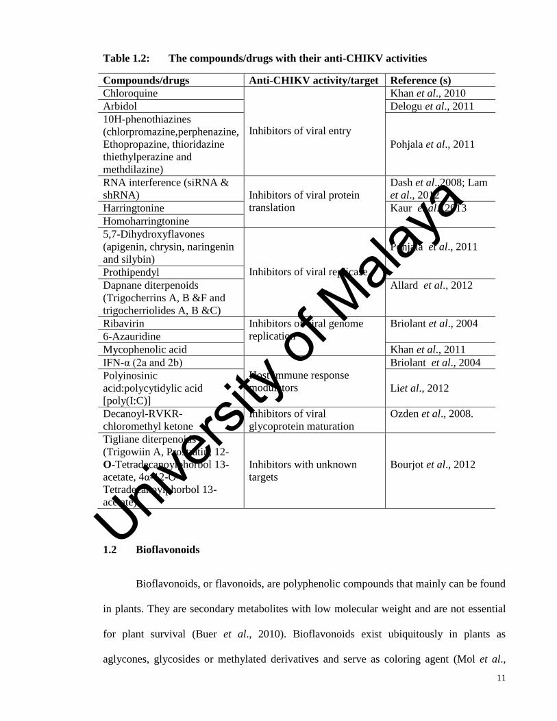
Some of the compounds and drugs which have been tested for anti-CHIKV activities were
summarized Table 1.2.
Univers
ity of
Mala
ya
11
Table 1.2: The compounds/drugs with their anti-CHIKV activities
Compounds/drugs Anti-CHIKV activity/target Reference (s)
Chloroquine
Inhibitors of viral entry
Khan et al., 2010
Arbidol Delogu et al., 2011
10H-phenothiazines
(chlorpromazine,perphenazine,
Ethopropazine, thioridazine
thiethylperazine and
methdilazine)
Pohjala et al., 2011
RNA interference (siRNA &
shRNA)
Inhibitors of viral protein
translation
Dash et al.,2008; Lam
et al., 2012
Harringtonine Kaur et al., 2013
Homoharringtonine
5,7-Dihydroxyflavones
(apigenin, chrysin, naringenin
and silybin)
Inhibitors of viral replicase
Pohjala et al., 2011
Prothipendyl
Dapnane diterpenoids
(Trigocherrins A, B &F and
trigocherriolides A, B &C)
Allard et al., 2012
Ribavirin Inhibitors of viral genome
replication
Briolant et al., 2004
6-Azauridine
Mycophenolic acid Khan et al., 2011
IFN-α (2a and 2b)
Host immune response
modulators
Briolant et al., 2004
Polyinosinic
acid:polycytidylic acid
[poly(I:C)]
Liet al., 2012
Decanoyl-RVKR-
chloromethyl ketone
Inhibitors of viral
glycoprotein maturation
Ozden et al., 2008.
Tigliane diterpenoids
(Trigowiin A, Prostratin, 12-
O-Tetradecanoylphorbol 13-
acetate, 4α-12-O-
Tetradecanoylphorbol 13-
acetate)
Inhibitors with unknown
targets
Bourjot et al., 2012
1.2 Bioflavonoids
Bioflavonoids, or flavonoids, are polyphenolic compounds that mainly can be found
in plants. They are secondary metabolites with low molecular weight and are not essential
for plant survival (Buer et al., 2010). Bioflavonoids exist ubiquitously in plants as
aglycones, glycosides or methylated derivatives and serve as coloring agent (Mol et al.,
Univers
ity of
Mala
ya

12
1998), auxin transport inhibitor (Peer & Murphy., 2007), allelopathy, defense (Bais et al.,
2006), as well as UV protection in plants (Treutter., 2005). The basic structure of
bioflavonoids are made up of 2-phenyl-benzo[α] pyrane or known as flavane nucleus
(Figure 1.2), and 14 classes of bioflavonoids differ from each other based on their chemical
nature as well as the substituents’ position on the A, B and C rings (Brown, 1980).
Found in various plants and fruits, bioflavonoids are being consumed daily in
significant quantities and they are one of the unavoidable components in our diet. Some of
the bioflavonoids were also used in traditional eastern medicine in ancient years (Havsteen
1983; Grange & Davey,1990; Bosio et al., 2000). Bioflavonoids have been studied
extensively in many fields of medical research. Many of them are known to have anti-
oxidant (Williams et al., 2004), anti-tumor (Garcia-Mediavilla et al., 2007), anti-
proliferative (Taylor & Grotewold., 2005), anti-inflammatory (Pandey et al., 2007) and pro-
apoptotic activities (Sung et al., 2007). Nonetheless, they also exhibited anti-fungal
(Wachter et al., 1999; Valsaraj et al., 1997; Zheng et al., 1996), antibacterial as well as
antiviral activities (Tereschuk et al., 1997; Bosio et al., 2000; Pepeljnjak et al., 1982).
The antiviral activity of bioflavonoids against wide range of viruses is undeniable.
Bioflavonoids such as baicalein, quercetin, kaempferol, luteolin and fisetin have shown
their promising antiviral activity against dengue virus (Moghaddam et al., 2014), Japanese
encephalitis virus (Zandi et al., 2012), enterovirus-71 (Evers et al., 2005), human
cytomegalovirus (Mehla et al., 2011), herpes simplex virus (Xu et al., 2014) and even
human immunodeficiency virus (Zhang et al., 2012).
There are many advantages of choosing bioflavonoid compounds as the candidates
for anti-CHIKV, and some of them are; low-toxicity in animal, rarely have any side effects,
Univers
ity of
Mala
ya

13
relatively long half-life, can easily be absorbed in the intestine, consumed daily and
unlimited sources for extraction (Skibola et al., 2000; Middleton et al., 2000). Thus, in this
study, 14 bioflavonoids from various groups were tested for possible anti-CHIKV
activities in vitro, being selected based on previous published data on their antiviral
activities against other viruses (please refer to the Chapter 2: Literature review for the
comprehensive assessment of the relevant literature explaining the studies done on related
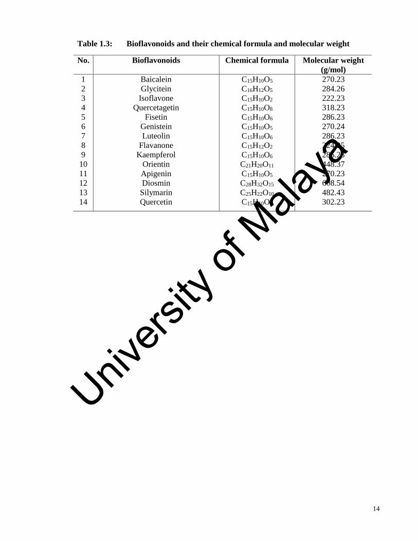
bioflavonoids). The structure of the 14 bioflavonoids is as shown in Figure 1.3. The
information of the 14 bioflavonoids with their chemical formula and molecular weight are
available in the Table 1.3.
Figure 1.2. The basic structure of the bioflavonoids. Drawn by using ChemDraw 7.0.
Univers
ity of
Mala
ya
14
Table 1.3: Bioflavonoids and their chemical formula and molecular weight
No. Bioflavonoids Chemical formula Molecular weight
(g/mol)
1
2
3
4
5
6
7
8
9
10
11
12
13
14
Baicalein
Glycitein
Isoflavone
Quercetagetin
Fisetin
Genistein
Luteolin
Flavanone
Kaempferol
Orientin
Apigenin
Diosmin
Silymarin
Quercetin
C15H10O5
C16H12O5
C15H10O2
C15H10O8
C15H10O6
C15H10O5
C15H10O6
C15H12O2
C15H10O6
C21H20O11
C15H10O5
C28H32O15
C25H22O10
C15H10O7
270.23
284.26
222.23
318.23
286.23
270.24
286.23
224.25
286.23
448.37
270.23
608.54
482.43
302.23
Univers
ity of
Mala
ya

15
Figure 1.3: The chemical structure of bioflavonoid compounds used in this study.
Drawn by using ChemDraw 7.0.
Univers
ity of
Mala
ya

16
1.3 Research objectives
Hence, the main objective of this research is to;
1. Explore the antiviral activity of 14 different types of bioflavonoids against
chikungunya virus replication in cell culture. The bioflavonoids were screened for
their antiviral activity in the primary antiviral screening. The bioflavonoids that
shown the antiviral activity in the primary antiviral screening were further analysed
for their specific method of actions by performing specific antiviral assays.
2. The cytotoxicity of each bioflavonoid in cell culture will be determined in order to
calculate the selectivity index. The cytotoxicity of the bioflavonoids on Vero cells
and BHK-21 cells were determined by the cell viability assay (MTS assay).
3. The step(s) of viral replication cycle that can be interfered by effective compound(s)
will also be investigated. The viral RNA copy number after the treatment with
bioflavonoids, in each antiviral assay, were determined by qRT-PCR. The IC50 was
calculated by comparing the treated CHIKV-infected sample with the CHIKV-
infected sample without treatment.
4. The role of bioflavonoids on CHIKV protein expression and CHIKV genome will
be studied. Immunofluorescence assay and immunoblot were performed to
determine the effects of bioflavonoids treatment on the CHIKV protein
accumulation.
Univers
ity of
Mala
ya

17
CHAPTER 2: LITERATURE REVIEW
2.1 Chikungunya virus life cycle and replication
2.1.1 Entry (attachment, penetration and uncoating)
Like other alphaviruses, chikungunya virus enters the susceptible cells by engaging
with a host receptor. Briefly during the process of entering, the virus E2 glycoprotein is
responsible for receptor interaction and the E1 protein is related to the receptor
engagement. The conformational changes occur in E2 and E1 glycoproteins as the
engagement occurs between the viruses with the host receptors (Meyer & Johnston, 1993).
Once they are bound to the receptor molecules, virions are endocytosed in a clathrin-
dependent manner (Helenius et al., 1980). The pH of the virus-containing endosome turns
acidic as it matures and this triggers the destabilization of the E1-E2 heterodimer (Lescar et
al., 2001; Gibbons et al., 2003). Subsequently, the insertion of the fusion peptide into the
late endosomal membrane occurs and trimerizes, leaving the mix of viral and endosomal
membranes and the nucleocapsids deposited in the host cell cytoplasm through the fusion
pores (Wahlberg et al., 1992). In the cytoplasm, the nucleocapsid disassembles and expose
the encapsidated genome for translation.
2.1.2 Virus assembly and budding
The binding of the virus nucleocapsid to the genome RNA and the recruitment of
membrane-associated envelope glycoproteins leads to the viral assembly. The icosahedral
core-containing virion budding out from the plasma membrane of the host cells. Effective
interaction between the nucleocapsid and the glycoproteins is crucial for this budding
process (Schwartz & Albert, 2010). The binding of nucleocapsid with cdE2 (cytoplasmic
Univers
ity of
Mala
ya

18
tail of E2) provides the free energy to propel the capsid across the plasma membrane and
the mature infectious virion is free to infect other susceptible cells (Tsetsarkin et al., 2011).
2.2 Difficulties in developing antiviral agents
The majority of registered antiviral drugs are nucleoside analogues. Though they are
undoubtedly effective, the shortcomings of these drugs are undeniable. Nucleoside
analogue antiviral drugs are potentially teratogenic, embryotoxic, carcinogenic and
possessed the anti-proliferative activities. The undesirable effects of these drugs are mostly
involving the bone marrow depression and neurotoxicity. Some of them must be
administered with care during pregnancy. One important point of establishing the antiviral
drug is to inhibit viral replication without harming the cells or in this case, the virus
machinery hosts (Morris, 1994).
A good potent antiviral candidate must possess certain characteristics through in
vitro study to qualify it for further study towards antiviral drug development. The most
important of these characteristics are; low toxicity towards the host cells, low half maximal
inhibitory concentration (IC50), high maximum non-toxic dose (MNTD), cheap and easy
preparation as well as unlimited sources. However, in this study, all of these characteristics
are present at the in vitro level, by selecting the compound of natural resources,
bioflavonoids.
2.3 Bioflavonoids and its potential
Apart from its importance listed in Chapter 1, bioflavonoids have its own
importance and role in antiviral research. All 14 bioflavonoids from various groups that are
used in this study have already extensively discovered for their antiviral activity against
various viruses.
Univers
ity of
Mala
ya

19
2.3.1 Baicalein (5,6,7-trihydroxyflavone)
Baicalein was extracted from the plant named Huangchin (Scutellaria baicalensis)
as well as Scutellaria lateriflora. It has been utilized for thousands of years in China’s
herbal medicine for its function to treat periodontal abscesses and infected oral wounds
(Cushnie & Lamb, 2005). In 2012, Cotin and his colleague have reported the potential of
baicalein as one of the antiviral candidates for human cytomegalovirus (HCMV) at the in
vitro level. It was the most effective compound out of four antivirals against HCMV in
vitro replication. They have shown that baicalein exhibited inhibitory effect against various
stages of HCMV replication cycle with IC50=2.2±0.5µM. They have found that baicalein
has reduced the expression of the HCMV immediate early gene (IE-1) as well as total
inhibition of IE-2 gene expression. Furthermore, baicalein was shown to inhibit the tyrosine
kinase activity of the EGF receptor. Nevertheless, it inhibits the early stage of the viral
cycle with the IC50 as low as 5µM (Cotin et al., 2012).
In the same year, Zandi and his colleagues discovered the dynamic roles of baicalein
in inhibiting many stages of dengue virus type-2 (DENV-2) replication. Baicalein was not
only shown the inhibitory effect against intracellular replication of DENV-2 with
IC50=6.46µg/ml; SI=17.8, but also interferes with early stages of DENV-2 replication cycle
such as adsorption phase with IC50=7.14µg/ml. It also possessed the direct virucidal
acitivity against DENV-2 extracellular particles with IC50=1.55µg/ml (Zandi et al., 2012).
A year after, Hour and his team demonstrated that baicalein extracted from
Scutellaria baicalensis by using ethyl acetate (EtOAc) and chloroform has better antiviral
activity against the pandemic 2009 H1N1 and seasonal Influenza A viruses compared to the
one that being extracted by using the methanol. The baicalein extracted with EtOAc inhibit
Univers
ity of
Mala
ya

20
the viral neuraminidase activity with the IC50 ranges from 73.16 to 487.40µg/ml and the
plaque reduction IC50 value ranges from 23.7 to 27.4µg/ml. The chloroform-extracted
baicalein have shown the plaque reduction ranges from 14.16 to 41.49µg/ml. They have
shown that the in vitro replication of H1N1 strain of influenza A virus was inhibited with
the IC50 as low as 0.018µM (Hour et al., 2013).
Additionally baicalin, which is the main metabolite of baicalein, has also became
one of the main antiviral candidates as it was shown to have the antiviral activity against
different viruses including DENV-2 and Influenza A (H1N1/H3N2), both in cell culture
and animal model as well (Moghaddam et al., 2014; Ding et al., 2014).
2.3.2 Isoflavones (2-phenyl-4H-1-benzopyr-4-one)
Isoflavones are differed from flavone in the location of their phenyl group. Apart
from legumes, grains and vegetables at which the isoflavones can be found in the small
amounts, soybeans has the highest concentration of isoflavones in human diet (Fletcher ,
2003; Munro et al., 2003). Common isoflavones that are found in soy products (Table 2.1)
are mainly in the form of glycosides and are not bioactive until fermentation or digestion
process transforms it into a bioactive form called aglycone (Cassidy et al., 2006; Dixon,
2004; Cornwell et al., 2004).
Table 2.1: Isoflavone glycosides and its aglycones
Glycosides Aglycones
Genistein Genistin
Daidzein Daidzin
Glycitein Glycitin
Univers
ity of
Mala
ya

21
In western countries, average consumption of isoflavones by adults was reported as
1-2mg/day which is lower compared to the Asian countries with 11-47mg/day. Genistein,
as one of the isoflavones, was found to be able to reduce the infectivity of non-enveloped
single-stranded RNA viruses such as coxsackie virus, poliovirus and echovirus; double-
stranded RNA virus such as rotavirus; double-stranded DNA viruses including adenovirus,
JC virus and Simian virus 40 (Andres et al., 2009).
In other studies, genistein exhibits anti-rotavirus activity and was suggested as a
biologically active isoflavone as it shows better inhibitory activities compared to other
isoflavones, which was individually tested in soy-based infant formula as well as by
comparing isoflavones mixtures (MIX). MIX somehow lost its anti-rotavirus activity with
the absence of genistein. Both genistein and MIX are able to reduce the rotavirus infectivity
by 33-62% and 66-74% respectively as assessed by using focus forming unit assay.
Genistein significantly reduces rotavirus infectivity in a dose-dependent manner at
concentrations as low as 30µmol/L. Genistein acts by modulating the post-binding step,
hence modulating the attachment of the virion to the host cells (Andres et al., 2007).
A cohort study was conducted from 1992 to 2008 involving 15, 607 women aged 35
years and above to find the association between soy and isoflavone intake with breast
cancer incidence in the population. Women with higher intakes of soy and isoflavones were
found to have lower relative risks of post-menopausal breast cancer compared to the
women having lower intakes of soy and isoflavones (Wada et al., 2013). In the search for
the broad-spectrum antiviral compound, isoflavones were tested in a high-throughput
screening study against RNA viruses. The isoflavone compounds exhibit a highly potent
activity against hepatitis C virus (HCV) and influenza virus by activating the interferon-
Univers
ity of
Mala
ya

22
stimulated gene (ISG54) promoter and mediating the nuclear translocation of interferon
regulatory factor (IRF-3) (Bedard et al., 2012).
2.3.3 Glycitein (4',7-Dihydroxy-6-methoxyisoflavone)
On average, 5-10% of the total isoflavones in soy products are glycitein. In some
soy supplements, glycitein content can be as high as 40% (Song et al., 1999). Although soy
products are common in our diet, antiviral research on this compound is scarce. Most of the
studies on glycitein mainly focused on the role of glycitein in the regulation of estrogen
hormone. However, glycitein only possessed a weak estrogenic activity as it was reported
to have low affinity towards the estrogen receptor at the in vitro level (Song et al., 1999;
Molzberger et al., 2013). Roh and Sung have attempted to explore the possibility of
antiviral activity in glycitein on the HCV. However, glycitein did not exhibit neither any
antiviral effects nor inhibit the activity of viral protein NS5B (Roh & Jo., 2011).
2.3.4 Quercetagetin (2-(3,4-dihydroxyphenyl)-3,5,6,7-tetrahydroxychromen-4-one)
Quercetagetin is mainly found in hydrolysates of the leaves from six Eriocaulon
genus (Bate-Smith and Harborne, 1969). Recently, quercetagetin has atrracted attention
from antiviral researchers. Though quercetagetin was found to inhibit HCMV replication,
the activity was not efficient at various stages (Cotin S et al., 2012). It has turned out to be
the most potent inhibitor of HCV RNA-dependent RNA polymerase (RdRp). The inhibition
of RNA binding to the viral polymerase by quercetagetin is considered as the mechanism
with broad genotypic activity and high barrier to resistance either by site-directed
mutagenesis or long-term selection experiments (Ahmed-Belkacem et al., 2014).
Univers
ity of
Mala
ya
23
2.3.5 Fisetin (2-(3,4-dihydroxyphenyl)-3,7-dihydroxychromen-4-one)
Fisetin is usually found in many fruits and vegetables including strawberries, apples,
onions and grapes (Fiorani and Accorsi, 2005; Maher et al., 2011; Arai et al., 2000). Lyu
and the team have discovered the moderate inhibitory effects of fisetin against herpes
simplex virus 1 (HSV-1) in 2005. In 2011, a research was initiated to evaluate the activity
of fisetin against dengue virus. It was shown that fisetin can significantly interfere with
dengue virus replication at the in vitro level with the treatment after virus adsorption (IC50
= 55 μg/ml) and treatment at 5 hours before virus infection (IC50 = 43.12 μg/ml) (Zandi et
al., 2011). A year later, fisetin was reported to also reduce enterovirus-A71-induced
cytopathic effect and viral titer (Lin et al., 2012).
2.3.6 Genistein (5,7-dihydroxy-3-(4-hydroxyphenyl)chromen-4-one)
Apart from the studies being discussed earlier in 2.2.2, genistein, a tyrosine kinase
inhibitor, was suggested to be able to reduce the level of HCMV DNA synthesis as well as
early and late proteins through the mechanism assumed as blocking HCMV immediate-
early protein functions (L. Evers et al., 2005). Genistein was also found to inhibit the
infection or transduction in cells infected with severely fatal viruses such as Ebola virus,
Marburg virus and Lassa virus in a study where the host cells were pre-treated with
genistein and tyrphostin AG1478 (A. Kolokoltsov et al., 2012). In 2013, an important
finding was made when the genistein was found to be able to inhibit the SDF-1-mediated
chemotaxis and HIV infection of resting CD4 T cells. Genistein inhibited the viral DNA
accumulation in resting CD4 T cells thereby interfering with SDF-1 and HIV-1 mediated
actin dynamics in CD4 T cells (Guo et al., 2013).
Univers
ity of
Mala
ya

24
2.3.7 Luteolin (2-(3,4-dihydroxyphenyl)-5,7-dihydroxy-4-chromenone)
In 2011, luteolin was found to be able to reduce the HIV-1 infection in the reporter
cells and primary lymphocytes. However, the activity of luteolin was only at the viral entry
level and unable to inhibit the reverse transcription of HIV-1 (Mehla et al., 2011). A year
later, Calland et al. studied antiviral activity of luteolin against HCV infection in a cell-
based assay system where NS5B polymerase was reported as the viral target with IC50 was
1.1 to 7.9µM (Calland et al., 2012). Luteolin was also listed as the most potent inhibitor of
EV71 and CA16 infection through cell viability and plaque reduction assays with EC50
about 10µM. Luteolin targeted the post-attachment stage of EV71 infection and inhibited
viral replication of CA16 (Xu et al., 2014). Murali et al. also have found the antiviral
activity of luteolin against chikungunya virus. However, ethanolic extract of Cynodon
dactylon was used, which differed from our study which used the commercial luteolin with
≥ 98% purity. The extract was able to reduce the viral mRNA synthesis and its CPE by
98% at a concentration of 50µg/ml (Murali et al., 2015).
2.3.8 Flavanone (2-Phenyl-2,3-dihydro-4H-chromen-4-one)
Flavanones are typical polyphenols in Citrus species. Naringenin, eriodictyol,
isosakuranetin and hesperetin are among well-studied flavanones (Khan & Dangles, 2014).
Flavanones draws attention mainly in the anti-inflammatory, anti-oxidant, anti-cancer and
anti-microbial investigations, least known in the area of the antiviral research. Hesperidin,
the glycosides of hesperetin, was found to exhibit antiviral activity against herpes simplex
virus, poliovirus and parainfluenza virus (Kaul et al., 1985). Hesperitin and narigenin
showed antiviral activity against Sindbis neurovirulent strain virus (NSV). In a dose-
dependent manner a decrease of viral plaque formation for both used flavanones, scored the
Univers
ity of
Mala
ya

25
ID50 as low as 20.5 and 14.9µg/ml respectively. Beyond hesperetin with only 50% of viral
replication inhibition at 25µg/ml, naringenin is the most effective anti-sindbis NSV with up
to 80% viral replication inhibition at the same concentration (Paredes et al., 2003).
2.3.9 Kaempferol (3,5,7-Trihydroxy-2-(4-hydroxyphenyl)-4H-chromen-4-one)
Kaempferol has shown about 40% of viral internal ribosome entry site (IRES)
activity by interfering with EV71 virus replication and pseudotyped virus (Tsai et al.,
2011). In addition, kaempferol is potent against the Japanese encephalitis virus (JEV) by
inactivating the virus through its frameshift site RNA (Zhang et al., 2012). The inhibitory
effect of kaempferol-7-O-glucoside on HIV-1 reverse transcriptase activity was more
effective than the kaempferol itself (Behbahani et al., 2014). Also, it has more planar
flavonol structure with only one C-4’ phenolic hydroxyl group in the B ring, which is
crucial for anti-influenza B virus activity in a structure-activity relationship study (Yang et
al., 2014).
2.3.10 Orientin (2-(3,4-dihydroxyphenyl)-5,7-dihydroxy-8-[(2S,3R,4R,5S,6R)-3,4, 5-
trihydroxy-6-(hydroxymethyl)oxan-2-yl]chromen-4-one)
There are not many studies involving the antiviral activity of orientin except about
the antiviral activity of crude extracts from the flower Trollius chinensis which contains
flavonoids, orientin, vitexin and proglobeflowery acid against the parainfluenza type 3. It
was also shown that orientin has potent antiviral activity against parainfluenza virus type 3
(Li et al., 2002).
Univers
ity of
Mala
ya

26
2.3.11 Apigenin (5,7-Dihydroxy-2-(4-hydroxyphenyl)-4H-1-benzopyran-4-one)
Recently, apigenin and enterovirus-71 (EV71) became closer partners in antiviral
research. Apigenin is able to inhibit the EV71-mediated CPE, virus replication efficiency,
viral protein expression and hindered EV71-induced cell apoptosis, intracellular reactive
oxygen species (ROS) generation and cytokines up-regulation. Apigenin exhibited its
activity very well after the viral entry and interfered with viral internal ribosome entry site
(IRES) activity as well as modulating the (c-Jun N-terminal kinase) JNK pathway (Lv et
al., 2014). Since EV71 infection requires the hnRNP proteins (trans-acting factor regulating
the EV71 translation), apigenin is able to disrupt the viral RNA association with hnRNP A1
and A2 proteins selectively (Zhang et al., 2014). Foot-and-mouth disease virus (FMDV)
translational activity was interfered as apigenin interrupts the IRES activity at the post-
entry stage with no direct extracellular virucidal activity (Qian et al., 2015). Coincidentally,
chikungunya virus was also targeted by apigenin. However, in this recent study, Murali and
colleagues were using the apigenin-rich fraction from Cynodon dactylon rather than the
pure apigenin compound. Apigenin and other major phytochemicals inhibit 98% of viral
activity at the concentration of 50 µg/ml through the observation of CPE reduction as well
as reduction in viral mRNA synthesis (Murali et al., 2015).
2.3.12 Diosmin (5-Hydroxy-2-(3-hydroxy-4-methoxyphenyl)- 7-[(2S,3R,4S,5S,6R)-
3,4,5-trihydroxy -6-[[(2R,3R,4R,5R,6S) -3,4,5-trihydroxy-6-methyloxan-2-
yl]oxymethyl]oxan-2-yl]oxychromen-4-one)
Up to now, there is no report on antiviral activity of diosmin. Diosmin which is the
alternative supplement taken in handling the venous diseases was found to have
cardioprotective effects by free radical scavenging and anti-hyperlipidaemic activity as well
Univers
ity of
Mala
ya

27
as protective effect on hepatic ischemia repefusion injury in rats (Queenthy and John, 2013;
Tanrikulu et al., 2013).
2.3.13 Silymarin ((2R,3S)-3,5,7-Trihydroxy-2-[(2R)-2-(4-hydroxy-3-methoxyphenyl)-
3-(hydroxymethyl)-2,3-dihydro-1,4-benzodioxin-6-yl]-2,3-dihydro-4H-chromen-4-one)
Silymarin is a complex of more than seven flavonolignans and one flavonoid that
made up 65%-80% of milk thistle initial extract together with 20%-35% fatty acid. The
seven flavonolignans are silybin A, silybin B, isosilybin A, isosilybin B, silychristin,
isosilychristin and silydianin. In literature, silybin A and silybin B are also referred as
silibinin A and silibinin B (Polyak et al., 2013). Silymarin is one of the most popular
natural product consumed by the Western society especially for those who suffered from
chronic hepatitis C (Polyak, Ferenci & Pawlotsky, 2013).
Silymarin was found to be able to inhibit the virus entry, RNA and protein
expression and infectious virus production in hepatitis C virus cell culture infection.
Silymarin did not block the binding of the virus to the cells but inhibited the entry of
several viral pseudoparticles (pp) and fusion of HCVpp with liposomes, as well as inhibited
microsomal triglyceride transfer protein activity, apolipoprotein B secretion and infectious
virion production into culture supernatants. It is worth noting that the genotype 2a NS5B
RNA-dependent RNA polymerase (RdRp) activity was inhibited by silymarin and not
silibinin, one of its flavonolignan. Silymarin also blocked cell-to-cell spread of the virus
(Wagoner et al., 2010). However, in a randomized controlled trial study, no evidence of
salutary effects of oral silymarin has been reported although silymarin is very well-tolerated
in chronic HCV-infected patients (Yang et al., 2014).
Univers
ity of
Mala
ya

28
Apart from hepatitis C virus, silymarin also showed anti-influenza virus activity by
reducing visible CPE by 98% at concentration of 100 μg/ml. It inhibited viral mRNA
synthesis and also affect late viral RNA synthesis (Song & Choi, 2011).
2.3.14 Quercetin (2-(3,4-dihydroxyphenyl)-3,5,7-trihydroxy-4H-chromen-4-one)
Quercetin is a well-studied flavonol. It was shown that it inhibited the DENV-2 in
vitro replication with IC50=35.7 μg/ml when the cells were treated after the virus
adsorption, where the IC50 has dropped to 28.9 μg/ml when the cells were continuously
treated for 5 hours before virus infection up to four days post-infection (Zandi et al., 2011).
In research pertaining hepatitis C virus, quercetin was found to be able to inhibit the NS3
activity in a specific dose-dependent manner in an in vitro catalysis assay and confirmed to
inhibit HCV RNA replication as analyzed in sub-genomic HCV RNA replicon system apart
from the direct inhibition on HCV NS3 protease and infectious virus production in HCV
cell culture system (Bachmetov et al., 2011). Quercetin is also able to improve the lung
function in the rhinovirus-infected mice and reduced the expression of pro-inflammatory
cytokines. The pre-treatment of the airway epithelial cells with quercetin decreased the Akt
phosphorylation, viral endocytosis and IL-8 responses. Six hours after infection with
rhinovirus, the IL-8 and IFN responses were decreased in airway epithelial cells (Ganesan
et al., 2012).
2.4 Recent news regarding the spread of CHIKV in America
For about 62 years, CHIKV outbreaks have occurred occasionally in Africa, Asia,
Europe as well as Indian and Pacific oceans. However, recently in late 2013, CHIKV was
found for the first time in the Americas on islands in Carribean as reported by the Centers
for Disease Control and Prevention (CDC, Atlanta, Georgia, U.S). The first local cases
Univers
ity of
Mala
ya

29
were reported from St. Martin and the outbreak was quickly spread to other locations in
Carribean and even beyond. In December 2013, over 750 travelers have returned to the
United States infected with CHIKV and the transmission was predicted to be continuing
throughout Americas. Since antiviral treatment has not yet existed, a symptomatic and
supportive treatment was offered. Other than resting, acetaminophen or paracetamol are
used to relieve fever. Also, ibuprofen, naproxen, or another non-steroidal anti-inflammatory
agents (NSAID) are used to relieve the arthritic component of the disease.
Univers
ity of
Mala
ya

30
CHAPTER 3: MATERIALS AND METHODS
3.1 Cell lines, virus and antibodies
The African green monkey kidney cell line or also known as Vero cells and the
baby hamster kidney cell line (BHK-21 cells) were obtained from the American Type
Culture Collection (ATCC, Virginia, USA). Both cell lines were cultured and maintained in
the Eagle’s Minimum Essential Medium (EMEM) (Thermo-Fisher Scientific, MA, USA)
supplemented with 10% fetal bovine serum (FBS) (Bovogen Biologicals, Vic, Australia), 2
mM L-glutamine (HiMedia Laboratories, Mumbai, India), 1x non-essential amino acid
(NEAA) (HiMedia Laboratories, Mumbai, India) and also 50 IU penicillin/streptomycin
(Sigma-Aldrich, MO, USA). Both cell lines were incubated at 37°C in the presence of 5%
CO2 and were sub-cultured or cryopreserved as they reached to 80% confluency.
The CHIKV replicon BHK-21 cell line was contributed by Dr. Andres Merits from
University of Tartu, Tartu, Estonia as published in Pohjala et al., 2011 (refer to Appendix
B). The CHIKV replicon containing the virus replicase proteins together with puromycin
acetyltransferase, EGFP and Renilla luciferase marker genes was transfected into BHK-21
cells to yield a stable cell line. The CHIKV replicon cell line was cultured and maintained
in Dulbecco’s Modified Eagle’s Medium (DMEM) (Thermo-Fisher Scientific, MA, USA)
supplemented with 8% FBS (Bovogen Biologicals, Vic, Australia), 2% tryptose-broth
phosphate (Sigma-Aldrich, MO, USA), 2 mM L-glutamine (HiMedia Laboratories,
Mumbai, India) and also 50 IU penicillin/streptomycin (Sigma-Aldrich, MO, USA). The
replicon cell line was incubated at 37°C in the presence of 5% CO2 and was sub-cultured or
cryopreserved as they reached to 80% confluency.
Univers
ity of
Mala
ya

31
Throughout the experiment, the CHIKV of ECSA genotype (accession number:
MY/065/08/FN295485) was used. The virus was isolated from the Johor’s outbreak in 2008
and was kindly provided by Assoc. Prof. Jamal I-Ching Sam from University of Malaya
(Sam et al., 2009).
3.1.1 CHIKV propagation in cell culture
In order to ensure the continuous supply of CHIKV for usage throughout the whole
experiment, propagation of CHIKV was performed. Since CHIKV grows in a variety of
non-human cell lines, including Vero cells and BHK-21 cells (Schwartz and Albert, 2010),
the propagation and harvesting of the CHIKV were performed using BHK-21 cells and the
subsequent antiviral assays were performed by using the Vero cells.
A monolayer of the BHK-21 cells were cultured in 75 cm2 cell culture flask using
Eagle’s Minimum Essential Medium (EMEM) (Thermo-Fisher Scientific, MA, USA)
supplemented with 10% fetal bovine serum (FBS) (Bovogen Biologicals, Vic, Australia), 2
mM L-glutamine (HiMedia Laboratories, Mumbai, India), 1x non-essential amino acid
(NEAA) (HiMedia Laboratories, Mumbai, India) and also 50 IU penicillin/streptomycin
(Sigma-Aldrich, MO, USA). The cultured cells were incubated at 37°C in the presence of
5% CO2 and infected CHIKV after attaining 80% confluency. The flask containing
CHIKV-infected BHK-21 cells was placed on the rocker for 30 minutes prior to the
incubation at 37°C in the presence of 5% CO2.
Before harvesting the CHIKV, the infected cells were incubated in the same
environment for three days until about 70%-80% cytopathic effect (CPE) shows up. The
CPE of alphaviruses in cell culture was characterized as lytic infection or apoptosis. On the
third day, the cells were scrapped by using the cell scraper, centrifuged at 4°C and 2000rpm
Univers
ity of
Mala
ya

32
to isolate the cell debris. The CHIKV was then aliquoted into screw cap tubes and stored at
-80°C for titration and further experiments.
3.1.2 CHIKV titration assay
The virus titration has been done based on Reed-Muench endpoint calculation
method to determine the tissue culture 50% infectious dose (TCID50) (Reed and Muench,
1938). A monolayer of 1x10⁴ Vero cells per well were grown in a 96-well plate in the
growth media made up of the HyClone™ Eagle’s Minimum Essential Medium (EMEM)
(Thermo-Fisher Scientific, MA, USA) supplemented with 10% fetal bovine serum (FBS)
(Bovogen Biologicals, Vic, Australia), 2 mM L-glutamine (HiMedia Laboratories,
Mumbai, India), 1x non-essential amino acid (NEAA) (HiMedia Laboratories, Mumbai,
India) and also 50 IU penicillin/streptomycin (Sigma-Aldrich, MO, USA). The cells were
then incubated at 37°C in the presence of 5% CO2 until it reached 80% confluency.
The CHIKV stock was then serially diluted tenfold EMEM (Thermo-Fisher
Scientific, MA, USA) supplemented with 2% fetal bovine serum (FBS) (Bovogen
Biologicals, Vic, Australia) and each dilution was used to infect the cells in respective
wells. The plate was then incubated at the same condition as mentioned earlier for virus
propagation. The plate was then examined daily for the presentation and recording of the
CPE in infected wells and the recording of the CPE presentation was performed until the
mock infected cells showed healthy morphology.
To titer the viral stock based on Reed-Muench endpoint calculation method, a score
was given to each of the dilution according to the amount of the wells showing CPE. The
interpolated value of TCID50 was then calculated by Reed-Muench formula as stated in the
following:-
Univers
ity of
Mala
ya

33
Reed-Muench formula
TCID 50/ml = 10 log total dilution above 50% - (α x log dilution factor)
Where α = (% of wells infected at dilution above 50% - 50%) ÷ (% of wells infected
at dilution above 50% - % of wells infected at dilution below 50%)
3.1.3 Anti-CHIKV antibodies
The monoclonal rabbit anti-E2 CHIKV antibody used in the immunofluorescence
assay and western blotting was provided by Dr. Justin Chu Jang Hann from National
University of Singapore. The monoclonal anti-nsP1 CHIKV and anti-nsP3 CHIKV used in
the immunofluorescence assay and western blotting was provided by Dr. Andres Merits
from University of Tartu, Tartu, Estonia.
3.2 Bioflavonoids and nucleoside analogue
13 bioflavonoids except quercetagetin were purchased from Sigma-Aldrich, MO,
USA. Quercetagetin was purchased from Indofine Chemical Co., NJ, USA. All of the
bioflavonoids are of ≥ 95% purity. Ribavirin, a nucleoside analogue known to inhibit the
CHIKV viral genome replication (Briolant et al., 2004) was used as the control for positive
anti-CHIKV activity in the post-entry assay, and purchased from Sigma-Aldrich, MO, USA
of ≥ 95% purity. The bioflavonoids and ribavirin stock solutions were prepared in 0.1%
dimethyl sulfoxide (DMSO) or methanol according to the solvent suggested by the
manufacturer. The stock solutions were then stored in -20°C for further experiments.
Univers
ity of
Mala
ya

34
3.2.1 Cytotoxicity assay
In order to determine the cytotoxicity of the bioflavonoids and ribavirin on Vero
cells and BHK-21 cells, the 3-(4,5-dimethylthiazol-2-yl)-5-(3-carboxymethoxyphenyl)-2-
(4-sulfophenyl)-2H-tetrazolium (MTS) assay was performed. Through this assay, the half
maximal cytotoxic concentration (CC50) and the maximum non-toxic dose (MNTD) of each
compound can be determined. Basically, in the CellTiter 96® AQueous One Solution Cell
Proliferation Assay kit (Promega, WI, USA), the MTS solution and an electron coupling
reagent (phenazine ethosulfate; PES) were combined to form a stable solution. The
NAD(P)H-dependent cellular oxidoreductase enzymes in the viable cells will reduce the
dye to its colored formazan and this reflects the amount of viable cells in the assay. This
assay was performed according to the manufacturer’s protocol.
A monolayer of 1x10⁴ Vero cells and BHK-21 cells per well were prepared in a
different 96-well cell culture plate (Nest Biotechnology Co., Ltd. Wuxi, China) and were
treated with different concentrations of each compound in triplicate together with negative
control (media containing 0.1% DMSO). The prepared bioflavonoid compounds were
filtered using 0.2μm syringe filter unit (Merck Millipore Ltd., MA, USA). The plate was
then incubated at 37 °C with 5% CO2 for 48 hours before the MTS assay was performed.
Treated and control cells were kept for two days at 37°C, under similar conditions and
duration until used for antiviral activity assay. After two days post-treatment, MTS solution
was added to the cells and incubated for 4 hours at 37°C with 5% CO2 prior to absorbance
detection at 495 nm wavelength using Infinite 200 Pro multiplate reader (Tecan,
Männedorf, Switzerland). All experiments were conducted in triplicate. The CC50 and
MNTD for each compound was determined through this assay using Graph Pad Prism 5
(Graph Pad Software Inc., San Diego, CA, USA, 2005).
Univers
ity of
Mala
ya

35
3.3 In vitro antiviral assays
3.3.1 Antiviral screening assay using CHIKV replicon cell line
To screen the compounds for their activity against CHIKV replicase complex, a
CHIKV BHK-21 replicon cell line has been used. This assay was modified and performed
according to Pohjala et al., 2011. A monolayer of 1x10⁴ CHIKV replicon cells were
prepared in 96-well white plate (Corning Inc., NY, USA) and treated with increasing
concentrations of each compound starting from MNTD concentration of each compound.
After 48 hours incubation at 37°C with 5% CO2, the activity of the Renilla luciferase (Rluc)
expressed by CHIKV replicon was detected by using Renilla luciferase assay (Promega,
WI, USA). The assay was performed according to the manufacturer’s protocols. The
luminescence signal was then measured by using the GloMAX 20/20 Luminometer
(Promega, WI, USA). The signal was plotted against the log transformation of the
concentrations of the compounds and a sigmoidal curve fit with variable slope was created
to obtain the half maximal inhibitory concentration (IC50) value for each compound. The
non-parametric one-way ANOVA was performed by using the Graph Pad Prism 5 (Graph
Pad Software Imn., San Diego, CA, USA, 2005) in order to determine the correlation
between the treatments and the Rluc activity.
3.3.2 Continuous treatment assay
As another antiviral screening assay using infectious CHIKV, a continuous
treatment assay was performed to determine any plausible antiviral activity of a compound
against any stage of virus life cycle. This assay was modified and performed according to
Zandi et al., 2014. A monolayer of 1x10⁴ Vero cells per well were prepared in 96-well cell
culture plate. The cells then were treated with increasing concentrations of each compound
Univers
ity of
Mala
ya

36
starting from MNTD concentration of each compound. Concurrently, cells were infected
with CHIKV of MOI=1 and the plate was then incubated for 2 hours at 37°C in the
presence of 5% CO2. After 2 hours of incubation, the supernatants were removed from each
well and the newly prepared bioflavonoid compound with the same concentrations as the
previous stage was added into the respective wells.
The plate was then incubated for 48 hours at the same condition as previously
described until the CPE appears in the virus control wells. The plate was examined daily for
the presentation of CPE and after two days, the percentage of CPE score was given based
on the degree of inhibition of the CPE. The MTS assay was also performed to confirm the
results. The non-parametric one-way ANOVA was performed by using the Graph Pad
Prism 5 (Graph Pad Software Imn., San Diego, CA, USA, 2005) in order to determine the
correlation between the treatments and the CHIKV RNA copy number. The bioflavonoid
compounds that show significant inhibition against CHIKV were selected to continue with
further experiments which are necessary to perform to determine the extent of its inhibitory
activities.
3.3.3 Time-of-addition assay
This assay was modified and performed according to Kaur et al., 2013. The time-of-
addition study was performed to determine the best acting time for the compounds against
the virus as well as to determine how long the addition of a compound could be postponed
before it loses its antiviral activity (Daelemans et al., 2011; Pauwels et al., 1990). In this
assay, the bioflavonoids that results in positive antiviral hits in the previous assays were
tested using the highest concentration of each compound that can be achieved below their
MNTDs.
Univers
ity of
Mala
ya

37
A monolayer of Vero cells were prepared in 96-well cell culture plate using EMEM
supplemented with 10% FBS. For the pre-treatment assays, which refers to the -2h and -1h
pre-infection; the cell monolayers were treated with the three compounds respectively at 2h
and 1h before the infection with CHIKV. For the co-treatment assay (0h), Vero cells were
treated with each compound and being infected at the same time. For the post-treatment
assay, Vero cells were infected with CHIKV (MOI=1) and subsequently the compounds
were added at 2h, 3h, 4h and 5h. The CHIKV for the positive controls were added at the 0h
and the negative controls were only containing EMEM with 2% FBS. The plate was then
incubated for 48 hours at 37°C in the presence of 5% CO2 until the CPE is shown.
The plate was then examined daily and after 2 days, the percentage of CPE score
was given based on the degree of inhibition of the CPE. The supernatants from each well
were collected and a viral yield assay were conducted using a specific quantitative real-time
polymerase chain reaction (qRT-PCR) for CHIKV, which will be elaborated in another
section. The non-parametric one-way ANOVA was performed by using the Graph Pad
Prism 5 (Graph Pad Software Imn., San Diego, CA, USA, 2005) in order to determine the
correlation between the treatments and the CHIKV RNA copy number.
3.3.4 Virucidal assay
This assay was modified and performed according to Johari et al., 2012. The
virucidal assay was performed to identify whether there is a possibility that the
bioflavonoids can inactivate the extracellular virus particles or in other words before
infections of the cells. Hence, in this assay, increasing concentrations of the compounds
ranges from 6.25 μg/ml to 100 μg/ml were mixed with the CHIKV (TCID50=104.5 EID50/ml)
and incubated for 2 hours at 37°C.
Univers
ity of
Mala
ya

38
The compounds were further diluted before the addition of the mixtures on Vero
cells. Treated viral mixtures were added to prepared monolayer of 6x10⁵ Vero cells in 24-
well cell culture plates in triplicates, where the virus controls the CHIKV without any
compound and the negative controls only contained cell culture medium with 2% FBS. The
plates were then incubated at 37°C in the presence of 5% CO2 for 48 hours. The plates were
examined daily for the presentation of CPE and after 2 days of incubation, the supernatants
were collected from their respective wells and the qRT-PCR assay were performed to
evaluate the virucidal activity of the compound. The IC50 was determined and the non-
parametric one-way ANOVA was performed by using the Graph Pad Prism 5 (Graph Pad
Software Imn., San Diego, CA, USA, 2005) in order to determine the correlation between
the treatments and the CHIKV RNA copy number.
3.3.5 Anti-adsorption assay
This assay was modified and performed according to Zandi et al., 2013. In order to
identify whether the compounds of interest could interfere with the specific binding of the
viral attachment proteins and the cellular receptors. A viral suspension from CHIKV with
MOI=1 and descending concentrations of bioflavonoids ranges from 100 μg/ml to 6.25
μg/ml were prepared in EMEM supplemented with 2% FBS and overlaid simultaneously on
monolayer of 6x10⁵ Vero cells per well in 24-well cell culture plates.
The plates were then incubated at 37°C in the presence of 5% CO2 for and after 2
days of incubation, the supernatants were collected from their respective wells and the
qRT-PCR assay were performed to confirm the results. The IC50 and the non-parametric
one-way ANOVA was performed by using the Graph Pad Prism 5 (Graph Pad Software
Imn., San Diego, CA, USA, 2005) in order to determine the correlation between the
treatments and the CHIKV RNA copy number.
Univers
ity of
Mala
ya

39
3.3.6 Anti-entry assay
The procedure of the anti-entry assay was modified and performed according to Lee
et al., 2013. Monolayers of Vero cells were grown in 96-well plate with EMEM
supplemented with 10% inactivated FBS. The Vero cells were then infected with CHIKV
and the plate was incubated for 1 hour at 4 °C. Non-adsorbed virus was then washed with
1xPBS. Tested compound was added in different concentrations and incubated at 37 °C
with 5% CO2 for 2 hours. The plate was again washed with 1xPBS and treated with citrate
buffer (pH = 3) to inactivate the non-internalized virus, before the plate was again washed
with 1xPBS. The EMEM supplemented with 2% inactivated FBS was added into every
wells and the plate was incubated for 48 hours at 37 °C with 5% CO2. After 2 days of
incubation, the supernatants were collected from their respective wells and the qRT-PCR
assay were performed to confirm the results. The IC50 and The non-parametric one-way
ANOVA was performed by using the Graph Pad Prism 5 (Graph Pad Software Imn., San
Diego, CA, USA, 2005) in order to determine the correlation between the treatments and
the CHIKV RNA copy number.
3.3.7 Post-adsorption assay
This assay was modified and performed according to Zandi et al., 2011. A
monolayer of 6x10⁵ Vero cells per well were grown in 24-well cell culture plates using
EMEM supplemented with 10% FBS. The Vero cells were then inoculated with CHIKV of
MOI= 1 and incubated for 2 hours at 37°C with 5% CO2 prior to the treatment with
different concentrations of bioflavonoid compounds ranging from 100 μg/ml to 6.25 μg/ml
were prepared in EMEM supplemented with 2% FBS.
Univers
ity of
Mala
ya

40
After the treatment, the plates were then incubated for 48 hours at 37°C with 5%
CO2. The plates were examined daily for the CPE presentation and after two days, the
quantitative real-time polymerase chain reaction (qRT-PCR) assay were performed to
confirm the results. The IC50 and the non-parametric one-way ANOVA was performed by
using the Graph Pad Prism 5 (Graph Pad Software Imn., San Diego, CA, USA, 2005) in
order to determine the correlation between the treatments and the CHIKV RNA copy
number.
3.4 Virus yield assay using quantitative reverse transcription PCR (qRT-PCR)
3.4.1 CHIKV RNA extraction and the generation of cDNA
The CHIKV RNA was extracted from supernatants collected from previous in vitro
antiviral assays with QIAamp Viral RNA Mini Kit (QIAGEN, Germany). The nsP3 primers
of 136bp were nsP3-F (5'-GCGCGTAAGTCCAAGGGAAT-3') and nsP3-R (5'-
AGCATCCAGGTCTGACGGG-3') as described by Chiam and colleagues (Chiam et al.,
2013). The nsP3-R primer was used to generate nsP3 positive-strand cDNA. The mixture of
500 nmol/L of primers, 50 nmol/L of dNTP mix (Promega, WI, USA), and 1 μL of RNA
was incubated at 65°C for 5 min and placed on ice for 4 min. The cDNA was synthesized
with 200 U of Superscript III Reverse Transcriptase (Life Technologies, USA), 0.1 mol/L
DTT (Life Technologies, USA), 40 U of RNaseOUT (Life Technologies, USA), and 1×
First Strand buffer (Life Technologies, USA) at 50 °C for 60min. Reverse transcriptase
enzyme was inactivated at 70°C for 15 min, then unincorporated primers were digested
with 20 U of Exonuclease I (New England Biolabs, MA, USA). The cDNA were stored at
−80 °C.
Univers
ity of
Mala
ya

41
3.4.2 qRT-PCR
A qRT-PCR assay was used to evaluate the virus yield reduction due to the
treatments through quantifying the extracellular CHIKV RNA copy number. For this
experiment, the amplification of 136 base region of nsP3 encoding sequence was performed
as described by Chiam et al., 2013. The qRT-PCR was performed in the Step-One Plus
Real-Time PCR System (Life Technologies, USA) using a 10 μL reaction volume
containing 1×Power SYBR Green PCR Master Mix (Life Technologies, USA), 1 μmol/L
sense and antisense primers, 1 μL of cDNA, and 3.8 μL of nuclease-free water. The qRT-
PCR was performed with a Step-OnePlus Real-Time PCR System (Life Technologies,
USA) following the manufacturer’s protocol, and using serially diluted standards. Cycling
parameters were 95ºC for 10 min, 40 cycles of 95ºC for 15 s and 60ºC for 1 min. The
amplified product was verified by melting curve analysis. The non-parametric one-way
ANOVA was performed by using the Graph Pad Prism 5 (Graph Pad Software Imn., San
Diego, CA, USA, 2005) in order to determine the correlation between the treatments and
the CHIKV RNA copy number.
3.5 Immunofluorescence assay
In order to quantify the CHIKV antigen production as a confirmatory test for other
antiviral assays, an immunofluorescence assay was modified and performed according to
Kaur et al., 2013. A monolayer of Vero cells were prepared in 96-well cell culture plate
using EMEM supplemented with 10% FBS. The monolayer of Vero cells were then
infected with CHIKV of MOI= 1 and incubated for 2 hours at 37°C with 5% CO2 prior to
the treatment with different concentrations of tested compounds ranging from 100 μg/ml to
6.25 μg/ml in respective wells. The plates were then incubated for 24 hours at 37°C with
5% CO2. After the overnight incubation, the cells were fixed using 100 µl of 4%
Univers
ity of
Mala
ya

42
paraformaldehyde for 30 minutes at room temperature. The cells were then washed three
times with 100 µl 1xPBS prior to the addition of 40 µl of monoclonal rabbit anti-E2
antibody (diluted to 1:300) and incubated for 1 hour at 37ºC.
After 1 hour, the plate was washed for 3 times with 1xPBS prior to the addition of
40 µl secondary antibody (diluted to 1:1000), the anti-rabbit IgG Fab2 conjugated with
Alexa Fluor (RT) 488 (Cell Signaling Technology, MA, USA) and incubated for one hour
at 37ºC. Nuclei were stained with 40 µl of DAPI (Thermo Scientific, MA, USA) for 15
minutes at room temperature before being washed again for 3 times with 1xPBS and dried.
The immunofluorescence signals were then measured and analysed using Harmony
Software Version 3.5.1 (Perkin-Elmer, Hamburg, Germany). The image acquisition was
captured by using the high content screening system (Operetta, Perkin-Elmer, Hamburg,
Germany).
3.6 Immunoblot assay
To determine the effects of compounds treatment on CHIKV protein expression,
western blotting assay has been developed for different structural and non-structural
proteins of CHIKV. Vero cells at the density of 3x10⁶ cells were seeded into a 75cm2 cell
culture flask and EMEM supplemented with 10% FBS was added to the flask to support the
Vero cells monolayer preparation. After Vero cell monolayer was established, each flask
was infected with CHIKV inoculum with MOI= 1 and then incubated at 37°C with 5% CO2
for 2h for virus attachment and internalization.
Then each flask was treated with different concentrations of compounds of interest
ranging from 100 μg/ml to 6.25 μg/ml in respective flasks, where the virus control flasks
were treated with working medium containing 0.1% DMSO. All the flasks were then
Univers
ity of
Mala
ya

43
incubated at 37°C with 5% CO2 until the CPE presentation appeared in the virus control
flask. Once CPE was observed, cells were then scraped, washed with PBS and lysed using
300 µl of 1% Triton X100 (Sigma-Aldrich, St. Louis, MO, USA) containing complete
protease inhibitor cocktail (Sigma-Aldrich, St. Louis, MO, USA) at 4°C for 45 min.
Cellular debris was pelleted out by centrifugation at 16,000 × g for 5 min. A Micro BCATM
Protein Assay Kit (Thermo Scientific, Rockford, IL) was used to quantify the protein
concentration for each sample.
A cell lysate containing 100 µg of protein was denatured using SDS-loading buffer
and proteins were separated using SDS-PAGE in 12% gels. The gels were then equilibrated
in Towbin buffer (0.025 M Tris, 0.192 M glycine 20% methanol) for 10 min and proteins
were transferred to a PVDF membrane using the Bio-Rad wet transfer system (Bio Rad,
San Francisco, CA). For detection of nsP1 and nsP3, membranes were blocked with 1X
PBS 1% Casein Blocker (Bio Rad, San Francisco, CA) for an hour at room temperature on
a shaker. The blots were rinsed three times with 1X PBS Tween 20 before being incubated
with primary anti-CHIKV nsP1, anti-CHIKV nsP3 or anti-CHIKV E2 rabbit polyclonal
antibodies in 1% casein solution. The blots were then washed three times with 1X PBS
Tween20 for 15 min each time.
This was followed by incubation with the secondary goat anti-rabbit IgG (Abcam,
Cambridge, UK) antibodies conjugated with horseradish peroxidase (HRP) for 1 hour at
room temperature on an orbital shaker. Membranes were washed three times with PBS
containing Tween 20 for 15 minutes each time. For the loading control, separate blots
containing the same samples were incubated with primary anti-β-actin mouse monoclonal
antibody conjugated with HRP (Cell Signaling Technology, MA, USA) dissolved in 1%
Casein for 1 hour at room temperature on shaker. The blots were then washed three times
Univers
ity of
Mala
ya

44
with 1XPBS Tween 20 for 15minutes each time. Membranes were developed by the
colorimetric method using appropriate substrates (Thermo Scientific, Rockford, IL).
3.7 Statistical analysis
All the statistical analysis appropriate for each result were performed by using the
Graph Pad Prism 5 (Graph Pad Software Inc., San Diego, CA, USA, 2005) and all the data
was presented as the mean with the standard error mean (±SEM). The non-parametric one-
way ANOVA was performed by using the Graph Pad Prism 5 (Graph Pad Software Imn.,
San Diego, CA, USA, 2005). This test allows one to determine if there are statistically
significant differences between two or more groups of an independent variable on a
continuous or ordinal. In order to determine the effective time of treatment in the time-of-
addition assay, a Dunnett’s multiple comparison posttest was performed after the one-way
ANOVA test. This test allows one to compare each time of treatment with the positive
control.
Univers
ity of
Mala
ya

45
CHAPTER 4: RESULTS
4.1 Cell viability on flavonoids treatment
The MTS assay was performed to determine the cytotoxicity of the bioflavonoids
and ribavirin on Vero cells. The CC50 and MNTD obtained from the assay are as presented
in the Table 4.1.
Table 4.1: The CC50 and MNTD values of the flavonoid compounds and nucleoside
analogue on Vero cells
No. Bioflavonoids Chemical formula CC50 (μg/ml) MNTD (μg/ml)
1 Baicalein C15H10O5 503.7 183.2
2 Glycitein C16H12O5 >400.0 80.52
3 Isoflavone C15H10O2 844.1 237.3
4 Quercetagetin C15H10O8 770.2 136.8
5 Fisetin C15H10O6 803.1 717.8
6 Genistein C15H10O5 690.4 49.89
7 Luteolin C15H10O6 769.9 269.0
8 Flavanone C15H12O2 397.8 131.9
9 Kaempferol C15H10O6 >400.0 128.4
10 Orientin C21H20O11 >800.0 >400.0
11 Apigenin C15H10O5 580.7 223.2
12 Diosmin C28H32O15 1242.0 284.1
13 Silymarin C25H22O10 425.1 262.5
14 Quercetin C15H10O7 >1000.0 >400.0
15 Ribavirin C8H12N4O5 >600 799.2
Since the Vero cells are viable at the concentration of 100 μg/ml and below for each
compound, the concentration used for screening and subsequent antiviral assays starts at
100 μg/ml except for glycitein and genistein.
Univers
ity of
Mala
ya

46
4.2 Four flavonoids show inhibitory activity against CHIKV on primary screening
Based on the continuous treatment assay results through the percentage of CPE
inhibition assay result of 14 compounds (Figure 4.1) and MTS assay (Figure 4.2), four
compounds; baicalein, fisetin, quercetagetin and silymarin have shown promising antiviral
activity against CHIKV. By referring to the Figure 4.1, baicalein (P< 0.0001), fisetin (P<
0.0001), quercetagetin (P< 0.0001) and silymarin (P< 0.0001), showed promising
significant inhibition against CHIKV in vitro replication in a dose-dependent manner. The
IC50 values can be achieved at the concentration of 36.39 μg/ml, 21.15 μg/ml, 22.70 μg/ml
and 24.84 μg/ml for baicalein, fisetin, quercetagetin and silymarin respectively. The IC90
values were 148.6 μg/ml, 103.8 μg/ml, 82.68 μg/ml and 85.49 μg/ml for baicalein, fisetin,
quercetagetin and silymarin respectively.
Referring to Figure 4.2, baicalein (P< 0.0001), fisetin (P< 0.0001), quercetagetin
(P< 0.0001) and silymarin (P< 0.0001), showed promising significant inhibition against
CHIKV in vitro replication in a dose-dependent manner by comparing the amount of viable
cells in the treated wells and the non-treated CHIKV-infected wells or the positive controls.
The 50% of the Vero cells are still viable at the concentration of 67.87 μg/ml, 45.14 μg/ml,
49.76 μg/ml and 44.79 μg/ml with the treatment of baicalein, fisetin, quercetagetin and
silymarin respectively. From the interpolated data graph, 90% of the viable Vero cells can
be achieved at the concentration of 443.2 μg/ml, 403.0 μg/ml, 334.1 μg/ml and 394.5 μg/ml
by the treatment with baicalein, fisetin, quercetagetin and silymarin respectively. Since the
compounds were showing potential inhibitory activity against CHIKV in vitro replication,
it is presumably worthwhile to perform further evaluation on the specific antiviral assays.
Univers
ity of
Mala
ya

47
0.000 3.125 6.250 12.500 25.000 50.000 100.0000
20
40
60
80
100
Baicalein
Glycitein
Isoflavone QuercetagetinFisetin
Genistein LuteolinFlavanone Kaempferol
Orientin
Apigenin
DiosminSilymarin
Quercetin
Concentration (µg/ml)
CP
E i
nh
ibit
ion
(%
)
Figure 4.1: The bioflavonoids inhibited CHIKV-induced cytotoxic effect in continuous
treatment. The results are normalized to the respective values obtained from the non-
treated CHIKV-infected control cells (“0” on the X-axis is referring to the non-treated
positive CHIKV-infected controls). Statistical significance is analyzed from a one-way
ANOVA, P<0.05. Error bars represent standard errors of triplicate means.
Univers
ity of
Mala
ya

48
0.00 6.25 12.50 25.00 50.00 100.000.0
0.2
0.4
0.6
0.8
1.0
BaicaleinFisetin
QuercetagetinSilymarin
Concentration (µg/ml)
OD
in
49
0n
m/6
20
nm
Figure 4.2: The cell viability results obtained through the MTS assay from the
continuous treatment. The results are normalized to the respective values obtained from
the non-treated CHIKV-infected control cells (“0” on the X-axis is referring to the non-
treated positive CHIKV-infected controls). Statistical significance is analyzed from a one-
way ANOVA, P<0.05. Error bars represent standard errors of triplicate means.
Univers
ity of
Mala
ya

49
4.3 The inhibitory effect of selected flavonoids on CHIKV replicon cell line
As another reliable antiviral screening method, the effects of four selected
compounds have been evaluated on CHIKV replicon cell line.The CHIKV replicon cell
line-based assay offers the insight of the antiviral activity by providing the marker genes
such as puromycin acetyltransferase, EGFP and Renilla luciferase marker genes together
with the CHIKV replicase proteins. The cytotoxicity assay was performed on the BHK-21
cells to evaluate the MNTD and CC50 of the baicalein, fisetin, quercetagetin and silymarin
before performing the CHIKV replicon cell-line based assay. The CC50 and MNTD values
obtained from the cytotoxicity assay are shown in the Table 4.2.
Table 4.2: The CC50 and MNTD values of the flavonoid compounds on BHK-21 cells
Bioflavonoids CC50 (μg/ml) MNTD (μg/ml)
Baicalein 474.5 197.2
Fisetin 212.3 72.68
Quercetagetin 52.42 32.1
Silymarin 305.4 >150.0
Since the MNTD of each bioflavonoid on BHK-21 is different, the concentrations
of the bioflavonoid used also varied. The result of baicalein treatment on CHIKV replicon
cell line is shown in Figure 4.3. Baicalein, ranging from 6.25 μg/ml to 100 μg/ml
significantly reduced the Rluc activity, P< 0.0001 and its IC50 value was 3.243 μg/ml. The
result of fisetin treatment on CHIKV replicon cell line is shown in Figure 4.4. Fisetin,
ranging from 6.25 μg/ml to 25 μg/ml also non-significantly reduced the Rluc activity,
P=0.7217. The IC50 value is higher compared to baicalein at the concentration of 44.27
μg/ml. The result of quercetagetin treatment on CHIKV replicon cell line is shown in
Figure 4.5. Quercetagetin, ranging from 6.25 μg/ml to 25 μg/ml, has significantly reduced
the percentage of Rluc activity (P=0.0029) with an IC50 = 13.53 μg/ml.
Univers
ity of
Mala
ya

50
0 10 20 30 40 50 60 70 80 90 1000
20
40
60
80
100
Baicalein (g/ml)
% o
f R
luc a
cti
vit
y
Figure 4.3: The Rluc activity has been reduced significantly by baicalein treatment on
the CHIKV replicon cell line. Statistical significance was analyzed from a one-way
ANOVA, P<0.05. Error bars represent standard errors of triplicate means.
Univers
ity of
Mala
ya

51
0 5 10 15 20 250
20
40
60
80
100
Fisetin (g/ml)
% o
fR
luc
act
ivit
y
Figure 4.4: The Rluc activity is not reduced significantly at all concentrations of fisetin
treatment in the CHIKV replicon cell line. Statistical significance was analyzed from a
one-way ANOVA (Kruskal-Wallis test), P<0.05. Error bars represent standard errors of
triplicate means.
Univers
ity of
Mala
ya

52
0 5 10 15 20 250
20
40
60
80
100
120
Quercetagetin (g/ml)
% o
f R
luc
act
ivit
y
Figure 4.5: The Rluc activity reduced significantly by quercetagetin treatment on the
CHIKV replicon cell line. Statistical significance was analyzed from a one-way ANOVA,
P<0.05. Error bars represent standard errors of triplicate means.
Univers
ity of
Mala
ya

53
The result of silymarin treatment on CHIKV replicon cell line is shown in Figure
4.6. Silymarin, ranging from 6.25 μg/ml to 100 μg/ml, has reduced the percentage of Rluc
activity significantly, P< 0.0001 with an IC50 = 21.08 μg/ml.
0 10 20 30 40 50 60 70 80 90 1000
20
40
60
80
100
Silymarin (g/ml)
% o
f R
luc a
cti
vit
y
Figure 4.6: The Rluc activity has reduced significantly by silymarin treatment on the
CHIKV replicon cell line. Statistical significance was analyzed from a one-way ANOVA,
P<0.05. Error bars represent standard errors of triplicate means.
4.4 The window in the CHIKV replication cycle when the flavonoids exert its
antiviral effect
Based on the time-of-addition assay results which were confirmed by qRT-PCR,
almost all four tested compounds; baicalein (Figure 4.7(a)), fisetin (Figure 4.7(b)),
Univers
ity of
Mala
ya

54
quercetagetin (Figure 4.7(c)) and silymarin (Figure 4.7(d)), showed good inhibitory
effects at the early stage of the CHIKV life cycle considering the reduction of the of the
RNA copy number during early hours of the treatment comparing to the non-treated
CHIKV-infected Vero cells or the positive controls.
By referring to the Figure 4.7(a), both baicalein treatment and the time of baicalein
treatment do affect the CHIKV RNA copy number and the interactions are considered as
significant. The treatment with baicalein at 100 μg/ml gave a significant reduction on
CHIKV RNA copy number, P< 0.0001, as confirmed by one-way ANOVA test. As
confirmed by using the Dunnett’s multiple comparison posttest, at the time of treatment (-2,
-1, 0, 2 and 3 hpi), the association between the time of baicalein addition and the reduction
of CHIKV RNA copy number is very potent, ***, whereas at other hours of treatment the
association is insignificant. This means that the best time of treatment with baicalein is up
to 3 hours after CHIKV infection.
Figure 4.7(b) showed that, the treatment with fisetin at 100 μg/ml gave a significant
reduction on CHIKV RNA copy number, P< 0.0001, as confirmed by one-way ANOVA
test. As confirmed by Dunnett’s multiple comparison posttest, all time of treatment used in
this assay has significantly reduced the CHIKV RNA copy number. The association
between the time of fisetin addition and the reduction of CHIKV RNA copy number is very
strong, ***. This means that the effective time of treatment with fisetin is up to 5 hours
after CHIKV infection. Univ
ersity
of M
alaya

55
Figure 4.7(a): The RNA copy numbers of CHIKV reduced significantly at the early
hours of baicalein treatment in the time-of-addition assay. “VC” is referring to the non-
treated CHIKV-infected controls. Statistical significance was analyzed from a one-way
ANOVA and Dunnett’s multiple comparison posttest. ***, P<0.05. “ns” is referring to not
significant. “hpi = hours post-infection”. Error bars represent standard errors of triplicate
means.
Univers
ity of
Mala
ya
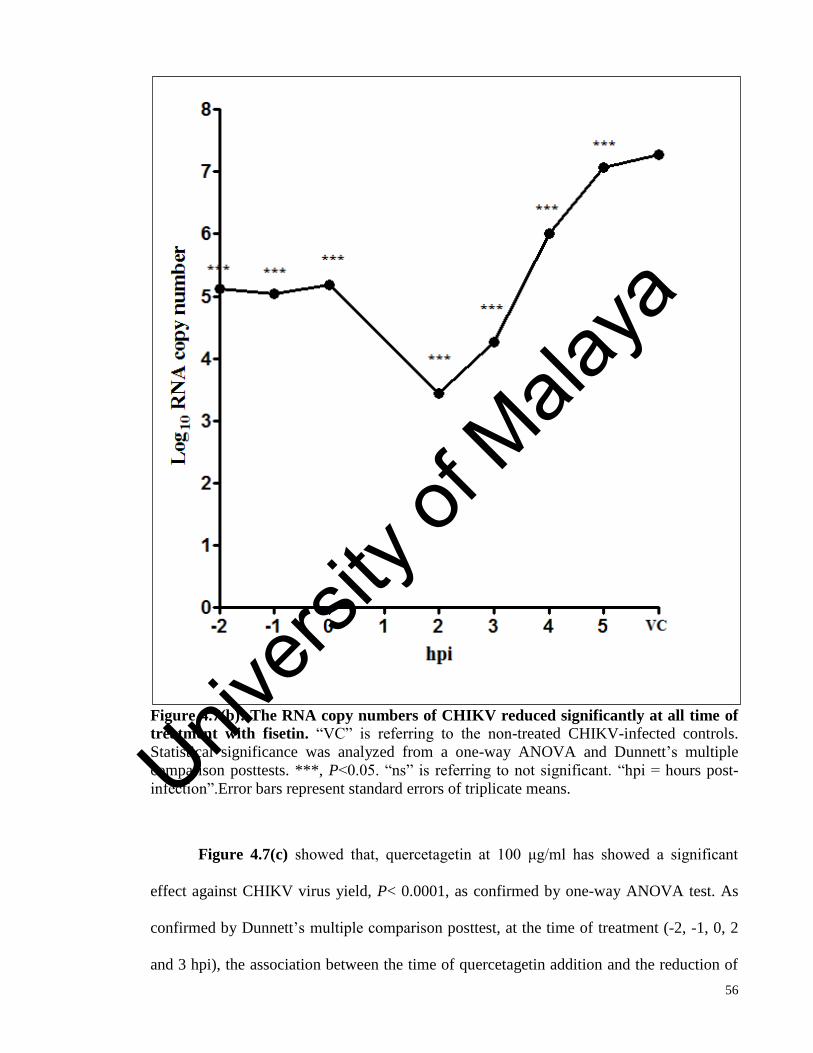
56
Figure 4.7(b): The RNA copy numbers of CHIKV reduced significantly at all time of
treatment with fisetin. “VC” is referring to the non-treated CHIKV-infected controls.
Statistical significance was analyzed from a one-way ANOVA and Dunnett’s multiple
comparison posttests. ***, P<0.05. “ns” is referring to not significant. “hpi = hours post-
infection”.Error bars represent standard errors of triplicate means.
Figure 4.7(c) showed that, quercetagetin at 100 μg/ml has showed a significant
effect against CHIKV virus yield, P< 0.0001, as confirmed by one-way ANOVA test. As
confirmed by Dunnett’s multiple comparison posttest, at the time of treatment (-2, -1, 0, 2
and 3 hpi), the association between the time of quercetagetin addition and the reduction of
Univers
ity of
Mala
ya

57
CHIKV RNA copy number is very strong, ***, whereas at other hours of treatment the
association is insignificant. This means that the effective time of treatment with
quercetagetin is up to 3 hours after CHIKV infection.
Figure 4.7(c): The RNA copy numbers of CHIKV reduced significantly up to 3 hpi of
quercetagetin treatment in the time-of-addition assay. “VC” is referring to the non-
treated CHIKV-infected controls. Statistical significance was analyzed from a one-way
ANOVA and Dunnett’s multiple comparison posttest. ***, P<0.05. “ns” is referring to not
significant. “hpi = hours post-infection”. Error bars represent standard errors of triplicate
means.
Univers
ity of
Mala
ya

58
Figure 4.7(d): The RNA copy numbers of CHIKV reduced significantly at all time of
silymarin treatment in the time-of-addition assay. “VC” is referring to the non-treated
CHIKV-infected controls. Statistical significance was analyzed from a one-way ANOVA
and Dunnett’s multiple comparison posttest. ***, P<0.05. “ns” is referring to not
significant. “hpi = hours post-infection”. Error bars represent standard errors of triplicate
means.
It was shown in Figure 4.7(d), that silymarin at 100 μg/ml has significant anti-
CHIKV activity, P< 0.0001, as confirmed by one-way ANOVA test. As confirmed by
Univers
ity of
Mala
ya

59
Dunnett’s multiple comparison posttest, all time of treatment used in this assay has
significantly reduced the CHIKV RNA copy number. The association between the time of
silymarin addition and the reduction of CHIKV RNA copy number is very strong, ***. This
means that the effective time of treatment with silymarin is up to 5 hours after CHIKV
infection. From the results of the time-of-addition assay, all four selected compounds seem
to be able to inhibit CHIKV in vitro replication at its early stage of life cycle. Thus,
individual antiviral assays were performed to determine at which stage of CHIKV life cycle
does the bioflavonoid compounds affecting.
4.5 Selected flavonoids inactivate CHIKV particles
Based on the the results of virucidal assay shown in Figure 4.8, quercetagetin (P<
0.0001) and baicalein (P< 0.0001) exert potent dose-dependent effect against extracellular
CHIKV particles with the IC50 values 9.934 μg/ml and 11.64 μg/ml respectively. Fisetin
(P< 0.0001) and silymarin (P< 0.0001) also showed significant virucidal activity against
CHIKV with the IC50 values of 52.71 μg/ml, 20.21 μg/ml respectively. The IC50 was
calculated by comparing the treated CHIKV-infected sample with the CHIKV-infected
sample without treatment.
Univers
ity of
Mala
ya

60
0.00 6.25 12.50 25.00 50.00 100.002
3
4
5
6
7
8
9
10
Fisetin Silymarin
Baicalein Quercetagetin
Concentration (µg/ml)
Lo
g1
0 R
NA
cop
y n
um
ber
Figure 4.8: The reduction of the RNA copy number by the treatment of baicalein,
fisetin, quercetagetin and silymarin in the virucidal assay. Statistical analysis was
performed by using one-way ANOVA where P<0.05 is significant. (“0” on the X-axis is
referring to the non-treated CHIKV-infected controls). Error bars represent standard errors
of triplicate means.
Univers
ity of
Mala
ya

61
4.6 Selected flavonoids interfere the adsorption of CHIKV on the cells
Based on the results of the anti-adsorption assay shown in Figure 4.9, baicalein (P<
0.0001), fisetin (P< 0.0001), silymarin (P< 0.0001) and quercetagetin (P< 0.0001) exert
dose-dependent inhibition against CHIKV replication when they added during the virus
attachment stage to the Vero cells. The IC50 values were 28.04 μg/ml, 176.0 μg/ml and
8.050 μg/ml for baicalein, silymarin and quercetagetin respectively.
Univers
ity of
Mala
ya

62
0.00 6.25 12.50 25.00 50.00 100.000
1
2
3
4
5
6
7
8
9
10
SilymarinBaicalein Quercetagetin
Fisetin
Concentration (µg/ml)
Log
10 R
NA
co
py
nu
mb
er
Figure 4.9: The reduction of the RNA copy number by the treatment of baicalein,
fisetin, silymarin and quercetagetin in the anti-adsorption assay. Statistical analysis
was performed by using one-way ANOVA where P<0.05 is significant. (“0” on the X-axis
is referring to the non-treated positive CHIKV-infected controls). Error bars represent
standard errors of triplicate means.
Univers
ity of
Mala
ya

63
4.7 Selected flavonoids interfere the entry of CHIKV into the cells
It was shown in Figure 4.10, that baicalein (P< 0.0001), fisetin (P< 0.0001) and
quercetagetin (P< 0.0001) exert significant dose-dependent inhibition against CHIKV after
its adsorption to the Vero cells. Silymarin did show only moderate significant dose-
dependent inhibition (P=0.0319) against CHIKV internalization. The IC50 values for
baicalein, fisetin and quercetagetin were 21.01 μg/ml, 56.04 μg/ml and 64.21 μg/ml
respectively. These results signified that baicalein is the strongest compound among the
other tested compounds which could interfere with the entry of the CHIKV particles into
Vero cells compared to the other three compounds.
Univers
ity of
Mala
ya

64
0.00 6.25 12.50 25.00 50.00 100.000
1
2
3
4
5
6
7
8
9
SilymarinQuercetagetin
FisetinBaicalein
Concentration (µg/ml)
Log
10 R
NA
co
py
nu
mb
er
Figure 4.10: The reduction of the RNA copy number by the treatment of
quercetagetin, baicalein, silymarin and fisetin in the anti-entry assay. Statistical
analysis was performed by using one-way ANOVA lwhere P<0.05 is significant. (“0” on
the X-axis is referring to the non-treated positive CHIKV-infected controls). Error bars
represent standard errors of triplicate means.
Univers
ity of
Mala
ya
65
4.8 Selected flavonoids interfere the intracellular CHIKV replication
Figure 4.11 showed that, baicalein (P< 0.0001), silymarin (P< 0.0001),
quercetagetin (P< 0.0001), fisetin (P< 0.0001) and ribavirin (P< 0.0001) as a positive anti-
CHIKV control exert significant dose-dependent inhibition against CHIKV in vitro
replication when they were added after virus internalization. The IC50 values obtained were
1.891 μg/ml, 12.98 μg/ml, 13.85 μg/ml, 8.444 μg/ml and 11.07 μg/ml for baicalein,
silymarin, quercetagetin, fisetin and ribavirin respectively. The IC90 values were 0.1812
μg/ml, 27.70 μg/ml, 142.7 μg/ml, 11.02 μg/ml and 16.63 μg/ml for baicalein, silymarin,
quercetagetin, fisetin and ribavirin respectively. These results suggested that all four tested
compounds could interfere with the specific binding of the viral attachment proteins and the
cellular receptors.
Univers
ity of
Mala
ya

66
0.00 6.25 12.50 25.00 50.00 100.000
1
2
3
4
5
6
7
8
9
RibavirinQuercetagetinFisetinBaicalein
Silymarin
Concentration (µg/ml)
Log
10 R
NA
co
py
nu
mb
er
Figure 4.11: The reduction of the RNA copy number by the treatment of
quercetagetin, baicalein, fisetin, silymarin and ribavirin at the post-adsorption assay. Statistical analysis was performed by using one-way ANOVA (Kruskal-Wallis test) where
P<0.05 is significant. (“0” on the X-axis is referring to the non-treated positive CHIKV-
infected controls). Error bars represent standard errors of triplicate means.
Univers
ity of
Mala
ya

67
4.9 Selectivity index value
The calculation of the selectivity index (SI) value is essential to determine the
potency of the treatment by dividing the concentration that reduces the cell viability by
50% to the concentration that offered the inhibitory effect by 50% (CC50/IC50). The
selectivity index value for the four selected compounds at each antiviral assays were
summarized in Table 4.3.
Table 4.3: Selectivity index of compounds for each antiviral assays.
Bioflavonoids Antiviral assay
Virucidal Adsorption Post-
adsorption
Entry
Baicalein 43.27 17.96 266.37 25.26
Fisetin 15.24 Undetermined 95.11 14.33
Quercetagetin 77.53 95.68 55.61 11.99
Silymarin 21.03 2.42 32.75 Undetermined
4.10 Selected flavonoids reduced the success rate of CHIKV infection
The immunofluorescence assay was performed to detect the alphaviral envelope
protein as an indication of successful CHIKV infection and replication. Ribavirin has
previously been shown to inhibit CHIKV replication (Briolant et al., 2004) and was
selected to serve as a positive control, as well as to assess the robustness of the assay.The
result of immunofluorescence assay for baicalein treatment is shown in Figure 4.12.
Univers
ity of
Mala
ya

68
Figure 4.12: Dose-dependent inhibition of baicalein on CHIKV infectivity shown using
immunofluorescence assay. Immunofluorescence detection of alphavirus envelope protein
is used as an indication of CHIKV infection. CHIKV infection of Vero cells is compared
untreated cells. Cell nuclei are stained with DAPI (blue), and CHIKV infection is indicated
by Alexa Fluor (green) staining. Mock-infected cells were stained with both primary and
secondary antibodies, as well as DAPI.
By referring to the Figure 4.12, dose-dependent reduction of viral antigen-positive
cells for the different baicalein concentrations is observed from immunofluorescence
images. Baicalein with 100 μg/ml concentration exerted about 70% inhibition against
CHIKV antigen presentation. The results of immunofluorescence assay for fisetin treatment
is shown in Figure 4.13.
Univers
ity of
Mala
ya

69
Figure 4.13: Dose-dependent inhibition of fisetin on CHIKV infectivity shown using
immunofluorescence assay. Immunofluorescence detection of alphavirus envelope protein
is used as an indication of CHIKV infection. CHIKV infection of Vero cells is compared
untreated cells. Cell nuclei are stained with DAPI (blue), and CHIKV infection is indicated
by Alexa Fluor (green) staining. Mock-infected cells were stained with both primary and
secondary antibodies, as well as DAPI.
As shown in Figure 4.13, fisetin has successfully inhibited the CHIKV antigen
production even by the lowest concentration. A dose-dependent reduction of viral antigen-
positive cells for the different fisetin concentrations is observed from immunofluorescence
images.. The lowest concentration of fisetin treatment, 6.25 μg/ml, offered more than 90%
protection against CHIKV infection.
Univers
ity of
Mala
ya

70
The results of immunofluorescence assay for quercetagetin treatment is shown in
Figure 4.14. A dose-dependent reduction of viral antigen-positive cells for the different
quercetagetin concentrations is observed from immunofluorescence images which indicates
successful inhibition of CHIKV antigen production. Quercetagetin with 100 μg/ml inhibited
the CHIKV antigen production for more than 80%.
Figure 4.14: Dose-dependent inhibition of quercetagetin on CHIKV infectivity shown
using immunofluorescence assay. Immunofluorescence detection of alphavirus envelope
protein is used as an indication of CHIKV infection. CHIKV infection of Vero cells is
compared untreated cells. Cell nuclei are stained with DAPI (blue), and CHIKV infection is
indicated by Alexa Fluor 488 (green) staining. Mock-infected cells were stained with both
primary and secondary antibodies, as well as DAPI.
Univers
ity of
Mala
ya

71
The results of immunofluorescence assay for silymarin treatment is shown in
Figure 4.15. A dose-dependent reduction of viral antigen-positive cells for the different
silymarin concentrations is observed from immunofluorescence images.which indicates
successful inhibition of CHIKV antigen production. At concentration of 100 μg/ml,
silymarin offered more than 80% inhibition against CHIKV antigen production.
Figure 4.15: Dose-dependent inhibition of silymarin on CHIKV infectivity shown
using immunofluorescence assay. Immunofluorescence detection of alphavirus envelope
protein is used as an indication of CHIKV infection. CHIKV infection of Vero cells is
compared untreated cells. Cell nuclei are stained with DAPI (blue), and CHIKV infection is
indicated by Alexa Fluor 488 (green) staining. Mock-infected cells were stained with both
primary and secondary antibodies, as well as DAPI.
Univers
ity of
Mala
ya

72
The results of immunofluorescence assay on the nucleoside analogue anti-CHIKV
control, ribavirin, is shown in Figure 4.16. A dose-dependent reduction of viral antigen-
positive cells for the different baicalein concentrations is observed from
immunofluorescence images.
Figure 4.16: Dose-dependent inhibition of ribavirin on CHIKV infectivity shown using
immunofluorescence assay. Immunofluorescence detection of alphavirus envelope protein
is used as an indication of CHIKV infection. CHIKV infection of Vero cells is compared
untreated cells. Cell nuclei are stained with DAPI (blue), and CHIKV infection is indicated
by Alexa Fluor 488 (green) staining. Mock-infected cells were stained with both primary
and secondary antibodies, as well as DAPI.
Univers
ity of
Mala
ya

73
4.11 Selected flavonoids reduced the efficiency of CHIKV proteins accumulation
In order to determine the effect of the bioflavonoid compounds on CHIKV proteins
synthesis, western blot analyses were performed. CHIKV proteins such as; pE2 (65 kDa),
E2 (50 kDa), nsP1 (59 kDa) and nsP3 (76-78kDa) were detected by using the colorimetric
method. β-actin was used to as a loading control in the experiment, as well as to ensure that
the concentrations of the tested compounds did not affect the synthesis and expression of
host cellular proteins, and also the integrity of the treated cells. The detection of the target
CHIKV proteins would reflect the effect of the selected compounds on the process of
CHIKV in vitro successful replication.
The results of western blotting upon baicalein treatment is as shown in Figure 4.17.
Baicalein with concentration of 100 μg/ml showed significant inhibition against nsP1 and
nsP3 protein accumulation. The E2 protein faded at the same concentration. By referring to
the relative band intensity, the sudden drop in the band intensity of all three proteins were
shown. This results reflect the ability of baicalein to suppress the efficiency of the process
of CHIKV in vitro replication.
The results of western blotting upon fisetin treatment is shown in Figure 4.18. The
nsP1 band intensity has been strongly affected by the treatment of fisetin with
concentrations of 12.5 μg/ml and above. The nsP3 protein faded and totally disappeared at
concentrations of 25 μg/ml and above. The E2 protein remained producing efficiently and
was not affected by fisetin treatment. By referring to the relative band intensity, the sudden
drop in the band intensity of nsP1 and nsP3 proteins were shown. This results reflect the
ability of fisetin to suppress the CHIKV in vitro replication.
Univers
ity of
Mala
ya

74
Figure 4.17: Baicalein suppressed the accumulation of CHIKV-encoded proteins.
(a) Bands of the specific proteins as observed on the blot. A dose dependent reduction of
CHIKV nsP1, nsP3 and pE2/E2 proteins were observed upon baicalein treatments for 48 h.
(b) The above mentioned observation confirmed by calculating the relative band intensity
(calculated from the band intensity of sample/band intensity of β-actin loading control)
using Image J. β-actin is used as a loading control for each set of samples.
Univers
ity of
Mala
ya

75
Figure 4.18: Fisetin suppressed the accumulation of CHIKV-encoded proteins.
(a) Bands of the specific proteins as observed on the blot. A dose dependent reduction of
CHIKV nsP1 and nsP3 proteins were observed upon fisetin treatments for 48 h.
(b) The above mentioned observation confirmed by calculating the relative band intensity
(calculated from the band intensity of sample/band intensity of β-actin loading control)
using Image J. β-actin is used as a loading control for each set of samples.
Univers
ity of
Mala
ya

76
The results of western blotting upon quercetagetin treatment is as shown in Figure
4.19. Quercetagetin, at all concentrations used, did not affect the accumulation of all
targeted CHIKV proteins. The intensity of CHIKV proteins bands are equivalent to the
positive controls. This result reflects the inability of quercetagetin to suppress the efficiency
of CHIKV negative-strand synthesis and CHIKV replicase unit functions but not the
process of glycoprotein maturation and transportation. These results indicate a possibility of
the antiviral activity of quercetagetin at the later stage of CHIKV life cycle such as the
assembly of the nucleocapsid core and genomic RNA packaging or even virus budding.
Whereas for silymarin (results shown in Figure 4.20), did affect the CHIKV
proteins accumulation in a dose-dependent manner. At silymarin concentration of 100
μg/ml, total disappearance of the nsP1 band can be seen after a dose-dependent reduction of
the protein accumulation. The E2 and nsP3 proteins faded at the same concentration. By
referring to the relative band intensity, sudden drop in the band intensity of all three
proteins were shown. This results reflect the ability of silymarin to suppress the efficiency
of the process of glycoprotein maturation and transportation, CHIKV negative-strand
synthesis and CHIKV replicase unit functions.
All the results supported the assumptions that four selected compounds; baicalein,
fisetin, quercetagetin and silymarin were able to inhibit and interfere CHIKV infection and
replication efficiency at various stages of CHIKV in vitro life cycle efficiently.
Univers
ity of
Mala
ya
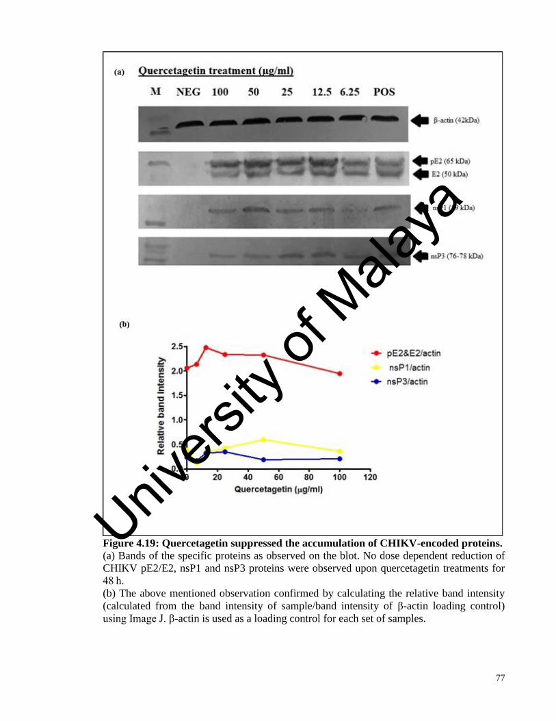
77
Figure 4.19: Quercetagetin suppressed the accumulation of CHIKV-encoded proteins.
(a) Bands of the specific proteins as observed on the blot. No dose dependent reduction of
CHIKV pE2/E2, nsP1 and nsP3 proteins were observed upon quercetagetin treatments for
48 h.
(b) The above mentioned observation confirmed by calculating the relative band intensity
(calculated from the band intensity of sample/band intensity of β-actin loading control)
using Image J. β-actin is used as a loading control for each set of samples.
Univers
ity of
Mala
ya

78
Figure 4.20: Silymarin suppressed the accumulation of CHIKV-encoded proteins.
(a) Bands of the specific proteins as observed on the blot. A dose dependent reduction of
CHIKV nsP1, nsP3 and pE2/E2 proteins were observed upon silymarin treatments for 48 h.
(b) The observation above is confirmed by calculating the relative band intensity
(calculated from the band intensity of sample/band intensity of β-actin loading control)
using Image J. β-actin is used as a loading control for each set of samples.
Univers
ity of
Mala
ya

79
CHAPTER 5: DISCUSSION
The year of 2005 remarked the importance of research regarding CHIKV antivirals
since the chikungunya fever epidemic in La Réunion, which was the worst in history. The
fatal outbreak was also accompanied by the mutation of alanine to valine in the CHIKV E1
glycoprotein (A226V). Subsequently, CHIKV was transmitted by Aedes albopictus, the
native mosquito vector in temperate regions (Schuffenecker et al., 2006). Many important
medical factors contributed to the urge of finding the antiviral for CHIKV. CHIKV has re-
emerged in recent years rapidly due to the efficiency of the mosquito vectors in establishing
the CHIKV infection to the wide geographical regions. Approved antivirals are needed to
control symptoms and minimize the complications in future epidemics (Kaur & Chu.,
2013).
The best treatments that can be offered now are usually symptomatic by the
administration of non-steroidal anti-inflammatory drugs or corticosteroids to soothe the
arthralgia and myalgia (Queyriaux et al., 2008). In order to cope with increased global
travels, wide distribution of vectors in many geographical regions as well as the risk of
future epidemic, active investigations are continuously conducted to find effective
antivirals.
Most antivirals such as harringtonine, arbidol, mycophenolic acids and many more
are only in the preliminary stages of antiviral discovery. Harringtonine inhibited the
production of CHIKV nsP3 and E2 proteins as well as positive- and negative-sense CHIKV
RNA (Kaur et al., 2013). Arbidol, the broad-spectrum antiviral, prevented the CHIKV
adsorption to target cells thus inhibited the viral entry. Mycophenolic acids inhibited
CHIKV replication in a similar mechanism to that of the nucleoside analogue, ribavirin.
Univers
ity of
Mala
ya

80
However, a number of these preliminary antiviral studies have provided an insight towards
understanding the biology of CHIKV.
Well-known broad-spectrum antivirals such as chloroquine, ribavirin and IFN-α
have shown the efficacy in vivo. Upon administration of ribavirin into patients experiencing
arthritis and lower limb pains after CHIKV infection, they were found to have reduced soft
tissue swelling and joint pains. However, a lot remains unknown regarding CHIKV biology
in vivo (Kaur & Chu., 2013). Furthermore, the shortcomings of nucleoside analogues or
synthesized drugs cannot be underestimated since they are potentially teratogenic,
embryotoxic, carcinogenic and possessed the anti-proliferative activities (Morris D.J.,
1994).
The undesirable side effects should be taken into account before a compound could
be considered an antiviral candidate. Continuous research is required to establish a suitable
and effective antiviral and diversifying potential compounds would broaden the chances of
getting one. In doing so, many researchers turn to natural products for antivirals because it
guarantees continuous supply, ease of preparation, relatively low toxicity and low side
effects. In this case, bioflavonoids have shown high degree of potential for anti-CHIKV
research.
Although these phenolic compounds are nonessential for plant survival, their
potentials in medical research fields should not be overlooked. Bioflavonoid compounds
played roles in plants such as defense (Mol et al., 1998), allelopathy (Peer & Murphy.,
2007), providing flower coloring to attract pollinators (Bais et al., 2006), modulating the
levels of reactive oxygen species and influenced the transport of the auxin (Treutter., 2005).
In medical research fields, bioflavonoids compounds were found to have anti-oxidant
(Williams R.J et al., 2004), anti-tumor (Garcia-Mediavilla et al., 2007), anti-proliferative
Univers
ity of
Mala
ya

81
(Taylor & Grotewold., 2005), anti-inflammatory (Pandey et al., 2007), anti-fungal (Sung et
al., 2007), anti-bacterial (Wachter et al., 1999) and even antiviral activity against many
viruses (Valsaraj et al., 1997; Zheng et al., 1996; Li et al., 2000; Ono et al.,1989). While
going through the research for antivirals against CHIKV, a number of researchers have
already started roaming across the library of bioflavonoid compounds regardless whether
they are pure compounds or a mixture of compounds. Among the bioflavonoid compounds
were naringenin, chrysin, silybin and apigenin, also investigated in this study.
Naringenin possessed anti-Sindbis virus activity which is an alphavirus similar to
CHIKV (Paredes et al., 2003). Thus predictably, naringenin (IC50=30.0 μM) was also
studied by using a stable CHIKV replicon cell line, together with silybin (IC50=59.8 μM),
chrysin (IC50=50.2 μM) and apigenin (IC50=28.3 μM), and was able to suppress the Rluc
marker gene activity expressed by CHIKV replicon (Pohjala et al., 2011). However, the
study did not involve infectious CHIKV. Silybin, on the other hand, is a semi-purified,
commercially available fraction of silymarin, the bioflavonoid that has also been
investigated in this study.
Though silybin exhibited antiviral activity against CHIKV, we recently published a
study suggesting that silymarin, as a whole complex of more than 7 flavonolignans
including silybin, possessed better activity against CHIKV. Moreover, in a previous study
IC50 of silybin, one of the major components of silymarin, was estimated as 59.8 μM
(approximately 30 μg/ml). Compared to this, silymarin was somewhat more efficient as
nearly three-fold inhibition was observed at 25 μg/ml indicating that other components of
silymarin likely contributed to its anti-CHIKV activity. It has been concluded that other
components of silymarin have enhanced its anti-CHIKV activity (Lani et al., 2015).
Univers
ity of
Mala
ya

82
In this study, 14 bioflavonoids were screened for any possible antiviral activity
against CHIKV. However, after performing continuous treatment as a screening method,
only four showed potential inhibition activity against CHIKV. A qualitative assay, CPE
inhibition assay was performed to validate the screening result. A quantitative assay, MTS
assay confirmed that the four compounds can inhibit CHIKV infection significantly from
the screening. Those four compounds were baicalein, fisetin, quercetagetin and silymarin.
Interestingly, these four compounds are of different chemical structure based on the flavone
ring. Silymarin is a flavonolignan which is a mixture of the flavonoid and lignan, baicalein
is a flavone whereas both fisetin and quercetagetin are flavonol. Their antiviral activities
against CHIKV are varied based on the antiviral assays that has been performed. These four
bioflavonoids interfere with different stages of CHIKV in vitro replication in Vero cells.
The findings of this study are summarized in Table 5.1.
Table 5.1: Summary of the result obtained from various assays performed in the
current study.
Bioflavonoids
T.O.A Antiviral assays Western blotting
Effective
time
Virucidal Adsorption Post-
adsorption
Entry Proteins
E2 nsP1 nsP3
Baicalein -2 to 3
hpi
+ + + + + + +
Fisetin All time
of
treatment
hpi
+ + + + + + +
Quercetagetin -2 to 3
hpi
+ + + + - - -
Silymarin All time
of
treatment
+ + + + + + +
Note that, “T.O.A: time of addition assay”, “hpi: hours post-infection”, “+: significant
inhibition activity” and “-: non-significant inhibition activity”.
In this study, a positive strand qRT-PCR has been performed to evaluate the virus
yield reduction as described by Chiam et al., 2013. The target gene was the nsP3 gene by
Univers
ity of
Mala
ya
83
using the designed nsP3 primer (136bp). The positive strand qRT-PCR assay was chosen
because the previous study showed that it was specific for CHIKV, possessed more
optimum high coefficients of determination (R2), slope, and efficiency. The positive strand
qRT-PCR was able to detect CHIKV RNA with highest sensitivity and appeared to be more
suitable for the detection of the high viral loads as expected in this study (Chiam et al.,
2013).
Focusing on the ability of the bioflavonoid compounds to interfere the CHIKV
replication, a CHIKV cell line-based replicon was chosen to depict the assumption. CHIKV
replicon cell lines offer a screening-friendly approach. By constructing replicon containing
CHIKV replicase proteins with puromycin acetyltransferase, EGFP and Renilla luciferase
marker genes was constructed for this objective. This enabled us to identify the potential
antiviral candidates for alphavirus entry and replication phase inhibitiors (Pohjala et al.,
2011). Through screening using CHIKV replicon, baicalein showed the best inhibitory
activity with IC50=3.243 μg/ml. However, the result was different from the antiviral assay
performed with infectious CHIKV most probably due to the structural glycoproteins of
CHIKV which also facilitated anti-CHIKV activity of the compounds.
In the time-of-addition assay, bioflavonoids were added to the CHIKV-infected
Vero cells as the pre-treatment (-2 and -1 hpi), co-treatement (0 hpi) and post-treatment (2,
3, 4, and 5 hpi) assays. Through this assay, the best time for the most potent anti-CHIKV
effects due to treatment with selected bioflavonoids have been investigated. The half-life of
a compound would probably be one of the ultimate reasons that would cause the effective
time for baicalein and quercetagetin to exert its antiviral effect up to 3 hpi, while fisetin and
silymarin performed better up to 5 hpi. Almost 100% of CHIKV replication efficiency can
be inhibited by baicalein and quercetagetin at -2 to 2 hpi whereas fisetin and silymarin at all
Univers
ity of
Mala
ya

84
time of treatment.It is highly certain that these bioflavonoid compounds acted at the early
hours and most possibly early stages of CHIKV infection. Thus, in the antiviral assays
composed of the assays that will portrayed the actions of bioflavonoids on early events of
CHIKV infections, the probable mechanism could be determined.
In virucidal assays, the four compounds have successfully inactivated the CHIKV
prior to the infection of the Vero cells. The reduction of CHIKV RNA yield in this assay
reflects that most of treated CHIKV particles were unable to infect the Vero cells. All four
bioflavonoids showed effective virucidal effect against CHIKV (the effectivity order as
quercetagetin>baicalein>silymarin>fisetin), but the most potent bioflavonoid was
quercetagetin with IC50=9.934 μg/ml and SI=77.53. However, there are no reported
virucidal activity of quercetagetin against other viruses. Baicalein showed virucidal activity
against JEV (IC50=3.44 μg/ml) and DENV-2 (IC50=1.55 μg/ml) (Johari et al., 2012; Zandi
et al., 2012). Fisetin, in the other hand, did not exhibit any virucidal activity against DENV-
2 and EV-71 (Zandi et al., 2011; Lin et al., 2012). Silibinin, the primary active component
of silymarin, showed virucidal activity against herpes virus with IC50=5.0 μg/ml (Cardile &
Mbuy., 2013).
We have found that all four selected bioflavonoids in this study showed more anti-
adsorption activity against the adsorption of CHIKV particles to the Vero cells. The
proteins that are most likely to be involved were E1 and E2 glycoproteins since these are
the proteins crucial for the host-receptor interaction during CHIKV adsorption (Jose,
Snyder & Kuhn., 2010). The reduction of the CHIKV adsorption efficiency can be
interpreted through the reduction of the CHIKV yield in the respective assay. Quercetagetin
again exhibited the best anti-adsorption activity against CHIKV with IC50=8.050 μg/ml and
SI=95.68. Quercetagetin has successfully interfered the interactions between CHIKV
Univers
ity of
Mala
ya

85
glycoproteins and host receptors. Among four bioflavonoids, baicalein showed anti-
adsorption activity against JEV (IC50=7.27 μg/ml) and DENV-2 (IC50=7.14 μg/ml) in
previously reported studies (Johari et al., 2012; Zandi et al., 2012).
After adsorption of CHIKV to the Vero cells, the host receptors induce
conformational changes in the E1 and E2 glycoproteins. This stage is known as the entry
stage. From the anti-entry assay, the reduction in the CHIKV RNA copy number can be
seen after the treatment with all compounds. These compounds are hypothesized to
interfere the conformational changes. The CHIKV bound to the receptor molecules were
then endocytosed in clathrin-dependent manner. In order to interfere this stage, a compound
must be able to either block the clathrin-coated pits or influence cells to ablate the ability of
forming functional clathrin-coated pits. The CHIKV yield has been reduced since CHIKV
cannot successfully enter Vero cells upon treatment with these three bioflavonoids. Three
compounds in the effectivity order as baicalein>fisetin>quercetagetin showed anti-entry
activity against CHIKV, baicalein being the most effective compound with IC50=21.01
μg/ml and SI=25.26. Silymarin did not exhibit any anti-entry activity against CHIKV.
The post-adsorption or post-entry assay is crucial to determine the intracellular
antiviral activity of compounds against CHIKV. Ribavirin was only relevant as anti-
CHIKV positive control in this assay because it is the RNA replication inhibitor and it acts
intracellularly. All selected compounds showed activity against post-entry activity of
CHIKV with the effectivity order baicalein>fisetin>ribavirin>silymarin>quercetagetin. The
best compound which exhibited post-entry antiviral activity against CHIKV was baicalein
with IC50=1.891 μg/ml and SI=266.37. Baicalein, fisetin, quercetagetin and silymarin have
shown promising CHIKV inhibitory results, comparable to the performance of ribavirin.
Univers
ity of
Mala
ya

86
These four compounds are highly recommended for further in vivo research since ribavirin
was able to reduce joint pain in a few in vivo studies.
The effects of the bioflavonoids’ treatment on production of the CHIKV E2 antigen
was determined using the immunofluorescence assay. The effectivity order of the treatment
determined from the intensity and quantity of the stained antigen is designated as
fisetin>quercetagetin>silymarin>baicalein. Fisetin showed 90% reduction in the intensity
and quantity of the antigen at even the lowest concentrations. This shows that the
bioflavonoids were able to interfere with the processing and maturation of the E2 antigen as
well as the transportation of the E2 antigen to the plasma membrane.
This study will be more beneficial if the production of the CHIKV proteins upon
treatment with bioflavonoid compounds can be determined. Thus, the western blotting
analyses were performed. All three of the compounds except quercetagetin, were able to
suppress the production or accumulation of the targeted proteins, pE2/E2, nsP1 and nsP3.
This effect could be due to inhibition of CHIKV RNA replication and/or transcription.
However, as in virus expression and replicon cell lines the synthesis of viral RNAs and
proteins are coupled further study is necessary to evaluate the direct effect of the
bioflavonoids on inhibition of newly synthesized CHIKV proteins.
It is possible that some of non-structural proteins of CHIKV represents direct target
for the bioflavonoids. The possibilities include nsP1 protein, which is involved in the
synthesis of the negative strand of viral RNA and RNA capping, and nsP3 protein, that is
another component of the viral replicase complex. Down regulation of E2 expression may
represent consequence of suppression of replication (directly or via inhibition of ns-
protein(s)). However, this down regulation is clearly relevant from point of view of
Univers
ity of
Mala
ya

87
development of effective antiviral as E2 protein is one of the important virion glycoproteins
and is essential for receptor binding
Baicalein, fisetin and silymarin were able to interfere the accumulation of CHIKV
proteins involved in negative-strand synthesis, replicase unit and the process of
glycoprotein maturation and transportation. Although quercetagetin did not show any
suppression, this result actually show that quercetagetin might interfere with the later stage
of the CHIKV life cycle which is the assembly of the nucleocapsid core (together with its
genomic RNA packaging) with the processed glycoproteins at the plasma membrane before
budding. It is worth noting that these four bioflavonoid compounds exhibited antiviral
activity against CHIKV with different strategies.
Univers
ity of
Mala
ya

88
CHAPTER 6: CONCLUSION
Throughout this study, all the objectives were achieved. The cytotoxicity assay of
14 bioflavonoid compounds towards Vero cells were determined. Four out of 14
bioflavonoid compounds; baicalein, fisetin, quercetagetin and silymarin exhibited
significant antiviral activity against CHIKV at different stages of CHIKV life cycle.
Baicalein, fisetin and silymarin interfered with the CHIKV proteins involved in negative-
strand synthesis and replicase unit.
Whereas quercetagetin, most probably interfered the later stage of CHIKV life cycle
which is the assembly of nucleocapsid core together with RNA genomic packaging and
processed glycoproteins. However, we have also successfully shown that quercetagetin can
interfere with the early stages of CHIKV replication cycle especially with virus attachment
to the Vero cells. These results are in conjunction with the ability of baicalein, fisetin and
silymarin to suppress the accumulation or production of pE2/E2, nsP1 and nsP3 proteins.
The selectivity index value for each effective compound in each antiviral assay have also
been identified.
In future, further investigations should be carried out to determine the relationship
between the differences of these four bioflavonoids chemical structure and the means of
their antiviral activity against CHIKV. Performing molecular docking analysis would
provide the prediction on predominant binding mode(s) of the compounds with a protein of
known three-dimensional structure as well as providing its mechanism of actions.
Performing the in vivo research by using animal models would be the second step towards
understanding the outcomes of the the bioflavonoids treatment, side effects and suitable
dosage in more complex organisms.
Univers
ity of
Mala
ya

89
REFERENCES
Adesina, O.A., and Odelola, H.A. (1991). Ecological distribution of chikungunya
haemagglutinaton inhibition antibodies in human and domestic animals in Nigeria. Tropical
and Geographical Medicine, 43:271-5.
Ahmed-Belkacem, A., Guichou, J. F., Brillet, R., Ahnou, N., Hernandez, E., Pallier, C., and
Pawlotsky, J. M. (2014). Inhibition of RNA binding to hepatitis C virus RNA-dependent
RNA polymerase: a new mechanism for antiviral intervention. Nucleic Acids Research,
42(14): 9399-9409.
Allard, P. M., Leyssen, P., Martin, M. T., Bourjot, M., Dumontet, V., Eydoux, C.,
Guillemot., J.C., and Litaudon, M. (2012). Antiviral chlorinated daphnane diterpenoid
orthoesters from the bark and wood of Trigonostemon cherrieri. Phytochemistry, 84:160-
168.
Alto, B.W., and Juliano, S.A. (2001). Precipitation and temperature effects on populations
of Aedes albopictus (Diptera: Culicidae): implications for range expension. Journal of
Medical Entomology, 38:646-656.
Andres, A., Donovan, S. M., and Kuhlenschmidt, M. S. (2009). Soy isoflavones and virus
infections. The Journal of Nutritional Biochemistry, 20(8): 563-569.
Andres, A., Donovan, S. M., Kuhlenschmidt, T. B., and Kuhlenschmidt, M. S. (2007).
Isoflavones at concentrations present in soy infant formula inhibit rotavirus infection in
vitro. The Journal of Nutrition, 137(9): 2068-2073.
Arai, Y., Watanabe, S., Kimira, M., Shimoi, K., Mochizuki, R., and Kinae, N. (2000).
Dietary intakes of flavonols, flavones and isoflavones by Japanese women and the inverse
correlation between quercetin intake and plasma LDL cholesterol concentration. The
Journal of Nutrition, 130(9): 2243-2250.
Bachmetov, L., Gal‐Tanamy, M., Shapira, A., Vorobeychik, M., Giterman‐Galam, T.,
Sathiyamoorthy, P., Golan‐Goldhirsh, A., Benhar, I., Tur‐Kaspa Zemel, R., R. (2012).
Suppression of hepatitis C virus by the flavonoid quercetin is mediated by inhibition of
NS3 protease activity. Journal of Viral Hepatitis, 19(2): e81-e88.
Univers
ity of
Mala
ya
90
Bais, H. P., Weir, T. L., Perry, L. G., Gilroy, S., and Vivanco, J. M. (2006). The role of root
exudates in rhizosphere interactions with plants and other organisms. Annual Review of
Plant Biology, 57:233-266.
Bate-Smith, E. C., and Harborne, J. B. (1969). Quercetagetin and patuletin in Eriocaulon.
Phytochemistry, 8(6): 1035-1037.
Bedard, K. M., Wang, M. L., Proll, S. C., Loo, Y. M., Katze, M. G., Gale, M., and
Iadonato, S. P. (2012). Isoflavone agonists of IRF-3 dependent signaling have antiviral
activity against RNA viruses. Journal of Virology, 86(13): 7334-7344.
Behbahani, S. S., Pourazar, A., and M Shanehsazzadeh, M. (2014). In vitro anti-HIV-1
activities of kaempferol and kaempferol-7-O-glucoside isolated from Securigera
securidaca. Research in Pharmaceutical Sciences, 9(6): 463-469.
Bosio, K., Avanzini, C., D'avolio, A., Ozino, O., and Savoia, D. (2000). In vitro activity of
propolis against Streptococcus pyogenes. Letters in Applied Microbiology, 31(2):174-177.
Bourjot, M., Delang, L., Nguyen, V. H., Neyts, J., Gueritte, F., Leyssen, P., and Litaudon,
M. (2012). Prostratin and 12-O-tetradecanoylphorbol 13-acetate are potent and selective
inhibitors of chikungunya virus replication. Journal of Natural Products, 75(12):2183-
2187.
Bréhin, A. C., Casadémont, I., Frenkiel, M. P., Julier, C., Sakuntabhai, A., and Desprès, P.
(2009). The large form of human 2′, 5′-Oligoadenylate Synthetase (OAS3) exerts antiviral
effect against Chikungunya virus. Virology, 384(1):216-222.
Briolant, S., Garin, D., Scaramozzino, N., Jouan, A., and Crance, J. M. (2004). In vitro
inhibition of Chikungunya and Semliki Forest viruses replication by antiviral compounds:
synergistic effect of interferon-α and ribavirin combination. Antiviral Research, 61(2):111-
117.
Brown, J. P. (1980). A review of the genetic effects of naturally occurring flavonoids,
anthraquinones and related compounds. Mutation Research/Reviews in Genetic
Toxicology, 75(3):243-277.
Univers
ity of
Mala
ya

91
Buer, C. S., Imin, N., and Djordjevic, M. A. (2010). Flavonoids: new roles for old
molecules. Journal of Integrative Plant Biology, 52(1):98-111.
Calland, N., Dubuisson, J., Rouillé, Y., and Séron, K. (2012). Hepatitis C virus and natural
compounds: a new antiviral approach?. Viruses, 4(10): 2197-2217.
Cardile, A. P., and Mbuy, G. K. (2013). Anti-herpes virus activity of silibinin, the primary
active component of Silybum marianum. Journal of Herbal Medicine, 3(4): 132-136.
Cassidy, A. (2006). Factors affecting the bioavailability of soy isoflavones in humans.
Journal of AOAC International, 89(4): 1182-1188.
Centers For Disease Control And Prevention (Cdc. (2015). CDC Health Information for
International Travel 2016. Oxford University Press.
Charrel, R.N., de Lamballerie, X., and Raoult, D. (2007). Chikungunya outbreaks- The
globalization of vectorborne diseases. The New England Journal of Medicine, 356(8):769-
771.
Chew, L.P., and Chua, H.H., (2009). Outbreak of Chikungunya in Johor Bahru, Malaysia:
Clinical and laboratory features of hospitalized patients. Medical Journal of Malaysia,
64(3):220-222.
Chiam, C. W., Chan, Y. F., Loong, S. K., Yong, S. S. J., Hooi, P. S., and Sam, I. C. (2013).
Real-time polymerase chain reaction for diagnosis and quantitation of negative strand of
chikungunya virus. Diagnostic Microbiology and Infectious Disease, 77(2): 133-137.
Chua, K.B. (2010). Epidemiology of Chikungunya in Malaysia: 2006-2009. Medical
Journal of Malaysia, 65(4):278-283.
Cornwell, T., Cohick, W., and Raskin, I. (2004). Dietary Phytoestrogens and Health.
Phytochemistry, 65(8): 995-1016.
Univers
ity of
Mala
ya

92
Cotin, S., Calliste, C. A., Mazeron, M. C., Hantz, S., Duroux, J. L., Rawlinson, W. D.,
Ploy, M-C., and Alain, S. (2012). Eight flavonoids and their potential as inhibitors of
human cytomegalovirus replication. Antiviral Research, 96(2): 181-186.
Couderc, T., Chrétien, F., Schilte, C., Disson, O., Brigitte, M., Guivel-Benhassine, F.,
Touret, Y., and Lecuit, M. (2008). A mouse model for Chikungunya: young age and
inefficient type-I interferon signaling are risk factors for severe disease. PLoS
Pathogens, 4(2):e29.
Cushnie, T. T., and Lamb, A. J. (2005). Antimicrobial activity of flavonoids. International
Journal of Antimicrobial Agents, 26(5): 343-356.
Daelemans, D., Pauwels, R., De Clercq, E., & Pannecouque, C. (2011). A time-of-drug
addition approach to target identification of antiviral compounds. Nature Protocols, 6(6):
925-933.
Dash, P. K., Tiwari, M., Santhosh, S. R., Parida, M., and Rao, P. L. (2008). RNA
interference mediated inhibition of Chikungunya virus replication in mammalian
cells. Biochemical and Biophysical Research Communications, 376(4):718-722.
de Lamballerie, X., Leroy, E., Charrel, R.N., Ttsetsarkin, K., Higgs, S., and Gould, E.A.
(2008). Chikungunya virus adapts to tiger mosquito via evolutionary convergence: a sign of
things to come? Virology, 5:33.
Delogu, I., Pastorino, B., Baronti, C., Nougairède, A., Bonnet, E., and de Lamballerie, X.
(2011). In vitro antiviral activity of arbidol against Chikungunya virus and characteristics
of a selected resistant mutant. Antiviral Research, 90(3):99-107.
Diallo, M., Thonnon, J., Traore-Lamizana, M., and Fontenille, D. (1999). Vectors of
chikungunya virus in Senegal: current data and transmission cycles. The American Journal
of Tropical Medicine and Hygiene, 60:281-286.
Ding, Y., Dou, J., Teng, Z., Yu, J., Wang, T., Lu, N., Wang, H., and Zhou, C. (2014).
Antiviral activity of baicalin against influenza A (H1N1/H3N2) virus in cell culture and in
mice and its inhibition of neuraminidase. Archives of Virology, 159(12): 3269-3278.
Univers
ity of
Mala
ya

93
Dixon, R. A. (2004). Phytoestrogens. Annual Review of Plant Biology, 55: 225-261.
Eckels, K.H., Harrison, V.R., and Hetrick, F.M. (1970). Chikungunya virus vaccine
prepared by Tween-ether extraction. Applied Microbiology, 19:321-325.
Edelman, R., Tacket, C. O., Wasserman, S. S., Bodison, S. A., Perry, J. G., and Mangiafico,
J. A. (2000). Phase II safety and immunogenicity study of live chikungunya virus vaccine
TSI-GSD-218. The American Journal of Tropical Medicine and Hygiene, 62(6):681-685.
Evers, D. L., Chao, C. F., Wang, X., Zhang, Z., Huong, S. M., and Huang, E. S. (2005).
Human cytomegalovirus-inhibitory flavonoids: studies on antiviral activity and mechanism
of action. Antiviral Research, 68(3): 124-134.
Fiorani, M., and Accorsi, A. (2005). Dietary flavonoids as intracellular substrates for an
erythrocyte trans-plasma membrane oxidoreductase activity. British Journal of Nutrition,
94(03): 338-345.
Fletcher, R. J. (2003). Food sources of phyto-oestrogens and their precursors in Europe.
British Journal of Nutrition, 89(S1): S39-S43.
Ganesan, S., Faris, A. N., Comstock, A. T., Wang, Q., Nanua, S., Hershenson, M. B., and
Sajjan, U. S. (2012). Quercetin inhibits rhinovirus replication in vitro and in vivo. Antiviral
Research, 94(3): 258-271.
García-Mediavilla, V., Crespo, I., Collado, P. S., Esteller, A., Sánchez-Campos, S., Tuñón,
M. J., and González-Gallego, J. (2007). The anti-inflammatory flavones quercetin and
kaempferol cause inhibition of inducible nitric oxide synthase, cyclooxygenase-2 and
reactive C-protein, and down-regulation of the nuclear factor kappaB pathway in Chang
Liver cells. European Journal of Pharmacology, 557(2):221-229.
Gerardin, P., Barau, G., Michault, A., Bintner, M., Randrianaivo, H., Choker, G., Lenglet,
Y., Touret, Y., Bouveret, A., Grivard, P., Le Roux, K., Blanc, S., Schuffenecker, I.,
Couderc, T., Arenzana-Seisdedos, F., Lecuit, M., and Robillard, P-Y. (2008).
Multidisciplinary prospective study of mother-to-child chikungunya virus infections on the
island of La Réunion. PLoS Medicine, 5(3): e60. doi:10. 1371/journal.pmed.0050060.
Univers
ity of
Mala
ya

94
Gibbons, D. L., Erk, I., Reilly, B., Navaza, J., Keilian, M., A. Rey, F., and Lepault, J.
(2003). Visualization of the target-membrane-inserted fusion protein of Semliki Forest
virus by combined electron microscopy and crystallography. Cell, 114:573-583.
Gibbons, D. L., Vaney, M-C., Roussel, A., Vigouroux, A., Reilly, B., Lepault, J., Keilian,
M., and Rey, F. A. (2004). Conformational change and protein-protein interactions of the
fusion protein of Semliki Forest virus. Nature, 427:320-325.
Gould, E.A., and Higgs, S. (2009). Impact of climate change and other factors on emerging
arbovirus diseases. Transactions of the Royal Society of Tropical Medicine and Hygiene,
103:109-121.
Grange, J. M., and Davey, R. W. (1990). Antibacterial properties of propolis (bee
glue). Journal of the Royal Society of Medicine, 83(3):159-160.
Griffin, D.E. (2007). Fields Virology 5th Edition. (eds Knipe, D.M., and Howley, P.M.).
Lippincott Williams & Wilkins, Philadelphia, 1023-1066.
Guo, J., Xu, X., Rasheed, T. K., Yoder, A., Yu, D., Liang, H., Yi, F., and Wu, Y. (2013).
Genistein interferes with SDF-1-and HIV-mediated actin dynamics and inhibits HIV
infection of resting CD4 T cells. Retrovirology, 10(1), 62.
Hammon, W. McD., Rudnick, A., and Sather, G.E. (1960).Viruses associated with
epidemic hemorrhagic fevers of the Philippines and Thailand. Science, 131: 1102-3.
Harrison, V. R., Binn, L. N., and Randall, R. (1967). Comparative immunogenicities of
chikungunya vaccines prepared in avian and mammalian tissues. The American Journal of
Tropical Medicine and Hygiene, 16(6):786-791.
Harrison, V. R., Eckels, K. H., Bartelloni, P. J., and Hampton, C. (1971). Production and
evaluation of a formalin-killed Chikungunya vaccine. The Journal of
Immunology, 107(3):643-647.
Univers
ity of
Mala
ya

95
Havsteen, B. (1983). Flavonoids, a class of natural products of high pharmacological
potency. Biochemical Pharmacology, 32(7):1141-1148.
Hegazi, A. G., Abd El Hady, F. K., and Abd Allah, F. A. (2000). Chemical composition
and antimicrobial activity of European propolis. Zeitschrift fur Naturforschung
C, 55(1/2):70-75.
Helenius, A., Kartenbeck, J., Simons, K., and Fries, E. (1980). On the entry of Semliki
Forest virus into BHK-21 cells. Journal of Cell Biology, 84(2):404-420.
Hour, M. J., Huang, S. H., Chang, C. Y., Lin, Y. K., Wang, C. Y., Chang, Y. S., and Lin, C.
W. (2013). Baicalein, ethyl acetate, and chloroform extracts of Scutellaria baicalensis
inhibit the neuraminidase activity of pandemic 2009 H1N1 and seasonal influenza A
viruses. Evidence-Based Complementary and Alternative Medicine, 2013.
Johari, J., Kianmehr, A., Mustafa, M. R., Abubakar, S., and Zandi, K. (2012). Antiviral
activity of baicalein and quercetin against the Japanese encephalitis virus. International
Journal of Molecular Sciences, 13(12): 16785-16795.
Johnston, R.E., and Peters, C. (1996). Alphaviruses associated primarily with fever and
polyarthritis. eds Fields, B.N., Knipe, D.M., and Howly, P.M: Raven Press, New York.
Jose, J, Snyder, J.E., and Kuhn, R.J. (2009). A structural and functional perspective of
alphavirus replication and assembly. Future Microbiology, 4:837-856.
Kaul, T. N., Middleton, E., and Ogra, P. L. (1985). Antiviral effect of flavonoids on human
viruses. Journal of Medical Virology, 15(1): 71-79.
Kaur, P., Thiruchelvan, M., Lee, R. C. H., Chen, H., Chen, K. C., Ng, M. L., and Chu, J. J.
H. (2013). Inhibition of chikungunya virus replication by harringtonine, a novel antiviral
that suppresses viral protein expression. Antimicrobial Agents and
Chemotherapy, 57(1):155-167.
Univers
ity of
Mala
ya

96
Keivan, Z., Boon-Teong, T., Sing-Sin, S., Pooi-Fong, W., Mustafa, M. R., and Sazaly, A.
B. (2011). In vitro antiviral activity of fisetin, rutin and naringenin against dengue virus
type-2. Journal of Medicinal Plants Research, 5(23): 5534-5539.
Khan, M. K., and Dangles, O. (2014). A comprehensive review on flavanones, the major
citrus polyphenols. Journal of Food Composition and Analysis, 33(1): 85-104.
Khan, M., Dhanwani, R., Patro, I. K., Rao, P. V. L., and Parida, M. M. (2011). Cellular
IMPDH enzyme activity is a potential target for the inhibition of Chikungunya virus
replication and virus induced apoptosis in cultured mammalian cells. Antiviral
Research, 89(1):1-8.
Khan, M., Santhosh, S. R., Tiwari, M., Lakshmana Rao, P. V., and Parida, M. (2010).
Assessment of in vitro prophylactic and therapeutic efficacy of chloroquine against
Chikungunya virus in vero cells. Journal of Medical Virology, 82(5): 817-824.
Kielian, M., and Rey, F. A. (2006). Virus membrane-fusion proteins: more than one way to
make a hairpin. Nature Reviews Microbiology, 4:67-76.
Kolokoltsov, A. A., Adhikary, S., Garver, J., Johnson, L., Davey, R. A., and Vela, E. M.
(2012). Inhibition of Lassa virus and Ebola virus infection in host cells treated with the
kinase inhibitors genistein and tyrphostin. Archives of Virology, 157(1): 121-127.
Kumarasamy, V., Prathapa, S., Zuridah, H., Chem, Y. K., Norizah, I., and Chua, K. B.
(2006). Re-emergence of Chikungunya virus in Malaysia. Medical Journal of
Malaysia, 61(2), 221.
Lam, S., Chen, K. C., Ng, M. M. L., and Chu, J. J. H. (2012). Expression of plasmid-based
shRNA against the E1 and nsP1 genes effectively silenced Chikungunya virus
replication. PLoS One, 7(10):e46396.
Lam, S.K., Chua, K.B., Hooi, P.S., Rahimah, M.A., Kumari, S., Tharmaratnam, M., Chuah,
S.K., Smith D.W., and Sampson, I.A. (2001). Chikungunya infection-an emerging disease
in Malaysia. The Southeast Asian Journal of Tropical Medicine and Public Health,
32(3):447-451.
Univers
ity of
Mala
ya

97
Lani, R., Hassandarvish, P., Chiam, C. W., Moghaddam, E., Chu, J. J. H., Rausalu, K.,
Merits, A., Higgs, S., Vanlandingham, D., AbuBakar, S., and Zandi, K. (2015). Antiviral
activity of silymarin against chikungunya virus. Scientific Reports, 5.
Lee, R. C., Hapuarachchi, H. C., Chen, K. C., Hussain, K. M., Chen, H., Low, S. L., Ng, L.
C., Lin, R., Ng, M. M., and Chu, J. J. (2013). Mosquito cellular factors and functions in
mediating the infectious entry of chikungunya virus. PLoS Negl Trop Dis, 7(2): e2050.
Lescar, J., Roussel, A., Wien, M. W., Navaza, J., Fuller, S. D., Wengler, G., Wengler, G.,
and Rey, F. A. (2001). The fusion glycoprotein shell of Semliki Forest virus: an icosahedral
assembly primed for fusogenic activation at endosomal pH. Cell, 105(1):137-148.
Li, B. Q., Fu, T., Dongyan, Y., Mikovits, J. A., Ruscetti, F. W., and Wang, J. M. (2000).
Flavonoid baicalin inhibits HIV-1 infection at the level of viral entry. Biochemical and
Biophysical Research Communications, 276(2):534-538.
Li, B. Q., Fu, T., Yan, Y. D., Baylor, N. W., Ruscetti, F. W., and Kung, H. F. (1992).
Inhibition of HIV infection by baicalin--a flavonoid compound purified from Chinese
herbal medicine. Cellular & Molecular Biology Research, 39(2):119-124.
Li, Y. G., Siripanyaphinyo, U., Tumkosit, U., Noranate, N., A-nuegoonpipat, A., Pan, Y.,
Kameoka, M. and Anantapreecha, S. (2012). Poly (I: C), an agonist of toll-like receptor-3,
inhibits replication of the Chikungunya virus in BEAS-2B cells. Virology
Journal, 9(1):114-121.
Li, Y. L., Ma, S. C., Yang, Y. T., Ye, S. M., and But, P. P. H. (2002). Antiviral activities of
flavonoids and organic acid from Trollius chinensis Bunge. Journal of Ethnopharmacology,
79(3): 365-368.
Lin, Y. J., Chang, Y. C., Hsiao, N. W., Hsieh, J. L., Wang, C. Y., Kung, S. H., Tsai, F. J.,
Lan, Y. C., and Lin, C. W. (2012). Fisetin and rutin as 3C protease inhibitors of enterovirus
A71. Journal of Virological Methods, 182(1): 93-98.
Univers
ity of
Mala
ya

98
Lv, X., Qiu, M., Chen, D., Zheng, N., Jin, Y., and Wu, Z. (2014). Apigenin inhibits
enterovirus 71 replication through suppressing viral IRES activity and modulating cellular
JNK pathway. Antiviral Research, 109: 30-41.
Maher, P., Dargusch, R., Ehren, J. L., Okada, S., Sharma, K., and Schubert, D. (2011).
Fisetin lowers methylglyoxal dependent protein glycation and limits the complications of
diabetes. PLoS One, 6(6): e21226.
Marchette, N.J., Rudnick, A., and Garcia, R. (1980). Alphaviruses in Peninsular Malaysia.
II: Serological evidence of human infection. The Southeast Asian Journal of Tropical
Medicine and Public Health, 2:14-23.
Marchette, N.J., Rudnick, A., Garcia, R., and MacVean, S. (1978). Alphaviruses in
Peninsular Malaysia. I. Virus isolations and animal serology. The Southeast Asian Journal
of Tropical Medicine and Public Health, 9:317-29.
Mavalankar, D., Shastri, P., Bandyopadhyay, T., Parmar, J., and Ramani, K.V. (2008).
Increased mortality rate associated with chikungunya epidemic, Ahmedabad, India.
Emerging Infectious Diseases, 14:412-415.
Mehla, R., Bivalkar-Mehla, S., and Chauhan, A. (2011). A flavonoid, luteolin, cripples
HIV-1 by abrogation of tat function. PloS One, 6(11): e27915.
Meyer, W.J., and Johnston, R.E. (1993). Structural rearrangement of infecting Sindbis
virions at the cell surface: mapping of newly accessible epitopes. Journal of Virology,
67(9):5117-5125.
Middleton, E., Kandaswami, C., and Theoharides, T. C. (2000). The effects of plant
flavonoids on mammalian cells: implications for inflammation, heart disease, and
cancer. Pharmacological Reviews, 52(4):673-751.
Ministry of Health, Malaysia. (2010). Current situation of dengue and Chikungunya in
Malaysia for epidemiological week 18, 2010 (2 to 8 May 2010). 2010 May 10 [cited 2010
May 17]. Available from http://www.moh.gov.my/pr_categories/1/press_Releases.
Univers
ity of
Mala
ya
99
Moghaddam, E., Teoh, B. T., Sam, S. S., Lani, R., Hassandarvish, P., Chik, Z., Yueh, A.,
AbuBakar, S., and Zandi, K. (2014). Baicalin, a metabolite of baicalein with antiviral
activity against dengue virus. Scientific Reports, 4.
Mol, J., Grotewold, E., and Koes, R. (1998). How genes paint flowers and seeds. Trends in
Plant Science, 3(6):212-217.
Molzberger, A. F., Soukup, S. T., Kulling, S. E., and Diel, P. (2013). Proliferative and
estrogenic sensitivity of the mammary gland are modulated by isoflavones during distinct
periods of adolescence. Archives of Toxicology, 87(6): 1129-1140.
Morisson, J. (1979). Chikungunya fever. International Journal of Dermatology, 18(8): 628-
629.
Morris, D. J. (1994). Adverse effects and drug interactions of clinical importance with
antiviral drugs. Drug Safety, 10(4): 281-291.
Mourya, D. T., and Mishra, A. C. (2006). Chikungunya fever. The Lancet, 368(9531): 186-
187.
Mourya, D.T., and Yadav, P. (2006) Vector biology of dengue and chikungunya viruses.
Indian Journal of Medical Research, 124:475-480.
Munro, I. C., Harwood, M., Hlywka, J. J., Stephen, A. M., Doull, J., Flamm, W. G., and
Adlercreutz, H. (2003). Soy isoflavones: a safety review. Nutrition Reviews-Washington,
61(1): 1-33.
Murali, K. S., Sivasubramanian, S., Vincent, S., Murugan, S. B., Giridaran, B., Dinesh, S.,
Gunasekaran, P., Krishnasamy, K., and Sathishkumar, R. (2015). Anti—chikungunya
activity of luteolin and apigenin rich fraction from Cynodon dactylon. Asian Pacific
Journal of Tropical Medicine, 8(5): 352-358.
Ng, L. F., Chow, A., Sun, Y. J., Kwek, D. J., Lim, P. L., Dimatatac, F., Ng, L.C., and Leo,
Y. S. (2009). IL-1β, IL-6, and RANTES as biomarkers of Chikungunya severity. PLoS
One, 4(1):e4261.
Univers
ity of
Mala
ya

100
Ono, K., Nakane, H., Fukushima, M., Chermann, J. C., and Barré-Sinoussi, F. (1989).
Inhibition of reverse transcriptase activity by a flavonoid compound, 5, 6, 7-
trihydroxyflavone. Biochemical and Biophysical Research Communications,160(3):982-
987.
Ozden, S., Lucas-Hourani, M., Ceccaldi, P. E., Basak, A., Valentine, M., Benjannet, S.,
Hamelin, J. and Seidah, N. G. (2008). Inhibition of Chikungunya Virus Infection in
Cultured Human Muscle Cells by Furin Inhibitors IMPAIRMENT OF THE
MATURATION OF THE E2 SURFACE GLYCOPROTEIN. Journal of Biological
Chemistry, 283(32):21899-21908.
Pandey, M. K., Sandur, S. K., Sung, B., Sethi, G., Kunnumakkara, A. B., and Aggarwal, B.
B. (2007). Butein, a tetrahydroxychalcone, inhibits nuclear factor (NF)-κB and NF-κB-
regulated gene expression through direct inhibition of IκBα kinase β on cysteine 179
residue. Journal of Biological Chemistry, 282(24):17340-17350.
Paredes, A., Alzuru, M., Mendez, J., and Rodríguez-Ortega, M. (2003). Anti-Sindbis
activity of flavanones hesperetin and naringenin. Biological and Pharmaceutical Bulletin,
26(1): 108-109.
Pauwels, R., Andries, K., Desmyter, J., Schols, D., Kukla, M. J., Breslin, H. J.,
Raeymaeckers, A., and Janssen, P. A. (1990). Potent and selective inhibition of HIV-1
replication in vitro by a novel series of TIBO derivatives. Nature, 343: 470 – 474.
Peer, W. A., and Murphy, A. S. (2007). Flavonoids and auxin transport: modulators or
regulators?. Trends in Plant Science, 12(12):556-563.
Pepeljnjak, S., Jalsenjak, I., and Maysinger, D. (1982). Growth inhibition of bacillus
subtilis and composition of various propolis extracts. Die Pharmazie, 37(12): 864-865.
Pohjala, L., Utt, A., Varjak, M., Lulla, A., Merits, A., Ahola, T., Tammela, P. (2011).
Inhibitors of Alphavirus entry and replication identified with a stable chikungunya replicon
cell line and virus-based assays. PloS One. 6(12):e28923.
doi:10.1371/journal.pone.0028923.
Univers
ity of
Mala
ya

101
Polyak, S. J., Ferenci, P., and Pawlotsky, J. M. (2013). Hepatoprotective and antiviral
functions of silymarin components in hepatitis C virus infection. Hepatology, 57(3): 1262-
1271.
Polyak, S. J., Oberlies, N. H., Pécheur, E. I., Dahari, H., Ferenci, P., and Pawlotsky, J. M.
(2013). Silymarin for hepatitis C virus infection. Antiviral Therapy, 18(2): 141.
Powers, A. M., Brault, A. C., Shirako, Y., Strauss, E. G., Kang, W., Strauss, J. H., and
Weaver, S. C. (2001). Evolutionary relationships and systematics of the
alphaviruses. Journal of Virology, 75(21):10118-10131.
Powers, A.M., and Logue, C.H. (2007). Changing patterns of chikungunya virus:re-
emergence of zoonotic arbovirus. Journal of General Virology, 88:2363-2377.
Presti, A. L., Lai, A., Cella, E., Zehender, G., and Ciccozzi, M. (2014). Chikungunya virus,
epidemiology, clinics and phylogenesis: A review. Asian Pacific Journal of Tropical
Medicine, 7(12):925-932.
Qian, S., Fan, W., Qian, P., Zhang, D., Wei, Y., Chen, H., and Li, X. (2015). Apigenin
Restricts FMDV Infection and Inhibits Viral IRES Driven Translational Activity. Viruses,
7(4): 1613-1626.
Queenthy, S. S., and John, B. (2013). Diosmin exhibits anti-hyperlipidemic effects in
isoproterenol induced myocardial infarcted rats. European Journal of Pharmacology,
718(1): 213-218.
Queyriaux, B., Simon, F., Grandadam, M., Michel, R., Tolou, H., and Boutin, J. P. (2008).
Clinical burden of chikungunya virus infection. The Lancet Infectious Diseases, 8(1):2-3.
Rao, T.R. (1971). Immunological surveys of arbovirus infections in South-East Asia, with
special reference to dengue, chikungunya, and Kyasanur Forest disease. Bulletin WHO,
44:585-91.
Univers
ity of
Mala
ya
102
Reed, L. J., and Muench, H. (1938). A simple method of estimating fifty per cent
endpoints. American Journal of Epidemiology, 27(3): 493-497.
Robin, S., Ramful, D., Zettor, J., Benhamou, L., Jaffar-Bandjee, M. C., Rivière, J. P.,
Marichy, J., Khaled, E., and Alessandri, J. L. (2010). Severe bullous skin lesions associated
with Chikungunya virus infection in small infants. European Journal of Pediatrics,
169(1):67-72.
Robinson, M. C. (1955). An epidemic of virus diseasein Southern Province, Tanganyika
Territory, in1952–1953 I. Clinical features. Trans. R. Soc.Trop. Med. Hyg. 49:28–32.
Roh, C., and Jo, S. K. (2011). (−)-Epigallocatechin gallate inhibits hepatitis C virus (HCV)
viral protein NS5B. Talanta, 85(5): 2639-2642.
Sam, I. C., Chan, Y. F., Chan, S. Y., Loong, S. K., Chin, H. K., Hooi, P. S., Ganeswrie, R.,
and AbuBakar, S. (2009). Chikungunya virus of Asian and central/east African genotypes
in Malaysia. Journal of Clinical Virology, 46(2), 180-183.
Sam, I. C., Chan, Y. F., Chan, S. Y., Loong, S. K., Chin, H. K., Hooi, P. S., Ganeswrie, R.,
and AbuBakar, S. (2009). Chikungunya virus of Asian and central/east African genotypes
in Malaysia. Journal of Clinical Virology, 46(2): 180-183.
Sam, I-C., Kamarulzaman, A., Ong, G.S.Y., Veriah, R.S., Ponnampalavanar, S., Chan, Y.F.
and AbuBakar, S. (2010). Chikungunya-virus associated death in Malaysia. Tropical
Biomedicine, 27(2):343-347.
Schuffenecker, I., Iteman, I., Michault, A., Murri, S., Frangeul, L., Vaney, M. C., Lavenir,
R., Pardigon, N., Reynes, J.M., Pettinelli, F., Biscornet, L., Diancourt, L., Michel, S.,
Duquerroy, S., Guigon, G., Frenkiel, M.P., Brehin, A.C., Cubito, N., Despres, P., Kunst, F.,
A Rey, F., Zeller, H., and Brisse, S. (2006). Genome microevolution of chikungunya
viruses causing the Indian Ocean outbreak. PLoS Med, 3(7): e263.
Schwartz, O., and Albert, M. L. (2010). Biology and pathogenesis of chikungunya virus.
Nature Reviews Microbiology, 8(7): 491-500.
Univers
ity of
Mala
ya

103
Simizu, B., Yamamoto, K., Hashimoto, K., and Ogata, T. (1984). Structural proteins of
Chikungunya virus. Journal of Virology, 51:254-258.
Skibola, C. F., and Smith, M. T. (2000). Potential health impacts of excessive flavonoid
intake. Free Radical Biology and Medicine, 29(3):375-383.
Song, J. H., and Choi, H. J. (2011). Silymarin efficacy against influenza A virus replication.
Phytomedicine, 18(10): 832-835.
Song, T. T., Hendrich, S., and Murphy, P. A. (1999). Estrogenic activity of glycitein, a soy
isoflavone. Journal of Agricultural and Food Chemistry, 47(4): 1607-1610.
Sourisseau, M., Schilte, C., Casartelli, N., Trouillet, C., Guivel-Benhassine, F., Rudnicka,
D., Sol-Foulon, N., and Schwartz, O. (2007). Characterization of reemerging chikungunya
virus. PLoS Pathogens, 3(6):e89.
Spurgers K.B., and Glass P.J. (2011). Vaccine Development for Biothreat Alpha viruses.
Journal of Bioterrorism and Biodefense S1:001. doi: 10.4172/2157-2526.S1-001
Strauss, J.H., and Strauss E.G. (1994). The alphaviruses: gene expression, replication, and
evolution. Microbiological Reviews, 58(3):491-562.
Sung, B., Pandey, M. K., and Aggarwal, B. B. (2007). Fisetin, an inhibitor of cyclin-
dependent kinase 6, down-regulates nuclear factor-κB-regulated cell proliferation,
antiapoptotic and metastatic gene products through the suppression of TAK-1 and receptor-
interacting protein-regulated IκBα kinase activation. Molecular Pharmacology, 71(6):1703-
1714.
Talarmin, F., Staïkowsky, F., Schoenlaub, P., Risbourg, A., Nicolas, X., Zagnoli, A., and
Boyer, P. (2007). [Skin and mucosal manifestations of chikungunya virus infection in
adults in Reunion Island]. Medecine Tropicale: revue du Corps de sante
colonial, 67(2):167-173.
Univers
ity of
Mala
ya

104
Tanrikulu, Y., Şahin, M., Kismet, K., Kilicoglu, S. S., Devrim, E., Tanrikulu, C. S.,
Erdemli, E., Erel, S., Bayraktar, K., and Akkus, M. A. (2013). The protective effect of
diosmin on hepatic ischemia reperfusion injury: an experimental study. Bosnian Journal of
Basic Medical Sciences, 13(4): 218.
Taylor, L. P., and Grotewold, E. (2005). Flavonoids as developmental regulators.Current
Opinion in Plant Biology, 8(3):317-323.
Tereschuk, M. L., Riera, M. V., Castro, G. R., and Abdala, L. R. (1997). Antimicrobial
activity of flavonoids from leaves of Tagetes minuta. Journal of
Ethnopharmacology, 56(3):227-232.
Tesh, R.B. (1982). Arthritides caused by mosquito-borne viruses. Annual Review of
Medicine, 33(1):31-40.
Thuang, U., Ming, C.K., Swe, T., and Thein, S. (1975). Epidemiological features of dengue
and chikungunya infections in Burma. The Southeast Asian Journal of Tropical Medicine
and Public Health, 6:276-83.
Treutter, D. (2005). Significance of flavonoids in plant resistance and enhancement of their
biosynthesis. Plant Biology, 7(6):581-591.
Tsai, F. J., Lin, C. W., Lai, C. C., Lan, Y. C., Lai, C. H., Hung, C. H., Hsueh, K. C., and
Lin, Y. J. (2011). Kaempferol inhibits enterovirus 71 replication and internal ribosome
entry site (IRES) activity through FUBP and HNRP proteins. Food Chemistry, 128(2): 312-
322.
Tsetsarkin, K.A., Chen, R., Sherman, M.B., and Weaver, S.C. (2011). Chikungunya virus:
evolution and genetic determinants of emergence. Current Opinion in Virology, 1(4):310-
317.
Tsetsarkin, K.A., Vanlandingham, D.L., McGee, C.E., and Higgs, S.A. (2007). A single
mutation in chikungunya virus affects vector specificity and epidemic potential. PLoS
Pathogens, 3:e201.
Univers
ity of
Mala
ya

105
Turell. M.J., Beaman, J.R., and Tammariello, R.F. (1992). Susceptibility of selected strains
of Aedes aegypti and Aedes albopictus (Diptera: Culicidae) to chikungunya virus. Journal
of Medical Entomology, 29:49-53.
Valsaraj, R., Pushpangadan, P., Smitt, U. W., Adsersen, A., Christensen, S. B., Sittie, A.,
Nyman, U., Nielsen, C., and Olsen, C. E. (1997). New anti-HIV-1, antimalarial, and
antifungal compounds from Terminalia bellerica. Journal of Natural Products,60(7):739-
742.
Vazeille, M., Jeannin, C., Martin, E., Schaffner, F., and Failloux, A.B. (2008).
Chikungunya: a risk for Mediterranean countries? Acta Tropica, 105:200-202.
Vazeille, M., Moutailler, S., Coudrier, D., Rousseaux, C., Khun, H., et al., (2007). Two
chikungunya isolates from the outbreak of La Réunion (Indian Ocean) exhibit different
patterns of infection in the mosquito, Aedes albopictus. PloS One, 2:e1168.
Wächter, G. A., Hoffmann, J. J., Furbacher, T., Blake, M. E., & Timmermann, B. N.
(1999). Antibacterial and antifungal flavanones from Eysenhardtia texana.
Phytochemistry, 52(8):1469-1471.
Wada, K., Nakamura, K., Tamai, Y., Tsuji, M., Kawachi, T., Hori, A., Takeyama, N., and
Nagata, C. (2013). Soy isoflavone intake and breast cancer risk in Japan: From the
Takayama study. International Journal of Cancer, 133(4): 952-960.
Wagoner, J., Negash, A., Kane, O. J., Martinez, L. E., Nahmias, Y., Bourne, N., Owen, D.
M., and Polyak, S. J. (2010). Multiple effects of silymarin on the hepatitis C virus lifecycle.
Hepatology, 51(6): 1912-1921.
Wahlberg, J.M., Bron, R., Wilschut, J., and Garoff, H. (1992). Membrane fusion of Semliki
Forest virus involves homotrimers of the fusion protein. Journal of Virology, 66(12):7309-
7318.
Watts, D.M., Burke, D.S., Harrison, B.A., Whitmire, R.E., and Nisalak, A. (1987). Effect of
temperature on the vector efficiency of Aedes aegypti for dengue 2 virus. The American
Journal of Tropical Medicine and Hygiene, 36:143-152.
Univers
ity of
Mala
ya

106
Weaver, S. C., and Reisen, W. K. (2010). Present and future arboviral threats. Antiviral
Research, 85(2):328-345.
Williams, R. J., Spencer, J. P., and Rice-Evans, C. (2004). Flavonoids: antioxidants or
signalling molecules?. Free Radical Biology and Medicine,36(7):838-849.
Xu, L., Su, W., Jin, J., Chen, J., Li, X., Zhang, X., Sun, M., and Jiang, C. (2014).
Identification of luteolin as enterovirus 71 and coxsackievirus A16 inhibitors through
reporter viruses and cell viability-based screening. Viruses, 6(7): 2778-2795.
Yang, Z. F., Bai, L. P., Huang, W. B., Li, X. Z., Zhao, S. S., Zhong, N. S., and Jiang, Z. H.
(2014). Comparison of in vitro antiviral activity of tea polyphenols against influenza A and
B viruses and structure–activity relationship analysis. Fitoterapia, 93: 47-53.
Yang, Z., Zhuang, L., Lu, Y., Xu, Q., and Chen, X. (2014). Effects and tolerance of
silymarin (milk thistle) in chronic hepatitis C virus infection patients: a meta-analysis of
randomized controlled trials. BioMed Research International, 2014.
Zandi, K., Lim, T. H., Rahim, N. A., Shu, M. H., Teoh, B. T., Sam, S. S., Danlami, M. B.,
Tan, K. K., and Abubakar, S. (2013). Extract of Scutellaria baicalensis inhibits dengue
virus replication. BMC Complementary and Alternative Medicine, 13(1): 91.
Zandi, K., Teoh, B. T., Sam, S. S., Wong, P. F., Mustafa, M. R., and AbuBakar, S. (2012).
Novel antiviral activity of baicalein against dengue virus. BMC Complementary and
Alternative Medicine, 12(1): 214.
Zandi, K., Teoh, B. T., Sam, S. S., Wong, P. F., Mustafa, M. R., and AbuBakar, S. (2011).
Antiviral activity of four types of bioflavonoid against dengue virus type-2. Virol J, 8: 560.
Univers
ity of
Mala
ya

107
Zhang, T., Wu, Z., Du, J., Hu, Y., Liu, L., Yang, F., and Jin, Q. (2012). Anti-Japanese-
encephalitis-viral effects of kaempferol and daidzin and their RNA-binding characteristics.
PLoS One, 7(1): e30259.
Zhang, W., Qiao, H., Lv, Y., Wang, J., Chen, X., Hou, Y., Tan, R., and Li, E. (2014).
Apigenin inhibits enterovirus-71 infection by disrupting viral RNA association with trans-
acting factors. PLoS One 9(10): e110429. doi:10.1371/journal.pone.0110429.
Zheng, W.F., Tan, R.X., Yang, L., and Liu Z.L. (1996). Two flavones from Artemisia
giraldii and their antimicrobial activity. Planta Medica, 62:160-2.
Univers
ity of
Mala
ya

108
LIST OF PUBLICATIONS AND PAPERS PRESENTED
Publications
1. Lani, R., Moghaddam, E., Haghani, A., Chang, L. Y., AbuBakar, S., and Zandi, K.
(2014). Tick-borne viruses: a review from the perspective of therapeutic
approaches. Ticks and tick-borne diseases, 5(5), 457-465.
2. Moghaddam, E., Teoh, B. T., Sam, S. S., Lani, R., Hassandarvish, P., Chik, Z.,
Yueh, A., AbuBakar, S., and Zandi, K. (2014). Baicalin, a metabolite of baicalein
with antiviral activity against dengue virus. Scientific reports, 4.
3. Marlina, S., Radzi, S. F. M., Lani, R., Sieng, K. C., Rahim, N. F. A., Hassan, H.,
Chang, L.Y., AbuBakar, S., and Zandi, K. (2014). Seroprevalence screening for the
West Nile virus in Malaysia’s Orang Asli population. Parasites & vectors, 7(1), 597.
4. Shukri, M. M., Kho, K. L., Kisomi, M. G., Lani, R., Marlina, S., Radzi, S. F. M.,
Tay, S.T., and Zandi, K. (2015). Seroprevalence report on tick-borne encephalitis
virus and Crimean-Congo hemorrhagic fever virus among Malaysian’s farm
workers. BMC public health, 15(1), 704.
5. Lani, R., Hassandarvish, P., Chiam, C. W., Moghaddam, E., Chu, J. J. H., Rausalu,
K., Merits, A., Higgs, S., Vanlandingham, D., AbuBakar, S., and Zandi, K. (2015).
Antiviral activity of silymarin against chikungunya virus. Scientific reports, 5.
6. Lani R, Mohd Rahim NF, Hassan H, Yaghoobi R, Chang LY, AbuBakar S, Zandi
K. (2015) First report on the seroprevalence of the Crimean-Congo haemorrhagic
fever virus, a tick-borne virus, in Malaysia's Orang Asli population. Eur Rev Med
Pharmacol Sci., 19(3):461-6.
Univers
ity of
Mala
ya

109
Papers Presented
1. “Evaluation of in vitro antiviral activity of silymarin against chikungunya virus”
was presented at Chikungunya 2013, Langkawi, Malaysia on 28 to 30 October
2013.
2. “The in vitro study of the antiviral activity of silymarin, quercetin and kaempferol
against chikungunya virus” was presented at InPRAS, University of Malaya,
Malaysia on 10 until 11 December 2014.
3. “Evaluation of in vitro antiviral activity of fisetin against chikungunya virus” was
presented at ICBHSR, Putrajaya, Malaysia on 25 until 27 February 2015.
Univers
ity of
Mala
ya
110
Univers
ity of
Mala
ya

111
Univers
ity of
Mala
ya
112
Univers
ity of
Mala
ya

113
Univers
ity of
Mala
ya

114
Univers
ity of
Mala
ya

115
Univers
ity of
Mala
ya

116
APPENDICES
APPENDIX A
Reproduce from Sam et al., 2009. The phylogenetic analysis of 837bp partial E1
CHIKV sequences.
Univers
ity of
Mala
ya

117
APPENDIX B
Reproduce from Pohjala et al., 2011. Schematic representation of the used CHIKV
replicon.
Univers
ity of
Mala
ya