explanations of figures and case reports with individual
TRANSCRIPT

39E Benenson Rheumatology Clinical ScenariosDOI 101007978-0-85729-240-7_2 copy Springer-Verlag London Limited 2011
Explanations of Figures and Case Reports with Individual Therapeutic Options 2
All figures clearly present the clinical radiological and morphological findings detected during everyday rheumatology practice In accordance with their approximation to the morphological substrate of a condition or disease they have been categorized into three divergent and loosely delimited groups
A Condition-specific syndromes when the depicted stable combination of the assembled symptoms represents a morphological substrate or morphological diagnosis of a condi-tion subsequently causal investigation should ensue
B Non-disease-specific syndromes when the figures can be assigned to more individual conditions and diseases
C Disease-specific syndromes when pathognomonic (Greek ldquoindicative of a diseaserdquo) symptoms or the relevant diagnostic criteria of a certain disease can be identified
The diagnostic value of all syndromes particularly of the most representative subgroup B of non-disease-specific syndromes can be decisively (but not absolutely) enhanced if they are considered together with the pertinent CS (Chap 112)
The explanations of these pictures with references to the relevant (sub) chapters of the text where the diagnosis is actually to be found (in RSS ) and the implemented con-cepts of individual therapy have been provided as teaching support in Chap 2
1 This picture best represents our discipline This classic pattern of targets in rheumatol-ogy should be visualized with the other structures not presented namely connective tissues of the internal organs nerves vessels immune and endocrine systems
2 (B) A clinical syndrome (RSS Chap 912 see also Figs 10 11 30 31) Linked with the clinical data (weight loss and exhaustion no organ involvement ANCA negative histo-logically involvement of the small arterial vessels in the subcutis with perivascular eosino-philia and granulomatous changes capillary microscopy ndash highly pathological pattern consistent with vasculitis) this syndrome is to be assessed as certain vasculitis though not disease-specific most likely PAN cutanea benigna Therapy immunosuppression (Urbasonreg 30 mg and Imurekreg 100 mg daily) nevertheless the course was resistant
3 (C) Two diseases present here (RSS Chaps 131 141 311) Therapy systemic (for primary disease) and as necessary local (cortisone injections)
4 (C) A disease (RSS Chaps 14 and 46 Fig 8) Therapy none since this patient had any arthralgia evidently because she had been on several years of moderate immunosup-pression (Urbasonreg 4 mg and Imurekreg 100 mg) for the underlying disease (PMDM)
40 2 Explanations of Figures and Case Reports with Individual Therapeutic Options
5 (C) A disease (RSS Chap 13 CS 1) with specific radiomorphological pattern (Fig 9a) Therapy during the last 8 years strong immunosuppression (at present Decortinreg 10 mg and MTX 15 mgweek SC earlier Enbrelreg and Remicadereg recently Rituximab and Humirareg) 18 orthopedic operations (some of which can be seen in Fig 9b) Prognosis (RSS Chap 74) and the course of the disease (only the 8-year-old medical history) in this case is extremely unfavorable in spite of modern therapy
6 (B) A clinical syndrome (RSS Chaps 321 941 see also Figs 27 71) can be explained by serological investigation (CS 1) Therapy doxycycline 100 mg for 3 weeks increase in cortisone doses These changes were reversible after 3 weeks
CS 1 Acute swelling and reddening in the ankle joint in immunosuppressed female patient 42 years
In this patient with advanced disease (Figs 5 9a b) an acute process with reddening pain and fever should probably be assessed first as sepsis or erysipelas (this was the suspected diagnosis also in CS 5 24 34 Fig 27) Septic arthritis of the ankle cannot be confirmed in this case on the following grounds non-synovialitic involvement (no dramatic functional deficits no effusion in the joint) more likely the periarticular structures are affected no septic fever only moderate increase in CRP Fresh Yersinia infection appears to be a good explanation for this process
7 (B) A clinical syndrome (RSS Chap 462 see also Figs 66 115) Only in association with the clinical data (CS 2) can it be correctly interpreted in causal terms Therapy Decortinreg 80 mg () more than 1 year (hardly appropriate in light of the activity and organ involvement hence marked Cushingrsquos syndrome developed) and azathioprine 150 mgday With additional administration of MTX 15 mgweek the symptoms and findings regressed and at present no cortisone normal CRP
CS 2 Puffy hands and high-titer ANA in 26-year-old female patient
In this case a difference between synovitis and non-synovialitic periarticular swelling of the joints (puffy hands) is first detectable (see also Fig 128c) The term synovitis (like arthritis) implies that the swelling originates from the synovial membrane That is not the case here Diagnosis is found in the disease-specific immunology (RSS Chap 111) namely the high ANA titers with U1-RNP and Sm pattern with no anti-CCP-Ab and RF Radiomorphological data (no enhancement on bone scan and no bone destruction on X-ray) also indicate an extra-articular cause for the swollen fingers A definable sys-temic disease is thereby involved Increased immunosuppression produced clinical remission
8 (C) A radiological syndrome (X-ray hands rarr RSS Chap 144 Fig 4 belongs to the same patient) The destructive involvement of the PIP joints is the same as in Fig 9a But here we have the other disease The polar differences between the two diseases
412 Explanations of Figures and Case Reports with Individual Therapeutic Options
which are the two most commonly confused can be seen as in Fig 9b particularly well in the MCP and wrist joints Therapy local as necessary
9a (C) A radiological syndrome (X-ray hands rarr RSS Chap 134 prior to surgery) belonging to the female patient in Fig 5 Therapy systemic medicinal (see CS 1) and orthopedic (see Fig 9b) treatment
9b (C) A radiological syndrome (X-ray hands following surgery see Fig 9a) Here a typical individual pattern of involvement (mainly MCP involvement) should be implied ie the PIP joints are typical of the condition for both diseases (OA and RA) though whether the MCP (in RA) or DIP (in OA) are affected at the same time is important
10 (C) A disease (RSS Chaps 912 922 see also Figs 2 30ndash32) Acute course of a disease developed 30 years ago ie before the era of immunosuppression The dra-matic traits of such a disease would today as at that time be prognostically unfavor-able Changes are suggestive clearly of inflammatory thrombotic involvement with necroses of the medium vessels There we have the entity already (see the nomencla-ture of systemic vasculitis Jennette et al 1994)
11 (C) A disease (RSS Chap 134) see comments on Fig 10 Which other diseases are possible
12 (A) A radiological syndrome Lerichersquos syndrome (aortic angiography RSS Chaps 925 927 929 ascertained shortly before surgery and in fact Y-prosthesis) Causal interpretation in conjunction with clinical (CS 3) and above all lab findings (RSS Chaps 111 114) only (the dramatic stenoses could also emerge from other diseases see also CS 21 Fig 132) Therapy emergency OP (Y-prosthesis)
CS 3 Acute Claudicatio arteriosa manifested as weakness in the legs and lupus-type immunology in 40-year-old female patient (56 years at present)
Of clinical interest here is the acute development of Claudicatio arteriosa which on exertion was not characterized by pain in the legs ndash as is the case with peripheral circulatory disorders ndash but by extreme weakness in the legs (ldquono strength in the legs not a single step further without having to stoprdquo) The acuteness of this condition and its morphological cor-relate can be seen in Fig 12 ie the extreme narrowing of the abdominal aorta and A iliaca The causes for this are certainly associated with the abnormal immunology ie the high ANA titers anti-dsDNA-Ab (but with negative Crithidia test) and ACLA with lupus anticoagulant Such a anti-constellation is at once to be regarded as a condition or in my opinion a disease (RSS Chap 114) Clinically the deciding factors are the lack of organsystem involvement and the recent development of livedo reticularis in the legs (see histological findings in RSS Chap 116) The lifelong therapy with phenprocoumon (16 years already) certainly appears to be optimal Earlier prescriptions of cortisone and Imurekreg were ultimately discontinued
13 (C) A disease (RSS Chap 131) on account of the pattern of involvement sudden developments (monoarthritis with redness widespread swelling intense pain with movement deficits) are already the diagnosis of a disease regardless of whether the
42 2 Explanations of Figures and Case Reports with Individual Therapeutic Options
patient has stable hyperuricemia (as in this case) and which joint is affected (cf Fig 68 where there is also florid arthritis of the same joint but with a completely different model) Pseudogout usually protects the schmall joints (RSS Chap 153) Therapy Mainly anti-inflammatory (Arcoxiareg 90 mg daily prednisone 30 mg later possibly colchicine) The patient cannot tolerate the allopurinol and benzbromarone so could be eligible for febuxostat Adenuricreg 80ndash120 mg daily a novel non-purine selective inhibitor of xanthine oxidase or pegloticase new uricosuric agent The options with IL-1 blockers (RSS Chap 732) are also open
14 (B) A clinical syndrome (RSS Chaps 912 923 925 see also Figs 47 52 113) explicable only on account of the detailed history (CS 4) suspicion of systemic vascu-litis could not be verified
CS 4 Acute peripheral gangrene of unknown etiology in 48-year old male patient
Diagnostics for vasculitis were initially instigated (Jennette et al 1994) In this regard no criteria were established ie (a) no organ involvement except for the lungs (COLD due to nicotine abuse) and liver (transaminase increase due to alcohol abuse) (b) negative CTD and vasculitis serology (c) negative capillaroscopy for CTD and vasculitis (d) no signs of inflammation Accordingly vasculitis was questioned as the appropriate diagno-sis When asked the patient claimed to have typical Claudicatio intermittens (cf Claudicatio arteriosa in CS 3) and the affected fingers had already been painful and blue during the last 2 years approximately The patient reported to have accidentally hit these fingers with a hammer Thus the patient was not rheumatic Therapy and prophylaxis to be managed at a practice for internal medicine or angiology
15a (C) A disease (CS 5 see also Figs 84a b) A condition prior to therapy conventional immunosuppression (Decortinreg 30ndash10 mg for 4 months and MTX 15 mgweekly SC) was effective but the baseline condition was still clearly visible Subsequently exper-imental therapy (double-blind randomized controlled study with Apremilastreg) as part of a study also ensued
15b (B) The disease in this picture is not identifiable (cf Fig 15a CS 5) The condition resulted following experimental therapy (double blind randomized controlled study with Apremilastreg)
CS 5 Pain and reddening in the toes and forefoot massive CRP increase 63-year-old male patient
The symmetrical dactylitis skin (reddening) and nail changes leaves no doubt as to the diagnosis of the underlying disease The suspected diagnosis of erysipelas seems rather unlikely on account of the symmetrical involvement The degree of MRI changes would in my opinion be commensurate with the high CRP values The response (ulti-mately complete regression of these changes due to intensified immunosuppression with MTX and Apremilastreg) is an indirect confirmation of the immunological cause behind the process
432 Explanations of Figures and Case Reports with Individual Therapeutic Options
16 (A) A clinical syndrome (RSS Chaps 321 and 941) emerged in a female patient (Fig 3) after subcutaneous Kineretreg injections this was completely reversible upon discontinuation of the medication
17 (B) A radiological syndrome (MRI of the brain native large cortical defects and atro-phic changes the demyelination areas periventricular and subcortical predominantly in the region of the posterior horns of lateral ventricle leukoencephalopathic changes in the frontal region clear dilatation of lateral ventricles particularly of 3rd ventricle microangiopathy) Such changes can be interpreted correctly only in conjunction with the medical history (CS 6) and immunology (RSS Chaps 106 and 114) Therapy initially on suspicion of an inflammatoryvasculitic etiology to the multi-infarct situa-tion anticoagulation (ASA and Clopedogrelreg) and cortisone therapy were initiated During such therapy media infarctions repeatedly developed necessitating the use of phenprocoumon (no new clinically distinguishable processes have since occurred) In my opinion there was no indication for extension of the immunosuppression (without signs of further organ manifestations and no ACLA) Furthermore life-long antico-agulation (phenprocoumon therapy) is being administered if necessary minimal doses of cortisone social and medicinal rehabilitation
CS 6 Several insults (since age of 17) normal delivery and at present (after 12 years) dementia deaf-muteness and spasticity 41-year-old female
The patient had residual symptoms following multiple infarctions such as anacusis apha-sia impaired vision intellectual deficit and spasticity emphasized on the left and symp-tomatic epilepsy These media infarcts could fundamentally be correlated to vasculitis thromboses or both Primary CNS vasculitis was at no time detected by MRI The lack of lupus-specific organ involvement and dsDNA-Ab almost certainly rules out the existence of SLE despite the histological examination of the skin (suspected LE) Immunology also revealed no ACLA in the blood These contradictory clinical and immunological data are in my opinion most likely to be interpreted within the context of primary APS (Sneddonrsquos syndrome) This concept and the phenprocoumon therapy given as a consequence achieved a certain degree of stability in the disease
18 (C) A disease with several syndromes and symptoms (RSS Chaps 133 314 461 914 933 and 943) On DD CREST syndrome could also be a possibility Therapy was determined from the activity (some signs of activity look at the right wrist the consequences of such inflammation contractures are visible) and organ involve-ment For the hands heat movement therapy Dolobene Gelreg
19 (A) A clinical syndrome (RSS Chaps 133 and 933 see also Figs 18 43 62 80 130a) here in a patient with tetraplegia Condition not active Therapy only move-ment and physiotherapy no surgery
20a (A) A clinical syndrome (RSS Chaps 911 921 and 136) is explicable only in conjunction with clinical findings (CS 7)
20b (B) A clinical syndrome following therapy (cf Fig 20a) Lasting regression of such symptoms from two cycles of doxycycline therapy infers causal involvement
44 2 Explanations of Figures and Case Reports with Individual Therapeutic Options
CS 7 Massive Livedo racemosa reversible with doxycyclines female 53 years
Causality is a major problem in this case Neither the biopsy nor CTD immunology was indicative The positive borreliosis serology (RSS Chap 1134) and curative effect of doxycyclines (Fig 20b) are most likely to be seen as criteria for Lymphadenitis cutis benigna in borreliosis (Fig 20a) (this is how the case was interpreted by Dr B Heilig)
21 (B) A clinical syndrome (RSS Chap 131) Only is explicable causally in the context of Figs 22ndash24 and CS 8 Monoarthritis is visible (where) Therapy biphosphonates and local cortisone injections at the spread of arthritis MTX option is to be considered
22 (B) A radiological syndrome (X-ray hands) cystic changes in the same joint23 (B) A radiological syndrome (CT hands) marked erosive changes in this joint
(two projections pictured) They can be interpreted only in conjunction with certain diseases (RSS Chap 134)
24 (B) A clinical syndrome MTP monoarthritis and oligoarthritis represent in an HLA-B27-positive female patient the typical pattern of involvement for the relevant diseases (RSS Chap 221) Namely not the individual clinical or radiological investigations but together with CS 8 such findings may be disease-specific
CS 8 Stable mono-oligoarthritis HLA-B27-positive female patient 18 years
Firstly the clinical and radiomorphological syndrome diagnostics are to be interpreted Clinical findings showed stable monoarthritis (more than 1 year) hand X-ray revealed suspected erosive arthritis (massive cystic changes) CT produced the most important feature of MCP 5 arthritis which with such clinical findings and radiology is only to be suspected In terms of cause attention is to be paid mostly to stable monoarthritic involve-ment and later onset of oligoarthritic involvement At such an age three diseases are possible One can be differentiated immunologically (see above negative RF and anti-CCP-Ab) The other two are to be suspected rather from the changes revealed by CT (RSS Chaps 134 and 221) Therapy should in my opinion first be local and later systemic (see comments on Fig 21)
25 (B) A clinical syndrome (RSS Chaps 131 147 and 4101) only to be correctly interpreted within the context of the clinical findings (CS 9) and MRI (Fig 26)
26 (C) A radiological syndrome (MRI of left knee rarr RSS Chap 146) clear chon-dromalacia in medial femorotibial joint compartment mild bone marrow edema of medial femoral condyle medially emphasized capsulitis manifest suprapatel-lar joint effusion This finding itself is a diagnosis and provides an exact explana-tion for CS 9 Therapy (local) Triamreg 40 N 4 with MTX 20 mg N 2 twice hyaluronic acid Synviscreg N 5 No pain during or after running a marathon (recently under 4 h)
452 Explanations of Figures and Case Reports with Individual Therapeutic Options
CS 9 Acute arthritis of the knee at 50-year-old marathon runner
In such a case MRI is highly appropriate for primary morphological diagnosis A next step was joint puncture with subsequent punctate testing (leukocytes lt1000ml mostly lymphocytes negative CRP and RF) To be thorough certain serological tests (RSS Chap 113) could be conducted The aforementioned therapy was functionally effective
27 (B) A clinical syndrome (RSS Chaps 321 and 941 see also Figs 6 71 and 73 following therapy) should in this case CS 34 be causally ascribed to the underlying disease as with CS 24 (on exclusion of a septic process) Therapy high-dose cortisone (Decortinreg 40 mgdaily) and MTX good response
28 (C) A disease (RSS Chaps 131 and 38 CS 10) in initial stage Therapy provided the patient agreed (desire for children) induction therapy was first undertaken for 4 months namely with cortisone (Decortinreg 30 mg to 0daily with MTX 15 mgweek) then maintenance therapy (hydroxychloroquine Quensylreg 200 mg) No routine therapy in the last 8 months (patient takes cortisone for any minor episodes)
CS 10 PIP bursitis and MCP 2 arthritis 26-year-old female
Stable (bursitis indicative thereof) PIP 2-3 arthritis in a young woman highly suspicious of a diagnosis which can be confirmed by immunological investigations Consequently on con-sulting the patient (due to the desire for children) pathogenetic therapy was undertaken result-ing in immediate remission (see above) Patient is presently in the 24th week of pregnancyNormal first-born
29 (C) A clinical syndrome (RSS Chaps 3116 and 98) with cartilaginous inflammation in typical areas that should be considered a rare condition In this case it first advances to a systemic inflammatory disease due to the symmetric infestation of the ears (5 months ago) and RA-like arthritis of the finger joints (2 months later) CRP increase (factor times 9) Other disorders (CTD systemic vasculitis or myelodysplastic syndrome) or typical organ involvement (nose trachea airways eyes heart) are not apparent Here or even generally a cartilage biopsy is not required to make the diagnosis (a traumatic event with morphological data which is also seen clinically) Therapy the patient a 66-year-old male with persistent disease has diabetes mellitus and did not respond to current therapy (prednisolone 25 mg daily) therefore he was prescribed MTX 15 mgwk SC Intensification of the immunosuppressive therapy as far as allogenic bone marrow transplantation cannot be ruled out
30ndash31 (A) A clinical syndrome (RSS Chaps 912 921 and 922 see also Fig 2 with histologically defined disease) with disease not clearly definable (CS 11) Therapy (from my perspective) strong immunosuppression (cortisone and CYC pulse therapies) certainly accompanied by symptom-oriented therapy for pul-monary hypertension ongoing at this time together with low doses of cortisone
46 2 Explanations of Figures and Case Reports with Individual Therapeutic Options
32 (A) A clinical syndrome (RSS Chap 912) after one year (complete scarring of the deep ulcerations presented in Figs 30ndash31) Therapy low-dose cortisone without immunosuppression
CS 11 Livedo racemosa and deep ulcerations with pulmonary and renal involvement ANCA negative 42-year-old male patient
It is a serious clinical problem Diagnosis The diagnosis was formulated thus at a hospital abroad primary pulmonary hypertension leg ulcer Dyspnoea Livedoid vasculopathy Nephrotic range proteinuria mild nephrosclerosis for biopsy pulmonary hypertension right ventricle dysfunction monoclonalgammopathy ANA positive 1160 Thereby all syndromes are compiled without consideration of the development of the disease (initially there were the changes in the legs) and without formulating a common nosological concept which could unite such syndromes pathogenetically and would permit therapy to be devised in more than a syndrome-oriented way Hence with the exception of prednisone only symptomatic therapy with a view to pulmonary hypertension (24 h) is being under-taken at present My interpretation Livedo racemosa and deep ulcerations which were the first manifestations of the disease signify the involvement of the small and medium ves-sels (establishment of this fact is virtually a diagnosis (RSS Chaps 911 921 922 and 929) although the biopsies performed in triplicate evidently were unable to furnish any clues regarding morphological diagnosis The above mentioned pulmonary and renal symptoms are to be assessed within the context of such graphic pathomorphology
In such patients four diseases or states are most likely (Jennette et al 1994) Two dis-eases (APS and Cry-V) can be identified from blood tests The non-specific morphological and immunological findings do not as is well known rule out vasculitis ie PAN If this concept which I favor does not fit such a situation can also be described as non- differentiated vasculitis without pathognomonic symptoms ndash which in the case of CTD (see CS 16) or vasculitis is often the case Such an assumption can justify the strong immu-nosuppression in similar not very distinct cases ndash which in this event appears to be neces-sary Even one state came into question although livedoid vasculopathy It has been recognized mainly as skin involvement and a nonvasculitic disorder of coagulation but authentic vasculitis in the underlying subcutis can occur in cases of CTD and PAN with organ involvement typical of these diseases as seen in this case The association of the non-inflammatory livedoid vasculopathy with progressive pulmonary hypertension has not yet been addressed Therapy (from my perspective in terms of the inflammatory nature of this event) strong immunosuppression (cortisone and CYC pulse therapies) accompanied certainly by symptom-oriented therapy In addition place the full regression of these ulcer-ations (Fig 32) and a certain stabilization of the disease (patient has been able to work for about 6 months) Over the course there was progressive right heart failure despite maxi-mal therapy for pulmonary hypertension and the patient died 3 years after the outbreak of the disease (December 2009 you can see the dates of the images)
33 (B) A clinical syndrome (RSS Chaps 911 921) appears in conjunction with CS 12 to certainly be disease-specific Systemic therapy to be conducted also with the present
472 Explanations of Figures and Case Reports with Individual Therapeutic Options
lack of organ involvement primarily on account of the massive spread in skin changes several medications were applied (CYC MTX Aravareg) without success moderate cortisone therapy (Decortinreg 40 mg daily for 2 weeks) conducted at present MMF therapy (up to about 4 months ago) so far achieving a certain stabilization of the disease and regression of myositis
CS 12 Localized erythema elevated CK and positive ANCA 45-year-old male
All the clinical data suggest in line with the criteria a rare form of prognostically unfavor-able vasculitis (most likely MPA) with cutaneous subcutaneous testicular and muscular involvement but to date with no organ involvement apart from selective hematuria with no renal insufficiency Nevertheless on account of the massive cutaneoussubcutaneous changes and the highly unpredictable course intensified immunosuppression should be initiated (see comments on Fig 33)
34 (B) A radiological syndrome (CT wrist bone left) A clear radiomorphological diag-nosis (RSS Chaps 134 3101 and 45) sharply demarcated cystic lesions around the bone of the wrist particularly the Os lunatum mainly on the left (small singular cysts on the right) Morphologically the bony tissue appears vital and free of inflam-mation the soft tissue has localized necrotizing florid inflammation with a predomi-nance of neutrophilic granulocytes In conjunction with pain in the areas of both hands and unstable swelling of the wrists for about 1 year the causes for such mul-tiple osteolyses in a 72-year-old woman are difficult to identify The extensive sclero-sis of the cyst walls and central localization of the cysts does not suggest bony lesions as part of gout (cf Fig 130b) A lack of immunological activity (anti-CCP-Ab RF) and clinical evidence (no other joints involved no previous illnesses including pso-riasis no organ involvement) also does not seem to provide any indications A col-league also believed this case to be RA It is difficult with a lack of RA criteria to accept such a concept With a localized problem eg intraosseous ganglion TB sarcoidosis possibly multiple myeloma lymphoma there was no indicative finding Consideration should more likely be given in my opinion to an osteolytic syndrome (etiologically unexplained bone absorption vanishing bone disease with ldquonon- reactiverdquo bone defects as in Fig 34)
35 (C) A disease (RSS Chaps 132 133 and 311) based on the pattern of involve-ment also with respect to symmetrical involvement Other radiomorphological syn-dromes in this patient (see below) can be interpreted only in the context of this picture and CS 13
36a (B) A radiological syndrome (X-ray chest rarr RSS Chaps 16 311 1022 and 133) such nodules should first be investigated from an oncology viewpoint Open lung biopsy ensued with diagnosis of ldquorheumatoid nodulesrdquo which were also visible in Fig 35
36b (B) A radiological syndrome (MRI tibia rarr RSS Chaps 311 133 and 3118) these local bone changes are clinically consistent with cysts enchondroma possibly chon-droblastoma or other tumors (eg villonodular synovitis) A morphological finding
48 2 Explanations of Figures and Case Reports with Individual Therapeutic Options
from the bioptate was described as granulomatous inflammation with necrosis and interpreted as bone TB Consequently tuberculostatic therapy was undertaken for 1frac12 years At present (2008) this morphological pattern is interpreted differently namely in association with the rheumatoid nodules (see histological reference finding in CS 13 Chap 112)
37 (B) A radiological syndrome (X-ray tibia rarr RSS Chap 133) Ostitis cystoides multi-plex Such a picture confirms the ultimate interpretation of the bone involvement (see above)
38 (B) A radiological syndrome (X-ray thorax rarr RSS Chaps 16 311 1022 and 133) Detection of disseminated and poorly demarcated rounded swelling projecting onto the whole right and left lung They are mostly consistent in terms of number and size with the previous examinations rounded transparency with horizontal reflection projecting onto the right mid lobes Here no radiomorphological differentiation can be made between disintegrated rheumatoid nodules and for example a tuberculous cavern or abscess cavity This picture poses the same questions as Fig 36a (2004 see comments) A chest CT was arranged (see Fig 39)
39 (B) A radiological syndrome (CT thorax rarr RSS Chaps 16 311 1022 and 133) enables confirmation of Fig 38 ldquoDetection of partly pleural nodules of a maximum of 3 cm in diameter Ventrally in the mid lobe and touching the pleura there is a cavern of 2 cm in diameter These pulmonary lesions are highly consistent with necrobiotic rheumatoid nodes Radiomorphologically there is a concurrent malignant growth which cannot be ruled out with absolute certaintyrdquo(all the more so in a chain-smoker) In comparison with Fig 85 where a small-cell carcinoma was morphologically con-firmed at almost the same localization as the nodules (dorsal paravertebral) hardly any difference is to be found One cavern (right front) is suggestive of TB (a diagnosis which was formulated in 2004) The third (my) version ndash involving several extra-articular rheumatoid nodules ndash can be justified over the course since the general health was by no means impaired during the last 3 years The patient refused a repeat lung biopsy
CS 13 Pulmonary foci left suspected to be tumors and tuberculosis of the bone (2004) foci with caverns right (2007) in 58-year-old chain-smoker with RA
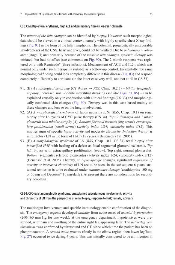
The diagnostic problems could and cannot be missed Initially there were nodules in the left lung (Fig 36a) in 2004 which were highly suspicious of a tumor or metastatic mass The morphology ndash ldquopulmonary abscesses consistent with rheumatoid nodules with vascu-litis in the marginal zonerdquo ndash was in fact surprising (the first morphological finding was non-specific) but easily explained in the context of seropositive RA and peripheral rheu-matoid nodules (Fig 35) The diagnosis of bone TB can be established from the remark-able changes in the tibia (Fig 36b) in addition there were the bone biopsy findings at this point (granulomatous inflammation with necrosis) leading to the conclusion of TB This second disease could quite clearly even rule out the use of the possible TNF blockers Another surprise was the negative outcome of in vivo and in vitro (QuantiFERONreg TB) TB specimens which actually questioned the history of the TB Incidentally on
492 Explanations of Figures and Case Reports with Individual Therapeutic Options
morphological examination of this finding this granulomatous tissue was interpreted as being different from TB ndash in fact it was associated with the rheumatoid nodules (see refer-ence finding CS 13 Chap 2) The third surprise emerged from the latest radiomorphology of the lungs in 2007 (Figs 38 39) The three major causes for several foci including one cavern are to be considered ndash namely the rheumatoid nodules (a priority for me in light of the good health of the patient) TB and ultimately the tumors (chain-smoker) When fur-ther invasive diagnostics were refused by the patient combination therapy (Aravareg 20 mgday with Enbrelreg 50 mgweek) was initiated and produced a good response in the joints after a further 3 months Over the course pulmonary infestation in the form of acute pneumothorax developed as a complication and necessitated mechanical ventilation
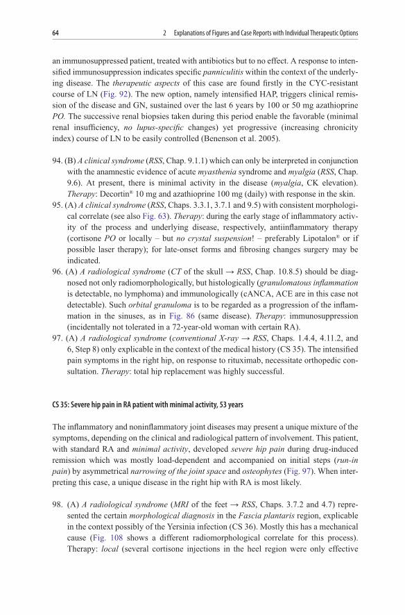
40 (C) A disease (RSS Chap 131 see also Fig 49a) based on the pattern of involve-ment Therapy immunosuppression (cortisone with MTX and possible biologics in this case Enbrelreg currently approved for early forms of this disease) as early as possible in order to achieve remission as applicable
41 (B) A clinical syndrome (RSS Chaps 131 and 463) in conjunction with CS 14 disease-specific Therapy immunosuppression (MTX with or without cortisone) as early as possible it was possible to use a biologic as part of a study
CS 14 Acute swelling of toe 5 on the left and emergency synovectomy with a suspected tumor female 52 years
This case should demonstrate the not untypical difficulties of early diagnosis of a classic disease Instead of instigating immunological examinations synovectomy of MTP 5 left was carried out upon suspicion of neoplasia (this is how severe arthritis see Figs 41 65 looks) Once the arthritis had spread especially symmetrically (Fig 41) rheumatological examina-tion ensued About synovectomy In the case of definable earlier RA with (a) symmetrical involvement of a knee (but not the small joints) such surgery could even be curative
42 (C) A disease (RSS Chaps 133 and 461) in light of the pattern of involvement the late sequelae or damage from a medical history spanning more than 20 years without basic therapy are visible (cf early forms of the same disease Figs 28 40) Telescopic finger (shortened slack and without function) Therapy all therapies (currently taking only Decortinreg 5 mgday) were refused by this patient due to potential side effects (compliance difficulties) Optimization of systemic (at the sign of activity possibly MTX with biologics Enbrelreg is approved without MTX) and local (cortisone injec-tions surgery due to risk of tendon rupture orthesis possibly TEP of PIP MCP MTP see Fig 9b) therapies should be considered however
43 (C) A disease (RSS Chaps 133 and 461) on account of the pattern of involvement (same patient as in Fig 42) here the late sequelae (fixed malpositioning of the toe joints) in the feet are visible (cf early forms of the same disease Figs 41 65) Therapy only orthopedic care as necessary
44 (A) A radiological syndrome (conventional X-ray of sacroiliac joints rarr RSS Chap 68) produced a certain morphological diagnosis of sacroiliac joints involvement
50 2 Explanations of Figures and Case Reports with Individual Therapeutic Options
bilaterally (RSS Chap 223) The nosological entity is to be considered in context with CS 15 and the radiological procedures (Figs 45 and 46) On account of this picture further diagnostics and antiinflammatory therapy (local essential possibly systemic) are indicated
CS 15 Asymmetrical pain and imaging of sacroiliac joints in HLA-B27-positive male patient 42 years
The criterium for radiology was the inflammatory back pain (RSS Chap 215) on initial diagnosis of AS (RSS Chap 22) Thereby the methods must be expected to provide both possibilities and limitations Several methods can reveal the various clinical aspects of back pain namely the presence of involvement (X-ray and bone scintigram Fig 44 show inflammatory or degenerative backgrounds) activity (Fig 45) and damage (Fig 46) which can be laterally different Depending on the issue at hand the relevant methods should be employed in a particular sequence Such methods are to be regarded as the starting points for optimal therapy initially local CT-controlled cortisone injection into the right sacro-iliac joints joint where the pain and activity are located then left where the erosions or damage are more to be found Such therapy was initially successful so that the need for biologics is not given at present (according to guidelines)
45 (A) A radiological syndrome (CT of sacroiliac joints rarr RSS Chap 6 Step 8) in the same patient with left-sided changes bilateral sacroiliitis with erosions and subchon-dral sclerosis emphasized on the left ldquocolorful picturerdquo but with acute inflammatory extensive corticalis destruction on the right
46 (A) A radiological syndrome (MRI of sacroiliac joints rarr RSS Chap 6 Step 8) in the same patient revealed more right-sided changes (bone edema in the right and increased sclerosis of the left sacroiliac joint) confirming the diagnosis of sacroiliitis (RSS Chap 223) with therapeutic consequences (see above)
47 (B) A clinical syndrome (RSS Chaps 912 923) even in conjunction with CS 16 difficult to assign causally during the short period of observation (3 months) Most such cases involve non-differentiated vasculitis associated with CTD most likely SSc or Buergerrsquos disease Therapy immunosuppression (high doses of cortisone with eg MTX SC) and vasodilation (Prostavasinreg Iloprost Bosentan)
CS 16 Acute acral gangrene high-titer ANA with no organ involvement 36-year-old female
In this case the main difficulty was finding the cause of the acute acral gangrene Initially this process is to be assessed as a true Raynaudrsquos phenomenon where the other ndash but not all (eg digits 4ndash5 not affected) ndash vessels of the hands (some with minor gangrene others with cyanosis) are affected Skin-muscle biopsy from the same side of the upper arm produced unremarkable findings Thereby there was no clinical involvement of the feet Another pattern of circulatory disorders in the fingers can be seen from Fig 14 No organ (endocarditis should be ruled out due to the risk of embolism) or connective tissue involve-ment could be ascertained Some evidence of such is to be seen in the capillaroscopic
512 Explanations of Figures and Case Reports with Individual Therapeutic Options
pattern (similar to SSc) and the high ANA titers but with no differentiation ruling out primary Raynaudrsquos phenomenon The slight CRP increase is attributable to the existing gangrene In men consideration is to be given more to Buergerrsquos disease which in this case is also relevant Another rare cause for such digital gangrene (APS cryoglobulinemia or other forms of vasculitis cholesterol embolism) was ruled out clinically immunologi-cally morphologically and angiographically The course could be stabilized by 2 weeks of therapy with Decortinreg and MTX as well as Prostavasinreg infusions The underlying diag-nosis was unclear during the follow-up period (6 months) Most likely this case is in my opinion the initial manifestation of SSc
48a (A) A radiological syndrome (MRI of thoracic spine before therapy (RSS Chap 222) Irregular contouring of the base and top plates with an indication of erosive character clear edematous zone in the vertebral body and barrier disruption of the vertebral disc or reduction in the intervertebral space and edematous signal changes in the medullary cavity At the same time the base and top plates are poorly outlined Such changes can be clarified causally only in the context of CS 17 The same changes developed after a 2-week break during years of maintenance therapy (CS 17 Chap 112)
48b (A) A radiological syndrome MRI-confirmed syndrome (after 3 months of therapy see CS 17) the patient had no symptoms clinical findings correlated with positive dynamics of the MRI changes in T67 and T89
CS 17 Localized pain small gibbus in thoracic spine and CRP elevation in HLA-B27-negative female 50 years
A deciding factor in the local diagnosis of the five years of medical history is the MRI finding (Fig 48a) The most common cause for this are infections (TB among others) and tumors They can be ruled out anamnestically for certain Indicative to the causal diagno-sis of such relatively rare but prognostically relevant local thorax spine changes was the sacroiliac jontsrsquos finding (as with CS 63 with isolierte arthritis of C1-C2 initially of unknown origin) in a similar way to Fig 124 The second feature of such a case is its dependency on the strength of immunosuppression only very intensive and sustained immunosuppression with cortisone was effective During a therapeutic interval of 2 weeks the same MRI changes developed as in Fig 48a and at the same time there was a dramatic rise in CRP (from normal to times 15) and moderate back pain (40 mm VAS) Therapy with Enbrelreg 50 mg and Decortinreg 40 mg left the patient free of symptoms in 3 days with nor-mal CRP Maintenance therapy (Decortinreg 5 mg and Enbrelreg 25 mg in 3 weeks) was initi-ated The further reduction of such therapy is noticeable clinically (from the mild back pain and rise in ESR and fibrinogen) hence treatment is continued
49a (C) A disease (RSS Chap 131 see also Fig 40) on account of the pattern of involve-ment pictured on a bone scan (Fig 49b) The highly active anti-CCP-positive female patient with a 2-year medical history did not respond to several basic medications (MTX Aravareg also in combination) biologics (Enbrelreg Rituximab RoActembrareg)
52 2 Explanations of Figures and Case Reports with Individual Therapeutic Options
and RSO high-dose cortisone is needed At present Remicadereg is used with uncon-vincing efficacy
49b (B) A radiological syndrome (bone scan rarr RSS Chap 6 Step 8) is in the same patient as Fig 49a Without clinical findings it is hardly possible to draw any diagnos-tic conclusions (could also be PsA) initially showing accumulation in the early phase as an expression of exudative inflammation (RSS Chap 131) secondly a pattern of involvement indicative to the distinctions between the various joint diseases and of the introduction of systemic andor local therapy
50 (A) A radiological syndrome (MRI of the skull) extensive demyelination zones bilat-erally parietooccipital with space-occupying nature and smaller peripheral areas in the left frontal region In relation to the medical history (CS 18) such CNS involve-ment is consistent with an immunological disease and also serves as a follow-up control (demyelination of cortical structures) Therapy relatively mild immunosup-pression (17-year-old female) with Imurekreg (100 mg daily) stabilized the disease quite rapidly but did not prevent the occasional mild epidose
CS 18 Recurrent neurological and visual deficits (since the age of 13) 3-year treatment of multiple sclerosis (MS) high-titer ANA CNS involvement stabilized with Imurekreg therapy female patient 17 years
This case of a 13-year-old girl is exquisite and is characterized by the episodic occurrence of CNS involvement with optic neuritis and transverse myelitis (all of which improved well with glucocorticoids) The case represents the syndromal similarity of a disease which is easily definable using immunological examinations (RSS Chap 111) on the one hand and MS on the basis of criteria on the other MRI was performed upon diagnosis of known MS extensive demyelination zones bilaterally parietooccipital with space-occupying nature and smaller peripheral areas in the left frontal region For 3 years these diseases could not be differentiated either neurologically or radiologically and were ultimately controllable only with high-dose cortisone (interferon was ineffective) This response to cortisone as well as the immunological (known since 2004) and histological (2006) aspects are to be regarded in discriminatory terms with a view to MS Consequently present therapy with Imurekreg 150 mg and Decortinreg 25 mgday appears to effect clinical remission with rare episodes
51 (C) A radiological syndrome (conventional X-ray rarr RSS Chaps 133 134 and 6 Step 8) revealed partially bony fusion of the carpalia massive cysts and usures (stage IV) also in a visually normal wrist (Fig 62) Bone destruction and erosion are a com-ponent of many types of articular diseases (see Figs 8 9a 23 59 130b) and can be differentiated only in conjunction with the medical history and first and foremost the pattern of involvement
52 (B) A clinical syndrome (RSS Chaps 914 and 923) easy to define in conjunction with CS 19 and Fig 53 Therapy (during the last 2 years) daily CYC 150 mg Decortinreg 10 mg Trentalreg 400 mg times 2 ASA 100 mg ACE inhibitors No episodes full regres-sion of sclerodactyly and sclerodermia on the thoracic wall
53 (B) A radiological syndrome (CT lungs rarr RSS Chap 1023) finely streakedreticular pattern in the sense of inflammatory fibrosing alveolitis (the finding on the left can be
532 Explanations of Figures and Case Reports with Individual Therapeutic Options
described as pneumonitis or pulmonary fibrosis) CT is the method of choice for inter-stitial diseases This syndrome can be clarified in conjunction with the medical history (CS 19 Fig 52 see also Figs 79 85 91) and is highly significant to therapy (see above) and prognosis
CS 19 Acute respiratory distress pleural effusions and cold hands male 58 years
This disease which is easy to define clinically and immunologically should on the one hand show the association between the acute onset with multiorgan involvement and on the other hand the unfavorable course Nevertheless therapy (systematic immunosuppres-sion with CYC and cortisone for 2 years) positively influenced the regression of sclerodac-tyly and sclerodermia on the trunk and the pulmonary fibrosis without the occurrence of any clinically relevant adverse effects
54 (A) A radiological syndrome (MRI of right ankle rarr RSS Chap 49) showing an extensive osteolytic process approx 3 cm distal to tip of fibula right resulting in a pathological fracture The 75-year-old female patient had suffered 5 months of pain-ful swelling in the lower leg (as formulated in the referral diagnosis) Extensive diagnostics (blood count immunology infectious serology conventional X-ray) produced no findings MRI performed by a practitioner (I was asked by radiologists to assess the usefulness of such a procedure) produced the suspicious malignant find-ing (bone tumor osteolytic metastasis with pathological fracture) Check-up revealed no signs of primary tumor metastasis or osteomyelitis (also possible radiomorpho-logically) The diagnosis of a stress fracture emerged during the surgery
55 (A) A clinical syndrome (RSS Chaps 912 and 922 see also Figs 71 101a) as if punched out Ulcera cruris in a patient with RA (could also be the case with CTD or vasculitis) Thereby massive extra-articular disease activity (RSS Chap 26) is to be expected Therapy systemic immunosuppression (see CS 11 24)
56ab (B) A clinical syndrome (RSS Chaps 911 and 921) only assignable precisely in context with CS 20 has the key role in the assessment of the activity of the immu-nological disease including renal involvement Therapy with existing HCV infec-tion and renal involvement there was a lack of efficacy with MTX and azathioprine and the patient could not tolerate CYC initially responded well to MMF (CellCeptreg 1500-2000 mgday with Decortinreg 75ndash10 mgday) In the last 2 years the course has been recurrent (skin changes abdominal pain ldquoblack stoolsrdquo anemia) neces-sitating several emergency hospital admissions with high-dose cortisone treatment
CS 20 Palpable purpura abdominal pain and renal insufficiency active hepatitis C female 50 years
This case should demonstrate that the rheumatological aspects (Figs 56a b) of the under-lying disease (in this case hepatitis C) could be predominant Such skin changes (palpable purpura of necrotizing venulitis) are associated clinically with the current renal involvement
54 2 Explanations of Figures and Case Reports with Individual Therapeutic Options
(stable hematuria mild albuminuria and elevated creatinine) and recurrent abdominal pain The cryoglobulinemia (mixed type 2) induced by the active hepatitis C caused the systemic affectation of small vessels dermatohistologically leukocytoclastic vasculitis At this time there is no evidence of a progression to lymphoproliferative disease (in type 2 roughly 15 of patients) It is best to instigate therapy for the underlying disease (specific therapy with interferon and Ribavarinreg was not effective and poorly tolerated) At the same time immunosuppression was administered (daily Decortinreg 10 mg and Cellceptreg 2000 mg) which proved to be ineffective (as above) In this refractory case therapy with 500 mg rituximab weekly (four times) was undertaken After the second infusion there was a recurrence of the skin changes The immediate effect was revealed after the fourth infusion (all were tolerated well) ie a symptom-free condition under reduction of Decortinreg to 5 mg and CellCeptreg to 1000 mg daily Clinical remission was sustained for the following 18 months with such therapy
57 (B) A clinical syndrome of ocular involvement (RSS Chap 1081) such cases should be clarified and managed together with an ophthalmologist In conjunction with the clinical findings (patient has similar changes to those in Fig 44 and HLA-B27 antigen) the causes mostly were easily identifiable Therapy ciclosporin 150 mgday Ultralanreg 10 mgday Humirareg 40 mg at intervals of 2 weeks careful mon-itoring of concomitant diseases (obesity class III arterial hypertension poorly controlled diabetes mellitus II drug-toxic liver damage) Ophthalmological exami-nation detected relatively rapid (within 6 weeks) positive dynamics of ocular involvement and vision At the same time there was an increase in serum creatinine (140 mgdl) After a break CellCeptreg was administered with renal values normal thereafter
58 (B) A clinical syndrome (RSS Chaps 132 and 153) namely MCP 2 involvement could be interpreted only in the context of the clinical findings (polyarthritis anti-CCP positivity) and radiomorphology (Fig 59 see CS 50 with identical involvement and different diagnosis) Therapy MTX and a biologic (Cimziaregcertolizumab as part of a study) resulted in remission in line with the criteria with the exception of the affected joint (Fig 59) Consequently several cortisone injections and ultimately RSO were administered with short-term regression of the local clinical activity
59 (B) A radiological syndrome (conventional X-ray rarr RSS Chap 134) revealing selective destruction of the right MCP 2 and a cyst in MCP 4 of this female patient (see Fig 58) Refer to the comments on CS 50
60 (C) A radiological syndrome (conventional X-rays of C1-C2) with absolute disease specificity (RSS Chap 222) fresh syndesmophyte left) in this case the early disease was documented Therapy according to disease activity
61 (C) A radiological syndrome (aortoangiogram rarr RSS Chaps 922 926 927 and 134) The pictured morphological diagnosis can be confirmed by the clinical data (CS 21) Therapy angiological treatment (3 stents inserted into A brachiocephalica A carotis and A iliaca) and immunosuppression (azathioprine then MTX with Decortinreg then Aravareg) The course was stable with no clinically relevant circulatory disorders
552 Explanations of Figures and Case Reports with Individual Therapeutic Options
CS 21 No pulse on one side (since the age of 20) cardiac arrhythmias and two acute myocardial infarctions female 36 years
Many years of disease with varied angiologicalcardiological symptoms not initially regarded as relevant in this young woman Only after two myocardial infarctions in quick succession was a rheumatic disease first postulated Angiological (4 stent implants into the coronary (1) supraaortic (2) vessels and A iliaca (1)) and immunosuppressive (MTX azathioprine cortisone) therapies enabled stabilization of the course apart from (a) the extremely painful red and swollen scar (post thoracotomy synchondrosis perichondritis manubriosternalis RSS Chaps 3117 and 424 suspected) and (b) the left thoracic pain during the last 4 years which increased with exercise These two syndromes are com-pletely reversible with Aravareg therapy
62 (C) A clinical syndrome (RSS Chap 133 see also Figs 35 42 80 129c where partly similar but less pronounced changes are to be seen) which is virtually disease specific button-hole deformity digits 2ndash5 left and 5 right flexion contractures in the distal interphalangeal joints 2ndash4 right so-called claw hand which here is arthrogenically fixed in Fig 18 dermatogenic in Fig 19 neurogenic in Fig 130a osteoarthritis-induced The hands in Fig 62 are a diagnosis particularly in conjunction with radiomorphology (Fig 51) and clinical findings (more than 20 years of symmetrical polyarthritis with restrictions to passive movement (why) of the fingers and wrists patient recently arrived from abroad) At the same time the patient currently has stable CRP elevations (times4ndash6) and therefore needs a more than moderate level of immuno-suppression (cortisone MTX + Aravareg if there is no response biologics should be used) despite stage IV disease
63 (A) A clinical syndrome (RSS Chap 331) with a clear morphological correlate (see Fig 95) associated with arthritis of PIP 3 (as in Fig 68) whereby two articular dis-eases are possible (serological diagnostics indicative namely anti-CCP positivity) Therapy basic therapy (Decortinreg 5 mgday and MTX 15 mgweek) were first intensi-fied (Decortinreg 30 mgday + Remicadereg N3) but unsuccessful suggesting that ortho-pedic measures would be required
64 (B) A radiological syndrome (bone scan rarr RSS Chaps 131 and 6 Step 8) revealing the activity of the disease (differing intensity in early-phase enhancement) and pattern of involvement (ldquohot-spotrdquo affectation both DIP and PIP joints to be seen in left dig 3) hence both RA and PsA would be possible Clinically these two diseases are mostly easily distinguishable Therapy according to nature and activity of disease
65a (B) A clinical syndrome causally identifiable only in the context of the clinical find-ings (RSS Chap 463) and imaging (RSS Chap 6 Step 8) (see Figs 15a 41) This case involves a young man with recurrent Acne vulgaris and Tietze syndrome (as in Fig 69) The dactylitis visible in Figs 65a b would fit with acne-induced arthropathy (roughly the same as SAPHO syndrome rarr RSS Chap 421 but no psoriasis) Therapy systemic (cortisone and MTX) response was registered
65b (B) A radiological syndrome (bone scan in the early phase rarr RSS Chaps 131 and 6 Step 8) confirms the dactylitis (rarrRSS Chap 463)
56 2 Explanations of Figures and Case Reports with Individual Therapeutic Options
66 (B) A clinical syndrome (RSS Chap 462 see also Figs 7 and 115) cannot in fact be clarified by the clinical findings (CS 22) no morphological correlate was found The good response maintained with cortisone (Fig 67) suggests that there is an (auto) immunological cause or cortisone-dependent disease (RSS Chap 133) most likely associated with vasculitis Therapy cortisone pulse therapy + MTX 15 mgweek with hospitalized treatment for the flares
67 (B) A clinical syndrome (see Fig 66) following cortisone pulse therapy
CS 22 Puffy hands and high CRP of unclear etiology in 17-year-old trainee painter
This case (Fig 66) demonstrated the difficulties of morphological diagnosis in rheumatology (what should be biopsied here) It is certainly an inflammatoryrheumatic disease namely acute swelling in both hands (see also Fig 7) massive CRP increase good response to corti-sone from the first episode (Fig 67) with sustained remission also during the second episode and identical therapy No organ involvement non-specific immunology Acute Sudeckrsquos syn-drome seems unlikely on account of the symmetrical involvement Remitting seronegative symmetrical Synovitis with Pitting Edema (RS3PE syndrome) also seems unlikely because of the missing pattern of arthritides young age of the patient and extremely high CRP
Vasculitis of the small vessels cannot be confirmed morphologically or angiologically An allergic process during painting work is not really consistent with elevated CRP The fact remains that the dramatic disease or condition was healed without having secured a diagnosis
68 (B) A clinical syndrome (RSS Chap 131 see also Figs 3 and 64 a radiomorphologi-cal correlate of such changes) It cannot be clearly explained in conjunction with the clinical findings (CS 23) This case is more likely the joint disease associated with HIV (RSS Chap 1243 see Fig 88) Therapy cortisone and MTX under CD4-level control produced a clear improvement
69 (B) A clinical syndrome (RSS Chap 421) Only in conjunction with CS 23 and Fig 68 can HIV-associated joint disease be postulated based on the almost specific pattern of joint involvement (RSS Chap 1243) Therapy (see comment to Fig 68)
CS 23 Acute arthritis and Tietze syndrome with HIV female 36 years
The rheumatological question was whether this illness (whereby HIV is not implied) can be attributed to an inflammatory rheumatic disease Most certainly is on account of the stable florid arthritis with extremely high CRP Which inflammatory and rheumatic dis-ease with such a pattern of involvement in HIV (the disease in Fig 84b) is involved Other rheumatic diseases can almost certainly be ruled out on the basis of the immunological tests performed The necessary and effective immunosuppression was administered in collaboration with infectiologists while carefully monitoring CD4
70 (B) A clinical syndrome (RSS Chap 39 461 and 934) described as Jaccoudrsquos arthropathy Only by taking an X-ray of the hands (Fig 72) and considering CS 24
572 Explanations of Figures and Case Reports with Individual Therapeutic Options
can such changes be correctly interpreted Therapy systemic namely several years of Decortinreg 5 mgday + MTX 10ndash15 mgweek
71 (B) A clinical syndrome (RSS Chaps 321 912 921 and 941) in the same female patient could only be interpreted on the basis of morphology and in the context of CS 24 An attempt was first made to consider this condition as an independent disease ndash erysipelas Therapy antibiotics for 3 weeks but with no success intensified immuno-suppression (Decortinreg 30ndash20 mgday + MTX 15 mgweek) produced a regression in the changes in 2 weeks and they were later (Fig 73) totally reversible
72 (B) A radiological syndrome (conventional X-ray rarr RSS Chaps 134 6 Step 8 and 139) indicating the characteristics of deformed hands (Fig 70) should be considered in conjunction with the medical history (CS 24)
73 (B) A clinical syndrome (RSS Chaps 321 and 941) showing the regressiveness of the changes (cf Fig 71) after cortisone therapy This conveys an important generic term in rheumatology namely cortisone dependency (RSS Chap 13 Step 3) which is to be regarded as an important criterium but of no nosological specificity for the pres-ence of an inflammatory rheumatic disease
CS 24 Deformities of the hands myalgia pacemaker red feet with ulceration female 52 years (some episodes during 12-year follow-up period)
Over the course of this disease (which) definable on the basis of clinical findings and immunological tests various clinical patterns emerge which at times can be interpreted as independent diseases eg are the problems with the hands related to RA (Fig 70 ulnar deviation contractures tendinitis rarr RSS Chaps 132 33 and 39) This should be strictly contradicted on the basis of a lack of erosions (Fig 72) but also RF and anti-CCP-Ab The other manifestation of the changes depicted in Fig 71 (RSS Chaps 321 922 and 941) was initially interpreted as erysipelas But there was no fever with chills no migratory ery-thema and no response to antibiotics - which thus contradict this Such changes are cer-tainly among the extra-articular symptoms of the disease The response to intensified immunosuppression (Fig 73) is indicative of the immunological basis of such changes
74 (C) A radiological syndrome (PET 2 positions rarr RSS Chaps 81 922 and 927 cf Fig 77b) assigned to fever of unknown origin and suspected inflammatory changes in the vessels (in vasculitis of the large vessels) and perivascular (in Ormondrsquos dis-ease) structures if other methods (RSS Chap 13 Step 9) furnished no evidence Causes can only be interpreted in conjunction with CS 25 Therapy relatively strong immunosuppression (moderate doses of cortisone and eg azathioprine or MMF) in this case brought satisfactory results
CS 25 Fever weight loss and increased inflammatory symptoms of initially unexplained origin in a 50-year-old female
This case presents the diagnostic problems and possibilities of causal investigation into months of unexplained B symptoms with fever elevated CRP and ESR weight loss
58 2 Explanations of Figures and Case Reports with Individual Therapeutic Options
especially in young women This PET method should come last in the sequence of staged diagnostics (possibly after ultrasound CT or MRI) for suspected vasculitis of the large vessels or Ormondrsquos disease Therapy immunosuppression with Decortinreg (40 mgday) and azathioprine (150 mgday) After a few days the remarkable symptoms were regres-sive and remained so long-term
75 (B) A radiological syndrome (chest X-ray rarr RSS Chap 102 cf Fig 79) multiple nodular pulmonary infiltrations with caverns and bronchoectasis fibrosis - causally explicable only in the context of the medical history (patient suffered for approx 10 years from cANCA-positive WG with involvement of the lungs) Therapy lung transplanta-tion was refused here on account of the advanced pulmonary and cardiac insufficiency (picture taken shortly before the fatal outcome) In other cases with pulmonary involve-ment more or less strong immunosuppression should be instigated depending on the entity activity organ involvement and concurrent diseases and conditions
76 (B) A clinical syndrome (RSS Chap 915) only to be interpreted in association with other syndromes and constellations (CS 26) Therapy immunosuppression colchicine sustained response generally to cortisone pulse therapy (Decortinreg 250 mg IV N3)
CS 26 Mouth ulcers heart attack severely increased ESR and CRP female 73 years
These common problems (acute myocardial infarction in a 73-year-old woman otherwise no risk factors) have remarkable clinical aspects namely known MB with recurrent mouth ulcers extremely elevated ESR and CRP during and shortly before the event Such constellations are suggestive of vasculitis of the large and medium (coronary) vessels possible in association with MB Therapy cortisone pulse therapy produced a stable response (no vascular symp-toms no increase in CRP or ESR) No further diagnostics were undertaken therefore It must be noted that continued elevation of CRP which is regarded as a significant trigger for athero-sclerosis (RSS Chap 1242) markedly increases the rate of myocardial infarction
77a (C) A radiological syndrome (MRI angiogram rarr RSS Chaps 922 and 13 Step 6) Irregularities of the external aortic wall and A iliaca bilaterally Lumen fluctuations of A iliaca communis bilaterally but somewhat greater on the right than the left but with no higher stages of stenosis Clinically there was severe back pain initially treated intensively (reportedly 60 injections in one month) as well as episodic fever and abdominal pain A temporalis biopsy (before therapy) was normal Such symp-toms are dependent upon the strength of immunosuppression No other large vessels affected Therapy relatively strong and systematic (for 5 years) immunosuppression (MTX 10 mgweek Aravareg 20 mgday Decortinreg 15 mgday) The thickness of the aortic wall (on MRI and ultrasound) could be reduced over the course Nevertheless intermittent abdominal pain and episodes of fever remain
77b (C) A radiological syndrome (PET rarr RSS Chaps 81 922 and 927 cf Fig 74) in the same patient (see Fig 77a) band-shaped enhancement in abdominal aorta ndash (level approx L 23) as with minimally active aortitis which is relatively rare (approx 10ndash15 cases in such a rare disease) particularly in men (cf Fig 61 CS 21)
592 Explanations of Figures and Case Reports with Individual Therapeutic Options
78 (A) A clinical syndrome rarr RSS Chaps 911 and 10102 cf Fig 33 where are the differences To be causally explained only in context (no organ involvement specific Ab spectrum cortisone dependency cf CS 27) Therapy immunosuppression (more with induction therapy less with maintenance therapy) Daily over the previous months Decortinreg 15ndash30 mg and CellCeptreg 2000-3000 mg which are resulting in a serious urinary tract infection Cellceptreg was stopped for 1 month and then reissued The above mentioned therapy at high doses was ineffective (platelet count was below 30000ndash50000ml whereupon rituximab (1000 mg in two infusions) was prescribed
CS 27 Skin and mucous membrane bleeding thrombocytopenia and high positive anti-dsDNA with no organ involvement female 54 years
The problem here is the causal classification of the selective thrombocytopenia with anti-thrombocytic Ab Indicative of such are the high ANA and dsDNA-Ab titers with positive Crithidia test In the event of disease with such specific serology rarr RSS Chap 111) intensified immunosuppression (cortisone and CYC pulse therapy azathioprine) should first be attempted in accordance with current guidelines Over the course following sple-nectomy the clinical findings under cortisone (Decortinreg 10 mg and azathioprine 150 mg daily) during the 8 years of follow-up did not remain entirely stable fluctuating thrombo-cytopenia dependent on the cortisone doses concurrent basic therapy and infections especially after CellCeptreg as is well known As a result close monitoring of organ involve-ment and platelet count as well as the potential complications of immunosuppression (one of which was described above) is essential In recent months by increasing the doses of Decortinreg (20 mgday) and Myforticreg 360 mg (4 tabsday) to way beyond those which are safe clinically relevant thrombocytopenia (below the critical count) resulted thereby necessitating introduction of label of use therapy with rituximab 1000 mg (two infusions at 2-weekly intervals) which has recently been positively evaluated in the literature Following such therapy which was tolerated well an improvement appeared to ensue and there was a rise in the platelet count up to 76000ml despite the reduction in maintenance therapy (Decortinreg to 75 mg and Myforticreg to 720 mg daily) Clinical remission was seen from such therapy during the following 18 months
79 (B) A radiological syndrome (CT thorax rarr RSS Chaps 1022 and 1023) Honeycomb lung massive multiple cystic changes confluent focal shadowing mainly in the upper lobe interstitial shadow pattern left pleural effusion massive bihilar lymphadenopa-thy The case involves a 70-year-old male patient known to have had respiratory distress for about 15 years and exophthalmos (until recently evidently of unknown etiology rarr RSS Chap 1085 though it belongs to the underlying disease) Histologically (lung 12 years ago) chronic granulomatous inflammation was estab-lished (RSS Chaps 1022 1023 and 116) and with ongoing cortisone therapy the patient was able to work until recently Rapid deterioration in general health and pulmonary function were ascertained on discontinuation of cortisone (chest X-ray taken shortly before death) The cortisone (Decortinreg 1000 mg ndash 5 days) and CYC (1000 mg ndash 3 days) pulse therapy was ineffective
60 2 Explanations of Figures and Case Reports with Individual Therapeutic Options
80 (C) A disease (RSS Chap 13 see Figs 3 5 35 42 62 104 129c) Can the differ-ences to Figs 4 13 81 89 where the other diseases was illustrated be seen Therapy mostly immunosuppression
81 (A) A clinical syndrome (RSS Chap 142) definable in conjunction with CS 28 (see Fig 80 where are the differences or Fig 4 where similar changes can be seen) Therapy prescription of MTX should not be deemed appropriate local therapy is to be applied (cortisone injections RSO or Dolobene Gelreg)
CS 28 Arthritis of the finger joints rheumatoid factors and appropriate () MTX therapy 64-year-old male
The patient was referred for continuation of basic therapy in existing RA The distinction from inflammatory osteoarthritis (OA) was the main problem here as well as generally in arthrol-ogy In terms of the pattern of involvement (MCP joints not affected see Figs 8 80 128a b) the arthritis of the individual PIP joints (Bouchardrsquos OA) is to be interpreted in conjunction with Heberdenrsquos OA clearly as OA of the fingers The patient does not need MTX
82 (B) A clinical syndrome (RSS Chap 916) definable as a sign of vasculitis of the small vessels only in conjunction with the clinical findings (CS 29) Therapy after the effective immunosuppression (see below) these changes disappeared completely
CS 29 Alopecia acute abdominal pain and massive proteinuria 27-year-old male
This case initially showed over the course peculiar clinical masks of a disease Acute onset of systemic organ involvement characterized mostly by an unfavorable course In such a case the non-typical symptoms predominated severe abdominal pain (due to pseudoileus on account of lymphadenopathy another time due to renal vein thromboses) severe headaches (due to cerebral edema) and edema of the legs (due to marked nephrotic syndrome) Therapy Cellceptreg 2000 mg and ciclosporin 50 mg acute 2 CYC azathioprine 4 acute rituximab 1000 mg The nephrotic syndrome proved resistant to all basic therapy hence autologous cell transplantation has been carried out successfully
83 (B) A clinical syndrome (RSS Chaps 914 and 941) in a male patient with tattoos can be considered only morphologically (in this case granulomatous inflammation large nodular-type Lupus pernio compared with small nodular disseminated type Fig 90) and radiologically (here bihilar lymphadenopathy was found but only on CT) Such a constellation is typical for sarcoidosis but also for lymphoma hence these diseases are hardly distinguishable The lack of B symptoms and only hilar involve-ment are more indicative of sarcoidosis despite the negative finding of ACE Such changes should be differentiated from pigment granuloma (the deciding factor thereby is the pulmonary involvement) Therapy due to the massive proliferation of skin changes cortisone therapy was instigated The skin changes and hilar lymphoma were reversible (except for the slight growth at the location of the Fig)
612 Explanations of Figures and Case Reports with Individual Therapeutic Options
84ab (C) A clinical syndrome (RSS Chap 911) which is of great diversity (a ndash Psoriasis hyperkeratotica and b ndash Psoriasis vulgaris) with great specificity and clinical sig-nificance on investigation of the joint and back pain Should also be checked as a potential occurrence in relatives (has the same indicative clinical value) Induration of the skin is similar to leukemic infiltration Therapy in collaboration with derma-tologists particularly for similar (as in Fig 84a) cases
85 (B) A radiological syndrome (CT thorax compare against Fig 53 CS 19) Radiomorphological diagnosis of new onset (when comparing with previous imag-ing from 062007) clearly pronounced mediastinal and hilar lymphoma as well as solid nodule of 25 cm subpleurally in S6 left The finely streaked reticular pattern of the lung parenchyma presented here was known The finding should be investi-gated immediately (see below) and compared against Fig 79
CS 30 Pulmonary fibrosis Raynaudrsquos syndrome Scl-70 positive HAP with response deterioration in general condition and drop in DLCO transfer abnormal thorax CT 66-year-old female
The present primary finding is seen in Fig 85 (see above) The moderate interstitial fibro-sis (increased reticular streaking emphasized hilus region bilaterally) reflects the underly-ing disease Consideration must be given thereby to multiorgan involvement without sclerodactyly and with positive Scl-70 tends to contradict SSc The extent of the pulmo-nary fibrosis is not proportionate to the highly impaired pulmonary function and general health deteriorating in the previous 3ndash4 months The decisive explanation for this was furnished by the perthoracal punch biopsy of the mass on the left namely the proof of small-cell anaplastic carcinoma Metastases were found in the liver Chemotherapy was instigated The decisive question should the entire medical case history be viewed as a paraneoplastic process or are two diseases involved (rarr RSS Chap 5 CS 73 with virtually the same issues as in the patient with WG and colon carcinoma) Thereby the other possible triggers are to be considered ie the systematic azathioprine therapy during the last 4 years
86 (A) A radiological syndrome (MRI of the skull for ENT rarr RSS Chap 1092 follow-ing CYC therapy) an almost circular soft-tissue structure in the left maxillary sinus (T2-T1-weighted hypointense) This is a radiomorphological diagnosis to be inter-preted causally only in conjunction with the medical history (CS 31) the maxillary sinuses were affected (sinusitis) after four cycles of CYC pulse therapy Induction therapy 6 cycles of HAP (Benenson et al 2005)
87 (A) A radiological syndrome (MRI of the skull rarr RSS Chap 1092 after HAP therapy) significant structures of soft-tissue density no longer detectable in sinus region The method would be suitable for follow-up control Maintenance therapy immunosup-pression (Decortinreg 10ndash5 mgday and MTX 10 mgweek) With 2-year clinical remission under the aforementioned therapy an attempt to stop MTX was made Shortly thereafter there was a cerebral insult which cannot be ruled out as the sign of a flare
62 2 Explanations of Figures and Case Reports with Individual Therapeutic Options
CS 31 Multiorgan involvement and MRI of the ENT region under immunosuppression 62-year-old male
This case with multiorgan involvement and pANCA positivity poses few diagnostic dif-ficulties Perhaps there is just one aspect with a view to the activity of the disease during many years of maintenance therapy (with MTX) and justification for such therapy is to be highlighted This question can be answered only by attempting to stop MTX as a detector for the initial activity Stroke after the 5-week break in MTX appears to be causally related to dormant minimal activity For this reason I would in future continue with the immuno-suppressive maintenance therapy in the remission of such (and perhaps other) diseases Another aspect is a new therapeutic option in WG with refractory ENT involvement HAP achieved full regression of the specific mass (Benenson et al 2005)
88 (B) A clinical syndrome (RSS Chap 464) explicable causally only in association with clinical findings The case involves a 29-year-old HIV-positive patient with no cardiac or lung disease no skin changes he was evaluated as one case (CS 23 Figs 68 69)
89 (B) A clinical syndrome (RSS Chaps 14 461 932) behind which are two other systemic diseases which can only be diagnosed by disease-specific immunology (CS 32) Therapy mild immunosuppression (daily Decortinreg 20 mg ndash at present 5 mg + Aravareg 20 mg) consequently stabilized
CS 32 Arthritis sicca syndrome and SS-A CenpB- and anti-CCP antibodies female 67 years
This case in fact involves all 4 rheumatic diseases One is to be seen in the DIP nodules (Fig 89 and radiologically as in Fig 8) The second is noticeable from the MCP swellings (MCP 2-3) and high-titer anti-CCP antibodies The third can be established from the clinical findings (Calcinosis cutis Raynaudrsquos syndrome Teleangiectases swallowing difficulties) and first and foremost from the CenpB-Ab (RSS Chap 111) The fourth can be postulated from the sicca syndrome and possibly SS-A-Ab but viewed as secondary All inflammatory rheumatic diseases have relatively minimal activity at present with no organ involvement and thus are easy to control with mild immunosuppression (Aravareg 20 mg + Decortinreg 5 mgday) The further reduction in such therapy was always associated with deterioration in gen-eral health arthritis and elevated CRP which can be described as drug-induced remission
90 (B) A clinical syndrome (RSS Chap 911) can be clarified nosologically only by the morphology (granulomatous changes small-nodule disseminated type compared with large-nodule type Lupus pernio Fig 83) though in association with the clinical findings chest findings (Fig 91) and lab findings (CS 33) Therapy strong immuno-suppression (due to broad spread of efflorescences and pulmonary involvement) High-dose cortisone therapy (70 mgday for 3 months) was administered by family doctor but without any effect and long-term adverse effects (diabetes mellitus requir-ing treatment) No response (during 2 years) to CYC MTX azathioprine PO and pulse therapy MMF Aravareg also in combination with Humirareg (3 months) Ultimately a clear response was achieved with Remicadereg (three infusions) with full regression of ACE and IL2r lasting no longer than 8 weeks
632 Explanations of Figures and Case Reports with Individual Therapeutic Options
CS 33 Multiple focal erythema high ACE and pulmonary fibrosis 42-year-old male
The nature of the skin changes can be identified by biopsy However such morphological data should be viewed in a clinical context namely with highly specific chest X-ray find-ings (Fig 91) in the form of the hilar lymphoma The potential prognostically unfavorable involvements of the CNS heart and liver could not be verified Due to pulmonary involve-ment (stage II) and primarily because of the massive skin changes systemic therapy was initiated but had no effect (see comments on Fig 90) The 2-month response was regis-tered only with Remicadereg (three infusions) Measurement of ACE and IL2r which was normal only under such therapy is suitable as a follow-up control Incidentally the same morphological finding could look completely different in this disease (Fig 83) and respond completely differently to cortisone (in the latter case very well and not at all in CS 33)
91 (B) A radiological syndrome (CT thorax rarr RSS Chap 1023) ndash bihilar lymphade-nopathy increased small-nodule interstitial streaking (see also Figs 53 85) ndash can be explained causally only in conduction with clinical findings (CS 33) and morphologi-cally confirmed skin changes (Fig 90) Therapy was in this case based mainly on these changes and less so on the lung involvement
92 (A) A morphological syndrome of lupus nephritis LN (RSS Chap 101) on renal biopsy after 16 cycles of CYC pulse therapy (CS 34) Top 2 damaged and 1 intact glomeruli with tubular atrophy (A) Bottom fibrinoid necrosis (big arrow) extracapil-lary proliferation (small arrow) (activity index 924 chronicity index 412) This implies signs of specific lupus activity and moderate chronicity Induction therapy is in refractory LN in the form of HAP (18 cycles) (Benenson et al 2005)
93 (B) A morphological syndrome of LN (RSS Chap 101 CS 34) renal biopsy after intensified HAP with healing of a defect as focal segmental glomerulosclerosis Top left biopsy with extracapillary proliferation (arrow) Top right normal glomerulus Bottom segmental sclerotic glomerulus (activity index 124 chronicity index 812) (Benenson et al 2005) Thereby no lupus-specific changes significant regression of activity or increased chronicity of LN are to be seen In the subsequent 6 years sus-tained remission is to be evaluated under maintenance therapy (azathioprine 100 mg or 50 mg and Decortinreg 10 mgdaily) At present there are no indications for second-ary neoplasia
CS 34 CYC-resistant nephrotic syndrome unexplained subcutaneous involvement activity and chronicity of LN from the perspective of renal biopsy response to HAP female 52 years
The multiorgan involvement and specific immunology enable confirmation of the diagno-sis The emergency aspects developed initially from acute onset of arterial hypertension (260160 mm Hg for one week) at the emergency department hypotensives were pre-scribed with pain and swelling of the entire right leg appearing later The pelvicleg vein thrombosis was confirmed by ultrasound and CT since which time the patient has been on phenprocoumon A second acute process (firstly in the elbow region then lower legfoot Fig 27) occurred twice during 4 years This was initially considered to be an infection in
64 2 Explanations of Figures and Case Reports with Individual Therapeutic Options
an immunosuppressed patient treated with antibiotics but to no effect A response to inten-sified immunosuppression indicates specific panniculitis within the context of the underly-ing disease The therapeutic aspects of this case are found firstly in the CYC-resistant course of LN (Fig 92) The new option namely intensified HAP triggers clinical remis-sion of the disease and GN sustained over the last 6 years by 100 or 50 mg azathioprine PO The successive renal biopsies taken during this period enable the favorable (minimal renal insufficiency no lupus-specific changes) yet progressive (increasing chronicity index) course of LN to be easily controlled (Benenson et al 2005)
94 (B) A clinical syndrome (RSS Chap 911) which can only be interpreted in conjunction with the anamnestic evidence of acute myasthenia syndrome and myalgia (RSS Chap 96) At present there is minimal activity in the disease (myalgia CK elevation) Therapy Decortinreg 10 mg and azathioprine 100 mg (daily) with response in the skin
95 (A) A clinical syndrome (RSS Chaps 331 371 and 95) with consistent morphologi-cal correlate (see also Fig 63) Therapy during the early stage of inflammatory activ-ity of the process and underlying disease respectively antiinflammatory therapy (cortisone PO or locally ndash but no crystal suspension ndash preferably Lipotalonreg or if possible laser therapy) for late-onset forms and fibrosing changes surgery may be indicated
96 (A) A radiological syndrome (CT of the skull rarr RSS Chap 1085) should be diag-nosed not only radiomorphologically but histologically (granulomatous inflammation is detectable no lymphoma) and immunologically (cANCA ACE are in this case not detectable) Such orbital granuloma is to be regarded as a progression of the inflam-mation in the sinuses as in Fig 86 (same disease) Therapy immunosuppression (incidentally not tolerated in a 72-year-old woman with certain RA)
97 (A) A radiological syndrome (conventional X-ray rarr RSS Chaps 144 4112 and 6 Step 8) only explicable in the context of the medical history (CS 35) The intensified pain symptoms in the right hip on response to rituximab necessitate orthopedic con-sultation Therapy total hip replacement was highly successful
CS 35 Severe hip pain in RA patient with minimal activity 53 years
The inflammatory and noninflammatory joint diseases may present a unique mixture of the symptoms depending on the clinical and radiological pattern of involvement This patient with standard RA and minimal activity developed severe hip pain during drug-induced remission which was mostly load-dependent and accompanied on initial steps (run-in pain) by asymmetrical narrowing of the joint space and osteophytes (Fig 97) When inter-preting this case a unique disease in the right hip with RA is most likely
98 (A) A radiological syndrome (MRI of the feet rarr RSS Chaps 372 and 47) repre-sented the certain morphological diagnosis in the Fascia plantaris region explicable in the context possibly of the Yersinia infection (CS 36) Mostly this has a mechanical cause (Fig 108 shows a different radiomorphological correlate for this process) Therapy local (several cortisone injections in the heel region were only effective
652 Explanations of Figures and Case Reports with Individual Therapeutic Options
short-term physiotherapy) or systemic (cortisone biologics would be possible only within the framework of AS)
99 (B) Radiological syndrome (MRI of the feet rarr RSS Chaps 372 and 47 CS 36) with full regression of the changes presented in Fig 98 which should be regarded as a parameter for follow-up control (after laser therapy) Subsequently the patient is free of symptoms (2 years)
CS 36 Refractory heel pain and MRI confirmed response to laser therapy 63-year-old female (Benenson et al 2006)
This is initially a diagnostic problem which can be solved by MRI Then it becomes a causal problem namely with the associations of such changes to the intestine and Yersinia infection possible linked to mechanical causes Resistant heel pain was treated with the new option namely curatively with soft laser therapy (confirmed clinically and by MRI)
100 (A) A clinical syndrome (RSS Chaps 26 and 911) palmar erythema associated with pain in the finger tips in this case in a 36-year-old female patient with RA Therapy these changes were regressive upon intensification of the immunosuppression
101a (B) A clinical syndrome (leg ulcers rarr RSS Chaps 26 and 912 see also Figs 2 30 31 55) which can be explained only in conjunction with the medical history (severe RA long-term cortisone therapy venous insufficiency resistance to TNF blockers and basic medication) Therapy strong immunosuppression (CYC rituximab)
101b (B) A clinical syndrome (RSS Chap 912) in the same patient (after rituximab therapy Dr A Rubbert) The scarring should be considered when making a diagno-sis just as much as the ulcerations (see also Fig 32)
102 (A) A radiological syndrome (MRI of the calcaneus rarr RSS Chaps 332 471 and 95) prior to laser therapy marked plantar fasciitis edema of calcaneus (osteitis) perifascial surrounding of soft tissue and m flexor digitorum brevis (Benenson et al 2006) This is a morphological diagnosis with resistant heel and Achilles ten-don pain Causes can only be interpreted in conjunction with CS 37 Therapy anti-inflammatory systemic andor local therapy (irrespective of cause extent and bone marrow involvement) indicated
103 (B) A radiological syndrome (MRI of the heel after therapy in CS 37 complete regression (restitution) of the changes which were to be seen in Fig 102) used as a radiomorphological parameter for follow-up control (Benenson et al 2006)
CS 37 Recurrent arthritis tendinitis enthesitis associated with Yersinia infection Response to laser therapy 32-year-old female
The clinical aspects of this case documented over 8 years consist of recurrent arthritis of the small joints (MCP MTP and PIP) dactylitis (Digito 2) tenosynovitis enthesitis and plantar fasciitis Only the association with the 3 years of serologically fresh Yersinia infection can be verified thereby Fig 102 shows the tendinitis with bone marrow edema
66 2 Explanations of Figures and Case Reports with Individual Therapeutic Options
around the heel Therapy systemic cortisone boosts and MTX for flares recently Aravareg Laser therapy was used for localized problems (Benenson et al 2006)
104 (C) A disease (RSS Chaps 132 311 and 33 see also Figs 3 35 80 112 122a b 129c) Therapy systemic (RSS Chaps 73 and 123)
105 (A) A radiological syndrome (CT of the abdomen rarr RSS Chaps 101 and 13 Step 9) a morphological diagnosis nephrocalcinosis with unclear clinical significance in a patient with SLE (CS 38) and status post acute renal failure In other cases namely in primary hyperparathyroidism and Sjoumlgrenrsquos syndrome this syndrome is associated with an unfavorable prognosis for the kidneys Clinically the renal problems present as a nephrotic syndrome (RSS Chaps 1012 and 1015) Therapy systemic ie daily Decortinreg (15 mg 5 mg at present) and MMF (CellCeptreg 3000 mg 1000 mg at present) patient currently has no symptoms proteinuria up to 500 mg24 h
CS 38 ANA-positive mastitis resistant nephrotic syndrome nephrocalcinosis questionable APS phenprocoumon therapy iatrogenic renal failure 27-year-old female
This case is of clinical interest first and foremost because of the untypical onset of the disease at the age of 16 with ANA-positive mastitis following exposure to the sun Six years later exudative pleuritis developed pronounced nephrotic syndrome (proteinuria up to 90 g per 24 h) thirdly iatrogenic renal damage resulting in acute renal failure after abdominal hemorrhage which occurred during the unnecessary phenprocoumon therapy Too systematic a therapy with ciclosporin is incidentally known to increase the creati-nine levels Furthermore it is highly likely that diagnosis of APS (due to proof of ACLA) was not standardized and hence the phenprocoumon therapy was stringent I also find the stringent therapy with erythropoietin for Coombs-positive anemia (this substance was too highly quantified in the serum) superfluous (in the absence of renal insufficiency) The positive dynamics of proteinuria were seen during CellCeptreg therapy Thereby mixed proteinuria is to be evaluated as glomerular on the one hand and tubular on the other (RSS Chap 1012) The latter could be connected to nephrocalcinosis (Fig 105) If so limitations to immunosuppressive therapy are possible (in the event of a flare) The switch to rituximab does not at this time seem appropriate due to the clearly reduced proteinuria (to 05 g24 h)
106 (A) A radiological syndrome (MRI of the shoulder rarr RSS Chaps 33 36 and 41) subdeltoid and subcoracoid bursitis in combination with tendinitis and peritendinitis of the supraspinatus tendon small amount of joint effusion (Benenson et al 2006) This is a radiomorphological diagnosis in resistant shoulder pain (CS 39) Therapy local (physiotherapy cortisone injections) was ineffective laser therapy was undertaken
107 (B) A radiological syndrome (MRI of the shoulder CS 39) following laser therapy (Benenson et al 2006) the inflammatory changes in the clinical and MRI findings regressed completely over the long term MRI should be employed in such a condi-tion for follow-up
672 Explanations of Figures and Case Reports with Individual Therapeutic Options
CS 39 Refractory shoulder pain MRI-assured response to laser therapy 61-year-old female
This is a presentation initially of an appropriate radiomorphological procedure for unex-plained shoulder pain Secondly we have a relatively new therapeutic option for a com-mon process ie mechanically induced persistent pain in the shoulder The complex inflammation was treated effectively with laser therapy confirmed clinically and by MRI (Benenson et al 2006)
108 (A) A radiological syndrome (X-ray of the heelrarr RSS Chaps 362 and 471) a radiomorphological diagnosis for fresh not yet calcified osteophytes In this case the patient could hardly walk in other cases the radiological changes are not consistent necessarily with the symptoms of pain Therapy local (physiotherapy cortisone injections radiotherapy laser therapy)
109 (B) A clinical syndrome (RSS Chaps 321 and 941) and clinical diagnosis respec-tively explicable causally only in relation to the medical history (CS 40) Therapy cortisone (Decortinreg 20ndash10 mgday approx 2 weeks) Discontinuation upon regres-sion of the symptoms
CS 40 Attacks of fever with high CRP and Erythema nodosum 26-year-old female
The problem was the causal investigation of the last 16 years of feverish episodes and skin changes (Fig 109) One of the most common causes thereof is sarcoidosis and streptococ-cal infection followed by Yersinia infection which in this case was detected (RSS Chap 1132) The diagnosis certainly could have been ascertained sooner Therapy most effec-tive is cortisone therapy with eg Decortinreg 30 mgday Antibiotic therapy (tetracyclinesquinolones) is recommended only if the infection is fresh (eg positive stool culture)
110 (A) and 111ab (A) A clinical syndrome (RSS Chaps 911 and 913 see also Figs 15a 84a b) developed suddenly with generalization 2 days after Humirareg injection (CS 41) in the extremities and the whole body except the face and particu-larly dense on the hands and soles of the feet in the following sequence first pustules (Fig 111a) after 2ndash3 days massive vasculitic and hemorrhagic (massive on the heels) epidermal patches of necrolysis with Urticaria vasculitis somewhat later psoriasis (Figs 110 and 111b) Therapy prescription by dermatologists of mostly local therapy with good effect
CS 41 Drug-induced generalized polymorphic efflorescences following TNF blockers female doctor 49 years
Such changes (Figs 110 111a b) are to be viewed as drug-induced (RSS Chap 913) they developed simultaneously without prior signs following injection of Humirareg in a patient with psoriatic SpA who was HLA-B27 positive There is a history of regular intake of this medication for 15 years with good response also in the plaques without any
68 2 Explanations of Figures and Case Reports with Individual Therapeutic Options
adverse effects Also remarkable was the extent and the step-wise progression of these changes initially pustulous efflorescences then the vasculitic component and ultimately in those areas the massive superficial plaques Also remarkable was the efficacy of the local therapy prescribed by dermatologists Despite the known associations of such changes with TNF blockers the question must be asked whether the other members of this TNF family should be prescribed upon increased activity of the underlying disease In one almost identical case a patient with AS was effectively treated with Remicadereg for 3 years Thereafter the changes on the hands and soles of the feet developed after the 4th injection when switching to Humirareg Fig 129b Such pustulous keratoderma blenorrhagica (RSS Chaps 911 and 913) could be regarded as another variant of pustulous psoriasis in SpA particularly in ReA or SAPHO syndrome A switch to Remicadereg (together with local therapy) produced a marked improvement in the skin symptoms The known psoriasis-inducing effect of TNF blockers however in the form of Psoriasis vulgaris appears from the last summary (Wollina et al 2008) to be family-specific Recently we have seen such changes using Cimziareg
112 (B) A radiological syndrome (CT thorax rarr RSS Chap 1022) in ANCA-positive female patient 63 years demonstrated a progression in size of bipulmonary and intrapulmonary infiltrates and caverns in the mid lobe clearly connected to the bron-chial system (caverns almost 6 times 5 cm) in the right dorsocaudal lower lobe (cavern approx 43 times 62 cm) The two granulomas in the left lungula were confluent and now measure 43 cm in diameter At the time of examination (after 3rd cycle of CYC bolus therapy) the general condition was seriously deteriorated CRP increased (to factor acute 30) and procalcitonin normal Having ruled out superinfection and neoplasia ritux-imab therapy (two infusions of 1000 mg each) and CellCeptreg 2000 mg per day were initiated (Dr A Rubbert) Complete regression of the aforementioned pulmonary changes ensued in 6 weeks
113 (B) A clinical syndrome (RSS Chaps 912 and 922 see also Fig 14) can be assessed causally only in the context of the medical history (CS 42) Therapy at present the disease appears to be self-limiting and not in need of therapy
CS 42 Depression and treatment-associated gangrene 50-year-old male
The diagnosis of vasculitis (2 years ago) had very certain criteria (status post amputation of toes 2 and 3 on the right shown in Fig 113) On the cause Previous history treatment with psychopharmaceuticals (for depression) ensued at the same time as the above men-tioned acute condition From the first event onwards Trevilolreg was prescribed but the correlation to the product was not evident or such a correlation was not questioned Prior to the second event other antidepressants were taken (Remergilreg and Edronaxreg) and the changes were not reversible On the nature of the vasculitis To obtain a true picture cortisone was first stopped Subsequently no criteria for systemic vasculitis were found neither Raynaudrsquos syndrome nor organ or connective tissue involvement nor laboratory parameters (CRP ANA ANCA coagulation problems were not indicative) The pulmo-nary hypertension (40 mmHg) which developed shortly after stopping cortisone was seen
692 Explanations of Figures and Case Reports with Individual Therapeutic Options
as related to COLD with diminished one-second capacity (48) and no diffusion disor-ders We could find no criteria as to whether the vasculitic symptoms existed whatsoever 2 years following surgery Such constellations could almost exclusively involve drug-induced vasculitis This form of hypersensivity angiitis often has as in this case a termi-nated self-limiting course (upon discontinuation of medication) as well as untypical symptoms eg no organ involvement (cerebellar infarction is to be seen as an independent problem) or Raynaudrsquos phenomenon Therefore it is not wise in my opinion to undertake further diagnostics eg muscle biopsy or MRI of the thighs The diagnosis could ulti-mately be established from the extensive history namely by determining the close time-related connections of the two episodes of bleeding disorders with the intake of psychopharmaceuticals The basic therapy appears on account of the lack of activity not to be indicated
114 (A) A clinical syndrome (RSS Chaps 312 and 47) post-operative status of 1st toe (CS 43)
CS 43 Acute puffy hand right after tophus ablation of first toe 56-year-old male patient
This patientrsquos problems emerged 3 days after outpatient surgery manifested as acute puffy hand-like changes (Fig 115 cf Figs 7 66) but accompanied by asymmetrical joint involvement () fever and extreme pain in the wrist as well as elevated CRP Such an association between surgery and distant arthritis should always raise the suspicion of septic arthritis Positive pathogen diagnostics ensued (from blood and joint punctate) Conservative therapy (see comments on Fig 115) is most commonly employed lasted 4 weeks and was effective
115 (B) A clinical syndrome (RSS Chaps 131 44 and 462) can be clarified causally only in the context of the medical history (CS 43) Therapy wrist puncture (repeated preferably with irrigation lavage of the joint) long-term antibiotic therapy if such therapy is inadequate synovectomy is indicated
116 (C) A radiological syndrome (X-ray spine rarr RSS Chaps 23 and 6 Step 8 see also Figs 118 123) radiomorphological diagnosis not necessarily consistent with the pain and other spinal syndromes Therapy (orthopedic) depending on concomitant syndromes (RSS Chaps 34 and 3121) could be surgical in the case of spinal canal stenosis for example
117 (C) A radiological syndrome (X-ray spine rarr RSS Chaps 221 and 6 Step 8 see also Figs 60 124) is absolutely specific (diagnostic criterium) in the causal investigation of back pain Therapy (rheumatological) according to disease activity
118 (C) A radiological syndrome (CT spine rarr RSS Chaps 152 23 and 6 Step 8) marked spondylophytes described as DISH (diffuse idiopathic skeletal hyperostosis) syndrome with high diagnostic value in the causal investigation of back pain is not necessarily consistent with its intensity common in patients with Diabetes mellitus often confused with syndesmophytes (cf Figs 116 117) Therapy (orthopedic) depends on concurrent symptoms (RSS Chaps 34 and 3121)
70 2 Explanations of Figures and Case Reports with Individual Therapeutic Options
119 (B) A radiological syndrome (conventional X-ray hip rarr RSS Chaps 133 241 4101 and 6 Step 8) radiomorphological diagnosis (concentric narrowing of joint space) which can be causally explained only in conjunction with the medical his-tory It is important to find symmetrical involvement (see also Fig 97) At the same time there is increased subchondral sclerosis in the symphysis region which at the time appeared together with pain in this area To be regarded as symphysitis (see Fig 120) Therapy surgical (Fig 120) Figs 119 120 124 belong to the same patient
120 (B) A radiological syndrome (conventional X-ray hip rarr RSS Chaps 133 24 411 and 6 Step 8) Postoperative status in patient in Fig 119 (total hip replacement bilat-erally and stabilization of lower sacroiliac joints) Can another radiomorphological syndrome be identified Yes
121 (A) A radiological syndrome (MRI of the hip rarr RSS Chaps 243 3101 and 411) highly specific morphological finding marked signal changes in left acetabulum following application of contrast agent the changes in the acetabular roof show strong enhancement entire left acetabulum clearly surrounded by bone marrow edema The finding can be clarified causally in relation to the medical history (CS 44) Is spontaneous osteonecrosis in the acetabulum or coxitis most likely Therapy primary disease not active at present hence cortisone should be discontinued In terms of the acute process MTX should be applied A good response could be achieved immediately the patient remains mobile and almost free of pain Prognosis of the process is unclear
CS 44 Polyarthritis cortisone therapy acute motion deficits in the hip female 25 years
Once the morphological correlate to severe hip pain has been clarified by MRI (Fig 121) the question of causes for this condition and the underlying disease arises Asymmetrical arthritis of the large joints and proven fresh Yersinia infection are the criteria for diagnosis of ReA rarr RSS Chap 1132 This was possibly treated too systematically with cortisone resulting in the new onset of left-sided hip pain influencing the clinical picture This is a common occurrence sometimes unavoidable with long-term cortisone therapy (RSS Chaps 3101 and 121) and should be dealt with separately - in our case with MTX and good functional results
122ab (B) A radiological syndrome (MRI of the right forearm 2 projections) extensive linear mass of soft-tissue density with a diameter of 17 times 25 times 3 cm inhomoge-neous contrast enhancement producing a wall around the distal ulna which partly causes destruction of bone as well as the immediate tendons) Recommendation schedule surgery Such tumor-type painless changes were first assessed as a malig-nant tumor eg sarcoma (CS 45) and biopsied three times () Thereafter surgery ensued with fusion of the right wrist joint (Fig 122c)
122c (C) A radiological syndrome (conventional X-ray of forearm rarr RSS Chaps 134 311 and 44) post arthrodesis of the wrist (stabilized with a plate) for instability of the joint (see comments on Figs 122a b) The underlying disease can be seen from the histological finding of the above mentioned tumorous changes (massive
712 Explanations of Figures and Case Reports with Individual Therapeutic Options
mass of rheumatoid nodules) and the considerable destruction of the wrist bones (also as in Fig 9a a different form of destruction and a different disease can be seen in Fig 34) Surgery ensued to prevent luxation in this joint with deficits in hand mobility The hand function was thus clearly improved
CS 45 Massive tumor on lower arm with bone destruction and ulnar compression syndrome in female patient with RA aged 55 years
This patient appears to be ldquoexquisiterdquo initially by diagnosis and then therapeutic outcome The seropositive RA without rheumatoid nodules and anamnestic evidence of bone TB revealed a massive linear mass in the lower arm suspected to be tumorous infiltrating the surrounding structures (nerves tendons muscles synovial membrane) and destroying the bones Hence there is a variety of rheumatological and neurological symptoms Painless soft-tissue swelling should generally lead to the suspicion of sarcoma During surgery an enormous histologically confirmed rheumatoid nodule was found as a liquid mass and was curatively removed the wrist and wrist bone were fused for the purposes of stabiliza-tion The peculiarities of this case are (1) the extent of the only linear rheumatoid nodule with necrosis which developed following the ineffective rituximab therapy or this therapy was not able to prevent growth of the nodule (2) the specific malignancy of the local symptoms (N ulnar compression syndrome bone destruction tendon damage) (3) local symptoms treated curatively by orthopedic colleagues As basic therapy other biologics ndash namely Enbrelreg (with a view to the history of TB) ndash are used successfully but not persis-tent The disease activity was minimized in the last 4 months with Simponireg (50 mg monthly SC)
123 (C) A radiological syndrome (conventional X-ray of the lumbar spine and sacroiliac joints) a radiomorphological diagnosis (RSS Chaps 23 and 6 Step 8 see also Fig 116) in the form of massive osteophytes sacroiliac joints have in this case a key role in the assessment of the lumbar problems (cf Fig 124) Therapy orthopedic
124 (C) A radiological syndrome (conventional X-ray of sacroiliac joints rarr RSS Chap 222) radiomorphological diagnosis in the form of massive syndesmophytes in lum-bar region no sacroiliac joints space (total ankylosis) Therapy predominantly rheu-matological and as necessary orthopedic
125 (B) A radiological syndrome (conventional X-ray of thoracic spine at level of thoracic vertebrae 6ndash8 rarr RSS Chap 3111) Post osteosynthesis of a fracture to the thoracic spine (1995) with incorporated osteosynthetic material (cage and pedicle screws)
126 (A) A radiological syndrome (CT of thoracic spine of 170108 rarr RSS Chaps 212 3111 and 3121) narrowing of the spinal canal at level T3 with collapsed posterior edge of vertebra from fracture at the same level of unexplained etiopathology
127a (B) A radiological syndrome (CT of thoracic spine of 170108 rarr RSS Chap 3111) osteoporotic sintering fracture of vertebra T3
127b (B) A radiological syndrome (CT of thoracic spine of 190808) status post emer-gency surgery spondylodesis T2 (thoracic vertebra)T4 open repositioning of spine by osteosynthesis computer-navigated (Drs W P Groszlig and G Schiffer Neurosurgery Cologne University)
72 2 Explanations of Figures and Case Reports with Individual Therapeutic Options
CS 46 Years of cortisone therapy in RA vertebral fracture and acute paraplegia 73-year-old male
Patient taking maintenance therapy for RA for 15 years (MTX and Decortinreg 5ndash10 mgday) also since surgery in 1995 (acute event at that time considered as related to osteopo-rosis) The patient has since been relatively well mobilized with regular outpatient follow-up lastly on 08012008 The acute paraplegia is causally related to a fall and osteoporosis-induced compression fracture of the spine clearly visible in Figs 127a b The case illustrates the almost unavoidable consequences of long-term cortisone therapy in an elderly patient with RA and restricted mobility The patient died 2 months after the event (probably as a result of pulmonary embolism)
128 (abc) Visual diagnosis hands129 (abc) Visual diagnosis handsfeet130 (abc) Visual diagnosis handsfeet131 Visual diagnosis of the systemic rheumatic disease132 Visual diagnosis of the systemic rheumatic disease
CS 47 Unclear arthritis and contractures of the fingers referral diagnosis of RA radiological signs of OA and gout 70-year-old male patient
The patient was referred several times in about 5 years for rheumatological consultation In terms of the pattern of involvement there is a mixture of clinical and radiological data most likely attributable to OA (flexor contractures Fig 130a) and chronic gout arthritis (intermittent florid asymmetrical arthritis in toe 1 wrist knee and ankle joints) tophi in toe 1 Fig 130b auricles (as in a 10-year-old girl CS 55) Seronegative RA appears rather unlikely
CS 48 Acute severe pain in the legs dramatic weight loss abdominal pain intestinal infarctions and positive cANCA 30-year-old male patient
This case of a life-threatening condition initially posed enormous difficulties with diag-nosis in a 30-year-old man Acute sensory and motor deficits developed in the legs and feet (polyneuropathy) then rapid weight loss (retrospectively it is clear that this was linked to malabsorption or with the involvement of the small intestine) and following abdominal pain with mild tension (of unclear etiology at that time) there were several intestinal perforations A further problem how is such intestinal involvement to be evaluated in an ANCA-positive patient on the one hand as ANCA-associated vasculitis with involvement of the medium and small vessels or as an entity namely PAN Subsequent therapy (40 cm of small intestine removed) basic therapy with CYC (discontinued due to liver damage) and rituximab then maintenance therapy with MMF resulted in complete restitution of the symptoms (normal weight no vasculitic symp-toms ability to work) In a further 3 years showed the increasing renal insufficiency
732 Explanations of Figures and Case Reports with Individual Therapeutic Options
CS 49 Exudative pleuritis lymphadenopathy and morphological diagnosis of lupus in skin biopsy female 40 years
This case demonstrated the significant value of immunological data as compared to mor-phological data when considering the diagnosis of CTD In this case the disease-specific immunology (ANA SS-A SS-B high RF) is pivotal to the diagnosis (RSS Chap 111) and morphological diagnosis must be considered for skin biopsy (of LE) This morpho-logical finding depicts the common changes typical to CTD and should not be described as an entity (RSS Chap 116) This also applies to the biopsy of the lymph nodes in this dis-ease which is not entirely indicated (was almost always carried out as in CS 66) particu-larly with such a constellation In such a symptom-free state the patient certainly does not require treatment
CS 50 MCP 2-3 and other forms of arthritis status post MTX therapy ferritin increase male 56 years
Stable polyarthritis is commensurate with RA which in this case had been treated by colleagues systematically for several years Whether this term ndash arthritis ndash is correct in this case depends on the cause of the condition A disease-specific laboratory value can explain the causal diagnosis Other keys to this case lead from the pattern of involve-ment namely asymmetrical oligoarthritis of the large joints as with inflammatory OA primarily MCP 2-3 (Figs 58 59) bilaterally with no inflammatory signs Isolated MCP 2-3 arthritis should always be indicative of a fairly common genetic disease in which incidentally primary arthrotic and secondary arthritic changes predominate Thus first identify the underlying disease and then the pattern of joint involvement Or vice versa In terms of therapy pathogenetic (what is meant by this) comes first (RSS Chap 154) and then symptom-oriented (joint punctures with cortisone injection RSO orthopedic measures) therapy
CS 51 Acute hemolytic anemia and pneumonia associated with Chlamydia and dsDNA antibodies female 25 years
This case first demonstrated the diagnostic problems in explaining the new onset of hemo-lytic anemia In a young woman with hemolytic anemia and pneumonia the systemic disease should primarily be ruled out immunologically In this case these syndromes were associated with Chlamydia infection This could well be the case or more specifically that hemolytic anemia presents as a mono symptom in chronic SLE Over the course the patient experienced involvement of other organs and systems namely the CNS kidneys lungs APS As a consequence the diagnosis of SLE appears to be certain The basic therapy required should be adjusted to the patientrsquos explicit wish to have children To treat the CNS involvement pulse therapy with cortisone then maintenance therapy (Decortinreg 10 mgday and for a short period over 5 weeks MTX 15 mgweek SC) was initiated Stabilization was achieved (Decortinreg 5 mg and azathioprine 100 mg daily)
74 2 Explanations of Figures and Case Reports with Individual Therapeutic Options
CS 52 Minimal RA activity excessively high CRP and ESR male 68 years
This case demonstrated how with RA already established diagnostic problems may arise if the deterioration in general health (fatigue massive myalgias and elevated CRP and ESR rarr RSS Chaps 84 and 85) is not proportionate to the (minimal) activity of RA At such an age tumor and infection screening should basically be initiated The next step was to clarify whether this condition was dependent on cortisone or not Therapy no response to intensified immunosuppression (MTX 25 mgweek SC combined with Aravareg 20 mgday Decortinreg 20 mgday Humirareg 40 mg2 weeks) The cortisone pulse therapy admin-istered twice (Fortecortinreg 8ndash2 mg over 3 weeks PO) was clearly effective clinically as well as in terms of CRP and ESR On reducing the cortisone dosage the neckshoulder pain returned along with minimal arthritis in individual PIP joints higher CRP and ESR values At present therapy is ongoing with Decortinreg 75 mg and Azathioprin 100 mg daily and MTX 10 mg per week with no arthritis or signs of inflammation
In summary a response to cortisone must be specifically evaluated This leads to the suspicion ndash particularly at the given age ndash of another inflammatoryrheumatic concomitant disease One of which involves shoulder and neck pain ndash of that I am certain
CS 53 Pain and swelling of the finger joints and high-titer RF ANA and ENA long-standing diagnosis of RA revised female 45 years
This case showed that periarticular arthralgias non-synovialitic swelling of the fingers and high-titer RF may have other causes than RA The lack of erosions and signs of arthritic activity on bone scan were the criteria for further causal investigation The unremarkable sicca symptoms (RSS Chap 107) and remarkable specific CTD serology (RSS Chap 111) produced other diagnostic aspects which could explain all the syndromes ascertained in one concept better than RA
CS 54 Raynaudrsquos syndrome massive increase in CK and TSH in Scl-70-positive female patient 30 years (some episodes during 5-year follow-up)
The 5-year period of follow-up in this patient showed that systematic immunosuppression (CYC and Decortinreg) for SSc can be effective the swelling (Fig 130c) induration livid discoloration and substance defects in the fingers as well as extensive sclerosis of the skin on the arms and shoulders receded Over the course two different masks of this disease were seen with virtually identical symptoms fatigue and myasthenia accompanied initially by a massive increase in CK (with normal TSH) Somewhat later there was a noticeable massive increase in TSH with low readings of free T3 and T4 but at that time no CK elevation These common syndromes of SSc were largely stabilized by increasing the immunosuppression for the myositis syndrome on the one hand and l-thyroxine therapy for the massive hypothyroidism (RSS Chap 1244)
752 Explanations of Figures and Case Reports with Individual Therapeutic Options
CS 55 Acute recurrent attacks of pain in toe 1 in 10-year-old girl polyarthritis with deformities and elevated creatinine (20 years later)
This case has been described under the heading ldquojuvenile gout in womenrdquo (Kurmanalieva et al 1986) It revealed the classic variant of ldquomalerdquo gout at that age and over the course characterized by the serious consequences of hyperuricemia as far as concerns the joints (destruction deformity motion deficits) and renal involve-ment (interstitial nephritis with renal insufficiency) The initial diagnosis of serone-gative RA certainly did not have a positive influence on such damage The potential pathogenetic background is evidently found in the endogenic hereditary enzyme defi-ciency (excess purine load excluded) The therapeutic options are the same as those for adults with gout
CS 56 Sicca syndrome enlarged parotid acute increase in serum calcium epistaxis (three emergency situations over a 12-year follow-up period female 65 years)
This case showed that lymphoma possibly developed from Sjoumlgrenrsquos syndrome The question is how to identify as early as possible the degeneration or relapse in the lymphoma This concept or postulation of such should be reached based on (1) the clinical (deteriorated general health increase in parotid mass and lymphadenopathy development of systemic vasculitis small vessel disease could at the same time be part of Sjoumlgrenrsquos syndrome) (2) the biochemical (hypercalcemia with no increase in parathormone increased anemia polyclonal B-activation b-2-microglobulins) and (3) the morphological (cell transformation in the parotid and bone marrow) data The case also demonstrates that complete remission is highly likely if lymphoma is diag-nosed early
CS 57 Known PsA with minimal activity polyarthralgias long-term incapacity to work discharge from cure therapy because of acute relapse engineer 46 years
The inflammatory joint disease appears certain but with minimal activity (no florid arthritis even on bone scan no increase in CRP or ESR) The many months of inca-pacity to work and sudden discharge from cure therapy have in my opinion other causes ndash namely the diffuse pain symptoms or undiscovered fibromyalgia which rep-resented the justification for such organizational measures Therapy MTX 15 mgweek SC appears to have an effect on the skin changes but less on the arthralgia The trial use of Remicadereg (three times) was ineffective in terms of the joint and back pain
The prognosis of PsA (RSS Chap 74) in this case seems to me to be better than that of fibromyalgia which is often associated with depression and disability (see CS 59)
76 2 Explanations of Figures and Case Reports with Individual Therapeutic Options
CS 58 Arthritis dramatic weight loss swallowing difficulties ANA with anti-Cenp differentiation female patient 68 years
Such a case should be viewed from two angles Firstly a systemic rheumatic disease (stable non-erosive arthritis swallowing difficulties calcinosis telenagiectases and above all anti-Cenp-Ab) is identifiable The paraneoplastic aspects are possible on account of the loss of up to 15 kg in weight without changes in appetite (over a period of 6 months) they could not be confirmed by tumor screening In such circumstances mas-sive esophageal motility disorders are involved in my opinion developing as part of the CREST syndrome and possibly affecting the small intestine by means of malabsorption The excessive increases in TSH and low values for T3 and T4 which are often association with such an illness are not insignificant when considering the general condition Such a condition is always to be found with CREST syndrome and always requires treatment
CS 59 31-year-old female patient with general symptoms fever and discrete lymphadenopathy (histology NHL) After 14 years of general symptoms ldquopain all overrdquo fatigue depression incapacitated
In this patient the two entirely distinct diseases with partly identical symptoms should not be overlooked 14 years ago the clinical pattern was entirely non-specific until a colleague (Dr E Erdmann Cardiology Cologne University) reached the unobvious conclusion to have a minimally enlarged iliac lymph node biopsied Consequently the diagnosis of B-cell NHL was ascertained and treated effectively
Another clinical pattern developed 14 years later also with general symptoms fever abdominal symptoms and mainly with diffuse pain leaving the patient reliant on strong analgesics Once a relapse of the underlying disease infections and CTD had been ruled out the condition was interpreted as fibromyalgia with diffuse pain syndrome This was certainly a secondary somatogenic fibromyalgia as a result of the previous serious disease with medicinal and social sequelae The referral diagnosis namely PMR (from age alone) appears to be ruled out
CS 60 Fever of unknown etiology polyarthritis positive Yersinia serology lack of RF and anti-CCP-Ab in 40-year-old male patient
The diagnostic problems were considerable with an unexplained clinical pattern over 6 months with septic pathology (fever nocturnal sweating asymmetrical arthritis of the large joints extremely high CRP) Polyarthritic involvement ensued with symmetrical arthritis of both wrist joints and individual PIP joints The initial clinical pattern can be explained in association with the later detection of Yersinia infection as its rare variant (RSS Chap 1132) The subsequent development of polyarthritis would be characteristic on account of the pattern of involvement of the second joint disease which did not reveal any immunological markers however Therapy initially resistance and in the last 2 years response to basic medication (Decortinreg 20 to 5 mgday MTX 15 mgwks SC Aravareg 20 mgday Remicadereg N2 Rituximab N2 several RSO) Hence the distinctions between
772 Explanations of Figures and Case Reports with Individual Therapeutic Options
seronegative RA and ReA are sometimes difficult particularly if two diseases exist as I would suggest in this case
CS 61 Minimal activity of RA diffuse pain stable requirement for Durogesicreg over the last 5 years female patient 80 years
This case represents a disease or condition causing the need for Durogesicreg Whatever the case it is not RA which is certainly very well controlled It is a diffuse pain syndrome which can possibly be described as secondary to RA Pain therapy with opiates should in my opinion be administered at a pain management clinic
CS 62 Fever polyserosis elevated CRP GGT LDH and anti-dsDNA antibodies in 70-year-old male patient
Two important criteria for further diagnosis and therapy were identified at the hospital namely the exclusion of other diseases and the cortisone dependency The individual syn-dromes of the disease are entirely non-specific and suggestive of many diseases but the combination of these syndromes is highly specific for an inflammatoryrheumatic disease which can be distinguished from SLE The minimal increase in anti-dsDNA-Ab in such constellations should not carry any lupus specificity since the other components of SLE namely connective tissue symptoms renal involvement and even vascular problems are absent (RSS Chaps 81 and 84) What is the concept for Stillrsquos in adults To be certain the quantity of cortisone should first be reduced if possible at least as far as the clinical threshold Rheumatological check-up should then be repeated Therapy Aravareg 40 mgday once per week then 20 mgday and Decortinreg for 2 months reducing from 70 to 10 mgday without any reactivation of the clinical symptoms
CS 63 Severe cervical syndrome and isolated C1-C2 arthritis initially of unknown etiology female 53 years
The initially apparently isolated C1-C2 involvement (cervical syndrome no other joint or back pain) was interpreted by myself as secondary inflammatory osteoarthritis with osteitis (thus explaining the high CRP and ESR) on the basis of the MRI finding and scheduled for local therapy (cortisone installation) A colleague devised and confirmed a different concept by veri-fying sacroiliitis but with HLA-B27-negativity I would still adhere to this procedure have the entire spine examined despite the local symptoms and commence with local therapy
CS 64 Acute myelitis with paraplegia mouth ulcers hemolytic anemia with thrombocytopenia and positive cANCA female 47 years
The systemic nature of the disease with extraordinarily varied clinical laboratory and immunological symptoms is evident The inflammatory nature of the disease is also clear What was unclear in particular upon admission was the question whether vasculitis or
78 2 Explanations of Figures and Case Reports with Individual Therapeutic Options
CTD was involved The atypical pneumonia and pronounced Moschkowitz syndrome with ANCA positivity are more suggestive of primary (which) or secondary vasculitis Ultimately this prognostically very unfavorable Moschkowitz syndrome was treated (cor-tisone vincristine and rituximab but also antibiotics) successfully Nevertheless the entity remains unexplained To this aim analysis of previous history should help The first ques-tion to be answered is which vessels are affected Acute brain stem encephalitis butterfly erythema GN with renal insufficiency oronasal ulcerations cytopenias all suggest involvement of the small vessels most likely as part of SLE But this concept is contra-dicted by the immunological (no anti-dsDNA-Ab but ANCA) and morphological (not a typical pattern for SLE in main bioptate rather fibrosis and sclerosis as is typical for SSc) data In the context of clinical findings such results should be assessed retrospectively as non-specific (ANCA) or common with SLE (ulcerations) They certainly do not rule out SLE Over the course therapy with Decortinreg 10ndash5 mg and CellCeptreg 1000 mg daily enabled stabilization and release from the symptoms which can be described as a clinical and immunological (no ANCA detectable) remission of SLE
CS 65 Pain and swelling in the ankles of a physically active woman 62 years with known psoriasis
When investigating the resistant pain in the feet and heels radiomorphological diagnosis should initially be the aim (with MRI) thereby revealing polydimensional inflammatory involvement of namely the tendon sheath (tendinitis) attachments (enthesitis) and bones (osteitis) The causal explanation is reliant on the distinction between mechanical and inflammatory pain Based on the pattern (at rest better than on exercise) history (psoriasis vulgaris arthritis of the knee) and clearly elevated CRP and ESR such constellations are more likely to be interpreted as inflammatory in nature due to the known psoriasis if sero-logical and immunological markers of other diseases are absent After therapy (5 weeks Decortinreg 20ndash0 mgday and MTX 15 mgweek SC then soft laser therapy 785 mm N10 (Benenson et al 2006)) the patient was free of symptoms (8-month follow-up)
CS 66 Fever of unknown etiology lymphadenopathy and cough in a male patient aged 15 at the time (six episodes during the last 12 years)
When the disease first appeared there were considerable diagnostic problems when inves-tigating the multitude of clinical and laboratory symptoms The exclusion of the most com-mon diseases in such constellations is to be seen as the first necessary step in which case the similar (infections such as sepsis or infectious mononucleosis) and the distinct (possi-ble malignancies) DD spectrum should be introduced in the case of younger (this case) and older (CS 62) patients When ruling out such diseases ldquoconsideration is necessaryrdquo namely of a relatively rare disease with no specific morphological radiological or immu-nological markers but luckily virtually stable clinical and biochemical constellations whereby fever of unknown etiology with elevated CRP and liver enzyme-LDH but also macular exanthem and arthralgias should be prioritized Such a disease is always an exclusion diagnosis The virtually stable clinical and biochemical constellations and
792 Explanations of Figures and Case Reports with Individual Therapeutic Options
cortisone (dose) dependency are helpful in this case Therapy initially high-dose corti-sone (pulse and then maintenance therapy for 3ndash4 months) then combined with MTX (good response) with no response and adverse effects (nausea) to such therapy Aravareg (initially 100 mg ndash 3 days then 20 mgday) without cortisone was used 2004ndash2005 with prompt response and full clinical remission (2 years) patient was convinced that Aravareg achieved more than high-dose cortisone
CS 67 Emergency hospitalization of 42-year-old RA patient female after outpatient attendance with increased serum calcium levels
This patient whose general health was not diminished was clinically diagnosed at the first visit (280204) as having seropositive RA (polyarthritis positive RF and anti-CCP-Ab) A couple of hours later increased levels of calcium in the blood (337 mmoll upper limit 26 mmoll) were ascertained Such an emergency situation developed during the asymp-tomatic phase of the newly discovered condition To avoid hypercalcemic crisis which without precursors could have been exacerbated (cardiac arrhythmias with sudden death polyuria and somnolence) such patients should be sent to a hospital immediately At a serum calcium level gt 3 mmoll surgery is indicated in an asymptomatic state Consideration should be given to the cause of the condition prior to surgery Paraneoplastic hypercalce-mia takes a different course (see CS 56) In such a case (CS 67) the patient was suffering from a primary disease (the myeloma was excluded) and was cured long-term by surgery
CS 68 Female patient with known RA long-term MTX therapy and leukopenia Response to CellCeptreg 62 years
To obtain the correct diagnosis it is important to establish the following constellations firstly confirmed RA then absolute pronounced neutropenia postulated as a result of one-year administration of MTX Subsequently isolated middler doses cortisone therapy (Decortinreg 20ndash10 mgday) was applied with no basic preparations This concept of drug-induced myeloplasia cannot be confirmed by bone-marrow biopsy (more a regenerative condition) Hepatolienal syndrome suggests activation of the reticuloendothelial system which is to be regarded as the target for immunosuppression
Determination of antineutrophil antibodies is pivotal to the diagnosis Therapy to begin with immunosuppressive therapy for neutropenia (by hematologists) was refused and on account of the high RA activity only high-dose cortisone (no basic therapy) was pre-scribed Addition of CellCeptreg 2000 mg per day for 2 months produced a successive rise in leukocytes (to 3500ml) and neutrophils (abs 1400ml) The antineutrophil antibodies were no longer detectable After reduction of therapy (to Decortinreg 5 mg and CellCeptreg 1000 mg daily) neutropenia again developed and the antineutrophil antibodies could again be detected Intensified immunosuppression for 2 years already had a positive influ-ence (incidentally without side effects) on the clinic state and relevant laboratory param-eters (currently under treatment CellCeptreg 1000 mg and Decortinreg 5ndash75 mg daily) If such therapy were ineffective rituximab would be a possibility
- 2 Explanations of Figures and Case Reports with Individual Therapeutic Options
-

40 2 Explanations of Figures and Case Reports with Individual Therapeutic Options
5 (C) A disease (RSS Chap 13 CS 1) with specific radiomorphological pattern (Fig 9a) Therapy during the last 8 years strong immunosuppression (at present Decortinreg 10 mg and MTX 15 mgweek SC earlier Enbrelreg and Remicadereg recently Rituximab and Humirareg) 18 orthopedic operations (some of which can be seen in Fig 9b) Prognosis (RSS Chap 74) and the course of the disease (only the 8-year-old medical history) in this case is extremely unfavorable in spite of modern therapy
6 (B) A clinical syndrome (RSS Chaps 321 941 see also Figs 27 71) can be explained by serological investigation (CS 1) Therapy doxycycline 100 mg for 3 weeks increase in cortisone doses These changes were reversible after 3 weeks
CS 1 Acute swelling and reddening in the ankle joint in immunosuppressed female patient 42 years
In this patient with advanced disease (Figs 5 9a b) an acute process with reddening pain and fever should probably be assessed first as sepsis or erysipelas (this was the suspected diagnosis also in CS 5 24 34 Fig 27) Septic arthritis of the ankle cannot be confirmed in this case on the following grounds non-synovialitic involvement (no dramatic functional deficits no effusion in the joint) more likely the periarticular structures are affected no septic fever only moderate increase in CRP Fresh Yersinia infection appears to be a good explanation for this process
7 (B) A clinical syndrome (RSS Chap 462 see also Figs 66 115) Only in association with the clinical data (CS 2) can it be correctly interpreted in causal terms Therapy Decortinreg 80 mg () more than 1 year (hardly appropriate in light of the activity and organ involvement hence marked Cushingrsquos syndrome developed) and azathioprine 150 mgday With additional administration of MTX 15 mgweek the symptoms and findings regressed and at present no cortisone normal CRP
CS 2 Puffy hands and high-titer ANA in 26-year-old female patient
In this case a difference between synovitis and non-synovialitic periarticular swelling of the joints (puffy hands) is first detectable (see also Fig 128c) The term synovitis (like arthritis) implies that the swelling originates from the synovial membrane That is not the case here Diagnosis is found in the disease-specific immunology (RSS Chap 111) namely the high ANA titers with U1-RNP and Sm pattern with no anti-CCP-Ab and RF Radiomorphological data (no enhancement on bone scan and no bone destruction on X-ray) also indicate an extra-articular cause for the swollen fingers A definable sys-temic disease is thereby involved Increased immunosuppression produced clinical remission
8 (C) A radiological syndrome (X-ray hands rarr RSS Chap 144 Fig 4 belongs to the same patient) The destructive involvement of the PIP joints is the same as in Fig 9a But here we have the other disease The polar differences between the two diseases
412 Explanations of Figures and Case Reports with Individual Therapeutic Options
which are the two most commonly confused can be seen as in Fig 9b particularly well in the MCP and wrist joints Therapy local as necessary
9a (C) A radiological syndrome (X-ray hands rarr RSS Chap 134 prior to surgery) belonging to the female patient in Fig 5 Therapy systemic medicinal (see CS 1) and orthopedic (see Fig 9b) treatment
9b (C) A radiological syndrome (X-ray hands following surgery see Fig 9a) Here a typical individual pattern of involvement (mainly MCP involvement) should be implied ie the PIP joints are typical of the condition for both diseases (OA and RA) though whether the MCP (in RA) or DIP (in OA) are affected at the same time is important
10 (C) A disease (RSS Chaps 912 922 see also Figs 2 30ndash32) Acute course of a disease developed 30 years ago ie before the era of immunosuppression The dra-matic traits of such a disease would today as at that time be prognostically unfavor-able Changes are suggestive clearly of inflammatory thrombotic involvement with necroses of the medium vessels There we have the entity already (see the nomencla-ture of systemic vasculitis Jennette et al 1994)
11 (C) A disease (RSS Chap 134) see comments on Fig 10 Which other diseases are possible
12 (A) A radiological syndrome Lerichersquos syndrome (aortic angiography RSS Chaps 925 927 929 ascertained shortly before surgery and in fact Y-prosthesis) Causal interpretation in conjunction with clinical (CS 3) and above all lab findings (RSS Chaps 111 114) only (the dramatic stenoses could also emerge from other diseases see also CS 21 Fig 132) Therapy emergency OP (Y-prosthesis)
CS 3 Acute Claudicatio arteriosa manifested as weakness in the legs and lupus-type immunology in 40-year-old female patient (56 years at present)
Of clinical interest here is the acute development of Claudicatio arteriosa which on exertion was not characterized by pain in the legs ndash as is the case with peripheral circulatory disorders ndash but by extreme weakness in the legs (ldquono strength in the legs not a single step further without having to stoprdquo) The acuteness of this condition and its morphological cor-relate can be seen in Fig 12 ie the extreme narrowing of the abdominal aorta and A iliaca The causes for this are certainly associated with the abnormal immunology ie the high ANA titers anti-dsDNA-Ab (but with negative Crithidia test) and ACLA with lupus anticoagulant Such a anti-constellation is at once to be regarded as a condition or in my opinion a disease (RSS Chap 114) Clinically the deciding factors are the lack of organsystem involvement and the recent development of livedo reticularis in the legs (see histological findings in RSS Chap 116) The lifelong therapy with phenprocoumon (16 years already) certainly appears to be optimal Earlier prescriptions of cortisone and Imurekreg were ultimately discontinued
13 (C) A disease (RSS Chap 131) on account of the pattern of involvement sudden developments (monoarthritis with redness widespread swelling intense pain with movement deficits) are already the diagnosis of a disease regardless of whether the
42 2 Explanations of Figures and Case Reports with Individual Therapeutic Options
patient has stable hyperuricemia (as in this case) and which joint is affected (cf Fig 68 where there is also florid arthritis of the same joint but with a completely different model) Pseudogout usually protects the schmall joints (RSS Chap 153) Therapy Mainly anti-inflammatory (Arcoxiareg 90 mg daily prednisone 30 mg later possibly colchicine) The patient cannot tolerate the allopurinol and benzbromarone so could be eligible for febuxostat Adenuricreg 80ndash120 mg daily a novel non-purine selective inhibitor of xanthine oxidase or pegloticase new uricosuric agent The options with IL-1 blockers (RSS Chap 732) are also open
14 (B) A clinical syndrome (RSS Chaps 912 923 925 see also Figs 47 52 113) explicable only on account of the detailed history (CS 4) suspicion of systemic vascu-litis could not be verified
CS 4 Acute peripheral gangrene of unknown etiology in 48-year old male patient
Diagnostics for vasculitis were initially instigated (Jennette et al 1994) In this regard no criteria were established ie (a) no organ involvement except for the lungs (COLD due to nicotine abuse) and liver (transaminase increase due to alcohol abuse) (b) negative CTD and vasculitis serology (c) negative capillaroscopy for CTD and vasculitis (d) no signs of inflammation Accordingly vasculitis was questioned as the appropriate diagno-sis When asked the patient claimed to have typical Claudicatio intermittens (cf Claudicatio arteriosa in CS 3) and the affected fingers had already been painful and blue during the last 2 years approximately The patient reported to have accidentally hit these fingers with a hammer Thus the patient was not rheumatic Therapy and prophylaxis to be managed at a practice for internal medicine or angiology
15a (C) A disease (CS 5 see also Figs 84a b) A condition prior to therapy conventional immunosuppression (Decortinreg 30ndash10 mg for 4 months and MTX 15 mgweekly SC) was effective but the baseline condition was still clearly visible Subsequently exper-imental therapy (double-blind randomized controlled study with Apremilastreg) as part of a study also ensued
15b (B) The disease in this picture is not identifiable (cf Fig 15a CS 5) The condition resulted following experimental therapy (double blind randomized controlled study with Apremilastreg)
CS 5 Pain and reddening in the toes and forefoot massive CRP increase 63-year-old male patient
The symmetrical dactylitis skin (reddening) and nail changes leaves no doubt as to the diagnosis of the underlying disease The suspected diagnosis of erysipelas seems rather unlikely on account of the symmetrical involvement The degree of MRI changes would in my opinion be commensurate with the high CRP values The response (ulti-mately complete regression of these changes due to intensified immunosuppression with MTX and Apremilastreg) is an indirect confirmation of the immunological cause behind the process
432 Explanations of Figures and Case Reports with Individual Therapeutic Options
16 (A) A clinical syndrome (RSS Chaps 321 and 941) emerged in a female patient (Fig 3) after subcutaneous Kineretreg injections this was completely reversible upon discontinuation of the medication
17 (B) A radiological syndrome (MRI of the brain native large cortical defects and atro-phic changes the demyelination areas periventricular and subcortical predominantly in the region of the posterior horns of lateral ventricle leukoencephalopathic changes in the frontal region clear dilatation of lateral ventricles particularly of 3rd ventricle microangiopathy) Such changes can be interpreted correctly only in conjunction with the medical history (CS 6) and immunology (RSS Chaps 106 and 114) Therapy initially on suspicion of an inflammatoryvasculitic etiology to the multi-infarct situa-tion anticoagulation (ASA and Clopedogrelreg) and cortisone therapy were initiated During such therapy media infarctions repeatedly developed necessitating the use of phenprocoumon (no new clinically distinguishable processes have since occurred) In my opinion there was no indication for extension of the immunosuppression (without signs of further organ manifestations and no ACLA) Furthermore life-long antico-agulation (phenprocoumon therapy) is being administered if necessary minimal doses of cortisone social and medicinal rehabilitation
CS 6 Several insults (since age of 17) normal delivery and at present (after 12 years) dementia deaf-muteness and spasticity 41-year-old female
The patient had residual symptoms following multiple infarctions such as anacusis apha-sia impaired vision intellectual deficit and spasticity emphasized on the left and symp-tomatic epilepsy These media infarcts could fundamentally be correlated to vasculitis thromboses or both Primary CNS vasculitis was at no time detected by MRI The lack of lupus-specific organ involvement and dsDNA-Ab almost certainly rules out the existence of SLE despite the histological examination of the skin (suspected LE) Immunology also revealed no ACLA in the blood These contradictory clinical and immunological data are in my opinion most likely to be interpreted within the context of primary APS (Sneddonrsquos syndrome) This concept and the phenprocoumon therapy given as a consequence achieved a certain degree of stability in the disease
18 (C) A disease with several syndromes and symptoms (RSS Chaps 133 314 461 914 933 and 943) On DD CREST syndrome could also be a possibility Therapy was determined from the activity (some signs of activity look at the right wrist the consequences of such inflammation contractures are visible) and organ involve-ment For the hands heat movement therapy Dolobene Gelreg
19 (A) A clinical syndrome (RSS Chaps 133 and 933 see also Figs 18 43 62 80 130a) here in a patient with tetraplegia Condition not active Therapy only move-ment and physiotherapy no surgery
20a (A) A clinical syndrome (RSS Chaps 911 921 and 136) is explicable only in conjunction with clinical findings (CS 7)
20b (B) A clinical syndrome following therapy (cf Fig 20a) Lasting regression of such symptoms from two cycles of doxycycline therapy infers causal involvement
44 2 Explanations of Figures and Case Reports with Individual Therapeutic Options
CS 7 Massive Livedo racemosa reversible with doxycyclines female 53 years
Causality is a major problem in this case Neither the biopsy nor CTD immunology was indicative The positive borreliosis serology (RSS Chap 1134) and curative effect of doxycyclines (Fig 20b) are most likely to be seen as criteria for Lymphadenitis cutis benigna in borreliosis (Fig 20a) (this is how the case was interpreted by Dr B Heilig)
21 (B) A clinical syndrome (RSS Chap 131) Only is explicable causally in the context of Figs 22ndash24 and CS 8 Monoarthritis is visible (where) Therapy biphosphonates and local cortisone injections at the spread of arthritis MTX option is to be considered
22 (B) A radiological syndrome (X-ray hands) cystic changes in the same joint23 (B) A radiological syndrome (CT hands) marked erosive changes in this joint
(two projections pictured) They can be interpreted only in conjunction with certain diseases (RSS Chap 134)
24 (B) A clinical syndrome MTP monoarthritis and oligoarthritis represent in an HLA-B27-positive female patient the typical pattern of involvement for the relevant diseases (RSS Chap 221) Namely not the individual clinical or radiological investigations but together with CS 8 such findings may be disease-specific
CS 8 Stable mono-oligoarthritis HLA-B27-positive female patient 18 years
Firstly the clinical and radiomorphological syndrome diagnostics are to be interpreted Clinical findings showed stable monoarthritis (more than 1 year) hand X-ray revealed suspected erosive arthritis (massive cystic changes) CT produced the most important feature of MCP 5 arthritis which with such clinical findings and radiology is only to be suspected In terms of cause attention is to be paid mostly to stable monoarthritic involve-ment and later onset of oligoarthritic involvement At such an age three diseases are possible One can be differentiated immunologically (see above negative RF and anti-CCP-Ab) The other two are to be suspected rather from the changes revealed by CT (RSS Chaps 134 and 221) Therapy should in my opinion first be local and later systemic (see comments on Fig 21)
25 (B) A clinical syndrome (RSS Chaps 131 147 and 4101) only to be correctly interpreted within the context of the clinical findings (CS 9) and MRI (Fig 26)
26 (C) A radiological syndrome (MRI of left knee rarr RSS Chap 146) clear chon-dromalacia in medial femorotibial joint compartment mild bone marrow edema of medial femoral condyle medially emphasized capsulitis manifest suprapatel-lar joint effusion This finding itself is a diagnosis and provides an exact explana-tion for CS 9 Therapy (local) Triamreg 40 N 4 with MTX 20 mg N 2 twice hyaluronic acid Synviscreg N 5 No pain during or after running a marathon (recently under 4 h)
452 Explanations of Figures and Case Reports with Individual Therapeutic Options
CS 9 Acute arthritis of the knee at 50-year-old marathon runner
In such a case MRI is highly appropriate for primary morphological diagnosis A next step was joint puncture with subsequent punctate testing (leukocytes lt1000ml mostly lymphocytes negative CRP and RF) To be thorough certain serological tests (RSS Chap 113) could be conducted The aforementioned therapy was functionally effective
27 (B) A clinical syndrome (RSS Chaps 321 and 941 see also Figs 6 71 and 73 following therapy) should in this case CS 34 be causally ascribed to the underlying disease as with CS 24 (on exclusion of a septic process) Therapy high-dose cortisone (Decortinreg 40 mgdaily) and MTX good response
28 (C) A disease (RSS Chaps 131 and 38 CS 10) in initial stage Therapy provided the patient agreed (desire for children) induction therapy was first undertaken for 4 months namely with cortisone (Decortinreg 30 mg to 0daily with MTX 15 mgweek) then maintenance therapy (hydroxychloroquine Quensylreg 200 mg) No routine therapy in the last 8 months (patient takes cortisone for any minor episodes)
CS 10 PIP bursitis and MCP 2 arthritis 26-year-old female
Stable (bursitis indicative thereof) PIP 2-3 arthritis in a young woman highly suspicious of a diagnosis which can be confirmed by immunological investigations Consequently on con-sulting the patient (due to the desire for children) pathogenetic therapy was undertaken result-ing in immediate remission (see above) Patient is presently in the 24th week of pregnancyNormal first-born
29 (C) A clinical syndrome (RSS Chaps 3116 and 98) with cartilaginous inflammation in typical areas that should be considered a rare condition In this case it first advances to a systemic inflammatory disease due to the symmetric infestation of the ears (5 months ago) and RA-like arthritis of the finger joints (2 months later) CRP increase (factor times 9) Other disorders (CTD systemic vasculitis or myelodysplastic syndrome) or typical organ involvement (nose trachea airways eyes heart) are not apparent Here or even generally a cartilage biopsy is not required to make the diagnosis (a traumatic event with morphological data which is also seen clinically) Therapy the patient a 66-year-old male with persistent disease has diabetes mellitus and did not respond to current therapy (prednisolone 25 mg daily) therefore he was prescribed MTX 15 mgwk SC Intensification of the immunosuppressive therapy as far as allogenic bone marrow transplantation cannot be ruled out
30ndash31 (A) A clinical syndrome (RSS Chaps 912 921 and 922 see also Fig 2 with histologically defined disease) with disease not clearly definable (CS 11) Therapy (from my perspective) strong immunosuppression (cortisone and CYC pulse therapies) certainly accompanied by symptom-oriented therapy for pul-monary hypertension ongoing at this time together with low doses of cortisone
46 2 Explanations of Figures and Case Reports with Individual Therapeutic Options
32 (A) A clinical syndrome (RSS Chap 912) after one year (complete scarring of the deep ulcerations presented in Figs 30ndash31) Therapy low-dose cortisone without immunosuppression
CS 11 Livedo racemosa and deep ulcerations with pulmonary and renal involvement ANCA negative 42-year-old male patient
It is a serious clinical problem Diagnosis The diagnosis was formulated thus at a hospital abroad primary pulmonary hypertension leg ulcer Dyspnoea Livedoid vasculopathy Nephrotic range proteinuria mild nephrosclerosis for biopsy pulmonary hypertension right ventricle dysfunction monoclonalgammopathy ANA positive 1160 Thereby all syndromes are compiled without consideration of the development of the disease (initially there were the changes in the legs) and without formulating a common nosological concept which could unite such syndromes pathogenetically and would permit therapy to be devised in more than a syndrome-oriented way Hence with the exception of prednisone only symptomatic therapy with a view to pulmonary hypertension (24 h) is being under-taken at present My interpretation Livedo racemosa and deep ulcerations which were the first manifestations of the disease signify the involvement of the small and medium ves-sels (establishment of this fact is virtually a diagnosis (RSS Chaps 911 921 922 and 929) although the biopsies performed in triplicate evidently were unable to furnish any clues regarding morphological diagnosis The above mentioned pulmonary and renal symptoms are to be assessed within the context of such graphic pathomorphology
In such patients four diseases or states are most likely (Jennette et al 1994) Two dis-eases (APS and Cry-V) can be identified from blood tests The non-specific morphological and immunological findings do not as is well known rule out vasculitis ie PAN If this concept which I favor does not fit such a situation can also be described as non- differentiated vasculitis without pathognomonic symptoms ndash which in the case of CTD (see CS 16) or vasculitis is often the case Such an assumption can justify the strong immu-nosuppression in similar not very distinct cases ndash which in this event appears to be neces-sary Even one state came into question although livedoid vasculopathy It has been recognized mainly as skin involvement and a nonvasculitic disorder of coagulation but authentic vasculitis in the underlying subcutis can occur in cases of CTD and PAN with organ involvement typical of these diseases as seen in this case The association of the non-inflammatory livedoid vasculopathy with progressive pulmonary hypertension has not yet been addressed Therapy (from my perspective in terms of the inflammatory nature of this event) strong immunosuppression (cortisone and CYC pulse therapies) accompanied certainly by symptom-oriented therapy In addition place the full regression of these ulcer-ations (Fig 32) and a certain stabilization of the disease (patient has been able to work for about 6 months) Over the course there was progressive right heart failure despite maxi-mal therapy for pulmonary hypertension and the patient died 3 years after the outbreak of the disease (December 2009 you can see the dates of the images)
33 (B) A clinical syndrome (RSS Chaps 911 921) appears in conjunction with CS 12 to certainly be disease-specific Systemic therapy to be conducted also with the present
472 Explanations of Figures and Case Reports with Individual Therapeutic Options
lack of organ involvement primarily on account of the massive spread in skin changes several medications were applied (CYC MTX Aravareg) without success moderate cortisone therapy (Decortinreg 40 mg daily for 2 weeks) conducted at present MMF therapy (up to about 4 months ago) so far achieving a certain stabilization of the disease and regression of myositis
CS 12 Localized erythema elevated CK and positive ANCA 45-year-old male
All the clinical data suggest in line with the criteria a rare form of prognostically unfavor-able vasculitis (most likely MPA) with cutaneous subcutaneous testicular and muscular involvement but to date with no organ involvement apart from selective hematuria with no renal insufficiency Nevertheless on account of the massive cutaneoussubcutaneous changes and the highly unpredictable course intensified immunosuppression should be initiated (see comments on Fig 33)
34 (B) A radiological syndrome (CT wrist bone left) A clear radiomorphological diag-nosis (RSS Chaps 134 3101 and 45) sharply demarcated cystic lesions around the bone of the wrist particularly the Os lunatum mainly on the left (small singular cysts on the right) Morphologically the bony tissue appears vital and free of inflam-mation the soft tissue has localized necrotizing florid inflammation with a predomi-nance of neutrophilic granulocytes In conjunction with pain in the areas of both hands and unstable swelling of the wrists for about 1 year the causes for such mul-tiple osteolyses in a 72-year-old woman are difficult to identify The extensive sclero-sis of the cyst walls and central localization of the cysts does not suggest bony lesions as part of gout (cf Fig 130b) A lack of immunological activity (anti-CCP-Ab RF) and clinical evidence (no other joints involved no previous illnesses including pso-riasis no organ involvement) also does not seem to provide any indications A col-league also believed this case to be RA It is difficult with a lack of RA criteria to accept such a concept With a localized problem eg intraosseous ganglion TB sarcoidosis possibly multiple myeloma lymphoma there was no indicative finding Consideration should more likely be given in my opinion to an osteolytic syndrome (etiologically unexplained bone absorption vanishing bone disease with ldquonon- reactiverdquo bone defects as in Fig 34)
35 (C) A disease (RSS Chaps 132 133 and 311) based on the pattern of involve-ment also with respect to symmetrical involvement Other radiomorphological syn-dromes in this patient (see below) can be interpreted only in the context of this picture and CS 13
36a (B) A radiological syndrome (X-ray chest rarr RSS Chaps 16 311 1022 and 133) such nodules should first be investigated from an oncology viewpoint Open lung biopsy ensued with diagnosis of ldquorheumatoid nodulesrdquo which were also visible in Fig 35
36b (B) A radiological syndrome (MRI tibia rarr RSS Chaps 311 133 and 3118) these local bone changes are clinically consistent with cysts enchondroma possibly chon-droblastoma or other tumors (eg villonodular synovitis) A morphological finding
48 2 Explanations of Figures and Case Reports with Individual Therapeutic Options
from the bioptate was described as granulomatous inflammation with necrosis and interpreted as bone TB Consequently tuberculostatic therapy was undertaken for 1frac12 years At present (2008) this morphological pattern is interpreted differently namely in association with the rheumatoid nodules (see histological reference finding in CS 13 Chap 112)
37 (B) A radiological syndrome (X-ray tibia rarr RSS Chap 133) Ostitis cystoides multi-plex Such a picture confirms the ultimate interpretation of the bone involvement (see above)
38 (B) A radiological syndrome (X-ray thorax rarr RSS Chaps 16 311 1022 and 133) Detection of disseminated and poorly demarcated rounded swelling projecting onto the whole right and left lung They are mostly consistent in terms of number and size with the previous examinations rounded transparency with horizontal reflection projecting onto the right mid lobes Here no radiomorphological differentiation can be made between disintegrated rheumatoid nodules and for example a tuberculous cavern or abscess cavity This picture poses the same questions as Fig 36a (2004 see comments) A chest CT was arranged (see Fig 39)
39 (B) A radiological syndrome (CT thorax rarr RSS Chaps 16 311 1022 and 133) enables confirmation of Fig 38 ldquoDetection of partly pleural nodules of a maximum of 3 cm in diameter Ventrally in the mid lobe and touching the pleura there is a cavern of 2 cm in diameter These pulmonary lesions are highly consistent with necrobiotic rheumatoid nodes Radiomorphologically there is a concurrent malignant growth which cannot be ruled out with absolute certaintyrdquo(all the more so in a chain-smoker) In comparison with Fig 85 where a small-cell carcinoma was morphologically con-firmed at almost the same localization as the nodules (dorsal paravertebral) hardly any difference is to be found One cavern (right front) is suggestive of TB (a diagnosis which was formulated in 2004) The third (my) version ndash involving several extra-articular rheumatoid nodules ndash can be justified over the course since the general health was by no means impaired during the last 3 years The patient refused a repeat lung biopsy
CS 13 Pulmonary foci left suspected to be tumors and tuberculosis of the bone (2004) foci with caverns right (2007) in 58-year-old chain-smoker with RA
The diagnostic problems could and cannot be missed Initially there were nodules in the left lung (Fig 36a) in 2004 which were highly suspicious of a tumor or metastatic mass The morphology ndash ldquopulmonary abscesses consistent with rheumatoid nodules with vascu-litis in the marginal zonerdquo ndash was in fact surprising (the first morphological finding was non-specific) but easily explained in the context of seropositive RA and peripheral rheu-matoid nodules (Fig 35) The diagnosis of bone TB can be established from the remark-able changes in the tibia (Fig 36b) in addition there were the bone biopsy findings at this point (granulomatous inflammation with necrosis) leading to the conclusion of TB This second disease could quite clearly even rule out the use of the possible TNF blockers Another surprise was the negative outcome of in vivo and in vitro (QuantiFERONreg TB) TB specimens which actually questioned the history of the TB Incidentally on
492 Explanations of Figures and Case Reports with Individual Therapeutic Options
morphological examination of this finding this granulomatous tissue was interpreted as being different from TB ndash in fact it was associated with the rheumatoid nodules (see refer-ence finding CS 13 Chap 2) The third surprise emerged from the latest radiomorphology of the lungs in 2007 (Figs 38 39) The three major causes for several foci including one cavern are to be considered ndash namely the rheumatoid nodules (a priority for me in light of the good health of the patient) TB and ultimately the tumors (chain-smoker) When fur-ther invasive diagnostics were refused by the patient combination therapy (Aravareg 20 mgday with Enbrelreg 50 mgweek) was initiated and produced a good response in the joints after a further 3 months Over the course pulmonary infestation in the form of acute pneumothorax developed as a complication and necessitated mechanical ventilation
40 (C) A disease (RSS Chap 131 see also Fig 49a) based on the pattern of involve-ment Therapy immunosuppression (cortisone with MTX and possible biologics in this case Enbrelreg currently approved for early forms of this disease) as early as possible in order to achieve remission as applicable
41 (B) A clinical syndrome (RSS Chaps 131 and 463) in conjunction with CS 14 disease-specific Therapy immunosuppression (MTX with or without cortisone) as early as possible it was possible to use a biologic as part of a study
CS 14 Acute swelling of toe 5 on the left and emergency synovectomy with a suspected tumor female 52 years
This case should demonstrate the not untypical difficulties of early diagnosis of a classic disease Instead of instigating immunological examinations synovectomy of MTP 5 left was carried out upon suspicion of neoplasia (this is how severe arthritis see Figs 41 65 looks) Once the arthritis had spread especially symmetrically (Fig 41) rheumatological examina-tion ensued About synovectomy In the case of definable earlier RA with (a) symmetrical involvement of a knee (but not the small joints) such surgery could even be curative
42 (C) A disease (RSS Chaps 133 and 461) in light of the pattern of involvement the late sequelae or damage from a medical history spanning more than 20 years without basic therapy are visible (cf early forms of the same disease Figs 28 40) Telescopic finger (shortened slack and without function) Therapy all therapies (currently taking only Decortinreg 5 mgday) were refused by this patient due to potential side effects (compliance difficulties) Optimization of systemic (at the sign of activity possibly MTX with biologics Enbrelreg is approved without MTX) and local (cortisone injec-tions surgery due to risk of tendon rupture orthesis possibly TEP of PIP MCP MTP see Fig 9b) therapies should be considered however
43 (C) A disease (RSS Chaps 133 and 461) on account of the pattern of involvement (same patient as in Fig 42) here the late sequelae (fixed malpositioning of the toe joints) in the feet are visible (cf early forms of the same disease Figs 41 65) Therapy only orthopedic care as necessary
44 (A) A radiological syndrome (conventional X-ray of sacroiliac joints rarr RSS Chap 68) produced a certain morphological diagnosis of sacroiliac joints involvement
50 2 Explanations of Figures and Case Reports with Individual Therapeutic Options
bilaterally (RSS Chap 223) The nosological entity is to be considered in context with CS 15 and the radiological procedures (Figs 45 and 46) On account of this picture further diagnostics and antiinflammatory therapy (local essential possibly systemic) are indicated
CS 15 Asymmetrical pain and imaging of sacroiliac joints in HLA-B27-positive male patient 42 years
The criterium for radiology was the inflammatory back pain (RSS Chap 215) on initial diagnosis of AS (RSS Chap 22) Thereby the methods must be expected to provide both possibilities and limitations Several methods can reveal the various clinical aspects of back pain namely the presence of involvement (X-ray and bone scintigram Fig 44 show inflammatory or degenerative backgrounds) activity (Fig 45) and damage (Fig 46) which can be laterally different Depending on the issue at hand the relevant methods should be employed in a particular sequence Such methods are to be regarded as the starting points for optimal therapy initially local CT-controlled cortisone injection into the right sacro-iliac joints joint where the pain and activity are located then left where the erosions or damage are more to be found Such therapy was initially successful so that the need for biologics is not given at present (according to guidelines)
45 (A) A radiological syndrome (CT of sacroiliac joints rarr RSS Chap 6 Step 8) in the same patient with left-sided changes bilateral sacroiliitis with erosions and subchon-dral sclerosis emphasized on the left ldquocolorful picturerdquo but with acute inflammatory extensive corticalis destruction on the right
46 (A) A radiological syndrome (MRI of sacroiliac joints rarr RSS Chap 6 Step 8) in the same patient revealed more right-sided changes (bone edema in the right and increased sclerosis of the left sacroiliac joint) confirming the diagnosis of sacroiliitis (RSS Chap 223) with therapeutic consequences (see above)
47 (B) A clinical syndrome (RSS Chaps 912 923) even in conjunction with CS 16 difficult to assign causally during the short period of observation (3 months) Most such cases involve non-differentiated vasculitis associated with CTD most likely SSc or Buergerrsquos disease Therapy immunosuppression (high doses of cortisone with eg MTX SC) and vasodilation (Prostavasinreg Iloprost Bosentan)
CS 16 Acute acral gangrene high-titer ANA with no organ involvement 36-year-old female
In this case the main difficulty was finding the cause of the acute acral gangrene Initially this process is to be assessed as a true Raynaudrsquos phenomenon where the other ndash but not all (eg digits 4ndash5 not affected) ndash vessels of the hands (some with minor gangrene others with cyanosis) are affected Skin-muscle biopsy from the same side of the upper arm produced unremarkable findings Thereby there was no clinical involvement of the feet Another pattern of circulatory disorders in the fingers can be seen from Fig 14 No organ (endocarditis should be ruled out due to the risk of embolism) or connective tissue involve-ment could be ascertained Some evidence of such is to be seen in the capillaroscopic
512 Explanations of Figures and Case Reports with Individual Therapeutic Options
pattern (similar to SSc) and the high ANA titers but with no differentiation ruling out primary Raynaudrsquos phenomenon The slight CRP increase is attributable to the existing gangrene In men consideration is to be given more to Buergerrsquos disease which in this case is also relevant Another rare cause for such digital gangrene (APS cryoglobulinemia or other forms of vasculitis cholesterol embolism) was ruled out clinically immunologi-cally morphologically and angiographically The course could be stabilized by 2 weeks of therapy with Decortinreg and MTX as well as Prostavasinreg infusions The underlying diag-nosis was unclear during the follow-up period (6 months) Most likely this case is in my opinion the initial manifestation of SSc
48a (A) A radiological syndrome (MRI of thoracic spine before therapy (RSS Chap 222) Irregular contouring of the base and top plates with an indication of erosive character clear edematous zone in the vertebral body and barrier disruption of the vertebral disc or reduction in the intervertebral space and edematous signal changes in the medullary cavity At the same time the base and top plates are poorly outlined Such changes can be clarified causally only in the context of CS 17 The same changes developed after a 2-week break during years of maintenance therapy (CS 17 Chap 112)
48b (A) A radiological syndrome MRI-confirmed syndrome (after 3 months of therapy see CS 17) the patient had no symptoms clinical findings correlated with positive dynamics of the MRI changes in T67 and T89
CS 17 Localized pain small gibbus in thoracic spine and CRP elevation in HLA-B27-negative female 50 years
A deciding factor in the local diagnosis of the five years of medical history is the MRI finding (Fig 48a) The most common cause for this are infections (TB among others) and tumors They can be ruled out anamnestically for certain Indicative to the causal diagno-sis of such relatively rare but prognostically relevant local thorax spine changes was the sacroiliac jontsrsquos finding (as with CS 63 with isolierte arthritis of C1-C2 initially of unknown origin) in a similar way to Fig 124 The second feature of such a case is its dependency on the strength of immunosuppression only very intensive and sustained immunosuppression with cortisone was effective During a therapeutic interval of 2 weeks the same MRI changes developed as in Fig 48a and at the same time there was a dramatic rise in CRP (from normal to times 15) and moderate back pain (40 mm VAS) Therapy with Enbrelreg 50 mg and Decortinreg 40 mg left the patient free of symptoms in 3 days with nor-mal CRP Maintenance therapy (Decortinreg 5 mg and Enbrelreg 25 mg in 3 weeks) was initi-ated The further reduction of such therapy is noticeable clinically (from the mild back pain and rise in ESR and fibrinogen) hence treatment is continued
49a (C) A disease (RSS Chap 131 see also Fig 40) on account of the pattern of involve-ment pictured on a bone scan (Fig 49b) The highly active anti-CCP-positive female patient with a 2-year medical history did not respond to several basic medications (MTX Aravareg also in combination) biologics (Enbrelreg Rituximab RoActembrareg)
52 2 Explanations of Figures and Case Reports with Individual Therapeutic Options
and RSO high-dose cortisone is needed At present Remicadereg is used with uncon-vincing efficacy
49b (B) A radiological syndrome (bone scan rarr RSS Chap 6 Step 8) is in the same patient as Fig 49a Without clinical findings it is hardly possible to draw any diagnos-tic conclusions (could also be PsA) initially showing accumulation in the early phase as an expression of exudative inflammation (RSS Chap 131) secondly a pattern of involvement indicative to the distinctions between the various joint diseases and of the introduction of systemic andor local therapy
50 (A) A radiological syndrome (MRI of the skull) extensive demyelination zones bilat-erally parietooccipital with space-occupying nature and smaller peripheral areas in the left frontal region In relation to the medical history (CS 18) such CNS involve-ment is consistent with an immunological disease and also serves as a follow-up control (demyelination of cortical structures) Therapy relatively mild immunosup-pression (17-year-old female) with Imurekreg (100 mg daily) stabilized the disease quite rapidly but did not prevent the occasional mild epidose
CS 18 Recurrent neurological and visual deficits (since the age of 13) 3-year treatment of multiple sclerosis (MS) high-titer ANA CNS involvement stabilized with Imurekreg therapy female patient 17 years
This case of a 13-year-old girl is exquisite and is characterized by the episodic occurrence of CNS involvement with optic neuritis and transverse myelitis (all of which improved well with glucocorticoids) The case represents the syndromal similarity of a disease which is easily definable using immunological examinations (RSS Chap 111) on the one hand and MS on the basis of criteria on the other MRI was performed upon diagnosis of known MS extensive demyelination zones bilaterally parietooccipital with space-occupying nature and smaller peripheral areas in the left frontal region For 3 years these diseases could not be differentiated either neurologically or radiologically and were ultimately controllable only with high-dose cortisone (interferon was ineffective) This response to cortisone as well as the immunological (known since 2004) and histological (2006) aspects are to be regarded in discriminatory terms with a view to MS Consequently present therapy with Imurekreg 150 mg and Decortinreg 25 mgday appears to effect clinical remission with rare episodes
51 (C) A radiological syndrome (conventional X-ray rarr RSS Chaps 133 134 and 6 Step 8) revealed partially bony fusion of the carpalia massive cysts and usures (stage IV) also in a visually normal wrist (Fig 62) Bone destruction and erosion are a com-ponent of many types of articular diseases (see Figs 8 9a 23 59 130b) and can be differentiated only in conjunction with the medical history and first and foremost the pattern of involvement
52 (B) A clinical syndrome (RSS Chaps 914 and 923) easy to define in conjunction with CS 19 and Fig 53 Therapy (during the last 2 years) daily CYC 150 mg Decortinreg 10 mg Trentalreg 400 mg times 2 ASA 100 mg ACE inhibitors No episodes full regres-sion of sclerodactyly and sclerodermia on the thoracic wall
53 (B) A radiological syndrome (CT lungs rarr RSS Chap 1023) finely streakedreticular pattern in the sense of inflammatory fibrosing alveolitis (the finding on the left can be
532 Explanations of Figures and Case Reports with Individual Therapeutic Options
described as pneumonitis or pulmonary fibrosis) CT is the method of choice for inter-stitial diseases This syndrome can be clarified in conjunction with the medical history (CS 19 Fig 52 see also Figs 79 85 91) and is highly significant to therapy (see above) and prognosis
CS 19 Acute respiratory distress pleural effusions and cold hands male 58 years
This disease which is easy to define clinically and immunologically should on the one hand show the association between the acute onset with multiorgan involvement and on the other hand the unfavorable course Nevertheless therapy (systematic immunosuppres-sion with CYC and cortisone for 2 years) positively influenced the regression of sclerodac-tyly and sclerodermia on the trunk and the pulmonary fibrosis without the occurrence of any clinically relevant adverse effects
54 (A) A radiological syndrome (MRI of right ankle rarr RSS Chap 49) showing an extensive osteolytic process approx 3 cm distal to tip of fibula right resulting in a pathological fracture The 75-year-old female patient had suffered 5 months of pain-ful swelling in the lower leg (as formulated in the referral diagnosis) Extensive diagnostics (blood count immunology infectious serology conventional X-ray) produced no findings MRI performed by a practitioner (I was asked by radiologists to assess the usefulness of such a procedure) produced the suspicious malignant find-ing (bone tumor osteolytic metastasis with pathological fracture) Check-up revealed no signs of primary tumor metastasis or osteomyelitis (also possible radiomorpho-logically) The diagnosis of a stress fracture emerged during the surgery
55 (A) A clinical syndrome (RSS Chaps 912 and 922 see also Figs 71 101a) as if punched out Ulcera cruris in a patient with RA (could also be the case with CTD or vasculitis) Thereby massive extra-articular disease activity (RSS Chap 26) is to be expected Therapy systemic immunosuppression (see CS 11 24)
56ab (B) A clinical syndrome (RSS Chaps 911 and 921) only assignable precisely in context with CS 20 has the key role in the assessment of the activity of the immu-nological disease including renal involvement Therapy with existing HCV infec-tion and renal involvement there was a lack of efficacy with MTX and azathioprine and the patient could not tolerate CYC initially responded well to MMF (CellCeptreg 1500-2000 mgday with Decortinreg 75ndash10 mgday) In the last 2 years the course has been recurrent (skin changes abdominal pain ldquoblack stoolsrdquo anemia) neces-sitating several emergency hospital admissions with high-dose cortisone treatment
CS 20 Palpable purpura abdominal pain and renal insufficiency active hepatitis C female 50 years
This case should demonstrate that the rheumatological aspects (Figs 56a b) of the under-lying disease (in this case hepatitis C) could be predominant Such skin changes (palpable purpura of necrotizing venulitis) are associated clinically with the current renal involvement
54 2 Explanations of Figures and Case Reports with Individual Therapeutic Options
(stable hematuria mild albuminuria and elevated creatinine) and recurrent abdominal pain The cryoglobulinemia (mixed type 2) induced by the active hepatitis C caused the systemic affectation of small vessels dermatohistologically leukocytoclastic vasculitis At this time there is no evidence of a progression to lymphoproliferative disease (in type 2 roughly 15 of patients) It is best to instigate therapy for the underlying disease (specific therapy with interferon and Ribavarinreg was not effective and poorly tolerated) At the same time immunosuppression was administered (daily Decortinreg 10 mg and Cellceptreg 2000 mg) which proved to be ineffective (as above) In this refractory case therapy with 500 mg rituximab weekly (four times) was undertaken After the second infusion there was a recurrence of the skin changes The immediate effect was revealed after the fourth infusion (all were tolerated well) ie a symptom-free condition under reduction of Decortinreg to 5 mg and CellCeptreg to 1000 mg daily Clinical remission was sustained for the following 18 months with such therapy
57 (B) A clinical syndrome of ocular involvement (RSS Chap 1081) such cases should be clarified and managed together with an ophthalmologist In conjunction with the clinical findings (patient has similar changes to those in Fig 44 and HLA-B27 antigen) the causes mostly were easily identifiable Therapy ciclosporin 150 mgday Ultralanreg 10 mgday Humirareg 40 mg at intervals of 2 weeks careful mon-itoring of concomitant diseases (obesity class III arterial hypertension poorly controlled diabetes mellitus II drug-toxic liver damage) Ophthalmological exami-nation detected relatively rapid (within 6 weeks) positive dynamics of ocular involvement and vision At the same time there was an increase in serum creatinine (140 mgdl) After a break CellCeptreg was administered with renal values normal thereafter
58 (B) A clinical syndrome (RSS Chaps 132 and 153) namely MCP 2 involvement could be interpreted only in the context of the clinical findings (polyarthritis anti-CCP positivity) and radiomorphology (Fig 59 see CS 50 with identical involvement and different diagnosis) Therapy MTX and a biologic (Cimziaregcertolizumab as part of a study) resulted in remission in line with the criteria with the exception of the affected joint (Fig 59) Consequently several cortisone injections and ultimately RSO were administered with short-term regression of the local clinical activity
59 (B) A radiological syndrome (conventional X-ray rarr RSS Chap 134) revealing selective destruction of the right MCP 2 and a cyst in MCP 4 of this female patient (see Fig 58) Refer to the comments on CS 50
60 (C) A radiological syndrome (conventional X-rays of C1-C2) with absolute disease specificity (RSS Chap 222) fresh syndesmophyte left) in this case the early disease was documented Therapy according to disease activity
61 (C) A radiological syndrome (aortoangiogram rarr RSS Chaps 922 926 927 and 134) The pictured morphological diagnosis can be confirmed by the clinical data (CS 21) Therapy angiological treatment (3 stents inserted into A brachiocephalica A carotis and A iliaca) and immunosuppression (azathioprine then MTX with Decortinreg then Aravareg) The course was stable with no clinically relevant circulatory disorders
552 Explanations of Figures and Case Reports with Individual Therapeutic Options
CS 21 No pulse on one side (since the age of 20) cardiac arrhythmias and two acute myocardial infarctions female 36 years
Many years of disease with varied angiologicalcardiological symptoms not initially regarded as relevant in this young woman Only after two myocardial infarctions in quick succession was a rheumatic disease first postulated Angiological (4 stent implants into the coronary (1) supraaortic (2) vessels and A iliaca (1)) and immunosuppressive (MTX azathioprine cortisone) therapies enabled stabilization of the course apart from (a) the extremely painful red and swollen scar (post thoracotomy synchondrosis perichondritis manubriosternalis RSS Chaps 3117 and 424 suspected) and (b) the left thoracic pain during the last 4 years which increased with exercise These two syndromes are com-pletely reversible with Aravareg therapy
62 (C) A clinical syndrome (RSS Chap 133 see also Figs 35 42 80 129c where partly similar but less pronounced changes are to be seen) which is virtually disease specific button-hole deformity digits 2ndash5 left and 5 right flexion contractures in the distal interphalangeal joints 2ndash4 right so-called claw hand which here is arthrogenically fixed in Fig 18 dermatogenic in Fig 19 neurogenic in Fig 130a osteoarthritis-induced The hands in Fig 62 are a diagnosis particularly in conjunction with radiomorphology (Fig 51) and clinical findings (more than 20 years of symmetrical polyarthritis with restrictions to passive movement (why) of the fingers and wrists patient recently arrived from abroad) At the same time the patient currently has stable CRP elevations (times4ndash6) and therefore needs a more than moderate level of immuno-suppression (cortisone MTX + Aravareg if there is no response biologics should be used) despite stage IV disease
63 (A) A clinical syndrome (RSS Chap 331) with a clear morphological correlate (see Fig 95) associated with arthritis of PIP 3 (as in Fig 68) whereby two articular dis-eases are possible (serological diagnostics indicative namely anti-CCP positivity) Therapy basic therapy (Decortinreg 5 mgday and MTX 15 mgweek) were first intensi-fied (Decortinreg 30 mgday + Remicadereg N3) but unsuccessful suggesting that ortho-pedic measures would be required
64 (B) A radiological syndrome (bone scan rarr RSS Chaps 131 and 6 Step 8) revealing the activity of the disease (differing intensity in early-phase enhancement) and pattern of involvement (ldquohot-spotrdquo affectation both DIP and PIP joints to be seen in left dig 3) hence both RA and PsA would be possible Clinically these two diseases are mostly easily distinguishable Therapy according to nature and activity of disease
65a (B) A clinical syndrome causally identifiable only in the context of the clinical find-ings (RSS Chap 463) and imaging (RSS Chap 6 Step 8) (see Figs 15a 41) This case involves a young man with recurrent Acne vulgaris and Tietze syndrome (as in Fig 69) The dactylitis visible in Figs 65a b would fit with acne-induced arthropathy (roughly the same as SAPHO syndrome rarr RSS Chap 421 but no psoriasis) Therapy systemic (cortisone and MTX) response was registered
65b (B) A radiological syndrome (bone scan in the early phase rarr RSS Chaps 131 and 6 Step 8) confirms the dactylitis (rarrRSS Chap 463)
56 2 Explanations of Figures and Case Reports with Individual Therapeutic Options
66 (B) A clinical syndrome (RSS Chap 462 see also Figs 7 and 115) cannot in fact be clarified by the clinical findings (CS 22) no morphological correlate was found The good response maintained with cortisone (Fig 67) suggests that there is an (auto) immunological cause or cortisone-dependent disease (RSS Chap 133) most likely associated with vasculitis Therapy cortisone pulse therapy + MTX 15 mgweek with hospitalized treatment for the flares
67 (B) A clinical syndrome (see Fig 66) following cortisone pulse therapy
CS 22 Puffy hands and high CRP of unclear etiology in 17-year-old trainee painter
This case (Fig 66) demonstrated the difficulties of morphological diagnosis in rheumatology (what should be biopsied here) It is certainly an inflammatoryrheumatic disease namely acute swelling in both hands (see also Fig 7) massive CRP increase good response to corti-sone from the first episode (Fig 67) with sustained remission also during the second episode and identical therapy No organ involvement non-specific immunology Acute Sudeckrsquos syn-drome seems unlikely on account of the symmetrical involvement Remitting seronegative symmetrical Synovitis with Pitting Edema (RS3PE syndrome) also seems unlikely because of the missing pattern of arthritides young age of the patient and extremely high CRP
Vasculitis of the small vessels cannot be confirmed morphologically or angiologically An allergic process during painting work is not really consistent with elevated CRP The fact remains that the dramatic disease or condition was healed without having secured a diagnosis
68 (B) A clinical syndrome (RSS Chap 131 see also Figs 3 and 64 a radiomorphologi-cal correlate of such changes) It cannot be clearly explained in conjunction with the clinical findings (CS 23) This case is more likely the joint disease associated with HIV (RSS Chap 1243 see Fig 88) Therapy cortisone and MTX under CD4-level control produced a clear improvement
69 (B) A clinical syndrome (RSS Chap 421) Only in conjunction with CS 23 and Fig 68 can HIV-associated joint disease be postulated based on the almost specific pattern of joint involvement (RSS Chap 1243) Therapy (see comment to Fig 68)
CS 23 Acute arthritis and Tietze syndrome with HIV female 36 years
The rheumatological question was whether this illness (whereby HIV is not implied) can be attributed to an inflammatory rheumatic disease Most certainly is on account of the stable florid arthritis with extremely high CRP Which inflammatory and rheumatic dis-ease with such a pattern of involvement in HIV (the disease in Fig 84b) is involved Other rheumatic diseases can almost certainly be ruled out on the basis of the immunological tests performed The necessary and effective immunosuppression was administered in collaboration with infectiologists while carefully monitoring CD4
70 (B) A clinical syndrome (RSS Chap 39 461 and 934) described as Jaccoudrsquos arthropathy Only by taking an X-ray of the hands (Fig 72) and considering CS 24
572 Explanations of Figures and Case Reports with Individual Therapeutic Options
can such changes be correctly interpreted Therapy systemic namely several years of Decortinreg 5 mgday + MTX 10ndash15 mgweek
71 (B) A clinical syndrome (RSS Chaps 321 912 921 and 941) in the same female patient could only be interpreted on the basis of morphology and in the context of CS 24 An attempt was first made to consider this condition as an independent disease ndash erysipelas Therapy antibiotics for 3 weeks but with no success intensified immuno-suppression (Decortinreg 30ndash20 mgday + MTX 15 mgweek) produced a regression in the changes in 2 weeks and they were later (Fig 73) totally reversible
72 (B) A radiological syndrome (conventional X-ray rarr RSS Chaps 134 6 Step 8 and 139) indicating the characteristics of deformed hands (Fig 70) should be considered in conjunction with the medical history (CS 24)
73 (B) A clinical syndrome (RSS Chaps 321 and 941) showing the regressiveness of the changes (cf Fig 71) after cortisone therapy This conveys an important generic term in rheumatology namely cortisone dependency (RSS Chap 13 Step 3) which is to be regarded as an important criterium but of no nosological specificity for the pres-ence of an inflammatory rheumatic disease
CS 24 Deformities of the hands myalgia pacemaker red feet with ulceration female 52 years (some episodes during 12-year follow-up period)
Over the course of this disease (which) definable on the basis of clinical findings and immunological tests various clinical patterns emerge which at times can be interpreted as independent diseases eg are the problems with the hands related to RA (Fig 70 ulnar deviation contractures tendinitis rarr RSS Chaps 132 33 and 39) This should be strictly contradicted on the basis of a lack of erosions (Fig 72) but also RF and anti-CCP-Ab The other manifestation of the changes depicted in Fig 71 (RSS Chaps 321 922 and 941) was initially interpreted as erysipelas But there was no fever with chills no migratory ery-thema and no response to antibiotics - which thus contradict this Such changes are cer-tainly among the extra-articular symptoms of the disease The response to intensified immunosuppression (Fig 73) is indicative of the immunological basis of such changes
74 (C) A radiological syndrome (PET 2 positions rarr RSS Chaps 81 922 and 927 cf Fig 77b) assigned to fever of unknown origin and suspected inflammatory changes in the vessels (in vasculitis of the large vessels) and perivascular (in Ormondrsquos dis-ease) structures if other methods (RSS Chap 13 Step 9) furnished no evidence Causes can only be interpreted in conjunction with CS 25 Therapy relatively strong immunosuppression (moderate doses of cortisone and eg azathioprine or MMF) in this case brought satisfactory results
CS 25 Fever weight loss and increased inflammatory symptoms of initially unexplained origin in a 50-year-old female
This case presents the diagnostic problems and possibilities of causal investigation into months of unexplained B symptoms with fever elevated CRP and ESR weight loss
58 2 Explanations of Figures and Case Reports with Individual Therapeutic Options
especially in young women This PET method should come last in the sequence of staged diagnostics (possibly after ultrasound CT or MRI) for suspected vasculitis of the large vessels or Ormondrsquos disease Therapy immunosuppression with Decortinreg (40 mgday) and azathioprine (150 mgday) After a few days the remarkable symptoms were regres-sive and remained so long-term
75 (B) A radiological syndrome (chest X-ray rarr RSS Chap 102 cf Fig 79) multiple nodular pulmonary infiltrations with caverns and bronchoectasis fibrosis - causally explicable only in the context of the medical history (patient suffered for approx 10 years from cANCA-positive WG with involvement of the lungs) Therapy lung transplanta-tion was refused here on account of the advanced pulmonary and cardiac insufficiency (picture taken shortly before the fatal outcome) In other cases with pulmonary involve-ment more or less strong immunosuppression should be instigated depending on the entity activity organ involvement and concurrent diseases and conditions
76 (B) A clinical syndrome (RSS Chap 915) only to be interpreted in association with other syndromes and constellations (CS 26) Therapy immunosuppression colchicine sustained response generally to cortisone pulse therapy (Decortinreg 250 mg IV N3)
CS 26 Mouth ulcers heart attack severely increased ESR and CRP female 73 years
These common problems (acute myocardial infarction in a 73-year-old woman otherwise no risk factors) have remarkable clinical aspects namely known MB with recurrent mouth ulcers extremely elevated ESR and CRP during and shortly before the event Such constellations are suggestive of vasculitis of the large and medium (coronary) vessels possible in association with MB Therapy cortisone pulse therapy produced a stable response (no vascular symp-toms no increase in CRP or ESR) No further diagnostics were undertaken therefore It must be noted that continued elevation of CRP which is regarded as a significant trigger for athero-sclerosis (RSS Chap 1242) markedly increases the rate of myocardial infarction
77a (C) A radiological syndrome (MRI angiogram rarr RSS Chaps 922 and 13 Step 6) Irregularities of the external aortic wall and A iliaca bilaterally Lumen fluctuations of A iliaca communis bilaterally but somewhat greater on the right than the left but with no higher stages of stenosis Clinically there was severe back pain initially treated intensively (reportedly 60 injections in one month) as well as episodic fever and abdominal pain A temporalis biopsy (before therapy) was normal Such symp-toms are dependent upon the strength of immunosuppression No other large vessels affected Therapy relatively strong and systematic (for 5 years) immunosuppression (MTX 10 mgweek Aravareg 20 mgday Decortinreg 15 mgday) The thickness of the aortic wall (on MRI and ultrasound) could be reduced over the course Nevertheless intermittent abdominal pain and episodes of fever remain
77b (C) A radiological syndrome (PET rarr RSS Chaps 81 922 and 927 cf Fig 74) in the same patient (see Fig 77a) band-shaped enhancement in abdominal aorta ndash (level approx L 23) as with minimally active aortitis which is relatively rare (approx 10ndash15 cases in such a rare disease) particularly in men (cf Fig 61 CS 21)
592 Explanations of Figures and Case Reports with Individual Therapeutic Options
78 (A) A clinical syndrome rarr RSS Chaps 911 and 10102 cf Fig 33 where are the differences To be causally explained only in context (no organ involvement specific Ab spectrum cortisone dependency cf CS 27) Therapy immunosuppression (more with induction therapy less with maintenance therapy) Daily over the previous months Decortinreg 15ndash30 mg and CellCeptreg 2000-3000 mg which are resulting in a serious urinary tract infection Cellceptreg was stopped for 1 month and then reissued The above mentioned therapy at high doses was ineffective (platelet count was below 30000ndash50000ml whereupon rituximab (1000 mg in two infusions) was prescribed
CS 27 Skin and mucous membrane bleeding thrombocytopenia and high positive anti-dsDNA with no organ involvement female 54 years
The problem here is the causal classification of the selective thrombocytopenia with anti-thrombocytic Ab Indicative of such are the high ANA and dsDNA-Ab titers with positive Crithidia test In the event of disease with such specific serology rarr RSS Chap 111) intensified immunosuppression (cortisone and CYC pulse therapy azathioprine) should first be attempted in accordance with current guidelines Over the course following sple-nectomy the clinical findings under cortisone (Decortinreg 10 mg and azathioprine 150 mg daily) during the 8 years of follow-up did not remain entirely stable fluctuating thrombo-cytopenia dependent on the cortisone doses concurrent basic therapy and infections especially after CellCeptreg as is well known As a result close monitoring of organ involve-ment and platelet count as well as the potential complications of immunosuppression (one of which was described above) is essential In recent months by increasing the doses of Decortinreg (20 mgday) and Myforticreg 360 mg (4 tabsday) to way beyond those which are safe clinically relevant thrombocytopenia (below the critical count) resulted thereby necessitating introduction of label of use therapy with rituximab 1000 mg (two infusions at 2-weekly intervals) which has recently been positively evaluated in the literature Following such therapy which was tolerated well an improvement appeared to ensue and there was a rise in the platelet count up to 76000ml despite the reduction in maintenance therapy (Decortinreg to 75 mg and Myforticreg to 720 mg daily) Clinical remission was seen from such therapy during the following 18 months
79 (B) A radiological syndrome (CT thorax rarr RSS Chaps 1022 and 1023) Honeycomb lung massive multiple cystic changes confluent focal shadowing mainly in the upper lobe interstitial shadow pattern left pleural effusion massive bihilar lymphadenopa-thy The case involves a 70-year-old male patient known to have had respiratory distress for about 15 years and exophthalmos (until recently evidently of unknown etiology rarr RSS Chap 1085 though it belongs to the underlying disease) Histologically (lung 12 years ago) chronic granulomatous inflammation was estab-lished (RSS Chaps 1022 1023 and 116) and with ongoing cortisone therapy the patient was able to work until recently Rapid deterioration in general health and pulmonary function were ascertained on discontinuation of cortisone (chest X-ray taken shortly before death) The cortisone (Decortinreg 1000 mg ndash 5 days) and CYC (1000 mg ndash 3 days) pulse therapy was ineffective
60 2 Explanations of Figures and Case Reports with Individual Therapeutic Options
80 (C) A disease (RSS Chap 13 see Figs 3 5 35 42 62 104 129c) Can the differ-ences to Figs 4 13 81 89 where the other diseases was illustrated be seen Therapy mostly immunosuppression
81 (A) A clinical syndrome (RSS Chap 142) definable in conjunction with CS 28 (see Fig 80 where are the differences or Fig 4 where similar changes can be seen) Therapy prescription of MTX should not be deemed appropriate local therapy is to be applied (cortisone injections RSO or Dolobene Gelreg)
CS 28 Arthritis of the finger joints rheumatoid factors and appropriate () MTX therapy 64-year-old male
The patient was referred for continuation of basic therapy in existing RA The distinction from inflammatory osteoarthritis (OA) was the main problem here as well as generally in arthrol-ogy In terms of the pattern of involvement (MCP joints not affected see Figs 8 80 128a b) the arthritis of the individual PIP joints (Bouchardrsquos OA) is to be interpreted in conjunction with Heberdenrsquos OA clearly as OA of the fingers The patient does not need MTX
82 (B) A clinical syndrome (RSS Chap 916) definable as a sign of vasculitis of the small vessels only in conjunction with the clinical findings (CS 29) Therapy after the effective immunosuppression (see below) these changes disappeared completely
CS 29 Alopecia acute abdominal pain and massive proteinuria 27-year-old male
This case initially showed over the course peculiar clinical masks of a disease Acute onset of systemic organ involvement characterized mostly by an unfavorable course In such a case the non-typical symptoms predominated severe abdominal pain (due to pseudoileus on account of lymphadenopathy another time due to renal vein thromboses) severe headaches (due to cerebral edema) and edema of the legs (due to marked nephrotic syndrome) Therapy Cellceptreg 2000 mg and ciclosporin 50 mg acute 2 CYC azathioprine 4 acute rituximab 1000 mg The nephrotic syndrome proved resistant to all basic therapy hence autologous cell transplantation has been carried out successfully
83 (B) A clinical syndrome (RSS Chaps 914 and 941) in a male patient with tattoos can be considered only morphologically (in this case granulomatous inflammation large nodular-type Lupus pernio compared with small nodular disseminated type Fig 90) and radiologically (here bihilar lymphadenopathy was found but only on CT) Such a constellation is typical for sarcoidosis but also for lymphoma hence these diseases are hardly distinguishable The lack of B symptoms and only hilar involve-ment are more indicative of sarcoidosis despite the negative finding of ACE Such changes should be differentiated from pigment granuloma (the deciding factor thereby is the pulmonary involvement) Therapy due to the massive proliferation of skin changes cortisone therapy was instigated The skin changes and hilar lymphoma were reversible (except for the slight growth at the location of the Fig)
612 Explanations of Figures and Case Reports with Individual Therapeutic Options
84ab (C) A clinical syndrome (RSS Chap 911) which is of great diversity (a ndash Psoriasis hyperkeratotica and b ndash Psoriasis vulgaris) with great specificity and clinical sig-nificance on investigation of the joint and back pain Should also be checked as a potential occurrence in relatives (has the same indicative clinical value) Induration of the skin is similar to leukemic infiltration Therapy in collaboration with derma-tologists particularly for similar (as in Fig 84a) cases
85 (B) A radiological syndrome (CT thorax compare against Fig 53 CS 19) Radiomorphological diagnosis of new onset (when comparing with previous imag-ing from 062007) clearly pronounced mediastinal and hilar lymphoma as well as solid nodule of 25 cm subpleurally in S6 left The finely streaked reticular pattern of the lung parenchyma presented here was known The finding should be investi-gated immediately (see below) and compared against Fig 79
CS 30 Pulmonary fibrosis Raynaudrsquos syndrome Scl-70 positive HAP with response deterioration in general condition and drop in DLCO transfer abnormal thorax CT 66-year-old female
The present primary finding is seen in Fig 85 (see above) The moderate interstitial fibro-sis (increased reticular streaking emphasized hilus region bilaterally) reflects the underly-ing disease Consideration must be given thereby to multiorgan involvement without sclerodactyly and with positive Scl-70 tends to contradict SSc The extent of the pulmo-nary fibrosis is not proportionate to the highly impaired pulmonary function and general health deteriorating in the previous 3ndash4 months The decisive explanation for this was furnished by the perthoracal punch biopsy of the mass on the left namely the proof of small-cell anaplastic carcinoma Metastases were found in the liver Chemotherapy was instigated The decisive question should the entire medical case history be viewed as a paraneoplastic process or are two diseases involved (rarr RSS Chap 5 CS 73 with virtually the same issues as in the patient with WG and colon carcinoma) Thereby the other possible triggers are to be considered ie the systematic azathioprine therapy during the last 4 years
86 (A) A radiological syndrome (MRI of the skull for ENT rarr RSS Chap 1092 follow-ing CYC therapy) an almost circular soft-tissue structure in the left maxillary sinus (T2-T1-weighted hypointense) This is a radiomorphological diagnosis to be inter-preted causally only in conjunction with the medical history (CS 31) the maxillary sinuses were affected (sinusitis) after four cycles of CYC pulse therapy Induction therapy 6 cycles of HAP (Benenson et al 2005)
87 (A) A radiological syndrome (MRI of the skull rarr RSS Chap 1092 after HAP therapy) significant structures of soft-tissue density no longer detectable in sinus region The method would be suitable for follow-up control Maintenance therapy immunosup-pression (Decortinreg 10ndash5 mgday and MTX 10 mgweek) With 2-year clinical remission under the aforementioned therapy an attempt to stop MTX was made Shortly thereafter there was a cerebral insult which cannot be ruled out as the sign of a flare
62 2 Explanations of Figures and Case Reports with Individual Therapeutic Options
CS 31 Multiorgan involvement and MRI of the ENT region under immunosuppression 62-year-old male
This case with multiorgan involvement and pANCA positivity poses few diagnostic dif-ficulties Perhaps there is just one aspect with a view to the activity of the disease during many years of maintenance therapy (with MTX) and justification for such therapy is to be highlighted This question can be answered only by attempting to stop MTX as a detector for the initial activity Stroke after the 5-week break in MTX appears to be causally related to dormant minimal activity For this reason I would in future continue with the immuno-suppressive maintenance therapy in the remission of such (and perhaps other) diseases Another aspect is a new therapeutic option in WG with refractory ENT involvement HAP achieved full regression of the specific mass (Benenson et al 2005)
88 (B) A clinical syndrome (RSS Chap 464) explicable causally only in association with clinical findings The case involves a 29-year-old HIV-positive patient with no cardiac or lung disease no skin changes he was evaluated as one case (CS 23 Figs 68 69)
89 (B) A clinical syndrome (RSS Chaps 14 461 932) behind which are two other systemic diseases which can only be diagnosed by disease-specific immunology (CS 32) Therapy mild immunosuppression (daily Decortinreg 20 mg ndash at present 5 mg + Aravareg 20 mg) consequently stabilized
CS 32 Arthritis sicca syndrome and SS-A CenpB- and anti-CCP antibodies female 67 years
This case in fact involves all 4 rheumatic diseases One is to be seen in the DIP nodules (Fig 89 and radiologically as in Fig 8) The second is noticeable from the MCP swellings (MCP 2-3) and high-titer anti-CCP antibodies The third can be established from the clinical findings (Calcinosis cutis Raynaudrsquos syndrome Teleangiectases swallowing difficulties) and first and foremost from the CenpB-Ab (RSS Chap 111) The fourth can be postulated from the sicca syndrome and possibly SS-A-Ab but viewed as secondary All inflammatory rheumatic diseases have relatively minimal activity at present with no organ involvement and thus are easy to control with mild immunosuppression (Aravareg 20 mg + Decortinreg 5 mgday) The further reduction in such therapy was always associated with deterioration in gen-eral health arthritis and elevated CRP which can be described as drug-induced remission
90 (B) A clinical syndrome (RSS Chap 911) can be clarified nosologically only by the morphology (granulomatous changes small-nodule disseminated type compared with large-nodule type Lupus pernio Fig 83) though in association with the clinical findings chest findings (Fig 91) and lab findings (CS 33) Therapy strong immuno-suppression (due to broad spread of efflorescences and pulmonary involvement) High-dose cortisone therapy (70 mgday for 3 months) was administered by family doctor but without any effect and long-term adverse effects (diabetes mellitus requir-ing treatment) No response (during 2 years) to CYC MTX azathioprine PO and pulse therapy MMF Aravareg also in combination with Humirareg (3 months) Ultimately a clear response was achieved with Remicadereg (three infusions) with full regression of ACE and IL2r lasting no longer than 8 weeks
632 Explanations of Figures and Case Reports with Individual Therapeutic Options
CS 33 Multiple focal erythema high ACE and pulmonary fibrosis 42-year-old male
The nature of the skin changes can be identified by biopsy However such morphological data should be viewed in a clinical context namely with highly specific chest X-ray find-ings (Fig 91) in the form of the hilar lymphoma The potential prognostically unfavorable involvements of the CNS heart and liver could not be verified Due to pulmonary involve-ment (stage II) and primarily because of the massive skin changes systemic therapy was initiated but had no effect (see comments on Fig 90) The 2-month response was regis-tered only with Remicadereg (three infusions) Measurement of ACE and IL2r which was normal only under such therapy is suitable as a follow-up control Incidentally the same morphological finding could look completely different in this disease (Fig 83) and respond completely differently to cortisone (in the latter case very well and not at all in CS 33)
91 (B) A radiological syndrome (CT thorax rarr RSS Chap 1023) ndash bihilar lymphade-nopathy increased small-nodule interstitial streaking (see also Figs 53 85) ndash can be explained causally only in conduction with clinical findings (CS 33) and morphologi-cally confirmed skin changes (Fig 90) Therapy was in this case based mainly on these changes and less so on the lung involvement
92 (A) A morphological syndrome of lupus nephritis LN (RSS Chap 101) on renal biopsy after 16 cycles of CYC pulse therapy (CS 34) Top 2 damaged and 1 intact glomeruli with tubular atrophy (A) Bottom fibrinoid necrosis (big arrow) extracapil-lary proliferation (small arrow) (activity index 924 chronicity index 412) This implies signs of specific lupus activity and moderate chronicity Induction therapy is in refractory LN in the form of HAP (18 cycles) (Benenson et al 2005)
93 (B) A morphological syndrome of LN (RSS Chap 101 CS 34) renal biopsy after intensified HAP with healing of a defect as focal segmental glomerulosclerosis Top left biopsy with extracapillary proliferation (arrow) Top right normal glomerulus Bottom segmental sclerotic glomerulus (activity index 124 chronicity index 812) (Benenson et al 2005) Thereby no lupus-specific changes significant regression of activity or increased chronicity of LN are to be seen In the subsequent 6 years sus-tained remission is to be evaluated under maintenance therapy (azathioprine 100 mg or 50 mg and Decortinreg 10 mgdaily) At present there are no indications for second-ary neoplasia
CS 34 CYC-resistant nephrotic syndrome unexplained subcutaneous involvement activity and chronicity of LN from the perspective of renal biopsy response to HAP female 52 years
The multiorgan involvement and specific immunology enable confirmation of the diagno-sis The emergency aspects developed initially from acute onset of arterial hypertension (260160 mm Hg for one week) at the emergency department hypotensives were pre-scribed with pain and swelling of the entire right leg appearing later The pelvicleg vein thrombosis was confirmed by ultrasound and CT since which time the patient has been on phenprocoumon A second acute process (firstly in the elbow region then lower legfoot Fig 27) occurred twice during 4 years This was initially considered to be an infection in
64 2 Explanations of Figures and Case Reports with Individual Therapeutic Options
an immunosuppressed patient treated with antibiotics but to no effect A response to inten-sified immunosuppression indicates specific panniculitis within the context of the underly-ing disease The therapeutic aspects of this case are found firstly in the CYC-resistant course of LN (Fig 92) The new option namely intensified HAP triggers clinical remis-sion of the disease and GN sustained over the last 6 years by 100 or 50 mg azathioprine PO The successive renal biopsies taken during this period enable the favorable (minimal renal insufficiency no lupus-specific changes) yet progressive (increasing chronicity index) course of LN to be easily controlled (Benenson et al 2005)
94 (B) A clinical syndrome (RSS Chap 911) which can only be interpreted in conjunction with the anamnestic evidence of acute myasthenia syndrome and myalgia (RSS Chap 96) At present there is minimal activity in the disease (myalgia CK elevation) Therapy Decortinreg 10 mg and azathioprine 100 mg (daily) with response in the skin
95 (A) A clinical syndrome (RSS Chaps 331 371 and 95) with consistent morphologi-cal correlate (see also Fig 63) Therapy during the early stage of inflammatory activ-ity of the process and underlying disease respectively antiinflammatory therapy (cortisone PO or locally ndash but no crystal suspension ndash preferably Lipotalonreg or if possible laser therapy) for late-onset forms and fibrosing changes surgery may be indicated
96 (A) A radiological syndrome (CT of the skull rarr RSS Chap 1085) should be diag-nosed not only radiomorphologically but histologically (granulomatous inflammation is detectable no lymphoma) and immunologically (cANCA ACE are in this case not detectable) Such orbital granuloma is to be regarded as a progression of the inflam-mation in the sinuses as in Fig 86 (same disease) Therapy immunosuppression (incidentally not tolerated in a 72-year-old woman with certain RA)
97 (A) A radiological syndrome (conventional X-ray rarr RSS Chaps 144 4112 and 6 Step 8) only explicable in the context of the medical history (CS 35) The intensified pain symptoms in the right hip on response to rituximab necessitate orthopedic con-sultation Therapy total hip replacement was highly successful
CS 35 Severe hip pain in RA patient with minimal activity 53 years
The inflammatory and noninflammatory joint diseases may present a unique mixture of the symptoms depending on the clinical and radiological pattern of involvement This patient with standard RA and minimal activity developed severe hip pain during drug-induced remission which was mostly load-dependent and accompanied on initial steps (run-in pain) by asymmetrical narrowing of the joint space and osteophytes (Fig 97) When inter-preting this case a unique disease in the right hip with RA is most likely
98 (A) A radiological syndrome (MRI of the feet rarr RSS Chaps 372 and 47) repre-sented the certain morphological diagnosis in the Fascia plantaris region explicable in the context possibly of the Yersinia infection (CS 36) Mostly this has a mechanical cause (Fig 108 shows a different radiomorphological correlate for this process) Therapy local (several cortisone injections in the heel region were only effective
652 Explanations of Figures and Case Reports with Individual Therapeutic Options
short-term physiotherapy) or systemic (cortisone biologics would be possible only within the framework of AS)
99 (B) Radiological syndrome (MRI of the feet rarr RSS Chaps 372 and 47 CS 36) with full regression of the changes presented in Fig 98 which should be regarded as a parameter for follow-up control (after laser therapy) Subsequently the patient is free of symptoms (2 years)
CS 36 Refractory heel pain and MRI confirmed response to laser therapy 63-year-old female (Benenson et al 2006)
This is initially a diagnostic problem which can be solved by MRI Then it becomes a causal problem namely with the associations of such changes to the intestine and Yersinia infection possible linked to mechanical causes Resistant heel pain was treated with the new option namely curatively with soft laser therapy (confirmed clinically and by MRI)
100 (A) A clinical syndrome (RSS Chaps 26 and 911) palmar erythema associated with pain in the finger tips in this case in a 36-year-old female patient with RA Therapy these changes were regressive upon intensification of the immunosuppression
101a (B) A clinical syndrome (leg ulcers rarr RSS Chaps 26 and 912 see also Figs 2 30 31 55) which can be explained only in conjunction with the medical history (severe RA long-term cortisone therapy venous insufficiency resistance to TNF blockers and basic medication) Therapy strong immunosuppression (CYC rituximab)
101b (B) A clinical syndrome (RSS Chap 912) in the same patient (after rituximab therapy Dr A Rubbert) The scarring should be considered when making a diagno-sis just as much as the ulcerations (see also Fig 32)
102 (A) A radiological syndrome (MRI of the calcaneus rarr RSS Chaps 332 471 and 95) prior to laser therapy marked plantar fasciitis edema of calcaneus (osteitis) perifascial surrounding of soft tissue and m flexor digitorum brevis (Benenson et al 2006) This is a morphological diagnosis with resistant heel and Achilles ten-don pain Causes can only be interpreted in conjunction with CS 37 Therapy anti-inflammatory systemic andor local therapy (irrespective of cause extent and bone marrow involvement) indicated
103 (B) A radiological syndrome (MRI of the heel after therapy in CS 37 complete regression (restitution) of the changes which were to be seen in Fig 102) used as a radiomorphological parameter for follow-up control (Benenson et al 2006)
CS 37 Recurrent arthritis tendinitis enthesitis associated with Yersinia infection Response to laser therapy 32-year-old female
The clinical aspects of this case documented over 8 years consist of recurrent arthritis of the small joints (MCP MTP and PIP) dactylitis (Digito 2) tenosynovitis enthesitis and plantar fasciitis Only the association with the 3 years of serologically fresh Yersinia infection can be verified thereby Fig 102 shows the tendinitis with bone marrow edema
66 2 Explanations of Figures and Case Reports with Individual Therapeutic Options
around the heel Therapy systemic cortisone boosts and MTX for flares recently Aravareg Laser therapy was used for localized problems (Benenson et al 2006)
104 (C) A disease (RSS Chaps 132 311 and 33 see also Figs 3 35 80 112 122a b 129c) Therapy systemic (RSS Chaps 73 and 123)
105 (A) A radiological syndrome (CT of the abdomen rarr RSS Chaps 101 and 13 Step 9) a morphological diagnosis nephrocalcinosis with unclear clinical significance in a patient with SLE (CS 38) and status post acute renal failure In other cases namely in primary hyperparathyroidism and Sjoumlgrenrsquos syndrome this syndrome is associated with an unfavorable prognosis for the kidneys Clinically the renal problems present as a nephrotic syndrome (RSS Chaps 1012 and 1015) Therapy systemic ie daily Decortinreg (15 mg 5 mg at present) and MMF (CellCeptreg 3000 mg 1000 mg at present) patient currently has no symptoms proteinuria up to 500 mg24 h
CS 38 ANA-positive mastitis resistant nephrotic syndrome nephrocalcinosis questionable APS phenprocoumon therapy iatrogenic renal failure 27-year-old female
This case is of clinical interest first and foremost because of the untypical onset of the disease at the age of 16 with ANA-positive mastitis following exposure to the sun Six years later exudative pleuritis developed pronounced nephrotic syndrome (proteinuria up to 90 g per 24 h) thirdly iatrogenic renal damage resulting in acute renal failure after abdominal hemorrhage which occurred during the unnecessary phenprocoumon therapy Too systematic a therapy with ciclosporin is incidentally known to increase the creati-nine levels Furthermore it is highly likely that diagnosis of APS (due to proof of ACLA) was not standardized and hence the phenprocoumon therapy was stringent I also find the stringent therapy with erythropoietin for Coombs-positive anemia (this substance was too highly quantified in the serum) superfluous (in the absence of renal insufficiency) The positive dynamics of proteinuria were seen during CellCeptreg therapy Thereby mixed proteinuria is to be evaluated as glomerular on the one hand and tubular on the other (RSS Chap 1012) The latter could be connected to nephrocalcinosis (Fig 105) If so limitations to immunosuppressive therapy are possible (in the event of a flare) The switch to rituximab does not at this time seem appropriate due to the clearly reduced proteinuria (to 05 g24 h)
106 (A) A radiological syndrome (MRI of the shoulder rarr RSS Chaps 33 36 and 41) subdeltoid and subcoracoid bursitis in combination with tendinitis and peritendinitis of the supraspinatus tendon small amount of joint effusion (Benenson et al 2006) This is a radiomorphological diagnosis in resistant shoulder pain (CS 39) Therapy local (physiotherapy cortisone injections) was ineffective laser therapy was undertaken
107 (B) A radiological syndrome (MRI of the shoulder CS 39) following laser therapy (Benenson et al 2006) the inflammatory changes in the clinical and MRI findings regressed completely over the long term MRI should be employed in such a condi-tion for follow-up
672 Explanations of Figures and Case Reports with Individual Therapeutic Options
CS 39 Refractory shoulder pain MRI-assured response to laser therapy 61-year-old female
This is a presentation initially of an appropriate radiomorphological procedure for unex-plained shoulder pain Secondly we have a relatively new therapeutic option for a com-mon process ie mechanically induced persistent pain in the shoulder The complex inflammation was treated effectively with laser therapy confirmed clinically and by MRI (Benenson et al 2006)
108 (A) A radiological syndrome (X-ray of the heelrarr RSS Chaps 362 and 471) a radiomorphological diagnosis for fresh not yet calcified osteophytes In this case the patient could hardly walk in other cases the radiological changes are not consistent necessarily with the symptoms of pain Therapy local (physiotherapy cortisone injections radiotherapy laser therapy)
109 (B) A clinical syndrome (RSS Chaps 321 and 941) and clinical diagnosis respec-tively explicable causally only in relation to the medical history (CS 40) Therapy cortisone (Decortinreg 20ndash10 mgday approx 2 weeks) Discontinuation upon regres-sion of the symptoms
CS 40 Attacks of fever with high CRP and Erythema nodosum 26-year-old female
The problem was the causal investigation of the last 16 years of feverish episodes and skin changes (Fig 109) One of the most common causes thereof is sarcoidosis and streptococ-cal infection followed by Yersinia infection which in this case was detected (RSS Chap 1132) The diagnosis certainly could have been ascertained sooner Therapy most effec-tive is cortisone therapy with eg Decortinreg 30 mgday Antibiotic therapy (tetracyclinesquinolones) is recommended only if the infection is fresh (eg positive stool culture)
110 (A) and 111ab (A) A clinical syndrome (RSS Chaps 911 and 913 see also Figs 15a 84a b) developed suddenly with generalization 2 days after Humirareg injection (CS 41) in the extremities and the whole body except the face and particu-larly dense on the hands and soles of the feet in the following sequence first pustules (Fig 111a) after 2ndash3 days massive vasculitic and hemorrhagic (massive on the heels) epidermal patches of necrolysis with Urticaria vasculitis somewhat later psoriasis (Figs 110 and 111b) Therapy prescription by dermatologists of mostly local therapy with good effect
CS 41 Drug-induced generalized polymorphic efflorescences following TNF blockers female doctor 49 years
Such changes (Figs 110 111a b) are to be viewed as drug-induced (RSS Chap 913) they developed simultaneously without prior signs following injection of Humirareg in a patient with psoriatic SpA who was HLA-B27 positive There is a history of regular intake of this medication for 15 years with good response also in the plaques without any
68 2 Explanations of Figures and Case Reports with Individual Therapeutic Options
adverse effects Also remarkable was the extent and the step-wise progression of these changes initially pustulous efflorescences then the vasculitic component and ultimately in those areas the massive superficial plaques Also remarkable was the efficacy of the local therapy prescribed by dermatologists Despite the known associations of such changes with TNF blockers the question must be asked whether the other members of this TNF family should be prescribed upon increased activity of the underlying disease In one almost identical case a patient with AS was effectively treated with Remicadereg for 3 years Thereafter the changes on the hands and soles of the feet developed after the 4th injection when switching to Humirareg Fig 129b Such pustulous keratoderma blenorrhagica (RSS Chaps 911 and 913) could be regarded as another variant of pustulous psoriasis in SpA particularly in ReA or SAPHO syndrome A switch to Remicadereg (together with local therapy) produced a marked improvement in the skin symptoms The known psoriasis-inducing effect of TNF blockers however in the form of Psoriasis vulgaris appears from the last summary (Wollina et al 2008) to be family-specific Recently we have seen such changes using Cimziareg
112 (B) A radiological syndrome (CT thorax rarr RSS Chap 1022) in ANCA-positive female patient 63 years demonstrated a progression in size of bipulmonary and intrapulmonary infiltrates and caverns in the mid lobe clearly connected to the bron-chial system (caverns almost 6 times 5 cm) in the right dorsocaudal lower lobe (cavern approx 43 times 62 cm) The two granulomas in the left lungula were confluent and now measure 43 cm in diameter At the time of examination (after 3rd cycle of CYC bolus therapy) the general condition was seriously deteriorated CRP increased (to factor acute 30) and procalcitonin normal Having ruled out superinfection and neoplasia ritux-imab therapy (two infusions of 1000 mg each) and CellCeptreg 2000 mg per day were initiated (Dr A Rubbert) Complete regression of the aforementioned pulmonary changes ensued in 6 weeks
113 (B) A clinical syndrome (RSS Chaps 912 and 922 see also Fig 14) can be assessed causally only in the context of the medical history (CS 42) Therapy at present the disease appears to be self-limiting and not in need of therapy
CS 42 Depression and treatment-associated gangrene 50-year-old male
The diagnosis of vasculitis (2 years ago) had very certain criteria (status post amputation of toes 2 and 3 on the right shown in Fig 113) On the cause Previous history treatment with psychopharmaceuticals (for depression) ensued at the same time as the above men-tioned acute condition From the first event onwards Trevilolreg was prescribed but the correlation to the product was not evident or such a correlation was not questioned Prior to the second event other antidepressants were taken (Remergilreg and Edronaxreg) and the changes were not reversible On the nature of the vasculitis To obtain a true picture cortisone was first stopped Subsequently no criteria for systemic vasculitis were found neither Raynaudrsquos syndrome nor organ or connective tissue involvement nor laboratory parameters (CRP ANA ANCA coagulation problems were not indicative) The pulmo-nary hypertension (40 mmHg) which developed shortly after stopping cortisone was seen
692 Explanations of Figures and Case Reports with Individual Therapeutic Options
as related to COLD with diminished one-second capacity (48) and no diffusion disor-ders We could find no criteria as to whether the vasculitic symptoms existed whatsoever 2 years following surgery Such constellations could almost exclusively involve drug-induced vasculitis This form of hypersensivity angiitis often has as in this case a termi-nated self-limiting course (upon discontinuation of medication) as well as untypical symptoms eg no organ involvement (cerebellar infarction is to be seen as an independent problem) or Raynaudrsquos phenomenon Therefore it is not wise in my opinion to undertake further diagnostics eg muscle biopsy or MRI of the thighs The diagnosis could ulti-mately be established from the extensive history namely by determining the close time-related connections of the two episodes of bleeding disorders with the intake of psychopharmaceuticals The basic therapy appears on account of the lack of activity not to be indicated
114 (A) A clinical syndrome (RSS Chaps 312 and 47) post-operative status of 1st toe (CS 43)
CS 43 Acute puffy hand right after tophus ablation of first toe 56-year-old male patient
This patientrsquos problems emerged 3 days after outpatient surgery manifested as acute puffy hand-like changes (Fig 115 cf Figs 7 66) but accompanied by asymmetrical joint involvement () fever and extreme pain in the wrist as well as elevated CRP Such an association between surgery and distant arthritis should always raise the suspicion of septic arthritis Positive pathogen diagnostics ensued (from blood and joint punctate) Conservative therapy (see comments on Fig 115) is most commonly employed lasted 4 weeks and was effective
115 (B) A clinical syndrome (RSS Chaps 131 44 and 462) can be clarified causally only in the context of the medical history (CS 43) Therapy wrist puncture (repeated preferably with irrigation lavage of the joint) long-term antibiotic therapy if such therapy is inadequate synovectomy is indicated
116 (C) A radiological syndrome (X-ray spine rarr RSS Chaps 23 and 6 Step 8 see also Figs 118 123) radiomorphological diagnosis not necessarily consistent with the pain and other spinal syndromes Therapy (orthopedic) depending on concomitant syndromes (RSS Chaps 34 and 3121) could be surgical in the case of spinal canal stenosis for example
117 (C) A radiological syndrome (X-ray spine rarr RSS Chaps 221 and 6 Step 8 see also Figs 60 124) is absolutely specific (diagnostic criterium) in the causal investigation of back pain Therapy (rheumatological) according to disease activity
118 (C) A radiological syndrome (CT spine rarr RSS Chaps 152 23 and 6 Step 8) marked spondylophytes described as DISH (diffuse idiopathic skeletal hyperostosis) syndrome with high diagnostic value in the causal investigation of back pain is not necessarily consistent with its intensity common in patients with Diabetes mellitus often confused with syndesmophytes (cf Figs 116 117) Therapy (orthopedic) depends on concurrent symptoms (RSS Chaps 34 and 3121)
70 2 Explanations of Figures and Case Reports with Individual Therapeutic Options
119 (B) A radiological syndrome (conventional X-ray hip rarr RSS Chaps 133 241 4101 and 6 Step 8) radiomorphological diagnosis (concentric narrowing of joint space) which can be causally explained only in conjunction with the medical his-tory It is important to find symmetrical involvement (see also Fig 97) At the same time there is increased subchondral sclerosis in the symphysis region which at the time appeared together with pain in this area To be regarded as symphysitis (see Fig 120) Therapy surgical (Fig 120) Figs 119 120 124 belong to the same patient
120 (B) A radiological syndrome (conventional X-ray hip rarr RSS Chaps 133 24 411 and 6 Step 8) Postoperative status in patient in Fig 119 (total hip replacement bilat-erally and stabilization of lower sacroiliac joints) Can another radiomorphological syndrome be identified Yes
121 (A) A radiological syndrome (MRI of the hip rarr RSS Chaps 243 3101 and 411) highly specific morphological finding marked signal changes in left acetabulum following application of contrast agent the changes in the acetabular roof show strong enhancement entire left acetabulum clearly surrounded by bone marrow edema The finding can be clarified causally in relation to the medical history (CS 44) Is spontaneous osteonecrosis in the acetabulum or coxitis most likely Therapy primary disease not active at present hence cortisone should be discontinued In terms of the acute process MTX should be applied A good response could be achieved immediately the patient remains mobile and almost free of pain Prognosis of the process is unclear
CS 44 Polyarthritis cortisone therapy acute motion deficits in the hip female 25 years
Once the morphological correlate to severe hip pain has been clarified by MRI (Fig 121) the question of causes for this condition and the underlying disease arises Asymmetrical arthritis of the large joints and proven fresh Yersinia infection are the criteria for diagnosis of ReA rarr RSS Chap 1132 This was possibly treated too systematically with cortisone resulting in the new onset of left-sided hip pain influencing the clinical picture This is a common occurrence sometimes unavoidable with long-term cortisone therapy (RSS Chaps 3101 and 121) and should be dealt with separately - in our case with MTX and good functional results
122ab (B) A radiological syndrome (MRI of the right forearm 2 projections) extensive linear mass of soft-tissue density with a diameter of 17 times 25 times 3 cm inhomoge-neous contrast enhancement producing a wall around the distal ulna which partly causes destruction of bone as well as the immediate tendons) Recommendation schedule surgery Such tumor-type painless changes were first assessed as a malig-nant tumor eg sarcoma (CS 45) and biopsied three times () Thereafter surgery ensued with fusion of the right wrist joint (Fig 122c)
122c (C) A radiological syndrome (conventional X-ray of forearm rarr RSS Chaps 134 311 and 44) post arthrodesis of the wrist (stabilized with a plate) for instability of the joint (see comments on Figs 122a b) The underlying disease can be seen from the histological finding of the above mentioned tumorous changes (massive
712 Explanations of Figures and Case Reports with Individual Therapeutic Options
mass of rheumatoid nodules) and the considerable destruction of the wrist bones (also as in Fig 9a a different form of destruction and a different disease can be seen in Fig 34) Surgery ensued to prevent luxation in this joint with deficits in hand mobility The hand function was thus clearly improved
CS 45 Massive tumor on lower arm with bone destruction and ulnar compression syndrome in female patient with RA aged 55 years
This patient appears to be ldquoexquisiterdquo initially by diagnosis and then therapeutic outcome The seropositive RA without rheumatoid nodules and anamnestic evidence of bone TB revealed a massive linear mass in the lower arm suspected to be tumorous infiltrating the surrounding structures (nerves tendons muscles synovial membrane) and destroying the bones Hence there is a variety of rheumatological and neurological symptoms Painless soft-tissue swelling should generally lead to the suspicion of sarcoma During surgery an enormous histologically confirmed rheumatoid nodule was found as a liquid mass and was curatively removed the wrist and wrist bone were fused for the purposes of stabiliza-tion The peculiarities of this case are (1) the extent of the only linear rheumatoid nodule with necrosis which developed following the ineffective rituximab therapy or this therapy was not able to prevent growth of the nodule (2) the specific malignancy of the local symptoms (N ulnar compression syndrome bone destruction tendon damage) (3) local symptoms treated curatively by orthopedic colleagues As basic therapy other biologics ndash namely Enbrelreg (with a view to the history of TB) ndash are used successfully but not persis-tent The disease activity was minimized in the last 4 months with Simponireg (50 mg monthly SC)
123 (C) A radiological syndrome (conventional X-ray of the lumbar spine and sacroiliac joints) a radiomorphological diagnosis (RSS Chaps 23 and 6 Step 8 see also Fig 116) in the form of massive osteophytes sacroiliac joints have in this case a key role in the assessment of the lumbar problems (cf Fig 124) Therapy orthopedic
124 (C) A radiological syndrome (conventional X-ray of sacroiliac joints rarr RSS Chap 222) radiomorphological diagnosis in the form of massive syndesmophytes in lum-bar region no sacroiliac joints space (total ankylosis) Therapy predominantly rheu-matological and as necessary orthopedic
125 (B) A radiological syndrome (conventional X-ray of thoracic spine at level of thoracic vertebrae 6ndash8 rarr RSS Chap 3111) Post osteosynthesis of a fracture to the thoracic spine (1995) with incorporated osteosynthetic material (cage and pedicle screws)
126 (A) A radiological syndrome (CT of thoracic spine of 170108 rarr RSS Chaps 212 3111 and 3121) narrowing of the spinal canal at level T3 with collapsed posterior edge of vertebra from fracture at the same level of unexplained etiopathology
127a (B) A radiological syndrome (CT of thoracic spine of 170108 rarr RSS Chap 3111) osteoporotic sintering fracture of vertebra T3
127b (B) A radiological syndrome (CT of thoracic spine of 190808) status post emer-gency surgery spondylodesis T2 (thoracic vertebra)T4 open repositioning of spine by osteosynthesis computer-navigated (Drs W P Groszlig and G Schiffer Neurosurgery Cologne University)
72 2 Explanations of Figures and Case Reports with Individual Therapeutic Options
CS 46 Years of cortisone therapy in RA vertebral fracture and acute paraplegia 73-year-old male
Patient taking maintenance therapy for RA for 15 years (MTX and Decortinreg 5ndash10 mgday) also since surgery in 1995 (acute event at that time considered as related to osteopo-rosis) The patient has since been relatively well mobilized with regular outpatient follow-up lastly on 08012008 The acute paraplegia is causally related to a fall and osteoporosis-induced compression fracture of the spine clearly visible in Figs 127a b The case illustrates the almost unavoidable consequences of long-term cortisone therapy in an elderly patient with RA and restricted mobility The patient died 2 months after the event (probably as a result of pulmonary embolism)
128 (abc) Visual diagnosis hands129 (abc) Visual diagnosis handsfeet130 (abc) Visual diagnosis handsfeet131 Visual diagnosis of the systemic rheumatic disease132 Visual diagnosis of the systemic rheumatic disease
CS 47 Unclear arthritis and contractures of the fingers referral diagnosis of RA radiological signs of OA and gout 70-year-old male patient
The patient was referred several times in about 5 years for rheumatological consultation In terms of the pattern of involvement there is a mixture of clinical and radiological data most likely attributable to OA (flexor contractures Fig 130a) and chronic gout arthritis (intermittent florid asymmetrical arthritis in toe 1 wrist knee and ankle joints) tophi in toe 1 Fig 130b auricles (as in a 10-year-old girl CS 55) Seronegative RA appears rather unlikely
CS 48 Acute severe pain in the legs dramatic weight loss abdominal pain intestinal infarctions and positive cANCA 30-year-old male patient
This case of a life-threatening condition initially posed enormous difficulties with diag-nosis in a 30-year-old man Acute sensory and motor deficits developed in the legs and feet (polyneuropathy) then rapid weight loss (retrospectively it is clear that this was linked to malabsorption or with the involvement of the small intestine) and following abdominal pain with mild tension (of unclear etiology at that time) there were several intestinal perforations A further problem how is such intestinal involvement to be evaluated in an ANCA-positive patient on the one hand as ANCA-associated vasculitis with involvement of the medium and small vessels or as an entity namely PAN Subsequent therapy (40 cm of small intestine removed) basic therapy with CYC (discontinued due to liver damage) and rituximab then maintenance therapy with MMF resulted in complete restitution of the symptoms (normal weight no vasculitic symp-toms ability to work) In a further 3 years showed the increasing renal insufficiency
732 Explanations of Figures and Case Reports with Individual Therapeutic Options
CS 49 Exudative pleuritis lymphadenopathy and morphological diagnosis of lupus in skin biopsy female 40 years
This case demonstrated the significant value of immunological data as compared to mor-phological data when considering the diagnosis of CTD In this case the disease-specific immunology (ANA SS-A SS-B high RF) is pivotal to the diagnosis (RSS Chap 111) and morphological diagnosis must be considered for skin biopsy (of LE) This morpho-logical finding depicts the common changes typical to CTD and should not be described as an entity (RSS Chap 116) This also applies to the biopsy of the lymph nodes in this dis-ease which is not entirely indicated (was almost always carried out as in CS 66) particu-larly with such a constellation In such a symptom-free state the patient certainly does not require treatment
CS 50 MCP 2-3 and other forms of arthritis status post MTX therapy ferritin increase male 56 years
Stable polyarthritis is commensurate with RA which in this case had been treated by colleagues systematically for several years Whether this term ndash arthritis ndash is correct in this case depends on the cause of the condition A disease-specific laboratory value can explain the causal diagnosis Other keys to this case lead from the pattern of involve-ment namely asymmetrical oligoarthritis of the large joints as with inflammatory OA primarily MCP 2-3 (Figs 58 59) bilaterally with no inflammatory signs Isolated MCP 2-3 arthritis should always be indicative of a fairly common genetic disease in which incidentally primary arthrotic and secondary arthritic changes predominate Thus first identify the underlying disease and then the pattern of joint involvement Or vice versa In terms of therapy pathogenetic (what is meant by this) comes first (RSS Chap 154) and then symptom-oriented (joint punctures with cortisone injection RSO orthopedic measures) therapy
CS 51 Acute hemolytic anemia and pneumonia associated with Chlamydia and dsDNA antibodies female 25 years
This case first demonstrated the diagnostic problems in explaining the new onset of hemo-lytic anemia In a young woman with hemolytic anemia and pneumonia the systemic disease should primarily be ruled out immunologically In this case these syndromes were associated with Chlamydia infection This could well be the case or more specifically that hemolytic anemia presents as a mono symptom in chronic SLE Over the course the patient experienced involvement of other organs and systems namely the CNS kidneys lungs APS As a consequence the diagnosis of SLE appears to be certain The basic therapy required should be adjusted to the patientrsquos explicit wish to have children To treat the CNS involvement pulse therapy with cortisone then maintenance therapy (Decortinreg 10 mgday and for a short period over 5 weeks MTX 15 mgweek SC) was initiated Stabilization was achieved (Decortinreg 5 mg and azathioprine 100 mg daily)
74 2 Explanations of Figures and Case Reports with Individual Therapeutic Options
CS 52 Minimal RA activity excessively high CRP and ESR male 68 years
This case demonstrated how with RA already established diagnostic problems may arise if the deterioration in general health (fatigue massive myalgias and elevated CRP and ESR rarr RSS Chaps 84 and 85) is not proportionate to the (minimal) activity of RA At such an age tumor and infection screening should basically be initiated The next step was to clarify whether this condition was dependent on cortisone or not Therapy no response to intensified immunosuppression (MTX 25 mgweek SC combined with Aravareg 20 mgday Decortinreg 20 mgday Humirareg 40 mg2 weeks) The cortisone pulse therapy admin-istered twice (Fortecortinreg 8ndash2 mg over 3 weeks PO) was clearly effective clinically as well as in terms of CRP and ESR On reducing the cortisone dosage the neckshoulder pain returned along with minimal arthritis in individual PIP joints higher CRP and ESR values At present therapy is ongoing with Decortinreg 75 mg and Azathioprin 100 mg daily and MTX 10 mg per week with no arthritis or signs of inflammation
In summary a response to cortisone must be specifically evaluated This leads to the suspicion ndash particularly at the given age ndash of another inflammatoryrheumatic concomitant disease One of which involves shoulder and neck pain ndash of that I am certain
CS 53 Pain and swelling of the finger joints and high-titer RF ANA and ENA long-standing diagnosis of RA revised female 45 years
This case showed that periarticular arthralgias non-synovialitic swelling of the fingers and high-titer RF may have other causes than RA The lack of erosions and signs of arthritic activity on bone scan were the criteria for further causal investigation The unremarkable sicca symptoms (RSS Chap 107) and remarkable specific CTD serology (RSS Chap 111) produced other diagnostic aspects which could explain all the syndromes ascertained in one concept better than RA
CS 54 Raynaudrsquos syndrome massive increase in CK and TSH in Scl-70-positive female patient 30 years (some episodes during 5-year follow-up)
The 5-year period of follow-up in this patient showed that systematic immunosuppression (CYC and Decortinreg) for SSc can be effective the swelling (Fig 130c) induration livid discoloration and substance defects in the fingers as well as extensive sclerosis of the skin on the arms and shoulders receded Over the course two different masks of this disease were seen with virtually identical symptoms fatigue and myasthenia accompanied initially by a massive increase in CK (with normal TSH) Somewhat later there was a noticeable massive increase in TSH with low readings of free T3 and T4 but at that time no CK elevation These common syndromes of SSc were largely stabilized by increasing the immunosuppression for the myositis syndrome on the one hand and l-thyroxine therapy for the massive hypothyroidism (RSS Chap 1244)
752 Explanations of Figures and Case Reports with Individual Therapeutic Options
CS 55 Acute recurrent attacks of pain in toe 1 in 10-year-old girl polyarthritis with deformities and elevated creatinine (20 years later)
This case has been described under the heading ldquojuvenile gout in womenrdquo (Kurmanalieva et al 1986) It revealed the classic variant of ldquomalerdquo gout at that age and over the course characterized by the serious consequences of hyperuricemia as far as concerns the joints (destruction deformity motion deficits) and renal involve-ment (interstitial nephritis with renal insufficiency) The initial diagnosis of serone-gative RA certainly did not have a positive influence on such damage The potential pathogenetic background is evidently found in the endogenic hereditary enzyme defi-ciency (excess purine load excluded) The therapeutic options are the same as those for adults with gout
CS 56 Sicca syndrome enlarged parotid acute increase in serum calcium epistaxis (three emergency situations over a 12-year follow-up period female 65 years)
This case showed that lymphoma possibly developed from Sjoumlgrenrsquos syndrome The question is how to identify as early as possible the degeneration or relapse in the lymphoma This concept or postulation of such should be reached based on (1) the clinical (deteriorated general health increase in parotid mass and lymphadenopathy development of systemic vasculitis small vessel disease could at the same time be part of Sjoumlgrenrsquos syndrome) (2) the biochemical (hypercalcemia with no increase in parathormone increased anemia polyclonal B-activation b-2-microglobulins) and (3) the morphological (cell transformation in the parotid and bone marrow) data The case also demonstrates that complete remission is highly likely if lymphoma is diag-nosed early
CS 57 Known PsA with minimal activity polyarthralgias long-term incapacity to work discharge from cure therapy because of acute relapse engineer 46 years
The inflammatory joint disease appears certain but with minimal activity (no florid arthritis even on bone scan no increase in CRP or ESR) The many months of inca-pacity to work and sudden discharge from cure therapy have in my opinion other causes ndash namely the diffuse pain symptoms or undiscovered fibromyalgia which rep-resented the justification for such organizational measures Therapy MTX 15 mgweek SC appears to have an effect on the skin changes but less on the arthralgia The trial use of Remicadereg (three times) was ineffective in terms of the joint and back pain
The prognosis of PsA (RSS Chap 74) in this case seems to me to be better than that of fibromyalgia which is often associated with depression and disability (see CS 59)
76 2 Explanations of Figures and Case Reports with Individual Therapeutic Options
CS 58 Arthritis dramatic weight loss swallowing difficulties ANA with anti-Cenp differentiation female patient 68 years
Such a case should be viewed from two angles Firstly a systemic rheumatic disease (stable non-erosive arthritis swallowing difficulties calcinosis telenagiectases and above all anti-Cenp-Ab) is identifiable The paraneoplastic aspects are possible on account of the loss of up to 15 kg in weight without changes in appetite (over a period of 6 months) they could not be confirmed by tumor screening In such circumstances mas-sive esophageal motility disorders are involved in my opinion developing as part of the CREST syndrome and possibly affecting the small intestine by means of malabsorption The excessive increases in TSH and low values for T3 and T4 which are often association with such an illness are not insignificant when considering the general condition Such a condition is always to be found with CREST syndrome and always requires treatment
CS 59 31-year-old female patient with general symptoms fever and discrete lymphadenopathy (histology NHL) After 14 years of general symptoms ldquopain all overrdquo fatigue depression incapacitated
In this patient the two entirely distinct diseases with partly identical symptoms should not be overlooked 14 years ago the clinical pattern was entirely non-specific until a colleague (Dr E Erdmann Cardiology Cologne University) reached the unobvious conclusion to have a minimally enlarged iliac lymph node biopsied Consequently the diagnosis of B-cell NHL was ascertained and treated effectively
Another clinical pattern developed 14 years later also with general symptoms fever abdominal symptoms and mainly with diffuse pain leaving the patient reliant on strong analgesics Once a relapse of the underlying disease infections and CTD had been ruled out the condition was interpreted as fibromyalgia with diffuse pain syndrome This was certainly a secondary somatogenic fibromyalgia as a result of the previous serious disease with medicinal and social sequelae The referral diagnosis namely PMR (from age alone) appears to be ruled out
CS 60 Fever of unknown etiology polyarthritis positive Yersinia serology lack of RF and anti-CCP-Ab in 40-year-old male patient
The diagnostic problems were considerable with an unexplained clinical pattern over 6 months with septic pathology (fever nocturnal sweating asymmetrical arthritis of the large joints extremely high CRP) Polyarthritic involvement ensued with symmetrical arthritis of both wrist joints and individual PIP joints The initial clinical pattern can be explained in association with the later detection of Yersinia infection as its rare variant (RSS Chap 1132) The subsequent development of polyarthritis would be characteristic on account of the pattern of involvement of the second joint disease which did not reveal any immunological markers however Therapy initially resistance and in the last 2 years response to basic medication (Decortinreg 20 to 5 mgday MTX 15 mgwks SC Aravareg 20 mgday Remicadereg N2 Rituximab N2 several RSO) Hence the distinctions between
772 Explanations of Figures and Case Reports with Individual Therapeutic Options
seronegative RA and ReA are sometimes difficult particularly if two diseases exist as I would suggest in this case
CS 61 Minimal activity of RA diffuse pain stable requirement for Durogesicreg over the last 5 years female patient 80 years
This case represents a disease or condition causing the need for Durogesicreg Whatever the case it is not RA which is certainly very well controlled It is a diffuse pain syndrome which can possibly be described as secondary to RA Pain therapy with opiates should in my opinion be administered at a pain management clinic
CS 62 Fever polyserosis elevated CRP GGT LDH and anti-dsDNA antibodies in 70-year-old male patient
Two important criteria for further diagnosis and therapy were identified at the hospital namely the exclusion of other diseases and the cortisone dependency The individual syn-dromes of the disease are entirely non-specific and suggestive of many diseases but the combination of these syndromes is highly specific for an inflammatoryrheumatic disease which can be distinguished from SLE The minimal increase in anti-dsDNA-Ab in such constellations should not carry any lupus specificity since the other components of SLE namely connective tissue symptoms renal involvement and even vascular problems are absent (RSS Chaps 81 and 84) What is the concept for Stillrsquos in adults To be certain the quantity of cortisone should first be reduced if possible at least as far as the clinical threshold Rheumatological check-up should then be repeated Therapy Aravareg 40 mgday once per week then 20 mgday and Decortinreg for 2 months reducing from 70 to 10 mgday without any reactivation of the clinical symptoms
CS 63 Severe cervical syndrome and isolated C1-C2 arthritis initially of unknown etiology female 53 years
The initially apparently isolated C1-C2 involvement (cervical syndrome no other joint or back pain) was interpreted by myself as secondary inflammatory osteoarthritis with osteitis (thus explaining the high CRP and ESR) on the basis of the MRI finding and scheduled for local therapy (cortisone installation) A colleague devised and confirmed a different concept by veri-fying sacroiliitis but with HLA-B27-negativity I would still adhere to this procedure have the entire spine examined despite the local symptoms and commence with local therapy
CS 64 Acute myelitis with paraplegia mouth ulcers hemolytic anemia with thrombocytopenia and positive cANCA female 47 years
The systemic nature of the disease with extraordinarily varied clinical laboratory and immunological symptoms is evident The inflammatory nature of the disease is also clear What was unclear in particular upon admission was the question whether vasculitis or
78 2 Explanations of Figures and Case Reports with Individual Therapeutic Options
CTD was involved The atypical pneumonia and pronounced Moschkowitz syndrome with ANCA positivity are more suggestive of primary (which) or secondary vasculitis Ultimately this prognostically very unfavorable Moschkowitz syndrome was treated (cor-tisone vincristine and rituximab but also antibiotics) successfully Nevertheless the entity remains unexplained To this aim analysis of previous history should help The first ques-tion to be answered is which vessels are affected Acute brain stem encephalitis butterfly erythema GN with renal insufficiency oronasal ulcerations cytopenias all suggest involvement of the small vessels most likely as part of SLE But this concept is contra-dicted by the immunological (no anti-dsDNA-Ab but ANCA) and morphological (not a typical pattern for SLE in main bioptate rather fibrosis and sclerosis as is typical for SSc) data In the context of clinical findings such results should be assessed retrospectively as non-specific (ANCA) or common with SLE (ulcerations) They certainly do not rule out SLE Over the course therapy with Decortinreg 10ndash5 mg and CellCeptreg 1000 mg daily enabled stabilization and release from the symptoms which can be described as a clinical and immunological (no ANCA detectable) remission of SLE
CS 65 Pain and swelling in the ankles of a physically active woman 62 years with known psoriasis
When investigating the resistant pain in the feet and heels radiomorphological diagnosis should initially be the aim (with MRI) thereby revealing polydimensional inflammatory involvement of namely the tendon sheath (tendinitis) attachments (enthesitis) and bones (osteitis) The causal explanation is reliant on the distinction between mechanical and inflammatory pain Based on the pattern (at rest better than on exercise) history (psoriasis vulgaris arthritis of the knee) and clearly elevated CRP and ESR such constellations are more likely to be interpreted as inflammatory in nature due to the known psoriasis if sero-logical and immunological markers of other diseases are absent After therapy (5 weeks Decortinreg 20ndash0 mgday and MTX 15 mgweek SC then soft laser therapy 785 mm N10 (Benenson et al 2006)) the patient was free of symptoms (8-month follow-up)
CS 66 Fever of unknown etiology lymphadenopathy and cough in a male patient aged 15 at the time (six episodes during the last 12 years)
When the disease first appeared there were considerable diagnostic problems when inves-tigating the multitude of clinical and laboratory symptoms The exclusion of the most com-mon diseases in such constellations is to be seen as the first necessary step in which case the similar (infections such as sepsis or infectious mononucleosis) and the distinct (possi-ble malignancies) DD spectrum should be introduced in the case of younger (this case) and older (CS 62) patients When ruling out such diseases ldquoconsideration is necessaryrdquo namely of a relatively rare disease with no specific morphological radiological or immu-nological markers but luckily virtually stable clinical and biochemical constellations whereby fever of unknown etiology with elevated CRP and liver enzyme-LDH but also macular exanthem and arthralgias should be prioritized Such a disease is always an exclusion diagnosis The virtually stable clinical and biochemical constellations and
792 Explanations of Figures and Case Reports with Individual Therapeutic Options
cortisone (dose) dependency are helpful in this case Therapy initially high-dose corti-sone (pulse and then maintenance therapy for 3ndash4 months) then combined with MTX (good response) with no response and adverse effects (nausea) to such therapy Aravareg (initially 100 mg ndash 3 days then 20 mgday) without cortisone was used 2004ndash2005 with prompt response and full clinical remission (2 years) patient was convinced that Aravareg achieved more than high-dose cortisone
CS 67 Emergency hospitalization of 42-year-old RA patient female after outpatient attendance with increased serum calcium levels
This patient whose general health was not diminished was clinically diagnosed at the first visit (280204) as having seropositive RA (polyarthritis positive RF and anti-CCP-Ab) A couple of hours later increased levels of calcium in the blood (337 mmoll upper limit 26 mmoll) were ascertained Such an emergency situation developed during the asymp-tomatic phase of the newly discovered condition To avoid hypercalcemic crisis which without precursors could have been exacerbated (cardiac arrhythmias with sudden death polyuria and somnolence) such patients should be sent to a hospital immediately At a serum calcium level gt 3 mmoll surgery is indicated in an asymptomatic state Consideration should be given to the cause of the condition prior to surgery Paraneoplastic hypercalce-mia takes a different course (see CS 56) In such a case (CS 67) the patient was suffering from a primary disease (the myeloma was excluded) and was cured long-term by surgery
CS 68 Female patient with known RA long-term MTX therapy and leukopenia Response to CellCeptreg 62 years
To obtain the correct diagnosis it is important to establish the following constellations firstly confirmed RA then absolute pronounced neutropenia postulated as a result of one-year administration of MTX Subsequently isolated middler doses cortisone therapy (Decortinreg 20ndash10 mgday) was applied with no basic preparations This concept of drug-induced myeloplasia cannot be confirmed by bone-marrow biopsy (more a regenerative condition) Hepatolienal syndrome suggests activation of the reticuloendothelial system which is to be regarded as the target for immunosuppression
Determination of antineutrophil antibodies is pivotal to the diagnosis Therapy to begin with immunosuppressive therapy for neutropenia (by hematologists) was refused and on account of the high RA activity only high-dose cortisone (no basic therapy) was pre-scribed Addition of CellCeptreg 2000 mg per day for 2 months produced a successive rise in leukocytes (to 3500ml) and neutrophils (abs 1400ml) The antineutrophil antibodies were no longer detectable After reduction of therapy (to Decortinreg 5 mg and CellCeptreg 1000 mg daily) neutropenia again developed and the antineutrophil antibodies could again be detected Intensified immunosuppression for 2 years already had a positive influ-ence (incidentally without side effects) on the clinic state and relevant laboratory param-eters (currently under treatment CellCeptreg 1000 mg and Decortinreg 5ndash75 mg daily) If such therapy were ineffective rituximab would be a possibility
- 2 Explanations of Figures and Case Reports with Individual Therapeutic Options
-
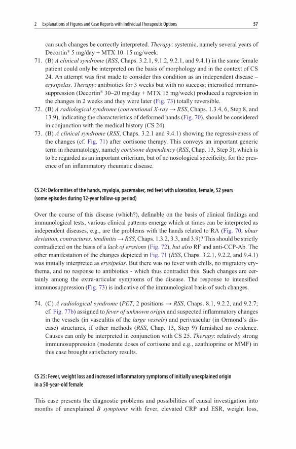