experimental supporting information - rsc.org · the organic layer was dried over mgso4, filtered...
TRANSCRIPT

S1
Experimental Supporting Information
Materials and general methods:
Chemicals: Fmoc-amino acids were obtained from GL Biochem (Shanghai, China). 2-Cl-trityl
chloride resin (1.0-1.2 mmol/g) was obtained from Nankai University Resin Co. Ltd. 1-
Naphthylacetic acid (NAA) was obtained from Ark Pharm. Tert-Butyl 2-hydroxyacetate was
obtained from TClchemicals. Agar sucrose was purchased from Sangon Biotech (Shanghai)
Co., Ltd. The model plant Arabidopsis thaliana (ecotype Columbia-0) and the auxin response
marker line ProDR5rev: GFP were obtained from the Arabidopsis Biological Resource Center
(ABRC). Commercially available reagents were used without further purification unless
otherwise noted. Nanopure water was used for all experiments. All other chemicals were
reagent grade or better.
General methods: High performance liquid chromatography (HPLC) was performed on a
LUMTECH HPLC (Germany) system using a C18 RP column with MeOH (0.1% of TFA) and
water (0.1% of TFA) as the eluents. LC-MS was conducted using an LCMS-2020 (Shimadzu)
system. The synthesized compounds were characterized by 1H NMR (Mercury Vx-300) using
DMSO-d6 as the solvent. HR-MS was performed using a VG ZAB-HS system (England). TEM
images were taken on a HITACHI HT7700 Exalens system operating at 120 kV. Rheology was
performed on an ARES 2000ex (TA instrument) system using parallel plates (40 mm) at a gap
of 500 µm. Phenotypes of seedlings were documented with a stereoscope (Leica 165FC,
Germany) equipped with a CCD camera or a scanner (Epson Perfection V33). The circular
dichroism (CD) spectrum was obtained on a BioLogic (MOS-450) system. Statistical analysis
of relative primary root elongation (%) and lateral roots were done with SPSS.
Synthesis and characterizations:
Electronic Supplementary Material (ESI) for ChemComm.This journal is © The Royal Society of Chemistry 2018

S2
Synthesis of NAA-HA: 1-Naphthylacetic acid(744 mg, 4 mmol), tert-Butyl 2-hydroxyacetate
(660.8 mg, 5 mmol), 4-dimethylaminopyridine (61.085 mg, 0.5 mmol) and
dicyclohexylcarbodiimide (2.1 g, 10 mmol) were dissolved in Anhydrous acetone, and the
resulting solution was stirred at room temperature for 24 h. The reaction solution was filtered
and the acetone was evaporated. The product was cleaved by using 75% DCM of with 25% of
trifluoroacetic acid and 5% of TIS for 2 h and the cutting fluid was removed by rotary
evaporation. 100 ml of 5% Na2CO3 was added under ultrasound while the white precipitate is
produced. The solution was extracted with ether (3×80 ml), and the water phase was kept. 1M
hydrochloric acid was added into the aqueous solution to adjust the final pH at 2 and
simultaneously white precipitation was produced. The fine white precipitate was washed with
EA (2×100 mL). The organic layer was dried over MgSO4, filtered and concentrated for rotary
evaporation to get crude product, which was directly reacted for next step without purification.
Scheme S-1. Synthesis of compounds NAA-HA.
Peptide systhesis: The peptide derivative NAA-G’FFY and NAA-GFFY were prepared by
solid phase peptide synthesis (SPPS) using 2-chlorotrityl chloride resin and the corresponding
N-Fmoc protected amino acids with side chains properly protected by a tert-butyl group or t-
Butyl oxy carbonylgroup. The first amino acid (Fmoc-Tyr(OtBu)-OH) was loaded on the resin
at the C-terminal with the loading efficiency about 1.0 mmol/g. 20% piperidine in anhydrous
N,N’-dimethylformamide (DMF) was used during deprotection of Fmoc group. Then the next
Fmoc-protected amino acid was coupled to the free amino group using O-(Benzotriazol-1-yl)-

S3
N,N,N’,N’-tetramethyluroniumhexafluorophosphate (HBTU) as the coupling reagent. The
growth of the peptide chain was according to the established Fmoc SPPS protocol. At the final
step, NAA-HA or NAA was used to couple with the peptide. After the last coupling step,
excessive reagents were removed by a single DMF wash for 5 times (5 ml per gram of resin),
followed by five steps of washing using DCM for 5 times (5 ml per gram of resin). The peptide
derivative was cleaved using 95% of trifluoroacetic acid with 2.5% of trimethylsilane (TIS) and
2.5% of H2O for 30 minutes. 20 mL per gram of resin of ice-cold diethylether was then added
to cleavage reagent. After rotary-evaporate process, 20 mL per gram of resin of ice-cold
diethylether was then added to cleavage reagent. Afterwards the supernatant was decanted and
the resulting crude product was purified by HPLC and lyophilized.
Characterization of compound 1: 1H NMR (400 MHz, DMSO) δ 12.71 (s, 1H), 9.21 (s, 1H),
8.22 (s, 1H), 7.98 (dd, J = 61.4, 21.1 Hz, 2H), 7.50 (d, J = 27.9 Hz, 2H), 7.19 (d, J = 32.0 Hz,
3H), 7.03 (d, J = 7.4 Hz, 1H), 6.66 (d, J = 7.2 Hz, 1H), 4.55 (s, 1H), 4.42 (d, J = 19.0 Hz, 1H),
4.20 (s, 1H), 3.08 – 2.89 (m, 2H), 2.87 – 2.66 (m, 2H).

S4
Figure S-1. LC-MS spectrum of compound 1.
Figure S-2. HR-MS of compound 1.
Figure S-3. 1H NMR of compound 1.
Characterization of compound 2: 1H NMR (400 MHz, DMSO) δ 12.69 (s, 1H), 9.20 (s, 1H),
8.24 (d, J = 35.4 Hz, 2H), 8.03 (t, J = 27.0 Hz, 2H), 7.87 (d, J = 36.1 Hz, 2H), 7.54 – 7.40 (m,
3H), 7.12 (dd, J = 73.8, 3.8 Hz, 9H), 6.66 (d, J = 6.8 Hz, 2H), 4.63 – 4.44 (m, 2H), 4.39 (s, 1H),
3.93 (s, 2H), 3.69 (d, J = 13.3 Hz, 1H), 3.56 (d, J = 6.5 Hz, 1H), 3.08 – 2.89 (m, 3H), 2.86 –
2.71 (m, 2H), 2.66 (d, J = 13.5 Hz, 1H).

S5
Figure S-4. LC-MS spectrum of compound 2.
Figure S-5. HR-MS of compound 2.

S6
Figure S-6. 1H NMR of compound 2.
Preparation of hydrogels: A 3 mg sample of the peptide derivative (compound 1 or 2) and
one equivalent of Na2CO3 (to neutralize the terminal carboxylic acid on the peptide) were
suspended in 1 mL of PBS buffer (pH = 7.4). The solution was heated to dissolve the powder
completely, and the hydrogel formed after cooling back to room temperature (22-25 ℃) within
10 min.
Rheology: A rheology test was done on an AR 2000ex (TA instrument) system, and 40 mm
parallel plates at a gap of 500 μm were used in the experiments. First, a dynamic strain sweep
was conducted to determine the linear region of strain for the following dynamic frequency
sweep. The dynamic frequency sweep was then characterized in the frequency region of 0.1-
100 rad/s at the strain value of 0.1%.
Preparation of TEM samples of gels: TEM samples were prepared at 25 ℃. A micropipet
was used to load 10 µL of sample of gels to a carbon coated copper grid for 1 minute. The

S7
excess solution was removed by apiece of filter paper. The samples were dyed by 15 µL uranyl
acetate for 1 minute and dried overnight in a desiccator before the TEM measurement.
Figure S-7. TEM image of a supramolecular hydrogel of compound 2 formed by the heating-cooling process (The scale bar represents 100 nm).
Releasing profile of NAA: The gel formed from 300 µL of PBS containing 0.3 wt% of
compound 1 was used for the determination of the releasing profile of the NAA. A volume of
250 µL of PBS (pH = 7.4) or 1/2 MS medium (pH = 5.7) was added on the top of the gel. A
volume of 200 µL of PBS or 1/2 Murashige and Skoog (1/2 MS) medium was removed at the
desired time for measurement of the accumulated release amount of NAA from the gel by LC-
MS, and 200 µL of the fresh buffer solution was then added. The experiment was performed
for 3 days with 3 parallel samples (n = 3).
Circular dichroism (CD) spectrum: The CD spectrum was recorded using a BioLogic (MOS-
450) system. All gels were placed in a 0.1 cm quartz spectrophotometer cell (20-C/Q/0.1). The
wavelength range was from 185 to 280 nm. The acquisition period was 0.5 s and the step was
0.5 nm. The resultant CD spectrum was acquired after subtracting the solvent background.
Formation of the coassembly gel I s: Different mass ratios of compound 1 and Fmoc-GFFY
were mixed in 300 µL of PBS buffer at a fixed combined concentration of 0.3 wt% of compound

S8
1 and Fmoc-GFFY. As shown in Table S-1, a series of coassembly gel Is and control were
prepared by heating and cooling.
Numbers 1 2 3 4 5 control
compound 1 1‰ 0.1‰ 0.01‰ 0.001‰ 0.0001‰ 0
Fmoc-GFFY 2‰ 2.9‰ 2.99‰ 2.999‰ 2.9999‰ 3‰
Table S-1. Different mass ratios of compound 1 and Fmoc-GFFY.
Formation of the coassembly Gel II -4: 0.001‰ of compound 2 and 2.999‰ of Fmoc-GFFY
were mixed in 300 µL of PBS buffer at a fixed combined concentration of 0.3 wt%. A
coassembly Gel II-4 was prepared by heating and cooling.
Formation of NAA in Agar/PBS: NAA was added into 0.1% (w/v) agar and PBS respectively,
the concentration of NAA in agar/PBS is 1.425 μM.
Formation of NAA evenly distributed: NAA was added into sterile 1/2MS media directly,
the concentration of NAA is 570 pM in the palte.
Figure S-8. Dynamic frequency sweep of A) Gel I-1, B) Gel I-2, C) Gel I-3, D) Gel I-4, and E) Gel I-5 with 0.3 wt% of the coassembly gels at the strain value of 0.1%.
Components

S9
Figure S-9. Release profile of NAA from A) Gel I-1, B) Gel I-2, C) Gel I-3, and D) Gel I-4 in the presence of PBS buffer (pH = 7.4) and 1/2 Murashige and Skoog (1/2 MS) medium (pH =
5.7).
Preparation and growth conditions of Arabidopsis: Generally, seeds were surface-sterilized
with 75 % ethanol for 5 min and 100 % ethanol for 1 min, rinsed with ddH2O five times, and
then stratified at 4 °C for 2 days before plating on 1/2 Murashige and Skoog (1/2 MS) medium
(pH = 5.7) containing 0.8 % (w/v) agar (Sigma-Aldrich, USA), and 1 % (w/v) sucrose (Sigma-
Aldrich, USA). The plants were grown vertically in a long-day condition[16 h (22 °C)/8 h (18
°C)] with a photosynthetic photon flux density at 90 µE m-2 sec-1.
Treatment with the hydrogels: Seven-day-old or four-day-old vertically grown Arabidopsis
seedlings (Ecotype Columbia-0) were transferred to plates (1450 mm in diameter) containing
50 mL of solid 1/2 MS media. Right before the transfer, five holes, interspersed by 5 mm, were
made on the medium with a plastic tube (5.5 mm in diameter). The seedlings were carefully
placed at the edges of the holes, with their primary root tips at the same level as the holes. Then,
20 µL of the hydrogels were injected into the holes.

S10
Fluorescence Microscopy: Seven-day-old vertically grown Arabidopsis seedlings
(ProDR5rev:GFP) were transferred to plates (1450 mm in diameter) containing 50 mL of solid
1/2 MS media. Right before the transfer, five holes, interspersed by 5 mm, were made on the
medium with a plastic tube (5.5 mm in diameter). The seedlings were carefully placed at the
edges of the holes, with their primary root tips at the same level as the holes. Then, 20 µL of
the hydrogels were injected into the holes. The seedlings treated with the hydrogels were
documented for their lateral root emergence in a consecutive three days. All images were
captured with a fluorescent stereoscope (Leica 165FC, Leica, Germany) equipped with a CCD
camera (λ excitation = 488 nm, λemission = 584 nm for GFP).
Quantification of relative primary root elongation (%):To quantify primary root elongation
on 1/2 MS supplied with different hydrogels or control gels/solutions, four-day-old vertically
grown Arabidopsis seedlings (Col-0) were transferred to plates (1450 mm in diameter)
containing 50 mL of solid 1/2 MS media. Right before the transfer, five holes, interspersed by
5 mm, were made on the medium with a plastic tube (5.5 mm in diameter). The seedlings were
carefully placed at the edges of the holes, with their primary root tips at the same level as the
holes. Then, 20 µL of the hydrogels were injected into the holes. The phenotypes of the
seedlings were documented with a scanner (Epson Perfection V33) at the same time daily for
three days. Primary root elongation in the three days were measured in image J from the scanned
images, and relative primary root enlongation (%) on each condition was compared against the
primary root elongation on Blank, which was set as 100%.
Quantification of number of lateral roots: To quantify number of lateral roots, seven-day-
old vertically grown Arabidopsis seedlings (Col-0) were transferred to plates (1450 mm in
diameter) containing 50 mL of solid 1/2 MS media. Right before the transfer, five holes,
interspersed by 5 mm, were made on the medium with a plastic tube (5.5 mm in diameter). The
seedlings were carefully placed at the edges of the holes, with their primary root tips at the same
level as the holes. Then, 20 µL of the hydrogels were injected into the holes. After three days,

S11
the phenotypes of the seedlings were documented with a scanner (Epson Perfection V33), and
number of lateral roots was calculated from the scanned images.
Figure S-10. Four-day-old vertically-grown Arabidopsis seedlings (Col-0) were transferred to new plates and grown vertically for 3 days in the presence of Blank, Control, Gel I-5, Gel I-4,
Gel I-3, Gel I-2, and Gel I-1, respectively. Seedlings were scanned at day 7 with a scanner (Epson Perfection V33). Bar =1 cm.
Figure S-11. Statistical analysis of relative primary root enlongation (%), as compared with Blank (set as 100%), over the 3 days. The data shown are mean ± SD (n = 15). One-way
ANOVA was done with SPSS.

S12
Figure S-12. Seven-day-old vertically-grown Arabidopsis seedlings (Col-0) were transferred to new plates and grown vertically for 3 days in the presence of Blank, Control, Gel I-5, Gel I-
4, Gel I-3, Gel I-2, and Gel I-1, respectively. Seedlings were scanned at day 10. Bar =1 cm.
Figure S-13. Quantification of numbers of lateral roots of 10-day-old seedlings. The data shown are mean ± SD (n = 15). One-way ANOVA was done with SPSS. Bar = 1 cm.
S13
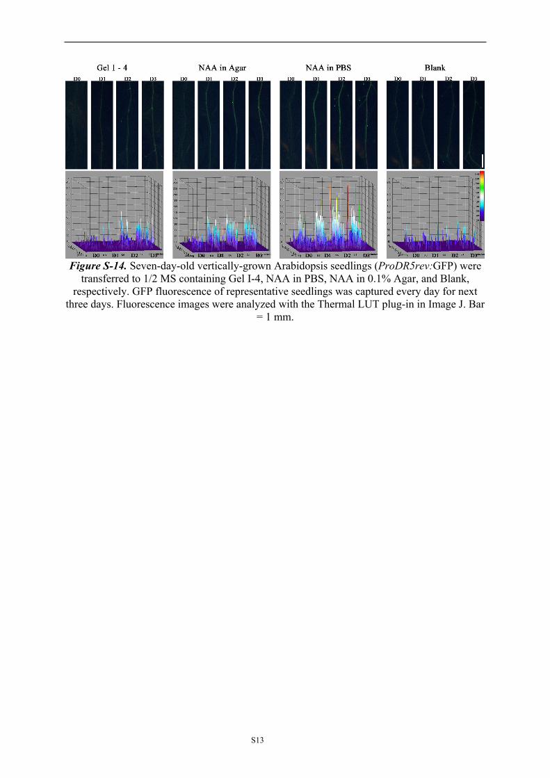
Figure S-14. Seven-day-old vertically-grown Arabidopsis seedlings (ProDR5rev:GFP) were transferred to 1/2 MS containing Gel I-4, NAA in PBS, NAA in 0.1% Agar, and Blank,
respectively. GFP fluorescence of representative seedlings was captured every day for next three days. Fluorescence images were analyzed with the Thermal LUT plug-in in Image J. Bar
= 1 mm.