experimental fossilisation of viruses from extremophilic archaea
TRANSCRIPT

Biogeosciences, 8, 1465–1475, 2011www.biogeosciences.net/8/1465/2011/doi:10.5194/bg-8-1465-2011© Author(s) 2011. CC Attribution 3.0 License.
Biogeosciences
Experimental fossilisation of viruses from extremophilic Archaea
F. Orange1,2, A. Chabin1, A. Gorlas3, S. Lucas-Staat4, C. Geslin3, M. Le Romancer3, D. Prangishvili4, P. Forterre4,and F. Westall1,2
1Centre de Biophysique Moleculaire, UPR4301, CNRS, Rue Charles Sadron, 45071 Orleans Cedex 2, France2Observatoire des Sciences de l’Univers en region Centre, UMS3116, 1A Rue de la Ferollerie, 45071 Orleans Cedex 2, France3Universite de Bretagne Occidentale, UMR 6539, CNRS – Institut Universitaire Europeen de la Mer,Technopole Brest-Iroise, Rue Dumont d’Urville, 29280 Plouzane, France4Molecular Biology of the Gene in Extremophiles Unit, Institut Pasteur, 25 rue du Docteur Roux,75724 Paris Cedex 15, France
Received: 16 February 2011 – Published in Biogeosciences Discuss.: 4 March 2011Revised: 20 May 2011 – Accepted: 26 May 2011 – Published: 9 June 2011
Abstract. The role of viruses at different stages of the originof life has recently been reconsidered. It appears that virusesmay have accompanied the earliest forms of life, allowingthe transition from an RNA to a DNA world and possiblybeing involved in the shaping of tree of life in the three do-mains that we know presently. In addition, a large varietyof viruses has been recently identified in extreme environ-ments, hosted by extremophilic microorganisms, in ecosys-tems considered as analogues to those of the early Earth.Traces of life on the early Earth were preserved by the pre-cipitation of silica on the organic structures. We presentthe results of the first experimental fossilisation by silica ofviruses from extremophilic Archaea (SIRV2 –Sulfolobus is-landicusrod-shaped virus 2, TPV1 –Thermococcus prieuriivirus 1, and PAV1 –Pyrococcus abyssivirus 1). Our resultsconfirm that viruses can be fossilised, with silica precipitat-ing on the different viral structures (proteins, envelope) overseveral months in a manner similar to that of other experi-mentally and naturally fossilised microorganisms. This studythus suggests that viral remains or traces could be preservedin the rock record although their identification may be chal-lenging due to the small size of the viral particles.
Correspondence to:F. Orange([email protected])
1 Introduction
The role of viruses in the early evolution of life has re-cently been reevaluated. Viruses were long considered asnon-living by-products of cellular activity and thus unableto have played a role in the origin and evolution of theirhosts. Over the last decade, however, our views on the originand nature of viruses have dramatically changed. Structuralsimilarities between viruses infecting phylogenetically dis-tant hosts suggest that viruses were already active at the timeof the Last Universal Common Ancestor (LUCA) and prob-ably before (Bamford et al., 2005; Koonin and Dolja, 2006;Krupovic and Bamford, 2008). Comparative genomics hasrevealed that most genes encoded by viruses have no cellularhomologues, testifying to the creativity of virocells (Forterre,2011) in inventing new proteins. Several theories and discov-eries have been made regarding the ancient nature of virusesand their possible key roles in the early evolution of life andin the origin of modern cells (Forterre, 2002; Bamford, 2003;Koonin et al., 2006; for recent reviews and alternative view-points, see Moreira and Lopez-Garcıa, 2009 and Forterreand Prangishvili, 2009a, b). For instance, viruses have beenevoked to explain the origin of DNA cells. Starting froman RNA world (cells with RNA genomes), DNA could haveappeared in viruses (DNA viruses) and then have been trans-ferred to cells (Forterre, 2002). One hypothesis also suggeststhat interactions between DNA viruses and RNA cells (fu-sion, gene transfers) could have resulted in the formation ofthe three domains (Eukaryotes, Bacteria, Archaea) (Forterre,2006). Finally, the discovery ofMimivirus, a giant DNA
Published by Copernicus Publications on behalf of the European Geosciences Union.

1466 F. Orange et al.: Experimental fossilisation of viruses from extremophilic Archaea
virus infectingAmoeba(La Scola et al., 2003), permits avirus genome to be included in the universal tree of life. Thissuggests that giant viruses could be intermediates betweencells and previously known viruses and could form a newdomain of life (Raoult et al., 2004).
A major development in recent years in virology has beenthe discovery of very diverse new viruses in extreme environ-ments. Such environments are particularly interesting as theyare often considered as analogues of the environments on theearly Earth in which life could have originated. Microorgan-isms found in these environments are also considered to bepossible analogues to the earliest forms of life on Earth andpossibly on Mars (Nisbet and Sleep, 2001; Konhauser et al.,2003). Recognition of the ancient nature of viruses suggeststhat early life forms could have hosted viruses. The studyof viruses of extremophilic microorganisms is a relatively re-cent field. Nevertheless, it has already allowed the identifi-cation of several thousands of viruses having different mor-phologies and characteristics, and also of new virus families(see review in Le Romancer et al., 2007). Viruses have beenidentified in all known extreme environments: hypersaline(Oren et al., 1997; Dyall-Smith et al., 2003; Pagaling et al.,2007; Sime-Ngando et al., 2010), alkaline lakes (Jiang et al.,2004), deserts (Prigent et al., 2005), polar regions (Marangeret al., 1994; Kepner et al., 1998; Borriss et al., 2003; Gow-ing, 2003), acid mine drainages (Kyle et al., 2008a), deepsubsurface rocks (Bird et al., 2001; Kyle et al., 2008b), andin hydrothermal environments. The search for new viruses inthe latter environment has been especially fruitful, follow-ing the pioneering work of Wolfram Zillig on the virusesof hyperthermophilic Archaea (Martin et al., 1984; Rice etal., 2001; Rachel et al., 2002). Many new viral familiesthat infect hyperthermophilic Archaea in terrestrial and ma-rine hot springs have been identified (Geslin et al., 2003b;Ortmann and Suttle, 2005; Geslin et al., 2005; Prangishviliet al., 2006a; Ortmann et al., 2006; Le Romancer et al.,2007). Although these viruses all have double-stranded DNAgenomes, they produce virions with very diverse morpholo-gies (e.g. rod-shaped, filamentous, spindle-shaped, ellipsoid,head and tail) and most proteins encoded in their genomeshave no homologues, except sometimes in other viral lin-eages (Prangishvili et al, 2006b).
Although structural and comparative genomics point to theantiquity of viruses, their fossil remains have yet to be de-tected in the rock record. Although challenging, consider-ing the size of viral particles, detection of possible fossilisedviral remains in the vicinity of putative fossilised microor-ganisms could be a direct proof of the antiquity of virusesand an additional clue to assessing the biogenicity of the ob-served structures. The oldest known fossil microorganisms,dating back to almost 3.5 billion years (Ga) ago, were pre-served as silicified remains (see review in Westall, 2010).The preservation by silica was due to the fact that the earlyEarth’s oceans were silica enriched compared to the under-saturated oceans of the present Earth, where siliceous organ-
isms are a sink for available silica. An additional sourceof silica in the early oceans came from the very active hy-drothermal processes cycling silica and other elements fromthe crust back into the ocean (see review in Westall andSoutham, 2006). In situ and experimental silicification ofmicroorganisms in these hydrothermally-influenced environ-ments has been studied in depth over the past two decades(Westall et al., 1995; Toporski et al., 2002; Konhauser etal., 2004; Orange et al., 2009 and references therein). Theseinvestigations have provided precious information regardingthe processes involved and helped the identification of sili-cified traces of life in ancient rocks (see review in Westall,2010). Recent studies have started to explore the ability ofviruses to be mineralised and the possibility that they couldbe preserved in the fossil record. Several studies have studiedinteractions between different viruses and iron (Daughney etal., 2004; You et al., 2005; Templeton et al., 2006; Kyle etal., 2008b). Daughney et al. (2004) have shown the abil-ity of a marine bacteriophage to interact with dissolved pro-tons, and to act as a site for iron binding, due to the presenceof negatively-charged functional groups in its capsid. Kyleet al. (2008b) observed the iron mineralisation of viruses inthe acid waters of Rio Tinto (Spain) and discussed the influ-ence of this mineralisation on the biogeochemical processesand on the possibility for viruses to be preserved in the rockrecord. Poinar and Poinar (2005) reported the possible pres-ence of preserved viruses in 15 to 100 Ma insect preservedin amber. Laidler and Stedman (2010) monitored the experi-mental fossilisation of bacteriophage T4 for a few days undersimulated hot spring silicifying conditions and showed thatsilica could precipitate around viral structures and preservethem.
Our study presents the results of the first long-term ex-perimental fossilisation of three viruses and the first experi-mental fossilisation of viruses hosted by hyperthermophilicArchaea. Among the great variety of viruses in hyperther-mophilic Archaea, we chose for this study viruses produc-ing virions (viral particles) with very different structures andmorphologies: the rod-shaped virus SIRV2 (Sulfolobus is-landicusrod-shaped virus 2) and the spindle-shaped TPV1(Thermococcus prieuriivirus 1) and PAV1 (Pyrococcusabyssivirus 1) viruses. These morphologies appear to bespecific for viruses infecting organisms living in extreme en-vironments, either at hot temperatures (rod-shaped, spindle-shaped) or with high salt concentrations (spindle-shaped),with the exception of some plant viruses which also producerod-shaped particles.
Our objectives were (1) to determine whether virusescould be fossilised by silica and whether they could be pre-served over relatively long periods of time (several months),and (2) to evaluate whether recognizable features in the viralstrains used could still be identified once silica had precipi-tated around them.
Biogeosciences, 8, 1465–1475, 2011 www.biogeosciences.net/8/1465/2011/

F. Orange et al.: Experimental fossilisation of viruses from extremophilic Archaea 1467
2 Materials and methods
2.1 Description of the viruses used for this study
SIRV2 (Sulfolobus islandicusrod-shaped virus 2) belongsto theRudiviridaefamily (Prangishvili et al., 1999; Bize etal., 2009) and is a lytic virus (i.e. which causes the death ofthe host cell) that infects a strain of the CrenarchaeotaSul-folobus islandicus, an acidophilic and hyperthermophilic Ar-chaea that was originally isolated from samples taken fromsolfataric fields in Iceland (Zillig et al., 1994). SIRV2 par-ticles are stiff rods up to 900 nm long and∼20 nm wide(Fig. 1a; Prangishvili et al., 1999) and consist of a super-helix including a protein with double-stranded linear DNA.In contrast to other viruses, such as rod-shaped members oftheLipothrixviridae, SIRV2 virions are not enveloped.
TPV1 (Thermococcus prieuriivirus 1) and PAV1 (Pyro-coccus abyssivirus 1) were isolated from the EuryarchaeotaThermococcus prieuriistrain Bio-pl-0405IT2 andPyrococ-cus abyssistrain GE23, respectively (Geslin et al., 2003a;Gorlas et al., 2009). The latter are two neutrophilic and hy-perthermophilic Archaea of the Thermococcales order (Er-auso et al., 1993; Gorlas et al., 2011). TPV1 and PAV1are non-lytic, spindle-shaped viruses (TPV1: 140×80 nm;PAV1: 120×80 nm), characterised by double-stranded circu-lar DNA, and are found either isolated or in groups (Fig. 1cand d). TPV1 and PAV1 have an envelope composed of viralproteins and lipids from the host. These viruses morpho-logically resemble members of theFuselloviridaefamily buthave not yet been classified.
2.2 Virus isolation
The viruses used in this study were harvested from fresh cul-tures of the host strains.
For the production of SIRV2, theSulfolobus islandicusstrain LAL14/1 was grown until the late exponential phasewas reached, as described by Zillig et al. (1994). Cells wereremoved by low-speed centrifugation (3500 g in a SorvallGS3 rotor). The viruses were precipitated from the super-natant by the addition of 1 M NaCl and 10 % polyethyleneglycol 6000 (PEG 6000) and incubated overnight at 4◦C. Apellet of viruses was collected by centrifugation in a SorvallGSA rotor at 23 000 g for 30 min and suspended in TA buffer(20 mM Tris-acecate, pH 6). SIRV2 were purified by cen-trifugation in a CsCl buoyant density gradient (0.45 g ml−1)in a Beckman SW41 rotor centrifuge at 250 000 g for 48 h.Fractions containing the viral particles were collected with asyringe then dialyzed against TA buffer for further analysis(Bettstetter et al., 2003). The viruses were stored at 4◦C in a20 mM Tris-acetate buffer (pH 6) until used.
Thermococcus prieuriistrain Bio-pl-0405IT2 (Gorlaset al., 2011) andPyrococcus abyssistrain GE23 (Er-auso et al., 1993; Marteinsson et al., 1995) were grownin the medium described by Geslin et al. (2003a), at
Fig. 1. (A–D): TEM micrographs showing examples of the virusesused for this study.(A) SIRV2 particle; note the central cavity,sometime discontinued.(B) Fragments of SIRV2 particle on an un-stained grid; note the absence of visible features, and a viral DNA-protein filament that links the two fragments (arrow).(C) Aggre-gate of TPV1 particles.(D) PAV1 particles (arrows); note the pres-ence of remnantP. abyssiflagellas in the sample.(E) TEM mi-crograph showing example of the silica precipitate formed sponta-neously in the control sample, which contained no viruses, observedafter 4 days; unstained grid. All TEM micrographs were made at200 kV on grids negatively stained with uranyl acetate, unless oth-erwise stated.
80◦C and 85◦C, respectively, up to the late exponentialphase. The cells were pelleted by low-speed centrifuga-tion at 6000 g for 15 min. TPV1 and PAV1 were precip-itated from the supernatant in 1 M NaCl with 10 % PEG6000 overnight at 4◦C with gentle stirring. The pre-cipitate was pelleted by centrifugation at 13 000 g for 1 h15 min, then drained and resuspended in a specific buffer
www.biogeosciences.net/8/1465/2011/ Biogeosciences, 8, 1465–1475, 2011

1468 F. Orange et al.: Experimental fossilisation of viruses from extremophilic Archaea
for each virus (TPV1: TPV1-buffer – 10 mM Tris-HCL,100 mM NaCl, 5 mM CaCl2; PAV1: TE buffer – 10 mM Tris-HCl and 1 mM EDTA, pH 8). A second precipitation withPEG 6000 (10 %) and 1 M NaCl was made during 1.5 h andthe precipitate was collected as described above. After cen-trifugation (5000 g for 10 min), the supernatant was kept andthe pellet was extracted two more times under the same con-ditions with reduced volumes of TPV1 or TE buffer. Thevirus-containing supernatants were pooled and stored for onenight at 4◦C. After centrifugation (5000 g for 15 min) to re-move residual cell debris, the supernatant was concentratedby ultracentrifugation at 33 000 g for 1 h 45 min (BeckmanOptima LE-80K 70.1Ti rotor) and the pellet was resuspendedin TPV1 or TE buffer. Viruses were purified from these sus-pensions by centrifugation in a CsCl buoyant density gradi-ent (TPV1: 1.32 g ml−1; PAV1: 1.298 g ml−1) in a BeckmanOptima LE-80K 70.1Ti centrifuge rotor (TPV1: 180 000 gfor 6 h; PAV1: 220 000 g for 24 h). Fractions containing thenucleic acids were detected at 254 nm and collected using adensity gradient fractionator (model 185, ISCO). These frac-tions were then dialyzed against a large volume of TPV1 orTE buffer and were stored at 4◦C until used.
2.3 Silicification procedure
The fossilisation experiments were made directly in thebuffering media of the viruses (SIRV2: Tris-acetate buffer –20 mM Tris-acetate, pH6; TPV1: TPV1 buffer – 10 mM Tris-HCl, 100 mM, 5 mM CaCl2, pH 8; PAV1: TE buffer – 10 mMTris-HCl, 1 mM EDTA, pH 8). This was done for practicalreasons as well as to maintain the viruses in a favourable en-vironment so that their evolution could be followed duringfossilisation over several months.
A stock silica solution (3200 ppm Si) was preparedfrom a pure sodium silicate solution (Riedel de Haen)containing ∼27 % SiO2 and ∼10 % NaOH (Na2Si3O7,M = 242 g mol−1). Its pH was adjusted to 8 before injectioninto the virus suspension.
19 µl of a suspension of purified SIRV2 were mixed with1 µl of the stock silica solution in a sealed glass vial to ob-tain a final concentration of∼160 ppm Si. For TPV1 andPAV1, 180 µl of suspensions of purified viruses were mixedwith 20 µl of the stock silica solution to obtain a final concen-tration of∼320 ppm Si. The SIRV2 fossilisation experimentvials were stored at room temperature (due to the small vol-ume, high temperatures led to evaporation). The TPV1 andPAV1 fossilisation experiment vials were placed in an ovenat 60◦C. A control sample without viruses, consisting of a∼160 ppm Si silica solution in distilled water, was also pre-pared and left at room temperature.
The small volumes involved in the experiments meant thatwe were not able to make precise monitoring of the pH andsilica concentration over time. We assumed that the injectionof the silica solution at pH 8 only slightly increased the pH
in the SIRV2 medium, while it did not change the pH of theTPV1 and PAV1 media.
2.4 Electron microscopy
The vials were sampled at different times (between 2 and60 days for the SIRV2 experiment; between 1 and 180 daysfor the TPV1 and PAV1 experiments; 4 days for the controlsample) by collecting∼1 µl of the sample and immediatelypreparing it for negatively-stained transmission electron mi-croscopy (Geslin et al., 2003a).
For negative staining, a droplet of sample (either unsili-cified or silicificed) was placed on a carbon-coated copperTEM grid. The sample was allowed to absorb to the carbonlayer for 2 min before removing the excess liquid with a pieceof filter paper. Some samples were stained to increased con-trast by placing a droplet of saturated uranyl acetate ethanolicsolution for 40 s and then removing the excess liquid. Somegrids were also observed unstained to better distinguish sil-ica deposition on the viral structures. The prepared sampleswere then air dried. They were observed and analysed with aPhilips CM20 Transmission Electron Microscope (Centre deMicroscopie Electronique, University of Orleans), equippedwith an EDX detector (Oxford Instruments).
3 Results
3.1 Experimental fossilisation of SIRV2
SIRV2 particles appear as stiff rods of several hundreds ofnanometers in length and∼20 nm in width and contain a cen-tral cavity (Fig. 1a), not visible on unstained grids (Fig. 1b).This virion is naturally fragile and readily breaks up intofragments that may be linked to each other by filaments,identified as the result of the degradation of the viral helixand made of DNA with remnants of the viral protein (Fig. 1b,arrow) (Prangishvili et al., 1999; Vertergaard et al., 2008).Apart from fragmentation, the preservation of SIRV2 frag-ments during fossilisation was generally good. In the firstdays of the fossilisation experiment, a granular and alveolar-textured silica precipitate formed spontaneously in both thecontrol experiment without viruses (Fig. 1e) and in the ex-periment with the viruses (Fig. 2a, b), indicating that nei-ther virus particles nor the buffering medium had any influ-ence on precipitation or on the structure of the silica. Ini-tially, after one week of fossilisation only a few SIRV2 par-ticles were observed in direct contact with, and sometimestrapped within, the silica precipitate (Fig. 2a) but most werenot (Fig. 2b). Where embedded in the silica deposit, the vi-ral helix appeared to have been compacted or decayed andthe virion was only visible as a faint, dark outline (Fig. 2a,arrows) that probably represents degraded viral DNA andprotein. Silica nucleation on the virion was observed af-ter 1 week (Fig. 2c, arrow) and, after 30 days, nanometric
Biogeosciences, 8, 1465–1475, 2011 www.biogeosciences.net/8/1465/2011/

F. Orange et al.: Experimental fossilisation of viruses from extremophilic Archaea 1469
Fig. 2. TEM micrographs showing progressive steps of the experimental fossilisation of SIRV2 at a∼160 ppm Si silica concentration.(A) 48 h, SIRV2 particle partially trapped inside the silica precipitate (Si); the arrows underline the shape of the virion inside the precipitate.(B) 7 days, fragments of SIRV2 particles in the vicinity of a silica precipitate (Si); the two separated filaments of viral DNA-protein are visible(arrow). (C) 7 days, unstained grid, fragment of SIRV2 particle; a dark∼10 nm particle is seen attached on the virion (arrow).(D) 30 days,SIRV2 particle; note the numerous dark silica particles in the central cavity.(E) 60 days, unstained grid, fragments of SIRV2 particle; notethe dark silica particles attached on the outer surface of the virion, and filling the central cavity.(F) 60 days, EDX spectra made on theSIRV2 particles of(E), showing a Si signal slightly higher than on the background; the Cs signal comes from the cesium chloride used forvirus purification and the Cu signal comes from the copper grid.(G) 60 days, unstrained grid, viral DNA-protein filament covered by silicaparticles. All TEM micrographs were made at 200 kV on grids negatively stained with uranyl acetate, unless otherwise stated.
dark particles had formed in the central cavity of some ofthe virions (Fig. 2d). This phenomenon was more evidentafter 60 days of fossilisation with virions showing, on un-stained grids, a strongly contrasted central cavity (Fig. 2e),suggesting that the individual particles observed previouslyhad continued their growth and eventually merged. Morepronounced silica nucleation and binding on the outside of
the viral particles was also observed. EDX analyses madeon these particles showed a silicium signal slightly strongerthan the signal from background noise (Fig. 2f). Fine chainsof silica particles (Fig. 2g) may represent silica directly pre-cipitated on isolated viral DNA-protein filaments, or the rem-nants of virion fragments completely covered by silica.
www.biogeosciences.net/8/1465/2011/ Biogeosciences, 8, 1465–1475, 2011
1470 F. Orange et al.: Experimental fossilisation of viruses from extremophilic Archaea
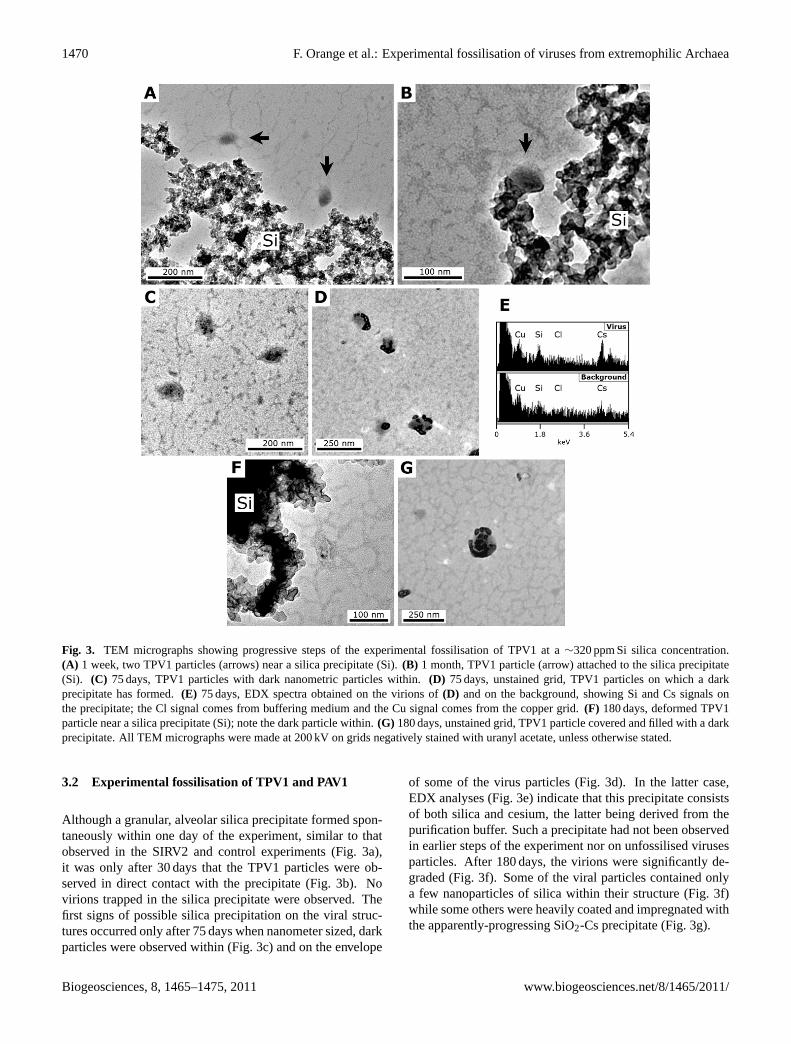
Fig. 3. TEM micrographs showing progressive steps of the experimental fossilisation of TPV1 at a∼320 ppm Si silica concentration.(A) 1 week, two TPV1 particles (arrows) near a silica precipitate (Si).(B) 1 month, TPV1 particle (arrow) attached to the silica precipitate(Si). (C) 75 days, TPV1 particles with dark nanometric particles within.(D) 75 days, unstained grid, TPV1 particles on which a darkprecipitate has formed.(E) 75 days, EDX spectra obtained on the virions of(D) and on the background, showing Si and Cs signals onthe precipitate; the Cl signal comes from buffering medium and the Cu signal comes from the copper grid.(F) 180 days, deformed TPV1particle near a silica precipitate (Si); note the dark particle within.(G) 180 days, unstained grid, TPV1 particle covered and filled with a darkprecipitate. All TEM micrographs were made at 200 kV on grids negatively stained with uranyl acetate, unless otherwise stated.
3.2 Experimental fossilisation of TPV1 and PAV1
Although a granular, alveolar silica precipitate formed spon-taneously within one day of the experiment, similar to thatobserved in the SIRV2 and control experiments (Fig. 3a),it was only after 30 days that the TPV1 particles were ob-served in direct contact with the precipitate (Fig. 3b). Novirions trapped in the silica precipitate were observed. Thefirst signs of possible silica precipitation on the viral struc-tures occurred only after 75 days when nanometer sized, darkparticles were observed within (Fig. 3c) and on the envelope
of some of the virus particles (Fig. 3d). In the latter case,EDX analyses (Fig. 3e) indicate that this precipitate consistsof both silica and cesium, the latter being derived from thepurification buffer. Such a precipitate had not been observedin earlier steps of the experiment nor on unfossilised virusesparticles. After 180 days, the virions were significantly de-graded (Fig. 3f). Some of the viral particles contained onlya few nanoparticles of silica within their structure (Fig. 3f)while some others were heavily coated and impregnated withthe apparently-progressing SiO2-Cs precipitate (Fig. 3g).
Biogeosciences, 8, 1465–1475, 2011 www.biogeosciences.net/8/1465/2011/

F. Orange et al.: Experimental fossilisation of viruses from extremophilic Archaea 1471
Fig. 4. TEM micrographs showing progressive steps of the experimental fossilisation of PAV1 at a∼320 ppm Si silica concentration.(A) 1 day, PAV1 particle (arrow) attached to the silica precipitate (Si).(B) 60 days, unstained grid, numerous PAV1 particles near the silicaprecipitate.(C) 60 days, unstained grid, aggregate of PAV1 particles trapped in a finely grained silica precipitate. All TEM micrographs weremade at 200 kV on grids negatively stained with uranyl acetate, unless otherwise stated.
Monitoring of the PAV1 fossilisation experiment was com-plicated by the presence of numerous fragments ofP. abyssiflagella as well as numerous artefacts in the preparation ofpurified PAV1 (Fig. 1d). PAV1 particles could be observedin direct contact with the silica precipitate after only 1 day(Fig. 4a). This precipitate was different from that formed inthe SIRV2 and TPV1 experiments and consisted of very fineparticles (compare Figs. 4c and 3b). The reason for this isunknown, although it possibly could be a consequence of thedifferent composition of the PAV1 buffering medium (whichincludes EDTA, a chelating and binding agent). No precip-itates formed inside or on free PAV1 particles after 60 days(Fig. 4b), although several virus particles were seen at theedge of the silica precipitate (Fig. 4c), suggesting that an im-portant number of PAV1 particles had been trapped in it.
4 Discussion
The viruses used in this experiment belong to different virusfamilies having completely different morphologies and struc-tures. This may partly explain why the results of the fossil-isation experiments were different. In the case of SIRV2, anon-enveloped rod-shaped virus, silica binding on the viralparticles was significant and progressive whereas only lim-ited silica binding occurred on TPV1 and PAV1, envelopedspindle-shaped viruses.
4.1 Fossilisation of the viruses
We assumed (but could not verify, due to the small volumesof the samples) that the silica behaviour during the experi-ments was similar to that described in previous experimentalfossilisation studies (review in Konhauser et al., 2004). Uponinjection into the vials, silica was in monomeric (Si(OH)4)
or slightly polymeric form and quickly spontaneously poly-merised as a colloidal amorphous silica precipitate. Dis-solved silica must have remained in the medium after this
initial polymerisation at a concentration close to the satura-tion concentration (62 ppm Si in distilled water; Gunnarssonand Arnorsson, 2000; Lalonde et al., 2005). In support ofthis, the silica precipitate formed in the SIRV2, TPV1 andcontrol experiments was similar to that observed in previousfossilisation experiments (Orange et al., 2009).
The fact that the silica precipitates in the SIRV2 experi-ment were invariably linked to viral DNA-protein filaments(Fig. 2b) suggests that the spontaneously-formed silica de-posit probably rapidly entrapped numerous SIRV2 particles,as was observed by Laidler and Stedman (2010). Some parti-cles were barely observable as dark outlines within the min-eral precipitate (Fig. 2a). The viral particles that remainedfree in the medium showed progressive silica binding withtime on their outer surface and in their central cavity. Ini-tial silica nucleation within the cavity of the SIRV2 particlesdid not appear to damage the body (Fig. 2d, e) although, atlater steps of the fossilisation process, strings of silica par-ticles observed on DNA-protein filaments (Fig. 2g) suggestthat viral particles may be heavily damaged by the growthof silica. The rarity of silicified DNA-protein filaments maybe due to the fact that DNA degrades rapidly when exposedto the environment (Schelble et al., 2008), while the remainsof DNA trapped in silica (Fig. 2a, g) may be preserved. Sil-ica binding to the outer surface of the virions appears to bethe result of two mechanisms: (i) the direct nucleation ofsilica (Fig. 2c) followed by continuing polymerisation, re-sulting in the formation of hemispherical silica particles thatprogressively grow and merge (Fig. 2c, e); (ii) the bindingof already formed silica particles on the virions, or on pre-viously bound silica particles (e.g. the middle silica particleamong the three attached to SIRV2 particle in Fig. 2e, whichhas a similar aspect to the free particles present around thevirion). These mechanisms have previously been describedin studies of the fossilisation of microorganisms (Birnbaumet al., 1989; Schultze-Lam et al., 1995; Westall et al., 1995;Toporski et al., 2002; Orange et al., 2009). Silica proba-bly binds via covalent or hydrogen bonds between the silica
www.biogeosciences.net/8/1465/2011/ Biogeosciences, 8, 1465–1475, 2011

1472 F. Orange et al.: Experimental fossilisation of viruses from extremophilic Archaea
particles and the hydroxyl and/or carboxyl functional groupscontained in the protein of the helix (see review of the fossil-isation processes in Konhauser et al., 2004).
Fossilisation of the viruses TPV1 and PAV1 differed fromthat of SIRV2. The former have an envelope that containslipids that derive from the hosts (the ArchaeaP. abyssiandT. prieurii, respectively) (Geslin et al., 2003a; Gorlas et al.,2011). Previous results from the experimental fossilisationof Archaea showed that the simple cell wall of these mi-croorganisms (S-Layer + plasma membrane) has only a lim-ited ability to bind silica (Orange et al., 2009). It was there-fore to be expected that, having a similar composition, theenvelopes of TPV1 and PAV1 would not bind significantamounts of silica. This was indeed observed for TPV1, al-though some virions occasionally occurred in direct contactwith silica (Fig. 3b), and, after 75 days of exposure to thesilica solution, nanometer-sized particles were ubiquitouslyobserved inside TPV1 (Fig. 3c, f). Only at this advancedstage in the fossilisation procedure (75 days), were nanopar-ticles of a SiO2-Cs precipitate first observed on the virion en-velopes (Fig. 3d). The source of the Cs was the cesium chlo-ride used for harvesting purified TPV1. Chelated metal ionsmay act as bridges in the fixation of mineral ions to organicmaterials. This has been demonstrated with Fe(III) chelatedto Bacteria and Archaea (Beveridge and Murray, 1976; Bev-eridge and Koval, 1981; Orange et al., 2011). However, asa monovalent cation, Cs+ cannot act as a direct intermediatebetween silica and the virion envelope or internal materials.Silica binding to the TPV1 particles most likely occurred inthe same way as with the SIRV2 particles, namely throughcovalent or hydrogen bonding of the silica. Similar resultsfor TPV1 and PAV1 were expected, since these two viruseshave similar structures. Instead, no silica precipitation wasobserved on free PAV1 particles (Fig. 4b) while numerousvirions were trapped in the silica precipitate (Fig. 4c). Theunknown factors that led to the formation of a different silicaprecipitate may also be responsible of these differences in thesilicification process.
4.2 Implication for the preservation and identificationof viral remains in rocks
The three viruses used in this artificial fossilisation studywere well preserved, apart from a slight deformation pos-sibly due to the length of the experiment rather than to theexposure to silica. The fossilisation was made directly onvirions stored in buffered solutions, which is obviously nota natural condition. The rationale for making the experi-ments on the microorganisms in a buffered solution is thepreviously noted importance for the cell to remain alive, orat least not to lyse, during the time necessary for silica toform a deposit around the cells thick enough to allow for fos-silisation and further preservation (Orange et al., 2009). Thisperiod of time varies depends on the strain or the type of mi-croorganism. Viral particles obviously do not lyse but their
physical integrity can quickly be affected due to their smallsize and constitution. Our results show that, if viral particlescan be conserved over several months, they can theoreticallybecome fossilised. In addition, our results suggest that viralparticles may have been trapped in the silica precipitate, withviral remains being possibly preserved, although the qualityof the preservation could not be precisely ascertained in thiscase.
The fact that viruses can be fossilised suggests that natu-rally fossilised viruses may occur in rocks in the same wayas fossilised microbial cells. The question is, how could weidentify them? Morphology is a criterion for their identifi-cation but their simple nanometric-sized structures may beconfused with minerals or other artifacts of the same size (ofthe order of∼100 nm). On the other hand, it may be possibleto identify filamentous or rod-shaped structures, such as theabout 500×20 nm-sized SIRV2 particles, in ancient rocks.These viruses are about the same size and shape as the so-called “nanobacteria” identified in 3.9 Ga old carbonate con-cretions in the Martian meteorite ALH 84001 (McKay et al.,1996 – note that it is now accepted that the latter are simplymineral precipitations; Gibson Jr. et al., 2001). Although sil-ica precipitated in the central cavity of SIRV2 (Fig. 2e) as astructure of less than 10 nm in width and several hundreds ofnanometers long, it would be difficult to distinguish such afeature from polymeric fibrils or filaments of other biologi-cal origin that had been silicified. Viral organic compounds,such as lipids and proteins, could also be preserved in verysmall amounts but they would rapidly be degraded, hencerendering their detection and identification as viral biomark-ers difficult. Thus, even though it is theoretically feasible,detection of virus remains in rocks will be highly challeng-ing.
5 Conclusions
This study is one of the first attempts to fossilise virusesand the first monitoring of the fossilisation of viruses of ex-tremophilic Archaea over a period of several months. Ourresults show that the viruses of extremophilic microorgan-isms can be fossilised by silica and that the structures have achance of being preserved if the process can take place over aperiod of several months. The virus structure and the molec-ular composition of their outer surface (lipid envelope, pro-tein) clearly play a role in the rate and manner of fossilisa-tion. Although faithful preservation of the viral structuresover geological time is probably unlikely, some traces maynevertheless remain in rocks. Our study thus contributes im-portant information to this new field of research. Due to thegreat diversity of viruses in terms of morphology and struc-ture, additional studies are needed to complete these resultswith the fossilisation of other kinds of virus in order to betterunderstand the mechanisms involved.
Biogeosciences, 8, 1465–1475, 2011 www.biogeosciences.net/8/1465/2011/

F. Orange et al.: Experimental fossilisation of viruses from extremophilic Archaea 1473
Acknowledgements.This research was funded by the CNRS – PIDOrigines des Planetes et de la Vie (2007–2009). Aurore Gorlas wasfunded through a Ph.D. grant from the Ministere de l’EnseignementSuperieur et de la Recherche. Dominique Jalabert is thanked forhis aid with the transmission electron microscope.
Edited by: J. Toporski
The publication of this article is financed by CNRS-INSU.
References
Bamford, D. H.: Do viruses form lineages across different domainsof life?, Res. Microbiol. 154, 231–236,doi:10.1016/S0923-2508(03)00065-2, 2003.
Bamford, D. H., Grimes, J. M., and Stuart, D. I.: What does struc-ture tell us about virus evolution?, Curr. Opin. Struc. Biol., 15,655–663,doi:10.1016/j.sbi.2005.10.012, 2005.
Bettstetter, M., Peng, X., Garrett, R. A., and Prangishvili, D.:AFV1, a novel virus infecting hyperthermophilic archaea of thegenusAcidianus, Virology, 315, 68–79, 2003.
Beveridge, T. J. and Koval, S. F.: Binding metals to cell envelopesof Escherichia coli, Appl. Environ. Microb., 42, 325–335, 1981.
Beveridge, T. J. and Murray, R. G. E.: Uptake and retention of met-als by cell walls ofBacillus subtilis, J. Bacteriol., 127, 1502–1518, 1976.
Bird, D. F., Juniper, S. K., Ricciardi-Rigault, M., Martineu, P.,Prairie, Y. T., and Calvert, S. E.: Subsurface viruses and bac-teria in Holocene/Late Pleistocene sediments of Saanich Inlet,BC: ODP Holes 1033B and 1034B, Leg 169S, Mar. Geol., 174,227–239,doi:10.1016/S0025-3227(00)00152-3, 2001.
Birnbaum, S. J., Wireman, J. W., and Borowski, R.: Silica precipi-tation by the anaerobic sulphate reducing bacteriumDesulfovib-rio desulfuricans: effects upon cellmorphology and implicationsfor preservation, in: Origin, Evolution, and Modern Aspects ofBiomineralization in Plants and Animals, Crick, R.E., PlenumPress, New York, USA, 507–516, 1989.
Bize, A., Karlsson, E. A., Ekefjard, K., Quax, T. E. F., Pina,M., Prevost, M. C., Forterre, P., Tenaillon, O., Bernan-der, R., and Prangishvili, D.: A unique virus release inthe Archaea, P. Natl. Acad. Sci. USA, 106, 11306–11311,doi:10.1073/pnas.0901238106, 2009.
Borriss, M., Helmke, E., Hanschke, R., and Schweder, T.: Iso-lation and characterization of marine psychrophilic phage–host systems from Arctic sea ice, Extremophiles, 7, 377–384,doi:10.1007/s00792-003-0334-7, 2003.
Daughney, C. J., Chatellier, X., Chan, A., Kenward, P., Fortin, D.,Suttle, C. A., and Fowle, D. A.: Adsorption and precipitationof iron from seawater on a marine bacteriophage (PWH3A-P1),Mar. Chem., 91, 101–115,doi:10.1016/j.marchem.2004.06.003,2004.
Dyall-Smith, M., Tang., S. L., and Bath, C.: Haloarchaealviruses: how diverse are they?, Res. Microbiol., 154, 309–313,doi:10.1016/S0923-2508(03)00076-7, 2003.
Erauso, G., Reysenbach, A. L., Godfroy, A., Meunier, J. R., Crump,B., Partensky, F., Baross, J. A., Marteinsson, V., Barbier, G.,Pace, N. R., and Prieur, D.:Pyrococcus abyssisp. nov., a newhyperthermophilic archaeon isolated from a deep-sea hydrother-mal vent, Arch. Microbiol., 160, 338–349, 1993.
Forterre, P.: A hot story from comparative genomics: reverse gyraseis the only hyperthermophile-specific protein, Trends Genet., 18,236–238,doi:10.1016/S0168-9525(02)02650-1, 2002.
Forterre, P.: Three RNA cells for ribosomal lineages and three DNAviruses to replicate their genomes : A hypothesis for the originof cellular domain, P. Natl. Acad. Sci. USA, 103, 3669–3674,doi:10.1073/pnas.0510333103, 2006.
Forterre, P.: Manipulation of cellular syntheses and the natureof viruses: The virocell concept, C.R. Chim., 14, 392–399,doi:10.1016/j.crci.2010.06.007, 2011.
Forterre, P. and Prangishvili, D.: The great billion-year war betweenribosome- and capsid-encoding organisms (cells and viruses) asthe major source of evolutionary novelties, Ann. NY. Acad. Sci.,1178, 65–77,doi:10.1111/j.1749-6632.2009.04993.x, 2009a.
Forterre, P. and Prangishvili, D.: The origin of viruses, Res. Micro-biol., 160, 466–472,doi:10.1016/j.resmic.2009.07.008, 2009b.
Geslin, C., Le Romancer, M., Erauso, G., Gaillard, M., Perrot, G.,and Prieur, D.: PAV1, the first virus-like particle isolated from ahyperthermophilic Euryarchaeote,Pyrococcus abyssi, J. Bacte-riol., 185, 3888–3894,doi:10.1128/JB.185.13.3888-3894.2003,2003a.
Geslin, C., Le Romancer, M., Gaillard, M., Erauso, G., and Prieur,P.: Observation of virus-like particles in high temperature enrich-ment cultures from deep-sea hydrothermal vents, Res Microbiol.,154, 303–307,doi:10.1016/S0923-2508(03)00075-5, 2003b.
Geslin, C., Le Romancer, M., Gaillard, M., and Prieur, D.: Diversitevirale associee auxecosystemes hydrothermaux oceaniques pro-fonds et aux sources chaudes terrestres, Virologie, 9, 357–366,2005.
Gibson Jr., E. K., McKay, D. S., Thomas-Keprta, K. L., Wentworth,S. J., Westall, F., Steele, A., Romanek, C. S., Bell, M. S., andToporski, J.: Life on Mars: evaluation of the evidence withinMartian meteorites ALH 84001, Nakhla and Shergotty, Precam-brian Res., 106, 15–34,doi:10.1016/S0301-9268(00)00122-4,2001.
Gorlas, A., Geslin, C., and Prieur, D.: TV1, the first virus-like par-ticle of Thermococcus, a hyperthermophilic Archaea genus, Ori-gins Life Evol. B., 39, p. 62, 2009.
Gorlas, A., Alain, K., Bienvenu, N., Isaac, S., and Geslin, C.:Ther-mococcus prieuriisp. nov., a novel hyperthermophilic archaeonisolated from a deep-sea hydrothermal vent at the East PacificRise, Int. J. Syst. Evol. Micr., submitted, 2011.
Gowing, M. M.: Large viruses and infected microeukaryotes inRoss Sea summer pack ice habitats, Mar. Biol., 142, 1029–1040,doi:10.1007/s00227-003-1015-x, 2003.
Gunnarsson, I. and Arnorsson, S.: Amorphous silica solubility andthe thermodynamic properties of H4SiO◦
4 in the range of 0◦
to 350◦C at Psat, Geochim. Cosmochim. Ac., 64, 2295–2307,doi:10.1016/S0016-7037(99)00426-3, 2000.
Jiang, S., Steward, G., Jellison, R., Chu, W., and Choi, S.:Abundance, distribution and diversity of viruses in alkaline
www.biogeosciences.net/8/1465/2011/ Biogeosciences, 8, 1465–1475, 2011
1474 F. Orange et al.: Experimental fossilisation of viruses from extremophilic Archaea
hypersaline Mono Lake, California, Microb. Ecol., 47, 9–17,doi:10.1007/s00248-003-1023-x, 2004.
Kepner, R. L., Wharton Jr., R. A., and Suttle, C. A.: Viruses inAntarctic lakes, Limnol. Oceanogr., 43, 1754–1761, 1998.
Konhauser, K. O., Jones, B., Reysenbach, A. L., and Renaut, R.W.: Hot spring sinters: Keys to understanding Earth’s earliestlife forms, Can. J. Earth Sci., 40, 1713–1724, 2003.
Konhauser, K. O., Jones, B., Phoenix, V. R., Ferris, G., and Renaut,R. W.: The microbial role in hot spring silicification, Ambio, 33,552–558, 2004.
Koonin, E. V. and Dolja, V. V.: Evolution of complexity in theviral world: the dawn of a new vision. Virus Res., 117, 1–4,doi:10.1016/j.virusres.2006.01.018, 2006.
Koonin, E. V., Senkevich, T. G., and Dolja, V. V.: The an-cient virus world and evolution of cells, Biol. Direct, 1, 29,doi:10.1186/1745-6150-1-29, 2006.
Krupovic, M. and Bamford, D. H.: Virus evolution: how far doesthe double beta-barrel viral lineage extend?, Nat. Rev. Micro-biol., 6, 941–9488,doi:10.1038/nrmicro2033, 2008.
Kyle, J. E., Eydal, H. S. C., Ferris, F. G., and Pedersen, K.:Viruses in granitic groundwater from 69 to 450 m depth ofthe Aspo hard rock laboratory, Sweden, ISME J., 2, 571–574,doi:10.1038/ismej.2008.18, 2008a.
Kyle, J. E., Pedersen, K., and Ferris, F. G.: Virus mineralization atlow pH in the Rio Tinto, Spain, Geomicrobiol. J., 25, 338–345,doi:10.1080/01490450802402703, 2008b.
Laidler, J. R. and Stedman, K. M.: Virus silicification undersimulated hot spring conditions, Astrobiology, 10, 569–576,doi:10.1089/ast.2010.0463, 2010.
Lalonde, S. V., Konhauser, K. O., Reysenbach, A. L., and Ferris, F.G.: The experimental silicification of Aquificales and their role inhot spring formation, Geobiology, 3, 41–52,doi:10.1111/j.1472-4669.2005.00042.x, 2005.
La Scola, B., Audic, S., Robert, C., Jungang, L., de Lambal-lerie, X., Drancourt, M., Birtles, R., Claverie, J. M., andRaoult, D.: A giant virus in Amoebae, Science, 299, p. 2033,doi:10.1126/science.1081867, 2003.
Le Romancer, M., Gaillard, M., Geslin, C., and Prieur, D.: Virusesin extremes environments, Reviews in Environmental Scienceand Biotechnology, 6, 17–31,doi:10.1007/s11157-006-0011-2,2007.
Maranger, R., Bird, D. F., and Juniper, S. K.: Viral and bacterialdynamics in Arctic sea ice during the spring algal bloom nearResolute, N.W.T., Canada, Mar. Ecol.-Prog. Ser., 111, 121–127,1994.
Marteinsson, V. T., Watrin, L., Prieur, D., Caprais, J. C., Raguenes,G., and Erauso G.: Phenotypic characterization, DNA simi-larities, and protein profiles of twenty sulfur-metabolizing hy-perthermophilic anaerobic Archaea isolated from hydrothermalvents in the southwestern Pacific Ocean, Int. J. Syst. Bacteriol.,45, 623–632,doi:10.1099/00207713-45-4-623, 1995.
Martin, A., Yeats, S., Janekovic, D., Reiter, W. D., Aicher, W., andZillig, W.: SAV1, a temperate u.v.-inducible DNA virus-like par-ticle from the archarbacteriumSulfolobus acidocaldariusisolateB12, EMBO J., 3, 2165–2168, 1984.
McKay, D. S., Gibson Jr., E. K., Thomas-Keprta, K. L., Vali, H.,Romanek, C. S., Clemett, S. J., Chillier, X. D. F., Maechling,C. R., and Zare, R. N.: Search for Past Life on Mars: PossibleRelic Biogenic Activity in Martian Meteorite ALH84001, Sci-
ence, 273, 924–930,doi:10.1126/science.273.5277.924, 1996.Moreira, D. and Lopez-Garcıa, P.: Ten reasons to exclude
viruses from the tree of life, Nat. Rev. Microbiol., 7, 306–311,doi:10.1038/nrmicro2108, 2009.
Nisbet, E. G. and Sleep, N. H.: The habitat and nature of early life,Nature, 409, 1083–1091,doi:10.1038/35059210, 2001.
Orange, F., Westall, F., Disnar, J. R., Prieur, D., Bienvenu, N., LeRomancer, M., and Defarge, C.: Experimental silicification ofthe extremophilic ArchaeaPyrococcus abyssiandMethanocal-dococcus jannaschii. Applications in the search for evidence oflife in early Earth and extraterrestrial rocks, Geobiology, 7, 403–418,doi:10.1111/j.1472-4669.2009.00212.x, 2009.
Orange, F., Disnar, J. R., Westall, F., Prieur, D., and Baillif, P.:Metal cation binding by the hyperthermophilic microorganism,ArchaeaMethanocaldococcus jannaschii, and its effects on sili-cification, Palaeontology, accepted, 2011.
Oren, A., Bratbak, G., and Heldal, M.: Occurrence of viruslike particles in the Dead Sea, Extremophiles, 1, 143–149,doi:10.1007/s007920050027, 1997.
Ortmann, A. C. and Suttle, C. A.: High abundances of virusesin deep-sea hydrothermal vent system indicate viral medi-ated microbial mortality, Deep-Sea Res. Pt. I, 52, 1515–1527,doi:10.1016/j.dsr.2005.04.002, 2005.
Ortmann, A. C., Wiedenheft, B., Douglas, T., and Young, M.: Hotcrenarchaeal viruses reveal deep evolutionary connections, Nat.Rev. Microbiol., 4, 520–528,doi:10.1038/nrmicro1444, 2006.
Pagaling, E., Haigh, R. D., Grant, W. D., Cowan, D. A., Jones,B. E., Ma, Y., Ventosa, A., and Heaphy, S.: Sequence analysisof an Archaeal virus isolated from a hypersaline lake in InnerMongolia, China, BMC Genomics, 8, 410,doi:10.1186/1471-2164-8-410, 2007.
Poinar, G. P. and Poinar, R.: Fossil evidence of insect pathogens,J. Invertebr. Pathol., 89, 243–250,doi:10.1016/j.jip.2005.05.007,2005.
Prangishvili, D., Arnold, H. P., Gotz, D., Ziese, U., Holz, I., Krist-jansson, J. K., and Zillig, W.: A novel virus family, theRudiviri-dae: Structure, virus-host like interactions and genome variabil-ity of the Sulfolobus viruses SIRV1 and SIRV2, Genetics, 152,1387–1396, 1999.
Prangishvili, D., Forterre, P., and Garrett, R. A.: Viruses of theArchaea: a unifying view, Nat. Rev. Microbiol., 4, 837–848,doi:10.1038/nrmicro1527, 2006a.
Prangishvili, D., Garrett, R. A., and Koonin, E. V.: Evolu-tionary genomics of archaeal viruses: unique viral genomesin the third domain of life, Virus Res., 117, 52–67,doi:10.1016/j.virusres.2006.01.007, 2006b.
Prigent, M., Leroy, M., Confalonieri, F., Dutertre, M., and DuBowM. S.: A diversity of bacteriophage forms and genomes canbe isolated from the surface sands of the Sahara Desert, Ex-tremophiles, 9, 289–296,doi:10.1007/s00792-005-0444-5, 2005.
Rachel, R., Bettstetter, M., Hedlund, B. P., Haring, M., Kessler, A.,Stetter, K. O., and Prangishvili, D.: Remarkable morphologicaldiversity of viruses and virus-like particles in hot terrestrial en-vironments, Arch. Virol., 147, 2419–2429,doi:10.1007/s00705-002-0895-2, 2002.
Raoult, D., Audic, S., Robert, C., Abergel, C., Renesto, P., OgataH., La Scola, B., Suzan, M., and Claverie, J. M.: The 1.2-megabase genome sequence of Mimivirus, Science, 306, 1344–1350,doi:10.1126/science.1101485, 2004.
Biogeosciences, 8, 1465–1475, 2011 www.biogeosciences.net/8/1465/2011/

F. Orange et al.: Experimental fossilisation of viruses from extremophilic Archaea 1475
Rice, G., Stedman, K., Snyder, J., Wiedenheft, B., Willits, D.,Brumfield, S., and McDermott, T.: Viruses from extreme ther-mal environments, P. Natl. Acad. Sci. USA, 98, 13341–13345,doi:10.1073/pnas.231170198, 2001.
Schelble, R. T., Hall, J. A., Nealson, K. H., and Steele, A.:DNA perseverance of microorganisms exposed to silica: an ex-perimental study, Geobiology, 6, 503–511,doi:10.1111/j.1472-4669.2008.00177.x, 2008.
Schultze-Lam, S., Ferris, F. G., Kohnauser, K. O., and Wiese, R.G.: In situ silicification of an Icelandic microbial mat: impli-cations for microfossil formation, Can. J. Earth Sci., 32, 2021–2026, 1995.
Sime-Ngando, T., Lucas, S., Robin, A., Pause Tucker, K., Colom-bet, J., Bettarel, Y., Desmond, E., Gribaldo, S., Forterre, P.,Breitbart, M., and Prangishvili, D.: Diversity of virus–host sys-tems in hypersaline Lake Retba, Senegal, Environ. Microbiol.,doi:10.1111/j.1462-2920.2010.02323.x, in press, 2010.
Templeton, M. R., Andrews, R. C., and Hofmann, R.: Impact ofiron particles in groundwater on the UV inactivation of bac-teriophages MS2 and T4, J. Appl. Microbiol., 101, 732–741,doi:10.1111/j.1365-2672.2006.02980.x, 2006.
Toporski, J. K. W., Steele, A., Westall, F., Thomas-Keprta, K. L.,and McKay, D. S.: The simulated silicification of bacteria – newclues to the modes and timing of bacterial preservation and im-plications for the search for extraterrestrial microfossils, Astro-biology, 2, 1–26, 2002.
Vestergaard, G., Shah, S. A., Bize, A., Reitberger, W., Reuteur, M.,Phan, H., Brigel, A., Rachel, R., Garrett, R. A., and Prangishvili,D.: StygiolobusRod-Shaped Virus and the interplay of Crenar-chaeal Rudiviruses with the CRISPR Antiviral System, J. Bacte-riol., 190, 6837–6845,doi:10.1128/JB.00795-08, 2008.
Westall, F.: Early life: nature, distribution and evolution, in: Ori-gins and evolution of life, an astrobiological perspective, editedby: Gargaud, M., Lopez-Garcıa, P., and Martin, H., CambridgeUniversity Press, Cambridge, United-Kingdom, 391–413, 2010.
Westall, F. and Southam, G.: The early record of life, Geoph.Monog. Series, 164, 283–304, 2006.
Westall, F., Boni, L., and Guerzoni, E.: The experimental silicifica-tion of microorganisms, Palaeontology, 38, 495–528, 1995.
You, Y., Han, J., Chiu, P. C., and Jin, Y.: Removal and inactiva-tion of waterborne viruses using zerovalent iron, Environ. Sci.Technol., 39, 9263–9269,doi:10.1021/es050829j, 2005.
Zillig, W., Kletzin, A., Schleper, C., Holz, I., Janekovic, D., Hain,J., Lanzendorfer, M., and Kristjansoon, J. K.: Screening for Sul-folobales, their plasmids, and their viruses in Icelandic solfataras,Syst. Appl. Microbiol., 16, 609–628, 1994.
www.biogeosciences.net/8/1465/2011/ Biogeosciences, 8, 1465–1475, 2011