expansion and characterization of human natural killer (nk
TRANSCRIPT

APPLICATION NOTE CTS NK-Xpander Medium
Expansion and characterization of human natural killer (NK) cellsOptimize your NK cell expansion protocol with CTS NK-Xpander Medium
IntroductionNatural killer (NK) cells are lymphocytes that either directly attack or activate the adaptive immune system in response to detection of malignant or virally infected cells. They have a well-known safety profile and the ability to act as allogeneic immune modulators, which makes NK cells an attractive option for off-the-shelf immunotherapeutics [1].
In the clinical setting, strategies are being developed to improve NK cell function, persistence, and trafficking to tumor sites [2]. These include inserting chimeric antigen receptors (CARs) and using complementary antibodies to enhance the identification and destruction of tumor cells, as well as ex vivo expansion and activation techniques.
• CTS NK-Xpander Medium
• Recombinant human IL-2
• Human AB serum
• Countess 3 FL Automated Cell Counter
• Attune NxT Acoustic Focusing Cytometer
• Staining solutions and monoclonal antibodies
AnalyzeExpand
Liu et al. [2] outline the many expansion and activation strategies under exploration, including cytokine-only expansion, autologous and allogeneic feeder strategies, engineered cell line feeder–based systems, and even strategies to engineer cell-free particles that mimic the activity of engineered feeder cells. Feeder-based protocols have historically provided the highest rates of expansion. The typical expansion range is less than 10-fold for cytokine-only systems, less than 500-fold for autologous and allogeneic feeder–based systems, and well over 1,000-fold for engineered cell line feeder–based systems [2].
Safety concerns about feeder cells, including the risk of contaminating cells in the final therapeutic product, make feeder-free systems a desirable choice for clinical research applications. However, lower expansion rates in feeder-free culture systems present a challenge to widespread adoption. This is a key reason why feeder-based expansion systems are still frequently used despite the safety and regulatory risks they pose.
Here we describe the expansion and characterization of NK cells grown in a feeder-free culture system using Gibco™ CTS™ NK-Xpander™ Medium, which can support an average 1,500-fold expansion of NK cells within two weeks.Figure 1. Optimized workflow for feeder-free expansion and
characterization of NK cells.

Materials and methods Enriched NK cells were expanded for 14 days in either CTS NK-Xpander Medium with supplementation, classic medium with supplementation, or an alternate feeder-free media system. NK cell cultures were fed every 2–3 days beginning on day 5. The level of expansion was measured on every feed day, and phenotypic and functional characterization was performed on day 14 (Figure 2).
Enriched NK cellsfrom PBMCs
25,000 cells platedin 96-well plate
CD56+ NK cells
CTS NK-Xpander Medium+ 5% hAB serum+ 500 U/mL IL-2
14 days culture feeding and splitting every 2–3 days
• Total nucleated cells
• Percent viability
• Flow phenotyping
• Cytotoxicity assessment
E�ector
Expanded NKcells
+CSFE-labeled
K562 cells
Target
Incubate expanded NK cellswith labeled K562 cells for 2 hr
• NK cell degranulation
• Measure K562 cell death
Figure 2. Primary NK cell expansion and characterization methodology.
Feeder-free NK cell expansionCD56⁺ NK cells enriched from 20 different donors of peripheral blood mononuclear cells (PBMCs) were plated at 1.25 x 10⁵ cells/mL in 200 µL per well in untreated Thermo Scientific™ Nunc™ 96-well plates (Cat. No. 268200). The cells were cultured for 14 days in one of four media: (1) CTS NK-Xpander Medium (Cat. No. A5019001) containing 500 U/mL recombinant human IL-2 (Cat. No. PHC0023) and 5% hAB serum (Fisher Scientific Cat. No. BP2525100); (2) a xeno-free NK specialty medium from supplier 1 supplemented with 500 U/mL recombinant human IL-2 and 5% hAB serum per product instructions; (3) Gibco™ RPMI 1640 Medium (Cat. No. 11875093) supplemented with 500 U/mL recombinant human IL-2 and 5% FBS (Cat. No. 16140063); and (4) RPMI 1640 Medium supplemented with 500 U/mL recombinant human IL-2 and 5% hAB serum. The cells were stained with trypan blue (Cat. No. 15250061) and counted using the Invitrogen™ Countess™ 2 FL Automated Cell Counter (Cat. No. AMQAX1000). Beginning on day 5, cultures were fed every 2–3 days to maintain an optimal cell density of 4–5 x 10⁵ cells/mL.
Figure 3. NK cell functionality was assessed by measurement of degranulation and the ability of the NK cells to kill K562 target cells.
NK cell phenotypic characterizationNK cells have traditionally been identified by the presence of CD56 and the absence of CD3 receptors on their surfaces via flow cytometry. NK cells can be further categorized into a variety of subsets based on many different cell surface markers, including CD16 and the KIR family of receptors. For these experiments, expanded NK cells were gated for live cells using the Invitrogen™ LIVE/DEAD™ Fixable Dead Cell Stain Kit (Cat. No. L34964). The levels of CD56, CD3, and CD16 were measured using labeled antibodies specific to these markers, and the Invitrogen™ Attune™ NxT Acoustic Focusing Cytometer (Cat. No. A42858).
NK cell functionalityFigure 3 illustrates the functional characterization strategy. NK cells (effector cells) expanded in CTS NK-Xpander Medium were co-incubated with K562 target cells labeled with the Invitrogen™ CellTrace™ CFSE Cell Proliferation Kit (Cat. No. C34570) for 2 hours. The ratios of NK cells to K562 cells were 0.625:1, 1.25:1, 2.5:1, and 5:1. Following incubation, degranulation was assessed by the expression of CD107a on CD56⁺ NK cells, measured using a labeled antibody specific to this marker, and the Attune NxT Acoustic Focusing Cytometer. NK cell cytotoxicity was assessed by measuring K562 cell death on the Attune NxT Flow Cytometer; the gate was set for CFSE-labeled K562 cells, and percentages of live and dead cells were determined using the LIVE/DEAD stain kit (Cat. No. L34964).

ResultsNK cells that were expanded using CTS NK-Xpander Medium maintained their phenotype and functionality.
Feeder-free NK cell expansionPBMC-derived NK cells from 20 different donors were expanded for 14 days in either CTS NK-Xpander Medium, classic media, or an NK specialty medium from supplier 1. As expected, expansion levels varied from donor to donor and between the different media systems tested. Cell expansion in the different media over 14 days is compared in Figure 4A, and Figure 4B shows a donor-to-donor comparison. Expansion in CTS NK-Xpander Medium was significantly higher than it was in any other medium system tested (p < 0.0001). In CTS NK-Xpander Medium, expansion was significantly higher than it was in the medium from supplier 1 for 80% of the donors tested. Expansion of cells from the 20 donors in supplier 1 medium was 732-fold on average after 14 days, whereas expansion of cells from the same donors in CTS NK-Xpander Medium was 1,508-fold on average after 14 days.
0
200
400
600
800
1,000
1,200
1,400
1,600
1,800
Day 5 Day 7 Day 10 Day 12 Day 14
Fold
exp
ansi
on
NK-Xpander
RPMI - FBS
RPMI - hAB
Supplier 1
Medium
0
500
1,000
1,500
2,000
2,500
3,000
3,500
Donor
1
Donor
2
Donor
3
Donor
4
Donor
5
Donor
6
Donor
7
Donor
8
Donor
9
Donor
10
Donor
11
Donor
12
Donor
13
Donor
14
Donor
15
Donor
16
Donor
17
Donor
18
Donor
19
Donor
20
Day
14
fold
exp
ansi
on
NK-Xpander
Supplier 1
Donor blinded
Medium
0
20
40
60
80
100
NK-Xpander RPMI-FBS RPMI-hAB Supplier 1
% C
ell t
ype
Day 14 cell purity
Medium
Cell type
NK cells
NK T cells
T cellsOther
NK cell phenotypic characterizationThe purity of NK cells expanded in CTS NK-Xpander Medium and the supplier 1 medium was comparable, while NK cells expanded in RPMI medium supplemented with either FBS or hAB serum had lower purity (Figure 5). Cells grown in CTS NK-Xpander Medium had significantly greater expansion but similar purity relative to cells grown in other NK cell–specific expansion systems.
Figure 4. Expansion of NK cell culture in CTS NK-Xpander Medium compared to expansion in other media systems. (A) Average expansion of cells from 20 different donors over 14 days. Error bars represent 1 standard error from the mean. (B) Donor-to-donor comparison after 14 days of expansion. Error bars represent 1 standard deviation from the mean.
A
A
B
B
C
0
200
400
600
800
1,000
1,200
1,400
1,600
1,800
NK-Xpander RPMI-FBS RPMI-hAB Supplier 1
Fold
exp
ansi
on
Medium
Figure 5. Phenotypic characterization of NK cells. Expansion of NK cells in CTS NK-Xpander Medium was significantly higher, with no loss in purity. (A) Gating strategy, (B) purity, and (C) expansion by day 14 in different media systems. Error bars represent 1 standard error from the mean.
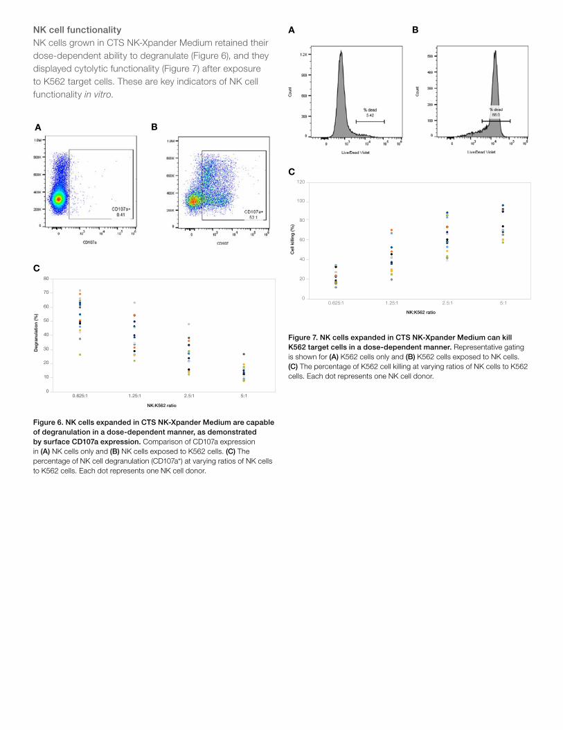
NK cell functionalityNK cells grown in CTS NK-Xpander Medium retained their dose-dependent ability to degranulate (Figure 6), and they displayed cytolytic functionality (Figure 7) after exposure to K562 target cells. These are key indicators of NK cell functionality in vitro.
0
10
20
30
40
50
60
70
80
0.625:1 1.25:1 2.5:1 5:1
Deg
ranu
latio
n (%
)
NK:K562 ratio
Donor 1 Donor 2 Donor 3 Donor 4 Donor 5 Donor 6 Donor 7 Donor 8 Donor 10
Donor 11 Donor 13 Donor 14 Donor 15 Donor 16 Donor 17 Donor 18 Donor 19 Donor 20Figure 6. NK cells expanded in CTS NK-Xpander Medium are capable of degranulation in a dose-dependent manner, as demonstrated by surface CD107a expression. Comparison of CD107a expression in (A) NK cells only and (B) NK cells exposed to K562 cells. (C) The percentage of NK cell degranulation (CD107a⁺) at varying ratios of NK cells to K562 cells. Each dot represents one NK cell donor.
Figure 7. NK cells expanded in CTS NK-Xpander Medium can kill K562 target cells in a dose-dependent manner. Representative gating is shown for (A) K562 cells only and (B) K562 cells exposed to NK cells. (C) The percentage of K562 cell killing at varying ratios of NK cells to K562 cells. Each dot represents one NK cell donor.
0
20
40
60
80
100
120
0.625:1 1.25:1 2.5:1 5:1
Cel
l kill
ing
(%)
NK:K562 ratio
Donor 1 Donor 2 Donor 3 Donor 4 Donor 5 Donor 6 Donor 7 Donor 8 Donor 10
Donor 11 Donor 13 Donor 14 Donor 15 Donor 16 Donor 17 Donor 18 Donor 19 Donor 20
A B
C
A B
C

For Research Use or Manufacturing of Cell, Gene, or Tissue- Based Products. CAUTION: Not intended for direct administration into humans or animals. © 2021 Thermo Fisher Scientific Inc. All rights reserved. All trademarks are the property of Thermo Fisher Scientific and its subsidiaries unless otherwise specified. COL34269 0821
Find out more at thermofisher.com/nkcelltherapy
Ordering information
Product Quantity Cat. No.
Expand
CTS NK-Xpander Medium500 mL bottle A5019001
5 L bag A5019002
Human IL-2 Recombinant Protein 1 mg PHC0023
CTS DPBS, without calcium chloride, without magnesium chloride 2 L bag A1285602
Human AB Serum 100 mL Fisher Scientific, BP2525100
Nunc non-treated 96-well plates Case of 160 268200
Nunc non-treated 48-well plates Case of 75 150787
Analyze
Countess 3 FL Automated Cell Counter 1 instrument AMQAF2000
Trypan Blue Solution, 0.4% 100 mL 15250061
CellTrace CFSE Cell Proliferation Kit 1 kit C34570
eBioscience Flow Cytometry Staining Buffer 600 mL 004222-26
Fc Receptor Binding Inhibitor Polyclonal Antibody 100 tests 14-9161-73
UltraComp eBeads Compensation Beads 100 tests 01-2222-42
Arc Amine Reactive Compensation Kit 1 kit A10346
Attune NxT Acoustic Focusing Cytometer 1 instrument A42858
CD56 Monoclonal Antibody (CMSSB), PE, eBioscience 100 tests 12-0567-42
CD3 Monoclonal Antibody (OKT3), FITC, eBioscience 100 tests 11-0037-42
CD16 Monoclonal Antibody (CB16), APC, eBioscience 100 tests 17-0168-42
CD107a Monoclonal Antibody (eBioH4A3), PE-Cyanine7, eBioscience
100 tests 25-1079-42
LIVE/DEAD Fixable Dead Cell Stain Kit 400 assays L34964
ConclusionsWhile feeder-free culture systems are typically seen as a safer choice for NK cell expansion than feeder-based culture systems, NK cell expansion in existing feeder-free systems like RPMI and serum is significantly lower. The recent development of NK cell–specific specialty media has begun the transition to truly feeder-free culture systems, but they still leave much to be desired in terms of expansion.
CTS NK-Xpander Medium offers a solution to this challenge: feeder-free NK cell culture with CTS NK-Xpander Medium can deliver up to an average 1,500-fold expansion of functional NK cells. CTS NK-Xpander Medium is a xeno-free NK cell culture medium formulated for feeder-free culture that can help reduce regulatory risks for your NK cell expansion protocol.
References1. Abel AM et al. (2018) Natural killer cells: development, maturation, and clinical
utilization. Front Immunol 9:1869.
2. Liu S et al. (2021) NK cell-based cancer immunotherapy: from basic biology to clinical development. J Hematol Oncol 14:7.