exercise 36 blood vessels. the wall of the blood vessels three distinct layers tunica intima lines...
TRANSCRIPT
Exercise 36
Blood vessels
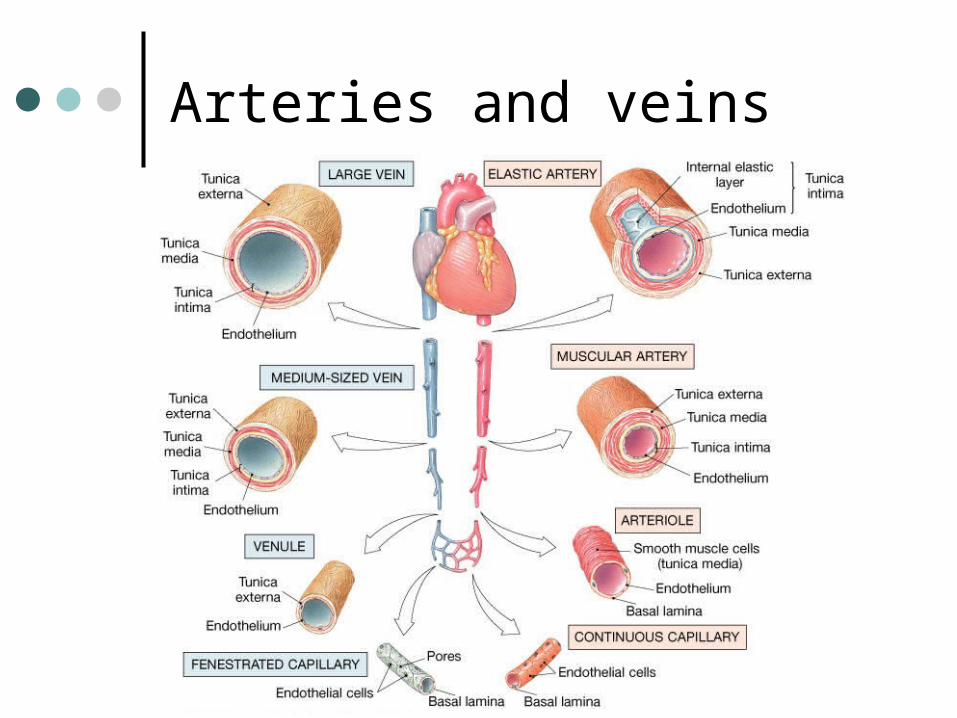
The wall of the blood vessels Three distinct layers
Tunica intima• Lines the lumen• Simple squamous epithelium• endothelium and endocardium
Tunica media• Smooth muscle, • collagen and elastic fibers• Regulates the diameter of the
blood vessels

The wall of the blood vessels
Tunica externa or adventitia• Areolar or fibrous connective
tissue• Supports the vessel• Protects the vessel

The wall of the blood vessels

Arteries
Blood vessels that conduct blood away from the heart and toward tissues. In the pulmonary circulation, pulmonary arteries conduct deoxygenated blood to the lungs. In the systemic circulation, the aorta and its branches conduct oxygenated blood toward the systemic tissues

Arteries
Small arteries are called arterioles. Arterioles conduct blood into a network of even smaller vessels, or capillaries.
Arteries
Subject to pressure fluctuations Thick walls Contain more smooth muscle and
elastic tissues Narrower lumen than veins

Arteries
Can be classified as: Elastic
Closer to the heartLarge arteriesMore elastic fibers than smooth
muscle
Arteries
MuscularFarther from the heartSmaller arteriesMore smooth muscle than elastic
fibers
Veins
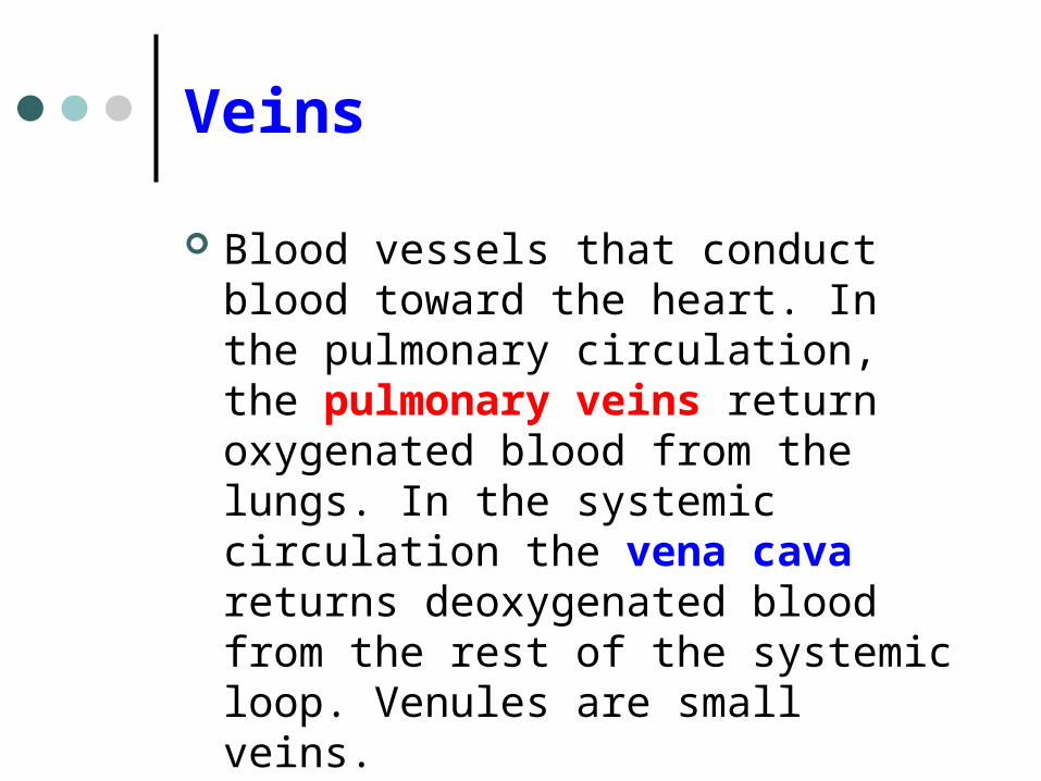
Blood vessels that conduct blood toward the heart. In the pulmonary circulation, the pulmonary veins return oxygenated blood from the lungs. In the systemic circulation the vena cava returns deoxygenated blood from the rest of the systemic loop. Venules are small veins.
Veins
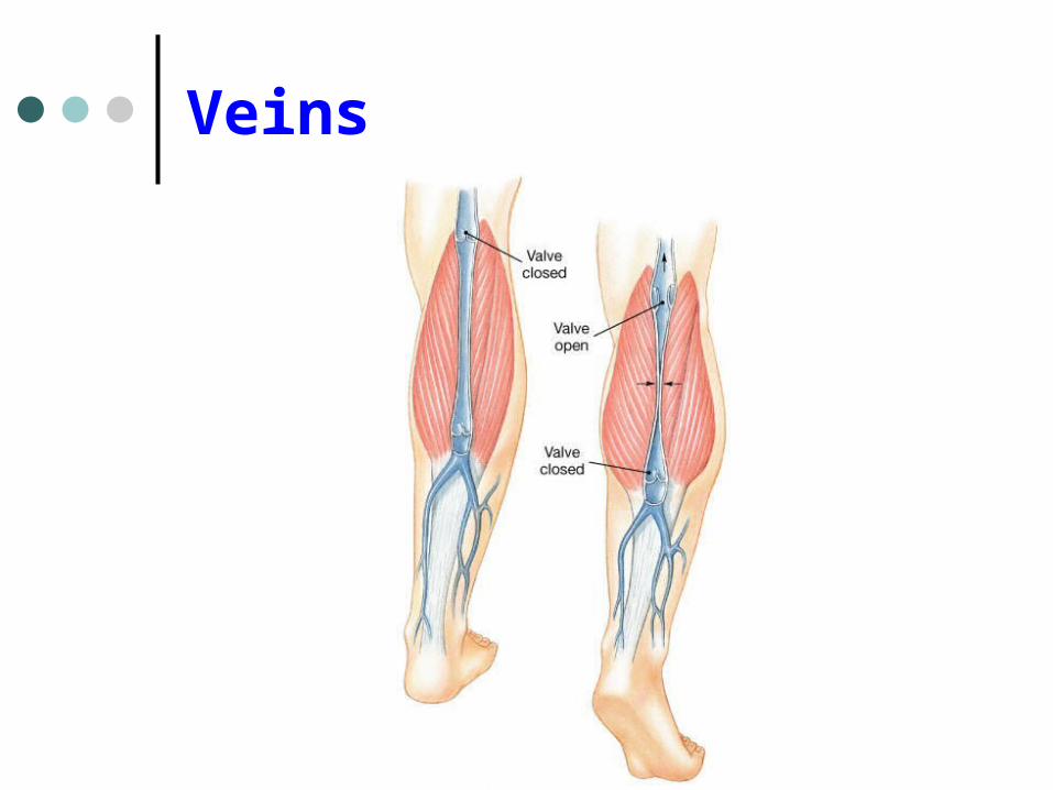
Far from the heart Not subjected to pressure fluctuations Thin walls Larger lumen than arteries Presence of valves Skeletal muscle pump
Veins
Arteries and veins
Aorta
Ascending aorta – first portion Aortic arch Descending aorta – within the thorax Abdominal aorta – within the
abdomen

Aorta
Ascending aorta Right and left coronary arteries
originate from base of aortic sinus Aortic arch
Brachiocephalic trunk – first branch• Right common carotid• Right subclavian
Left common carotid – second branch
Left subclavian – third branch
Aorta
Descending aortaThoracic and abdominal aortas
Head and neck
Internal Carotid External carotid Vertebral Basilar – formed by the fusion of the
vertebral arteries

Head and neck
Head and neck
Circle of WillisPosterior cerebralPosterior communicatingMiddle cerebralAnterior cerebralAnterior communicating
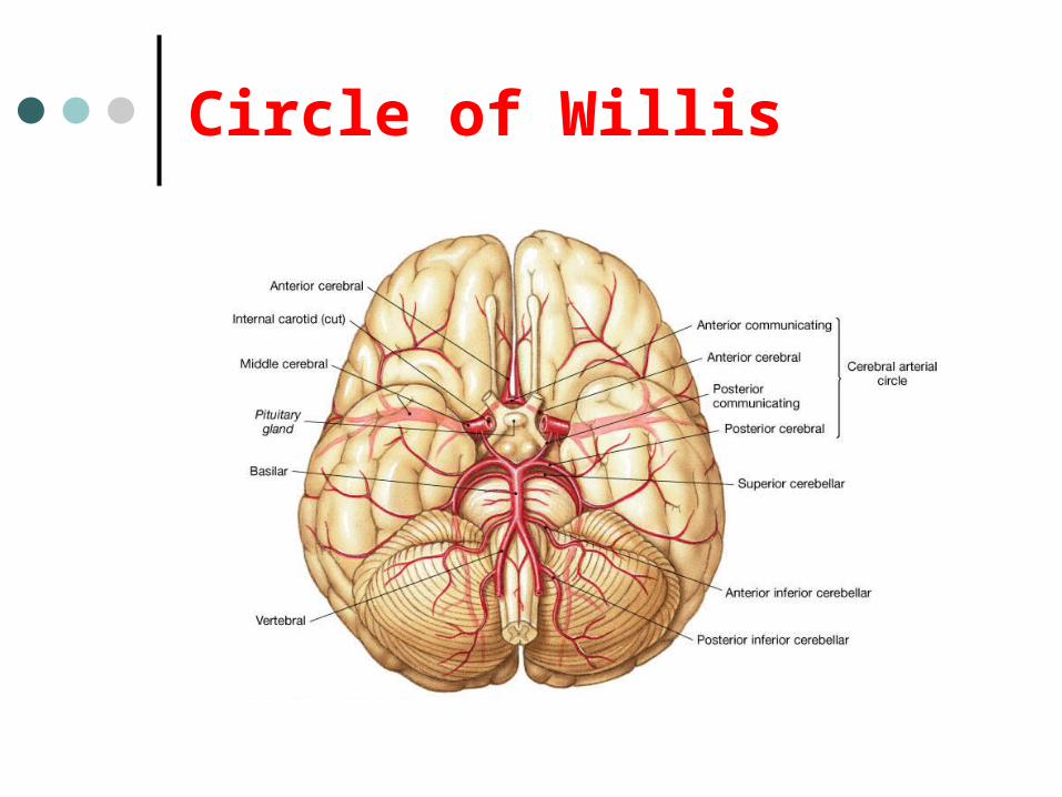
Circle of Willis

Upper limb
Axillary – continuation of the subclavian
Brachial – continuation of the axillary Ulnar – branch of the brachial Radial – branch of the brachial

Upper limb
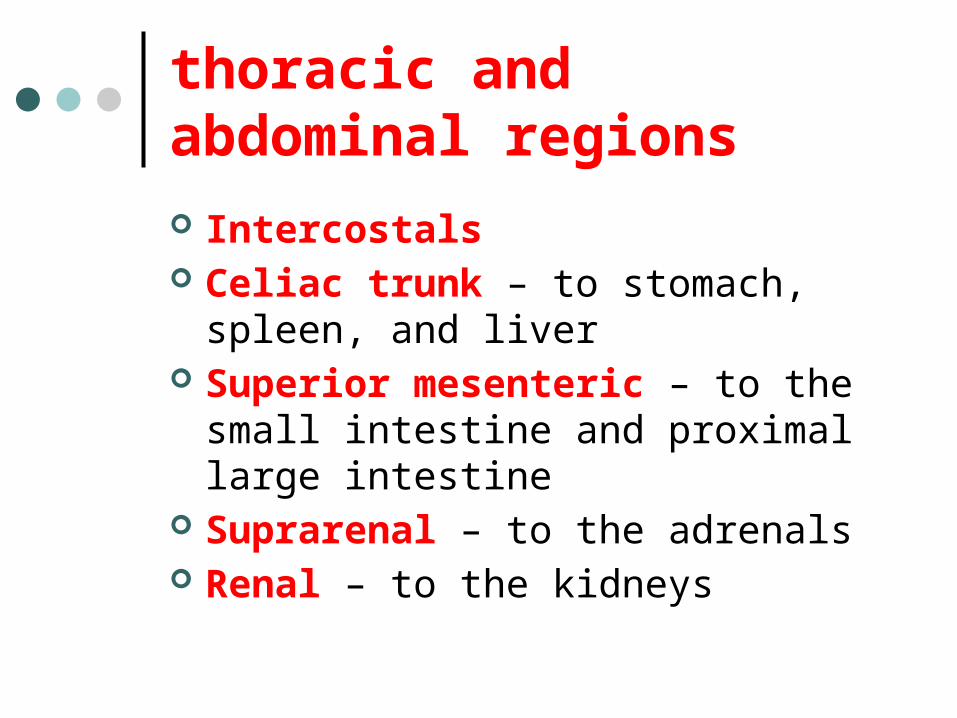
thoracic and abdominal regions
Intercostals Celiac trunk – to stomach, spleen, and
liver Superior mesenteric – to the small
intestine and proximal large intestine Suprarenal – to the adrenals Renal – to the kidneys

thoracic and abdominal regions
GonadalsTesticularOvarian
Inferior mesenteric – to the distal large intestine
Common iliac – branches from the inferior end of the abdominal aorta

thoracic and abdominal regions
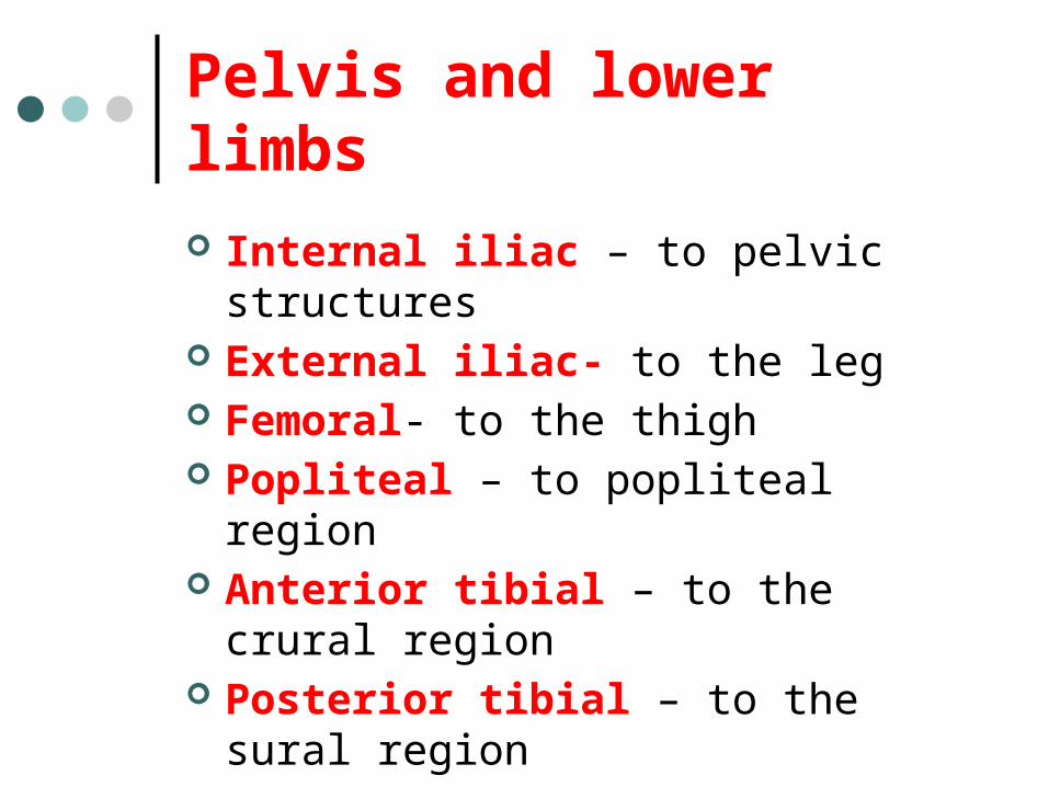
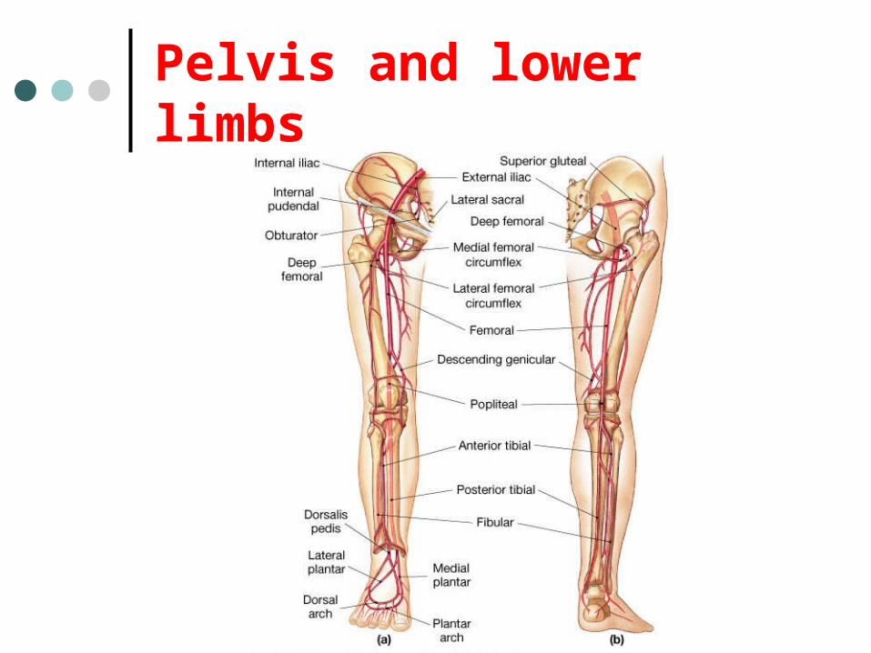
Pelvis and lower limbs
Internal iliac – to pelvic structures External iliac- to the leg Femoral- to the thigh Popliteal – to popliteal region Anterior tibial – to the crural region Posterior tibial – to the sural region Fibular – to fibular region
Pelvis and lower limbs

Chapter 33
PART B

Veins – head and neck
Brachiocephalic – into sup.vena cava Subclavian – lateral branch of
brachiocephalic Internal jugular – medial branch into
the brachiocephalic vein External jugular – external vein of the
neck that returns blood to the subclavian

Head and neck
Head and neck
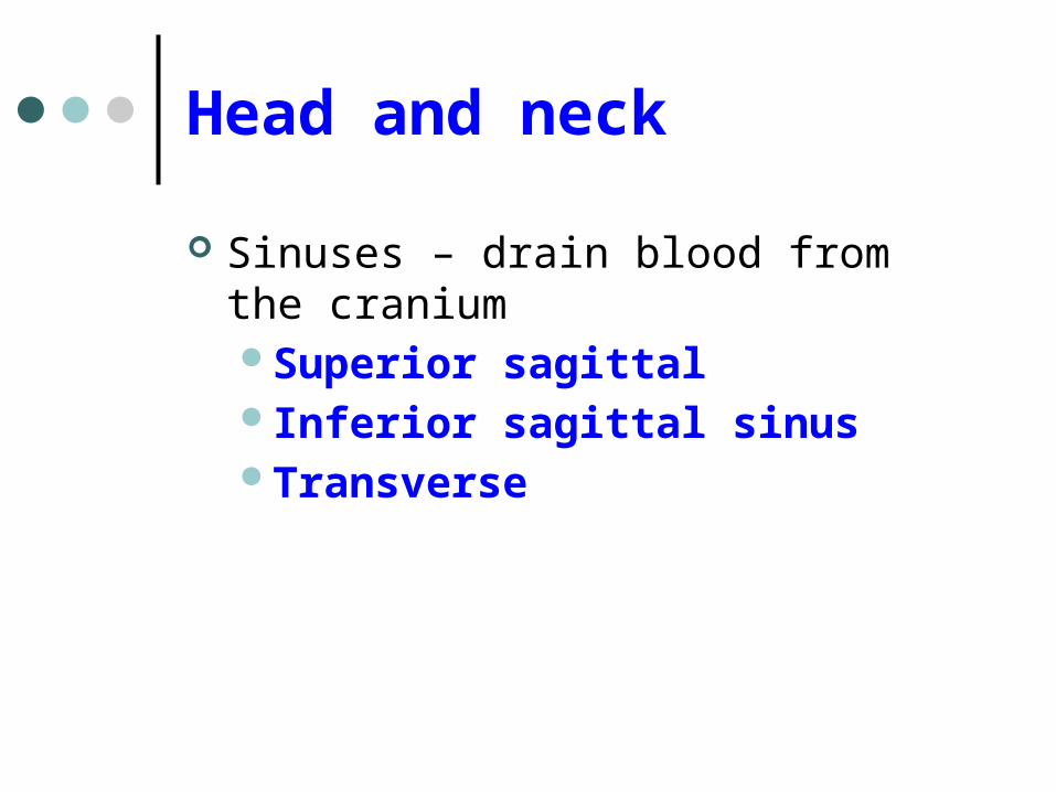
Sinuses – drain blood from the craniumSuperior sagittal Inferior sagittal sinusTransverse
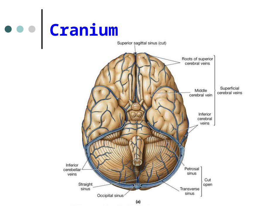
Cranium

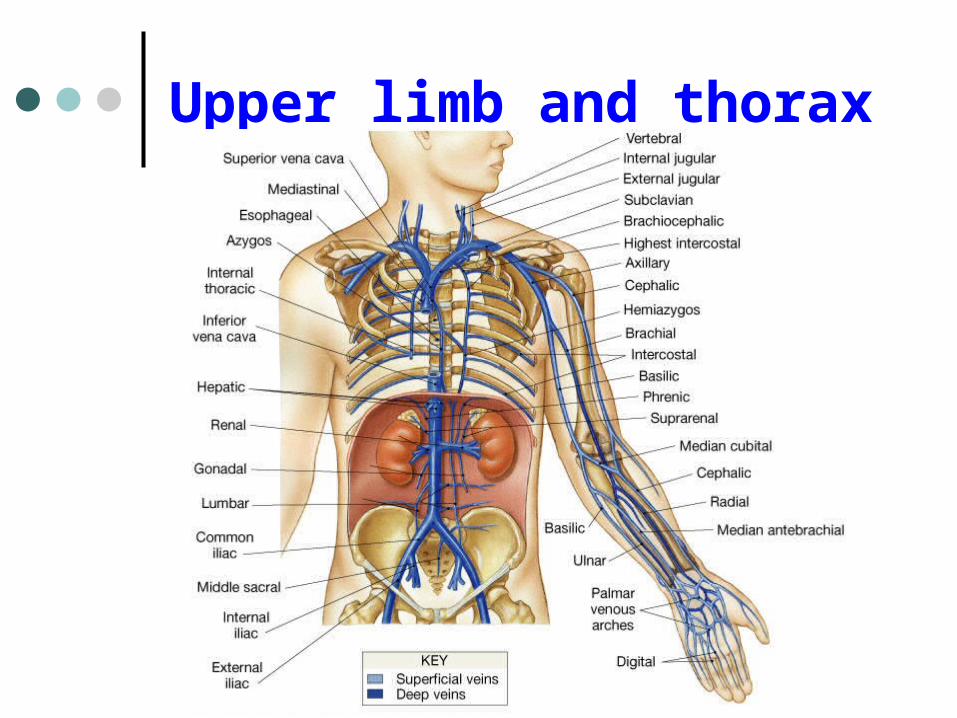
Upper limb and thorax
Axillary – it is a continuation of the subclavian
Basilic – medial and empties into the brachial
Brachial – continuation of axillary Cephalic – lateral and empties into the
axillary Medial cubital – connects basilic to
cephalic

Upper limb and thorax
Ulnar Median Radial Azygos – unpaired branch into the sup. Vena
cava. Drains the right side of the thorax. Hemiazygos, and accessory hemiazygos -2
sets of multiple veins that empty into the azygos and drain the left side of the thorax.
Intercostals
Upper limb and thorax
Abdominal veins
Hepatic Renal Gonadal – testicular or ovarian
Right side empties into inf. Vena cavaLeft side joins with left renal
Common iliac – two branches that fuse to become inf. Vena cava

Hepatic portal system
Portal circulation is a set of vessels that begins and ends with capillary networks.
It returns blood from the digestive system to the liver
From the liver the blood flows to the inf. Vena cava

Hepatic portal system
Hepatic portal vein – going to the liver Liver Hepatic vein - from the liver to inferior
vena cava

Hepatic portal system
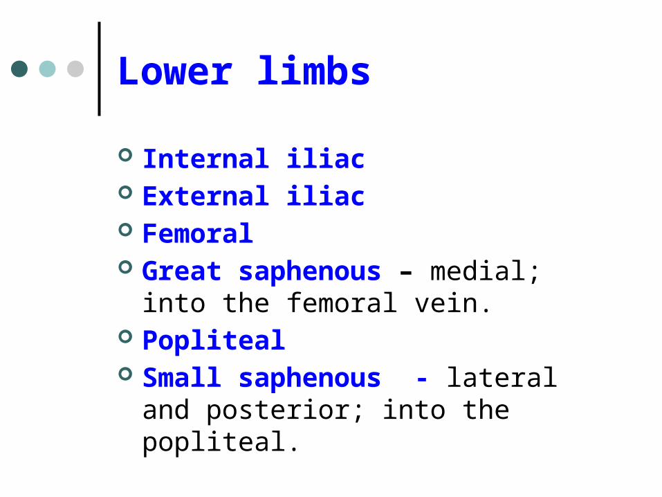
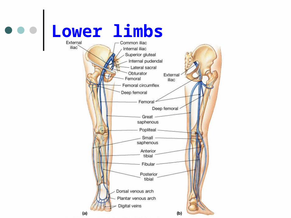
Lower limbs
Internal iliac External iliac Femoral Great saphenous – medial; into the
femoral vein. Popliteal Small saphenous - lateral and
posterior; into the popliteal.

Lower limbs
Anterior tibial - branch into the popliteal
Posterior tibial – branch into the popliteal
Fibular
Lower limbs
Vessels on the Cat Thoracic Cavity and Arm
ArteriesAortaBrachiocephalic trunkCommon carotid- right and leftSubclavian – right and leftAxillaryBrachial
Vessels on the Cat Thoracic Cavity and Arm
VeinsBrachiocephalic trunksJugular –internal and externalSubclavianBrachial
Vessels on the Cat Abdominal Cavity and Leg
ArteriesAbdominal aortaCeliac trunkSuperior mesentericRenalInferior mesentericIliac – common, external and internalFemoral

Vessels on the Cat Abdominal Cavity and Leg
VeinsInferior vena cavaHepatic portal veinRenalIliac – common, internal and externalGreat saphenousFemoral