examining the fingernails - uvaexamining the fingernails mark e. williams, m.d. university of...
TRANSCRIPT
Examining the Fingernails
Mark E. Williams, M.D.University of Virginia School of Medicine
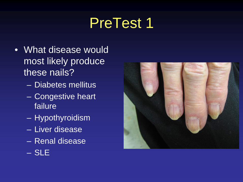
PreTest 1
• What disease would most likely produce these nails?– Diabetes mellitus– Congestive heart
failure– Hypothyroidism– Liver disease– Renal disease– SLE

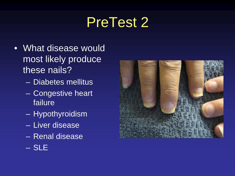
PreTest 2
• What disease would most likely produce these nails?– Diabetes mellitus– Congestive heart
failure– Hypothyroidism– Liver disease– Renal disease– SLE

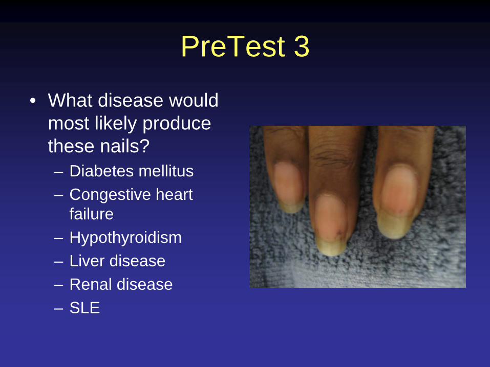
PreTest 3
• What disease would most likely produce these nails?– Diabetes mellitus– Congestive heart
failure– Hypothyroidism– Liver disease– Renal disease– SLE

What information is available from examining the fingernails?
• Overall vitality• Inner emotional state• Cerebral dominance• Occupations and
hobbies• Past medical history
• Nutritional status• Cardiovascular
function• Rheumatic conditions• Dermatological
problems

Sequence of the Examination
• Check the nail shape• Examine the nail color• Survey processes around the nails• Compare hands• Note skin conditions

Observing the Nail Shape

Normal Nail Findings• Softness and flexibility of
free edge • Shape and color• Quality of paronychial tissue• Growth rate
– Six months from cuticle to free edge
– Time of events can be estimated from location
• Nail polish– Distance from base and line
of polish gives approximate date of application (nails grow 0.1mm/ day
– Toenail polish suggests unusual flexibility, a friendly helper, or pedicure

Clubbed fingernails• Causes of clubbing (not exhaustive)
– Pulmonary and Cardiovascular causes (80%)
• Lung cancer, pulmonic abscess, interstitial pulmonary fibrosis, sarcoidosis, beryllium poisoning, pulmonary arteriovenous fistula, subacute bacterial endocarditis, infected arterial grafts, aortic aneurysm
– Gastrointestinal causes (about 5%)• Inflammatory bowel disease, sprue,
neoplasms (esophagus, liver, bowel)– Hyperthyroidism (about 1%)– Note: Chronic Obstructive Pulmonary
Disease does not cause clubbing!
Image courtesy of www.dermnet.comUsed with permission
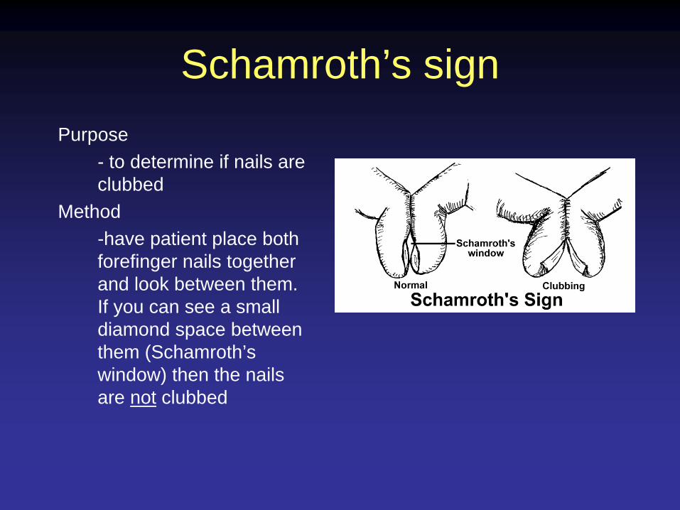
Schamroth’s signPurpose
- to determine if nails are clubbed
Method-have patient place both forefinger nails together and look between them. If you can see a small diamond space between them (Schamroth’s window) then the nails are not clubbed

Spooned nails (Koilonychia)• Water drop test
– Imagine placing a drop of water on the nail with a medicine dropper. If a drop of water would not roll off the nail, it is spooned
• Causes– iron deficiency– diabetes mellitus– Protein deficiency especially in
sulfur-containing amino acids (cysteine or methionine)
Koilonchychia comes from the Greek words for “spoon” and “nail”.
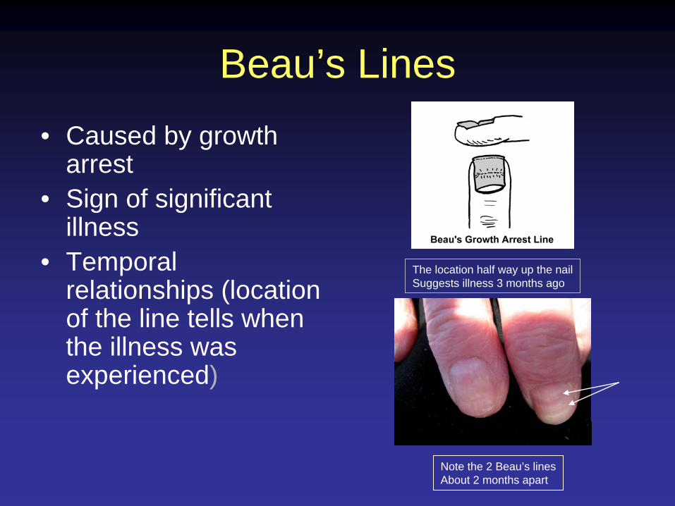
Beau’s Lines• Caused by growth
arrest • Sign of significant
illness • Temporal
relationships (location of the line tells when the illness was experienced)
The location half way up the nailSuggests illness 3 months ago
Note the 2 Beau’s linesAbout 2 months apart

Thin Brittle Nails• Metabolic bone disease
– Nail thinness is correlated with osteopenia
• Thyroid disorder• Systemic amyloidosis
– Yellow waxy flaking• Severe malnutrition
Note the thin nails in this woman with severe osteopenia
Systemic Amyloidosis

Central Nail Ridge
• Causes– Iron deficiency– Folic acid deficiency – Protein deficiency
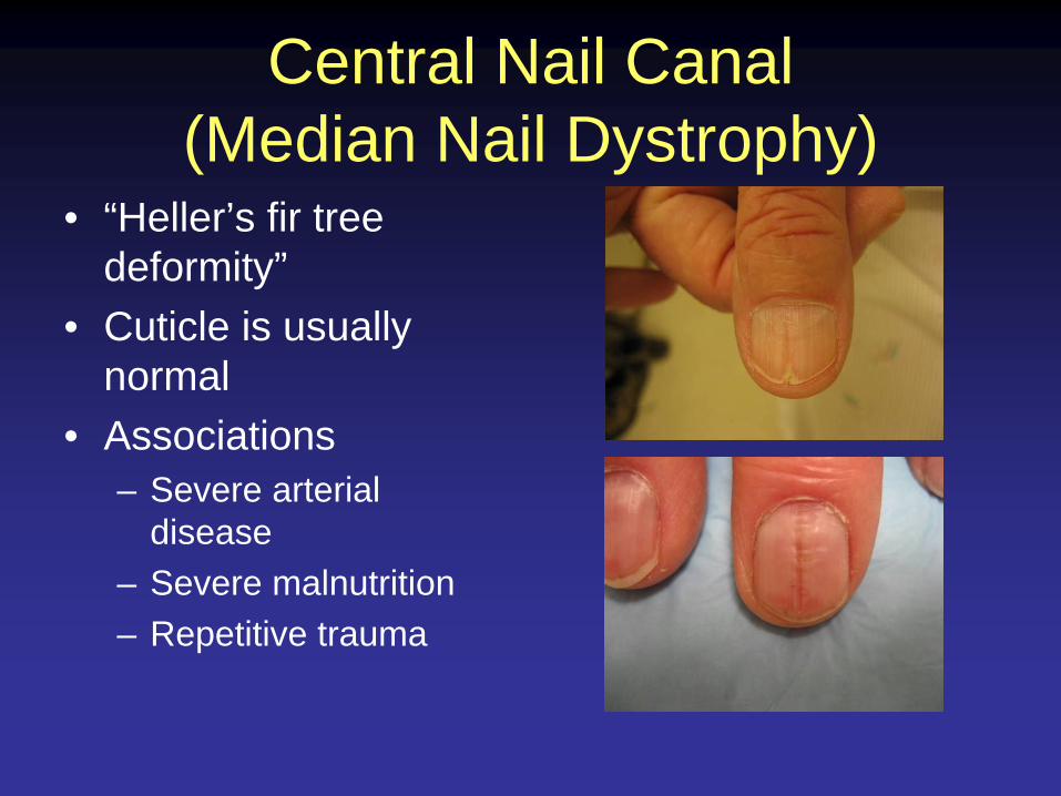
Central Nail Canal (Median Nail Dystrophy)
• “Heller’s fir tree deformity”
• Cuticle is usually normal
• Associations– Severe arterial
disease– Severe malnutrition– Repetitive trauma

Nail Pitting
• Cause is nail matrix inflammation
• Conditions– Psoriasis (random
appearance of pits)– Alopecia areata
(geometric rippled grid)
– Eczema– Lichen planus
Images courtesy of www.dermnet.comUsed with permission

Nail Beading
• Beads seem to drip down the nail like wax
• Associated with endocrine conditions– Diabetes mellitus– Thyroid disorders– Addison’s disease
Image courtesy of www.dermnet.comUsed with permission

Rough Nail Surface
• Nails look sandpapered and dull
• Consider:– autoimmune disease– Psoriasis– Chemical exposure– Lichen planus
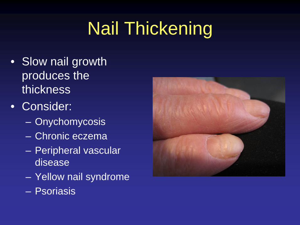
Nail Thickening
• Slow nail growth produces the thickness
• Consider:– Onychomycosis– Chronic eczema– Peripheral vascular
disease– Yellow nail syndrome– Psoriasis

Separation of the Nail Plate (Onycholysis)
• Caused by lifting of the nail plate
• Associations– Thyrotoxicosis– Psoriasis– Trauma– Contact dermatitis– Toxic exposures (solvents)– Porphyria cutanea tarda
(onycholysis and blistering of sun exposed skin)
Traumatic onycholysis(Only involving one nail)
PsoriasisNote the jagged border

Severe Curvature
• Curved or beaked nails– Caused by resorption
of distal digit– Consider
• Hyperparathyroidism• Renal failure• Psoriasis• Systemic sclerosis
Complete Nail Destruction
Local mechanisms:– Trauma– Paronychia
Generalized conditions:– Toxic epidermal necrolysis– Chemotherapy– Bullous diseases– Vasculitis

Observing Nail Color

Abnormalities of the Lunula• Absent
– Anemia– Malnutrition
• Pyramidal– Excessive manicure– Trauma
• Red Discoloration– Cardiovascular disease– Collagen vascular disease– Hematological malignancy– Others
Focal Discolorations of the Nail Plate

Transverse White Lines• Mee’s lines
– Can time the event from location on nail
– Significant illness– Heavy metal toxicity– Chemotherapy
• Muehrcke’s lines– Parallel white irregular lines– Caused by edema to nail plate– Sign of hypoalbuminemia– Lines do not migrate and
disappear when albumin increases
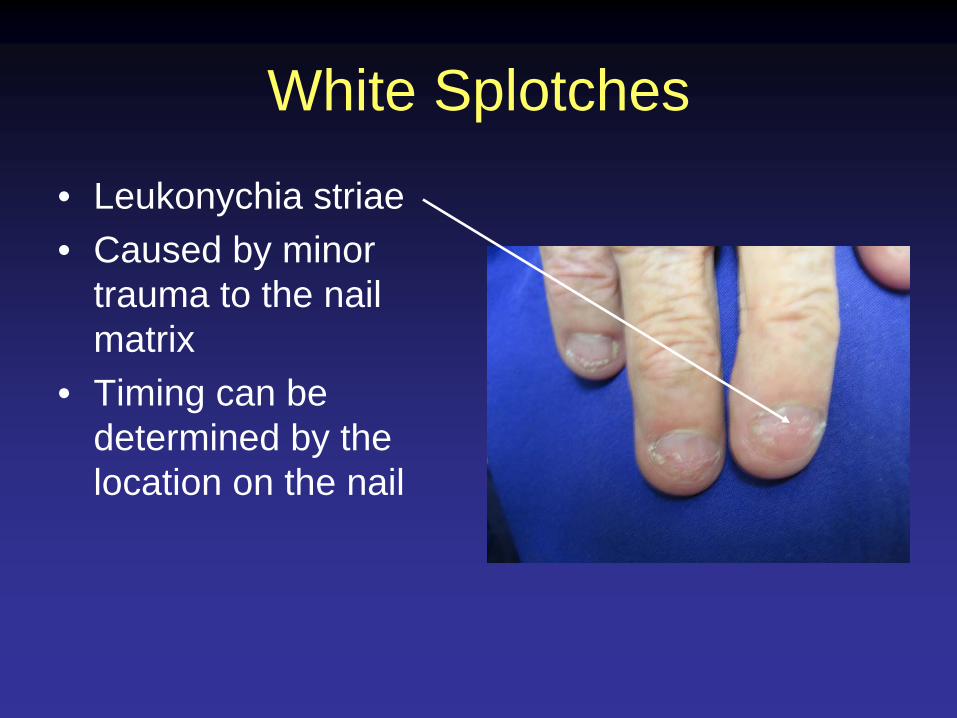
White Splotches
• Leukonychia striae• Caused by minor
trauma to the nail matrix
• Timing can be determined by the location on the nail

Longitudinal Brown Lines• Mechanism
– Increased melanin production by nail matrix melanocytes
• Associations– Addison’s disease– Nevus at nail base– Breast cancer– Melanoma (check for
periungal pigmentation)– Trauma
Image courtesy of www.dermnet.comUsed with permission

Splinter Hemorrhages• Caused by
hemorrhage of distal capillary loops
• Note thickness• Associations
– SBE– SLE– Trichinosis– Pityriasis rubra pilaris– Psoriasis– Renal failure Splinter hemorrhages tend to be fat.

Terry’s Half and Half Nails• Proximal portion is white
(edema and anemia) and the distal portion is dark
• These nails imply either renal or liver disease
• In renal disease there is a brown band at the junction of the erythema and the free edge
Liver disease (no brown line)
Renal disease (brown line)Lower Image courtesy of www.dermnet.com used with permission
Generalized Discolorations of the Nail Plate

White Nails
• Caused by anemia, edema or vascular conditions
• Consider:– Anemia– Renal failure– Cirrhosis– Diabetes mellitus– Chemotherapy– Hereditary (rare)

Pink or Red Nails
• Consider:– Polycythemia (dark)– SLE– Carbon monoxide
(cherry red)– Angioma– Malnutrition

Brown Grey Nails
• Consider:– Cardiovascular
disease– Diabetes mellitus– Vitamin B12 deficiency– Breast cancer– Malignant melanoma– Lichen planus– Syphilis– Topical agents

Yellow Nails
• Consider:– Amyloidosis– Lymphedema and
bronchiectasis (yellow nail syndrome)
– Median/Ulnar nerve injury
– Thermal injury– Jaundice– Diabetes mellitus

Green or Black Nails
• Topical preparations• Chronic Pseudomonas
infection• Trauma

Processes Around the Nail
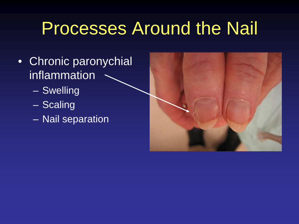
Processes Around the Nail
• Chronic paronychial inflammation– Swelling– Scaling– Nail separation

Periungal telangeictasia
• Dilated capillary loops and atrophy of cuticle
• Strongly associated with collagen vascular disease– SLE– Dermatomyositis– Scleroderma
Image courtesy of www.dermnet.comUsed with permission

Swelling Around the Nail
• Mucus cyst• Fibroma• Malignant
melanoma

Masses• Pyogenic granuloma
• Warts
• Fibroma
• Malignant melanoma

PreTest 1
• What disease would most likely produce these nails?– Diabetes mellitus– Congestive heart
failure– Hypothyroidism– Liver disease– Renal disease– SLE
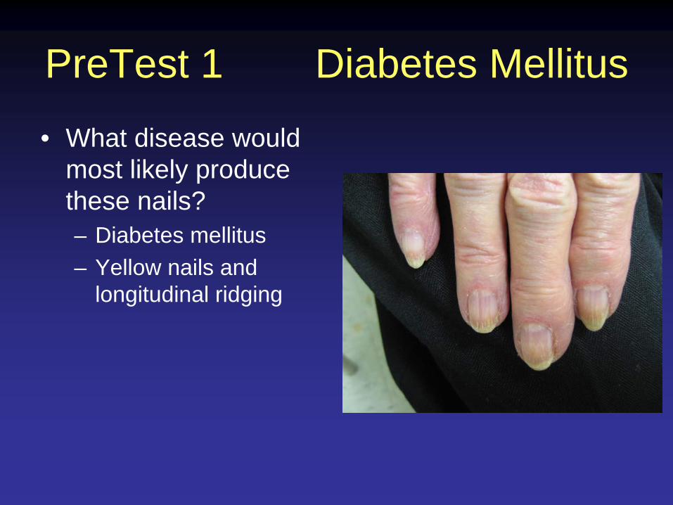
PreTest 1 Diabetes Mellitus
• What disease would most likely produce these nails?– Diabetes mellitus– Yellow nails and
longitudinal ridging
PreTest 2
• What disease would most likely produce these nails?– Diabetes mellitus– Congestive heart
failure– Hypothyroidism– Liver disease– Renal disease– SLE
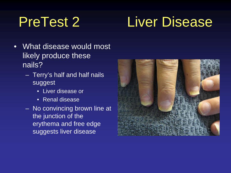
PreTest 2 Liver Disease• What disease would most
likely produce these nails?– Terry’s half and half nails
suggest• Liver disease or• Renal disease
– No convincing brown line at the junction of the erythema and free edge suggests liver disease
PreTest 3
• What disease would most likely produce these nails?– Diabetes mellitus– Congestive heart
failure– Hypothyroidism– Liver disease– Renal disease– SLE

PreTest 3 SLE
• What disease would most likely produce these nails?– SLE– Note the fat splinter
hemorrhage and the periungal telangeictasia
Summary
• Considerable useful information is available from careful examination of the nails
• Starting with the nail examination immediately communicates a sense of diligence and thoroughness
• Remain attentive and continually add to your diagnostic repertoire

Post Test 1
• 71 year old man with lethargy, fatigue, and anorexia
• What is your diagnosis and the evidence that supports it?
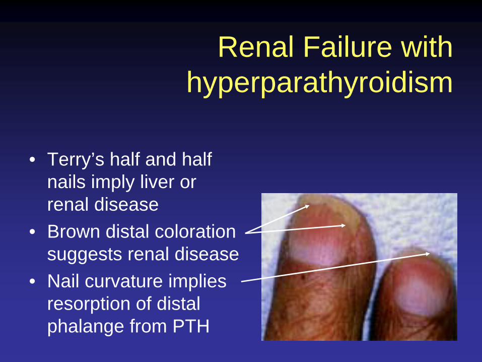
Renal Failure with hyperparathyroidism
• Terry’s half and half nails imply liver or renal disease
• Brown distal coloration suggests renal disease
• Nail curvature implies resorption of distal phalange from PTH
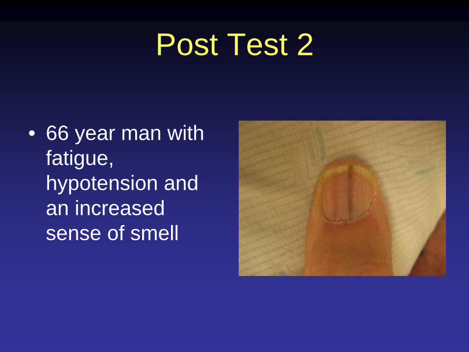
Post Test 2
• 66 year man with fatigue, hypotension and an increased sense of smell
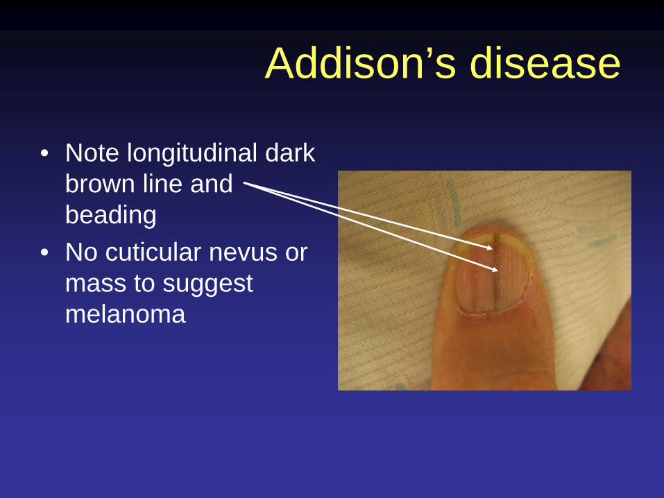
Addison’s disease
• Note longitudinal dark brown line and beading
• No cuticular nevus or mass to suggest melanoma

Post Test 3
• 78 year old with diabetes mellitus, anemia, congestive heart failure and peripheral vascular insufficiency
• What is the evidence?

Post Test 3• Red lunula can imply
CHF• Heller’s line suggests
peripheral vascular disease
• Ridging suggests diabetes or another endocrine condition
• Overall pallor suggests anemia
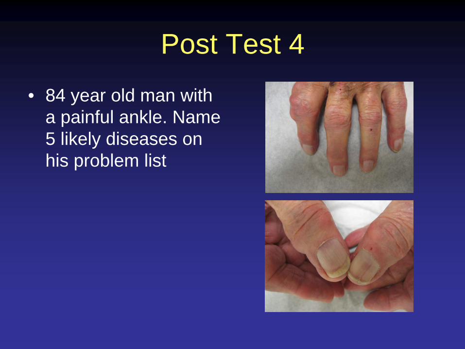
Post Test 4
• 84 year old man with a painful ankle. Name 5 likely diseases on his problem list

Post Test 4
• Gout (tophi)• CHF (red lunula)• Anemia (pallor)• Peripheral arterial
disease (longitudinal red line)
• Chronic kidney disease (distal brown pigmentation)

Post Test 5
• 68 year old man with weight loss and dysphagia
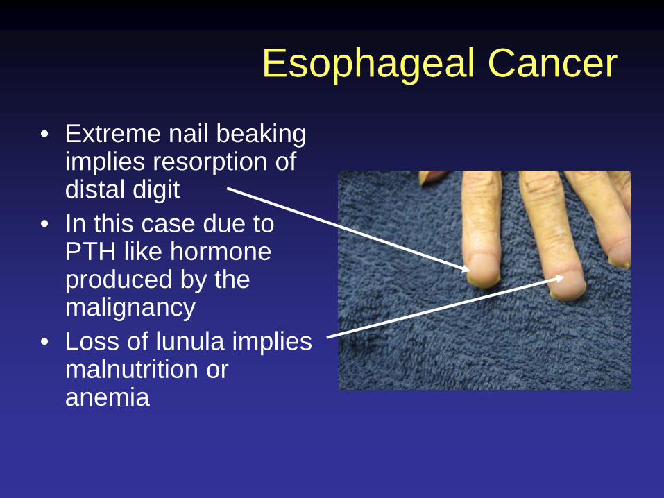
Esophageal Cancer• Extreme nail beaking
implies resorption of distal digit
• In this case due to PTH like hormone produced by the malignancy
• Loss of lunula implies malnutrition or anemia

Post Test 6
• 62 year old woman with proximal muscle weakness, dysphagia and weight loss
Image courtesy of www.dermnet.comUsed with permission

Dermatomyositis• Cuticular atrophy and
periungal telangiectasias suggest collagen vascular disease
• Gottron’s papules over the knuckles imply dermatomyositis
Image courtesy of www.dermnet.comUsed with permission
Systemic lupus affecting the skin over the hands tends to spare the knuckles while
Dermatomyositis tends to involve the knuckles
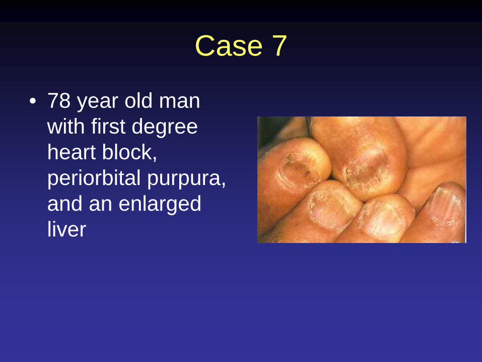
Case 7
• 78 year old man with first degree heart block, periorbital purpura, and an enlarged liver

Case 7 Systemic Amyloidosis
• Thinned, ragged edges
• Yellow discoloration
• Ridging
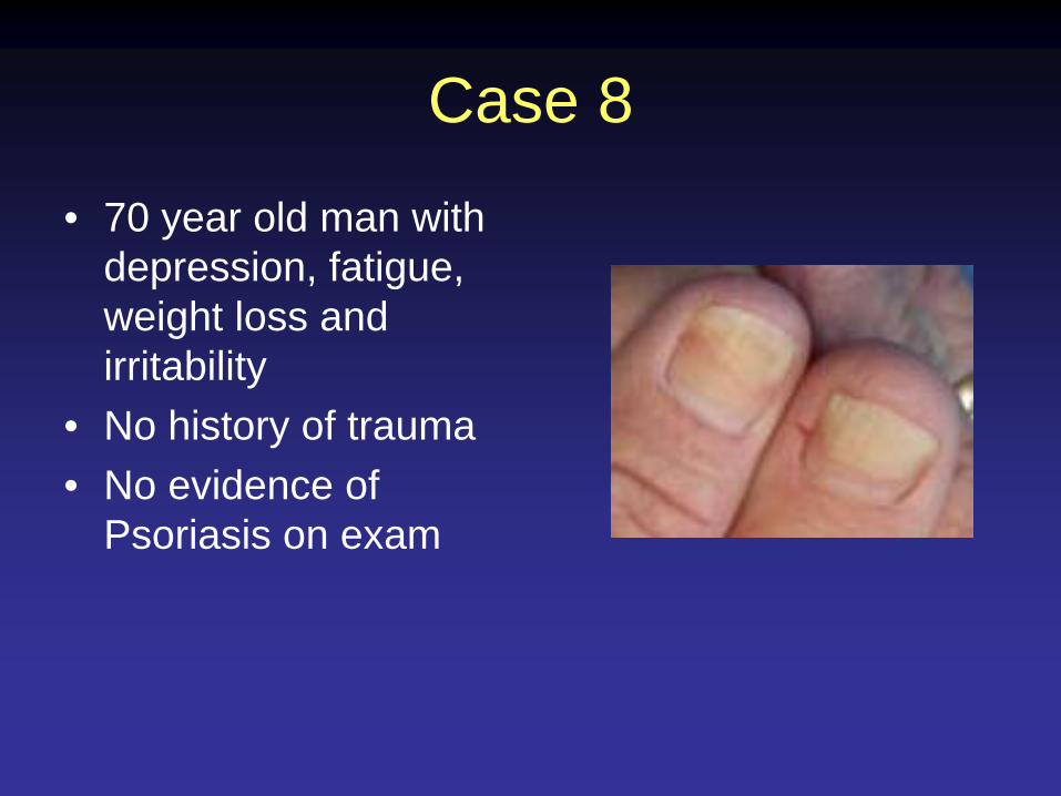
Case 8
• 70 year old man with depression, fatigue, weight loss and irritability
• No history of trauma• No evidence of
Psoriasis on exam
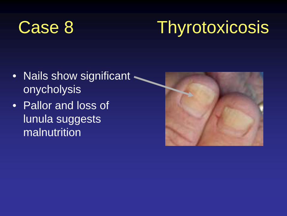
Case 8 Thyrotoxicosis
• Nails show significant onycholysis
• Pallor and loss of lunula suggests malnutrition

Case 9
• 60 year old with painful fingers

Case 9 Psoriasis
• Onycholysis• Splitting of nail
plate• Salmon patch• Chronic
paronychia• Nail disease is
associated with psoriatic arthritis

Case 10
• 75 year old man with weight loss and shortness of breath

Case 10 Lung cancer
• Significant clubbing• Nicotine staining from
cigarette smoking

Case 11
• Ill appearing 60 year old man with fever, malaise, weight loss and painful testicles

Case 11 Polyarteritis Nodosa
• Splitting• Thinning• Ridging• Nail plate infarction• Periungal telangiectasia

Case 12
• 55 year old woman with fatigue, muscle pain and pleuritic chest pain

Case 12 Systemic Lupus
• Periungal telangiectasis and cuticular atrophy suggest collagen vascular disease
• Rash tends to spare the knuckle
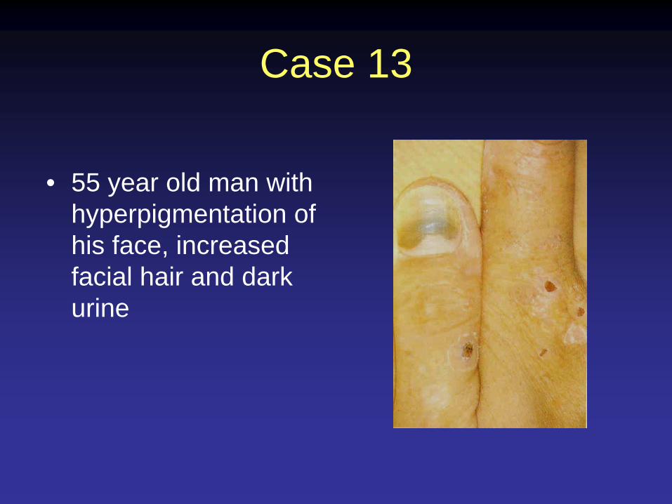
Case 13
• 55 year old man with hyperpigmentation of his face, increased facial hair and dark urine

Case 13 Porphyria Cutanea Tarda
• Onycholysis and blistering
• Due to defective liver uroporphyrinogen decarboxylase
• Urine has coral pink fluorescence under a Wood's lamp

Acknowledgements
• The UVA GME Office for funding support• Dr. Vladimir Goodkovski for technical
assistance• Dr. Jim Thomas of www.Dermnet.com for
permission to use images from their extensive dermatological atlas
• Internal Medicine residents at UVA for pre- testing and helpful feedback