examination of the heart. examination of the heart in the present era of technological advances,...
TRANSCRIPT

Examination Examination
of the Heartof the Heart
Examination of the HeartExamination of the HeartExamination of the HeartExamination of the Heart
In the present era of technological In the present era of technological advances, particularly in the various advances, particularly in the various imaging modalities, there is a growing imaging modalities, there is a growing conception among practicing physicians conception among practicing physicians in cardiovascular medicine that bedside in cardiovascular medicine that bedside physical examination is unnecessary and physical examination is unnecessary and does not provide useful information. does not provide useful information.


It should be emphasized, however, that It should be emphasized, however, that
for proper application and interpretation for proper application and interpretation of various new and old tests that are of various new and old tests that are available for cardiovascular evaluation available for cardiovascular evaluation
in a given patient.in a given patient.
Bedside clinical examination should Bedside clinical examination should
be performed and practiced in the be performed and practiced in the
same way following similar same way following similar
sequences.sequences.
Preparing the patientPreparing the patient
The heart examination should be The heart examination should be made as easy as possible for the made as easy as possible for the patient, who usually expects it to be patient, who usually expects it to be a relatively distasteful experience. If a relatively distasteful experience. If the physician is considerate and the physician is considerate and gentle, the patient should feel when it gentle, the patient should feel when it is all over, that most of his or her is all over, that most of his or her fears on that score were unfounded.fears on that score were unfounded.

The ideal examining room is private, The ideal examining room is private, warm enough to avoid chilling, and warm enough to avoid chilling, and free from distracting noise and sources free from distracting noise and sources of interruption. Adequate (preferably of interruption. Adequate (preferably fluorescent or natural) light is fluorescent or natural) light is essential. essential.

The examining table may be placed The examining table may be placed with its head against the wall, but with its head against the wall, but both sides (particularly the right) and both sides (particularly the right) and the foot should be accessible to the the foot should be accessible to the examiner. And the results should be examiner. And the results should be recorded carefully.recorded carefully.

Landmarks and topographic anatomy
Landmarks and topographic anatomy
Certain basic landmarksCertain basic landmarks
midsternal line(midsternal line( 前正中线前正中线 )) midclavicular lines(midclavicular lines( 锁骨中线锁骨中线 )) anterior, middle, and posterioranterior, middle, and posterior axillary lines(axillary lines( 腋前、中、后线腋前、中、后线 ))

suprasternal notchsuprasternal notch (胸骨上窝)(胸骨上窝) identification of various ribs andidentification of various ribs and
intercostal spaceintercostal space
precordiumprecordium (心前区)(心前区)

InspectionInspection
Inspection of the precordium shoInspection of the precordium should begin at the foot of the bed. The uld begin at the foot of the bed. The subject should be supine with the lesubject should be supine with the leg horizontal and the head and trunk g horizontal and the head and trunk elevated to approximately 15-30 deelevated to approximately 15-30 degrees. grees.


Asymmetry of the thoracic cage due tAsymmetry of the thoracic cage due to a convex bulging of the precordium o a convex bulging of the precordium suggests the presence of heart disease suggests the presence of heart disease since childhood, such as congenital hesince childhood, such as congenital heart disease and rheumatic heart diseasart disease and rheumatic heart disease, with skeletal molding to accommode, with skeletal molding to accommodate cardiac enlargement.ate cardiac enlargement.
In the adult, precordial bulge may be In the adult, precordial bulge may be
produced from the massive pericardiaproduced from the massive pericardia
l effusionl effusion (心包积液)(心包积液) ..
apical impulse (心尖搏动)apical impulse (心尖搏动)
Most part of apex is left ventricle. The Most part of apex is left ventricle. The apex strikes the chest during systole. apex strikes the chest during systole.

The apex impulse is normally located in The apex impulse is normally located in or about the fifth costal interspace insidor about the fifth costal interspace inside the left midclavicular line when the pe the left midclavicular line when the patient is supine. The extent of impulse is atient is supine. The extent of impulse is about 2~2.5 cm.about 2~2.5 cm.

Normal apical impulse : Normal apical impulse : It’s location It’s location
durationduration
intensityintensity
amplitudeamplitude
Usually it is detectable in only one intUsually it is detectable in only one intercostal space and is less than 2-2.5 cercostal space and is less than 2-2.5 cm in diameter. The normal apex impum in diameter. The normal apex impulse is characterized by a brief early sylse is characterized by a brief early systolic out ward thrust of moderate amstolic out ward thrust of moderate amplitude, which ends before the second plitude, which ends before the second heart sound. heart sound.

The apical impulse is normally exagThe apical impulse is normally exag
gerated in thin, young individuals and gerated in thin, young individuals and
when the subject is in the left lateral dewhen the subject is in the left lateral de
cubitus position(cubitus position( 左侧卧位左侧卧位 ). ).
When a patient takes a deep inspiration When a patient takes a deep inspiration and holds his breath, the apical impulse and holds his breath, the apical impulse moves downward from the fifth to the moves downward from the fifth to the sixth interspace.sixth interspace.

When the patient lies on his right side, When the patient lies on his right side, it moves slightly toward the right (1~ it moves slightly toward the right (1~ 2.5cm), and when he lies on his left 2.5cm), and when he lies on his left side it moves about 2~3 cm toward the side it moves about 2~3 cm toward the left.left.

The absence of mobility leads one to The absence of mobility leads one to suspect an adherent pericardium. suspect an adherent pericardium. However, a deep inspiration may bring However, a deep inspiration may bring the lungs over the heart so that the the lungs over the heart so that the impulse disappears altogether.impulse disappears altogether.

Diastolic movements are not Diastolic movements are not
perceptible in most cases, but in perceptible in most cases, but in
children and young adults an early children and young adults an early
diastolic F wave is occasionally diastolic F wave is occasionally
present.present.
Displacement of the apical impulseDisplacement of the apical impulseDisplacement of the apical impulseDisplacement of the apical impulse
Heart diseaseHeart disease
Thoracic diseaseThoracic disease
Abdominal diseaseAbdominal disease

Some heart diseases cause the left Some heart diseases cause the left
ventricular dilatationventricular dilatation (增大)(增大) , the a, the a
pical impulse is displaced laterally and pical impulse is displaced laterally and
inferiorly and sustained ,inferiorly and sustained ,
Heart diseaseHeart disease
and it may be shifted to the left and and it may be shifted to the left and
upward in right ventricular dilatation . upward in right ventricular dilatation .

In mitral diseIn mitral disease the impulase the impulse is displacese is displaced laterally.d laterally.
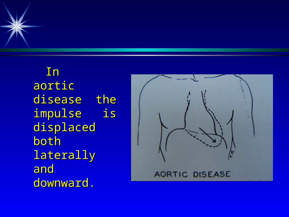
In aortic In aortic disease the disease the impulse is impulse is displaced both displaced both laterally and laterally and downward.downward.
It can be found at the right fifth inIt can be found at the right fifth intercostal space in dextrocardiatercostal space in dextrocardia ((右位心) 右位心) and can not be found iand can not be found in massive pericardial effusion.n massive pericardial effusion.

PneumothoraxPneumothorax (气胸) (气胸) and pleural eand pleural effusionffusion (胸腔积液) (胸腔积液) will displace thwill displace the apical impulse to the normal side. Plee apical impulse to the normal side. Pleural-adhesionural-adhesion (胸膜粘连) (胸膜粘连) and ateleand atelectasisctasis (肺不张) (肺不张) will result in a dispwill result in a displacement of impulse toward the diseaselacement of impulse toward the diseased side.d side.
Thoracic diseaseThoracic disease
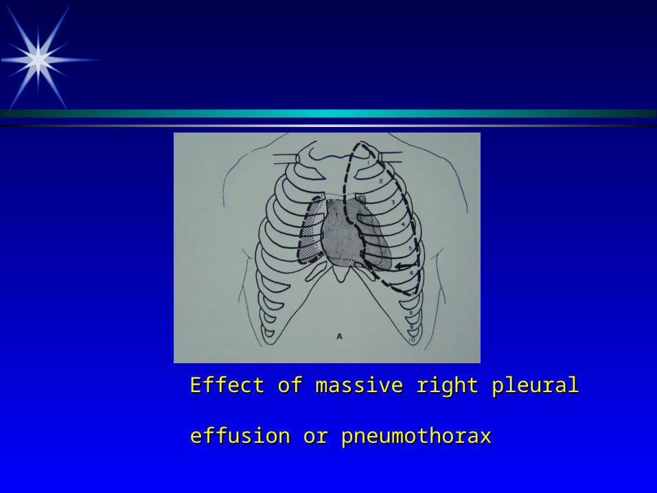
Effect of massive right pleural Effect of massive right pleural effusion or pneumothoraxeffusion or pneumothorax
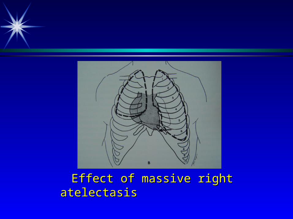
Effect of massive right atelectasisEffect of massive right atelectasis
The examiner should always observe the shape and contour of patient’s chest. Depressions of the sternum, Kyphosis of dorsal spine (驼背) , scoliosis (脊柱侧凸) often alter the shape and position of the apical impulse.

The apical impulse also can be displaceThe apical impulse also can be displaced by large mass(d by large mass( 肿瘤肿瘤 ), massive ascite), massive ascites(s( 腹水腹水 ).).
Abdominal diseaseAbdominal disease
The apical impulse may have increased amplitude and duration in those persons with a thin chest, anemia( 贫血 ), fever, hyperthyroidism ( 甲亢 ) and anxiety.

Inward impulse(Inward impulse( 负性心尖搏动负性心尖搏动 ): the a): the apex depress far from the chest instead opex depress far from the chest instead of strikes the chest during systole. Broadf strikes the chest during systole. Broadbent’s sign is of value in the diagnosis bent’s sign is of value in the diagnosis of adherent pericardium(of adherent pericardium( 粘连性心包粘连性心包炎炎 ). It is also seen in RVH.). It is also seen in RVH.

Abnormal pulsations in the other areas:Abnormal pulsations in the other areas:Abnormal pulsations in the other areas:Abnormal pulsations in the other areas:
Right vertricular hypertophy (RVH). TRight vertricular hypertophy (RVH). The impulse is clearly seen in left third fhe impulse is clearly seen in left third fourth intercostal space.ourth intercostal space.

Pulmonary emphysema(Pulmonary emphysema( 肺气肿肺气肿 ) with ) with RVH, usually the pulsation can be founRVH, usually the pulsation can be found inferior the xiphoid process(d inferior the xiphoid process( 剑突下剑突下搏动搏动 ).).
In ascending or arch aortic aneurysm(In ascending or arch aortic aneurysm(主动脉瘤主动脉瘤 ), one may detect abnormal ), one may detect abnormal pulsations in aortic area, with bulging opulsations in aortic area, with bulging or pulsation in systole.r pulsation in systole.
Pulmonary hypertension with dilatation Pulmonary hypertension with dilatation the pulsation in systole may be detectethe pulsation in systole may be detected in left second intercostal space to the d in left second intercostal space to the edge of sternum.edge of sternum.

Marked pulsation at the base of the heaMarked pulsation at the base of the heart is seen in aortic insufficiency(rt is seen in aortic insufficiency( 主闭主闭 )), in a dilated aorta or a saccular aneurys, in a dilated aorta or a saccular aneurysm.m.
ReviewReviewReviewReview
Precordial bulgePrecordial bulge ( ( 心前区隆起心前区隆起 ))
congenital heart diseasecongenital heart disease
rheumatic heart diseaserheumatic heart disease
(before puberty)(before puberty)
pericardical effusionpericardical effusion
(adult life)(adult life)

Normal apical impulseNormal apical impulseNormal apical impulseNormal apical impulse
The apex impulse is normally located iThe apex impulse is normally located in or about the fifth costal interspace insn or about the fifth costal interspace inside the left midclavicular line when thide the left midclavicular line when the patient is supine. The extent of impule patient is supine. The extent of impulse is about 2~2.5 cm.se is about 2~2.5 cm.

Displacement of the apical impulseDisplacement of the apical impulseDisplacement of the apical impulseDisplacement of the apical impulse
Heart diseaseHeart disease LVDLVD
displaced todisplaced to
lateral and inferiorlateral and inferior
Heart diseaseHeart disease LVDLVD
displaced todisplaced to
lateral and inferiorlateral and inferior

Displacement of the apical impulseDisplacement of the apical impulseDisplacement of the apical impulseDisplacement of the apical impulse
RVDRVD
displaced todisplaced to
left and upwardleft and upward
RVDRVD
displaced todisplaced to
left and upwardleft and upward

Displacement of the apical impulseDisplacement of the apical impulseDisplacement of the apical impulseDisplacement of the apical impulse
Congenital dextrocardiacCongenital dextrocardiac
right right
CHF, myocarditis, myocardiopathyCHF, myocarditis, myocardiopathy
apical impulseapical impulse
decrease intensitydecrease intensity
Congenital dextrocardiacCongenital dextrocardiac
right right
CHF, myocarditis, myocardiopathyCHF, myocarditis, myocardiopathy
apical impulseapical impulse
decrease intensitydecrease intensity

Displacement of the apical impulseDisplacement of the apical impulseDisplacement of the apical impulseDisplacement of the apical impulse
Massive pericardial effusionMassive pericardial effusion
apical impulseapical impulse
disappeardisappear
Massive pericardial effusionMassive pericardial effusion
apical impulseapical impulse
disappeardisappear
Displacement of the apical impulseDisplacement of the apical impulseDisplacement of the apical impulseDisplacement of the apical impulse
Thoracic diseaseThoracic disease
pneumothorax, pleural effusionpneumothorax, pleural effusion
shifted toshifted to
healthy sidehealthy side
Thoracic diseaseThoracic disease
pneumothorax, pleural effusionpneumothorax, pleural effusion
shifted toshifted to
healthy sidehealthy side
Displacement of the apical impulseDisplacement of the apical impulseDisplacement of the apical impulseDisplacement of the apical impulse
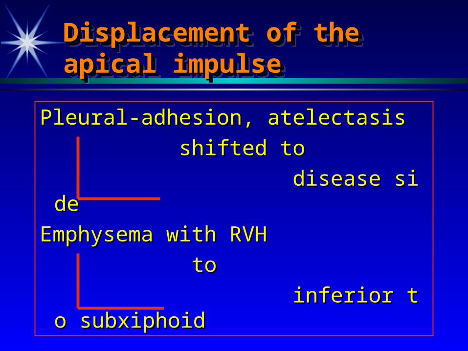
Pleural-adhesion, atelectasisPleural-adhesion, atelectasis
shifted to shifted to
disease sidedisease side
Emphysema with RVHEmphysema with RVH
toto
inferior to subxiphoid inferior to subxiphoid

What’s the meaning ofWhat’s the meaning of
Apical ImpulseApical ImpulseInward ImpulseInward ImpulseBroadbent’ signBroadbent’ sign
单选题单选题
正常成人心尖搏动位于正常成人心尖搏动位于A. A. 第四肋间第四肋间 ,, 左锁骨中线内侧左锁骨中线内侧 0.1~0.5cm0.1~0.5cm
B. B. 第五肋间第五肋间 ,, 左锁骨中线内侧左锁骨中线内侧 0.5~1.0cm0.5~1.0cm
C. C. 第五肋间第五肋间 ,, 右锁骨中线内侧右锁骨中线内侧 0.5~1.0cm0.5~1.0cm
D. D. 第四肋间第四肋间 ,, 左锁骨中线内侧左锁骨中线内侧 1.0~1.5cm1.0~1.5cm
E. E. 第五肋间第五肋间 ,, 右锁骨中线内侧右锁骨中线内侧 2.0~2.5cm2.0~2.5cm
正常成人心尖搏动范围以直径计算为正常成人心尖搏动范围以直径计算为A. 1.0~1.5cmA. 1.0~1.5cm
B. 1.5~2.0cmB. 1.5~2.0cm
C. 2.0~2.5cmC. 2.0~2.5cm
D. 2.5~3.0cmD. 2.5~3.0cm
E. E. 以上都不是以上都不是
心尖搏动的论述心尖搏动的论述 ,, 错误的是错误的是A. A. 搏动范围以直径计算为搏动范围以直径计算为 1.0~1.5cm1.0~1.5cm
B. B. 可位于第五肋间左锁骨中线内可位于第五肋间左锁骨中线内 0.5cm0.5cm
C. C. 可位于第四肋间可位于第四肋间D. D. 可位于第六肋间可位于第六肋间E. E. 体位、体型对心尖搏动位置有影响体位、体型对心尖搏动位置有影响

心尖搏动移位的论述心尖搏动移位的论述 ,, 错误的是错误的是A. A. 肥胖体型者肥胖体型者 ,, 心尖搏动可上移至第四肋间心尖搏动可上移至第四肋间B. B. 瘦长体型者瘦长体型者 ,, 心尖搏动可下移至第六肋间心尖搏动可下移至第六肋间C. C. 左心室增大时心尖搏动向左下移位左心室增大时心尖搏动向左下移位D. D. 右心室增大时心尖搏动向右移位右心室增大时心尖搏动向右移位E. E. 一侧胸膜粘连、增厚、心尖搏动向患侧移位一侧胸膜粘连、增厚、心尖搏动向患侧移位

心前区搏动错误的是心前区搏动错误的是A. A. 胸骨左缘第胸骨左缘第 3~43~4 肋间搏动可见于右心室肥大肋间搏动可见于右心室肥大B. B. 剑突下搏动可见于右心室肥大剑突下搏动可见于右心室肥大 ,, 亦可见于腹主亦可见于腹主
动脉瘤动脉瘤C. C. 胸骨左缘第胸骨左缘第 22 肋间收缩期搏动可见于肺动脉肋间收缩期搏动可见于肺动脉
高压高压D. D. 胸骨右缘第胸骨右缘第 22 肋间收缩期搏动可见于主动脉肋间收缩期搏动可见于主动脉
弓动脉瘤弓动脉瘤E. E. 以上都不是以上都不是