examination of the ear - the hillingdon hospitals examination of the ear otoscopy. anatomy of the...
TRANSCRIPT
Examination of the ear
Otoscopy

Anatomy of the ear
Eustachian tube = auditory tube

Examining the earWash your handsGRIP
Greet, Rapport, Introduce and Identify, explain Procedure, ensure Privacy
EquipmentOtoscopeOtoscope speculum512Hz Tuning fork
Position patientSeated at same level as youAccess to both ears
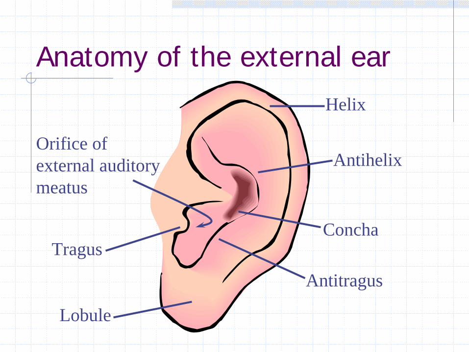
Anatomy of the external ear
Tragus
Helix
Antihelix
Lobule
Antitragus
Orifice of external auditory meatus
Concha

The external ear
External ear = external auditory meatus (EAM) + pinna (mobile part of external ear a.k.a. auricle)Begin with the pinna.Examine for any deformity or skin changes.Look for scars or pits in front of, or behind ear.Hearing aids should be noted then removed.Tug the pinna gently for any tenderness.

Congenital abnormalities
Skin tag / accessory
auricle
Preauricular pit

Acquired abnormalities
Mastoiditis Perichondral haematoma
Erysipelasmore info

Palpate for pre/post-auricular lymph nodes

OtoscopeOtoscope is an instrument which is used to view the earEnsure the batteries are workingUse a clean speculum for each patientUse the largest speculum that will comfortably fit in EAM
Bulb
Speculum
Pneumatic bulb attaches here
Viewing lens

Examination with an otoscopeTurn on the lightGrip the otoscope like a pencilFor right ear, hold in your right hand, handle pointing forwardsFor left ear, hold in your left hand, handle pointing forwardsPull the pinna up and back to straighten EAM

Examination with an otoscope
Look in the ‘good’ ear first
That way, if there is infection present in the ‘bad’ ear you will not transfer it to the other side

Look inside!
Insert speculum tip into EAMGradually move the speculum into EAM under direct vision through the instrumentLook at
External auditory meatusFB, wax, skin, shape
Tympanic membrane (TM)‘Behind’ TM

External auditory meatus
Zoster Foreign body
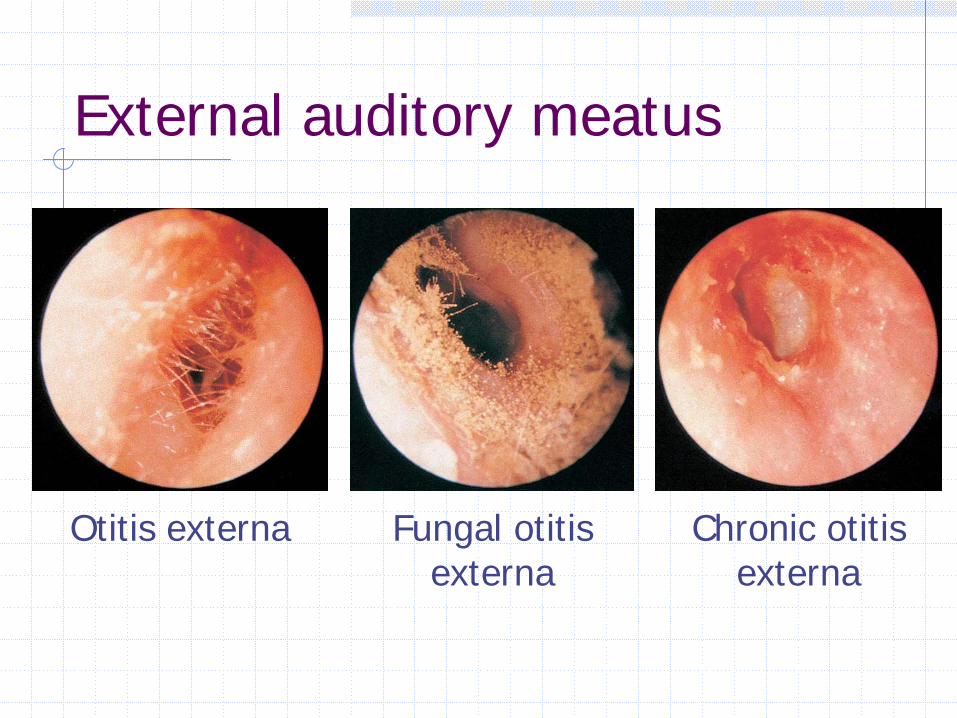
External auditory meatus
Otitis externa Fungal otitis externa
Chronic otitis externa
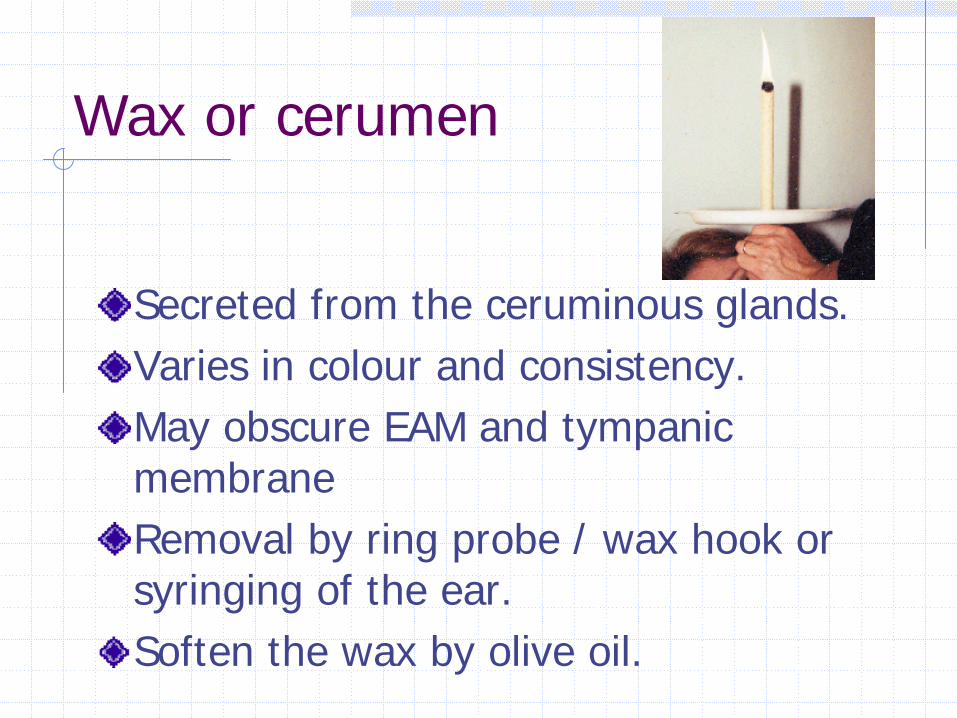
Wax or cerumen
Secreted from the ceruminous glands.Varies in colour and consistency.May obscure EAM and tympanic membraneRemoval by ring probe / wax hook or syringing of the ear.Soften the wax by olive oil.

Anatomy of tympanic membrane
Umbo
Chorda tympaniShort process/ handle -MalleusIncusRound window
Auditory tubeLight conePromontory
Features behind TM not always visibleFeatures behind TM not always visible
Pars tensaPars tensa
Pars flaccida
Pars flaccida

Anatomy of the ear

The Tympanic Membrane.
Anatomical features sought and notedPars tensaPars flaccidaHandle of malleusShort process of malleusUmboCone of lightAnterior and posterior malleolar folds

TM disease
Otitis media Cholesteatoma Tympanosclerosis

TM perforation

Tympanostomy tube
•Ventilates middle ear•Does not drain “glue”

Tuning fork tests
Rinné and Weber testsDetect and differentiate conductive and sensorineural hearing lossGive you something to do whilst waiting for the pure tone audiogram (PTA)

Rinné test
Tap 512Hz tuning fork against bony part, or heel of Gucci loafersPlace base firmly on mastoid processAsk patient to tell you when sound disappearsHold fork tips 2cm from EAM
Can the patient hear it now?
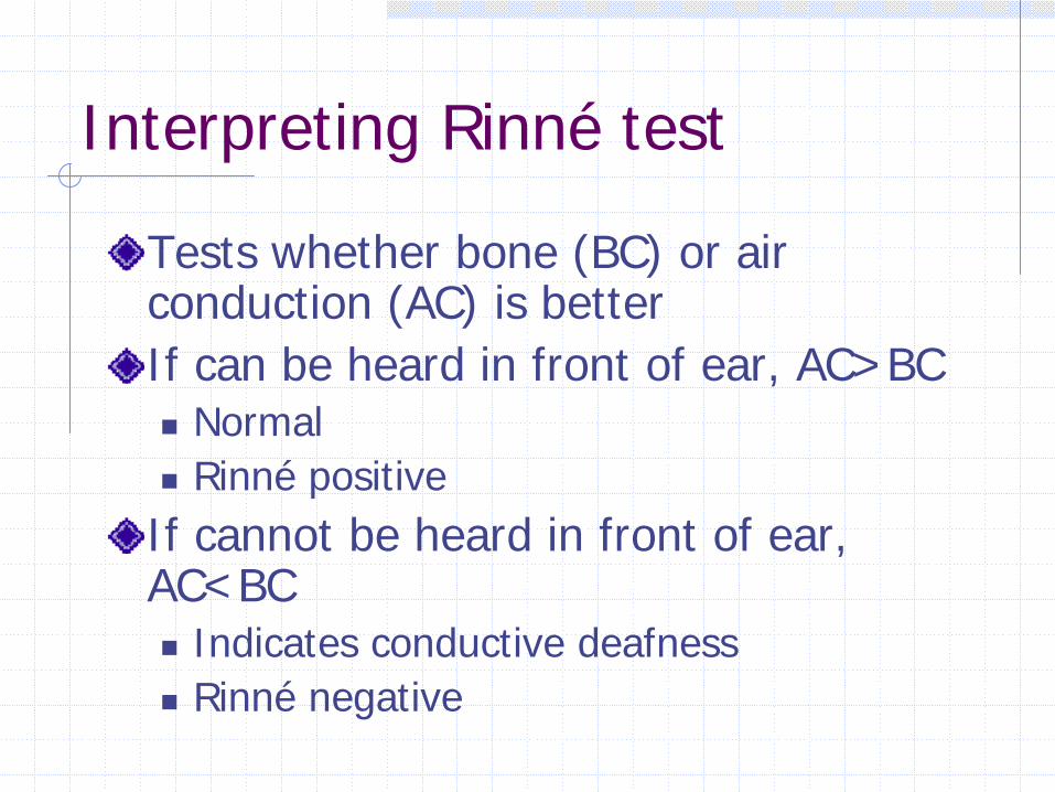
Interpreting Rinné test
Tests whether bone (BC) or air conduction (AC) is betterIf can be heard in front of ear, AC>BC
NormalRinné positive
If cannot be heard in front of ear, AC<BC
Indicates conductive deafnessRinné negative

Weber test
Strike tuning fork againHold somewhere on the head in the midline (firmly)
Usually vertex or foreheadCan use upper incisors
Which side is louder?

Interpreting Weber test
Conductive deafnesslocalises to abnormal ear
Sensorineural deafnesslocalises to normal ear
Try it on yourself with a finger in an earWhere does the sound localise?Have you caused conductive or sensorineural deafness?

PracticalInspect external earLook at
External auditory meatusFB, wax, skin, shape
Tympanic membrane (TM)‘Behind’ TM
Anatomical features sought and noted
Pars tensaPars flaccidaHandle of malleusShort process of malleusUmboCone of lightAnterior and posterior malleolar folds