examination of cardiovascular system · examination of cardiovascular system 1. iap ug teaching...
TRANSCRIPT
IAP UG Teaching slides 2015-16
EXAMINATION OF CARDIOVASCULAR SYSTEM
1

IAP UG Teaching slides 2015-16
CHIEF COMPLAINTS
• Breathlessness(Grading)• Poor feeding , ‘suck‐ rest‐ suck’ cycle• Palpitation • Chest pain• Cough• Edema • Failure to thrive • Joint pain / swelling • Syncope
2

IAP UG Teaching slides 2015-16
GRADING OF DYSPNOEA(NYHA)
Class Activity
I Vigorous exercise(eg:climbing stairs)
II Routine activity
III Minimal activity(walking from one room to another)
IV Dyspnea at rest
3
IAP UG Teaching slides 2015-16
HISTORY • Feeding habits
• Cyanotic spells
• Squatting episodes
• Sore throat
• Head sweating
• Pink frothy sputum
• Convulsions
• Recurrent LRTIs
4

IAP UG Teaching slides 2015-16
ANTENATAL/NATAL HISTORY
• Maternal Infection: ‐Rubella(PDA,Pulm branch stenosis) ‐Mumps(EFE) ‐Diabetes(Septal hypertrophy,TGV) ‐SLE(Complete heart block) ‐PKU(TOF, VSD)• Maternal drugs: Eg: Alcohol, Phenytoin,Lithium• Preterm baby: PDA
5

IAP UG Teaching slides 2015-16
GENERAL EXAMINATION
• Anemia • Cyanosis• Clubbing• Oedema • Signs of infective endocarditis • Signs of cardiac failure • Signs of Rheumatic fever• Peripheral signs of aortic regurgitation• In Syndromes – congenital heart disease
6
IAP UG Teaching slides 2015-16
FACIES• Mitral Facies• Moon Face• Elfin Face• Typical syndromes‐eg: Downs syndrome• Apprehensive facies produced by pain, anxiety and respiratory distress
• PE• Arrhythmias as VT, fast AF
7
IAP UG Teaching slides 2015-16
EYES & LIDS
• Blue sclera ‐ marfan syndrome, Ehlers‐Danlos syndrome, (associated with AR, MVP, ASD)
• Lens - (subluxation in Marfan‐ superior;homocystenuria ‐ inferior)- Cataract‐ Congenital Rubella , Down syndrome
• Fundus: ‐ Roth's spots [small red hemorrhage with pale center, due to vasculitis] (endocarditis). ‐ Hypertensive changes
8
IAP UG Teaching slides 2015-16
HANDS• Cyanosis• Clubbing of the fingers • Features of Anemia
– Koilonychia– Pallor of palmar creases
• Signs of Infective endocarditis– Osler nodes [0.5‐1 cm red‐brown painful subcutaneous papules on
fingertips or toes, palmar eminences and plantar surface of foot] – Janeway lesions [rare, painless flat erythematous macules on thenar
and hypothenar eminences]– Splinter hemorrhage (bacterial endocarditis)
• Wrist: tendon xanthoma [yellow deposit over extensors] (type II hyperlipidemia).
• Tremor & Heat (thyrotoxicosis)
9

IAP UG Teaching slides 2015-16
CYANOSIS
• Bluish discolouraton of skin, mucous membrane ; reduced Hb >5 gm/dl
• Central – tip of tongue, lips, oral mucosa etc., in cyanotic heart disease‐ Rt‐Lt shunts, heart failure, shock etc.,
• Peripheral –vasoconstriction due to hypothermia e.g unwrapped neonates
• Differential cyanosis – pink upper extremities and cyanosed lower extremities e.g COA, PDA with reversal
• Intermittent Cyanosis – Ebsteins anomaly
10
IAP UG Teaching slides 2015-16
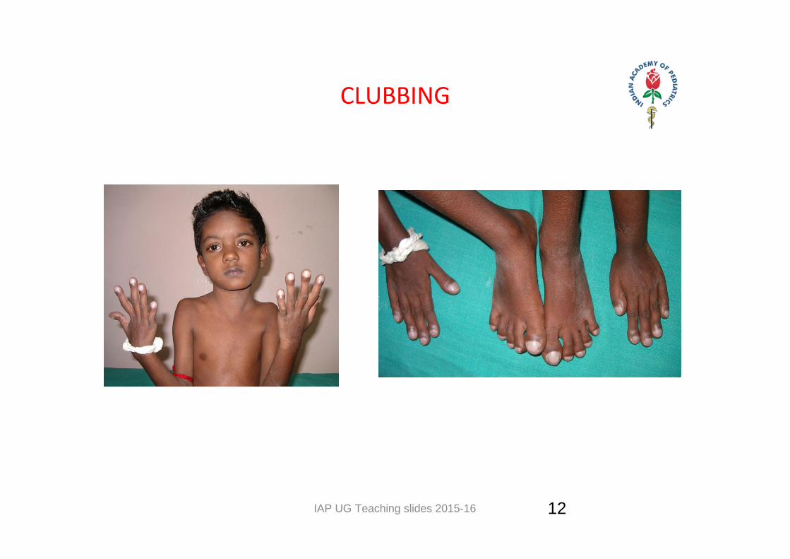
CLUBBING
• Normal angle between nail and nail plate (160°) LOVIBOND angle is lost
• Grading – I – softening of nail bed – II – obliteration of angle of nail plate and bed – lll – parrot beak – lV – hypertrophic osteo arthropathy
• Types – Unidigital – gout, local injury– Unilateral – Aneurysmal dilatation of aorta – Differential – clubbing in lower limbs only ,PDA with reversal– Bilateral – Rt Lt shunts, infectious endocarditis, atrial myxoma
11
IAP UG Teaching slides 2015-16
CLUBBING
12

IAP UG Teaching slides 2015-16
CAPILLARY REFILL
• Blanch the nail bed with sustained pressure for several seconds on a toenail or fingernail.
• Release the pressure• Observe the time elapsed before the nail regains full color– Should occur almost instantly – in less than 2 seconds.
– Longer than 2 seconds implies circulatory system compromise (ie: arterial occlusion, hypovolemic shock, hypothermia).
13
IAP UG Teaching slides 2015-16
SIGNS & SYMPTOMS OF INFECTIVE ENDOCARDITIS
• H/o CHD or any procedures• Fever • Chills • Chest & abdominal pain• Dyspnea• Night sweats• Weight loss• CNS Manifestations
• Elevated temperature• Anemia • Tachycardia• Embolic Phenomena – Roth spots,
Osler nodes, petechiae, splinter nail bed hemorrhages
• Janeway lesions• New or changing murmurs• Splenomegaly• Arthritis • Heart failure• Clubbing • Metastatic infection
14
IAP UG Teaching slides 2015-16
SIGNS OF FAILURE • Tender hepatomegaly • Basal crepitation• Edema in dependant region
– Infants – periorbital puffiness, flanks, sacrum– Older child – pedal edema
• Elevated JVP• Cardiomegaly
15
IAP UG Teaching slides 2015-16
SIGNS OF RHEUMATIC FEVER • Arthritis – migratory polyarthritis• Subcutaneous nodules ‐ at elbows, shin of tibia, occiput, spine• Chorea ‐ rapid, involuntary, purposeless, non repetitive, jerky movements aggravated during work relieved during sleep• Erythema marginatum – skin lesions with erythematous ring, central clearing
16
IAP UG Teaching slides 2015-16
PERIPHERAL SIGNS OF AORTIC REGURGITATION
• Head nodding‐ De Musset sign• Corrigans carotid sign• Dancing brachialis• Pulsatile Uvula‐ Mullers sign• Pulsatile nail beds‐ Quinckes sign• Pistol shot femorals• Hills sign• Water hammer pulse• Rosenbachs and Gerhards sign• Landolfi’s sign• Becker’s sign
17
IAP UG Teaching slides 2015-16
SYNDROMES• Typical syndromic features ‐ Be alert!
• Downs syndrome – Endocardial cushion defects, VSD
• Congenital rubella syndrome – PDA
• Turner’s syndrome – COA
• Trisomy 13 – VSD, ASD, PDA, Dextrocardia
• Trisomy 18 – VSD
• Noonan Syndrome‐PS
18
IAP UG Teaching slides 2015-16
ANTHROPOMETRY
• Short stature – Down, Noonan
• Microcephaly‐ Down,Congenital Rubella
• US/ LS ratio, armspan – Marfan’s syndrome
• Failure to thrive
19
IAP UG Teaching slides 2015-16
CVS ‐ VITAL SIGNS
Temperature
Respiration
Pulse
Blood pressure
20

IAP UG Teaching slides 2015-16
TEMPERATURE• Fever, chills and rigor
• IE• RF• Myxoma• Pericarditis, myocarditis• Pulmonary embolism• Pneumonia sec to large left to right shunt
21
IAP UG Teaching slides 2015-16
RESPIRATION• Rate, rhythm, Type
• Effortless tachypnoea
• Breathlessness decreased in propped up position/while putting on shoulder
22

IAP UG Teaching slides 2015-16
PULSE
• Rate• Rhythm • Volume• Character• Radio – radial delay• Radio femoral delay• Palpable Peripheral pulse
23
IAP UG Teaching slides 2015-16
WHERE TO PALPATE FOR ARTERIAL PULSE?
• Radial – fore arm slightly pronated and wrist slightly flexed. Examine for rate and rhythm.• Carotid – medial to sterno mastoid muscle. Examine for character and volume.• Femoral – midway between iliac crest and pubic ramus • Popliteal – knees flexed at 120° fingertips at popliteal fossa• Dorsalis pedis – lateral to proximal 1/3 rd of extensor hallucis longus• Anterior and posterior tibial
24

IAP UG Teaching slides 2015-16
NORMAL ARTERIAL PULSE
Note – steeper upstroke, higher systolic peak as pulse is transmitted to the peripheryUse central vessels to feel for the contour of the pulse
25
IAP UG Teaching slides 2015-16
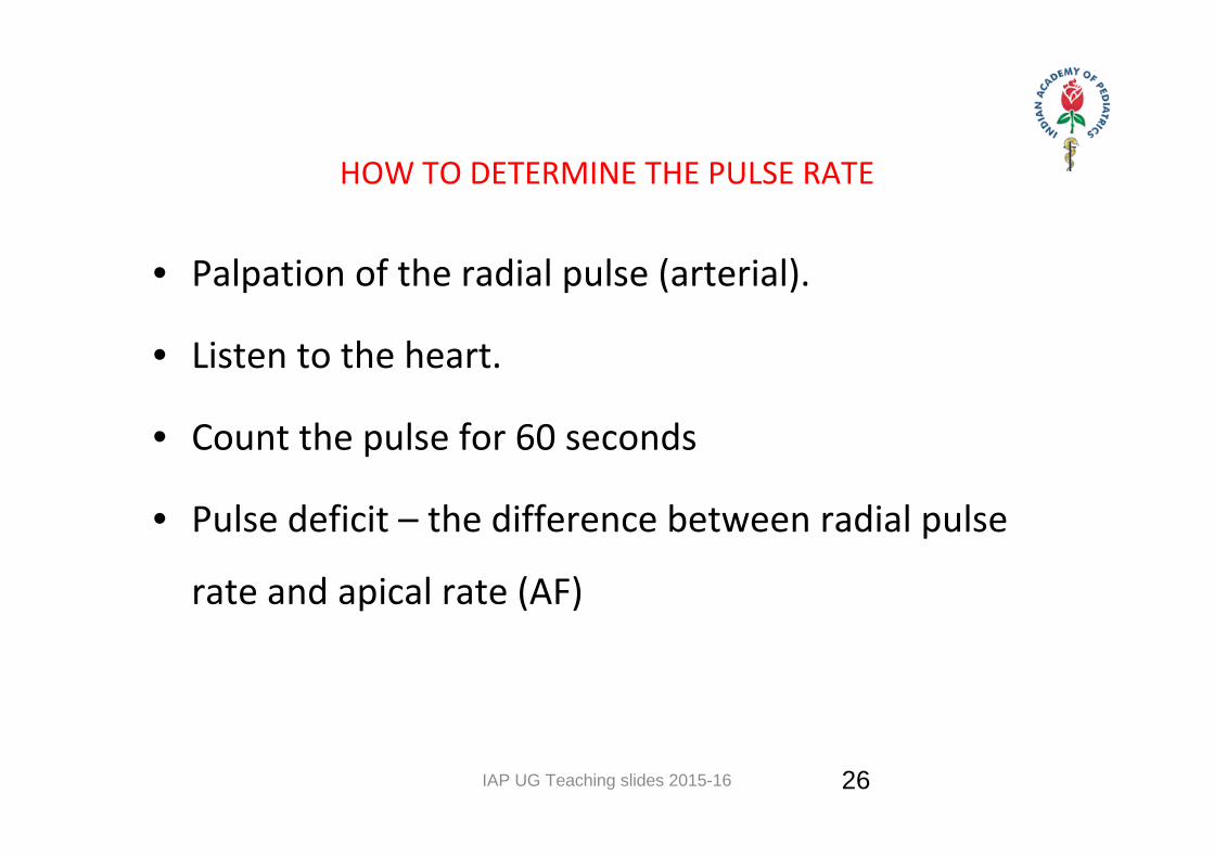
HOW TO DETERMINE THE PULSE RATE
• Palpation of the radial pulse (arterial).
• Listen to the heart.
• Count the pulse for 60 seconds
• Pulse deficit – the difference between radial pulse
rate and apical rate (AF)
26
IAP UG Teaching slides 2015-16
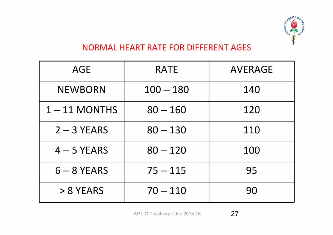
NORMAL HEART RATE FOR DIFFERENT AGES
AGE RATE AVERAGE
NEWBORN 100 – 180 140
1 – 11 MONTHS 80 – 160 120
2 – 3 YEARS 80 – 130 110
4 – 5 YEARS 80 – 120 100
6 – 8 YEARS 75 – 115 95
> 8 YEARS 70 – 110 90
27
IAP UG Teaching slides 2015-16
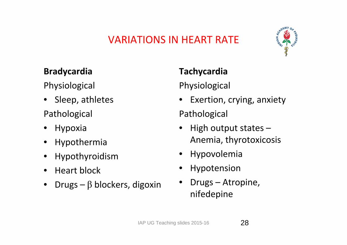
VARIATIONS IN HEART RATE
BradycardiaPhysiological• Sleep, athletesPathological• Hypoxia• Hypothermia• Hypothyroidism• Heart block• Drugs – β blockers, digoxin
TachycardiaPhysiological• Exertion, crying, anxietyPathological• High output states –
Anemia, thyrotoxicosis• Hypovolemia• Hypotension • Drugs – Atropine,
nifedepine
28

IAP UG Teaching slides 2015-16
PULSE – RHYTHM • Regular ‐ Normal• Irregular
– Regularly: Atrial Tachyarrythmia with fixed AV block– Irregularly: Atrial or ventricular ectopics, AF, Atrial tachyarrythmia with varied AV block
29

IAP UG Teaching slides 2015-16
PULSE ‐ CHARACTER
1. Collapsing pulse (water hammer pulse) jerky pulse with full expansion followed by sudden collapse (AR, PDA, A‐V fistulas, fever, thyrotoxicosis, anemia)
2. Alternating pulse pulses alternans (regular rate, amplitude varies from beat to beat) seen in LVF
3. Pulses bisferiens (two strong systolic peaks separated by a midsystolic dip) seen in HOCM, AS/AI
4. Anacrotic pulse slow rising pulse in A.S. (Parvus et tardus)5. Dicrotic pulse, two systolic and diastolic peaks (sepsis,
hypovolemic, cardiogenic shock)6. Pulsus paradoxus (amplitude decreases with inspiration
and increases during expiration) seen in cardiac tamponade, COPD, massive P.E.
30

IAP UG Teaching slides 2015-16
NORMAL
LV OUTFLOW OBSTUCTIONPULSUS BISFERIENS - AR
DICROTIC PULSE
31
IAP UG Teaching slides 2015-16
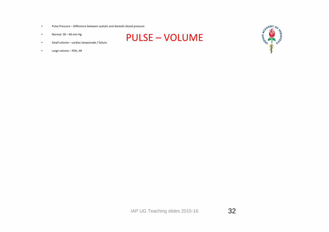
PULSE – VOLUME • Pulse Pressure – Difference between systolic and diastolic blood pressure
• Normal: 30 – 60 mm Hg
• Small volume – cardiac tamponade / failure
• Large volume – PDA, AR
32
IAP UG Teaching slides 2015-16
PULSE ‐ DELAY• Normally femorals felt just before radial
• Radio – radial – pre subclavian COA
• Radio femoral – post subclavian COA
33

IAP UG Teaching slides 2015-16
BLOOD PRESSURE
• Definition – Pressure exerted by the column of blood on the arterial wall
• Instrument ‐ Sphygmomanometer• Korotkoff Sound – Phase I – Phase V • Different methods to record BP
– Palpatory – Auscultatory – Flush method – Oscillometry – Non invasive Doppler
• In COA – all 4 limbs BP recorded 34
IAP UG Teaching slides 2015-16
PULSE PRESSURE
• Difference between the systolic and diastolic
pressure
• Mean arterial pressure = Diastolic pressure + 1/3 of
pulse pressure
35
IAP UG Teaching slides 2015-16
WHAT IS THE RELATIONSHIP BETWEEN THE BLOOD PRESSURE IN THE LEGS AND ARMS?
• To measure the blood pressure in the legs, place the cuff around the thigh and listen or palpate over the popliteal artery.
• Indirect measurement – the SBP in the legs is 10 – 15 mm hg higher than in the arms.
• Direct measurement – no difference• Hill’s sign ‐ > 20mm Hg difference between the arms and the legs (AR).
• Coarctation of the aorta – BP in legs is much less than in the arms.
36
IAP UG Teaching slides 2015-16
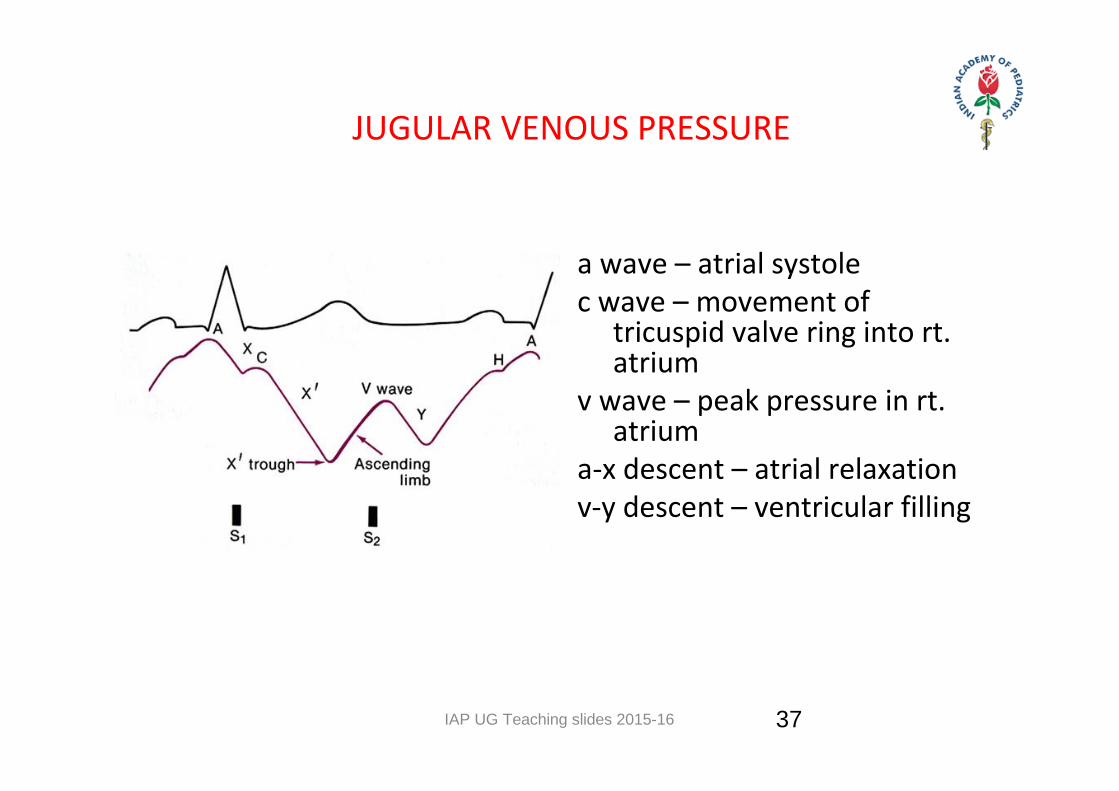
JUGULAR VENOUS PRESSURE
a wave – atrial systolec wave – movement of
tricuspid valve ring into rt. atrium
v wave – peak pressure in rt. atrium
a‐x descent – atrial relaxation v‐y descent – ventricular filling
37
IAP UG Teaching slides 2015-16
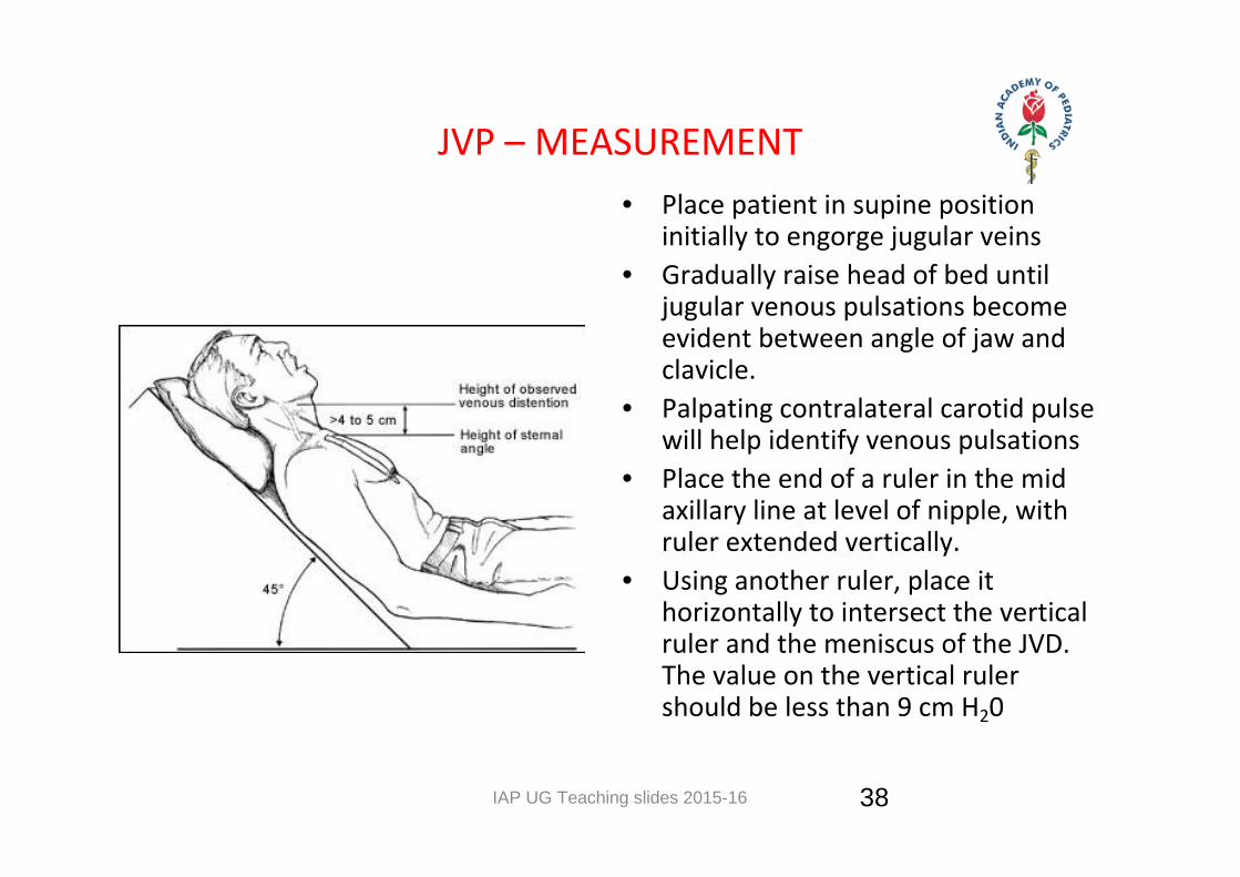
JVP – MEASUREMENT • Place patient in supine position
initially to engorge jugular veins• Gradually raise head of bed until
jugular venous pulsations become evident between angle of jaw and clavicle.
• Palpating contralateral carotid pulse will help identify venous pulsations
• Place the end of a ruler in the mid axillary line at level of nipple, with ruler extended vertically.
• Using another ruler, place it horizontally to intersect the vertical ruler and the meniscus of the JVD. The value on the vertical ruler should be less than 9 cm H20
38

IAP UG Teaching slides 2015-16
HOW TO MEASURE JVP
39
IAP UG Teaching slides 2015-16
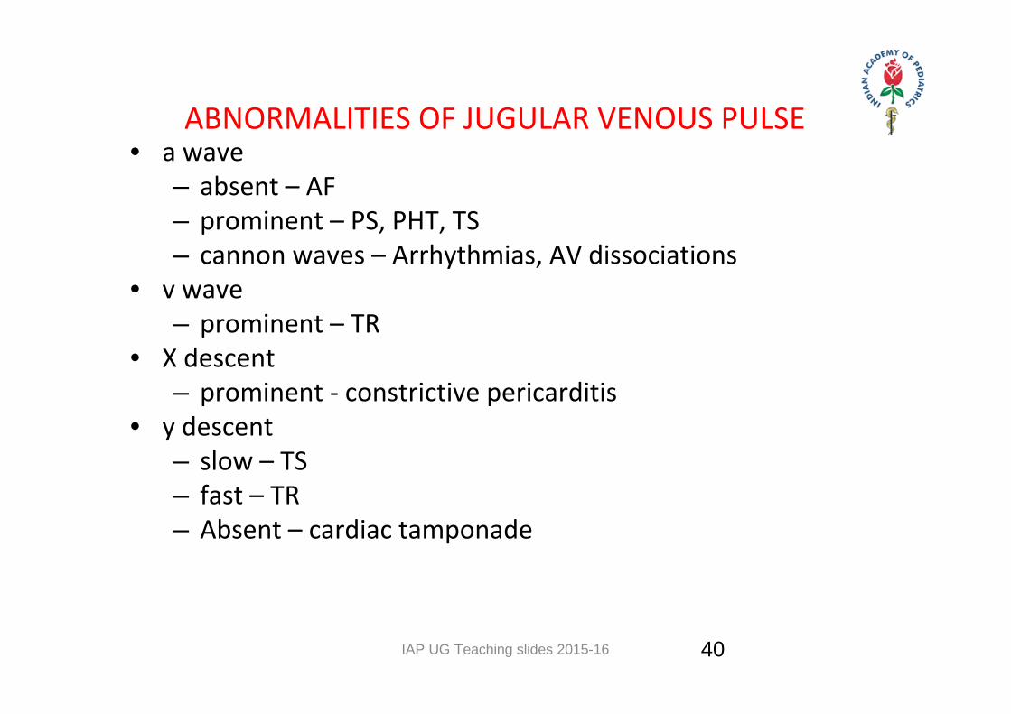
ABNORMALITIES OF JUGULAR VENOUS PULSE• a wave
– absent – AF – prominent – PS, PHT, TS – cannon waves – Arrhythmias, AV dissociations
• v wave – prominent – TR
• X descent – prominent ‐ constrictive pericarditis
• y descent – slow – TS– fast – TR– Absent – cardiac tamponade
40

IAP UG Teaching slides 2015-16
ABNORMALITIES OF JUGULAR VENOUS PULSE CONT…
• Low jugular venous pressure – Hypovolemia.
• Elevated jugular venous pressure – Intravascular volume overload conditions due to valvular disease
(tricuspid or pulmonic stenosis or regurgitation), right ventricular ischemia or infarction, cardiomyopathy or secondary to left heart failure (mitral stenosis/regurgitation, aortic stenosis/regurgitation, cardiomyopathy, myocardial ischaemia/ infarction).
– Right ventricular failure. – Constrictive pericarditis. – Pericardial effusion with tamponade physiology. – Obstructive atrial myxoma. – Superior vena caval obstruction.
41
IAP UG Teaching slides 2015-16
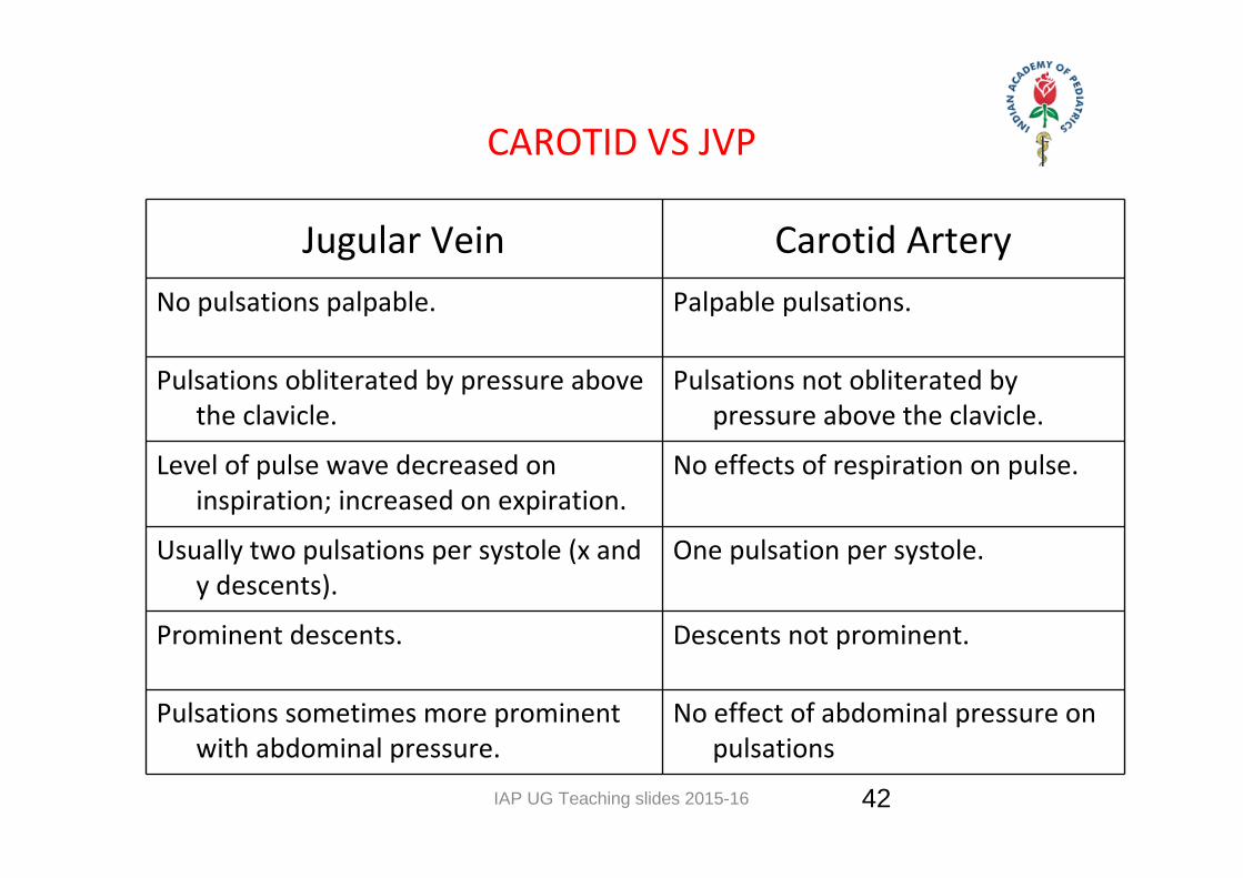
CAROTID VS JVP
Jugular Vein Carotid ArteryNo pulsations palpable. Palpable pulsations.
Pulsations obliterated by pressure above the clavicle.
Pulsations not obliterated by pressure above the clavicle.
Level of pulse wave decreased on inspiration; increased on expiration.
No effects of respiration on pulse.
Usually two pulsations per systole (x and y descents).
One pulsation per systole.
Prominent descents. Descents not prominent.
Pulsations sometimes more prominent with abdominal pressure.
No effect of abdominal pressure on pulsations
42

IAP UG Teaching slides 2015-16
CVS – SYSTEMIC EXAMINATION
•Inspection
•Palpation
•Percussion
•Auscultation
43

IAP UG Teaching slides 2015-16
INSPECTION • Position of trachea • Precordial bulge – left of sternum, seen from leg end child lying supine => cardiomegaly • Hyper dynamic precordium – thin patient, volume over load, LtRt shunt • Silent Precordium – obese child, pericardial effusion, severe cardio myopathy • Parasternal Lift ‐ RVE or severe MR
44
IAP UG Teaching slides 2015-16
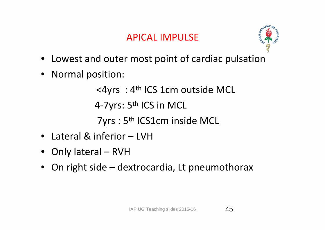
APICAL IMPULSE
• Lowest and outer most point of cardiac pulsation• Normal position:
<4yrs : 4th ICS 1cm outside MCL 4‐7yrs: 5th ICS in MCL 7yrs : 5th ICS1cm inside MCL • Lateral & inferior – LVH• Only lateral – RVH• On right side – dextrocardia, Lt pneumothorax
45

IAP UG Teaching slides 2015-16
OTHER PULSATIONS
• Aortic – AR, aortic aneurysm, dilatation of ascending aorta• Pulmonary – pulmonary hypertension• Carotid – hyperdynamic states, COA , AR• Supra clavicular ‐ AR • Supra sternal • Inter & infra scapular – COA (Suzman’s sign) • Epigastric – AR, RVH• Hepatic – TR, TS
46

IAP UG Teaching slides 2015-16
PALPATION
• Character of apex beat– Tapping ‐ MS– Heaving – force full, well sustained ‐ LVH, pressure over load – AS, Systemic HTN,COA
– Hyper dynamic – ill sustained ‐ Volume over load – MR,AR,VSD,PDA
• Para sternal heave – Right ventricular enlargement – ASD, VSD – Left atrial enlargement – MS, MR
47
IAP UG Teaching slides 2015-16
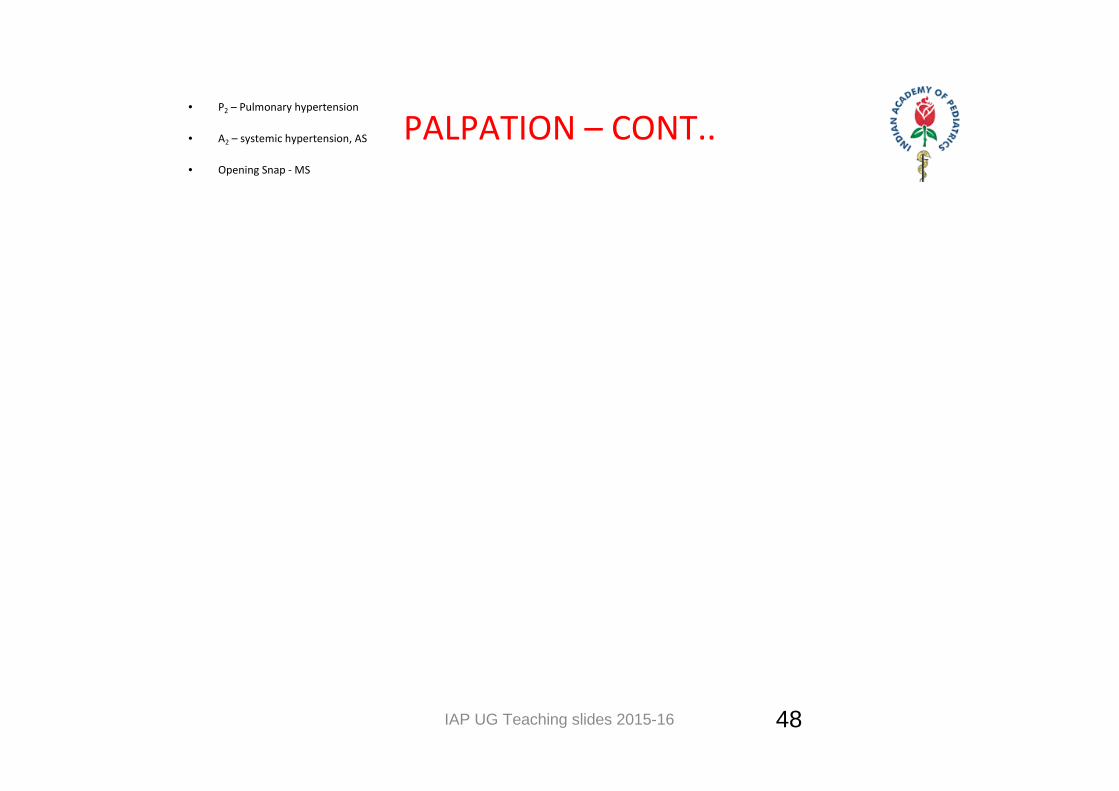
PALPATION – CONT..• Palpable heart sound
• P2 – Pulmonary hypertension
• A2 – systemic hypertension, AS
• Opening Snap ‐ MS
48
IAP UG Teaching slides 2015-16
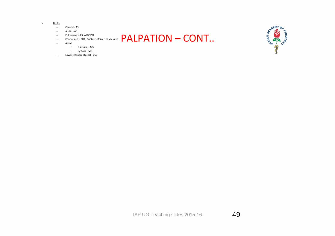
PALPATION – CONT..
• Thrills – Carotid ‐ AS– Aortic ‐ AS– Pulmonary – PS, ASD,VSD– Continuous – PDA, Rupture of Sinus of Valsalva– Apical
• Diastolic – MS• Systolic ‐ MR
– Lower left para sternal ‐ VSD
49

IAP UG Teaching slides 2015-16
PERCUSSION • Outline cardiac borders • Useful in
– Pericardial effusion – Dullness beyond apex– Pulmonary hypertension ‐ Dull 2nd Left ICS ,also in left atrial enlargement, pericardial effusion– Dextrocardia – Dilated cardiomyopathy
50

IAP UG Teaching slides 2015-16
AUSCULTATION • Method – Bell & Diaphragm • Areas – M T A P • Normal heart sounds • Abnormal heart sounds • Additional sounds• Murmurs
51
IAP UG Teaching slides 2015-16
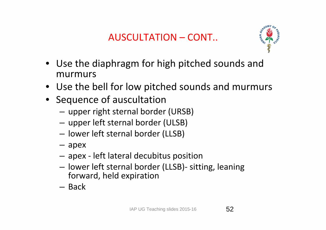
AUSCULTATION – CONT..
• Use the diaphragm for high pitched sounds and murmurs
• Use the bell for low pitched sounds and murmurs• Sequence of auscultation
– upper right sternal border (URSB)– upper left sternal border (ULSB) – lower left sternal border (LLSB)– apex– apex ‐ left lateral decubitus position– lower left sternal border (LLSB)‐ sitting, leaning forward, held expiration
– Back
52

IAP UG Teaching slides 2015-16 53
NORMAL HEART SOUNDS• S1 – Closure of AV valves “Lub”, Low pitched, Prolonged
• S2 – Closure of semilunar valves “Dub”, High pitched, Short, has two components (A2, P2)
• Physiological split – Normal splitting between A2 & P2 which varies with inspiration and expiration
IAP UG Teaching slides 2015-16
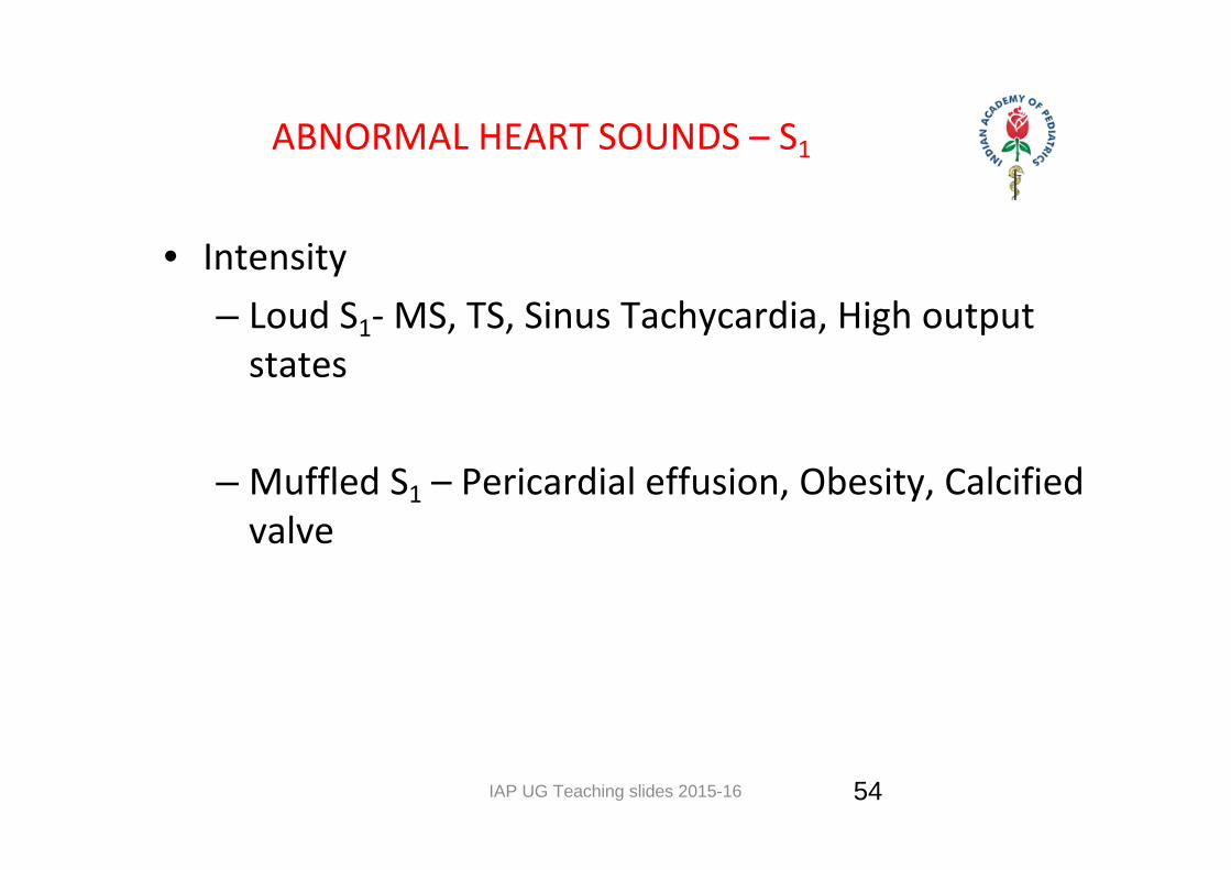
ABNORMAL HEART SOUNDS – S1
• Intensity – Loud S1‐ MS, TS, Sinus Tachycardia, High output states
– Muffled S1 – Pericardial effusion, Obesity, Calcified valve
54

IAP UG Teaching slides 2015-16
ABNORMAL HEART SOUNDS – S2
S2
A2 P2
Accentuated
Diminished
Delayed
Early
SH, AR
Calc.AV, Aortic Atresia
AS, PDA, AR, LVF, LBBB
VSD, MR
PAH
PS, PA
PS, ASD, TAPVC, RBBB
55
IAP UG Teaching slides 2015-16
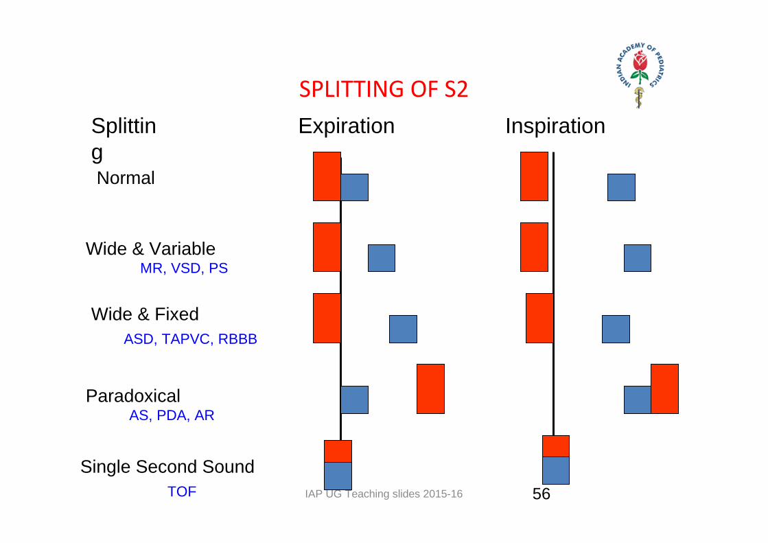
SPLITTING OF S2Expiration InspirationSplittin
gNormal
Wide & Variable
Paradoxical
Wide & Fixed
Single Second Sound
MR, VSD, PS
ASD, TAPVC, RBBB
AS, PDA, AR
TOF 56
IAP UG Teaching slides 2015-16
ABNORMAL HEART SOUNDS – CONT..
• 3rd heart sound – due to maximal ventricular filling – S3 Gallop – Myocarditis, CCF
• 4th heart sound – due to rapid emptying of atrium – Occurs in constrictive pericarditis, hypertrophic cardiomyopathy
57

IAP UG Teaching slides 2015-16
ADDITIONAL SOUNDS
• Click – arise due to semi lunar valves • Ejection systolic clicks – AS, PS • Aortic‐ bicuspid aortic valve
• Opening snap – due to abnormal mitral & tricuspid leaflets • Occurs in ASD, VSD, RHD – MS / TS
• Mid systolic Click – MVPS• Multiple Clicks – Ebstein’s Anomaly
• Pericardial rub – Acute rheumatic fever, pericarditis
58
IAP UG Teaching slides 2015-16
MURMURS
• Caused by normal flow through a abnormal valve or abnormal flow through a normal valve
• Types – Organic, Flow, Innocent • Description
– Intensity ‐ Grading– Pitch– Timing – Variation with respiration / posture– Area of maximum intensity– Conduction to other areas
59
IAP UG Teaching slides 2015-16
CHARACTERISTICS OF A “FUNCTIONAL” MURMUR• Short and soft ESM• Not radiating • Grade I – II, No thrill• Normal S1 and S2• Normal cardiac impulse• No evidence for any hemodynamic abnormality
60

IAP UG Teaching slides 2015-16 61
CHARACTERISTICS OF A PATHOLOGICAL MURMUR• Pansystolic murmur and all diastolic murmurs• Murmur associated with thrill• Harsh• With abnormal heart sound• Radiating

IAP UG Teaching slides 2015-16
SYSTOLIC MURMURSGrading I – VI1 – Barely audible 2 – Medium intensity3 – Loud but no thrill4 – Loud with thrill5 – Very loud still needs steth on the chest6 – Audible with steth off chest
62
IAP UG Teaching slides 2015-16
SYSTOLIC MURMURS
• Types– Early : AR, PR– MDM : MS, TS– Functional : Graham Steel, Carey Coombs, Austin Flint
63
IAP UG Teaching slides 2015-16
DIASTOLIC MURMURS‐GRADINGGrading I – IV1. Very Soft2. Soft3. Loud4. Loud with thrill
64

IAP UG Teaching slides 2015-16
COMMON MURMURS AND TIMING
Systolic Murmurs• Aortic stenosis• Mitral insufficiency• Mitral valve prolapse• Tricuspid insufficiency Diastolic Murmurs• Aortic insufficiency• Mitral stenosis
S1 S2 S165
IAP UG Teaching slides 2015-16 66
MURMUR ‐ VARIATION WITH RESPIRATION / POSTURE
• Variation with respiration– Left sided murmurs well heard in expiration– Right sided murmurs well heard in inspiration
• Variation with posture– MDM of MS best heard in left lateral position – EDM of AR best heard in sitting and leaning forward
IAP UG Teaching slides 2015-16
AUSCULTATION ‐ AORTIC AREA
• 2nd right intercostal space (URSB)– compare S1 to S2‐S1 should be softer. If the same, think Mitral Stenosis
– identify ejection murmur‐time the peak intensity in relation to systole
– identify ejection click if present
67
IAP UG Teaching slides 2015-16 68
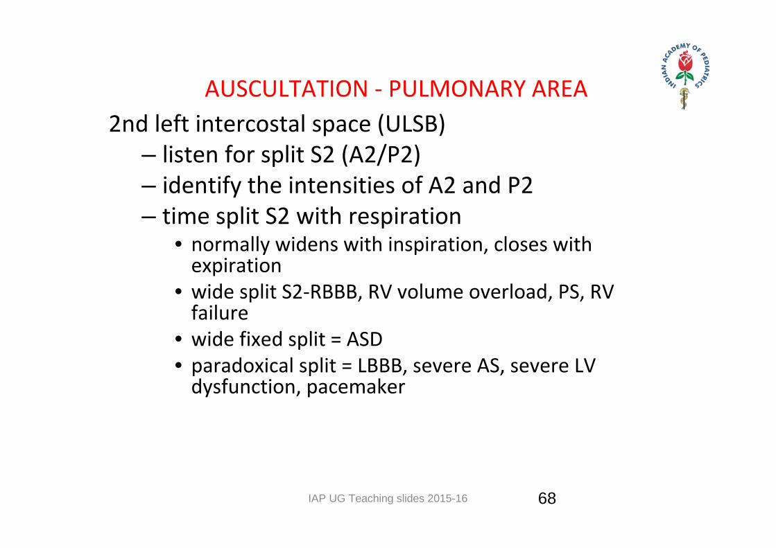
AUSCULTATION ‐ PULMONARY AREA2nd left intercostal space (ULSB)
– listen for split S2 (A2/P2)– identify the intensities of A2 and P2– time split S2 with respiration
• normally widens with inspiration, closes with expiration
• wide split S2‐RBBB, RV volume overload, PS, RV failure
• wide fixed split = ASD• paradoxical split = LBBB, severe AS, severe LV dysfunction, pacemaker
IAP UG Teaching slides 2015-16 69
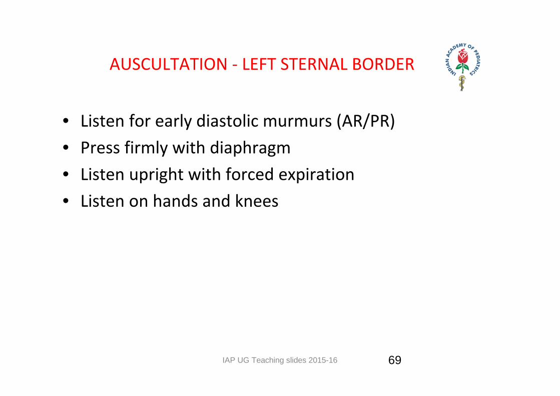
AUSCULTATION ‐ LEFT STERNAL BORDER
• Listen for early diastolic murmurs (AR/PR)• Press firmly with diaphragm• Listen upright with forced expiration• Listen on hands and knees
IAP UG Teaching slides 2015-16 70
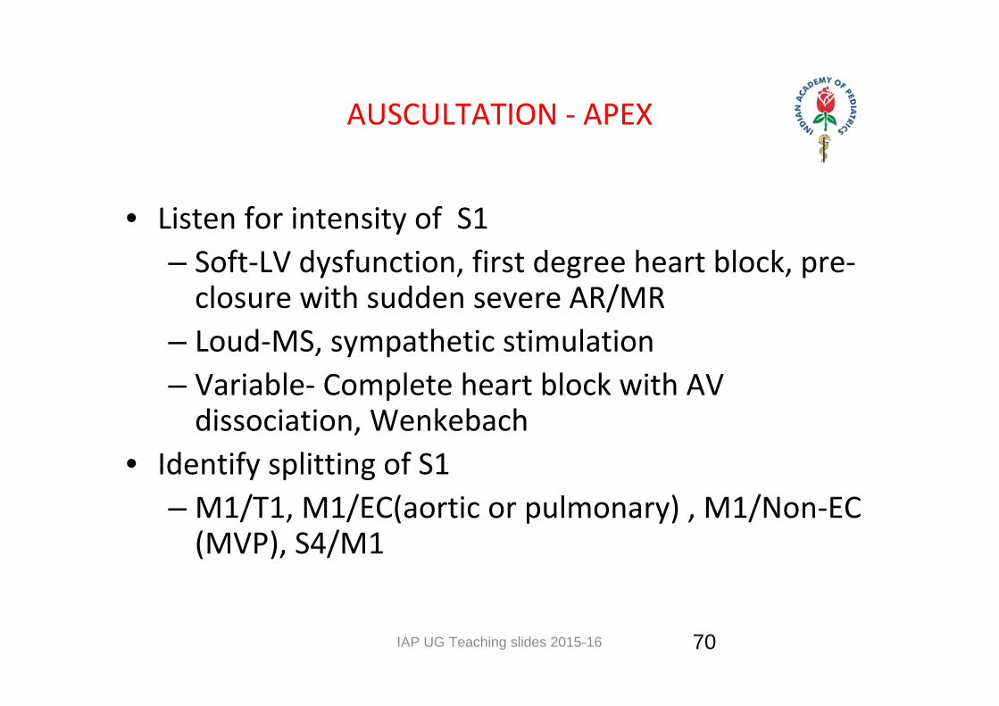
AUSCULTATION ‐ APEX
• Listen for intensity of S1– Soft‐LV dysfunction, first degree heart block, pre‐closure with sudden severe AR/MR
– Loud‐MS, sympathetic stimulation– Variable‐ Complete heart block with AV dissociation, Wenkebach
• Identify splitting of S1– M1/T1, M1/EC(aortic or pulmonary) , M1/Non‐EC (MVP), S4/M1
IAP UG Teaching slides 2015-16 71
AUSCULTATION – APEX – CONT..• Identify quality, timing and intensity of systolic murmurs
– ejection quality vs regurgitant quality– pansystolic vs early or mid to late systolic murmur

IAP UG Teaching slides 2015-16 72
AUSCULTATION – APEX – CONT..
–Listen for S3 and S4–Consider differential diagnosis of S3
• A2‐wide P2, A2‐OS, A2‐PK, A2‐S3
–Identify diastolic rumble–Determine radiation of murmur e.g.. MR to axilla

IAP UG Teaching slides 2015-16 73
MURMURS – CONT.. • Continuous
– PDA, Tricuspid atresia• To & fro
– AS with AR, VSD with AR
IAP UG Teaching slides 2015-16
VENOUS HUM• Low Pitched • Soft• Continuous• Accentuated in early diastole,exercise• Obliterated by compression of neck veins,valsalva maneuver• Present in Children, young adults and anemia
74
IAP UG Teaching slides 2015-16
SOME SPECIFIC CONDITIONS • ASD – Accentuated S1, wide fixed S2• VSD – muffled S1, wide variable S2, PSM• PDA ‐ Accentuated M1, Continuous murmur• TOF – only A2, ESM• PS – Delayed & muffled P2, ESM • AS – reverse splitting, ESM
75
IAP UG Teaching slides 2015-16
Thank You
76