evolution the evolution of the cytoskeleton - jcbjcb.rupress.org/content/jcb/194/4/513.full.pdf ·...
TRANSCRIPT

The Rockefeller University PressJ. Cell Biol. Vol. 194 No. 4 513–525www.jcb.org/cgi/doi/10.1083/jcb.201102065 JCB 513
JCB: Review
Correspondence to Bill Wickstead: [email protected] used in this paper: ARP, actin-related protein; DHC, dynein heavy chain; ESCRT-III, endosomal sorting complex required for transport, type III; IF, intermediate filament; LECA, last eukaryotic common ancestor; WACA, Walker A cytoskeletal ATPase.
IntroductionThe view that the cytoskeleton was a feature unique to eukary-otes was dramatically overturned about 20 years ago by the discovery that bacteria possess homologues of both tubulin (de Boer et al., 1992; RayChaudhuri and Park, 1992; Mukherjee et al., 1993) and actin (Bork et al., 1992). Since that time, a com-bination of bioinformatics, structural data, and advanced cell imaging has cemented the idea that both bacteria and archaea have active and dynamic cytoskeletons. However, as more infor-mation has emerged regarding the function of prokaryotic fila-ments and the distribution of cytoskeletal components, it has become clear that there is no simple relationship between the cytoskeletons of prokaryotes and eukaryotes. Moreover, there is considerable diversity in both composition and function between cytoskeletons in different lines of prokaryotes and eukaryotes.
Like eukaryotic actin-based microfilaments and tubulin-based microtubules, several of the filaments of the bacterial cytoskeleton are intrinsically “cytomotive” (Löwe and Amos, 2009); i.e., the filaments themselves can act as linear motors driven by the kinetics of polymerization/depolymerization. In eukaryotes, this activity has been hugely augmented by the
evolution of multiple classes of motors, as well as a menag-erie of nucleators, severing agents, tip-binding factors, and (de)polymerases. Other cytoskeletal filaments appear to be more structural in function, providing resistance to external force or act-ing as a scaffold. Such filaments may still be dynamic in cells without being intrinsically cytomotive. The most closely stud-ied of these are the intermediate filaments of animal cells, but a protein of bacteria, crescentin, also builds intermediate filament-like structures that function in cell shape determination.
In this review, we discuss the relationships between the major components of the bacterial, archaeal, and eukaryotic cytoskeletons. We compare the function of filaments in these three groups and also interrogate the distribution of key compo-nents across the tree of life. Finally, we examine what can be inferred with respect to the origins of cytoskeletal components and discuss the means by which the simple prokaryotic cyto-skeleton might have evolved into the elaborate system of fila-ments, motors, and accessory proteins that is characteristic of the eukaryotic cell.
Filaments I: tubulin-related proteinsEukaryotic microtubules are constructed from protofilaments resulting from the polymerization of heterodimers of - and -tubulin. Most microtubules consist of 13 protofilaments that interact laterally to form a hollow tube. Heterodimers added to the plus end of microtubules contain GTP in both subunits. Sub-sequent hydrolysis of GTP bound to the subunit encourages a conformational change in the heterodimer that is resisted by the geometry of the microtubule, thus trapping energy in the lattice (Howard and Hyman, 2003). This difference in the free energy of GTP- and GDP-bound polymers is the cause of microtubule dynamic instability—whereby the presence of unhydrolyzed GTP at the plus end of microtubules promotes further polymeriza-tion, but cap loss induces rapid depolymerization (Erickson and O’Brien, 1992; Howard and Hyman, 2009).
The first evidence for a bacterial homologue of tubulin came with the finding that FtsZ, an essential cell division pro-tein of Escherichia coli, bound and hydrolyzed GTP and pos-sesses a conserved seven-residue sequence nearly identical to
The cytoskeleton is a system of intracellular filaments cru-cial for cell shape, division, and function in all three do-mains of life. The simple cytoskeletons of prokaryotes show surprising plasticity in composition, with none of the core filament-forming proteins conserved in all lineages. In contrast, eukaryotic cytoskeletal function has been hugely elaborated by the addition of accessory proteins and extensive gene duplication and specialization. Much of this complexity evolved before the last common ances-tor of eukaryotes. The distribution of cytoskeletal filaments puts constraints on the likely prokaryotic line that made this leap of eukaryogenesis.
Evolution
The evolution of the cytoskeleton
Bill Wickstead1,2 and Keith Gull1
1Sir William Dunn School of Pathology, University of Oxford, Oxford OX1 3RE, England, UK2Centre for Genetics and Genomics, University of Nottingham, Nottingham NG7 2UH, England, UK
© 2011 Wickstead and Gull This article is distributed under the terms of an Attribution–Noncommercial–Share Alike–No Mirror Sites license for the first six months after the pub-lication date (see http://www.rupress.org/terms). After six months it is available under a Creative Commons License (Attribution–Noncommercial–Share Alike 3.0 Unported license, as described at http://creativecommons.org/licenses/by-nc-sa/3.0/).
TH
EJ
OU
RN
AL
OF
CE
LL
BIO
LO
GY
on May 13, 2018jcb.rupress.org Downloaded from http://doi.org/10.1083/jcb.201102065Published Online: 22 August, 2011 | Supp Info:

JCB • VOLUME 194 • NUMBER 4 • 2011 514
might be evidence that the protein predates the completion of the 20–amino acid standard code itself (Davis, 2002).
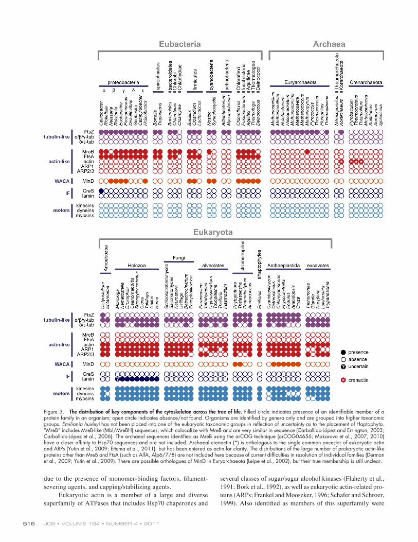
FtsZ is also encoded by many archaeal genomes, but it is not ubiquitous in prokaryotes. Most notably, FtsZ (and other tubulin homologues) are entirely absent from one of the major clades of archaea, the Crenarchaeota (Margolin, 2000; Vaughan et al., 2004)—with the possible exception of a highly divergent FtsZ-like gene in Sulfolobus solfataricus probably acquired by horizontal gene transfer (Makarova et al., 2010). FtsZ is also absent from at least one sequenced euryarchaeon (Picrophilus torridus; Fig. 3), as well as the bacterial groups Chlamydiae, Planctomycetes, and some Mycoplasmataceae (Vaughan et al., 2004; Adams and Errington, 2009). In the Crenarchaeota, FtsZ-independent division is possible because of an alternative cyto-kinesis machinery provided by ESCRT-III (endosomal sorting complex required for transport, type III; Lindås et al., 2008; Samson et al., 2008). ESCRT-III proteins are conserved between archaea and eukaryotes, and eukaryotic proteins of this complex form dynamic polymers that have a role in mem-brane scission events, including cell abscission during cyto-kinesis (Hurley and Hanson, 2010). In contrast to the lack of FtsZ in Crenarchaeota, the repertoire in Euryarchaeota has been duplicated to form three monophyletic families (FtsZ1, 2, and 3), with FtsZ3 being the most divergent and least widely distrib-uted (Vaughan et al., 2004).
an N-terminal tubulin signature motif (de Boer et al., 1992; RayChaudhuri and Park, 1992; Mukherjee et al., 1993). Subse-quent alignments showed that the similarities between FtsZ and tubulin sequences extended beyond the tubulin signature motif (Mukherjee and Lutkenhaus, 1994). Nevertheless, tubulin and FtsZ are very divergent in primary sequence, sharing only 10% identity compared with >40% between most FtsZ se-quences (Vaughan et al., 2004; Erickson, 2007). Despite low sequence similarity, the determination of the crystal structures of tubulin and FtsZ revealed proteins with near identical folds (Fig. 1; Löwe and Amos, 1998; Nogales et al., 1998a,b).
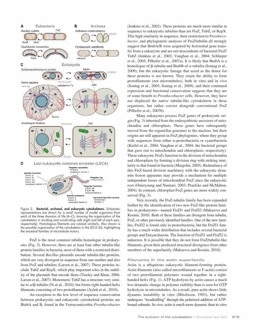
Unlike tubulin, FtsZ does not assemble into microtubules, but it does form a range of other structures in vitro using lateral interactions between protofilaments (Bramhill and Thompson, 1994; Mukherjee and Lutkenhaus, 1994; Erickson et al., 1996; Löwe and Amos, 1999, 2000). In vivo, FtsZ forms a dynamic “Z ring” between prospective daughter cells during cytokinesis (Fig. 2 A). This ring acts as a scaffold for the recruitment of the “divisome”, which gradually constricts to divide the cell (Adams and Errington, 2009). This system of cytokinesis was clearly an evolutionary success because FtsZ is both widely dis-tributed and highly conserved. Its presence in most lineages of bacteria is indicative of it being an ancient protein. It has even been suggested that an over-representation of amino acids with simpler biosynthetic pathways in conserved positions of FtsZ
Figure 1. Homology between prokaryotic and eukaryotic cytoskeletal filaments. Despite low levels of sequence similarity, the homologous cytoskeletal proteins FtsZ/TubZ/tubulin (top) and MreB/ParM/actin (bottom) have considerable conservation of folding and also longitudinal interaction. ParM and actin form similar helical filaments, but with opposite chirality (Orlova et al., 2007). Structures of filament subunits are derived from the following Protein Data Bank accession numbers: 1W5A (FtsZ; Oliva et al., 2004), 1JFF (/-tubulin; Löwe et al., 2001), 1JCG (MreB; van den Ent et al., 2001), and 1YAG (actin; Vorobiev et al., 2003).
515The evolution of the cytoskeleton • Wickstead and Gull
(Jenkins et al., 2002). These proteins are much more similar in sequence to eukaryotic tubulins than are FtsZ, TubZ, or RepX. This high similarity in sequence, their restriction to Prostheco-bacter, and phylogenetic analyses of FtsZ/tubulin all strongly suggest that BtubA/B were acquired by horizontal gene trans-fer from a eukaryote and are not descendents of bacterial FtsZ/TubZ (Jenkins et al., 2002; Vaughan et al., 2004; Schlieper et al., 2005; Pilhofer et al., 2007a). It is likely that BtubA is a homologue of -tubulin and BtubB of -tubulin (Sontag et al., 2009), but the eukaryotic lineage that acted as the donor for these proteins is not known. They retain the ability to form protofilaments (not microtubules), both in vitro and in vivo (Sontag et al., 2005; Sontag et al., 2009), and their continued expression and functional conservation suggests that they are of some benefit to Prosthecobacter cells. However, they have not displaced the native tubulin-like cytoskeleton in these organisms, but rather coexist alongside conventional FtsZ (Pilhofer et al., 2007b).
Many eukaryotes possess FtsZ genes of prokaryotic ori-gin (Fig. 3) inherited from the endosymbiotic ancestors of mito-chondria and chloroplasts. These genes have subsequently moved from the organellar genomes to the nucleus, but their origins are still apparent in FtsZ phylogenies, where they group with sequences from either -proteobacteria or cyanobacteria (Kiefel et al., 2004; Vaughan et al., 2004; the bacterial groups that gave rise to mitochondria and chloroplasts, respectively). These eukaryotic FtsZs function in the division of mitochondria and chloroplasts by forming a division ring with striking simi-larity to that found in bacteria (Margolin, 2005). Redundancy of this FtsZ-based division machinery with the eukaryotic dyna-min fission apparatus may provide a mechanism for multiple independent losses of mitochondrial FtsZ since the eukaryotic root (Osteryoung and Nunnari, 2003; Praefcke and McMahon, 2004). In contrast, chloroplast FtsZ genes are more widely con-served (Fig. 3).
Very recently, the FtsZ-tubulin family has been expanded further by the identification of two new FtsZ-like protein fami-lies in prokaryotes—named FtsZl1 and FtsZl2 (Makarova and Koonin, 2010). Both of these families are divergent from tubulin, FtsZ, or other previously identified families. One of the new fami-lies, FtsZl2 is found only in proteobacteria, but the FtsZl1 fam-ily has a much wider distribution that includes several bacterial groups and Euryarchaeota. The function of FtsZl1 and FtsZl2 is unknown. It is possible that they do not form FtsZ/tubulin-like filaments, given their predicted structural divergence from other members of the superfamily (Makarova and Koonin, 2010).
Filaments II: the actin superfamilyActin is a ubiquitous eukaryotic filament-forming protein. Actin filaments (also called microfilaments or F-actin) consist of two protofilament polymers wound together in a right-handed helix (Fig. 1). ATP hydrolysis by actin causes a much less dramatic change in polymer stability than is seen for GTP hydrolysis in microtubules. As a result, pure actin shows little dynamic instability in vitro (Mitchison, 1992), but rather undergoes “treadmilling” through the polarized addition of ATP-bound subunits. In vivo, actin is much more dynamic than in vitro
FtsZ is the most common tubulin homologue in prokary-otes (Fig. 3). However, there are at least four other tubulin-like protein families in bacteria, most of them with a restricted distri-bution. Several Bacillus plasmids encode tubulin-like proteins, which are very divergent in sequence from one another and also from FtsZ and tubulins (Larsen et al., 2007). These proteins in-clude TubZ and RepX, which play important roles in the stabil-ity of the plasmids that encode them (Tinsley and Khan, 2006; Larsen et al., 2007). Monomeric TubZ has a structure very simi-lar to /-tubulin (Ni et al., 2010), but forms right-handed helix filaments consisting of two protofilaments (Aylett et al., 2010).
An exception to the low level of sequence conservation between prokaryotic and eukaryotic cytoskeletal proteins are BtubA and B, found in the Verrucomicrobia Prosthecobacter
Figure 2. Bacterial, archaeal, and eukaryotic cytoskeletons. Schematic representations are shown for a small number of model organisms from each of the three domains of life (A–C), showing the organization of the cytoskeleton in dividing and nondividing cells (right and left of each pair, respectively). Homologous filaments are colored similarly. Also shown is the possible organization of the cytoskeleton in the LECA (D), highlighting the ancestral families of microtubule motors.
JCB • VOLUME 194 • NUMBER 4 • 2011 516
several classes of sugar/sugar alcohol kinases (Flaherty et al., 1991; Bork et al., 1992), as well as eukaryotic actin-related pro-teins (ARPs; Frankel and Mooseker, 1996; Schafer and Schroer, 1999). Also identified as members of this superfamily were
due to the presence of monomer-binding factors, filament- severing agents, and capping/stabilizing agents.
Eukaryotic actin is a member of a large and diverse superfamily of ATPases that includes Hsp70 chaperones and
Figure 3. The distribution of key components of the cytoskeleton across the tree of life. Filled circle indicates presence of an identifiable member of a protein family in an organism; open circle indicates absence/not found. Organisms are identified by genera only and are grouped into higher taxonomic groups. Emiliania huxleyi has not been placed into one of the eukaryotic taxonomic groups in reflection of uncertainty as to the placement of Haptophyta. “MreB” includes MreB-like (Mbl/MreBH) sequences, which colocalize with MreB and are very similar in sequence (Carballido-López and Errington, 2003; Carballido-López et al., 2006). The archaeal sequences identified as MreB using the arCOG technique (arCOG04656; Makarova et al., 2007, 2010) have a closer affinity to Hsp70 sequences and are not included. Archaeal crenactin (*) is orthologous to the single common ancestor of eukaryotic actin and ARPs (Yutin et al., 2009; Ettema et al., 2011), but has been entered as actin for clarity. The distributions of the large number of prokaryotic actin-like proteins other than MreB and FtsA (such as AlfA, Alp6/7/8) are not included here because of current difficulties in resolution of individual families (Derman et al., 2009; Yutin et al., 2009). There are possible orthologues of MinD in Euryarchaeota (Leipe et al., 2002), but their true membership is still unclear.

517The evolution of the cytoskeleton • Wickstead and Gull
(Makarova et al., 2007, 2010). These sequences have a closer affinity to Hsp70 sequences than to MreB from bacteria or Methanopyrus and their grouping in arCOG04656 may be an artifact of the technique. These sequences are not considered as true MreB members herein (Fig. 3).
Several other actin superfamily members exist in bacteria—notably: MamK, which is required for magnetosome organiza-tion in Magnetospirillium (Komeili et al., 2006; Pradel et al., 2006); AlfA, which is involved in plasmid segregation in Bacil-lus subtilis in a manner similar to ParM (Becker et al., 2006); and Ta0583 from the euryarchaeon Thermoplasma acidophilum (Roeben et al., 2006; Hara et al., 2007). These proteins have either a limited phylogenetic distribution or their families have yet to be well delimited (Derman et al., 2009; Yutin et al., 2009). The possibility that Ta0583 might be the closest prokaryotic homologue to eukaryotic actin (Hara et al., 2007) has not been supported by subsequent analyses (Yutin et al., 2009; Ettema et al., 2011). However, recently an archaeal actin-like family has been described that is monophyletic with eukaryotic actin and actin-related proteins (Yutin et al., 2009; Ettema et al., 2011). This protein family, dubbed “crenactin,” has a localization in Pyrobaculum cells very similar to MreB in bacteria (Fig. 2 B; Ettema et al., 2011), but is more closely related to eukaryotic actin than to MreB or ParM. The possible implications of this family are discussed further below.
Filaments III: intermediate filamentsEukaryotic intermediate filaments (IFs) are unlike microtubules and microfilaments in structure, biochemistry, and phylogenetic distribution. Unlike actin and tubulin, which are globular pro-teins that form polarized protofilaments, IF proteins are extended dimers that overlap to form unpolarized cables. There are many types of IF protein in vertebrates, most of which can be grouped into five classes: (1) type I (acidic) keratins; (2) type II (basic) keratins; (3) vimentin and desmin; (4) -internexin and neurofil-ament proteins; and (5) lamins (Fuchs and Weber, 1994). Lam-ins are also present in protosomes, suggesting that all IF protein families are derived from a single lamin-like sequence in the common metazoan ancestor (Weber et al., 1989; Dodemont et al., 1994; Bovenschulte et al., 1995). Significantly, however, eukaryotic IF proteins have only been found unambiguously in animals and their relatives (Erber et al., 1998), suggesting that they are an innovation specific to this lineage and not present in the last eukaryotic common ancestor (LECA; see Fig. 3).
The bacterium Caulobacter crescentus encodes a protein with a predicted arrangement of coiled-coils similar to that in animal lamin A (Ausmees et al., 2003). This protein, CreS or crescentin, forms helical filaments that are necessary for the vibrioid or helical cell shapes adopted by Caulobacter. Although crescentin was originally described only as “IF-like”, the function and predicted secondary structure of the protein have often been interpreted as evidence that bacteria possess ancient homologues of eukaryotic IFs. However, there are several argu-ments against such an interpretation. First, CreS has a very restricted distribution (Fig. 3); to date, it has only been found in Caulobacter. Given this restricted range, if lamin A and crescentin are truly homologous, then CreS would most likely
several other bacterial proteins, including MreB, FtsA, and ParM (Bork et al., 1992). Since initial identification, the superfamily of actin-like proteins in bacteria has proved to be very complex, encompassing more than 20 classes of protein (Derman et al., 2009).
The most common prokaryotic homologue of actin is MreB. As for tubulin/FtsZ, actin and MreB are very divergent in primary sequence but have similar structures (Fig. 1), based on the “actin-fold” that unites the superfamily (Kabsch and Holmes, 1995). In vitro MreB forms assemblies of two proto-filaments that are similar in structure to F-actin, but lack the helical twist (van den Ent et al., 2001; Esue et al., 2005). The conserved function of bacterial MreB (and closely related pro-teins such as Mbl and MreBH) appears to be in maintenance of cell shape (Jones et al., 2001; Graumann, 2007; Margolin, 2009). MreB filaments form a helix below the cell membrane and influence the position of cell wall synthesis (Fig. 2 A; Jones et al., 2001; Daniel and Errington, 2003). Consistent with this function, MreB is generally conserved only among rod-shaped bacteria, but absent from spherical cocci. Because extant rod-shaped bacterial lineages are probably more ancient, it has been suggested that the coccal forms have been derived from them multiple times by the loss of MreB and associated genes (Margolin, 2009). However, the correlation between prokaryote shape and MreB is not strict: some cocci still possess MreB and there are rod-shaped bacteria that lack MreB (Margolin, 2009).
ParM (previously StbA) is a plasmid-borne bacterial actin homologue with a filament-forming role. ParM is only 20% identical to MreB, which is comparable to the degree of conser-vation between actin and either ParM or MreB. Nonetheless, ParM assembles twisted polymers in vitro that are very reminis-cent of F-actin (van den Ent et al., 2002), but with a left-handed rather than right-handed twist (Orlova et al., 2007). The energy of polymerization of ParM is used in the segregation of the R1 plasmid (and others containing the parMRC operon) by pushing newly synthesized plasmid to the cell poles (Garner et al., 2007; Salje and Löwe, 2008).
To date, unambiguous ParM homologues are restricted to a close group of -proteobacteria, the Enterobacteriaceae. It is likely that the annotated “ParM” genes in the genomes of Firmicutes, -proteobacteria, and cyanobacteria represent other classes of bacterial actin-like proteins, rather than true ParM orthologues. In support of this interpretation, “ParM” encoded on the Staphylococcus aureus pSK41 plasmid shares only 19% identity with -proteobacterial ParM sequences and appears to be more structurally related to archaeal actin-like proteins (Popp et al., 2010).
Both MreB and FtsA are almost exclusively restricted to bacteria (Fig. 3). Clear examples of MreB are also found in euryarchea of the genera Methanopyrus, Methanobrevibacter, and Methanothermobacter (Yutin et al., 2009). Given their limited distribution and close similarity to bacterial sequences, these are most likely the result of horizontal gene transfer from bacte-ria. However, it cannot be entirely ruled out that they are rare but highly conserved genes of linear descent. Several other ar-chaeal sequences have been identified as possible MreB ortho-logues using the “archaeal cluster of orthologous groups” technique

JCB • VOLUME 194 • NUMBER 4 • 2011 518
Prokaryotic division: a common problem with many solutionsIf there is an overarching theme running through the evolution of the prokaryotic cytoskeleton, it appears to be this: plastic-ity. Each of the major families of cytomotive filament-forming proteins consists of several paralogues, which are often as di-vergent from each other as they are from eukaryotic homo-logues. Some of the classes—such as FtsA (from the MreB/actin superfamily) or the newly identified FtsZ-like families—may not form filaments in vivo at all. However, among those families that do polymerize, lateral interactions in the core filament appear to be quite malleable over evolutionary time-scales without disrupting polymerization (for example, straight MreB filaments, against left-handed twisting ParM and right-handed actin).
There is also plasticity in the biological function for which filaments are used—FtsZ and MreB are involved in cell division and morphology, while their homologues TubZ and ParM play roles in plasmid segregation. The result is that pro-karyotes use different systems to solve common problems. In bacteria, active DNA segregation can be achieved by at least three types of segregation machinery (Ebersbach and Gerdes, 2005; Hayes and Barillà, 2006). Type I systems use WACA proteins, such as ParA and Soj. Type II systems are based on the actin homologue ParM. Type III systems are those from Bacillus that use tubulin-like homologues, such as TubZ and RepX. In a striking example of apparent convergence, both ParM (type II) and TubZ (type III) form similar helical fila-ments that probably act to push apart plasmids (van den Ent et al., 2002; Aylett et al., 2010). However, it is type I systems that are by far the most wide spread in bacteria. It is interest-ing, then, that filaments based on WACA proteins are not conserved in eukaryotes, and may not be widely used in chro-mosome segregation in archaea (Bernander, 2000; Makarova et al., 2010). Significantly, none of the cytoskeletal protein families is ubiquitous to all prokaryotes, or even one of the two prokaryotic domains of life. The role for FtsZ in cytokine-sis appears to be the function under most selective pressure for retention (Erickson, 2007), but even this protein has been lost from several lineages of bacteria and the entire crenar-chaeal clade.
Eukaryogenesis: families and specializationDespite being based on homologous filaments, the eukaryotic cytoskeleton is not simply a more extensive version of the pro-karyotic one (Fig. 2 C). The complex eukaryotic cytoskeleton is actually based on a smaller set of ancestral cytomotive filaments than that of prokaryotes. With the notable exception of the pro-karyote-like division machineries associated with some plas-tids, only one paralogue of an MreB/crenactin family protein and one FtsZ/TubZ protein seems to have founded the eukary-otic cytoskeleton. However, this small selection has undergone several rounds of gene duplication and specialization from this ancestral set.
Heterodimers of - and -tubulin make up the vast major-ity of tubulin in eukaryotic cells. They are sufficient for the pro-duction of microtubules in vitro, and it is very likely that they
represent a lateral transfer of a eukaryotic gene to Caulobacter rather than a true bacterial homologue. Second, identifiable homologues of eukaryotic IF proteins are restricted to metazoa (or possibly holozoa; Fig. 3) and may not have been present in the LECA—precluding vertical inheritance from prokaryotes. Third, similarities in predicted protein architecture (in this in-stance, coiled-coil positions) are not equivalent to fold homol-ogy. At present, it is not possible to compare eukaryotic IF proteins and crescentin for evidence of fold homology, as there are no representatives of either family for which full structures have been determined. Moreover, although orthologues of cres-centin have not been identified outside of Caulobacter, there is evidence that CreS is a member of a larger family of bacterial proteins (Bagchi et al., 2008). All of these proteins are predicted to contain long stretches of coiled-coils, but the vast majority have no striking architectural similarity to lamin A (or other vertebrate IF proteins). Hence, the distribution of coiled-coil regions in crescentin and lamin A is more likely an example of convergence than a reflection of shared ancestry.
Filaments IV: WACA proteinsProkaryotes have a fourth class of filament-forming proteins known as Walker A cytoskeletal ATPases (WACAs; Michie and Löwe, 2006). WACA proteins are a diverse family of ATPases (Koonin, 1993), which are themselves part of the extremely large superclass of P-loop proteins including signal recognition particle proteins, Rho/Ras GTPases and cytoskeletal motors (Leipe et al., 2002). The WACA MinD is an active ATPase (de Boer et al., 1991) that forms dynamic filaments around the cell periphery in E. coli and inhibits Z ring formation (Pichoff and Lutkenhaus, 2001; Shih et al., 2003). In Bacillus subtilis, MinD is statically associated with the cell poles (Marston et al., 1998; Marston and Errington, 1999), making it unclear if dy-namic polymerization is an evolutionarily conserved feature of MinD biology. MinD sequences are found in many groups of bacteria (Fig. 3) and there are putative orthologues identified in Euryarchaeota (Leipe et al., 2002). However, the distinctions be-tween the prokaryotic WACA families are yet to be fully resolved, so it is presently unclear if these archaeal sequences are mono-phyletic with bacterial MinD. Even if not strict MinD ortho-logues, examples of WACA proteins do appear to be present in both Euryarcheaota and Crenarchaeota (Makarova et al., 2010).
Several other bacterial WACA proteins have been de-scribed. ParA, ParF, and Soj all play roles in DNA segregation by different mechanisms (Pogliano, 2008; Löwe and Amos, 2009), demonstrating an apparent versatility of this system for segregation. It is perhaps surprising, therefore, that there are no eukaryotic WACA filaments. The fold of MinD and Soj is dis-tantly related to that of eukaryotic septins (Cordell and Löwe, 2001; Leonard et al., 2005; Löwe and Amos, 2009), but this most likely reflects the distant relationship shared by all P-loop GTPases (Leipe et al., 2002). There are, however, MinD genes of bacterial ancestry in Viridiplantae (green algae and land plants) and some stramenopiles (Fig. 3), which play a role in plastid division (Marrison et al., 1999; Colletti et al., 2000; Kanamaru et al., 2000). It is noteworthy that all eukaryotes that possess MinD genes also encode endosymbiont-derived FtsZ.

519The evolution of the cytoskeleton • Wickstead and Gull
for the most part, present in the LECA. Although rare divergent types do exist (for example - and -tubulin), they do not have the same level of divergence from core types as is seen for di-vergent prokaryotic proteins. Moreover, their biological role seems more static. Divergence between the distribution of fami-lies in extant eukaryotes is largely a product of loss of function (e.g., loss of dynactin complex and ARP1; loss of centrioles and /-tubulin).
Cytoskeletal motorsProkaryotes use the intrinsic cytomotive capacity of cytoskele-tal filaments to do work. Eukaryotic cells also use the energy of polymerization/depolymerization (McIntosh et al., 2010). How-ever, in eukaryotes the intrinsic dynamics of the filaments has been augmented by the addition of the cytoskeletal motors—dyneins, kinesins, and myosins—which derive energy from ATP hydrolysis to perform various cellular tasks. Each of these motors is a superfamily containing multiple classes of special-ized proteins.
Kinesins and myosins share a similar fold structure, sug-gesting that they have common ancestry (Kull et al., 1996, 1998). This is rather surprising given that they now act exclu-sively on microtubules and F-actin, respectively, and presents something of a conundrum in terms of evolutionary cell biol-ogy. It is only plausible for motors to evolve after the filaments on which they act have been established. As has been discussed above, filaments made from tubulin/FtsZ and actin/MreB homologues were already well established in prokaryotes before the emergence of eukaryotes, yet both motor classes evolved only later in the proto-eukaryote. This requires that either: (1) a single “ur-kinesin-myosin” originally walked on both tubulin- and actin-based filaments and only later specialized into sepa-rate families; (2) a motor evolved for one type of filament and subsequently developed an ability to move on the other; or (3) both motor families evolved independently from the same fam-ily of NTPases. The large differences in structure and sequence between tubulin/FtsZ and actin/MreB filaments suggest that the last scenario is most plausible, even if less parsimonious. There is also a precedent for this because this family of P-loop NTPases also gave rise to many eukaryote-specific GTPases (including Arf, Ras/Rab, Rho/Ran, and G protein families; Leipe et al., 2002).
Dyneins have a very different structure to that of myosins and kinesins. The core of the dynein complex is the dynein heavy chain (DHC), which is a member of another large and di-verse superclass of NTPases, the AAA+ proteins (a family in-cluding ATPases associated with various cellular activities). DHCs contain six AAA+ domains that form an intramolecular hexameric ring (Samsó et al., 1998; King, 2000; Burgess et al., 2003), implying that DHCs originally evolved by either intra-gene domain duplication or fusion of genes encoding AAA+ proteins. Because all classes of DHC have the same structure, this duplication occurred before the creation of functional para-logues. The dynein superfamily is also different from kinesins and myosins in that all but one of the nine classes are associated with one specific organelle—the cilium. Only cytoplasmic dynein 1 has a conserved role outside of the cilium; the other
were the first types to evolve from the single proto-tubulin ancestor. However, they are not the only ancestral tubulins. Analyses suggest that the tubulin family encompasses at least six classes, named , , , , , and , with a further two divergent types ( and ) being found in some organisms (Vaughan et al., 2000; Dutcher, 2003). -Tubulin plays an essential role in micro-tubule nucleation (through the action of the conserved -tubulin ring complex) and is, like - and -, ubiquitous to all eukary-otes. In contrast, - and -tubulin have centriolar roles (Dutcher and Trabuco, 1998; Chang and Stearns, 2000; Ruiz et al., 2000) and are conserved in nearly all organisms that build centrioles/basal bodies and absent from organisms that have lost cilia/flagella (Hodges et al., 2010). At least five of the classes of tu-bulin (, , , , and ) can be traced back to the last common eukaryotic common ancestor (Fig. 3). All of the tubulin types are more closely related to one another than to FtsZ/TubZ (Vaughan et al., 2000; Dutcher, 2003), implying that they all descended from a common proto-tubulin by gene duplication. Both duplication and specialization into functionally distinct classes must have occurred before the LECA.
There are striking parallels between the proto-eukaryotic evolution of tubulins and that of actin. The proteins most similar in sequence to conventional actin are the ARPs. The ARPs cover at least eight major families (Sehring et al., 2007) that are found only in eukaryotes. Four ARP families (ARP4, 5, 6/7, and 8/9) are not associated with cytoplasmic actin, but are nuclear pro-teins involved in chromatin remodeling (Chen and Shen, 2007; Dion et al., 2010). The remaining families—ARP1, 2, 3, and 10/11—have important roles in modification or extension of cyto-plasmic actin function. ARP1 is an integral part of the dynactin complex, which links the actin- and tubulin-based cytoskeleton (Schroer, 2004). Yeast Arp10p and metazoan Arp11 (ARP10/11 family members) are also part of the dynactin complex (Eckley and Schroer, 2003; Clark and Rose, 2006). In contrast, a complex of seven subunits formed around a heterodimer of ARP2 and ARP3 is a major F-actin nucleator in most eukaryotes (Pollard and Beltzner, 2002; Goley and Welch, 2006). The only ARP that appears to form filaments is Arp1p (Schafer et al., 1994; Bingham and Schroer, 1999). These filaments are much shorter than those seen for actin, but have similar morphology.
As for tubulin, there was a single actin-like protein in the proto-eukaryote which evolved into multiple paralogous forms before the LECA. In doing so, both the actin- and tubulin-based cytoskeleton independently invented complexes containing di-vergent forms of filament subunits that were specialized for nucleation (ARP2/3 and -tubulin ring complexes). However, in the case of the actin-based cytoskeleton, this nucleating complex appears to be expendable in some circumstances be-cause orthologues of ARP2 and ARP3 have been lost multiple times across eukaryotes (Fig. 3; note that organisms always lose both, as would be expected for a complex). Cross talk be-tween microtubules and microfilaments via the dynactin com-plex has also been lost multiple times, as can be seen from the distribution of ARP1.
The history of these core eukaryotic cytoskeletal families also shows some degree of plasticity, but it is rather different from that seen in prokaryotes. The eukaryotic paralogues were,

JCB • VOLUME 194 • NUMBER 4 • 2011 520
(Grimstone and Cleveland, 1965; McIntosh, 1973), presumably being assembled in an IFT-independent manner as are some axonemes today (Witman, 2003; Briggs et al., 2004).
The proto-eukaryotic revolutionOne of the most surprising results of our increasing ability to probe the characteristics of the LECA has been how much of the biological complexity in extant cells can be traced back to this ancestral cell. The LECA possessed much of the complex-ity now seen in the replisome (Liu et al., 2009), the spliceosome (Collins and Penny, 2005), and the endocytic system (Dacks et al., 2009), as well as the machineries necessary for meiosis (Ramesh et al., 2005) and phagotrophy (Cavalier-Smith, 2002b; Yutin et al., 2009). Moreover, comparative analysis of the genome of the free-living excavate Naegleria gruberi identified 4,000 protein groups that probably were present in the LECA (Fritz-Laylin et al., 2010).
This “complexity early” model of eukaryotic evolution is mirrored in the cytoskeleton (Fig. 2 D). Somewhere in the evo-lutionary space between prokaryotes and the LECA, single proto-tubulin and proto-actin molecules diversified into multi-ple specialized forms. Three classes of motors arose indepen-dently, and evolved to include at least nine classes of dynein, eleven classes of kinesin, and three classes of myosin (Richards and Cavalier-Smith, 2005; Wickstead and Gull, 2007; Wickstead et al., 2010). As well as these, the axoneme formed, with 100–200 associated proteins (Avidor-Reiss et al., 2004; Pazour et al., 2005; Broadhead et al., 2006), many of which have no prokary-otic orthologues. Between the prokaryotes and the LECA, a revolution occurred in cytoskeletal biology.
Such complexity cannot have appeared fully formed, but arose by stepwise elaborations of cell structure (and genetic repertoire). However, the large number of simpler intermediate forms that must have existed appear to have left no descendants. This is perhaps because a great many of these changes occurred in a relatively short time, with one innovation creating a favor-able landscape for the evolution of the next (Cavalier-Smith, 2006). Alternatively, all descendants of these intermediate forms have been simply out-competed by the arrival of the LECA, with its mitochondrial endosymbiont, endomembrane system, and sophisticated cytoskeleton. What is clear is that since this complex LECA, the diversification into many eukaryotic lineages has often been accompanied not by the addition of fur-ther classes, but by loss of ancestral ones. Some of these losses are associated with loss of specific structures or functions (such as axonemal motility), but there appears to be a remarkable flex-ibility in the precise repertoire of many of these ancient families that is required for eukaryotic cell function.
Although they are constructed from homologous proteins, the functions of prokaryotic and eukaryotic filaments are not broadly homologous. In eukaryotes, DNA segregation is ubiq-uitously performed by the tubulin-based cytoskeleton, whereas cytokinesis involves actin–myosin. In contrast, most prokary-otic cytokinesis is based on the tubulin homologue FtsZ, whereas actin-like, tubulin-like, or WACA proteins may be used for DNA segregation. This suggests that a switch must have occurred in cytoskeleton function in the proto-eukaryote (Löwe and
families being the retrograde motor of intraflagellar transport (for historic reasons known as cytoplasmic dynein 2) and seven families of dyneins built into motile axonemes.
Given their central role in the eukaryotic cell, it was sur-prising to find from post-genomic analyses of motor repertoires that none of the families is ubiquitous to all eukaryotes. Indeed, myosin or dynein superfamilies have been lost in their entirety from some lineages (see Fig. 3; Lawrence et al., 2001; Matsuzaki et al., 2004; Richards and Cavalier-Smith, 2005; Wickstead and Gull, 2007). To date, kinesins are encoded by all sequenced genomes, but the repertoire in each lineage can be very different (Wickstead and Gull, 2006; Wickstead et al., 2010)—for exam-ple, Entamoeba histolytica encodes only members of kinesin-5, -14, and -15 families (and no dynein at all), whereas the api-complexan Theileria parva encodes kinesin-8 and -13.
The axonemeOne of the iconic structures of the eukaryotic cytoskeleton is the axoneme—canonically, nine microtubule doublets radially placed around a central apparatus containing two singlet micro-tubules (Haimo and Rosenbaum, 1981). The axoneme is the structure around which all eukaryotic cilia and flagella are formed. The evolution of flagella/cilia forms part of a review in this series by Carvalho-Santos et al. (2011) and will not be discussed at length here. However, the axoneme represents a major cytoskeletal innovation and aspects of its evolution are highly pertinent to the emergence of the cytoskeleton in the proto-eukaryote.
From its distribution in extant organisms, it is clear that the axoneme evolved before the last common ancestor of eu-karyotes. Since that time, it has been independently lost from multiple eukaryotic lineages (notably, from many lineages of plants, fungi, and amoebae). These losses are closely correlated to losses of ancestral kinesin and dynein families. The axoneme evolved from the microtubules of the cytoplasm (Cavalier-Smith, 1978, 1982, 2006). It is usually inferred that the earliest axoneme-like structures were microtubule-based protrusions from the cell body with a solely sensory function, similar to the im-motile cilia found on many types of differentiated animal cells (Rosenbaum and Witman, 2002; Jékely and Arendt, 2006; Satir et al., 2007). Axonemal motility, therefore, only arises later with the evolution of the specialized axonemal dynein classes. If it is assumed that cytoplasmic dynein 1 evolved before the other classes, then dynein phylogenies support this hypothesis, suggesting the evolution of intraflagellar transport (IFT) before axonemal dynein (Wilkes et al., 2008; Hartman and Smith, 2009). This rooting is also consistent with evolution of the proto-cilium as a motile organelle, but where movement was originally based on gliding driven by the IFT machinery (Mitchell, 2004, 2007). However, rooting of DHC phylogenies with the closest eukaryotic relative of dynein, midasin (Garbarino and Gibbons, 2002; Iyer et al., 2004), suggests that microtubule sliding was much closer to the origin of ciliary evolution, emerging before the specialized machinery for IFT (Wickstead and Gull, 2011). This implies that the proto-cilium evolved not from an immotile protrusion, but from a motile cytoplasmic microtubule bundle analogous to the axostyles of oxymonads

521The evolution of the cytoskeleton • Wickstead and Gull
during eukaryogenesis (Cavalier-Smith, 2002b; Yutin et al., 2009), although the presence of prokaryotes that live in other pro-karyotes demonstrates that phagocytosis is not necessarily a pre-requisite for acquisition (von Dohlen et al., 2001). The origin for tubulin is less clear. However, at least initially FtsZ/TubZ fila-ments from the incoming -proteobacterium would have been trapped on the “wrong side” of both the phagosome and bacterial membranes. This would make them unavailable for functions in DNA or organelle movements in the host until gene transfer to the host genome and subsequent translation in the host cytoplasm. The fact that several eukaryotic lines also still possess identifiable mitochondrial FtsZ with functions in organelle division suggests that FtsZ from the endosymbiont was constrained by a conserved function and did not evolve directly into cytoplasmic tubulin.
Phylogenomic studies variously support the origin of the archaea-like part of the eukaryotic genome from within Crenar-chaeota (Cox et al., 2008; Foster et al., 2009), Euryarchaeota (Pisani et al., 2007), or basal to either clade (Yutin et al., 2008; Kelly et al., 2010). Alternatively, both Archaea and eukaryotes might be derived from an extinct “neomuran” line (Brown and Doolittle, 1997; Cavalier-Smith, 2002a). If both eukaryotic actin and tubulin are derived from an archaea-like host rather than the endosymbiont, then what does this tell us about the phylo-genetic position of the proto-eukaryote? Because FtsZ (and all other identified tubulin homologues) are missing from Crenar-chaeota (Fig. 3), this would exclude an origin for eukaryotes from within this group (the “eocytes”; Lake, 1988). Conversely, the recently identified archaeal protein crenactin, which is mono-phyletic with eukaryotic actin/ARPs and is the closest extant homologue of eukaryotic actin, is absent from Euryarchaeota (Yutin et al., 2009). Given these data, the presence of both cren-actin and FtsZ in Korarcheota, deep-branched archaea that appear to be neither Euryarchaeota nor Crenarchaeota (Barns et al., 1996; Elkins et al., 2008), is particularly interesting (see Fig. 3). Significantly, the sequenced korarchaeon also contains key components of eukaryotic RNA polymerases (Koonin et al., 2007) and histones (Bell and White, 2010). Such inferences based on distribution are by no means definitive (it is note-worthy that the sequenced korarchaeon lacks ESCRT-III [Makarova et al., 2010], which is found in eukaryotes). How-ever, they do lend support to a root for the eukaryotes either embedded in a basal archaeal line, or as an earlier neomuran cell.
Over the last 20 years our model of the evolution of the cytoskeleton has changed greatly. The prokaryotic cytoskeleton has been shown not only to exist, but to be dynamic and diverse. Surprisingly, it has also turned out to be expendable—at least in its canonical forms. During eukaryogenesis a huge amount of complexity was built up before the LECA, but the composition in individual extant organisms shows a remarkable flexibility. As more biological data are gathered and it becomes possible to analyze sequences from a greater range of genomes, it seems likely that more surprises will emerge. For prokaryotes, it may be that some of the players have yet to join the stage.
The authors wish to thank Flavia Moreira-Leite (University of Oxford, Oxford, UK) for assistance in development of the manuscript and Stephen D. Bell (Uni-versity of Oxford) for helpful critical review.
This work was supported by the Wellcome Trust.
Amos, 2009). However, with several divergent forms occurring in both filament families, and increasing evidence for plasticity in prokaryotic cytoskeletal function, this was perhaps not such a dramatic transition as it might at first appear.
FtsZ and tubulin are highly conserved proteins that are constrained in sequence, yet are very divergent from one another (Erickson, 2007). Because this is the case, how did FtsZ (or a prokaryotic homologue thereof) evolve into eukaryotic tubulin? It has been suggested that eukaryotic tubulins evolved from a TubZ-like sequence (Cavalier-Smith, 2010). Such a scenario has much to recommend it. Because TubZ functions in DNA segre-gation, there is a plausible evolutionary transition to the develop-ment of the tubulin-based mitosis of eukaryotes. Moreover, TubZ and FtsZ coexist in bacterial cells, meaning that a tubulin-like function might evolve for a descendant of TubZ without the loss of FtsZ-based cell scission function. Alternatively, a redun-dant FtsZ, resulting from either gene duplication or the evolution of actin-based cytokinesis, could have provided the necessary relaxation of sequence constraints such that FtsZ might evolve into eukaryotic tubulin (Doolittle, 1995; Erickson, 2007).
Interestingly, the increase in cell size that accompanied eukaryogenesis may have been one of the factors favoring the replacement of FtsZ-based cytokinesis with an actin-based system. To date, no Z ring has been described that is much larger than 1 µm in diameter. Even very large bacteria require only these small Z rings for division because they form new cells by budding off small endospores (Angert and Losick, 1998; Robinow and Angert, 1998). This has led to the tentative suggestion that there may be a maximum diameter beyond which the Z ring cannot function efficiently (Erickson, 2007).
Eukaryotic originsAlongside the many genes that seem to have specifically arisen during the proto-eukaryotic revolution, eukaryotic genomes con-tain a mixture of genes with apparent archaeal ancestry and genes of bacterial origin (Koonin, 2010). Genes of apparent bacterial origin are more numerous (Esser et al., 2004; Rivera and Lake, 2004; Makarova et al., 2005). Because all extant eukaryotes are descended from a single ancestor that already contained the mito-chondrial endosymbiont, this provides a good candidate source for many of the genes of bacterial origin (although the phylogenetic affinities of these genes are actually rather mixed and not only from the -proteobacteria; Koonin, 2010). In contrast, much of the machinery for replication, transcription, and translation appears to be fundamentally archaeal, or rather archaeal-like (Ribeiro and Golding, 1998; Rivera et al., 1998; Yutin et al., 2008), suggesting that the eukaryotic nuclear genome originated either from within Archaea or from a sister group to it.
The eukaryotic cytoskeleton more likely evolved from the archaeal-like host than a bacterial endosymbiont. If we assume that the mitochondrion was acquired by a phagocytosis-like pro-cess in a proto-eukaryote, then the close association between actin and endomembrane systems in eukaryotes would strongly suggest that a primitive actin cytoskeleton was already in existence (and therefore the actin cytoskeleton is host- and not endosymbiont-derived). Such a phagocytic origin provides a biologically plausible mechanism for acquisition of mitochondria

JCB • VOLUME 194 • NUMBER 4 • 2011 522
Cavalier-Smith, T. 1982. The evolutionary origin and phylogeny of eukaryote flagella. Symp. Soc. Exp. Biol. 35:465–493.
Cavalier-Smith, T. 2002a. The neomuran origin of archaebacteria, the negibacte-rial root of the universal tree and bacterial megaclassification. Int. J. Syst. Evol. Microbiol. 52:7–76.
Cavalier-Smith, T. 2002b. The phagotrophic origin of eukaryotes and phylogenetic classification of Protozoa. Int. J. Syst. Evol. Microbiol. 52:297–354.
Cavalier-Smith, T. 2006. Cell evolution and Earth history: stasis and revolution. Philos. Trans. R. Soc. Lond. B Biol. Sci. 361:969–1006. doi:10.1098/ rstb.2006.1842
Cavalier-Smith, T. 2010. Origin of the cell nucleus, mitosis and sex: roles of intracellular coevolution. Biol. Direct. 5:7. doi:10.1186/1745-6150-5-7
Chang, P., and T. Stearns. 2000. Delta-tubulin and epsilon-tubulin: two new human centrosomal tubulins reveal new aspects of centrosome structure and function. Nat. Cell Biol. 2:30–35. doi:10.1038/71350
Chen, M., and X. Shen. 2007. Nuclear actin and actin-related proteins in chro-matin dynamics. Curr. Opin. Cell Biol. 19:326–330. doi:10.1016/j.ceb .2007.04.009
Clark, S.W., and M.D. Rose. 2006. Arp10p is a pointed-end-associated com-ponent of yeast dynactin. Mol. Biol. Cell. 17:738–748. doi:10.1091/mbc .E05-05-0449
Colletti, K.S., E.A. Tattersall, K.A. Pyke, J.E. Froelich, K.D. Stokes, and K.W. Osteryoung. 2000. A homologue of the bacterial cell division site- determining factor MinD mediates placement of the chloroplast division apparatus. Curr. Biol. 10:507–516. doi:10.1016/S0960-9822(00)00466-8
Collins, L., and D. Penny. 2005. Complex spliceosomal organization ances-tral to extant eukaryotes. Mol. Biol. Evol. 22:1053–1066. doi:10.1093/ molbev/msi091
Cordell, S.C., and J. Löwe. 2001. Crystal structure of the bacterial cell division regu-lator MinD. FEBS Lett. 492:160–165. doi:10.1016/S0014-5793(01)02216-5
Cox, C.J., P.G. Foster, R.P. Hirt, S.R. Harris, and T.M. Embley. 2008. The archaebacterial origin of eukaryotes. Proc. Natl. Acad. Sci. USA. 105: 20356–20361. doi:10.1073/pnas.0810647105
Dacks, J.B., A.A. Peden, and M.C. Field. 2009. Evolution of specificity in the eukaryotic endomembrane system. Int. J. Biochem. Cell Biol. 41:330–340. doi:10.1016/j.biocel.2008.08.041
Daniel, R.A., and J. Errington. 2003. Control of cell morphogenesis in bacte-ria: two distinct ways to make a rod-shaped cell. Cell. 113:767–776. doi:10.1016/S0092-8674(03)00421-5
Davis, B.K. 2002. Molecular evolution before the origin of species. Prog. Biophys. Mol. Biol. 79:77–133. doi:10.1016/S0079-6107(02)00012-3
de Boer, P.A., R.E. Crossley, A.R. Hand, and L.I. Rothfield. 1991. The MinD protein is a membrane ATPase required for the correct placement of the Escherichia coli division site. EMBO J. 10:4371–4380.
de Boer, P., R. Crossley, and L. Rothfield. 1992. The essential bacterial cell-division protein FtsZ is a GTPase. Nature. 359:254–256. doi:10.1038/ 359254a0
Derman, A.I., E.C. Becker, B.D. Truong, A. Fujioka, T.M. Tucey, M.L. Erb, P.C. Patterson, and J. Pogliano. 2009. Phylogenetic analysis identifies many uncharacterized actin-like proteins (Alps) in bacteria: regulated polymerization, dynamic instability and treadmilling in Alp7A. Mol. Microbiol. 73:534–552. doi:10.1111/j.1365-2958.2009.06771.x
Dion, V., K. Shimada, and S.M. Gasser. 2010. Actin-related proteins in the nu-cleus: life beyond chromatin remodelers. Curr. Opin. Cell Biol. 22:383–391. doi:10.1016/j.ceb.2010.02.006
Dodemont, H., D. Riemer, N. Ledger, and K. Weber. 1994. Eight genes and alternative RNA processing pathways generate an unexpectedly large diversity of cytoplasmic intermediate filament proteins in the nematode Caenorhabditis elegans. EMBO J. 13:2625–2638.
Doolittle, R.F. 1995. The origins and evolution of eukaryotic proteins. Philos. Trans. R. Soc. Lond. B Biol. Sci. 349:235–240. doi:10.1098/rstb.1995.0107
Dutcher, S.K. 2003. Long-lost relatives reappear: identification of new members of the tubulin superfamily. Curr. Opin. Microbiol. 6:634–640. doi:10 .1016/j.mib.2003.10.016
Dutcher, S.K., and E.C. Trabuco. 1998. The UNI3 gene is required for assem-bly of basal bodies of Chlamydomonas and encodes delta-tubulin, a new member of the tubulin superfamily. Mol. Biol. Cell. 9:1293–1308.
Ebersbach, G., and K. Gerdes. 2005. Plasmid segregation mechanisms. Annu. Rev. Genet. 39:453–479. doi:10.1146/annurev.genet.38.072902.091252
Eckley, D.M., and T.A. Schroer. 2003. Interactions between the evolutionarily conserved, actin-related protein, Arp11, actin, and Arp1. Mol. Biol. Cell. 14:2645–2654. doi:10.1091/mbc.E03-01-0049
Elkins, J.G., M. Podar, D.E. Graham, K.S. Makarova, Y. Wolf, L. Randau, B.P. Hedlund, C. Brochier-Armanet, V. Kunin, I. Anderson, et al. 2008. A kor-archaeal genome reveals insights into the evolution of the Archaea. Proc. Natl. Acad. Sci. USA. 105:8102–8107. doi:10.1073/pnas.0801980105
Submitted: 11 February 2011Accepted: 20 June 2011
ReferencesAdams, D.W., and J. Errington. 2009. Bacterial cell division: assembly, main-
tenance and disassembly of the Z ring. Nat. Rev. Microbiol. 7:642–653. doi:10.1038/nrmicro2198
Angert, E.R., and R.M. Losick. 1998. Propagation by sporulation in the guinea pig symbiont Metabacterium polyspora. Proc. Natl. Acad. Sci. USA. 95:10218–10223. doi:10.1073/pnas.95.17.10218
Ausmees, N., J.R. Kuhn, and C. Jacobs-Wagner. 2003. The bacterial cytoskel-eton: an intermediate filament-like function in cell shape. Cell. 115:705–713. doi:10.1016/S0092-8674(03)00935-8
Avidor-Reiss, T., A.M. Maer, E. Koundakjian, A. Polyanovsky, T. Keil, S. Subramaniam, and C.S. Zuker. 2004. Decoding cilia function: defining specialized genes required for compartmentalized cilia biogenesis. Cell. 117:527–539. doi:10.1016/S0092-8674(04)00412-X
Aylett, C.H.S., Q. Wang, K.A. Michie, L.A. Amos, and J. Löwe. 2010. Filament structure of bacterial tubulin homologue TubZ. Proc. Natl. Acad. Sci. USA. 107:19766–19771. doi:10.1073/pnas.1010176107
Bagchi, S., H. Tomenius, L.M. Belova, and N. Ausmees. 2008. Intermediate fila-ment-like proteins in bacteria and a cytoskeletal function in Streptomyces. Mol. Microbiol. 70:1037–1050.
Barns, S.M., C.F. Delwiche, J.D. Palmer, and N.R. Pace. 1996. Perspectives on archaeal diversity, thermophily and monophyly from environmental rRNA sequences. Proc. Natl. Acad. Sci. USA. 93:9188–9193. doi:10.1073/ pnas.93.17.9188
Becker, E., N.C. Herrera, F.Q. Gunderson, A.I. Derman, A.L. Dance, J. Sims, R.A. Larsen, and J. Pogliano. 2006. DNA segregation by the bacterial actin AlfA during Bacillus subtilis growth and development. EMBO J. 25:5919–5931. doi:10.1038/sj.emboj.7601443
Bell, S.D., and M.F. White. 2010. Archaeal chromatin organization. In Bacterial Chromatin. Dame, R.T., and C.J. Dorman, editors. Springer. 205–217.
Bernander, R. 2000. Chromosome replication, nucleoid segregation and cell division in archaea. Trends Microbiol. 8:278–283. doi:10.1016/S0966- 842X(00)01760-1
Bingham, J.B., and T.A. Schroer. 1999. Self-regulated polymerization of the actin-related protein Arp1. Curr. Biol. 9:223–226. doi:10.1016/S0960- 9822(99)80095-5
Bork, P., C. Sander, and A. Valencia. 1992. An ATPase domain common to prokaryotic cell cycle proteins, sugar kinases, actin, and hsp70 heat shock proteins. Proc. Natl. Acad. Sci. USA. 89:7290–7294. doi:10.1073/ pnas.89.16.7290
Bovenschulte, M., D. Riemer, and K. Weber. 1995. The sequence of a cytoplas-mic intermediate filament (IF) protein from the annelid Lumbricus ter-restris emphasizes a distinctive feature of protostomic IF proteins. FEBS Lett. 360:223–226. doi:10.1016/0014-5793(95)00108-L
Bramhill, D., and C.M. Thompson. 1994. GTP-dependent polymerization of Escherichia coli FtsZ protein to form tubules. Proc. Natl. Acad. Sci. USA. 91:5813–5817. doi:10.1073/pnas.91.13.5813
Briggs, L.J., J.A. Davidge, B. Wickstead, M.L. Ginger, and K. Gull. 2004. More than one way to build a flagellum: comparative genomics of parasitic protozoa. Curr. Biol. 14:R611–R612. doi:10.1016/j.cub.2004.07.041
Broadhead, R., H.R. Dawe, H. Farr, S. Griffiths, S.R. Hart, N. Portman, M.K. Shaw, M.L. Ginger, S.J. Gaskell, P.G. McKean, and K. Gull. 2006. Flagellar motility is required for the viability of the bloodstream trypano-some. Nature. 440:224–227. doi:10.1038/nature04541
Brown, J.R., and W.F. Doolittle. 1997. Archaea and the prokaryote-to-eukaryote transition. Microbiol. Mol. Biol. Rev. 61:456–502.
Burgess, S.A., M.L. Walker, H. Sakakibara, P.J. Knight, and K. Oiwa. 2003. Dynein structure and power stroke. Nature. 421:715–718. doi:10.1038/ nature01377
Carballido-López, R., and J. Errington. 2003. The bacterial cytoskeleton: in vivo dynamics of the actin-like protein Mbl of Bacillus subtilis. Dev. Cell. 4:19–28. doi:10.1016/S1534-5807(02)00403-3
Carballido-López, R., A. Formstone, Y. Li, S.D. Ehrlich, P. Noirot, and J. Errington. 2006. Actin homolog MreBH governs cell morphogenesis by localization of the cell wall hydrolase LytE. Dev. Cell. 11:399–409. doi:10.1016/j.devcel.2006.07.017
Carvalho-Santos, Z., J. Azimzadeh, J.B. Pereira-Leal, and M. Bettencourt-Dias. 2011. Origin and evolution of the centriole, cilia, and flagella. J. Cell Biol. 194:165–175. doi:10.1083/jcb.201011152
Cavalier-Smith, T. 1978. The evolutionary origin and phylogeny of micro-tubules, mitotic spindles and eukaryote flagella. Biosystems. 10:93–114. doi:10.1016/0303-2647(78)90033-3

523The evolution of the cytoskeleton • Wickstead and Gull
Iyer, L.M., D.D. Leipe, E.V. Koonin, and L. Aravind. 2004. Evolutionary his-tory and higher order classification of AAA+ ATPases. J. Struct. Biol. 146:11–31. doi:10.1016/j.jsb.2003.10.010
Jékely, G., and D. Arendt. 2006. Evolution of intraflagellar transport from coated vesicles and autogenous origin of the eukaryotic cilium. Bioessays. 28:191–198. doi:10.1002/bies.20369
Jenkins, C., R. Samudrala, I. Anderson, B.P. Hedlund, G. Petroni, N. Michailova, N. Pinel, R. Overbeek, G. Rosati, and J.T. Staley. 2002. Genes for the cytoskeletal protein tubulin in the bacterial genus Prosthecobacter. Proc. Natl. Acad. Sci. USA. 99:17049–17054. doi:10.1073/pnas.012516899
Jones, L.J., R. Carballido-López, and J. Errington. 2001. Control of cell shape in bacteria: helical, actin-like filaments in Bacillus subtilis. Cell. 104:913–922. doi:10.1016/S0092-8674(01)00287-2
Kabsch, W., and K.C. Holmes. 1995. The actin fold. FASEB J. 9:167–174.
Kanamaru, K., M. Fujiwara, M. Kim, A. Nagashima, E. Nakazato, K. Tanaka, and H. Takahashi. 2000. Chloroplast targeting, distribution and transcriptional fluctuation of AtMinD1, a Eubacteria-type factor critical for chloroplast division. Plant Cell Physiol. 41:1119–1128. doi:10.1093/pcp/pcd037
Kelly, S., B. Wickstead, and K. Gull. 2010. Archaeal phylogenomics pro-vides evidence in support of a methanogenic origin of the Archaea and a thaumarchaeal origin for the eukaryotes. Proc. Biol. Sci. 10.1098/ rspb.2010.1427.
Kiefel, B.R., P.R. Gilson, and P.L. Beech. 2004. Diverse eukaryotes have re-tained mitochondrial homologues of the bacterial division protein FtsZ. Protist. 155:105–115. doi:10.1078/1434461000168
King, S.M. 2000. AAA domains and organization of the dynein motor unit. J. Cell Sci. 113:2521–2526.
Komeili, A., Z. Li, D.K. Newman, and G.J. Jensen. 2006. Magnetosomes are cell membrane invaginations organized by the actin-like protein MamK. Science. 311:242–245. doi:10.1126/science.1123231
Koonin, E.V. 1993. A superfamily of ATPases with diverse functions containing either classical or deviant ATP-binding motif. J. Mol. Biol. 229:1165–1174. doi:10.1006/jmbi.1993.1115
Koonin, E.V. 2010. The origin and early evolution of eukaryotes in the light of phylogenomics. Genome Biol. 11:209. doi:10.1186/gb-2010- 11-5-209
Koonin, E.V., K.S. Makarova, and J.G. Elkins. 2007. Orthologs of the small RPB8 subunit of the eukaryotic RNA polymerases are conserved in hyperthermophilic Crenarchaeota and “Korarchaeota”. Biol. Direct. 2:38. doi:10.1186/1745-6150-2-38
Kull, F.J., E.P. Sablin, R. Lau, R.J. Fletterick, and R.D. Vale. 1996. Crystal structure of the kinesin motor domain reveals a structural similarity to myosin. Nature. 380:550–555. doi:10.1038/380550a0
Kull, F.J., R.D. Vale, and R.J. Fletterick. 1998. The case for a common ancestor: kinesin and myosin motor proteins and G proteins. J. Muscle Res. Cell Motil. 19:877–886. doi:10.1023/A:1005489907021
Lake, J.A. 1988. Origin of the eukaryotic nucleus determined by rate-invariant analy-sis of rRNA sequences. Nature. 331:184–186. doi:10.1038/331184a0
Larsen, R.A., C. Cusumano, A. Fujioka, G. Lim-Fong, P. Patterson, and J. Pogliano. 2007. Treadmilling of a prokaryotic tubulin-like protein, TubZ, required for plasmid stability in Bacillus thuringiensis. Genes Dev. 21:1340–1352. doi:10.1101/gad.1546107
Lawrence, C.J., N.R. Morris, R.B. Meagher, and R.K. Dawe. 2001. Dyneins have run their course in plant lineage. Traffic. 2:362–363. doi:10.1034/j.1600-0854.2001.25020508.x
Leipe, D.D., Y.I. Wolf, E.V. Koonin, and L. Aravind. 2002. Classification and evolution of P-loop GTPases and related ATPases. J. Mol. Biol. 317:41–72. doi:10.1006/jmbi.2001.5378
Leonard, T.A., P.J. Butler, and J. Löwe. 2005. Bacterial chromosome segrega-tion: structure and DNA binding of the Soj dimer—a conserved biologi-cal switch. EMBO J. 24:270–282. doi:10.1038/sj.emboj.7600530
Lindås, A.C., E.A. Karlsson, M.T. Lindgren, T.J.G. Ettema, and R. Bernander. 2008. A unique cell division machinery in the Archaea. Proc. Natl. Acad. Sci. USA. 105:18942–18946. doi:10.1073/pnas.0809467105
Liu, Y., T.A. Richards, and S.J. Aves. 2009. Ancient diversification of eu-karyotic MCM DNA replication proteins. BMC Evol. Biol. 9:60. doi:10.1186/1471-2148-9-60
Löwe, J., and L.A. Amos. 1998. Crystal structure of the bacterial cell-division protein FtsZ. Nature. 391:203–206. doi:10.1038/34472
Löwe, J., and L.A. Amos. 1999. Tubulin-like protofilaments in Ca2+-induced FtsZ sheets. EMBO J. 18:2364–2371. doi:10.1093/emboj/18.9.2364
Löwe, J., and L.A. Amos. 2000. Helical tubes of FtsZ from Methanococcus jan-naschii. Biol. Chem. 381:993–999. doi:10.1515/BC.2000.122
Löwe, J., and L.A. Amos. 2009. Evolution of cytomotive filaments: the cyto-skeleton from prokaryotes to eukaryotes. Int. J. Biochem. Cell Biol. 41: 323–329. doi:10.1016/j.biocel.2008.08.010
Erber, A., D. Riemer, M. Bovenschulte, and K. Weber. 1998. Molecular phy-logeny of metazoan intermediate filament proteins. J. Mol. Evol. 47:751–762. doi:10.1007/PL00006434
Erickson, H.P. 2007. Evolution of the cytoskeleton. Bioessays. 29:668–677. doi:10.1002/bies.20601
Erickson, H.P., and E.T. O’Brien. 1992. Microtubule dynamic instability and GTP hydrolysis. Annu. Rev. Biophys. Biomol. Struct. 21:145–166. doi:10.1146/ annurev.bb.21.060192.001045
Erickson, H.P., D.W. Taylor, K.A. Taylor, and D. Bramhill. 1996. Bacterial cell division protein FtsZ assembles into protofilament sheets and minirings, structural homologs of tubulin polymers. Proc. Natl. Acad. Sci. USA. 93:519–523. doi:10.1073/pnas.93.1.519
Esser, C., N. Ahmadinejad, C. Wiegand, C. Rotte, F. Sebastiani, G. Gelius-Dietrich, K. Henze, E. Kretschmann, E. Richly, D. Leister, et al. 2004. A genome phylogeny for mitochondria among alpha-proteobacteria and a predominantly eubacterial ancestry of yeast nuclear genes. Mol. Biol. Evol. 21:1643–1660. doi:10.1093/molbev/msh160
Esue, O., M. Cordero, D. Wirtz, and Y. Tseng. 2005. The assembly of MreB, a prokaryotic homolog of actin. J. Biol. Chem. 280:2628–2635. doi:10.1074/ jbc.M410298200
Ettema, T.J.G., A.C. Lindås, and R. Bernander. 2011. An actin-based cytoskel-eton in archaea. Mol. Microbiol. 80:1052–1061. doi:10.1111/j.1365-2958.2011.07635.x
Flaherty, K.M., D.B. McKay, W. Kabsch, and K.C. Holmes. 1991. Similarity of the three-dimensional structures of actin and the ATPase fragment of a 70-kDa heat shock cognate protein. Proc. Natl. Acad. Sci. USA. 88:5041–5045. doi:10.1073/pnas.88.11.5041
Foster, P.G., C.J. Cox, and T.M. Embley. 2009. The primary divisions of life: a phylogenomic approach employing composition-heterogeneous methods. Philos. Trans. R. Soc. Lond. B Biol. Sci. 364:2197–2207. doi:10.1098/ rstb.2009.0034
Frankel, S., and M.S. Mooseker. 1996. The actin-related proteins. Curr. Opin. Cell Biol. 8:30–37. doi:10.1016/S0955-0674(96)80045-7
Fritz-Laylin, L.K., S.E. Prochnik, M.L. Ginger, J.B. Dacks, M.L. Carpenter, M.C. Field, A. Kuo, A. Paredez, J. Chapman, J. Pham, et al. 2010. The genome of Naegleria gruberi illuminates early eukaryotic versatility. Cell. 140:631–642. doi:10.1016/j.cell.2010.01.032
Fuchs, E., and K. Weber. 1994. Intermediate filaments: structure, dynamics, function, and disease. Annu. Rev. Biochem. 63:345–382. doi:10.1146/ annurev.bi.63.070194.002021
Garbarino, J.E., and I.R. Gibbons. 2002. Expression and genomic analysis of midasin, a novel and highly conserved AAA protein distantly related to dynein. BMC Genomics. 3:18. doi:10.1186/1471-2164-3-18
Garner, E.C., C.S. Campbell, D.B. Weibel, and R.D. Mullins. 2007. Reconstitution of DNA segregation driven by assembly of a prokaryotic actin homolog. Science. 315:1270–1274. doi:10.1126/science.1138527
Goley, E.D., and M.D. Welch. 2006. The ARP2/3 complex: an actin nuclea-tor comes of age. Nat. Rev. Mol. Cell Biol. 7:713–726. doi:10.1038/ nrm2026
Graumann, P.L. 2007. Cytoskeletal elements in bacteria. Annu. Rev. Microbiol. 61:589–618. doi:10.1146/annurev.micro.61.080706.093236
Grimstone, A.V., and L.R. Cleveland. 1965. The fine structure and function of the contractile axostyles of certain flagellates. J. Cell Biol. 24:387–400. doi:10.1083/jcb.24.3.387
Haimo, L.T., and J.L. Rosenbaum. 1981. Cilia, flagella, and microtubules. J. Cell Biol. 91:125s–130s. doi:10.1083/jcb.91.3.125s
Hara, F., K. Yamashiro, N. Nemoto, Y. Ohta, S. Yokobori, T. Yasunaga, S. Hisanaga, and A. Yamagishi. 2007. An actin homolog of the archaeon Thermoplasma acidophilum that retains the ancient characteristics of eu-karyotic actin. J. Bacteriol. 189:2039–2045. doi:10.1128/JB.01454-06
Hartman, H., and T.F. Smith. 2009. The evolution of the cilium and the eukary-otic cell. Cell Motil. Cytoskeleton. 66:215–219. doi:10.1002/cm.20344
Hayes, F., and D. Barillà. 2006. The bacterial segrosome: a dynamic nucleopro-tein machine for DNA trafficking and segregation. Nat. Rev. Microbiol. 4:133–143. doi:10.1038/nrmicro1342
Hodges, M.E., N. Scheumann, B. Wickstead, J.A. Langdale, and K. Gull. 2010. Reconstructing the evolutionary history of the centriole from protein components. J. Cell Sci. 123:1407–1413. doi:10.1242/jcs.064873
Howard, J., and A.A. Hyman. 2003. Dynamics and mechanics of the microtubule plus end. Nature. 422:753–758. doi:10.1038/nature01600
Howard, J., and A.A. Hyman. 2009. Growth, fluctuation and switching at mi-crotubule plus ends. Nat. Rev. Mol. Cell Biol. 10:569–574. doi:10.1038/ nrm2713
Hurley, J.H., and P.I. Hanson. 2010. Membrane budding and scission by the ESCRT machinery: it’s all in the neck. Nat. Rev. Mol. Cell Biol. 11:556–566. doi:10.1038/nrm2937

JCB • VOLUME 194 • NUMBER 4 • 2011 524
Osteryoung, K.W., and J. Nunnari. 2003. The division of endosymbiotic organ-elles. Science. 302:1698–1704. doi:10.1126/science.1082192
Pazour, G.J., N. Agrin, J. Leszyk, and G.B. Witman. 2005. Proteomic analy-sis of a eukaryotic cilium. J. Cell Biol. 170:103–113. doi:10.1083/ jcb.200504008
Pichoff, S., and J. Lutkenhaus. 2001. Escherichia coli division inhibitor MinCD blocks septation by preventing Z-ring formation. J. Bacteriol. 183:6630–6635. doi:10.1128/JB.183.22.6630-6635.2001
Pilhofer, M., A.P. Bauer, M. Schrallhammer, L. Richter, W. Ludwig, K.H. Schleifer, and G. Petroni. 2007a. Characterization of bacterial operons con-sisting of two tubulins and a kinesin-like gene by the novel two-step gene walking method. Nucleic Acids Res. 35:e135. doi:10.1093/nar/gkm836
Pilhofer, M., G. Rosati, W. Ludwig, K.H. Schleifer, and G. Petroni. 2007b. Coexistence of tubulins and ftsZ in different Prosthecobacter species. Mol. Biol. Evol. 24:1439–1442. doi:10.1093/molbev/msm069
Pisani, D., J.A. Cotton, and J.O. McInerney. 2007. Supertrees disentangle the chimerical origin of eukaryotic genomes. Mol. Biol. Evol. 24:1752–1760. doi:10.1093/molbev/msm095
Pogliano, J. 2008. The bacterial cytoskeleton. Curr. Opin. Cell Biol. 20:19–27. doi:10.1016/j.ceb.2007.12.006
Pollard, T.D., and C.C. Beltzner. 2002. Structure and function of the Arp2/3 complex. Curr. Opin. Struct. Biol. 12:768–774. doi:10.1016/S0959- 440X(02)00396-2
Popp, D., W. Xu, A. Narita, A.J. Brzoska, R.A. Skurray, N. Firth, U. Ghoshdastider, Y. Maéda, R.C. Robinson, and M.A. Schumacher. 2010. Structure and filament dynamics of the pSK41 actin-like ParM protein: implications for plasmid DNA segregation. J. Biol. Chem. 285:10130–10140. doi:10.1074/jbc.M109.071613
Pradel, N., C.L. Santini, A. Bernadac, Y. Fukumori, and L.F. Wu. 2006. Biogenesis of actin-like bacterial cytoskeletal filaments destined for po-sitioning prokaryotic magnetic organelles. Proc. Natl. Acad. Sci. USA. 103:17485–17489. doi:10.1073/pnas.0603760103
Praefcke, G.J.K., and H.T. McMahon. 2004. The dynamin superfamily: univer-sal membrane tubulation and fission molecules? Nat. Rev. Mol. Cell Biol. 5:133–147. doi:10.1038/nrm1313
Ramesh, M.A., S.B. Malik, and J.M.J. Logsdon Jr. 2005. A phylogenomic inven-tory of meiotic genes; evidence for sex in Giardia and an early eukaryotic origin of meiosis. Curr. Biol. 15:185–191.
RayChaudhuri, D., and J.T. Park. 1992. Escherichia coli cell-division gene ftsZ encodes a novel GTP-binding protein. Nature. 359:251–254. doi:10.1038/ 359251a0
Ribeiro, S., and G.B. Golding. 1998. The mosaic nature of the eukaryotic nu-cleus. Mol. Biol. Evol. 15:779–788.
Richards, T.A., and T. Cavalier-Smith. 2005. Myosin domain evolution and the primary divergence of eukaryotes. Nature. 436:1113–1118. doi:10.1038/ nature03949
Rivera, M.C., and J.A. Lake. 2004. The ring of life provides evidence for a ge-nome fusion origin of eukaryotes. Nature. 431:152–155. doi:10.1038/ nature02848
Rivera, M.C., R. Jain, J.E. Moore, and J.A. Lake. 1998. Genomic evidence for two functionally distinct gene classes. Proc. Natl. Acad. Sci. USA. 95:6239–6244. doi:10.1073/pnas.95.11.6239
Robinow, C., and E.R. Angert. 1998. Nucleoids and coated vesicles of “Epulopiscium” spp. Arch. Microbiol. 170:227–235. doi:10.1007/s002030050637
Roeben, A., C. Kofler, I. Nagy, S. Nickell, F.U. Hartl, and A. Bracher. 2006. Crystal structure of an archaeal actin homolog. J. Mol. Biol. 358:145–156. doi:10.1016/j.jmb.2006.01.096
Rosenbaum, J.L., and G.B. Witman. 2002. Intraflagellar transport. Nat. Rev. Mol. Cell Biol. 3:813–825. doi:10.1038/nrm952
Ruiz, F., A. Krzywicka, C. Klotz, A. Keller, J. Cohen, F. Koll, G. Balavoine, and J. Beisson. 2000. The SM19 gene, required for duplication of basal bodies in Paramecium, encodes a novel tubulin, eta-tubulin. Curr. Biol. 10:1451–1454. doi:10.1016/S0960-9822(00)00804-6
Salje, J., and J. Löwe. 2008. Bacterial actin: architecture of the ParMRC plas-mid DNA partitioning complex. EMBO J. 27:2230–2238. doi:10.1038/ emboj.2008.152
Samsó, M., M. Radermacher, J. Frank, and M.P. Koonce. 1998. Structural characterization of a dynein motor domain. J. Mol. Biol. 276:927–937. doi:10.1006/jmbi.1997.1584
Samson, R.Y., T. Obita, S.M. Freund, R.L. Williams, and S.D. Bell. 2008. A role for the ESCRT system in cell division in archaea. Science. 322:1710–1713. doi:10.1126/science.1165322
Satir, P., C. Guerra, and A.J. Bell. 2007. Evolution and persistence of the cilium. Cell Motil. Cytoskeleton. 64:906–913. doi:10.1002/cm.20238
Schafer, D.A., and T.A. Schroer. 1999. Actin-related proteins. Annu. Rev. Cell Dev. Biol. 15:341–363. doi:10.1146/annurev.cellbio.15.1.341
Löwe, J., H. Li, K.H. Downing, and E. Nogales. 2001. Refined structure of alpha beta-tubulin at 3.5 A resolution. J. Mol. Biol. 313:1045–1057. doi:10.1006/ jmbi.2001.5077
Makarova, K.S., and E.V. Koonin. 2010. Two new families of the FtsZ-tubulin protein superfamily implicated in membrane remodeling in diverse bacte-ria and archaea. Biol. Direct. 5:33. doi:10.1186/1745-6150-5-33
Makarova, K.S., Y.I. Wolf, S.L. Mekhedov, B.G. Mirkin, and E.V. Koonin. 2005. Ancestral paralogs and pseudoparalogs and their role in the emergence of the eukaryotic cell. Nucleic Acids Res. 33:4626–4638. doi:10.1093/ nar/gki775
Makarova, K.S., A.V. Sorokin, P.S. Novichkov, Y.I. Wolf, and E.V. Koonin. 2007. Clusters of orthologous genes for 41 archaeal genomes and implications for evolutionary genomics of archaea. Biol. Direct. 2:33. doi:10.1186/ 1745-6150-2-33
Makarova, K.S., N. Yutin, S.D. Bell, and E.V. Koonin. 2010. Evolution of di-verse cell division and vesicle formation systems in Archaea. Nat. Rev. Microbiol. 8:731–741. doi:10.1038/nrmicro2406
Margolin, W. 2000. Themes and variations in prokaryotic cell division. FEMS Microbiol. Rev. 24:531–548. doi:10.1111/j.1574-6976.2000.tb00554.x
Margolin, W. 2005. FtsZ and the division of prokaryotic cells and organelles. Nat. Rev. Mol. Cell Biol. 6:862–871. doi:10.1038/nrm1745
Margolin, W. 2009. Sculpting the bacterial cell. Curr. Biol. 19:R812–R822. doi:10.1016/j.cub.2009.06.033
Marrison, J.L., S.M. Rutherford, E.J. Robertson, C. Lister, C. Dean, and R.M. Leech. 1999. The distinctive roles of five different ARC genes in the chlo-roplast division process in Arabidopsis. Plant J. 18:651–662. doi:10.1046/ j.1365-313x.1999.00500.x
Marston, A.L., and J. Errington. 1999. Selection of the midcell division site in Bacillus subtilis through MinD-dependent polar localization and activa-tion of MinC. Mol. Microbiol. 33:84–96. doi:10.1046/j.1365-2958.1999 .01450.x
Marston, A.L., H.B. Thomaides, D.H. Edwards, M.E. Sharpe, and J. Errington. 1998. Polar localization of the MinD protein of Bacillus subtilis and its role in selection of the mid-cell division site. Genes Dev. 12:3419–3430. doi:10.1101/gad.12.21.3419
Matsuzaki, M., O. Misumi, T. Shin-I, S. Maruyama, M. Takahara, S.Y. Miyagishima, T. Mori, K. Nishida, F. Yagisawa, K. Nishida, et al. 2004. Genome sequence of the ultrasmall unicellular red alga Cyanidioschyzon merolae 10D. Nature. 428:653–657. doi:10.1038/nature02398
McIntosh, J.R. 1973. The axostyle of Saccinobaculus. II. Motion of the micro-tubule bundle and a structural comparison of straight and bent axostyles. J. Cell Biol. 56:324–339. doi:10.1083/jcb.56.2.324
McIntosh, J.R., V. Volkov, F.I. Ataullakhanov, and E.L. Grishchuk. 2010. Tubulin depolymerization may be an ancient biological motor. J. Cell Sci. 123:3425–3434. doi:10.1242/jcs.067611
Michie, K.A., and J. Löwe. 2006. Dynamic filaments of the bacterial cytoskel-eton. Annu. Rev. Biochem. 75:467–492. doi:10.1146/annurev.biochem.75 .103004.142452
Mitchell, D.R. 2004. Speculations on the evolution of 9+2 organelles and the role of central pair microtubules. Biol. Cell. 96:691–696. doi:10.1016/ j.biolcel.2004.07.004
Mitchell, D.R. 2007. The evolution of eukaryotic cilia and flagella as motile and sensory organelles. Adv. Exp. Med. Biol. 607:130–140. doi:10.1007/ 978-0-387-74021-8_11
Mitchison, T.J. 1992. Compare and contrast actin filaments and microtubules. Mol. Biol. Cell. 3:1309–1315.
Mukherjee, A., and J. Lutkenhaus. 1994. Guanine nucleotide-dependent assem-bly of FtsZ into filaments. J. Bacteriol. 176:2754–2758.
Mukherjee, A., K. Dai, and J. Lutkenhaus. 1993. Escherichia coli cell division protein FtsZ is a guanine nucleotide binding protein. Proc. Natl. Acad. Sci. USA. 90:1053–1057. doi:10.1073/pnas.90.3.1053
Ni, L., W. Xu, M. Kumaraswami, and M.A. Schumacher. 2010. Plasmid pro-tein TubR uses a distinct mode of HTH-DNA binding and recruits the prokaryotic tubulin homolog TubZ to effect DNA partition. Proc. Natl. Acad. Sci. USA. 107:11763–11768. doi:10.1073/pnas.1003817107
Nogales, E., K.H. Downing, L.A. Amos, and J. Löwe. 1998a. Tubulin and FtsZ form a distinct family of GTPases. Nat. Struct. Biol. 5:451–458. doi:10.1038/nsb0698-451
Nogales, E., S.G. Wolf, and K.H. Downing. 1998b. Structure of the alpha beta tubulin dimer by electron crystallography. Nature. 391:199–203. doi:10.1038/34465
Oliva, M.A., S.C. Cordell, and J. Löwe. 2004. Structural insights into FtsZ pro-tofilament formation. Nat. Struct. Mol. Biol. 11:1243–1250. doi:10.1038/ nsmb855
Orlova, A., E.C. Garner, V.E. Galkin, J. Heuser, R.D. Mullins, and E.H. Egelman. 2007. The structure of bacterial ParM filaments. Nat. Struct. Mol. Biol. 14:921–926. doi:10.1038/nsmb1300

525The evolution of the cytoskeleton • Wickstead and Gull
Schafer, D.A., S.R. Gill, J.A. Cooper, J.E. Heuser, and T.A. Schroer. 1994. Ultrastructural analysis of the dynactin complex: an actin-related pro-tein is a component of a filament that resembles F-actin. J. Cell Biol. 126:403–412. doi:10.1083/jcb.126.2.403
Schlieper, D., M.A. Oliva, J.M. Andreu, and J. Löwe. 2005. Structure of bacte-rial tubulin BtubA/B: evidence for horizontal gene transfer. Proc. Natl. Acad. Sci. USA. 102:9170–9175. doi:10.1073/pnas.0502859102
Schroer, T.A. 2004. Dynactin. Annu. Rev. Cell Dev. Biol. 20:759–779. doi:10 .1146/annurev.cellbio.20.012103.094623
Sehring, I.M., J. Mansfeld, C. Reiner, E. Wagner, H. Plattner, and R. Kissmehl. 2007. The actin multigene family of Paramecium tetraurelia. BMC Genomics. 8:82. doi:10.1186/1471-2164-8-82
Shih, Y.L., T. Le, and L. Rothfield. 2003. Division site selection in Escherichia coli involves dynamic redistribution of Min proteins within coiled struc-tures that extend between the two cell poles. Proc. Natl. Acad. Sci. USA. 100:7865–7870. doi:10.1073/pnas.1232225100
Sontag, C.A., J.T. Staley, and H.P. Erickson. 2005. In vitro assembly and GTP hydrolysis by bacterial tubulins BtubA and BtubB. J. Cell Biol. 169:233–238. doi:10.1083/jcb.200410027
Sontag, C.A., H. Sage, and H.P. Erickson. 2009. BtubA-BtubB heterodimer is an essential intermediate in protofilament assembly. PLoS ONE. 4:e7253. doi:10.1371/journal.pone.0007253
Tinsley, E., and S.A. Khan. 2006. A novel FtsZ-like protein is involved in repli-cation of the anthrax toxin-encoding pXO1 plasmid in Bacillus anthracis. J. Bacteriol. 188:2829–2835. doi:10.1128/JB.188.8.2829-2835.2006
van den Ent, F., L.A. Amos, and J. Löwe. 2001. Prokaryotic origin of the actin cytoskeleton. Nature. 413:39–44. doi:10.1038/35092500
van den Ent, F., J. Møller-Jensen, L.A. Amos, K. Gerdes, and J. Löwe. 2002. F-actin-like filaments formed by plasmid segregation protein ParM. EMBO J. 21:6935–6943. doi:10.1093/emboj/cdf672
Vaughan, S., T. Attwood, M. Navarro, V. Scott, P. McKean, and K. Gull. 2000. New tubulins in protozoal parasites. Curr. Biol. 10:R258–R259. doi:10.1016/S0960-9822(00)00414-0
Vaughan, S., B. Wickstead, K. Gull, and S.G. Addinall. 2004. Molecular evolu-tion of FtsZ protein sequences encoded within the genomes of archaea, bacteria, and eukaryota. J. Mol. Evol. 58:19–29. doi:10.1007/s00239- 003-2523-5
von Dohlen, C.D., S. Kohler, S.T. Alsop, and W.R. McManus. 2001. Mealybug -proteobacterial endosymbionts contain -proteobacterial symbionts. Nature. 412:433–436. doi:10.1038/35086563
Vorobiev, S., B. Strokopytov, D.G. Drubin, C. Frieden, S. Ono, J. Condeelis, P.A. Rubenstein, and S.C. Almo. 2003. The structure of nonvertebrate actin: implications for the ATP hydrolytic mechanism. Proc. Natl. Acad. Sci. USA. 100:5760–5765. doi:10.1073/pnas.0832273100
Weber, K., U. Plessmann, and W. Ulrich. 1989. Cytoplasmic intermediate fila-ment proteins of invertebrates are closer to nuclear lamins than are ver-tebrate intermediate filament proteins; sequence characterization of two muscle proteins of a nematode. EMBO J. 8:3221–3227.
Wickstead, B., and K. Gull. 2006. A “holistic” kinesin phylogeny reveals new kinesin families and predicts protein functions. Mol. Biol. Cell. 17:1734–1743. doi:10.1091/mbc.E05-11-1090
Wickstead, B., and K. Gull. 2007. Dyneins across eukaryotes: a comparative genomic analysis. Traffic. 8:1708–1721. doi:10.1111/j.1600-0854.2007 .00646.x
Wickstead, B., and K. Gull. 2011. The evolutionary biology of dyneins. In Dyneins: Structure, biology and disease. King, S., editor. Elsevier. 89–121.
Wickstead, B., K. Gull, and T.A. Richards. 2010. Patterns of kinesin evolution reveal a complex ancestral eukaryote with a multifunctional cytoskeleton. BMC Evol. Biol. 10:110. doi:10.1186/1471-2148-10-110
Wilkes, D.E., H.E. Watson, D.R. Mitchell, and D.J. Asai. 2008. Twenty-five dyneins in Tetrahymena: A re-examination of the multidynein hypoth-esis. Cell Motil. Cytoskeleton. 65:342–351. doi:10.1002/cm.20264
Witman, G.B. 2003. Cell motility: deaf Drosophila keep the beat. Curr. Biol. 13:R796–R798. doi:10.1016/j.cub.2003.09.047
Yutin, N., K.S. Makarova, S.L. Mekhedov, Y.I. Wolf, and E.V. Koonin. 2008. The deep archaeal roots of eukaryotes. Mol. Biol. Evol. 25:1619–1630. doi:10.1093/molbev/msn108
Yutin, N., M.Y. Wolf, Y.I. Wolf, and E.V. Koonin. 2009. The origins of phagocytosis and eukaryogenesis. Biol. Direct. 4:9. doi:10.1186/1745- 6150-4-9