evolution of protein synthesis from an rnaworld
TRANSCRIPT

Evolution of Protein Synthesis froman RNA World
Harry F. Noller
Center for Molecular Biology of RNA and Department of Molecular, Cell, and Developmental Biology,Sinsheimer Laboratories, University of California at Santa Cruz, Santa Cruz, California 95064
Correspondence: [email protected]
SUMMARY
Because of the molecular complexity of the ribosome and protein synthesis, it is a challengeto imagine how translation could have evolved from a primitive RNA World. Two specificsuggestions are made here to help to address this, involving separate evolution of the peptidyltransferase and decoding functions. First, it is proposed that translation originally arose not tosynthesize functional proteins, but to provide simple (perhaps random) peptides that bound toRNA, increasing its available structure space, and therefore its functional capabilities. Second,it is proposed that the decoding site of the ribosome evolved from a mechanism for duplicationof RNA. This process involved homodimeric “duplicator RNAs,” resembling the anticodonarms of tRNAs, which directed ligation of trinucleotides in response to an RNA template.
Outline
1 Introduction
2 Translation out of an RNA World
3 Peptidyl transferase: the ribosomeis a ribozyme
4 Aminoacyl-tRNA selection:the 30S subunit a site
5 The 30S subunit p site: anotherfunction of rRNA
6 RNA molecular mechanics andtranslocation
7 What are ribosomal proteins for?
8 “Stop tRNAs” and the evolution oftype I release factors
9 dRNA and the origins of the ribosomaldecoding site
10 The driving force for evolution oftranslation from an RNA world
11 Conclusions
References
Editors: John F. Atkins, Raymond F. Gesteland, and Thomas R. Cech
Additional Perspectives on RNA Worlds available at www.cshperspectives.org
Copyright # 2010 Cold Spring Harbor Laboratory Press; all rights reserved.
Advanced Online Article. Cite this article as Cold Spring Harb Perspect Biol doi: 10.1101/cshperspect.a003681
1
on November 2, 2021 - Published by Cold Spring Harbor Laboratory Press http://cshperspectives.cshlp.org/Downloaded from

1 INTRODUCTION
Translation links the nucleotide sequences of genes tothe amino acid sequences of proteins, establishing at themolecular level the correspondence between genotypeand phenotype. The basic underlying mechanisms oftranslation must have arisen early in the history of molec-ular evolution, in some primitive form, before the existenceof any genetically encoded protein. To understand how theribosome, one of the most complex molecular structures inall of biology, and its associated translational ligands, couldhave emerged from an RNAworld presents one of the mostchallenging problems in molecular evolution. Thanks tonumerous fresh insights into the structure and functionof ribosomes (and RNA in general), many of which aredescribed in this collection, this once impenetrable prob-lem can now be viewed as merely extraordinarily difficult.Among the central problems in reconstructing the mo-lecular evolution of translation are : (1) The chicken-or-the-egg problem: If the ribosome requires proteins to func-tion, where did the proteins come from to make the firstribosome and its translation factors? (2) What was the driv-ing force for evolution of the ribosome? and (3) How didcoding arise? Thanks to numerous advances in this field,we now have a likely answer to the first question, and aplausible answer to the second question (Noller 2004)Although the origins of coding remain a puzzle in spiteof many decades of thought and speculation, a possibleRNA World origin for the codon recognition function ofthe modern ribosome is suggested here. Another question,implicit in the RNA World hypothesis, is: (4) Can weaccount for all of the basic functions of translation in termsof RNA? The answer to this last question seems to bemainly “yes,” although some proteins, such as the type Irelease factors, may have taken over functional roles thatwere once played by RNA.
2 TRANSLATION OUT OF AN RNA WORLD
We begin with the question of how the first translationalsystem could have arisen without proteins, a questionthat was raised in the years following the elucidation ofthe genetic code and the discovery of the general propertiesof the translational apparatus (Woese 1967; Crick 1968;Orgel 1968). The simplest ribosomes (those from bacteriaand archaea) contain about 50 different proteins and threerRNAs (16S, 23S, and 5S rRNAs) comprising about 4500nucleotides and two-thirds of the mass of the ribosome.In addition to the ribosomal proteins, many nonribosomalprotein factors are required for the steps of initiation, elon-gation, termination, and ribosome recycling. But howcould the first ribosome have depended on proteins forits function? The overall process of translation was from
the outset recognized to be centered around RNA—mRNA, tRNA, and the ribosome. In view of the fact thatribosomes contain large amounts of ribosomal RNA(rRNA), Crick asked whether the first ribosomes mighthave been made exclusively of RNA. Crick’s conjecturenotwithstanding, the overwhelming preponderance ofopinion in the translation field was that the functions ofthe ribosome were determined by its proteins, and by thetranslation factors.
The first proteins shown to be dispensable were thetranslation factors. Polypeptide synthesis could be initi-ated in the absence of initiation factors, by manipula-ting the ionic conditions (Nirenberg and Leder 1964).Aminoacyl-tRNA could be bound to the ribosome in theabsence of elongation factor EF-Tu, albeit at greatly reducedrates (Lill et al. 1986). Peptide bond formation was shownto be catalyzed by the large ribosomal subunit itself (Monro1967). And translocation of tRNA could occur withoutEF-G (Pestka 1968; Gavrilova et al. 1976). The isolationof deletion mutants showed that at least 17 ribosomal pro-teins were individually dispensable (Dabbs 1986). More-over, early in vitro reconstitution studies showed thatmany small-subunit ribosomal proteins could be singlyomitted without abolishing function (Nomura et al.1969). Conversely, although mutations in certain proteinswere known to confer antibiotic resistance or affect transla-tional accuracy (Davies and Nomura 1972), no exampleswere found in which mutation or chemical modificationof a ribosomal protein caused loss of ribosome function.
Around the same time, findings from several laborato-ries began to point to the possibility of a functional role forrRNA. Inactivation of ribosomes on cleavage of a singlephosphodiester bond of 16S rRNA by colicin E3 (Bowmanet al. 1971; Senior and Holland 1971), resistance to theantibiotic kasugamycin conferred by the absence of meth-ylation of two bases in 16S rRNA (Helser et al. 1972), inac-tivation of ribosomes by kethoxal modification of a fewbases in 16S rRNA (Noller and Chaires 1972), and the un-usually high conservation of sequences within the rRNAs(Woese et al. 1975) were early warning signs. Crosslinkingof the anticodon and acceptor ends of tRNA with surpris-ingly high efficiency to 16S and 23S rRNA, respectively,placed the two most important functional features oftRNA in close proximity to universally conserved featuresof the two large rRNAs (Prince et al. 1982; Barta et al.1984). Inactivation of ribosomes by cleavage of a singlephosphodiester bond in the large subunit rRNA bya-sarcin (Endo and Wool 1982) and the dominant lethalphenotype of point mutations of G530 of 16S rRNA(Powers and Noller 1990) were more in keeping with thenotion of a functional rRNA than of a mere structural scaf-fold. The technique of chemical footprinting of RNA
H.F. Noller
2 Advanced Online Article. Cite this article as Cold Spring Harb Perspect Biol doi: 10.1101/cshperspect.a003681
on November 2, 2021 - Published by Cold Spring Harbor Laboratory Press http://cshperspectives.cshlp.org/Downloaded from

quickly showed that tRNA, elongation factors, initiationfactors and all major classes of ribosome-directed antibiot-ics interacted with 16S and/or 23S rRNA, often at univer-sally conserved nucleotides [summarized in (Noller et al.1990)].
In spite of the nearly overwhelming body of evidence,the idea that rRNA was a functional molecule, let alonethe functional molecule of the ribosome, was met withwidespread skepticism. The sole functional role for rRNAthat was generally accepted was the Shine-Dalgarno mech-anism for mRNA start-site selection (Shine and Dalgarno1974), because of convincing supporting evidence (Steitzand Jakes 1975), but perhaps also because its straight-forward base-pairing interactions put the mechanism in acomfortable context, in keeping with well-known proper-ties of nucleic acids.
3 PEPTIDYL TRANSFERASE: THE RIBOSOMEIS A RIBOZYME
To many outside observers of the field, the main function ofthe ribosome was considered to be the peptidyl transferasereaction, the sole chemical reaction known to be cayalyzedby the ribosome itself. Although other ribosomal func-tions, including the crucial processes of aminoacyl-tRNAselection and translocation would seem to merit at leastas much mechanistic interest, peptide bond formation isalso a symbolic event—the point of entry of an aminoacid into the protein world. Footprinting and crosslinkingof tRNA and its CCA end (Barta et al. 1984; Moazed andNoller 1989; Moazed and Noller 1991), and localizationof the sites of interaction of several peptidyl transferaseinhibitors (Moazed and Noller 1987), had unambiguouslyplaced 23S rRNA at the “scene of the crime,” althoughcrosslinking studies had also shown that proteins L2 andL16 were nearby (reviewed in Wower et al. 1993). In vitroreconstitution experiments had eliminated all but a hand-ful of large-subunit ribosomal proteins (Moore et al. 1975;Schulze and Nierhaus 1982). In one attempt to show therole of rRNA in catalysis of peptide bond formation,ribosomes were subjected to stringent protein-extractionprocedures. Thermus thermophilus 50S subunits treatedwith 0.5% SDS and extensive digestion with protease K,followed by continuous vortexing for an hour or morewith phenol, retained their full peptidyl transferase activity(Noller et al. 1992). Most, but not all, of the protein wasremoved by this procedure, leaving open the mainquestion, but forcefully calling attention to the probablecatalytic functionality of rRNA.
When the crystal structures of the ribosome and itssubunits were solved, any remaining doubts about thefunctional role of rRNA were dispelled. Structures of the
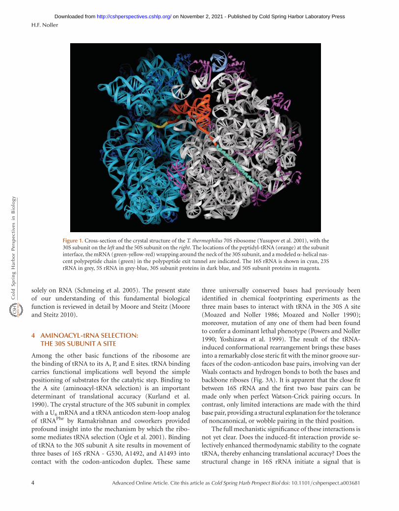
30S and 50S subunits at 3.0 A and 2.4 A resolution, respec-tively, provided detailed descriptions of the folding of theRNA and protein components of the ribosome for the firsttime (Ban et al. 2000; Schluenzen et al. 2000; Wimberlyet al. 2000). The 5.5 A resolution structure of the complete70S ribosome, with mRNA and tRNAs bound, showed howthe subunits fit together, and revealed the interactions be-tween the ribosome and the P- and E-site tRNAs (Yusupovet al. 2001) (Fig. 1). A complex containing the completeA-site tRNA bound to the 70S ribosome at 7.0 A, andanother complex of a tRNA anticodon stem-loop boundto the 30S subunit at 3.1–3.3 A provided the details of theinteractions between the ribosome and the A-site tRNA(Ogle et al. 2001). Several different complexes containingthe 50S subunit bound with a variety of tRNA acceptor-endmimics, including a transition-state analogue, providedinsight into how the aminoacyl and peptidyl ends of thetRNAs interact with the peptidyl transferase catalytic site(Nissen et al. 2000).
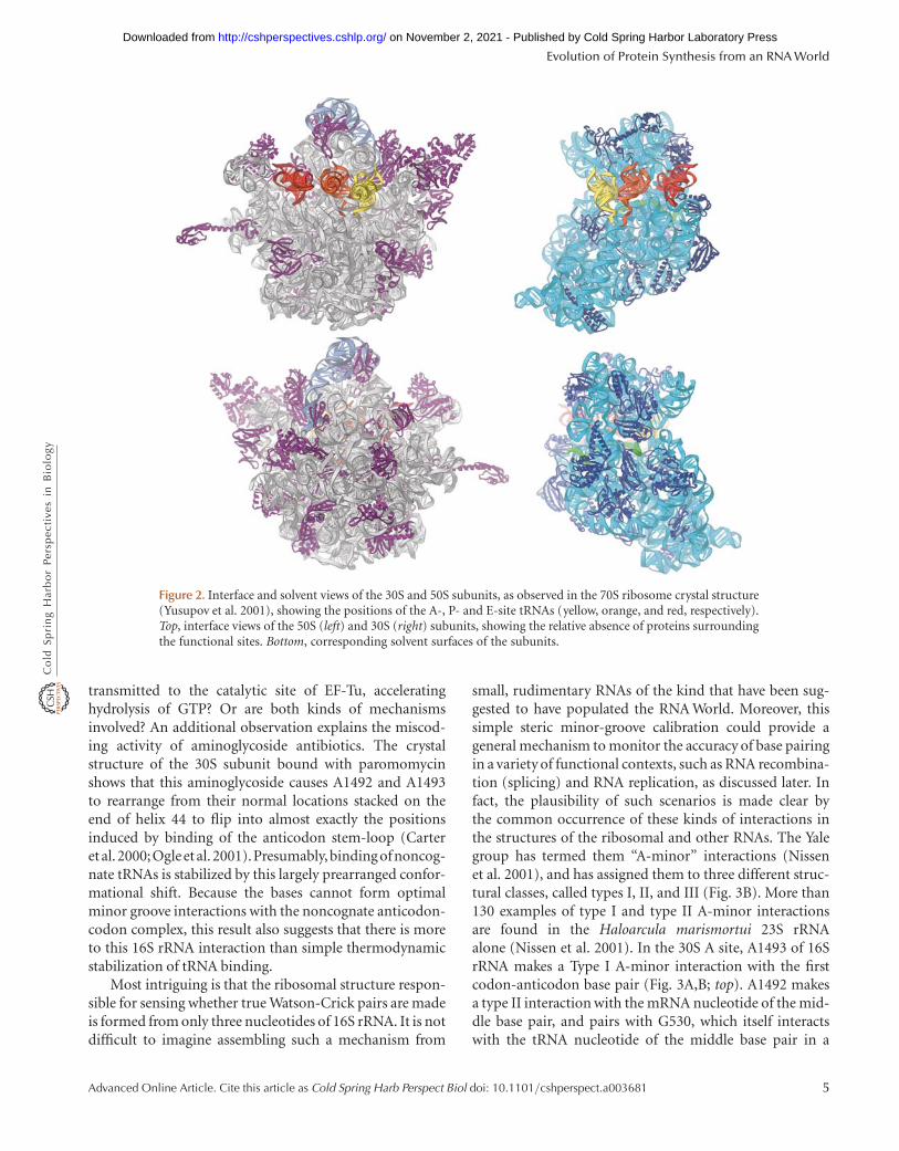
The distribution of the rRNA and protein moieties onthe surface of the ribosomal subunits (Fig. 2) makes a clearcase for the functional importance of rRNA. Proteins aredistributed more or less evenly over the external surfaceof the ribosome, filling nooks and crannies in the rRNA(Fig. 2B,D), but the subunit interface surface, which con-tains the tRNA binding sites as well as other functionalfeatures, is made up mainly of rRNA (Fig. 2A,C). The over-all impression is that of an RNA structure that has graduallyincorporated a number of proteins over evolutionarytime, but has not allowed them to impinge on its crucialfunctional centers.
The high-resolution structure of the 50S subunit (Banet al. 2000), together with the knowledge of the positionsof the acceptor ends of the tRNAs (Nissen et al. 2000;Yusupov et al. 2001), provided the first look at the structureof the peptidyl transferase center. No protein moieties werefound with 17 A of the catalytic site, definitively demon-strating that peptide bond formation is indeed catalyzedby RNA. Although a more recent high-resolution structureof the T. thermophilus 70S ribosome bound with tRNAsshows interactions between the amino-terminal tail of pro-tein L27 and the backbone of the 30 CCA end of the P-sitetRNA (Selmer et al. 2006), no part of the protein is closeenough to the catalytic site to play a direct (chemical)role in the reaction. Furthermore, early studies showedthat Escherichia coli 50S subunits reconstituted in vitrowithout L27 were active in catalyzing peptide bond forma-tion (Moore et al. 1975). Also, no counterpart to L27 isfound in archaeal 50S subunits (Nissen et al. 2000). Com-parison of structures of 50S complexes containing varioussusbstrate and transition-state analogs support an induced-fit model for catalysis of peptide bond formation based
Evolution of Protein Synthesis from an RNA World
Advanced Online Article. Cite this article as Cold Spring Harb Perspect Biol doi: 10.1101/cshperspect.a003681 3
on November 2, 2021 - Published by Cold Spring Harbor Laboratory Press http://cshperspectives.cshlp.org/Downloaded from
solely on RNA (Schmeing et al. 2005). The present stateof our understanding of this fundamental biologicalfunction is reviewed in detail by Moore and Steitz (Mooreand Steitz 2010).
4 AMINOACYL-tRNA SELECTION:THE 30S SUBUNIT A SITE
Among the other basic functions of the ribosome arethe binding of tRNA to its A, P, and E sites. tRNA bindingcarries functional implications well beyond the simplepositioning of substrates for the catalytic step. Binding tothe A site (aminoacyl-tRNA selection) is an importantdeterminant of translational accuracy (Kurland et al.1990). The crystal structure of the 30S subunit in complexwith a U6 mRNA and a tRNA anticodon stem-loop analogof tRNAPhe by Ramakrishnan and coworkers providedprofound insight into the mechanism by which the ribo-some mediates tRNA selection (Ogle et al. 2001). Bindingof tRNA to the 30S subunit A site results in movement ofthree bases of 16S rRNA - G530, A1492, and A1493 intocontact with the codon-anticodon duplex. These same
three universally conserved bases had previously beenidentified in chemical footprinting experiments as thethree main bases to interact with tRNA in the 30S A site(Moazed and Noller 1986; Moazed and Noller 1990);moreover, mutation of any one of them had been foundto confer a dominant lethal phenotype (Powers and Noller1990; Yoshizawa et al. 1999). The result of the tRNA-induced conformational rearrangement brings these basesinto a remarkably close steric fit with the minor groove sur-faces of the codon-anticodon base pairs, involving van derWaals contacts and hydrogen bonds to both the bases andbackbone riboses (Fig. 3A). It is apparent that the close fitbetween 16S rRNA and the first two base pairs can bemade only when perfect Watson-Crick pairing occurs. Incontrast, only limited interactions are made with the thirdbase pair, providing a structural explanation for the toleranceof noncanonical, or wobble pairing in the third position.
The full mechanistic significance of these interactions isnot yet clear. Does the induced-fit interaction provide se-lectively enhanced thermodynamic stability to the cognatetRNA, thereby enhancing translational accuracy? Does thestructural change in 16S rRNA initiate a signal that is
Figure 1. Cross-section of the crystal structure of the T. thermophilus 70S ribosome (Yusupov et al. 2001), with the30S subunit on the left and the 50S subunit on the right. The locations of the peptidyl-tRNA (orange) at the subunitinterface, the mRNA (green-yellow-red) wrapping around the neck of the 30S subunit, and a modeleda-helical nas-cent polypeptide chain (green) in the polypeptide exit tunnel are indicated. The 16S rRNA is shown in cyan, 23SrRNA in grey, 5S rRNA in grey-blue, 30S subunit proteins in dark blue, and 50S subunit proteins in magenta.
H.F. Noller
4 Advanced Online Article. Cite this article as Cold Spring Harb Perspect Biol doi: 10.1101/cshperspect.a003681
on November 2, 2021 - Published by Cold Spring Harbor Laboratory Press http://cshperspectives.cshlp.org/Downloaded from
transmitted to the catalytic site of EF-Tu, acceleratinghydrolysis of GTP? Or are both kinds of mechanismsinvolved? An additional observation explains the miscod-ing activity of aminoglycoside antibiotics. The crystalstructure of the 30S subunit bound with paromomycinshows that this aminoglycoside causes A1492 and A1493to rearrange from their normal locations stacked on theend of helix 44 to flip into almost exactly the positionsinduced by binding of the anticodon stem-loop (Carteret al. 2000;Ogle etal. 2001).Presumably, binding of noncog-nate tRNAs is stabilized by this largely prearranged confor-mational shift. Because the bases cannot form optimalminor groove interactions with the noncognate anticodon-codon complex, this result also suggests that there is moreto this 16S rRNA interaction than simple thermodynamicstabilization of tRNA binding.
Most intriguing is that the ribosomal structure respon-sible for sensing whether true Watson-Crick pairs are madeis formed from only three nucleotides of 16S rRNA. It is notdifficult to imagine assembling such a mechanism from
small, rudimentary RNAs of the kind that have been sug-gested to have populated the RNA World. Moreover, thissimple steric minor-groove calibration could provide ageneral mechanism to monitor the accuracy of base pairingin a variety of functional contexts, such as RNA recombina-tion (splicing) and RNA replication, as discussed later. Infact, the plausibility of such scenarios is made clear bythe common occurrence of these kinds of interactions inthe structures of the ribosomal and other RNAs. The Yalegroup has termed them “A-minor” interactions (Nissenet al. 2001), and has assigned them to three different struc-tural classes, called types I, II, and III (Fig. 3B). More than130 examples of type I and type II A-minor interactionsare found in the Haloarcula marismortui 23S rRNAalone (Nissen et al. 2001). In the 30S A site, A1493 of 16SrRNA makes a Type I A-minor interaction with the firstcodon-anticodon base pair (Fig. 3A,B; top). A1492 makesa type II interaction with the mRNA nucleotide of the mid-dle base pair, and pairs with G530, which itself interactswith the tRNA nucleotide of the middle base pair in a
Figure 2. Interface and solvent views of the 30S and 50S subunits, as observed in the 70S ribosome crystal structure(Yusupov et al. 2001), showing the positions of the A-, P- and E-site tRNAs (yellow, orange, and red, respectively).Top, interface views of the 50S (left) and 30S (right) subunits, showing the relative absence of proteins surroundingthe functional sites. Bottom, corresponding solvent surfaces of the subunits.
Evolution of Protein Synthesis from an RNA World
Advanced Online Article. Cite this article as Cold Spring Harb Perspect Biol doi: 10.1101/cshperspect.a003681 5
on November 2, 2021 - Published by Cold Spring Harbor Laboratory Press http://cshperspectives.cshlp.org/Downloaded from

type II-like interaction (Fig. 3A,B; middle). The 30S subu-nit A site presents compelling evidence for the likelihoodthat the ribosome evolved from a purely RNA structure,and helps to explain why its function continues to be basedon RNA.
5 THE 30S SUBUNIT P SITE: ANOTHER FUNCTIONOF rRNA
One of the earliest indications of the functional role ofrRNA was the implication of 16S rRNA in binding tRNAto the 30S P site. Inactivation of P-site binding by kethoxalmodification of 16S rRNA (Noller and Chaires 1972),direct crosslinking of the wobble base of tRNA to C1400
of 16S rRNA (Prince et al. 1982), chemical footprintingof 16S rRNA by P-site tRNA (Moazed and Noller 1986;Moazed and Noller 1990) and modification-interferenceexperiments (von Ahsen and Noller 1995) all pointed tothe involvement of a constellation of 16S rRNA nucleotidesin this function. The crystal structures directly showed theparticipation of 16S rRNA in tRNA binding to the 30S Psite, providing further evidence for the RNA character ofthe ribosome (Wimberly et al. 2000; Yusupov et al. 2001).However, any hopes for a simple RNA-World picturewere clouded by the intrusion of proteins S9 and S13into the tRNA binding site (Wimberly et al. 2000). Bothproteins contain extended carboxy-terminal tails, whichcontact phosphate 35 of the anticodon loop and the
G530
G34U3
A35 U2
A1492
G530
A1493
A36 U1
A521
G1364 C637
Type I
A520
G1363C638
Type II
Figure 3. Steric minor-groove calibration of Watson-Crick codon-anticodon pairing by three conserved bases of 16SrRNA. (left) Contacts between G530, A1492, and A1493 of 16S rRNA and the codon-anticodon base pairs in the 30SA site (Ogle et al. 2001). (right) Type I and type II A-minor interactions (Nissen et al. 2001).
H.F. Noller
6 Advanced Online Article. Cite this article as Cold Spring Harb Perspect Biol doi: 10.1101/cshperspect.a003681
on November 2, 2021 - Published by Cold Spring Harbor Laboratory Press http://cshperspectives.cshlp.org/Downloaded from

backbone of the anticodon stem, respectively. Both tailscontain basic amino-acid side-chains that appear to makeelectrostatic interactions with the tRNA backbone.
An RNA-World impression of S9 and S13 is one of twoproteins that landed on and took root in 16S rRNA as a laterevolutionary refinement of the basic RNA structure of thesubunit. The functional requirement for their C-terminaltails was tested directly by replacing the genomic copiesof the E. coli genes encoding S9 and S13 with versions inwhich their carboxy-terminal tails were deleted (Hoanget al. 2004). The result was that mutant strains bearingthe deleted versions of the two proteins were viable, includ-ing strains in which the tails of both proteins were deleted.The phenotypes were relatively mild, amounting to a 40%reduction in growth rate for the double deletion. In thisstrain, all of the cellular proteins are synthesized by ribo-somes whose 30S P sites are composed purely of RNA.Thus, 16S rRNA is able to support all of the essential func-tions of the 30S P site, including translational initiation,P-site tRNA binding, and maintenance of the translationalreading frame.
6 RNA MOLECULAR MECHANICS ANDTRANSLOCATION
Perhaps the most demanding step of translation isthe coupled movement of mRNA and tRNA, called trans-location, which follows formation of each new peptidebond. This step depends on elongation factor EF-G andis coupled to hydrolysis of GTP. It is coupled to large-scalemolecular movements, including relative rotation ofthe two ribosomal subunits, emphasizing the structuraldynamics of the ribosome. Because the pioneer ribosomesmust have been capable of translocation, we can ask:(a) How could such a fundamental process have operatedin the absence of EF-G, which is commonly referred to asthe “translocase” of protein synthesis? And, what wasthe source of energy to drive movement of mRNA andtRNA, and intersubunit rotation? Studies by Pestka (Pest-ka 1968) and Spirin (Gavrilova et al. 1976) showed manyyears ago that poly(U)-dependent synthesis of polypheny-lalanine could proceed in the absence of EF-G, undercertain in vitro conditions, or by modification of ribo-somes with thiol-directed reagents. Criticisms that theobserved synthesis might have involved some sort of“slippage” of the poly(U) mRNA were addressed by Greenand coworkers using a defined mRNA (Southworth et al.2002). The requirement for GTP was shown not to beabsolute by the demonstration that the peptidyl transferaseinhibitor sparsomycin can trigger a single round oftranslocation in vitro, with high efficiency and accuracy,in the complete absence of EF-G or GTP (Fredrick and
Noller 2003). These studies suggest that translocationcould have originated as a purely ribosomal, factor-independent process.
But in the absence of GTP hydrolysis, what is the sourceof free energy to drive the translocation reaction, and whatkeeps it from going backwards? The most obvious source ofenergy comes from peptide bond formation; peptidyltransfer results in formation of a peptide amide bond froman activated ribose ester linkage, accompanied by a largechange in free energy. How can this free energy change becoupled to translocation? The answer most likely lies inthe changing chemical nature of the acceptor end of thetRNA as it moves through the ribosome (Spirin 1985).It enters as an aminoacyl-tRNA, is transformed into apeptidyl-tRNA and then becomes completely deacylated.The 50S subunit contains three tRNA binding sites, theA, P, and E sites, which have specific affinities for the threeforms of tRNA, providing at the same time a downhillenergetic pathway and a unidirectional movement for thetRNA during translocation.
Translocation also appears to depend on rotation of the�850 kDa 30S subunit relative to the 50S subunit for eachstep (Frank and Agrawal 2000; Frank et al. 2007; Horan andNoller 2007). In the absence of GTP hydrolysis, what is thesource of energy to drive this massive intermolecularmovement? Recent single-molecule FRET studies showthat spontaneous intersubunit rotation can occur in awide variety of mRNA-tRNA-ribosome complexes in theabsence of EF-G or GTP, or even peptide bond formation(Cornish et al. 2008). This finding shows that thermalenergy alone is sufficient to drive the intersubunit rotationunderlying translocation. Translocation is also coupled tomovement of a feature of the 50S subunit called the L1 stalk,which maintains contact with the elbow of the deacylatedtRNA as it moves from the P/E to the E/E state. Single-molecule FRETexperiments show that the L1 stalk can tra-verse through three different positions during transloca-tion, corresponding to the P/E, E/E and vacant states ofthe E site (Cornish et al. 2009). Again, the L1 stalk wasfound to be able to move spontaneously in the absence ofEF-G or GTP. These findings show that even the complex,large-scale molecular movements associated with translo-cation can be driven by thermal energy, obviating theneed for special energy-generating steps in protein synthe-sis by the first ribosomes.
7 WHAT ARE RIBOSOMAL PROTEINS FOR?
If, as we suspect, the fundamental functions of the ribo-some are based on its rRNA, why are there so many riboso-mal proteins, some of which are highly conserved? So far,no one has reported observation of a ribosomal function
Evolution of Protein Synthesis from an RNA World
Advanced Online Article. Cite this article as Cold Spring Harb Perspect Biol doi: 10.1101/cshperspect.a003681 7
on November 2, 2021 - Published by Cold Spring Harbor Laboratory Press http://cshperspectives.cshlp.org/Downloaded from

being carried out by a rigorously protein-free preparationof rRNA. One explanation for this is that rRNA does notfold into its functional state in the absence of r-proteins(Nomura et al. 1969; Stern et al. 1989). Another reasonfor the presence of proteins in ribosomes is that theyimprove the efficiency and accuracy of translation. For ex-ample, mutations in proteins S4 and S5 have long beenknown to cause increased translational error frequencies,implying that they help to improve the accuracy of transla-tion (Davies and Nomura 1972; Kurland et al. 1990). Thepresence of the carboxy-terminal tails of S9 and S13 isnot essential for 30S P-site function, as discussed above,but improves tRNA binding and increases the growth rateof E. coli (Hoang et al. 2004). A further point is that evensmall improvements in the speed and accuracy of transla-tion bring strong selective advantages.
In contrast to histones, for example, the structures ofthe ribosomal proteins are extremely heterogeneous, repre-senting a large number of different domain types, includ-ing helical bundles, a/b RRM folds, all-b OB folds, andso on. Many contain long unstructured tails that penetratethe structure of the rRNA (Ban et al. 2000; Wimberly et al.2000). Some are essential for correct overall folding andassembly, whereas others are not (Nomura et al. 1969). Afew are positioned at or near the subunit interface, wherethey can influence ribosomal function, whereas the major-ity are located on the solvent surface, remote from any func-tional site. Thus, the ribosomal proteins clearly did notarise by duplication of one another, to play a single role,or related roles, but give the impression that they wereadded one at a time over the course of evolution, as incre-mental refinements of an essentially RNA-based ribosome.
8 “STOP tRNAs” AND THE EVOLUTION OFTYPE I RELEASE FACTORS
All but three of the 64 possible triplet codons are recognizedby tRNAs. The remaining three, the stop codons UAG,UAA and UGA are recognized by the type I release factors(RF1 and RF2 in bacteria). Recent crystal structures of 70Sribosome termination complexes show that the stop co-dons are directly recognized by the release factors and sug-gest that catalysis of peptidyl-tRNA hydrolysis is catalyzedby the –NH group of the polypeptide backbone of a con-served Gln in the conserved GGQ motif (Korostelev et al.2008; Laurberg et al. 2008; Weixlbaumer et al. 2008). Theposition of this –NH group superimposes with the posi-tion of the 30-OH group of a deacylated tRNA bound tothe A site. This raises the possibility that stop codonswere originally recognized by deacylated tRNAs, whichwere replaced during evolution by the type I release factors(Laurberg et al., 2008). In fact, it has been shown that
binding of deacylated tRNA to the A site catalyzes polypep-tide release (Caskey et al. 1971). A potential shortcoming ofa “stop tRNA” is that deacylated tRNAs are unable to bindelongation factor EF-Tu, which is critical for the accuracy oftRNA binding; the resulting high error frequency of trans-lation termination may thus have driven the evolution oftype I release factors.
9 dRNA AND THE ORIGINS OF THE RIBOSOMALDECODING SITE
The molecular interactions involved in codon recognitionshown in Figure 3 could, in principle, serve to monitorthe accuracy of Watson-Crick base pairing in other RNAcontexts. An obvious possible application of this type ofquality control in the RNAWorld is the critical process ofRNA replication. A-minor interactions are so simple andso widespread that it would be surprising if they were notput to use for this purpose. Is it possible that the 30S decod-ing site is a relic of an RNA replication mechanism from theRNAWorld? The following is a suggestion for how such areplication system may have worked, extrapolating fromwhat we have learned from the ribosome.
Our starting assumption is that codon-anticodoninteraction occurs at the site of the template–productinteraction in an RNA World replicase, in which similarA-minor interactions were used to ensure the accuracy ofRNA replication. The fact that the decoding site mediatestriplet–triplet base pairing suggests that its RNA Worldrole would have been to stabilize ligation of oligonucleotidetriplets base-paired to a template; i.e., the replicase wouldhave taken the form of an RNA ligase. The first criticalquestion is whether the mRNA codon corresponds to thetemplate (as it does in protein synthesis) or the product(in which case, the anticodon would be the template).Examination of the structure of the decoding site providesan unambiguous answer. The distance between phosphateand ribose groups of adjacent anticodons bound to the Aand P sites is more than 30 A, ruling out models in whichthe mRNA serves as template. Accordingly, we concludethat anticodons serve as short templates for positioningRNA triplets in the replicase active site.
This raises a second critical question: How can RNA bereplicated using such short templates? Here, we introducethe idea of “duplicator RNA” (dRNA). dRNAs are small,tRNA-like structures that mediate duplication (as opposedto replication) of an RNA template (Fig. 4). dRNAs have aloop resembling an anticodon loop (here, it is shown as aseven-nucleotide loop, but loops of other sizes would bepossible), and an unpaired self-complementary four-nu-cleotide tail, that allows dRNAs to form homodimers.One “anticodon” end of the dimer base pairs with a triplet
H.F. Noller
8 Advanced Online Article. Cite this article as Cold Spring Harb Perspect Biol doi: 10.1101/cshperspect.a003681
on November 2, 2021 - Published by Cold Spring Harbor Laboratory Press http://cshperspectives.cshlp.org/Downloaded from

sequence in the template RNA, and the other anticodonend pairs with either the nascent product RNA or the in-coming triplet substrate (Fig. 5). The product interactionresembles the strong binding of the ribosomal P site and
the substrate interaction corresponds to that of the decod-ing site. Ligation of the substrate oligomer to the growingproduct chain occurs at the junction between these twobinding sites.
Because there are 16 possible self-complementary tet-ramers, there are 16 possible different dRNAs. Interestingly,the same number of dRNAs is predicted from a completelyindependent argument. The geometry of the A-minorinteractions used in the decoding site creates the basis forthe degeneracy of the genetic code (Fig. 3); adjacent basepairs can interact with adjacent adenosines to form type Iand type II interactions, but this does not extend to a thirdbase pair (which, in the decoding site is partially taken overby a separate guanine, G530). Degeneracy in the third po-sition of the replicase triplet-triplet interaction would thuslimit the number of dRNA anticodons (which could them-selves be degenerate) to 16. Thus, each dRNA has two dis-tinct identity elements—a self-complementary tetramertail, and a (degenerate) triplet anticodon—which establishthe link between the template and product RNAs. (Implicitin this discussion is the idea that dRNAs are precursors totRNAs. Besides their anticodon-like features, their self-complementary tails are reminiscent of tRNA identity ele-ments that are often present in the acceptor stems of tRNAs.)
There are several properties of this form of indirecttemplating that distinguish it from normal RNA replica-tion, some of which seem especially well suited to the
C U
GCGC
N
C U
GCGC
CGCG
N
C U N
Self-complementarytetramer tail
Degenerate anticodontriplet
dRNA monomer dRNA homodimer
Figure 4. Duplicator RNA. (left) A schematic cartoon representingthe structure of a duplicator RNA (dRNA) monomer, showing itstwo identity elements: A self-complementary tetramer tail and a de-generate anticodon triplet. There are 16 possible dRNAs. (right) AdRNA homodimer, formed by base pairing of its self-complementarytail. The wavy line indicates that other details of the structure betweenthe anticodon and tail are not intended to be explicit.
G G
CUAG
GAUC
N
C C N
G G N
C C N
C U
GCGC
CGCG
N
G A N
C U N
G5¢
5¢
3¢Template
Product
A N
A SITEP SITE
Figure 5. Indirect templating of RNA duplication mediated by dRNA homodimers. One dRNA dimer (left) is boundto the last triplet (GAN) in the product RNA, stabilized by a structure resembling the 30S ribosomal P site (blue box).A second dRNA dimer (right), bound to the next (CCN) triplet in the template by pairing with one of its GGN anti-codons, binds the incoming substrate CCN trimer via base pairing with its other GGN anticodon. Discrimination ofcorrect pairing with the incoming substrate trimer is promoted by A-minor interactions by a structure resemblingthe 30S ribosomal A site (red box). Binding of the upper anticodon triplets to the template RNA also uses structuresresembling the 30S A and P sites (not shown).
Evolution of Protein Synthesis from an RNA World
Advanced Online Article. Cite this article as Cold Spring Harb Perspect Biol doi: 10.1101/cshperspect.a003681 9
on November 2, 2021 - Published by Cold Spring Harbor Laboratory Press http://cshperspectives.cshlp.org/Downloaded from
challenges of the RNA World. First, the template is dupli-cated in a parallel fashion, eliminating the formation ofan intermediate RNA duplex and the associated problemof unwinding a long duplex to allow release and foldingof the product RNA. Second, the substrates are tripletoligonucleotides, which bind more stably to their templatesthan single nucleotides, yet are readily disrupted at ambienttemperatures. These can be sourced from random-sequencetriplet pools; because of third-position degeneracy, there areeffectively only 16 different substrate oligomers.
The general scheme described here raises many detailedmechanistic questions. How are the substrate oligomerssynthesized and activated? What is the mechanism ofcatalysis? How is the nascent chain translocated followingaddition of each oligomer? How is the secondary structureof the RNA template disrupted to allow dRNA binding?Would accidental binding of longer oligomers disruptRNA duplication? Finally, the details of the structure ofdRNA in the region linking the anticodon to the self-complementary tail (unspecified here) will be critical, sothat the resulting geometry will allow binding of adjacentdRNAs to adjacent triplets and their parallel translocationwith respect to the template and product RNAs at oppositeends of the dRNA dimers. These and other aspectsof dRNA-mediated duplication are discussed elsewhere(Noller 2010 in preparation).
10 THE DRIVING FORCE FOR EVOLUTION OFTRANSLATION FROM AN RNA WORLD
It is challenging to ask how the structurally and function-ally complex process of translation could have evolvedfrom an RNAWorld. Most encouraging is the demonstra-tion that small RNAs evolved in vitro are able to catalyzeall of the principal chemical reactions of protein synthesis,including amino acid activation, aminoacylation of RNA,and peptidyl transfer (Zhang and Cech 1997; Illangasekareand Yarus 1999; Lee et al. 2000; Kumar and Yarus 2001). Butquite apart from the mind-boggling prospect of evolving astructure as complex as the ribosome (even without itsproteins), the probability of an early translational systemproducing a functionally capable protein, such as an en-zyme, is vanishingly small (Woese 1967). Given these pros-pects, what was the driving force that led to the evolutionaryselection of protein synthesis in the context of an RNAWorld? If we assume that some sort of Darwinian selectionwas in place, it must have provided a selective advantage toan RNAworld system.
The diversity and efficiency of RNA functiondepends on the possible types of structure into whichRNA can fold. Structural studies on naturally occurringand in vitro-selected RNAs have revealed a rich diversity
of RNA folds. Nevertheless, it can already be seen that therange of RNA structures is limited, probably because ofthe relatively modest chemical differences between thefour nucleotide monomers, compounded by the strong in-herent tendency for ribonucleotides to adopt conforma-tions resembling those that are found in A-type doublehelices (Saenger 1984). We would therefore expect an ex-pansion of the range of possible RNA structures to confera strong selective advantage.
Studies on the structures of RNA-ligand complexesshow that the binding of even quite small molecules toRNA can cause large-scale structural changes. RNAs thatappear to be unstructured adopt well-defined three-dimensional folds, and structured RNAs undergo confor-mational rearrangements in the presence of bound ligands.An example is the AMP-dependent structuring of an invitro-selected aptamer, in which the 11-nucleotide RNAwraps around the nucleotide into an intricate, well-definedfold, such that the AMP plays the role of the conserved A ina GNRA tetraloop (Dieckmann et al. 1996). Another exam-ple is the discovery of “riboswitches,” ligand-induced RNAstructures that are found in naturally occurring mRNAs,influencing the expression of the mRNAs in which theyare embedded (Mandal and Breaker 2004; Serganov et al.2004; Breaker 2010). Thus, binding of small-moleculeligands is likely to have played an important part in expand-ing the structural repertoire of RNA.
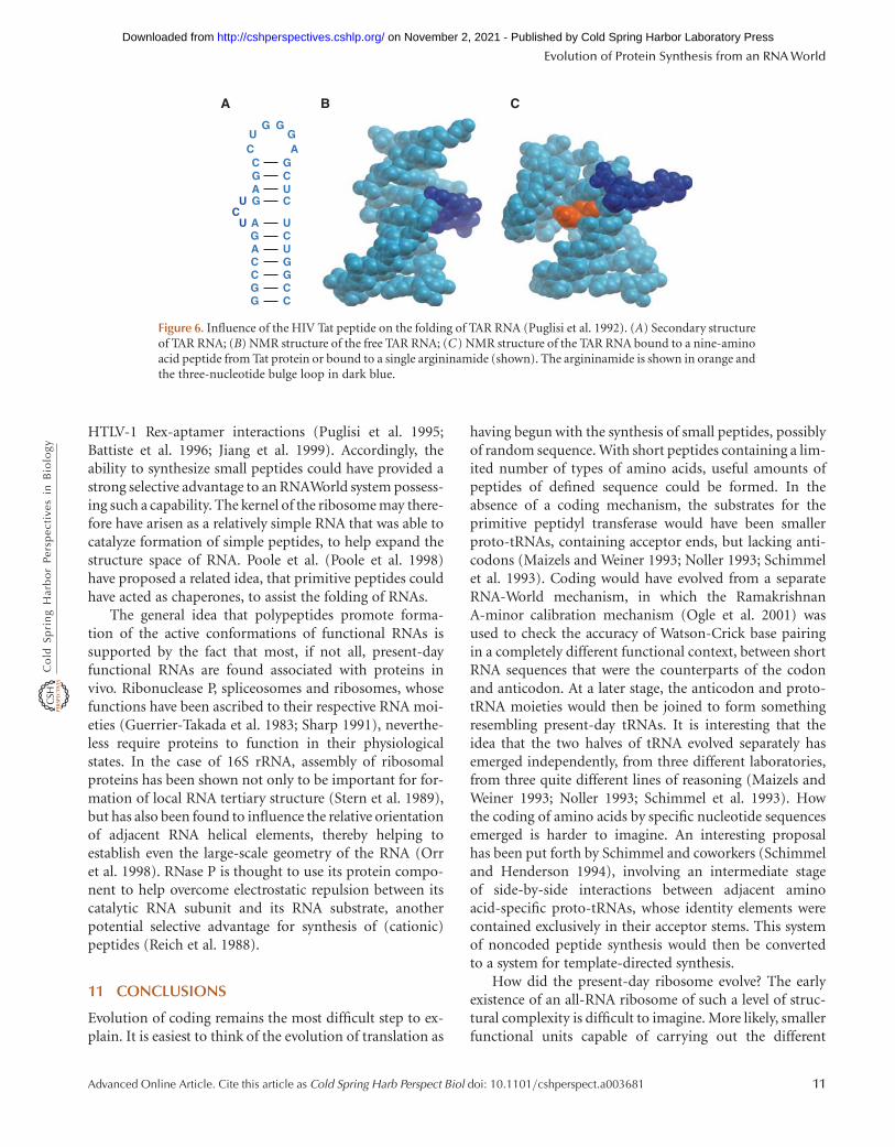
Binding of peptides has also been shown to causerearrangement of RNA structure. A vivid example is basedon the HIV Tat-TAR complex, a protein-RNA interactionthat is essential for viral function. Binding of the Tat pro-tein to the TAR RNA could be mimicked by a nine-aminoacid arginine-rich peptide derived from the binding site ofthe protein (Puglisi et al. 1992). Binding of this peptidecauses a dramatic rearrangement of the structure ofthe TAR RNA, from a conventional hairpin stem-loopstructure into a structure containing a bulge loop, resultingin formation of an A-U-U triple base pair tertiary interac-tion. At the core of the structure is an arginine side-chainthat appears to play a crucial role in stabilizing the newfold. Remarkably, it was found that this same structuralrearrangement occurred in the presence of a single argini-namide (Puglisi et al. 1992) (Fig. 6), although the mono-mer bound with an affinity that was lower by five or sixorders of magnitude. These findings show two points: (1)Simple peptides or even amino acid monomers candramatically influence the structure of RNA, and (2) Theextended structures provided by incorporating aminoacids into peptides confer higher binding affinity (andconcomitantly, increased specificity). Further examples ofpeptide-induced structural rearrangements of RNA havebeen observed for the HIV Rev-RRE, BIV Tat-TAR, and
H.F. Noller
10 Advanced Online Article. Cite this article as Cold Spring Harb Perspect Biol doi: 10.1101/cshperspect.a003681
on November 2, 2021 - Published by Cold Spring Harbor Laboratory Press http://cshperspectives.cshlp.org/Downloaded from
HTLV-1 Rex-aptamer interactions (Puglisi et al. 1995;Battiste et al. 1996; Jiang et al. 1999). Accordingly, theability to synthesize small peptides could have provided astrong selective advantage to an RNAWorld system possess-ing such a capability. The kernel of the ribosome may there-fore have arisen as a relatively simple RNA that was able tocatalyze formation of simple peptides, to help expand thestructure space of RNA. Poole et al. (Poole et al. 1998)have proposed a related idea, that primitive peptides couldhave acted as chaperones, to assist the folding of RNAs.
The general idea that polypeptides promote forma-tion of the active conformations of functional RNAs issupported by the fact that most, if not all, present-dayfunctional RNAs are found associated with proteins invivo. Ribonuclease P, spliceosomes and ribosomes, whosefunctions have been ascribed to their respective RNA moi-eties (Guerrier-Takada et al. 1983; Sharp 1991), neverthe-less require proteins to function in their physiologicalstates. In the case of 16S rRNA, assembly of ribosomalproteins has been shown not only to be important for for-mation of local RNA tertiary structure (Stern et al. 1989),but has also been found to influence the relative orientationof adjacent RNA helical elements, thereby helping toestablish even the large-scale geometry of the RNA (Orret al. 1998). RNase P is thought to use its protein compo-nent to help overcome electrostatic repulsion between itscatalytic RNA subunit and its RNA substrate, anotherpotential selective advantage for synthesis of (cationic)peptides (Reich et al. 1988).
11 CONCLUSIONS
Evolution of coding remains the most difficult step to ex-plain. It is easiest to think of the evolution of translation as
having begun with the synthesis of small peptides, possiblyof random sequence. With short peptides containing a lim-ited number of types of amino acids, useful amounts ofpeptides of defined sequence could be formed. In theabsence of a coding mechanism, the substrates for theprimitive peptidyl transferase would have been smallerproto-tRNAs, containing acceptor ends, but lacking anti-codons (Maizels and Weiner 1993; Noller 1993; Schimmelet al. 1993). Coding would have evolved from a separateRNA-World mechanism, in which the RamakrishnanA-minor calibration mechanism (Ogle et al. 2001) wasused to check the accuracy of Watson-Crick base pairingin a completely different functional context, between shortRNA sequences that were the counterparts of the codonand anticodon. At a later stage, the anticodon and proto-tRNA moieties would then be joined to form somethingresembling present-day tRNAs. It is interesting that theidea that the two halves of tRNA evolved separately hasemerged independently, from three different laboratories,from three quite different lines of reasoning (Maizels andWeiner 1993; Noller 1993; Schimmel et al. 1993). Howthe coding of amino acids by specific nucleotide sequencesemerged is harder to imagine. An interesting proposalhas been put forth by Schimmel and coworkers (Schimmeland Henderson 1994), involving an intermediate stageof side-by-side interactions between adjacent aminoacid-specific proto-tRNAs, whose identity elements werecontained exclusively in their acceptor stems. This systemof noncoded peptide synthesis would then be convertedto a system for template-directed synthesis.
How did the present-day ribosome evolve? The earlyexistence of an all-RNA ribosome of such a level of struc-tural complexity is difficult to imagine. More likely, smallerfunctional units capable of carrying out the different
A B C
GUG G
ACGC
G CA UG C
UC
CC
UGG
A
U
UC
G
GG
ACC
Figure 6. Influence of the HIV Tat peptide on the folding of TAR RNA (Puglisi et al. 1992). (A) Secondary structureof TAR RNA; (B) NMR structure of the free TAR RNA; (C) NMR structure of the TAR RNA bound to a nine-aminoacid peptide from Tat protein or bound to a single argininamide (shown). The argininamide is shown in orange andthe three-nucleotide bulge loop in dark blue.
Evolution of Protein Synthesis from an RNA World
Advanced Online Article. Cite this article as Cold Spring Harb Perspect Biol doi: 10.1101/cshperspect.a003681 11
on November 2, 2021 - Published by Cold Spring Harbor Laboratory Press http://cshperspectives.cshlp.org/Downloaded from
translational steps such as peptidyl transferase, decodingand so on evolved. These small functional RNA unitsthen merged to form larger structures, which were incre-mentally refined by incorporation of additional RNA struc-tural elements. Two features of RNA make such a processplausible. First is the fact that small RNAs, unlike most pep-tides, tend to retain their local structures when excised fromlarger RNA structures. For example, the anticodon stem-loop of tRNA interacts efficiently and accurately with theribosome in the absence of the rest of the tRNA structure,and is even capable of accurate codon recognition andtranslocation (Rose et al. 1983; Joseph and Noller 1998;Ogle et al. 2001). This may be because RNAs do not havehydrophobic cores that are essential for their three-dimensional folding. Thus, building a large functionalRNA out of pre-existing small ones can be accomplishedwhile preserving structural and functional properties ofthe latter. Second, RNAs can readily interact with each oth-er to form complexes, not only by base pairing, but by ro-bust tertiary interactions such as the A-minor interactionsdescribed previously. RNAs that have associated noncova-lently in this way can then become ligated together toform covalently stable RNAs of increasing size, by well-known ribozyme-catalyzed mechanisms. An explicit pro-posal for the hierarchical evolution of 23S rRNA was re-cently described by (Bokov and Steinberg 2009), whoanalyzed the distribution of donor adenosines and acceptorhelices for the A-minor interactions of the 50S ribosomalsubunit. They observed a striking assymetry in domain Vof 23S rRNA, which contains the elements of the peptidyltransferase center: in all but one case, domain V contrib-uted the acceptor helices for adenosines from other do-mains. This suggests that 23S rRNA evolved by gradualaddition of ancillary RNA domains to the catalytic corein domain V, exploiting A-minor interactions in position-ing the added elements. Importantly, in an RNAWorld, anewly constructed RNA becomes its own gene, whose rep-lication directly provides multiple copies of new functionalRNAs. This cycle of noncovalent complex formation, fol-lowed by ligation, can then be repeated, while selectingfor improvement of ribosome-like function. Finally, the in-fluence of the peptides produced by a primitive ribosomeon its own structure and assembly would also begin to im-pact its own evolution. Ribosome-binding peptides maythus have played an early role in shaping the ultimateform (and function) of the ribosome.
ACKNOWLEDGMENTS
I thank Bill Scott, Melissa Moore, Andrei Korostelev, GerryJoyce, Jim Dahlberg, Carl Woese, Leslie Orgel and themembers of my laboratory for many stimulating
discussions. This work was supported by grants from theNIH and NSF, and by a grant from the W.M. Keck Founda-tion to the Center for Molecular Biology of RNA at UCSC.
REFERENCES
Ban N, Nissen P, Hansen J, Moore PB, Steitz TA. 2000. The completeatomic structure of the large ribosomal subunit at 2.4 A resolution.Science 289: 905–920.
Barta A, Steiner G, Brosius J, Noller HF, Kuechler E. 1984. Identificationof a site on 23S ribosomal RNA located at the peptidyl transferase cen-ter. Proc Natl Acad Sci 81: 3607–3611.
Battiste JL, Mao H, Rao NS, Tan R, Muhandiram DR, Kay LE, FrankelAD, Williamson JR. 1996. a helix-RNA major groove recognition inan HIV-1 rev peptide-RRE RNA complex. Science 273: 1547–1551.
Bokov K, Steinberg SV. 2009. A hierarchical model for evolution of 23Sribosomal RNA. Nature 457: 977–980.
Bowman CM, Dahlberg JE, Ikemura T, Konisky J, Nomura M. 1971. Spe-cific inactivation of 16S ribosomal RNA induced by colicin E3 in vivo.Proc Natl Acad Sci 68: 964–968.
Breaker RR. 2010. Riboswitches and the RNA world. Cold Spring HarbPerspect Biol 2: a003566.
Carter AP, Clemons WM, Brodersen DE, Morgan-Warren RJ, WimberlyBT, Ramakrishnan V. 2000. Functional insights from the structure ofthe 30S ribosomal subunit and its interactions with antibiotics [seecomments]. Nature 407: 340–348.
Caskey CT, Beaudet AL, Scolnick EM, Rosman M. 1971. Hydrolysis offMet-tRNA by peptidyl transferase. Proc Natl Acad Sci 68: 3163–3167.
Cornish PV, Ermolenko DN, Noller HF, Ha T. 2008. Spontaneous inter-subunit rotation in single ribosomes. Mol Cell 30: 578–588.
Cornish PV, Ermolenko DN, Staple DW, Hoang L, Hickerson RP, NollerHF, Ha T. 2009. Following movement of the L1 stalk between threefunctional states in single ribosomes. Proc Natl Acad Sci 106:2571–2576.
Crick FHC. 1968. The origin of the genetic code. J Mol Biol 38: 367–379.Dabbs ER. 1986. Mutant studies on the prokaryotic ribosome. in Struc-
ture, function and genetics of ribosomes (ed. B. Hardesty, G. Kramer),pp. 733–748. Springer-Verlag, N.Y.
Davies J, Nomura M. 1972. The genetics of bacterial ribosomes. Ann RevGenet 6: 203–234.
Dieckmann T, Suzuki E, Nakamura GK, Feigon J. 1996. Solution struc-ture of an ATP-binding RNA aptamer reveals a novel fold. RNA 2:628–640.
Endo Y, Wool IG. 1982. The site of action of a-sarcin on eukaryotic ribo-somes. The sequence at the a-sarcin cleavage site in 28 S ribosomalribonucleic acid. J Biol Chem 257: 9054–9060.
Frank J, Agrawal RK. 2000. A ratchet-like inter-subunit reorganization ofthe ribosome during translocation. Nature 406: 318–322.
Frank J, Gao H, Sengupta J, Gao N, Taylor DJ. 2007. The process ofmRNA-tRNA translocation. Proc Natl Acad Sci 104: 19671–19678.
Fredrick K, Noller HF. 2003. Catalysis of ribosomal translocation by spar-somycin. Science 300: 1159–1162.
Gavrilova LP, Kostiashkina OE, Koteliansky VE, Rutkevitch NM, SpirinAS. 1976. Factor-free (“non-enzymic”) and factor-dependent systemsof translation of polyuridylic acid by Escherichia coli ribosomes. J MolBiol 101: 537–552.
Guerrier-Takada C, Gardiner K, Marsh T, Pace N, Altman S. 1983. TheRNA moiety of ribonuclease P is the catalytic subunit of the enzyme.Cell 35: 849–857.
Helser TL, Davies JE, Dahlberg JE. 1972. Mechanism of kasugamycinresistance in Escherichia coli. Nat New Biol 235: 6–9.
Hoang L, Fredrick K, Noller HF. 2004. Creating ribosomes with anall-RNA 30S subunit P site. Proc Natl Acad Sci 101: 12439–12443.
Horan LH, Noller HF. 2007. Intersubunit movement is required for ribo-somal translocation. Proc Natl Acad Sci 104: 4881–4885.
H.F. Noller
12 Advanced Online Article. Cite this article as Cold Spring Harb Perspect Biol doi: 10.1101/cshperspect.a003681
on November 2, 2021 - Published by Cold Spring Harbor Laboratory Press http://cshperspectives.cshlp.org/Downloaded from

Illangasekare M, Yarus M. 1999. Specific, rapid synthesis of Phe-RNA byRNA. Proc Natl Acad Sci 96: 5470–5475.
Jiang F, Gorin A, Hu W, Majumdar A, Baskerville S, Xu W, Ellington A,Patel DJ. 1999. Anchoring an extended HTLV-1 Rex peptide withinan RNA major groove containing junctional base triples. StructureFold Des 7: 1461–1472.
Joseph S, Noller HF. 1998. EF-G-catalyzed translocation of anticodonstem-loop analogs of transfer RNA in the ribosome. EMBO J 17:3478–3483.
Korostelev A, Asahara H, Lancaster L, Laurberg M, Hirschi A, Zhu J, Tra-khanov S, Scott WG, Noller HF. 2008. Crystal structure of a translationtermination complex formed with release factor RF2. Proc Natl AcadSci 105: 19684–19689.
Kumar RK, Yarus M. 2001. RNA-catalyzed amino acid activation. Bio-chemistry 40: 6998–7004.
Kurland CG, Jorgensen F, Richter AA, Ehrenberg M, Bilgin N, Rojas AM.1990. Through the Accuracy Window. in The Ribosome: Structure,function, and evolution (ed. WE. Hill, A. Dahlberg, RA. Garrett, PB.Moore, D. Schlessinger, JR. Warner), pp. 513–526. American Societyof Microbiology, Washington, D.C.
Laurberg M, Asahara H, Korostelev A, Zhu J, Trakhanov S, Noller HF.2008. Structural basis for translation termination on the 70S ribo-some. Nature 454: 852–857.
Lee N, Bessho Y, Wei K, Szostak JW, Suga H. 2000. Ribozyme-catalyzedtRNA aminoacylation. Nat Struct Biol 7: 28–33.
Lill R, Robertson JM, Wintermeyer W. 1986. Affinities of tRNA bindingsites of ribosomes from Escherichia coli. Biochemistry 25: 3245–3255.
Maizels N, Weiner AM. 1993. The genomic tag hypothesis: Modernviruses as molecular fossils of ancient strategies for genomic replica-tion. in The RNA world (ed. RF. Gesteland JF. Atkins), pp. 577–602.Cold Spring Harbor Laboratory Press, Cold Spring Harbor, NY.
Mandal M, Breaker RR. 2004. Gene regulation by riboswitches. Nat RevMol Cell Biol 5: 451–463.
Moazed D, Noller HF. 1986. Transfer RNA shields specific nucleotides in16S ribosomal RNA from attack by chemical probes. Cell 47: 985–994.
Moazed D, Noller HF. 1987. Chloramphenicol, erythromycin, carbomy-cin and vernamycin B protect overlapping sites in the peptidyl trans-ferase region of 23S ribosomal RNA. Biochimie 69: 879–884.
Moazed D, Noller HF. 1989. Interaction of tRNA with 23S rRNA in theribosomal A, P, and E sites. Cell 57: 585–597.
Moazed D, Noller HF. 1990. Binding of tRNA to the ribosomal A and Psites protects two distinct sets of nucleotides in 16S rRNA. J Mol Biol211: 135–145.
Moazed D, Noller HF. 1991. Sites of interaction of the CCA end ofpeptidyl-tRNA with 23S rRNA. Proc Natl Acad Sci 88: 3725–3728.
Monro RE. 1967. Catalysis of peptide bond formation by 50 S ribosomalsubunits from Escherichia coli. J Mol Biol 26: 147–151.
Moore PB, Steitz TA. 2010. The roles of RNA in the synthesis of protein.Cold Spring Harb Perspect Biol 2: a003780.
Moore VG, Atchison RE, Thomas G, Moran M, Noller HF. 1975. Identi-fication of a ribosomal protein essential for peptidyl transferase activ-ity. Proc Natl Acad Sci 72: 844–848.
Nirenberg M, Leder P. 1964. RNA code words and protein synthesis. Sci-ence 145: 1399–1407.
Nissen P, Hansen J, Ban N, Moore PB, Steitz TA. 2000. The structural basisof ribosome activity in peptide bond synthesis. Science 289: 920–930.
Nissen P, Ippolito JA, Ban N, Moore PB, Steitz TA. 2001. RNA tertiaryinteraction in the large ribosomal subunit: The A-minor motif. ProcNatl Acad Sci 98: 4899–4903.
Noller HF. 1993. On the origin of the ribosome: Coevolution of subdo-mains of tRNA and rRNA. in The RNA world (ed. RF. Gesteland, TR.Cech, JF. Atkins), pp. 137–156. Cold Spring Harbor Laboratory Press,Cold Spring Harbor, NY.
Noller HF. 2004. The driving force for molecular evolution of translation.RNA 10: 1833–1837.
Noller HF, Chaires JB. 1972. Functional modification of 16S ribosomalRNA by kethoxal. Proc Natl Acad Sci 69: 3113–3118.
Noller HF, Hoffarth V, Zimniak L. 1992. Unusual resistance of peptidyltransferase to protein extraction procedures. Science 256: 1416–1419.
Noller HF, Moazed D, Stern S, Powers T, Allen PN, Robertson JM, Weiser B,Triman K. 1990. Structure of rRNA and its functional interactions intranslation. in The ribosome: structure, function, and evolution (ed. WE.Hill, A. Dahlberg, RA. Garrett, PB. Moore, D. Schlessinger, JR. Warner),pp. 73–92. American Society of Microbiology, Washington, D.C.
Nomura M, Mizushima S, Ozaki M, Traub P, Lowry CV. 1969. Structureand function of ribosomes and their molecular components. ColdSpring Harbor Symp Quant Biol 34: 49–61.
Ogle JM, Brodersen DE, Clemons WM, Tarry MJ, Carter AP, Ramak-rishnan V. 2001. Recognition of cognate transfer RNA by the 30S ribo-somal subunit. Science 292: 897–902.
Orgel LE. 1968. Evolution of the genetic apparatus. J Mol Biol 38:381–393.
Orr JW, Hagerman PJ, Williamson JR. 1998. Protein and Mg(2þ)-induced conformational changes in the S15 binding site of 16 S ribo-somal RNA. J Mol Biol 275: 453–464.
Pestka S. 1968. Studies on the formation of transfer ribonucleic acid-ribosome complexes. 3. The formation of peptide bonds by ribosomesin the absence of supernatant enzymes. J Biol Chem 243: 2810–2820.
Poole AM, Jeffares DC, Penny D. 1998. The path from the RNA world.J Mol Evol 46: 1–17.
Powers T, Noller HF. 1990. Dominant lethal mutations in a conservedloop in 16S rRNA. Proc Natl Acad Sci 87: 1042–1046.
Prince JB, Taylor BH, Thurlow DL, Ofengand J, Zimmermann RA. 1982.Covalent crosslinking of tRNA1Val to 16S RNA at the ribosomalP site: Identification of crosslinked residues. Proc Natl Acad Sci 79:5450–5454.
Puglisi JD, Chen L, Blanchard S, Frankel AD. 1995. Solution structureof a bovine immunodeficiency virus Tat-TAR peptide-RNA complex.Science 270: 1200–1203.
Puglisi JD, Tan R, Calnan BJ, Frankel AD, Williamson JR. 1992. Confor-mation of the TAR RNA-arginine complex by NMR spectroscopy.Science 257: 76–80.
Reich C, Olsen GJ, Pace B, Pace NR. 1988. Role of the protein moiety ofribonuclease P, a ribonucleoprotein enzyme. Science 239: 178–181.
Rose SJ 3rd, Lowary PT, Uhlenbeck OC. 1983. Binding of yeast tRNAPheanticodon arm to Escherichia coli 30 S ribosomes. J Mol Biol 167:103–117.
Saenger W. 1984. Principles of nucleic acid structure Springer-Verlag,New York.
Schimmel P, Giege R, Moras D, Yokoyama S. 1993. An operational RNAcode for amino acids and possible relationship to genetic code. ProcNatl Acad Sci 90: 8763–8768.
Schimmel P, Henderson B. 1994. Possible role of aminoacyl-RNA com-plexes in noncoded peptide synthesis and origin of coded synthesis.Proc Natl Acad Sci 91: 11283–11286.
Schluenzen F, Tocilj A, Zarivach R, Harms J, Gluehmann M, Janell D,Bashan A, Bartels H, Agmon I, Franceschi F, et al. 2000. Structure offunctionally activated small ribosomal subunit at 3.3 angstroms reso-lution. Cell 102: 615–623.
Schmeing TM, Huang KS, Strobel SA, Steitz TA. 2005. An induced-fitmechanism to promote peptide bond formation and exclude hydrol-ysis of peptidyl-tRNA. Nature 438: 520–524.
Schulze H, Nierhaus KH. 1982. Minimal set of ribosomal componentsfor reconstitution of the peptidyltransferase activity. EMBO J 1:609–613.
Selmer M, Dunham CM, Murphy FVt, Weixlbaumer A, Petry S, KelleyAC, Weir JR, Ramakrishnan V. 2006. Structure of the 70S ribosomecomplexed with mRNA and tRNA. Science 313: 1935–1942.
Senior BW, Holland IB. 1971. Effect of colicin E3 upon the 30S ribosomalsubunit of Escherichia coli. Proc Nat Acad Sci 69: 959–964.
Serganov A, Yuan YR, Pikovskaya O, Polonskaia A, Malinina L, Phan AT,Hobartner C, Micura R, Breaker RR, Patel DJ. 2004. Structural basis
Evolution of Protein Synthesis from an RNA World
Advanced Online Article. Cite this article as Cold Spring Harb Perspect Biol doi: 10.1101/cshperspect.a003681 13
on November 2, 2021 - Published by Cold Spring Harbor Laboratory Press http://cshperspectives.cshlp.org/Downloaded from

for discriminative regulation of gene expression by adenine- and gua-nine-sensing mRNAs. Chem Biol 11: 1729–1741.
Sharp PA. 1991. “Five easy pieces”. Science 254: 663.Shine J, Dalgarno L. 1974. The 3’-terminal sequence of E coli 16S riboso-
mal RNA complementarity to nonsense triplets and ribosome bindingsites. Proc Nat Acad Sci 71: 1342–1346.
Southworth DR, Brunelle JL, Green R. 2002. EFG-independenttranslocation of the mRNA:tRNA complex is promoted by modifi-cation of the ribosome with thiol-specific reagents. J Mol Biol 324:611–623.
Spirin AS. 1985. Ribosomal translocation: Facts and models. Prog NucleicAcid Res Mol Biol 32: 75–114.
Steitz JA, Jakes K. 1975. How ribosomes select initiator regions in mRNA:Base pair formation between the 3’ terminus of 16S rRNA and themRNA during initiation of protein synthesis in Escherichia coli. ProcNatl Acad Sci 72: 4734–4738.
Stern S, Powers T, Changchien LM, Noller HF. 1989. RNA-proteininteractions in 30S ribosomal subunits: Folding and function of 16SrRNA. Science 244: 783–790.
von Ahsen U, Noller HF. 1995. Identification of bases in 16S rRNAessential for tRNA binding at the 30S ribosomal P site. Science 267:234–237.
Weixlbaumer A, Jin H, Neubauer C, Voorhees RM, Petry S, Kelley AC,Ramakrishnan V. 2008. Insights into translational termination fromthe structure of RF2 bound to the ribosome. Science 322: 953–956.
Wimberly BT, Brodersen DE, Clemons WM Jr, Morgan-Warren RJ, Car-ter AP, Vonrhein C, Hartsch T, Ramakrishnan V. 2000. Structure of the30S ribosomal subunit. Nature 407: 327–339.
Woese CR. 1967. The genetic code. The molecular basis for genetic expres-sion Harper & Row, New York.
Woese CR, Fox GE, Zablen L, Uchida T, Bonen L, Pechman K, Lewis BJ,Stahl D. 1975. Conservation of primary structure in 16S ribosomalRNA. Nature 254: 83–86.
Wower J, Sylvers LA, Rosen KV, Hixson SS, Zimmermann RA. 1993.A model of the tRNA binding sites on the Escherichia coli ribosome.in The translational apparatus: Structure, function, regulation, evolution(ed. KH. Nierhaus), pp. 455–464. Plenum Press, New York.
Yoshizawa S, Fourmy D, Puglisi JD. 1999. Recognition of thecodon-anticodon helix by ribosomal RNA. Science 285: 1722–1725.
Yusupov M, Yusupova G, Baucom A, Lieberman K, Earnest TN, Cate JH,Noller HF. 2001. Crystal structure of the ribosome at 5.5 A resolution.Science 292: 883–896.
Zhang B, Cech TR. 1997. Peptide bond formation by in vitro selectedribozymes. Nature 390: 96–100.
H.F. Noller
14 Advanced Online Article. Cite this article as Cold Spring Harb Perspect Biol doi: 10.1101/cshperspect.a003681
on November 2, 2021 - Published by Cold Spring Harbor Laboratory Press http://cshperspectives.cshlp.org/Downloaded from

published online July 7, 2010Cold Spring Harb Perspect Biol Harry F. Noller Evolution of Protein Synthesis from an RNA World
Subject Collection RNA Worlds
Alternate RNA StructuresMarie Teng-Pei Wu and Victoria D'Souza
Structural Biology of TelomeraseYaqiang Wang, Lukas Susac and Juli Feigon
Expressionof Long Noncoding RNA Regulation of Gene Approaches for Understanding the Mechanisms
Patrick McDonel and Mitchell Guttman
Splicing in Higher EukaryotesStructural Insights into Nuclear pre-mRNA
Berthold Kastner, Cindy L. Will, Holger Stark, et al.
Act on ChromatinThatExperiments to Study Long Noncoding RNAs
Principles and Practices of Hybridization Capture
Matthew D. Simon and Martin Machyna
UTRs Doing?′What Are 3Christine Mayr
Sequencerson Massively Parallel High-Throughput Linking RNA Sequence, Structure, and Function
Sarah K. Denny and William J. Greenleaf
Transcriptase EnzymesSingle-Molecule Analysis of Reverse
Linnea I. Jansson and Michael D. Stone
Insights into Eukaryotic TranslationComplexity: Ribosomal Structures Provide Extensions, Extra Factors, and Extreme
Melanie Weisser and Nenad Ban
RegulationCRISPR Tools for Systematic Studies of RNA
Gootenberg, et al.Jesse Engreitz, Omar Abudayyeh, Jonathan
with TranscriptionNascent RNA and the Coordination of Splicing
Karla M. Neugebauer
Relating Structure and Dynamics in RNA Biology
al.Kevin P. Larsen, Junhong Choi, Arjun Prabhakar, et
ComplexesRNA−Integrative Structural Biology of Protein
Magnetic Resonance (NMR) Spectroscopy for Combining Mass Spectrometry (MS) and Nuclear
AllainAlexander Leitner, Georg Dorn and Frédéric H.-T.
Synthetic GeneticsBeyond DNA and RNA: The Expanding Toolbox of
HolligerAlexander I. Taylor, Gillian Houlihan and Philipp
http://cshperspectives.cshlp.org/cgi/collection/ For additional articles in this collection, see
Copyright © 2010 Cold Spring Harbor Laboratory Press; all rights reserved
on November 2, 2021 - Published by Cold Spring Harbor Laboratory Press http://cshperspectives.cshlp.org/Downloaded from
of mRNANucleotides That Comprise the Epitranscriptome Discovering and Mapping the Modified
Bastian Linder and Samie R. Jaffrey
Lessons from YeastStructural Basis of Nuclear pre-mRNA Splicing:
NagaiClemens Plaschka, Andrew J. Newman and Kiyoshi
http://cshperspectives.cshlp.org/cgi/collection/ For additional articles in this collection, see
Copyright © 2010 Cold Spring Harbor Laboratory Press; all rights reserved
on November 2, 2021 - Published by Cold Spring Harbor Laboratory Press http://cshperspectives.cshlp.org/Downloaded from