evidencethatintravenouslyderivedmurinepulmonarymelanomamÃ...
TRANSCRIPT
[CANCER RESEARCH 46, 5167-5171, October 1986)
Evidence That Intravenously Derived Murine Pulmonary Melanoma MétastasesCan Originate from the Expansion of a Single Tumor CellIsaiah J. Fidler1 and James E. Talmadge
Department of Cell Biology, The University of Texas, M. D. Anderson Hospital and Tumor institute, Houston, Texas 77030 ¡I.J. F.], and Preclinical Screen Laboratory,Program Resources, inc., NCI-Frederick Cancer Research Facility, Frederick, Maryland 21701 [J. E. TJ
ABSTRACT
The purpose of these studies was to determine whether hematogenousdonai pulmonary melanoma métastasesoriginate from the expansion ofa single cell and if so, by extrapolation, metastasis can be considered acloning process. Three different cell lines of murine K-1735 melanomawith different metastatic properties and unique karyotypes were injectedi.v. into syngeneic C3H/HeN mice as multiceli aggregates of individualcell lines or combinations of cell lines. Resultant solitary lung métastaseswere isolated in culture as individual lines and then karyotyped. Evenwhen heterogeneous clumps of tumor cells were injected, the individualmétastasesexhibited a karyotype unique to one metastatic cell type.Furthermore, when cellular aggregates were composed of metastatic cellsadmixed with cells that were tumorigenic but nonmetastatic, the resultantmétastasesexhibited only the karyotype of the metastatic cells. Thisfinding suggests that the presence of metastatic cells did not change theinability of nonmetastatic cells to proliferate in a distant organ. Collectively, the results indicate that the resultant métastaseswere of clonalorigin owing to the expansion of a single metastatic tumor cell in the lungparenchyma.
INTRODUCTION
Multiple métastases,even those within the same organ, oftenexhibit a diversity in their various biological properties, such asantigenicity, immunogenicity, hormone receptors, metastaticpotential, or response to various chemotherapeutic agents (1-
6). This diversity is due to both the nature of the metastaticprocess (7-9) and to the evolution and progression of the tumorcells (10-14). Malignant neoplasms are heterogeneous and
therefore contain cells with different biological and metastaticproperties (1-9). To produce a metastasis, a tumor cell must
complete a series of steps which are potentially lethal to thecells. The outcome of this process is therefore dependent onboth the responses of the host and the intrinsic properties ofthe tumor cells (7-9). Metastasis begins with the detachment
of tumor cells from the primary lesion. After they invade theblood vessels and/or the lymphatics, single tumor cells ormulticeli emboli circulate and eventually reach the capillarybeds of distant organs, where they arrest. After tumor cellsextravasate into the organ parenchyma, they must proliferatein order to give rise to secondary lesions. Métastasesare notproduced by the random survival of circulating tumor embolibut rather by the selective growth of specialized subpopulationsof metastatic cells that preexist in the parent tumor (15-17).
We recently demonstrated that individual lung métastasesproduced from a mouse melanoma can be clonal in origin (18).We based this conclusion on karyotype analyses of cells fromindividual lung métastasesproduced in syngeneic C3H/HeNmice by f.p.2 growth of the K-1735 melanoma. We irradiated
Received 4/7/86; revised 6/12/86; accepted 6/13/86.The costs of publication of this article were defrayed in part by the payment
of page charges. This article must therefore be hereby marked advertisement inaccordance with 18 U.S.C. Section 1734 solely to indicate this fact.
1To whom requests for reprints should be addressed, at Department of Cell
Biology, University of Texas System Cancer Center, M. D. Anderson Hospitaland Tumor Institute, 6723 Bertner Avenue (173), Houston, TX 77030.
2The abbreviations used are: f.p., footpad; M-2, K-1735-M-2 line; X-21, K-1735-X-met-21 line; C-19, K-1735 clone 19.
the parent'tumor prior to f.p. implantation with 650 R to
induce random chromosome breaks and rearrangements (19).Although the results of this experiment demonstrated that somemétastaseshave a clonal origin and that different métastasescan originate from different progenitor cells, it did not resolvethe question as to whether clonal growth at the metastatic siteresults from a single cell or from the implantation of a homogeneous clump of cells. Primary neoplasms are known to bezonal (20, 21), and tumor emboli entering the circulation canbe either homogeneous (composed of one subpopulation) orheterogeneous (composed of multiple subpopulations). Theclonality of melanoma métastasescould therefore be merely thereflection of the zonal origin of the tumor embolus. Since it isunlikely that only homogeneous tumor cells circulate, wewished to determine whether the process of metastasis per secould be a cloning process; that is, if the clonal origin of ametastasis could be due to the multiplication of a single cellwithin a heterogeneous tumor embolus arrested in the capillarybed of a distant organ. Experiments presented in this reportaddress these possibilities and demonstrate that, in the murineK-1735 melanoma system, métastasescan result from a prolif
eration of a single cell.
MATERIALS AND METHODS
Animals. Adult C3H/HeN mammary tumor virus-negative mice, 6to 8 weeks old, were obtained from the Animal Production Area of theNCI-Frederick Cancer Research Facility.
Cell Lines. The original K-1735 melanoma (22) was a gift from Dr.Margaret L. Kripke, University of Texas, M. D. Anderson Hospitaland Tumor Institute, Houston, TX. The K-1735-M-2 line was derivedfrom a spontaneous pulmonary metastasis that had been produced bythe K-1735 parent tumor growing at a f.p. site (17). The K-1735-M-2cells were exposed to 650 R of X-radiation to induce random chromosome breakage and rearrangements (18). These X-irradiated cells werethen implanted into the footpads of syngeneic mice. When the resultingf.p. tumors reached an average diameter of 1 cm, the tumor-bearingleg, including the popliteal lymph node, was resected at midfemur. Sixweeks later, the mice were killed, and well-isolated, solitary spontaneouspulmonary métastaseswere aseptically removed, grown in culture asindividual lines, and karyotyped (18). Within certain métastases,thesame chromosomal abnormalities were detected in all of the spreadsexamined, suggesting that these métastaseswere of clonal origin. Oneof these karyotyped lines, X-21, was used in the present study. Theoriginal K-1735 melanoma was cloned in vitro by a double cloningmethod. Clone 19, thus derived, was characterized previously to benonmetastatic (22) and was used in this study.
All tumor cell lines (M-2, X-21, and C-19) were maintained in culturein Eagle's minimal essential medium supplemented with 10% fetal
bovine serum, sodium pyruvate, nonessential amino acids, L-glutamine,and 2-fold vitamin solution (Flow Laboratories, Rockville, MD). Cellcultures were maintained on plastic and were incubated in 5% CO2 at37°C.Cultures were free of Mycoplasma and the following murineviruses: reovirus type 3; pneumonia; K; Theiler's encephalitis; Sendai;
minute; mouse adenovirus; mouse hepatitis; lymphocytic choriomen-ingitis; ectromelia; and lactate dehydrogenase (assayed by M. A. Bio-products, Walkersville, MD).
5167
on June 23, 2018. © 1986 American Association for Cancer Research. cancerres.aacrjournals.org Downloaded from
METASTASIS AS A CLONING PROCESS
Kxperimcntal Metastasis Assay. To produce experimental métastases, tumor cells in exponential growth phase were harvested by a briefexposure to 0.25% trypsin:0.02% EDTA solution (w/v). The flask wastapped sharply to dislodge the cells, medium was added, and the cellsuspension was pipeted again to produce a single-cell suspension. Thecells were washed and resuspended in Ca2+- and Mg2*-free Hanks'
balanced salt solution to the desired cell concentration. Cell viabilitywas determined by trypan blue exclusion, and only suspensions of singlecells with greater than 90% viability were used. Aliquots (0.2 ml) oftumor cell suspensions were then injected into the lateral tail vein ofC3H/HeN mice. Four weeks after injection the mice were killed; thelungs were removed, washed, and fixed in Bouin's solution to differ
entiate the neoplastic lesions from the organ parenchyma. The lungnodules were counted by visualization with a dissecting microscope.
Experimental Design. Individual cultures of K-1735-M-2, K-1735-X-met-21, and K-1735 clone 19 in exponential growth phase were harvested after short exposure to 0.25% trypsin:0.02% EDTA solution.Medium was added, and the cells were incubated in separate test tubesor admixed at a 1:1 ratio for 30 min on a rotating platform at 37°Cto
produce homogeneous or heterogeneous aggregates (M-2; X-21; C-19;X-21 and M-2; X-21 and C-19). After this incubation period, the testtubes were placed in a rack, and large tumor cell aggregates (10 to 20cells) were recovered after a 30-min sedimentation at 37°C.Parallel
experiments using autoradiography demonstrated that the X-21 andM-2 aggregates and the X-21 and C-19 mixed aggregates were indeedheterogeneous. To demonstrate this heterogeneity, X-21 cells werecultured for 24 h in medium containing 0.2 ¿(Ci[3H]leucine/ml (NewEngland Nuclear, Boston. MA). The aggregates of X-21 cells alone, X-21 plus M-2 cells, and X-21 plus C-19 cells were plated onto glassslides. After 1 h, the slides were fixed in 10% buffered formalin andprepared for autoradiography (23).
To produce lung métastases,C3H/HeN mice were given injectionsi.v. of 1,000 to 2,000 tumor cell aggregates totaling 10,000 to 20,000tumor cells. The injections lasted 2 to 3 minutes to avoid pulmonaryembolism and death. Preliminary studies showed that this long injectiontime also reduced the possibility of reducing the size of the clumps dueto shear forces as the aggregates passed through the injection needle.Six weeks later, the mice were killed and individual well-isolated lungmétastases(I mm in diameter) were recovered aseptically, dispersedthrough a stainless steel sieve (Cellector, E. C. Apparatus, St. Petersburg, FL), and cultured. Individual cell lines were established from eachmetastatic lesion, as described previously (16, 17). Chromosome analyses of at least 60 consecutive spreads from each individual line werecarried out by two independent observers as described in detail previously (24). Briefly, confluent cultures were split 1:3 and harvested 48 hlater. Cell cultures were exposed to Colcemid (2.5 jig/ml) for 1 h. Afterbrief (3 min) trypsinization, the suspended cells were swollen in 0.38%potassium chloride, fixed with 2 x Carnoy's fixative (methanol:glacial
acetic acid) and dropped onto cold, wet slides.Whenever possible, 60 to 80 cells from each tumor population were
analyzed completely for breakage and recombinant formation. Includedamong unstable breaks are acentric fragments, chromatid and chromosome breaks, and complex rearrangements. Slides were stained withGiemsa, and metaphases were examined by phase microscopy at amagnification of x 1250. While this limited the definition of recombi
nants, it optimized recognition of breakage. Small unmatched centricchromosomes were scored as minutes and included in the chromosomecounts. Double minute fragments were scored but not counted aschromosome. Dicentric and multicentric chromosomes were countedas single chromosomes.
RESULTS
The tumorigenic and metastatic properties of the 3 K-1735lines used in these studies are summarized in Table 1. By thecriteria used, C-19 cells are tumorigenic but nonmetastatic,whereas cells of the M-2 and X-21 lines are both tumorigenicand highly metastatic. Karyotype analyses of the 3 culturedlines revealed that M-2 cells have a fairly stable mode of 44chromosomes, most of which are telocentric; while C-19 ispseudotetraploid with a mode of 54 chromosomes, most ofwhich are telocentric (18). The X-21 cells have a chromosomemode of 42, and all of the cells (in >1000 spreads) express thesame unique submetacentric chromosome marker with a 3:4arm length ratio (1, 25).
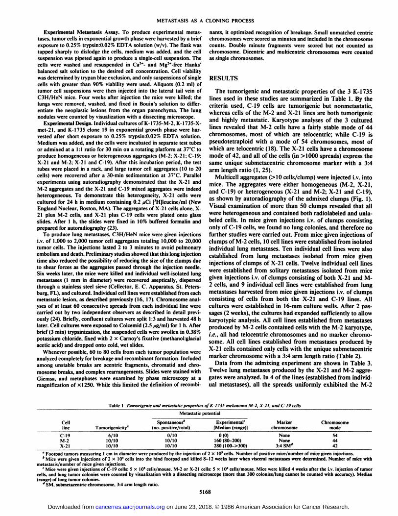
Multiceli aggregates (> 10 cells/clump) were injected i.v. intomice. The aggregates were either homogeneous (M-2, X-21,and C-19) or heterogeneous (X-21 and M-2; X-21 and C-19),as shown by autoradiography of the admixed clumps (Fig. 1).Visual examination of more than 50 clumps revealed that allwere heterogeneous and contained both radiolabeled and unla-beled cells. In mice given injections i.v. of clumps consistingonly of C-19 cells, we found no lung colonies, and therefore nofurther studies were carried out. From mice given injections ofclumps of M-2 cells, 10 cell lines were established from isolatedindividual lung métastases.Ten individual cell lines were alsoestablished from lung métastasesisolated from mice giveninjections of clumps of X-21 cells. Twelve individual cell lineswere established from solitary métastasesisolated from micegiven injections i.v. of clumps consisting of both X-21 and M-2 cells, and 9 individual cell lines were established from lungmétastasesharvested from mice given injections i.v. of clumpsconsisting of cells from both the X-21 and C-19 lines. Allcultures were established in 16-mm culture wells. After 2 passages (2 weeks), the cultures had expanded sufficiently to allowkaryotypic analysis. All cell lines established from métastasesproduced by M-2 cells contained cells with the M-2 karyotype,i.e., all had telocentric chromosomes and no marker chromosome. All cell lines established from métastasesproduced byX-21 cells contained only cells with the unique submetacentricmarker chromosome with a 3:4 arm length ratio (Table 2).
Data from the admixing experiment are shown in Table 3.Twelve lung métastasesproduced by the X-21 and M-2 aggregates were analyzed. In 4 of the lines (established from individual métastases),all the spreads uniformly exhibited the M-2
Table I Tumorigenic and metastatic properties of K-1735 melanoma M-2, X-21, and C-19 cells
MetastaticpotentialCell
lineC-19M-2
X-21Tumorigenicity"6/1010/1010/10Spontaneous*
(no. positive/total)0/10
10/1010/10Experimental
(Median(range)]0(0)
160(80-200)280(100->300)Marker
chromosomeNone
None3:4 SM*Chromosome
mode54
4442
" Footpad tumors measuring 1 cm in diameter were produced by the injection of 2 x III' cells. Number of positive mice/number of mice given injections.* Mice were given injections of 2 x 10* cells into the hind footpad and killed 8-12 weeks later when visceral métastaseswere determined. Number of mice with
metastasis/number of mice given injections.c Mice were given injections of C-19 cells: 5 x 10*cells/mouse. M-2 or X-21 cells: 5x10* cells/mouse. Mice were killed 4 weeks after the i.v. injection of tumor
cells, and lung tumor colonies were counted by visualization with a dissecting microscope (more than 300 colonies/lung cannot be counted with accuracy). Median(range) of lung tumor colonies.
* SM. submetacentric chromosome. 3:4 arm length ratio.
5168
on June 23, 2018. © 1986 American Association for Cancer Research. cancerres.aacrjournals.org Downloaded from

METASTASIS AS A CLONING PROCESS
Fig. 1. Autoradiography of multiceli aggregates. Unlabeled M-2 cells (A),radioactive labeled X-21 cells (B), and a mixture of M-2 cells and X-21 cells (Q.Cells of the X-21 line were incubated for 24 h with medium containing 0.2 »iCiof [3H]leucine/ml. Aggregates were produced as described in "Materials andMethods." Cell aggregates were deposited on glass slides and fixed. The slideswere covered with NBT-2 emulsion (Eastman Kodak. Rochester, NY) and developed 10 days later.
karyotype. In the remaining 8 of the 12 isolated lines, all thespreads uniformly exhibited the X-21 karyotype.
In the final set of experiments, mice were given injections i.v.of tumor cell aggregates that consisted of both C-19 (nonme-tastatic) and X-21 (metastatic) cells. Karyotype analyses of celllines established from 9 solitary lung métastasesrevealed thepresence of only X-21 cells, i.e., cells containing a submetacen-
tric marker chromosome with a 3:4 arm length ratio (Table 4).
Table 2 Karyotype analysis of cells from experimental lung métastasesproducedby the i.v. injection of multiceli aggregates consisting of only M-2 or only X-21
cellsKaryotype0
1 UllllHHill,M-2
parentMet*1Met
2Met3Met4MetSMet
6Met7MetSMet
9Met10X-21
parentMetIMet2Met3Met4MetSMetoMet?MetSMet
9Met10Telocentric6063636664666465646600000010000Submetacentric
marker00064
tetraploid00000006765646565626067645363
" M-2 cells have a chromosome mode of 44, and all the chromosomes aretelocentric. X-21 cells have a chromosome mode of 42, and all the cells expressthe same Submetacentric chromosome (3:4 arm length ratio). The numbersrepresent the number of spreads analyzed for each line.
* Met. metastasis.
Table 3 Karyotype analysis of cells from experimental lung métastasesproducedby the i.v. injection of multiceli aggregates consisting of both X-21 and M-2 cells
Karyotype0
TumorlineMet*lMet
2Met3Met
4MetSMet
6Met?MetSMet
9Met10Met11Met
12M-26060000000006060X-2100636065606143616100°Number of spreads per line. M-2 karyotype: all telocentric chromosomes. X-
21 karyotype: all spreads with Submetacentric marker chromosome (3:4 armlength ratio).
Met, metastasis.
Table 4 Karyotype analysis of cells from experimental lung métastasesproducedby the i.v. injection of multiceli aggregates consisting of X-21 and C-19 cells
Karyotype0
Tumor line C-19 X-21Met*l
Met 2Met 3Met 4MetSMetoMet?MetSMet 9
606267646463616564
'Number of spreads per line. C-19 karyotype: all telocentric chromosomes.X-21 karyotype: all spreads with Submetacentric marker chromosome (3:4 armlength ratio).
* Met, metastasis.
DISCUSSION
In a previous series of experiments we showed that murineK-1735 melanoma métastasesoften have a clonal origin andthat different métastasescan originate from different progenitorcells (6, 8, 18). Poste et al. (26, 27) have published similar data
5169
on June 23, 2018. © 1986 American Association for Cancer Research. cancerres.aacrjournals.org Downloaded from

METASTASIS AS A CLONING PROCESS
for the B16 melanoma system. None of these experimentshowever, resolve the question of whether the process of metastasis is a cloning process per se. That is, do métastasesarise asa consequence of the expansion of individual cells surviving inthe blood stream, or do they arise from the growth of homogeneous clumps of cells reaching an organ parenchyma? Thepresent studies were undertaken to clarify this question, andthe results suggest that in the K-1735 murine melanoma system,lung métastasesresult from the expansion of a single cell.
We used three established cell lines of the K-1735 melanomawith defined metastatic properties and took advantage of theirunique karyotypic phenotypes. The X-21 cell line consistentlyexhibits a submetacentric marker chromosome, the nonmeta-static C-19 is pseudotetraploid, and the M-2 cell line consistsof telocentric chromosomes with a mode of 42. The analysis ofcell lines established from discrete lung métastasesthat resultedfrom injection of multicellular emboli consisting of both X-21cells and M-2 cells, or X-21 cells and C-19 cells, revealed thatthe cultures were of clonal origin, i.e., all chromosome spreadsexhibited an uniform X-21 or M-2 karyotype (Table 2). Sincethe multiceli emboli injected i.v.'were heterogeneous (Fig. 1),
we conclude that the lung métastasesresulted from the expansion of a single cell or from a few cells of clonal origin subsequent to the arrest of the embolus in the capillary bed of thelung. It is noteworthy that of the 12 lung métastasesproducedfollowing the i.v. injection of X-21 and M-2 aggregates, 8 werethe progeny of X-21 cells and 4 were the progeny of the M-2cells. Although the cell aggregates injected i.v. were heterogeneous, the degree of this heterogeneity could well have variedfrom one aggregate to another. Variation in the ratio of oneclone to another could make the presence of one clone difficultto detect by in vitro growth techniques (28). Visual examinationof a large number of aggregates, however, did not reveal aggregates with a great excess of clone X-21. Therefore, we favor theexplanation that the disparity between the metastatic lines couldbe due to the high metastatic potential of X-21 tumor cells ascompared to M-2 tumor cells (Table 1; Refs. 6 and 17).
Studies from our laboratory (29, 30) and from others (25,31) have clearly demonstrated that circulating multiceli tumoremboli are more likely to give rise to experimental lung métastases than single cells. In this study, the i.v. injection of heterogeneous X-21 and C-19 aggregates produced lung métastasesthat consisted exclusively of X-21 cells (Table 4). It appearsthat cells of the C-19 line cannot proliferate in the lung parenchyma of C3H/HeN mice (32). Thus, despite the fact thatinjected tumor aggregates contained C-19 cells, they did notproduce a metastasis. These data reemphasize that the formation of a metastasis occurs subsequent to completion of amultistep process by metastatic cells. Merely reaching the organparenchyma does not ensure that a metastasis will occur (33).There are many reasons for the failure of tumor cells to producemétastases.C-19 cells are incapable of growing in the lungparenchyma and this deficiency cannot be compensated for bythe presence of metastatic X-21 cells. If the deficiency of C-19cells were associated with their inability to reach the capillarybed of the lungs, the results could have been different. Indeed,in a mouse mammary tumor system one metastatic subpopulation can induce some (34), but not all,3 nonmetastatic cells,
to produce métastases.Primary neoplasms which are larger than 1 cm3 shed millions
of tumor cells into the circulation on a daily basis (8). Our earlystudies on the distribution and fate of radioactively labeled
metastatic tumor cells established that the majority (>99.9%)of circulating cells die before they can produce a metastasis,and this suggested that a metastasis could develop from fewsurviving cells (33). The present data demonstrate that theprocess of metastasis is a cloning process. Although manytumor cells can enter the circulation and reach the cpaillary bedof distant organs, the linai phase in the metastatic process,proliferation in distant organs, may be the result of the clonalexpansion of a single cell.
REFERENCES
2.3.
4.
5.
6.
7.
8.
9.
10.
11.
12.
13.
14.
IS.
16.
17.
18.
19.
20.
21.
22.
23.
24.
25.
26.
27.
28.3 F. R. Miller, personal communication.
Fidler, I. J., and Hart, I. R. Biological diversity in metastatic neoplasms:origins and implications. Science (Wash. DC), 217:998-1003, 1982.Heppner, G. Tumor heterogeneity. Cancer Res., 44: 2259-2265, 1984.Fidler, I. J., and Poste, G. The cellular heterogeneity of malignant neoplasms:implications for adjuvant chemotherapy. Semin. Oncol., 12: 207-222, 1985.Dexter, D. 1... and I .oidi. J. T. Tumor heterogeneity and drug resistance. J.Clin. Oncol., 4: 244-257, 1986.Nicolson, G.L. Generation of phenotypic diversity and progression in metastatic tumors. Cancer Metastasis Rev., 3: 25-42, 1984.Talmadge, J. \... Benedict, k.. Madsen, J, and Fidler, I. J. Development ofbiological diversity and susceptibility to chemotherapy in murine cancermétastases.Cancer Res., 44: 3801-3805, 1984.Poste, G., and Fidler, I. J. The pathogenesis of cancer metastasis. Nature(Lond.), 283: 139-145, 1980.Fidler, I. J. The evolution of biological heterogeneity in metastatic neoplasm.In: G. L. Nicolson and L. Milas (als.). Cancer Invasion and Metastasis:Biologic and Therapeutic Aspects, p. 5. New York: Raven Press, 1984.Talmadge, J. E. The selective nature of metastasis. Cancer Metastasis Rev.,2:25-41, 1983.Foulds, L. The experimental study of tumor progression: a review. CancerRes., 14: 327-339, 1954.Nowell, P. S. The clonal evolution of tumor cell subpopulations. Science(Wash. DC), 194: 23-28, 1976.Cifone, M. A., and Fidler, I. J. Increasing metastatic potential is associatedwith increasing genetic instability of clones isolated from murine neoplasms.Proc. Nati. Acad. Sci. USA, 78:6949-6952, 1982.Bosslet, K., and Schirrmacher, V. High frequency generation of new inuminoresistant tumor variants during metastasis of a cloned murine tumor line(Esb). Int. J. Cancer, 29: 195-202, 1982.Harris, J. F., Chambers, A. F., Hill, R. P., el al. Metastatic variants aregenerated spontaneously at a high rate in mouse Kill tumor. Proc. Nati.Acad. Sci. USA, 79: 5547-5551, 1982.Fidler, I. J., and Kripke, M. L. Metastasis results from preexisting variantcells within a malignant tumor. Science (Wash. DC), 197: 893-895, 1977.Talmadge, J. E., and Fidler, I. J. Cancer metastasis is selective or randomdepending on the parent tumor population. Nature (Lond.), 27: 593-594,1982.Talmadge, J. E., and Fidler, I. J. Enhanced metastatic potential of tumorcells harvested from spontaneous métastasesof heterogeneous murine tumors. J. Nati. Cancer Inst., 69: 975-980, 1982.Talmadge, J. E., Wolman, S. R., and Fidler, I. J. Evidence for the clonalorigin of spontaneous métastases.Science (Wash. DC), 217: 361-363, 1982.Becker, A. J., McCulloch, E. A., and Till, J. E. Cytological demonstrationof the clonal nature of spleen colonies derived from transplanted mousemarrow cells. Nature (Lond.), ¡97:452-455, 1963.Trope, C. Different susceptibilities of tumor cell subpopulations to cytotoxicagents. In: I. J. Fidler and R. J. White (eds.), Design of Models for TestingCancer Chemotherapeutic Agents, pp. 64-79. New York: D. Van NostrandCo., 1982.Fidler, I. J., and Hart, I. R. Biological and experimental consequences of thezonal composition of solid tumors. Cancer Res., 41: 3266-3267, 1981.Kripke, M. L. Speculations on the role of ultraviolet radiation in the development of malignant melanoma. J. Nati. Cancer Inst., 63: 541-548, 1979.Fidler, I. J. Metastasis: quantitative analysis of distribution and fate of tumorcells emboli labelled with '"I-5-iododeoxyuridine. J. Nati. Cancer Inst., 45:773-782, 1970.Wolman, S. R., McMorrow, L. E., Fidler, I. J., and Talmadge, J. E.Development and progression of karyotypic variability in melanoma K1735following X-irradiation. Cancer Res., 45: 1839-1844, 1985.Liotta, L. A., Kleinerman, J., and Saidel, G. M. The significance of hema-togenous tumor cell clumps in the metastatic process. Cancer Res., 36: 889-894, 1976.Poste, G., Doll, J., Brown, A. E., Tzeng, J., and Zeidman, I. Comparison ofthe metastatic properties of B16 melanoma clones isolated from cultured celllines, subcutaneous tumors, and individual lung métastases.Cancer Res., 42:2270-2778, 1982.Poste, G., Tzeng, J., Doll, J., Greig, R., Rieman, D., and Zeidman, I.Evolution of tumor cell heterogeneity during progressive growth of individuallung métastases.Proc. Nati. Acad. Sci. USA, 79:6574-6578, 1982.Leith, J. T., Faulkner, L. E., Bliven, S. F., Lee, E. S., Glicksman, A. S., andDexter, D. L. Disaggregation studies of xenograft solid tumors grown from
5170
on June 23, 2018. © 1986 American Association for Cancer Research. cancerres.aacrjournals.org Downloaded from

METASTASIS AS A CLONING PROCESS
pure or admixed clonal subpopulations from a heterogeneous human colon exhibited by metastatic melanoma cells selected for reduced homotypicadenocarcinoma. Invasion & Metastasis, 5: 317-335, 1985. aggregation. Cancer Res., 43: 2088-2093, 1983.
29. Fidler, I. J. The relationship of embolie homogeneity, number, size and 32- Aukerman. S L., Price, J. E., and Fidler I J. Different deficiencies preventviability to the incidence of experimental metastasis. Eur. J. Cancer, 9: 223- tumongenic-low metastatic murine. K-1735 melanoma cells from producing
.Q.., métastases.J. Nati. Cancer Inst., in press, 1986.•"'•ly/J- , , 33. Fidler, I. J., Gruys, E., Cifone, M. A., Barnes. Z., and Bucana, C. Demon-
30. Hart. I. R., Talmadge, J. E., and Fidler, I. J. Metastatic behavior of a murine s(ration of multip|e phenotypic diversity in a murine melanoma of recentreticulum cell sarcoma exhibiting organ-specific growth. Cancer Res., 41: origin. J. Nati. Cancer Inst., 67: 947-956, 1981.1281-1287, 1981. 34. Miller, F. R. Tumor subpopulation interaction in metastasis. Invasion &
31. Lotan, R.. and Raz, A. Low colony formation in vivo and in culture as Metastasis, 3: 234-242, 1983.
5171
on June 23, 2018. © 1986 American Association for Cancer Research. cancerres.aacrjournals.org Downloaded from

1986;46:5167-5171. Cancer Res Isaiah J. Fidler and James E. Talmadge Single Tumor CellMelanoma Metastases Can Originate from the Expansion of a Evidence That Intravenously Derived Murine Pulmonary
Updated version
http://cancerres.aacrjournals.org/content/46/10/5167
Access the most recent version of this article at:
E-mail alerts related to this article or journal.Sign up to receive free email-alerts
Subscriptions
Reprints and
To order reprints of this article or to subscribe to the journal, contact the AACR Publications
Permissions
Rightslink site. Click on "Request Permissions" which will take you to the Copyright Clearance Center's (CCC)
.http://cancerres.aacrjournals.org/content/46/10/5167To request permission to re-use all or part of this article, use this link
on June 23, 2018. © 1986 American Association for Cancer Research. cancerres.aacrjournals.org Downloaded from