evidence relevant to guidelines for the investigation of breast
TRANSCRIPT

EVIDENCE RELEVANT TO GUIDELINES FOR THE
INVESTIGATION OF BREAST SYMPTOMS
SECOND EDITION FEBRUARY 2006
PREPARED BY THE NATIONAL BREAST CANCER CENTRE
FUNDED BY THE AUSTRALIAN GOVERNMENT DEPARTMENT OF HEALTH AND AGEING

Evidence relevant to GUIDELINES FOR THE INVESTIGATION OF BREAST SYMPTOMS 2
Evidence Relevant to Guidelines for the Investigation of Breast Symptoms. Second Edition.
was prepared and produced by:
The National Breast Cancer Centre
92 Parramatta Road, Camperdown NSW, Australia
Locked Bag 16, Camperdown NSW 1450
Telephone: +61 2 9036 3030
Fax: +61 2 9036 3077
Website: www.nbcc.org.au
Email: [email protected]
© National Breast Cancer Centre 2006 This work is copyright. Apart from any use as permitted under the Copyright Act 1968, no part might be reproduced by any process without prior written permission from the National Breast Cancer Centre. Requests and enquiries concerning reproduction and rights should be addressed to the Communications Manager, National Breast Cancer Centre, Locked Bag 16, Camperdown NSW 1450 Australia. Recommended citation National Breast Cancer Centre. Evidence Relevant to Guidelines for the Investigation of Breast Symptoms. Second Edition. National Breast Cancer Centre, Camperdown, NSW, 2006.
DISCLAIMER The National Breast Cancer Centre does not accept any liability for any injury, loss or damage incurred by use of or reliance on the information. The National Breast Cancer Centre develops material based on the best available evidence, however it cannot guarantee and assumes no legal liability or responsibility for the currency or completeness of the information.
Copies of this report can be downloaded from the National Breast Cancer Centre website: www.nbcc.org.au or ordered by telephone: 1800 624 973
The National Breast Cancer Centre is funded by the Australian Government Department of Health and Ageing.

Evidence relevant to GUIDELINES FOR THE INVESTIGATION OF BREAST SYMPTOMS 3
CONTENTS
List of tables 5
List of figures 6
Acknowledgements 7
Executive summary 8
Introduction 11
Methods 13
Searching the literature 13
Appraising quality and validity 15
Objective 1: Appraisal of international guidelines 17
Objective 2: The accuracy and clinical utility of the triple test in women presenting with a breast lump or asymmetrical prominence 19
The accuracy of a positive and negative triple test 20
The accuracy of combinations of components of the triple test 21
The accuracy of combined results for mammography and fine needle aspiration cytology 21
Objective 3: The value of core biopsy versus fine needle aspiration cytology as a component of the triple test in diagnosing breast cancer in women presenting with a breast lump or asymmetrical prominence 24
Diagnostic performance 25
Choosing between core biopsy and fine needle aspiration cytology 25
Objective 4: The value of ultrasound in diagnosing breast cancer in women presenting with a breast lump or asymmetrical prominence 28
Study quality 29
Diagnostic performance 29
Age-specific results 31

Evidence relevant to GUIDELINES FOR THE INVESTIGATION OF BREAST SYMPTOMS 4
Objective 5: The clinical significance of nipple discharge and the utility of cytology in diagnosing breast cancer 33
The clinical significance of nipple discharge 33
The diagnostic accuracy of cytology of discharge smears in the diagnosis of breast cancer 35
Conclusions 36
References 37
List of abbreviations 39
APPENDIX A: Search strategies 40
APPENDIX B: Websites 41
APPENDIX C: List of Included Studies 42
APPENDIX D: Second-stage Exclusions 43
APPENDIX E: Tables for ultrasound section 83

Evidence relevant to GUIDELINES FOR THE INVESTIGATION OF BREAST SYMPTOMS 5
LIST OF TABLES
Table 1. Electronic databases used in the search of the primary literature 13
Table 2. Entry criteria used to assess eligibility of published material 14
Table 3. Structure and content of the AGREE Instrument 16
Table 4. Items used to assess the quality of diagnostic test studies 17
Table 5. Standardised domain scores for clinical practice guidelines according to AGREE criteria 19
Table 6. True positive rate and false positive rate for the triple test and each component 21
Table 7. Distribution of results for clinical examination, mammography and fine needle aspiration with women with and without cancer 22
Table 8. Distribution of results for mammography and fine needle aspiration of women with and without cancer 24
Table 9. Reported diagnostic performance of studies examining the accuracy of core biopsy versus FNAC 26
Table 10. Reported diagnostic performance of mammography and ultrasonography in the diagnosis of breast cancer 31
Table 11. True and false positive rates for mammography with or without ultrasonography by age group from Zonderland and colleagues 32
Table 12. Studies examining the association between nipple discharge and breast cancer 33
Table 13. Distributions of nipple discharge characteristics as percentages of all discharges and of those cases found to have cancer 34
Table 14. Probability of cancer by age and characteristic of discharge 34
Table 15. Reported diagnostic performance of cytology of nipple discharge in the diagnosis of breast cancer 35

Evidence relevant to GUIDELINES FOR THE INVESTIGATION OF BREAST SYMPTOMS 6
LIST OF FIGURES
Figure 1. The probability of breast cancer based on results of the triple test 21
Figure 2. The probabilities of breast cancer based on results of individual components of the triple test 23
Figure 3. The probabilities of breast cancer based on results of mammography and fine needle aspiration cytology 24

Evidence relevant to GUIDELINES FOR THE INVESTIGATION OF BREAST SYMPTOMS 7
ACKNOWLEDGEMENTS
The National Breast Cancer Centre gratefully acknowledges the support of all the individuals and groups who contributed to the development of this report.
Working Group This report was developed with input from a multidisciplinary Working Group:
Dr Jane Armes (The Royal College of Pathologists of Australasia)
Dr Kathleen Burns (Australian Divisions of General Practice)
Dr Bronwyn Kennedy (The Royal Australian College of General Practitioners)
Dr Marjorie Kossoff (The Royal Australian and New Zealand College of Radiologists)
Ms Ros Lawson (Breast Cancer Network Australia)
Dr Warwick Lee (radiologist with expertise in breast imaging)
Mr David Oliver (Royal Australasian College of Surgeons)
Dr Wendy Raymond (The Royal College of Pathologists of Australasia)
Dr Julie Thompson (general practitioner with expertise in rural health)
This report was developed by Dr Elmer V Villanueva. Dr Adele Weston of Health Technology Analysts P/L provided technical input into the systematic review of evidence on ultrasonography. Ms Caroline Nehill and Dr Helen Zorbas contributed to the conception and design of the report and provided substantial critical comments on its intellectual content.
Funding Funding for the development of this report was provided by the Australian Government Department of Health and Ageing.
Evidence relevant to GUIDELINES FOR THE INVESTIGATION OF BREAST SYMPTOMS 8
EXECUTIVE SUMMARY
This report builds, replicates and extends the findings of the National Breast Cancer Centre (NBCC) review Evidence Relevant to Guidelines for the Investigation of Breast Symptoms, First Edition released in 1997. The review was used to support a number of NBCC publications, including The Investigation of a New Breast Symptom: A Guide for General Practitioners (the Guide).
The present edition has the following specific objectives:
1. To assess guidelines produced by international agencies for the quality of their evidence base.
2. To examine the evidence of the accuracy and clinical utility of the triple test in women presenting with a breast lump or asymmetrical prominence.
3. To evaluate the value of core biopsy versus fine needle aspiration cytology (FNAC) as a component of the triple test in women presenting with a breast lump or asymmetrical prominence.
4. To evaluate the value of ultrasound in diagnosing breast cancer in women presenting with a breast lump or asymmetrical prominence.
5. To examine the clinical significance of nipple discharge in terms of the crude and age-specific probabilities of breast cancer and the use and interpretation of clinical features and cytology in the assessment of cancer risk.
Standard techniques of systematic reviewing were applied to evaluate the evidence published since 1996, while retaining as much as possible of the approach used in the first edition.
APPRAISAL OF INTERNATIONAL GUIDELINES
Three guidelines on the diagnosis of breast disease were identified. These were released by the following organisations: NHS Cancer Screening Programmes (Cancer Research UK), Institute for Clinical Systems Improvement (USA) and the Brigham and Women’s Hospital (USA). While each showed strengths in specific areas, none adequately described the guidelines development process or provided sufficient coverage of issues relevant to the Australian setting. Guideline development groups focusing on the management of women with breast symptoms in the general practice setting should consider these guidelines in the light of local patterns of care, differences in health and financing systems, and end user and patient behaviour.

Evidence relevant to GUIDELINES FOR THE INVESTIGATION OF BREAST SYMPTOMS 9
THE ACCURACY AND CLINICAL UTILITY OF THE CLASSIC TRIPLE TEST
The triple test result may significantly modify the pre-test probability of a patient. The triple test is considered positive if any one of its component tests results in a positive result (i.e. “indeterminate”, “suspicious” or “malignant”). A positive finding on one of the components of the triple test will steadily increase the probability of cancer across the entire range of pre-test probabilities. A negative triple test suggests that the probability of breast cancer is less than 1%, unless the pre-test probability is greater than 60%.
The components of the triple test may be found to be positive or negative in a correlated manner. In the presence of positive FNAC, a single additional clinical or mammographic finding increases the odds of breast cancer by approximately equivalent amounts. Similarly, isolated clinical or mammographic findings in the presence of a negative FNAC result will decrease the odds of breast cancer by equivalent amounts. There is very little change in the post-test odds if clinical and mammographic findings agree but both disagree with FNAC results.
THE VALUE OF CORE BIOPSY VERSUS FINE NEEDLE ASPIRATION CYTOLOGY AS A COMPONENT OF THE TRIPLE TEST
Two studies conducted in patients who were referred for breast symptoms or triple test assessment showed that core biopsy is at least as sensitive and specific as FNAC in the diagnostic setting.
CHOOSING BETWEEN CORE BIOPSYAND FINE NEEDLE ASPIRATION CYTOLOGY
Core biopsy and FNAC are complementary procedures. The decision to use FNAC, core biopsy or both will be influenced by various factors including the size and clinical characteristics of the lesion, the characteristics of the lesion identified on imaging, the likelihood of achieving a definitive diagnosis, the woman’s ability to tolerate more than one procedure, the expertise of the clinician performing the procedure, the preference of the managing clinician, the availability of pathologists with experience in cytology, the need for hormone receptor assay or tumour marker studies in inoperable tumours, consideration of subsequent surgical management, and the need for a rapid result.

Evidence relevant to GUIDELINES FOR THE INVESTIGATION OF BREAST SYMPTOMS 10
THE VALUE OF ULTRASOUND IN DIAGNOSING BREAST CANCER
The quality of the evidence from five studies is variable. The sensitivity of mammography increases with age. Three studies indicate that the use of ultrasound in addition to mammography improves sensitivity and specificity when compared to the use of mammography alone. The results of two studies suggest that the sensitivity of ultrasound alone is superior to that of mammography alone in younger women. Differences in false positive rates are not consistent across all age groups.
It is unlikely that the addition of ultrasound will appreciably add to the detection of breast cancer in older women who have undergone the triple test. In younger women, ultrasound is the preferred imaging technique due to the increased sensitivity and specificity in comparison to mammography. There have been no studies that prospectively address the question of the optimal age at which one imaging modality is superior to another. Expert consensus opinion suggests that mammography and ultrasound be used as complementary modalities for the evaluation of symptomatic women, particularly those aged between 35 and 50 years.
THE CLINICAL SIGNIFICANCE OF NIPPLE DISCHARGE
Nipple discharge most commonly presents with serous or milky characteristics in women younger than 60 years. Bloody discharge accounts for approximately one-tenth of all nipple discharge.
The risk of cancer varies with age and the characteristic of the discharge. In women younger than 60 years, the risk of cancer is less than 1% for those with non-bloody discharge, increasing to 3% if blood is present. Bloody discharge is associated with a higher risk for cancer in those aged 60 years and older.
The utility of guaiac-based tests (e.g. Haemoccult™) in the detection of blood in nipple discharge and cancer is contentious. Research suggests that a negative guaiac-based test result may be used to rule out the presence of blood, but that a positive finding may need further investigation. The result of guaiac-based tests should not be used to determine the presence of breast cancer and further workup should be performed to reach a definitive diagnosis.
CONCLUSIONS
Additional evidence of the effectiveness of diagnostic procedures published since 1996 has been identified and evaluated. Guides to clinical practice in the investigation of women presenting with breast symptoms should incorporate these findings in order to maximise the detection of breast cancer in these women.

Evidence relevant to GUIDELINES FOR THE INVESTIGATION OF BREAST SYMPTOMS 11
INTRODUCTION
In 1997, the National Breast Cancer Centre (NBCC) produced a resource for general practitioners that detailed the recommended steps to be taken in determining the risk of breast cancer in patients presenting with breast symptoms. The Investigation of a New Breast Symptom: A Guide for General Practitioners (the Guide)1 was pioneering work on three fronts. First, the Guide was the first attempt to apply evidence-based principles to the diagnosis of breast cancer in the setting of general practice in Australia. The Guide was based on the findings of a Technical Report2 entitled Evidence Relevant to Guidelines for the Investigation of Breast Symptoms. The Technical Report was developed on the basis of systematic reviews of the evidence at a time when the principles of structured reviewing of the diagnostic test literature were in its infancy. Several techniques used in the visionary approach to the development of the Guide are used and taught today as standard methodological approaches.
Second, the Guide and Technical Report were comprehensive in their scope. Both accomplished four clearly stated objectives:
• searching and appraising guidelines existing at the time of development to establish rigour and acceptability in the Australian context
• evaluating the use of the triple test in diagnosing breast cancer in women presenting with a breast lump or asymmetrical prominence
• evaluating the value of ultrasound in diagnosing breast cancer in women presenting with a breast lump or asymmetrical prominence
• estimating the age-specific probability of breast cancer in women presenting with nipple discharge, as well as the utility and interpretation of clinical and cytological findings.
Third, the Guide was widely distributed and remains one of the most popular resources of the NBCC. Endorsement by the Royal Australian College of General Practitioners (RACGP), as well as targeted implementation strategies used in disseminating the resource, were pivotal in the Guide gaining general acceptance in the community of health professionals.

Evidence relevant to GUIDELINES FOR THE INVESTIGATION OF BREAST SYMPTOMS 12
Subsequent studies on the approach of general practitioners to the investigation of breast symptoms conducted after the release of the Guide showed that there were significant improvements in five of six areas. Compared to practice prior to dissemination of the resource and the institution of implementation strategies (including seminars, practice feedback and the opportunity to earn Practice Assessment Points as part of the RACGP Quality Assurance and Continuing Education program), the following changes were noted:3
• findings not consistent with normal breast tissue or hormonal changes that were referred for imaging increased from 69% to 90% (p=0.005)
• indeterminate, suspicious or malignant findings on imaging that prompted surgical referral increased from 64% to 85% (p=0.005)
• imaging studies performed prior to fine needle aspiration (FNA) or fine needle aspiration biopsy (FNAB) increased from 72% to 98% (p=0.02)
• FNAB or core biopsies (or surgical referrals) conducted in the case of normal imaging results not consistent with clinical findings increased from 29% to 51% (p=0.05)
• mammographic studies in the event of suspicious or malignant findings on clinical examination or ultrasound increased from 57% to 83% (p=0.005).
In the seven years since the release of the Guide and the Technical Report, there have been many advances in the understanding of breast cancer that have resulted in improvements in diagnosis, management and supportive care afforded to women.
The present report describes evidence published since the Guide was released in 1997. It builds, replicates and extends the findings of a previous version of the Technical Report. The findings contained here will be used to produce an update of the Guide.
The aim of this report is to evaluate the evidence relevant to critical decisions about appropriate investigations used to detect cancer in women who present to their general practitioners with a breast lump or asymmetrical prominence or with a nipple discharge. This report has the following specific objectives:
1. To assess guidelines produced by international agencies for the quality of their evidence base.
2. To examine the evidence of the accuracy and clinical utility of the triple test in women presenting with a breast lump or asymmetrical prominence.
3. To evaluate the value of core biopsy versus fine needle aspiration cytology as a component of the triple test in women presenting with a breast lump or asymmetrical prominence.
4. To evaluate the value of ultrasound in diagnosing breast cancer in women presenting with a breast lump or asymmetrical prominence.
5. To examine the clinical significance of nipple discharge in terms of the crude and age-specific probabilities of breast cancer and the use and interpretation of clinical features and cytology in the assessment of cancer risk.

Evidence relevant to GUIDELINES FOR THE INVESTIGATION OF BREAST SYMPTOMS 13
METHODS
We applied standard techniques of systematic reviewing to identify, appraise and report on the evidence supporting each of the five objectives enumerated above. Where applicable, we retained as much of the original approach outlined previously.2
SEARCHING THE LITERATURE
For searches of the primary literature, we used the electronic databases listed in Table 1. The first edition searched the primary literature to 1997. To minimise duplication while ensuring some degree of overlap in order to refine search strategies, we identified all peer-reviewed articles published in English from 1996 to 2004.
Table 1. Electronic databases used in the search of the primary literature*
Database Period Searched
ACP Journal Club (Ovid) 1996 to September/October 2004 Biological Abstracts (Ovid) 1996 to 2001 Cancer Library (NCI) 1996 to September 2004 CancerLit (NCI) 1996 to 2002 CINAHL (Ovid) 1996 to September 2004 Cochrane Central Register of Controlled Trials† Issue 2, 2004 Cochrane Database of Systematic Reviews† Issue 2, 2004 Database of Abstracts of Reviews of Effects† Issue 2, 2004 EMBASE (Ovid) December week 2 2004 Health Technology Assessment Database† Issue 2, 2004 Journals@Ovid 1996 to September Medline (Ovid) 1996 to December week 2 2004 NHS Economic Evaluation Database† Issue 2, 2004 PubMed 1996 to September 2004
* Abbreviations: CINAHL, Cumulative Index to Nursing and Allied Health Literature; NCI, National Cancer Institute US). † Available as part of the Cochrane Library.
We used search terms tailored to the particular objective; these are listed in Appendix A. In addition, we searched websites of international organisations and professional bodies for technical reports; these websites are listed in Appendix B.
All materials were collected and appraised using entry criteria listed in Table 2. We applied the eligibility criteria using a two-stage process. Titles, abstracts or executive summaries of all materials were evaluated for general concordance with the topic, population and outcomes of interest. Publications that failed to meet eligibility criteria at this stage were excluded from further consideration. Those that were considered in the second stage, in which the full text was appraised, included publications whose titles,

Evidence relevant to GUIDELINES FOR THE INVESTIGATION OF BREAST SYMPTOMS 14
abstracts or executive summaries suggested that they meet eligibility criteria or publications in which a decision could not be made from reading the summaries.
Studies that were found to meet eligibility criteria after review of the full text provide the basis of this report and are listed in Appendix C. Second stage exclusions are listed in Appendix D.
Table 2. Entry criteria used to assess eligibility of published material
Objective Entry criteria
General criteria Original primary research, excluding duplicate publications, reviews or opinion pieces
Guideline assessment (Obj 1)
The report or guideline was endorsed by, or prepared under the auspices of, a professional body or similar organisation
Triple test (Obj 2) Consecutive patients recruited from a general practice setting Quantitative information on the true and false positive rates was available or easily derived Results for individual components and combinations of components were reported
Core biopsy (Obj 3) Results were reported for a single group of at least 100 patients for core biopsy and FNAC Consecutive patients recruited from a clinical setting, excluding such studies conducted in restricted subsets of patients such as those: where the population was limited to women already known to have breast cancer where the study population comprised only those patients who had had, or were scheduled for, lump or breast removal surgery because of a high degree of suspicion of breast cancer on the basis of their diagnostic imaging results where the study population comprised only those patients who had biopsy because of a high degree of suspicion of breast cancer Quantitative information on the true and false positive rates was available or easily derived
Ultrasound (Obj 4) Results were reported for a single group of at least 100 patients for at least two of the listed diagnostic modalities in the same patients: mammography, ultrasonography or a combined interpretation of the mammography and ultrasound findings Consecutive patients recruited from a clinical setting, excluding such studies conducted in restricted subsets of patients such as those: where the population was limited to women already known to have breast cancer where the study population comprised only those patients who had had, or were scheduled for, lump or breast removal surgery because of a high degree of suspicion of breast cancer on the basis of their diagnostic imaging results where the study population comprised only those patients who had biopsy because of a high degree of suspicion of breast cancer Quantitative information on the true and false positive rates was available or easily derived
Nipple discharge (Obj 5)
Results were reported for a group of patients presenting with unilateral or bilateral nipple discharge Determinations of the presence or absence of cancer utilised accepted procedures

Evidence relevant to GUIDELINES FOR THE INVESTIGATION OF BREAST SYMPTOMS 15
APPRAISING QUALITY AND VALIDITY
GUIDELINES AND ALGORITHMS
We used the Appraisal of Guidelines for Research and Evaluation (AGREE) Instrument4 to assess the validity and quality of clinical practice guidelines (CPGs) and algorithms. The AGREE Instrument, developed on the basis of previous work in the area of CPG development and implementation,5-7 was created specifically for the purpose of assessing the extent to which potential biases in CPG development had been addressed by guideline developers, the degree to which recommendations were internally and externally valid, and the impact and feasibility of other factors linked to uptake and dissemination.
The AGREE Instrument consists of 23 items under six key domains (Table 3). Each item was rated on a four-point scale from 4 (“Strongly Agree”) and 1 (“Strongly Disagree”). Scores of each domain were standardised to produce a composite score for that domain representing the percentage of the maximum possible score. Given that domains were independent, no single score was calculated. As of the publication date of this report, the AGREE Instrument was available at www.agreecollaboration.org/instrument.4
Table 3. Structure and content of the AGREE Instrument.*
Domain Number of items
Scope and purpose 3 Stakeholder involvement 4 Rigour of development 7 Clarity and presentation 4 Applicability 3 Editorial independence 2
* Abbreviations: AGREE, Appraisal of Guidelines for Research and Evaluation
DIAGNOSTIC TEST STUDIES
Methodological information relating to the study population, the nature of the intervention and the definition of outcomes were extracted from the included studies. Particular attention was paid to methodological factors known to influence the quality of diagnostic studies.
A detailed assessment of study quality was undertaken using a modification of the diagnostic-specific checklist published by the Cochrane Screening and Diagnostic Tests Methods Group (Table 4). Quality assessment was undertaken on the basis of the information clearly enunciated in the published paper. No attempt was made to contact authors to seek clarification.

Evidence relevant to GUIDELINES FOR THE INVESTIGATION OF BREAST SYMPTOMS 16
Table 4. Items used to assess the quality of diagnostic test studies
Item
Was the test compared with a valid reference measure? Were the test and the reference standard measured independently (blind) of each other? Were all patients assessed by the reference standard or was the choice of patients who were assessed by the reference standard independent of the test results (avoidance of verification bias)? Was the test performed independently of all other clinical information? Was the reference standard measured before any treatment interventions were started with knowledge of test results (avoidance of treatment paradox)? Were the mammography results independent of the ultrasound results? Were mammography and ultrasound measured at a similar point in time? Did the study include a consecutive sample of patients with breast symptoms who were referred for imaging? Was a representative spectrum of disease captured? Was the disease prevalence indicative of the target population?

Evidence relevant to GUIDELINES FOR THE INVESTIGATION OF BREAST SYMPTOMS 17
OBJECTIVE 1: APPRAISAL OF INTERNATIONAL GUIDELINES
We identified three guidelines for the diagnosis of breast disease published by organisations other than the NBCC.8-10 The first set of guidelines8 was a revision of the only set of recommendations identified in the first edition of this report. The guidelines were published by NHS Cancer Screening Programmes, Cancer Research UK. Targeted towards general practitioners, they outline the referral pathways for women with breast problems including lumps, pain, nipple discharge, changes to the nipple or skin and family history.
While, on the whole, the overall objectives and target users of the guidelines were specifically described, the set of patients and clinical questions to which it was meant to apply was not. It was not clear whether individuals from relevant professional groups were consulted in its development, whether patients’ views and preferences were sought, or whether the guidelines were piloted in targeted users. The development of the guidelines was likewise poorly described. There was no information about the methods used to search for evidence, the criteria used to select evidence or the methods used to formulate recommendations, making decisions about its external validity difficult. The different options for management were clearly presented, but not supported by tools for its application.
Guidelines published by the Institute for Clinical Systems Improvement (USA)9 clearly defined the overall objectives, clinical questions, target users and patients. The development included representatives from a wide range of professional groups, although it was unclear whether patients’ views and preferences were sought. The rigour of development, clarity and presentation and applicability of these guidelines were the most transparent of the three.
The final set of guidelines identified from the electronic search were published by The Brigham and Women’s Hospital (USA),10 ostensibly to apply to the clinical services within particular hospitals. The objectives, clinical questions, target users and patients to whom the guidelines were to apply were relatively clearly described, although it was unclear whether the views and preferences of patients were specifically sought and incorporated. The method of development was vague, making the links between specific recommendations and its supporting evidence difficult to assess. Moreover, there was no discussion about the potential organisational barriers, cost implications or audit activities involved in implementing the guidelines.
Evidence relevant to GUIDELINES FOR THE INVESTIGATION OF BREAST SYMPTOMS 18
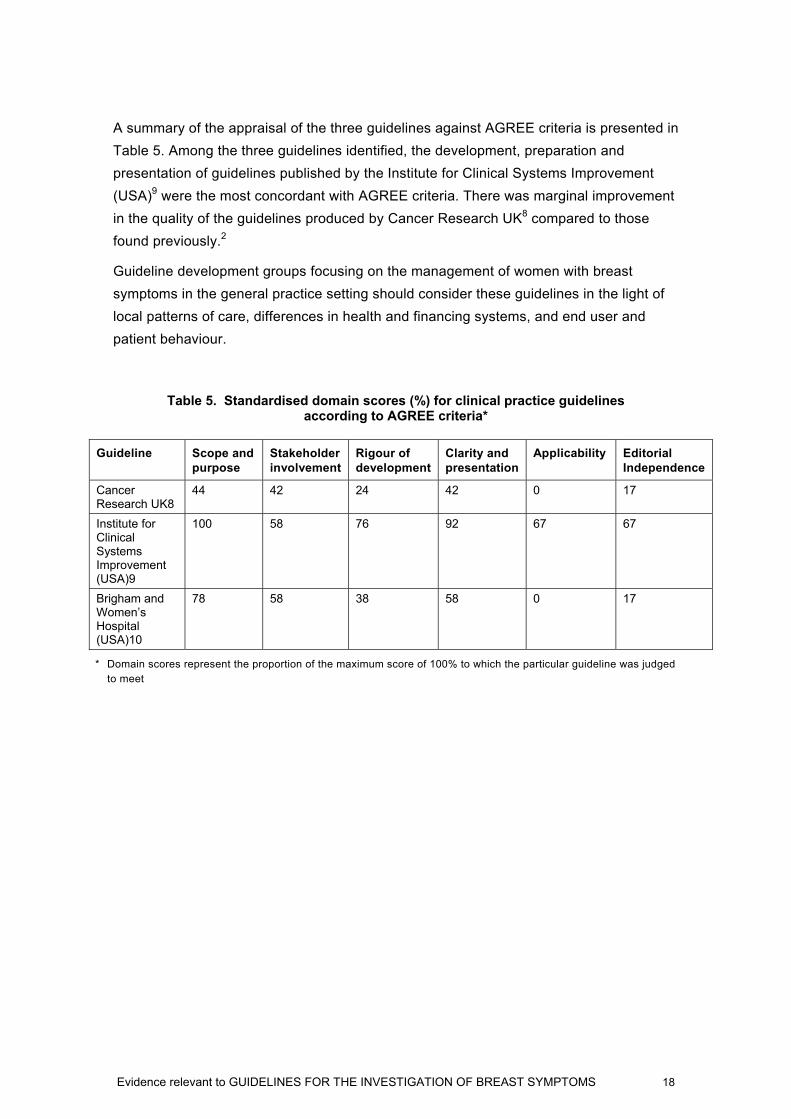
A summary of the appraisal of the three guidelines against AGREE criteria is presented in Table 5. Among the three guidelines identified, the development, preparation and presentation of guidelines published by the Institute for Clinical Systems Improvement (USA)9 were the most concordant with AGREE criteria. There was marginal improvement in the quality of the guidelines produced by Cancer Research UK8 compared to those found previously.2
Guideline development groups focusing on the management of women with breast symptoms in the general practice setting should consider these guidelines in the light of local patterns of care, differences in health and financing systems, and end user and patient behaviour.
Table 5. Standardised domain scores (%) for clinical practice guidelines according to AGREE criteria*
Guideline Scope and purpose
Stakeholder involvement
Rigour of development
Clarity and presentation
Applicability Editorial Independence
Cancer Research UK8
44 42 24 42 0 17
Institute for Clinical Systems Improvement (USA)9
100 58 76 92 67 67
Brigham and Women’s Hospital (USA)10
78 58 38 58 0 17
* Domain scores represent the proportion of the maximum score of 100% to which the particular guideline was judged to meet

Evidence relevant to GUIDELINES FOR THE INVESTIGATION OF BREAST SYMPTOMS 19
OBJECTIVE 2: THE ACCURACY AND CLINICAL UTILITY OF THE TRIPLE TEST IN WOMEN PRESENTING WITH A BREAST LUMP OR ASYMMETRICAL PROMINENCE
The triple test traditionally consists of the triad of clinical examination (C), mammography (M) and fine needle aspiration cytology (FNAC). Generally, the triple test is deemed to give a positive result if any of its components is reported as “suspicious” or “malignant”. The use of other diagnostic modalities in place of or in addition to the traditional components has been reported. While we focused on the classical components of the triple test in this report, we also described the effectiveness of core biopsy in place of FNAC. Additional information about the effectiveness of ultrasound is given in the subsequent section.
The initial search identified 1,093 citations. Preliminary application of the inclusion and exclusion criteria reduced the number to 156 requiring assessment of the full text. For the determination of the diagnostic characteristics of the classic triple test, none of the studies met entry criteria. Most were excluded on the basis of failing to provide information about individual components of the triple test or various combinations of the triple test components in a single study population. Two available studies examined the accuracy of core biopsy. Second stage exclusions are listed in Appendix D and article flow is shown in Appendix E.
A systematic review11 published in 2002, updating data from the first edition of the Technical Report,2 confirms this general lack of published information fulfilling the eligibility criteria (Les Irwig, Personal Communication, 2005). In the absence of additional information, results from the first edition of the Technical Report2 are described.

Evidence relevant to GUIDELINES FOR THE INVESTIGATION OF BREAST SYMPTOMS 20
THE ACCURACY OF A POSITIVE AND NEGATIVE TRIPLE TEST
The triple test is considered positive if any one of its component tests results in a positive result (i.e. “suspicious” or “malignant”). Six studies12-17 provided sufficient information to estimate the true positive rate (TPR) and false positive rate (FPR) of the triple test (Table 6).
Table 6. True positive rate (TPR) and false positive rate (FPR) for the triple test and each component
Triple test Clinical examination
Mammography Fine needle aspiration cytology
TPR, %* 99.6 85 90 91 FPR, %† 38 20 27 7 Specificity, %† 62 80 73 93
* Based on 735 cancers † Based on 1128 non-cancers
The triple test result may significantly modify the pre-test probability of a patient (Figure 1). A negative triple test (C-M-F-) suggests that the probability of breast cancer is less than 1%, unless the pre-test probability is greater than 60%. A positive finding on one of the components of the triple test will steadily increase the probability of cancer across the entire range of pre-test probabilities. For instance, a patient with a pre-test probability of 5% will, on receipt of a positive triple test, have a post-test probability of 12%.
Figure 1. The probability of breast cancer based on results of the triple test.
0
0.1
0.2
0.3
0.4
0.5
0.6
0.7
0.8
0.9
1
0 0.1 0.2 0.3 0.4 0.5 0.6 0.7 0.8 0.9 1
Post
-Tes
t Pro
babi
lity
of C
ance
r
Pre-Test Probability of Breast Cancer
C+ or M+ or F+
C-M-F-
Abbreviations: -, a negative test result (i.e. “benign”); +, a positive test result (i.e. “suspicious” or “malignant”); C, clinical examination; F, fine needle aspiration cytology; M, mammography

Evidence relevant to GUIDELINES FOR THE INVESTIGATION OF BREAST SYMPTOMS 21
THE ACCURACY OF COMBINATIONS OF COMPONENTS OF THE TRIPLE TEST
The components of the triple test may be found to be positive or negative in a correlated manner. Empirically, the results of the six studies providing information on all combinations of the three components are summarised in Table 7.
Table 7. Distribution of results for clinical examination, mammography and fine needle aspiration with women with and without cancer*
Component and result Combined data (%)†
C M F Cancer No cancer Likelihood ratio (+)
+ + + 73.3 1.6 45.8 + + - 5.3 8.5 0.6 + - + 5.7 1.0 5.7 - + + 8.7 2.1 4.1 + - - 1.1 8.5 0.1 - + - 2.4 14.3 0.2 - - + 3.0 2.1 1.4 - - - 0.4 61.9 0.007
* Abbreviations: -, a negative test result (i.e. “benign”); +, a positive test result (i.e. “suspicious” or “malignant”); C, clinical examination; F, fine needle aspiration cytology; M, mammography † Results are pooled from six studies12-17 providing information on all three tests (individually and in combination) in a single study population
The key item, likelihood ratio (+), expresses the extent to which the pre-test odds of disease would be modified by the test result. As expected, the biggest increase in pre-test odds occurs when all three components record a positive result while the biggest decrease occurs when all three components are negative. In the presence of positive FNAC, a single additional clinical or mammographic finding increases the odds of breast cancer by approximately equivalent amounts (5.7 and 4.1 times the pre-test level, respectively). Similarly, isolated clinical or mammographic findings in the presence of a negative FNAC result will decrease the odds of breast cancer by equivalent amounts (10% and 20% of the pre-test level, respectively). There is very little change in the post-test odds if clinical and mammographic findings agree but both disagree with FNAC results (i.e. C+M+F- or C-M-F+). A visual representation of the effect is given in Figure 2 for a range of pre-test probabilities.

Evidence relevant to GUIDELINES FOR THE INVESTIGATION OF BREAST SYMPTOMS 22
Figure 2. The probabilities of breast cancer based on results of individual components of the triple test.
0
0.1
0.2
0.3
0.4
0.5
0.6
0.7
0.8
0.9
1
0 0.1 0.2 0.3 0.4 0.5 0.6 0.7 0.8 0.9 1
Post
-Tes
t Pro
babi
lity
of C
ance
r
Pre-Test Probability of Cancer
C+M+F+ C+M-F+
C-M+F+
C-M-F+
C+M+F-
C-M+F-C+M-F-
C-M-F-
Abbreviations: -, a negative test result (i.e. “benign”); +, a positive test result (i.e. “suspicious” or “malignant”); C, clinical examination; F, fine needle aspiration cytology; M, mammography The accuracy of combined results for mammography and fine needle aspiration cytology
If the results of the clinical examination and medical history were incorporated into the pre-test probabilities prior to the application of imaging modalities and invasive procedures, all information would serve as a new pre-test probability that is modified by the results of mammography and fine needle aspiration cytology. Such results are presented in Table 8 and Figure 3.
Table 8. Distribution of results for mammography and fine needle aspiration of women with and without cancer*
Component and result Combined data (%)†
M F Cancer No Cancer Likelihood ratio (+)
+ + 82.0 3.7 22.0
+ - 7.8 22.8 0.3
- + 8.7 3.1 2.8
- - 1.5 70.4 0.02
* Abbreviations: -, a negative test result (i.e. “benign”); +, a positive test result (i.e. “suspicious” or “malignant”); F, fine needle aspiration cytology; M, mammography † Results are pooled from six studies12-17 providing information on all three tests (individually and in combination) in a single study population.

Evidence relevant to GUIDELINES FOR THE INVESTIGATION OF BREAST SYMPTOMS 23
Figure 3. The probabilities of breast cancer based on results of mammography and fine needle aspiration cytology.
0
0.1
0.2
0.3
0.4
0.5
0.6
0.7
0.8
0.9
1
0 0.1 0.2 0.3 0.4 0.5 0.6 0.7 0.8 0.9 1
Post
-Tes
t Pro
babi
lity
of C
ance
r
Pre-Test Probability of Cancer
M+F+
M-F+
M+F-
M-F-
Abbreviations: -, a negative test result (i.e. “benign”); +, a positive test result (i.e. “suspicious” or “malignant”); F, fine needle aspiration cytology; M, mammography.

Evidence relevant to GUIDELINES FOR THE INVESTIGATION OF BREAST SYMPTOMS 24
OBJECTIVE 3: THE VALUE OF CORE BIOPSY VERSUS FINE NEEDLE ASPIRATION CYTOLOGY AS A COMPONENT OF THE TRIPLE TEST IN DIAGNOSING BREAST CANCER IN WOMEN PRESENTING WITH A BREAST LUMP OR ASYMMETRICAL PROMINENCE
The research question addressed was whether the use of core biopsy in place of FNAC as a component of the triple test improves the diagnosis of breast cancer presenting with breast symptoms. Sensitivity, specificity and positive predictive value (PPV) of the imaging data were extracted.
Of a total 49 studies identified, two18,19 were available to determine the diagnostic characteristics of core biopsy in place of FNAC. All were conducted in patients who were referred for breast symptoms or triple test assessment. Exclusions are listed in Appendix D.
Shannon and colleagues18 presented the details of 1768 consecutive patients referred for triple test assessment, 822 (46.5%) of whom presented because of a breast symptom. Core biopsies or FNAC were performed on the basis of the clinician’s best judgment. All test results, including imaging and clinical findings, were discussed in multidisciplinary meetings. Westenend and colleagues19 described the results of FNAC and core biopsy performed on 286 breast lesions, 81.1% of which were palpable on presentation. The authors did not distinguish between symptomatic and screen-detected lesions.

Evidence relevant to GUIDELINES FOR THE INVESTIGATION OF BREAST SYMPTOMS 25
DIAGNOSTIC PERFORMANCE
Table 9 summarises the diagnostic characteristics of FNAC and core biopsy reported in the two studies. Shannon and colleagues reported that core biopsy produced statistically significant improvements in TPR compared to FNAC but Westenend and colleagues failed to detect such a difference.
Table 9. Reported diagnostic performance of studies examining the accuracy of core biopsy versus FNAC*
Study and test N Prevalence (%) TPR FPR PPV (M) PPV (S)
Shannon 200118 822 19.8† CB 99.5 14.5 99.5 ---‡ FNAC 88.8 52.4 100.0 84.0 Westenend 200119 286 27.6 CB 88.0 10.0 99.0 100.0 FNAC 92.0 18.0 100.0 78.0
* Abbreviations: CB, core biopsy; FNAC, fine needle aspiration cytology; FPR, false positive rate; N, total sample size; PPV (M), positive predictive value for malignant result; PPV (S), positive predictive value for suspicious result † Limited to the subset of patients attending for breast symptoms ‡ Data not provided
These results show that core biopsy is at least as sensitive and specific as fine needle aspiration cytology in the diagnostic setting.
CHOOSING BETWEEN CORE BIOPSY AND FINE NEEDLE ASPIRATION CYTOLOGY20
Both FNA cytology and core biopsy can be performed as outpatient procedures. Hormone receptor studies can be performed on specimens derived from both techniques. While core biopsy and FNAC are complementary procedures, one or another may be more appropriate in a particular clinical situation.
The decision to use FNA cytology, core biopsy or both will be influenced by various factors including the size and clinical characteristics of the lesion, the characteristics of the lesion identified on imaging, the likelihood of achieving a definitive diagnosis, the woman’s ability to tolerate more than one procedure, the expertise of the clinician performing the procedure, the preference of the managing clinician, the availability of pathologists with experience in cytology, the need for hormone receptor assay or tumour marker studies in inoperable tumours, consideration of subsequent surgical management, and the need for a rapid result.
Fine needle aspiration cytology is quicker and less expensive to perform than core biopsy. It is generally less traumatic than core biopsy, is associated with a low complication rate and, in most instances, does not require local anaesthesia. The results for FNAC are available relatively rapidly (within a few hours in some centres) and the presence of a cytopathologist may facilitate an immediate result. It is generally more

Evidence relevant to GUIDELINES FOR THE INVESTIGATION OF BREAST SYMPTOMS 26
appropriate for women taking anticoagulant medication but is inappropriate for the assessment of microcalcifications.
However, FNAC has its disadvantages. Proper training is required in the preparation of quality smears and considerable expertise in cytology is required to interpret specimens. Fine needle aspiration cytology does not enable the pathologist to distinguish between ductal carcinoma in situ (DCIS) and invasive cancer. In some cases, a definitive diagnosis may be difficult to reach in some lesions. Fine needle aspiration cytology may not be the sampling technique of choice for lesions that are relatively hypocellular and yield scanty epithelial material.
Core biopsy yields tissue fragments useful in the determination of the presence of DCIS or invasive carcinoma; tissue may also be available for adjunctive tests. It is useful in the evaluation of lesions likely to be of low histological grade and in those presenting as architectural distortions for which FNAC may fail or has failed to provide a diagnosis. More generally, core biopsy can be used when FNAC fails to correlate with clinical findings or imaging studies.
Core biopsy is the investigation of choice in the evaluation of microcalcifications. It may be preferred when appropriate cytological expertise is not available. Core biopsies are associated with an increased risk of complications, including haematoma and haemorrhage. A lesion previously identified on mammography may not be identified in subsequent open biopsy due to complete removal of the lesion or in the presence of inflammation and fibrosis due to biopsy. In addition, core biopsy may interfere with the interpretation of the subsequent excision biopsy, particularly with grading and the estimation of the size of the lesion. This is particularly relevant in the case of small lesions.
The reliability of FNAC and core biopsy as diagnostic procedures generally depends on the skill of the operator. It is not always possible to immediately assess the adequacy of core biopsy performed for a mass lesion or architectural distortion. Core biopsy specimens require adequate fixation and processing, usually taking a minimum of one working day before results can be available. Core biopsies require local anaesthesia and are generally more expensive than FNAC.
There are no absolute rules determining whether FNAC or core biopsy is the more appropriate investigation. General guidance is provided in Breast Fine Needle Aspiration Cytology and Core Biopsy: A Guide for Practice by the NBCC to inform clinical decisions in the context of symptomatic breast lesions.20

Evidence relevant to GUIDELINES FOR THE INVESTIGATION OF BREAST SYMPTOMS 27
INDICATIONS FOR FINE NEEDLE ASPIRATION CYTOLOGY
• investigation of palpable masses, regardless of whether they are considered benign or malignant
• investigation of impalpable image-detected masses that are considered likely to be benign or with typically malignant features
• evaluation of cystic lesions with atypical imaging features • confirmation of a diagnosis of breast cancer when core biopsy is not available, not
possible, or contraindicated.
INDICATIONS FOR CORE BIOPSY
• investigation of lesions with suspicious features identified on imaging that cannot be identified on ultrasound
• further evaluation of a benign cytological pattern in the presence of a suspicious lesion on imaging
• further evaluation of a lesion for which cytology results are atypical or suspicious • when a single surgical procedure is the desired outcome (for example, wide excision
and axillary dissection). It must be noted that a core biopsy showing only DCIS does not exclude the possibility of the presence of invasive carcinoma in the lesion
• evaluation of microcalcifications that are radiologically indeterminate, suspicious or typically malignant. In such cases, core biopsies should be radiographed prior to histological processing to confirm adequate sampling of the lesion.

Evidence relevant to GUIDELINES FOR THE INVESTIGATION OF BREAST SYMPTOMS 28
OBJECTIVE 4: THE VALUE OF ULTRASOUND IN DIAGNOSING BREAST CANCER IN WOMEN PRESENTING WITH A BREAST LUMP OR ASYMMETRICAL PROMINENCE
The research addressed the following questions:
• Does the use of ultrasound in addition to mammography improve the diagnosis of breast cancer in women presenting with breast symptoms?
• Does the use of ultrasound instead of mammography improve the diagnosis of breast cancer in women presenting with breast symptoms?
In addition, the impact of age upon the relative diagnostic performance of ultrasound and mammography was of particular interest. The review did not focus on the role of mammography and ultrasound in screening or in the monitoring of women who had been diagnosed with breast cancer.
Sensitivity, specificity, positive predictive value (PPV), negative predictive value (NPV), diagnostic accuracy and diagnostic odds ratio based upon dichotomous interpretation of the imaging data were extracted. If results were reported in the publication using alternative cut-points, these were also extracted.
After the elimination of duplicate citations and the addition of further citations identified from the reference lists of key publications, a total of 613 unique citations remained. Application of the inclusion and exclusion criteria reduced the number of articles to four.21-24 Several studies were excluded because they reported results only for a highly selected subgroup of the patient cohort in question. The patients included in such studies were not representative of the cohort referred for the initial investigation of a breast symptom, which would have distorted the diagnostic results if applied to the broader group of patients referred for diagnostic mammography and ultrasound. Confirmation that these studies represented the wrong patient population is evident from the higher prevalence of breast cancer reported in these studies compared to those conducted in consecutive referred populations.25,26 Second-stage exclusions are listed in Appendix D.

Evidence relevant to GUIDELINES FOR THE INVESTIGATION OF BREAST SYMPTOMS 29
STUDY QUALITY
In general, the design of the included studies addressed the research questions appropriately. In all studies, the population consisted of adult women referred for diagnostic imaging on the basis of a breast lump or other symptom. In one study21 the contralateral breast was also imaged. As this breast was asymptomatic, the results contributed by the second breast should be considered as screening.
In several of the studies, the patients undergoing both mammography and ultrasound represent a subset of all referred patients. As this subgroup was selected on the basis of demographic or clinical characteristics, the results may not be generalisable to the entire population of referred patients (Table A5-1 and Table A5-2).
Several of the studies22-24 used a five-point scoring system for level of certainty of diagnosis, however this was ultimately dichotomised to calculated diagnostic performance.
The studies generally were of adequate quality to address the research questions (Table A5-2). They represented an improvement in quality from many of the earlier studies. In particular, all studies attempt to include patients with negative imaging results and determine their outcomes. In three of the four studies,21,23,24 the ultrasound result was determined with knowledge of the mammography result.
DIAGNOSTIC PERFORMANCE
Table 10 presents the calculated diagnostic results. Houssami and colleagues22 designed their study in such a way as to match disease-positive patients with controls, resulting in a prevalence of disease of 50.7%, when the actual disease prevalence in the referred population was 1.9%. Similarly, the inclusion of the imaging results from the contralateral breast in the study by Flobbe and colleagues21 underestimated the calculated prevalence of disease due to dilution by results from paired-organ data. When calculated on the basis of individual patients, the prevalence of breast cancer was 6.3% and is likely to be a truer reflection of disease prevalence among the referred, symptomatic population. The prevalence of breast cancer in these studies ranged from 1.9% to 24.7%, with an overall prevalence of 3.9% (671 cases in 17,117 referrals).
ULTRASOUND INSTEAD OF MAMMOGRAPHY
Both studies reporting results for mammography alone versus ultrasound alone found that ultrasound gave a greater TPR and diagnostic odds ratio (DOR) and smaller FPR compared to mammography.21,22 While the earlier edition of this report did not find any consistency in TPR and FPR between ultrasound and mammography in six studies, five of the six studies reported that the DOR of ultrasound was greater than that of mammography.2

Evidence relevant to GUIDELINES FOR THE INVESTIGATION OF BREAST SYMPTOMS 30
ULTRASOUND IN ADDITION TO MAMMOGRAPHY
Three studies compared the incremental value of ultrasound in this setting.21,23,24 Two studies compared ultrasound plus mammography to mammography alone,23,24 while the other study reported results for clinical examination and mammography with or without ultrasound.21 All three studies reported increases in TPR and DOR and decreases in FPR when ultrasound was combined with mammography or ultrasound combined with clinical examination and mammography. Some increases in DOR were substantial: from 72.1 to 565.7 in the report by Flobbe and colleagues21,and from 50.7 to 223.5 in the study by Zonderland and colleagues.24 While there were modest increases in the likelihood ratios positive, all three studies reported likelihood ratios negative of 0.05 or less.
Table 10. Reported diagnostic performance of mammography and ultrasonography in the diagnosis of breast cancer*
Study and basis of test result
N Prevalence (%)
TPR FPR PPV NPV DOR LR(+) LR(-)
Flobbe 200321 3,835 3.4† M only 82.9 8.1 26.2 99.4 55.0 10.21 0.19 U only 87.6 4.5 40.4 99.6 150.4 19.53 0.13 C+M 91.5 13.0 19.7 99.7 72.1 7.06 0.10 C+M+U 96.9 5.2 39.2 99.9 565.7 18.51 0.03 Houssami 2003‡22 473 50.7 (1.9)§ M only 75.8 12.4 86.3 77.9 22.1 6.09 0.28 U only 81.7 12.0 87.5 82.3 32.6 6.80 0.21 Shetty 200323 411 3.4 M only 85.7 32.2 8.6 99.3 12.6 2.66 0.21 M+U 100.0 11.6 23.3 100.0 N/A|| 8.63 0.00 Zonderland 199924 1,103 24.7 M only 86.0 10.8 72.2 95.1 50.7 7.94 0.16 M+U 95.2 8.2 79.2 98.3 223.5 11.64 0.05
* Abbreviations: C, clinical examination; DOR, diagnostic odds ratio; FPR, false positive rate; LR(-), likelihood ratio negative; LR(+), likelihood ratio positive; M, mammography; N, total sample size; NPV, negative predictive value; PPV, positive predictive value; U, ultrasound † Prevalence was 3.4% when asymptomatic contralateral breasts were included. Prevalence was 6.3% (127/2020) on a per patient basis, which is likely to be a truer reflection of prevalence in the symptomatic population ‡ Data for the same patients were reported in an earlier publication27
§ The prevalence of breast cancer is artificial, as matched sampling of controls was conducted || Value is undefined

Evidence relevant to GUIDELINES FOR THE INVESTIGATION OF BREAST SYMPTOMS 31
AGE-SPECIFIC RESULTS
Through the use of receiver operator characteristic (ROC) curves to compare clinical examination and mammography versus clinical examination, mammography and ultrasound in two distinct age groups —those 50 years and younger, and those older than 50 years—Flobbe and colleagues21 found that the addition of ultrasound increased the area under the curve (AUC) for both age groups (0.87 to 0.96 for patients 50 years and younger; 0.95 to 0.99 for patients older than 50 years; p=0.03 for both groups) in patients referred specifically for a breast lump. The improvements were largely due to ultrasound correctly 'downgrading' positive results found on clinical examination and mammography. The magnitude of the improvement was most pronounced in the younger group. These findings contrast with those found in the group of patients referred due to other breast symptoms such as pain, nipple discharge and skin abnormalities. The authors found no statistically significant difference in the AUC with or without ultrasound.
Houssami and colleagues22 also compared the TPR and FPR of mammography and ultrasound by age category. The TPR of ultrasound was better than mammography in all age groups up to 45 yrs, but in none of the a priori defined age groups did the difference reach statistical significance. In both the 36–40 and 41–45 age groups, the lower 95% CI was close to zero, and the magnitude of the absolute difference in TPR was sizeable (15.4% and 14.3%, respectively). The large number of age categories (and, therefore, smaller number of patients in each category) may have prohibited these differences reaching statistical significance. When the results were calculated in a post hoc fashion for women 45 years or younger, the TPR of ultrasound was significantly higher than mammography (84.9% versus 71.7%, p=0.035).
There was no obvious relationship between age and FPR for either mammography or ultrasound in the study by Houssami and colleagues.22 The investigators found minimal differences between the specificities of mammography and ultrasound across the age range, except for those between the ages of 36 and 40 years. In this age group, the specificity was 8.1% better with ultrasound (91.9% vs 83.8%, 95% CI for difference 0.7%, 16.9%).
Zonderland and colleagues24 provided a comprehensive report of the diagnostic characteristics of mammography alone and mammography combined with ultrasound across a range of age groups (Table 11). The addition of ultrasound to mammography improved the true positive rate of breast cancer diagnosis, particularly among women below the age of 50 years. In contrast, the addition of ultrasound had minimal impact upon the false positive rate.

Evidence relevant to GUIDELINES FOR THE INVESTIGATION OF BREAST SYMPTOMS 32
Table 11. True and false positive rates for mammography with or without ultrasonography by age group from Zonderland and colleagues*24
Age group Characteristic and test
30–39 40–49 50–59 60–69 ≥70
True positive rate (%) M only 72 78 82 94 92 M and U 94 96 89 99 97 False positive rate (%) M only 4 6 19 23 29 M and U 6 4 12 18 17
* Abbreviations: M, mammography; U, ultrasound
It is unlikely that the addition of ultrasound will appreciably add to the detection of breast cancer in older women who have had a thorough clinical examination and mammography. In younger women, ultrasound is the preferred imaging technique due to the increased true positive rate and similar (or reduced) false positive rate in comparison to mammography. The optimum age cut-point is still uncertain.

Evidence relevant to GUIDELINES FOR THE INVESTIGATION OF BREAST SYMPTOMS 33
OBJECTIVE 5: THE CLINICAL SIGNIFICANCE OF NIPPLE DISCHARGE AND THE UTILITY OF CYTOLOGY IN DIAGNOSING BREAST CANCER
THE CLINICAL SIGNIFICANCE OF NIPPLE DISCHARGE
From a total of 151 citations, we identified six studies28-33 that examined the relationship between nipple discharge and the probability of breast cancer (Table 12). As none provided the extensive cross-tabulations of findings found in Ciatto and colleagues’ study34 (identified in the first edition of this report), or its sample size or patient spectrum, some relevant results are presented here.
Table 12. Studies examining the associationbetween nipple discharge and breast cancer*
Study N Setting
Cabioglu 200328 146 Women with nipple discharge presenting to a university clinic over seven years
Florio 199929 1,251 Women with nipple discharge presenting to a university clinic over ten years
Gupta 200430 1,530† Presentations to breast clinics for nipple discharge Hou 200231 487 Women with unilateral nipple discharge without palpable breast
masses Pritt 200432 395‡ Presentations to a single breast clinic for nipple discharge Sauter 200433 175 Women scheduled for diagnostic breast surgery
* Abbreviations: N, total sample size † Includes 36 males ‡ Includes 3 males
The most common presentations for nipple discharge are those with serous or milky characteristics occurring in women younger than 60 years. Bloody nipple discharge accounts for approximately one-tenth of all nipple discharge (Table 13).

Evidence relevant to GUIDELINES FOR THE INVESTIGATION OF BREAST SYMPTOMS 34
Table 13. Distributions of nipple discharge characteristics as percentages of all discharges and of those cases found to have cancer34
Age Characteristic of nipple discharge
< 40 years 40 to 59 years ≥ 60 years All Ages
All discharges Serous 25 25 2 52 Milky 20 11 0 31 Purulent 2.5 4 <1 7 Bloody 3 6 2 11 Total 51 47 4 100
All cancers Serous 0 6 6 12 Milky 3 3 0 6 Purulent 0 3 6 9 Bloody 13 29 32 74 Total 16 39 44 100
In those younger than 60 years, the risk of cancer is less than one percent for those with non-bloody discharge. This increases to three percent if blood is present. Bloody discharge is also associated with a higher risk for cancer in those aged 60 years and older, with the probability approaching 10 percent (Table 14).
Table 14. Probability (%) of cancer by age and characteristic of discharge34
Age Discharge
< 60 ≥ 60
Other < 1 * Serous <1 3 Bloody 3 9
* Few cases available to allow precise estimation
The utility of Hemoccult™ in the detection of blood in nipple discharge and cancer is contentious. Only three available studies provided evidence. An early study by Salmon and colleagues35 showed good correlation between Hemoccult™ results and the presence of blood in discharge specimens as determined by cytology. The TPR and FPR were 100% and 12.5%, respectively. This suggests that a negative Hemoccult™ result may be used to rule out the presence of blood, but that a positive finding may need further investigation.
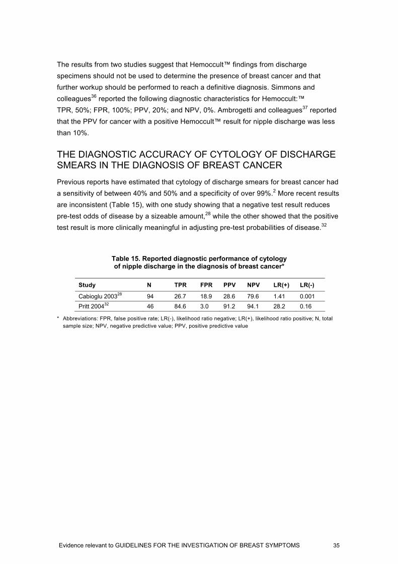
Evidence relevant to GUIDELINES FOR THE INVESTIGATION OF BREAST SYMPTOMS 35
The results from two studies suggest that Hemoccult™ findings from discharge specimens should not be used to determine the presence of breast cancer and that further workup should be performed to reach a definitive diagnosis. Simmons and colleagues36 reported the following diagnostic characteristics for Hemoccult:™ TPR, 50%; FPR, 100%; PPV, 20%; and NPV, 0%. Ambrogetti and colleagues37 reported that the PPV for cancer with a positive Hemoccult™ result for nipple discharge was less than 10%.
THE DIAGNOSTIC ACCURACY OF CYTOLOGY OF DISCHARGE SMEARS IN THE DIAGNOSIS OF BREAST CANCER
Previous reports have estimated that cytology of discharge smears for breast cancer had a sensitivity of between 40% and 50% and a specificity of over 99%.2 More recent results are inconsistent (Table 15), with one study showing that a negative test result reduces pre-test odds of disease by a sizeable amount,28 while the other showed that the positive test result is more clinically meaningful in adjusting pre-test probabilities of disease.32
Table 15. Reported diagnostic performance of cytology of nipple discharge in the diagnosis of breast cancer*
Study N TPR FPR PPV NPV LR(+) LR(-)
Cabioglu 200328 94 26.7 18.9 28.6 79.6 1.41 0.001 Pritt 200432 46 84.6 3.0 91.2 94.1 28.2 0.16
* Abbreviations: FPR, false positive rate; LR(-), likelihood ratio negative; LR(+), likelihood ratio positive; N, total sample size; NPV, negative predictive value; PPV, positive predictive value

Evidence relevant to GUIDELINES FOR THE INVESTIGATION OF BREAST SYMPTOMS 36
CONCLUSIONS
Additional evidence of the effectiveness of diagnostic procedures published since the release of The Investigation of a New Breast Symptom: A Guide for General Practitioners has been identified and evaluated. The implications of these findings for clinical practice should be considered in updates to the Guide with the goal of maximising the detection of breast cancer in women presenting with breast symptoms.
Evidence relevant to GUIDELINES FOR THE INVESTIGATION OF BREAST SYMPTOMS 37
REFERENCES
1. National Breast Cancer Centre. The Investigation of a New Breast Symptom: A Guide for General Practitioners. Woollomooloo: National Breast Cancer Centre, 1997.
2. Irwig L, Macaskill P. Evidence Relevant to Guidelines for the Investigations of Breast Symptoms. Woolloomooloo: National Breast Cancer Centre, 1997.
3. Pit S, Cockburn J, Zorbas H. Investigation of a New Breast Symptom: An Audit in General Practice. Woolloomooloo: National Breast Cancer Centre, 1999.
4. The Agree Collaboration. Appraisal of Guidelines for Research and Evaluation (AGREE) Instrument. London: St. George's Hospital Medical School, 2001.
5. Cluzeau FA, Littlejohns P, Grimshaw JM, Feder G, Moran SE. Development and application of a generic methodology to assess the quality of clinical guidelines. Int J Qual Health Care. 1999;11(1):21-8.
6. Littlejohns P, Cluzeau F, Bale R, Grimshaw J, Feder G, et al. The quantity and quality of clinical practice guidelines for the management of depression in primary care in the UK. Br J Gen Pract. 1999;49(440):205-10(6).
7. Grol R, Dalhuijsen J, Thomas S, in‘t Veld C, Rutten G, et al. Attributes of clinical guidelines that influence use of guidelines in general practice: observational study. BMJ. 1998;317(7162):858-61.
8. Austoker J, Mansel R, Baum M, Sainsbury R, Hobbs R, et al. Guidelines for Referral of Patients with Breast Problems. 2nd ed (with amendments). Sheffield: NHS Cancer Screening Programmes, 2003.
9. Institute for Clinical Systems Improvement. Health Care Guideline: Diagnosis of Breast Disease. 10th ed. Bloomington, MN: Institute for Clinical Systems Improvement, 2003.
10. Kaelin CM, Smith DN, Garber JE, Christian R, Rhei E, et al. Breast Disease: Guide to Prevention, Diagnosis and Treatment. Boston, MA: Brigham and Women's Hospital, 2001.
11. Irwig L, Macaskill P, Houssami N. Evidence relevant to the investigation of breast symptoms: the triple test. Breast. 2002;11(3):215-20.
12. Hermansen C, Skovgaard Poulsen H, Jensen J, et al. Diagnostic reliability of combined physical examination, mammography, and fine-needle puncture ("triple-test") in breast tumors. A prospective study. Cancer. 1987;60(8):1866-71.
13. Kaufman Z, Shpitz B, Shapiro M, Rona R, Lew S, et al. Triple approach in the diagnosis of dominant breast masses: combined physical examination, mammography, and fine-needle aspiration. J Surg Oncol. 1994;56(4):254-7.
14. Kreuzer G, Boquoi E. Aspiration biopsy cytology, mammography and clinical exploration: a modern set up in diagnosis of tumors of the breast. Acta Cytol. 1976;20(4):319-23.
15. Layfield LJ, Glasgow BJ, Cramer H. Fine-needle aspiration in the management of breast masses. Pathol Annu. 1989;24 Pt 2:23-62.
16. Negri S, Bonetti F, Capitanio A, Bonzanini M. Preoperative diagnostic accuracy of fine-needle aspiration in the management of breast lesions: comparison of specificity and sensitivity with clinical examination, mammography, echography, and thermography in 249 patients. Diagn Cytopathol. 1994;11(1):4-8.
17. Thomas JM, Fitzharris BM, Redding WH, Williams JE, Trott PA, et al. Clinical examination, xeromammography, and fine-needle aspiration cytology in diagnosis of breast tumours. Br Med J. 1978;2(6145):1139-41.
18. Shannon J, Douglas-Jones AG, Dallimore NS. Conversion to core biopsy in preoperative diagnosis of breast lesions: is it justified by results? J Clin Pathol. 2001;54(10):762-5.
19. Westenend PJ, Sever AR, Beekman-De Volder HJ, Liem SJ. A comparison of aspiration cytology and core needle biopsy in the evaluation of breast lesions. Cancer. 2001;93(2):146-50.

Evidence relevant to GUIDELINES FOR THE INVESTIGATION OF BREAST SYMPTOMS 38
20. National Breast Cancer Centre. Breast Fine Needle Aspiration Cytology and Core Biopsy: A Guide for Practice. Camperdown: National Breast Cancer Centre, 2004.
21. Flobbe K, Bosch AM, Kessels AG, Beets GL, Nelemans PJ, et al. The additional diagnostic value of ultrasonography in the diagnosis of breast cancer. Arch Intern Med. 2003;163(10):1194-9.
22. Houssami N, Irwig L, Simpson JM, McKessar M, Blome S, et al. Sydney Breast Imaging Accuracy Study: Comparative sensitivity and specificity of mammography and sonography in young women with symptoms. AJR Am J Roentgenol. 2003;180(4):935-40.
23. Shetty MK, Shah YP, Sharman RS. Prospective evaluation of the value of combined mammographic and sonographic assessment in patients with palpable abnormalities of the breast. J Ultrasound Med. 2003;22(3):263-8; quiz 9-70.
24. Zonderland HM, Coerkamp EG, Hermans J, van de Vijver MJ, van Voorthuisen AE. Diagnosis of breast cancer: contribution of US as an adjunct to mammography. Radiology. 1999;213(2):413-22.
25. Duijm LE, Guit GL, Zaat JO, Koomen AR, Willebrand D. Sensitivity, specificity and predictive values of breast imaging in the detection of cancer. Br J Cancer. 1997;76(3):377-81.
26. Flobbe K, van der Linden ES, Kessels AG, van Engelshoven JM. Diagnostic value of radiological breast imaging in a non-screening population. Int J Cancer. 2001;92(4):616-8.
27. Houssami N, Irwig L, Loy C. Accuracy of combined breast imaging in young women. Breast. 2002;11(1):36-40.
28. Cabioglu N, Hunt KK, Singletary SE, Stephens TW, Marcy S, et al. Surgical decision making and factors determining a diagnosis of breast carcinoma in women presenting with nipple discharge. J Am Coll Surg. 2003;196(3):354-64.
29. Florio MG, Manganaro T, Pollicino A, Scarfo P, Micali B. Surgical approach to nipple discharge: a ten-year experience. J Surg Oncol. 1999;71(4):235-8.
30. Gupta RK, Gaskell D, Dowle CS, Simpson JS, King BR, et al. The role of nipple discharge cytology in the diagnosis of breast disease: a study of 1948 nipple discharge smears from 1530 patients. Cytopathology. 2004;15(6):326-30.
31. Hou MF, Tsai KB, Ou-Yang F, Lin HJ, Liu CS, et al. Is a one-step operation for breast cancer patients presenting nipple discharge without palpable mass feasible? Breast. 2002;11(5):402-7.
32. Pritt B, Pang Y, Kellogg M, St John T, Elhosseiny A. Diagnostic value of nipple cytology: study of 466 cases. Cancer. 2004;102(4):233-8.
33. Sauter ER, Schlatter L, Lininger J, Hewett JE. The association of bloody nipple discharge with breast pathology. Surgery. 2004;136(4):780-5.
34. Ciatto S, Bravetti P, Cariaggi P. Significance of nipple discharge clinical patterns in the selection of cases for cytologic examination. Acta Cytol. 1986;30(1):17-20.
35. Salmon RJ, Merle S, Boue P. [Demonstration of blood in nipple discharges using the Hemoccult]. J Gynecol Obstet Biol Reprod (Paris). 1987;16(5):595-8.
36. Simmons R, Adamovich T, Brennan M, Christos P, Schultz M, et al. Nonsurgical evaluation of pathologic nipple discharge. Ann Surg Oncol. 2003;10(2):113-6.
37. Ambrogetti D, Berni D, Catarzi S, Ciatto S. [The role of ductal galactography in the differential diagnosis of breast carcinoma]. Radiol Med (Torino). 1996;91(3):198-201.
38. Shetty MK, Shah YP. Prospective evaluation of the value of negative sonographic and mammographic findings in patients with palpable abnormalities of the breast. J Ultrasound Med. 2002;21(11):1211-6; quiz 1217-9.

Evidence relevant to GUIDELINES FOR THE INVESTIGATION OF BREAST SYMPTOMS 39
LIST OF ABBREVIATIONS
AGREE Appraisal of Guidelines for Research and Evaluation
AUC area under the curve
C clinical examination (in the context of the triple test)
CB core biopsy
CINAHL Cumulative Index to Nursing and Allied Health Literature
CPG clinical practice guideline
DCIS ductal carcinoma in situ
DOR diagnostic odds ratio
FNA fine needle aspiration
FNAC fine needle aspiration cytology (F, in the context of the triple test)
FPR false positive rate
LR likelihood ratio
M mammography (in the context of the triple test)
N total sample size
NBCC National Breast Cancer Centre
NCI National Cancer Institute (US)
NPV negative predictive value
PPV positive predictive value
RACGP Royal Australian College of General Practitioners
ROC receiver operator characteristic
TPR true positive rate
U ultrasound

Evidence relevant to GUIDELINES FOR THE INVESTIGATION OF BREAST SYMPTOMS 40
APPENDIX A: SEARCH STRATEGIES
The following search strategies were constructed for the PubMed search interface. They were changed to suit the particular search engine (Table 1).
TRIPLE TEST
(Ultrasonography, Mammary[MESH] OR Mammography[MESH] OR (needle AND aspirat*)) AND (sensitiv*[Title/Abstract] OR sensitivity and specificity[MeSH Terms] OR diagnos*[Title/Abstract] OR diagnosis[MeSH:noexp] OR diagnostic * [MeSH:noexp] OR diagnosis,differential[MeSH:noexp] OR diagnosis[Subheading:noexp]) AND "breast neoplasms"[MESH]
CORE BIOPSY VERSUS FINE NEEDLE ASPIRATION CYTOLOGY
(("Breast Neoplasms"[MeSH] AND ((fine AND needle AND (aspiration or cytology)) OR "aspiration cytology" OR FNA OR FNAC) AND ((core AND biops*) OR CB OR CNB)) AND (compar* OR "triple test" OR palpable OR suspicious)) AND (specificity[Title/Abstract])
ULTRASOUND (USING EMBASE.COM)
(('breast cancer'/exp/dm_di AND [english]/lim AND [humans]/lim AND [1996-2005]/py) AND ((('mammography'/exp OR 'mammography') AND ('echomammography'/exp OR 'ultrasound' OR 'ultrasonography' OR 'sonography' OR 'echography')) AND [english]/lim AND [humans]/lim AND [1996-2005]/py))
NIPPLE DISCHARGE
((nipple* OR breast*) AND discharg*) AND ("Breast Neoplasms"[MeSH]) AND ((relative[Title/Abstract] AND risk*[Title/Abstract]) OR (relative risk[Text Word]) OR risks[Text Word] OR cohort studies[MeSH:noexp] OR (cohort[Title/Abstract] AND stud*[Title/Abstract]))

Evidence relevant to GUIDELINES FOR THE INVESTIGATION OF BREAST SYMPTOMS 41
APPENDIX B: WEBSITES
AUSTRALIAN SITES
Australia and New Zealand Horizon Scanning Network - http://www.horizonscanning.gov.au
Medicare Services Advisory Committee - http://www.health.gov.au/msac
INTERNATIONAL SITES
Agence d’évaluation des technologies et des modes d’intervention en santé - http://www.aetmis.gouv.qc.ca
Agency for Healthcare Research and Quality - http://www.ahrq.gov
Alberta Heritage Foundation for Medical Research - http://www.ahfmr.ab.ca
Canadian Coordinating Office Health Technology Assessment - http://www.ccohta.ca
Centre for Reviews and Dissemination - http://www.york.ac.uk/inst/crd
ECRI - http://www.ecri.org/
Finnish Office for Health Care Technology Assessment - http://www.stakes.fi/finohta
Institute for Clinical Systems Improvement - http://www.icsi.org
National Coordinating Centre for health technology Assessment - http://www.hta.nhsweb.nhs.uk
National Guidelines Clearninghouse – http://www.guideline.gov
National Horizon Scanning Centre - http://www.publichealth.bham.ac.uk/horizon
National Institute for Clinical Excellence - http://www.nice.org.uk
New Zealand Guidelines Group - http://www.nzgg.org.nz
New Zealand Health Technology Assessment - http://nzhta.chmeds.ac.nz
NHS Quality Improvement Scotland - http://www.nhshealthquality.org/nhsqis
Oregon Health Policy and Research - http://www.ohppr.state.or.us
Swedish Council on Technology Assessment in Health Care - http://www.sbu.se
Veterans Affairs Technology Assessment Program - http://www.va.gov/vatap/

Evidence relevant to GUIDELINES FOR THE INVESTIGATION OF BREAST SYMPTOMS 42
APPENDIX C: LIST OF INCLUDED STUDIES 1. Austoker J, Mansel R, Baum M, et al. Guidelines for Referral of Patients with Breast Problems.
2nd ed (with amendments). Sheffield: NHS Cancer Screening Programmes, 2003.
2. Cabioglu N, Hunt KK, Singletary SE, et al. Surgical decision making and factors determining a diagnosis of breast carcinoma in women presenting with nipple discharge. J Am Coll Surg. 2003;196(3):354-64.
3. Flobbe K, Bosch AM, Kessels AG, et al. The additional diagnostic value of ultrasonography in the diagnosis of breast cancer. Arch Intern Med. 2003;163(10):1194-9.
4. Florio MG, Manganaro T, Pollicino A, et al. Surgical approach to nipple discharge: a ten-year experience. J Surg Oncol. 1999;71(4):235-8.
5. Gupta RK, Gaskell D, Dowle CS, et al. The role of nipple discharge cytology in the diagnosis of breast disease: a study of 1948 nipple discharge smears from 1530 patients. Cytopathology. 2004;15(6):326-30.
6. Hou MF, Tsai KB, Ou-Yang F, et al. Is a one-step operation for breast cancer patients presenting nipple discharge without palpable mass feasible? Breast. 2002;11(5):402-7.
7. Houssami N, Irwig L, Simpson JM, et al. Sydney Breast Imaging Accuracy Study: Comparative sensitivity and specificity of mammography and sonography in young women with symptoms. AJR Am J Roentgenol. 2003;180(4):935-40.
8. Institute for Clinical Systems Improvement. Health Care Guideline: Diagnosis of Breast Disease. 10th ed. Bloomington, MN: Institute for Clinical Systems Improvement, 2003.
9. Kaelin CM, Smith DN, Garber JE, et al. Breast Disease: Guide to Prevention, Diagnosis and Treatment. Boston, MA: Brigham and Women's Hospital, 2001.
10. Pritt B, Pang Y, Kellogg M, et al. Diagnostic value of nipple cytology: study of 466 cases. Cancer. 2004;102(4):233-8.
11. Sauter ER, Schlatter L, Lininger J, Hewett JE. The association of bloody nipple discharge with breast pathology. Surgery. 2004;136(4):780-5.
12. Shannon J, Douglas-Jones AG, Dallimore NS. Conversion to core biopsy in preoperative diagnosis of breast lesions: is it justified by results? J Clin Pathol. 2001;54(10):762-5.
13. Shetty MK, Shah YP, Sharman RS. Prospective evaluation of the value of combined mammographic and sonographic assessment in patients with palpable abnormalities of the breast. J Ultrasound Med. 2003;22(3):263-8; quiz 9-70.
14. Westenend PJ, Sever AR, Beekman-De Volder HJ, Liem SJ. A comparison of aspiration cytology and core needle biopsy in the evaluation of breast lesions. Cancer. 2001;93(2):146-50.
15. Zonderland HM, Coerkamp EG, Hermans J, et al. Diagnosis of breast cancer: contribution of US as an adjunct to mammography. Radiology. 1999;213(2):413-22.

Evidence relevant to GUIDELINES FOR THE INVESTIGATION OF BREAST SYMPTOMS 43
APPENDIX D: SECOND-STAGE EXCLUSIONS
TRIPLE TEST 1. Abu-Salem OT. Fine needle aspiration biopsy (FNAB) of breast lumps: comparison study between
pre- and post-operative histological diagnosis. Arch Inst Pasteur Tunis. 2002;79(1-4):59-63.
2. Agarwal T, Patel B, Rajan P, et al. Core biopsy versus FNAC for palpable breast cancers. Is image guidance necessary? Eur J Cancer. 2003;39(1):52-6.
3. Apesteguia L, Pina L, Inchusta M, et al. Nonpalpable, well-defined, probably benign breast nodule: management by fine-needle aspiration biopsy and long-interval follow-up mammography. Eur Radiol. 1997;7(8):1235-9.
4. Ariga R, Bloom K, Reddy VB, et al. Fine-needle aspiration of clinically suspicious palpable breast masses with histopathologic correlation. Am J Surg. 2002;184(5):410-3.
5. Arisio R, Cuccorese C, Accinelli G, et al. Role of fine-needle aspiration biopsy in breast lesions: analysis of a series of 4,110 cases. Diagn Cytopathol. 1998;18(6):462-7.
6. Arslan N, Ozturk E, Ilgan S, et al. 99Tcm-MIBI scintimammography in the evaluation of breast lesions and axillary involvement: a comparison with mammography and histopathological diagnosis. Nucl Med Commun. 1999;20(4):317-25.
7. Arya V. Is mammographic screening for breast cancer losing ground? Natl Med J India. 2000;13(4):203-4.
8. Ballo MS, Sneige N. Can core needle biopsy replace fine-needle aspiration cytology in the diagnosis of palpable breast carcinoma. A comparative study of 124 women. Cancer. 1996;78(4):773-7.
9. Barlow WE, Lehman CD, Zheng Y, et al. Performance of diagnostic mammography for women with signs or symptoms of breast cancer. J Natl Cancer Inst. 2002;94(15):1151-9.
10. Bauer RL, Sung J, Eckhert KH, Jr., et al. Comparison of histologic diagnosis between stereotactic core needle biopsy and open surgical biopsy. Ann Surg Oncol. 1997;4(4):316-20.
11. Berg WA, Gilbreath PL. Multicentric and multifocal cancer: whole-breast US in preoperative evaluation. Radiology. 2000;214(1):59-66.
12. Berner A, Davidson B, Sigstad E, Risberg B. Fine-needle aspiration cytology vs. core biopsy in the diagnosis of breast lesions. Diagn Cytopathol. 2003;29(6):344-8.
13. Birdwell RL, Ikeda DM, Jeffrey SS, Jeffrey RB, Jr. Preliminary experience with power Doppler imaging of solid breast masses. AJR Am J Roentgenol. 1997;169(3):703-7.
14. Biscotti CV, Shorie JH, Gramlich TL, Easley KA. ThinPrep vs. conventional smear cytologic preparations in analyzing fine-needle aspiration specimens from palpable breast masses. Diagn Cytopathol. 1999;21(2):137-41.
15. Boerner S, Fornage BD, Singletary E, Sneige N. Ultrasound-guided fine-needle aspiration (FNA) of nonpalpable breast lesions: a review of 1885 FNA cases using the National Cancer Institute-supported recommendations on the uniform approach to breast FNA. Cancer. 1999;87(1):19-24.
16. Bojia F, Demisse M, Dejane A, Bizuneh T. Comparison of fine-needle aspiration cytology and excisional biopsy of breast lesions. East Afr Med J. 2001;78(5):226-8.
17. Bondeson L, Lindholm K. Prediction of invasiveness by aspiration cytology applied to nonpalpable breast carcinoma and tested in 300 cases. Diagn Cytopathol. 1997;17(5):315-20.
18. Bone B, Pentek Z, Perbeck L, Veress B. Diagnostic accuracy of mammography and contrast-enhanced MR imaging in 238 histologically verified breast lesions. Acta Radiol. 1997;38(4 Pt 1):489-96.

Evidence relevant to GUIDELINES FOR THE INVESTIGATION OF BREAST SYMPTOMS 44
19. Buchberger W, DeKoekkoek-Doll P, Springer P, et al. Incidental findings on sonography of the breast: clinical significance and diagnostic workup. AJR Am J Roentgenol. 1999;173(4):921-7.
20. Buchbinder SS, Gurell DS, Tarlow MM, et al. Role of US-guided fine-needle aspiration with on-site cytopathologic evaluation in management of nonpalpable breast lesions. Acad Radiol. 2001;8(4):322-7.
21. Butler RS, Venta LA, Wiley EL, et al. Sonographic evaluation of infiltrating lobular carcinoma. AJR Am J Roentgenol. 1999;172(2):325-30.
22. Cangiarella J, Mercado CL, Symmans WF, et al. Stereotaxic aspiration biopsy in the evaluation of mammographically detected clustered microcalcification. Cancer. 1998;84(4):226-30.
23. Chaiwun B, Settakorn J, Ya-In C, et al. Effectiveness of fine-needle aspiration cytology of breast: analysis of 2,375 cases from northern Thailand. Diagn Cytopathol. 2002;26(3):201-5.
24. Chan MC, Lam HS, Gwi E, et al. Stereotactic fine needle aspiration in the management of mammographic abnormalities detected in breast screening. Aust N Z J Surg. 1996;66(9):595-7.
25. Chapellier C, Balu-Maestro C, Bleuse A, et al. Ultrasonography of invasive lobular carcinoma of the breast: sonographic patterns and diagnostic value: report of 102 cases. Clin Imaging. 2000;24(6):333-6.
26. Charnvises S. The accuracy of mammography: a comparison with fine needle aspiration cytology. J Med Assoc Thai. 1996;79(1):27-30.
27. Chen DR, Jeng LB, Kao A, et al. Usefulness of mammoscintigraphy with thallium-201 single photon emission computed tomography to differentiate palpable breast masses of young Taiwanese women when comparing with mammography. Neoplasma. 2002;49(5):334-7.
28. Chen SC, Cheung YC, Lo YF, et al. Sonographic differentiation of invasive and intraductal carcinomas of the breast. Br J Radiol. 2003;76(909):600-4.
29. Chew SB, Hughes M, Kennedy C, et al. Mammographically negative breast cancer at the Strathfield Breast Centre. Aust N Z J Surg. 1996;66(3):134-7.
30. Ciatto S, Rosselli Del Turco M, Ambrogetti D, et al. Solid nonpalpable breast lesions. Success and failure of guided fine-needle aspiration cytology in a consecutive series of 2444 cases. Acta Radiol. 1997;38(5):815-20.
31. Cilotti A, Bagnolesi P, Moretti M, et al. Comparison of the diagnostic performance of high-frequency ultrasound as a first- or second-line diagnostic tool in non-palpable lesions of the breast. Eur Radiol. 1997;7(8):1240-4.
32. Clarke D, Sudhakaran N, Gateley CA. Replace fine needle aspiration cytology with automated core biopsy in the triple assessment of breast cancer. Ann R Coll Surg Engl. 2001;83(2):110-2.
33. Collaco LM, de Lima RS, Werner B, Torres LF. Value of fine needle aspiration in the diagnosis of breast lesions. Acta Cytol. 1999;43(4):587-92.
34. Cote JF, Klijanienko J, Meunier M, et al. Stereotactic fine-needle aspiration cytology of nonpalpable breast lesions: Institut Curie's experience of 243 histologically correlated cases. Cancer. 1998;84(2):77-83.
35. Cox BA, Kelly KM, Ko P, et al. Ultrasound characteristics of breast carcinoma. Am Surg. 1998;64(10):934-8.
36. Crosby JH. The role of fine-needle aspiration biopsy in the diagnosis and management of palpable masses. J Med Assoc Ga. 1996;85(1):33-6.
37. Cwikla JB, Buscombe JR, Kelleher SM, et al. Comparison of accuracy of scintimammography and X-ray mammography in the diagnosis of primary breast cancer in patients selected for surgical biopsy. Clin Radiol. 1998;53(4):274-80.
38. Demirkazik FB, Gulsun M, Firat P. Mammographic features of nonpalpable spiculated lesions. Clin Imaging. 2003;27(5):293-7.
39. Dennison G, Anand R, Makar SH, Pain JA. A prospective study of the use of fine-needle aspiration cytology and core biopsy in the diagnosis of breast cancer. Breast J. 2003;9(6):491-3.

Evidence relevant to GUIDELINES FOR THE INVESTIGATION OF BREAST SYMPTOMS 45
40. Dominguez F, Riera JR, Tojo S, Junco P. Fine needle aspiration of breast masses. An analysis of 1,398 patients in a community hospital. Acta Cytol. 1997;41(2):341-7.
41. Dray M, Mayall F, Darlington A. Improved fine needle aspiration (FNA) cytology results with a near patient diagnosis service for breast lesions. Cytopathology. 2000;11(1):32-7.
42. Duijm LE, Guit GL, Zaat JO, et al. Sensitivity, specificity and predictive values of breast imaging in the detection of cancer. Br J Cancer. 1997;76(3):377-81.
43. Dutta SK, Chattopadhyaya A, Roy S. Evaluation of fine needle aspiration and imprint cytology in the early diagnosis of breast lesions with histopathological correlation. J Indian Med Assoc. 2001;99(8):421-3.
44. Eltahir A, Jibril JA, Squair J, et al. The accuracy of “one-stop” diagnosis for 1,110 patients presenting to a symptomatic breast clinic. J R Coll Surg Edinb. 1999;44(4):226-30.
45. Evans N, Lyons K. The use of ultrasound in the diagnosis of invasive lobular carcinoma of the breast less than 10 mm in size. Clin Radiol. 2000;55(4):261-3.
46. Farshid G, Rush G. The use of fine-needle aspiration cytology and core biopsy in the assessment of highly suspicious mammographic microcalcifications: analysis of outcome for 182 lesions detected in the setting of a population-based breast cancer screening program. Cancer. 2003;99(6):357-64.
47. Feichter GE, Haberthur F, Gobat S, Dalquen P. Breast cytology. Statistical analysis and cytohistologic correlations. Acta Cytol. 1997;41(2):327-32.
48. Fenlon HM, Phelan N, Tierney S, et al. Tc-99m tetrofosmin scintigraphy as an adjunct to plain-film mammography in palpable breast lesions. Clin Radiol. 1998;53(1):17-24.
49. Flobbe K, Nelemans PJ, Kessels AG, et al. The role of ultrasonography as an adjunct to mammography in the detection of breast cancer. a systematic review. Eur J Cancer. 2002;38(8):1044-50.
50. Flobbe K, van der Linden ES, Kessels AG, van Engelshoven JM. Diagnostic value of radiological breast imaging in a non-screening population. Int J Cancer. 2001;92(4):616-8.
51. Furnival C. Needle sampling for breast diagnosis. Aust N Z J Surg. 2000;70(9):633-4.
52. Georgian-Smith D, Taylor KJ, Madjar H, et al. Sonography of palpable breast cancer. J Clin Ultrasound. 2000;28(5):211-6.
53. Gilchrist AM. Should fine needle aspiration cytology (FNAC) in breast assessment be abandoned? Clin Radiol. 2002;57(9):863.
54. Gufler H, Buitrago-Tellez CH, Madjar H, et al. Ultrasound demonstration of mammographically detected microcalcifications. Acta Radiol. 2000;41(3):217-21.
55. Hata T, Takahashi H, Watanabe K, et al. Magnetic resonance imaging for preoperative evaluation of breast cancer: a comparative study with mammography and ultrasonography. J Am Coll Surg. 2004;198(2):190-7.
56. Hatada T, Aoki I, Okada K, et al. Usefulness of ultrasound-guided, fine-needle aspiration biopsy for palpable breast tumors. Arch Surg. 1996;131(10):1095-8.
57. Hatada T, Ishii H, Ichii S, et al. Diagnostic value of ultrasound-guided fine-needle aspiration biopsy, core-needle biopsy, and evaluation of combined use in the diagnosis of breast lesions. J Am Coll Surg. 2000;190(3):299-303.
58. Hindle WH, Chen EC. Accuracy of mammographic appearances after breast fine-needle aspiration. Am J Obstet Gynecol. 1997;176(6):1286-90; discussion 90-2.
59. Hlawatsch A, Teifke A, Schmidt M, Thelen M. Preoperative assessment of breast cancer: sonography versus MR imaging. AJR Am J Roentgenol. 2002;179(6):1493-501.
60. Houssami N, Irwig L, Simpson JM, et al. Sydney Breast Imaging Accuracy Study: Comparative sensitivity and specificity of mammography and sonography in young women with symptoms. AJR Am J Roentgenol. 2003;180(4):935-40.

Evidence relevant to GUIDELINES FOR THE INVESTIGATION OF BREAST SYMPTOMS 46
61. Houssami N, Irwig L, Simpson JM, et al. The contribution of work-up or additional views to the accuracy of diagnostic mammography. Breast. 2003;12(4):270-5.
62. Houssami N, Irwig L, Simpson JM, et al. The influence of clinical information on the accuracy of diagnostic mammography. Breast Cancer Res Treat. 2004;85(3):223-8.
63. Ibrahim AE, Bateman AC, Theaker JM, et al. The role and histological classification of needle core biopsy in comparison with fine needle aspiration cytology in the preoperative assessment of impalpable breast lesions. J Clin Pathol. 2001;54(2):121-5.
64. Joseph L, Edwards JM, Nicholson CM, et al. An audit of the accuracy of fine needle aspiration using a liquid-based cytology system in the setting of a rapid access breast clinic. Cytopathology. 2002;13(6):343-9.
65. Kacl GM, Liu P, Debatin JF, et al. Detection of breast cancer with conventional mammography and contrast-enhanced MR imaging. Eur Radiol. 1998;8(2):194-200.
66. Kamphausen BH, Toellner T, Ruschenburg I. The value of ultrasound-guided fine-needle aspiration cytology of the breast: 354 cases with cytohistological correlation. Anticancer Res. 2003;23(3C):3009-13.
67. Kanchanabat B, Kanchanapitak P, Thanapongsathorn W, Manomaiphiboon A. Fine-needle aspiration cytology for diagnosis and management of palpable breast mass. Aust N Z J Surg. 2000;70(11):791-4.
68. Kim A, Lee J, Choi JS, et al. Fine needle aspiration cytology of the breast. Experience at an outpatient breast clinic. Acta Cytol. 2000;44(3):361-7.
69. Kitchen PR, Cawson JN. The evolving role of fine needle cytology and core-biopsy in the diagnosis of breast cancer. Aust N Z J Surg. 1996;66(9):577-9.
70. Klijanienko J, Cote JF, Thibault F, et al. Ultrasound-guided fine-needle aspiration cytology of nonpalpable breast lesions: Institut Curie's experience with 198 histologically correlated cases. Cancer. 1998;84(1):36-41.
71. Lam WW, Chu WC, Tse GM, Ma TK. Sonographic appearance of mucinous carcinoma of the breast. AJR Am J Roentgenol. 2004;182(4):1069-74.
72. Leconte I, Feger C, Galant C, et al. Mammography and subsequent whole-breast sonography of nonpalpable breast cancers: the importance of radiologic breast density. AJR Am J Roentgenol. 2003;180(6):1675-9.
73. Lee HC, Ooi PJ, Poh WT, Wong CY. Impact of inadequate fine-needle aspiration cytology on outcome of patients with palpable breast lesions. Aust N Z J Surg. 2000;70(9):656-9.
74. Lees A, Gabos A, Jenkins H. Investigations for staging and follow-up of breast cancer patients. Cancer Prev Control. 1997;1(2):157-60.
75. Leifland K, Lagerstedt U, Svane G. Comparison of stereotactic fine needle aspiration cytology and core needle biopsy in 522 non-palpable breast lesions. Acta Radiol. 2003;44(4):387-91.
76. Leifland K, Lundquist H, Lagerstedt U, Svane G. Comparison of preoperative simultaneous stereotactic fine needle aspiration biopsy and stereotactic core needle biopsy in ductal carcinoma in situ of the breast. Acta Radiol. 2003;44(2):213-7.
77. Lewin JM, Hendrick RE, D'Orsi CJ, et al. Comparison of full-field digital mammography with screen-film mammography for cancer detection: results of 4,945 paired examinations. Radiology. 2001;218(3):873-80.
78. Lioe TF, Elliott H, Allen DC, Spence RA. A 3 year audit of fine needle aspirates from a symptomatic breast clinic. Ulster Med J. 1997;66(1):24-7.
79. Lister D, Evans AJ, Burrell HC, et al. The accuracy of breast ultrasound in the evaluation of clinically benign discrete, symptomatic breast lumps. Clin Radiol. 1998;53(7):490-2.
80. Ljung BM, Drejet A, Chiampi N, et al. Diagnostic accuracy of fine-needle aspiration biopsy is determined by physician training in sampling technique. Cancer. 2001;93(4):263-8.
81. Lopez-Ferrer P, Jimenez-Heffernan JA, Vicandi B, et al. Fine needle aspiration cytology of breast fibroadenoma. A cytohistologic correlation study of 405 cases. Acta Cytol. 1999;43(4):579-86.

Evidence relevant to GUIDELINES FOR THE INVESTIGATION OF BREAST SYMPTOMS 47
82. Lumachi F, Marzola MC, Zucchetta P, et al. Breast cancer detection with 99m-Tc-sestamibi scintigraphy, mammography, and fine-needle aspiration cytology: comparative study in 64 surgically treated patients. Ann Surg Oncol. 1999;6(6):568-71.
83. Malur S, Wurdinger S, Moritz A, et al. Comparison of written reports of mammography, sonography and magnetic resonance mammography for preoperative evaluation of breast lesions, with special emphasis on magnetic resonance mammography. Breast Cancer Res. 2001;3(1):55-60.
84. Margolin FR, Leung JW, Jacobs RP, Denny SR. Percutaneous imaging-guided core breast biopsy: 5 years' experience in a community hospital. AJR Am J Roentgenol. 2001;177(3):559-64.
85. Marini C, Traino C, Cilotti A, et al. Differentiation of benign and malignant breast microcalcifications: mammography versus mammography-sonography combination. Radiol Med (Torino). 2003;105(1-2):17-26.
86. May DS, Lee NC, Nadel MR, et al. The National Breast and Cervical Cancer Early Detection Program: report on the first 4 years of mammography provided to medically underserved women. AJR Am J Roentgenol. 1998;170(1):97-104.
87. Mehmood A, Ahmed M, Jamal S. Role of cytological grading in the management of breast lump. J Coll Physicians Surg Pak. 2003;13(3):150-2.
88. Mitnick JS, Gianutsos R, Pollack AH, et al. Tubular carcinoma of the breast: sensitivity of diagnostic techniques and correlation with histopathology. AJR Am J Roentgenol. 1999;172(2):319-23.
89. Mitnick JS, Vazquez MF, Pressman PI, et al. Stereotactic fine-needle aspiration biopsy for the evaluation of nonpalpable breast lesions: report of an experience based on 2,988 cases. Ann Surg Oncol. 1996;3(2):185-91.
90. Mok PM, Keepin Y. Stereotactic breast biopsies for lesions discovered on routine mammography: experience at the North Shore Hospital. N Z Med J. 2000;113(1113):273-4.
91. Morris A, Pommier RF, Schmidt WA, et al. Accurate evaluation of palpable breast masses by the triple test score. Arch Surg. 1998;133(9):930-4.
92. Morris AM, Flowers CR, Morris KT, et al. Comparing the cost-effectiveness of the triple test score to traditional methods for evaluating palpable breast masses. Med Care. 2003;41(8):962-71.
93. Morris KT, Pommier RF, Morris A, et al. Usefulness of the triple test score for palpable breast masses; discussion 1012-3. Arch Surg. 2001;136(9):1008-12.
94. Moss HA, Britton PD, Flower CD, et al. How reliable is modern breast imaging in differentiating benign from malignant breast lesions in the symptomatic population? Clin Radiol. 1999;54(10):676-82.
95. Moy L, Slanetz PJ, Moore R, et al. Specificity of mammography and US in the evaluation of a palpable abnormality: retrospective review. Radiology. 2002;225(1):176-81.
96. Muller-Schimpfle M, Stoll P, Stern W, et al. Do mammography, sonography, and MR mammography have a diagnostic benefit compared with mammography and sonography? AJR Am J Roentgenol. 1997;168(5):1323-9.
97. Mumtaz H, Davidson T, Hall-Craggs MA, et al. Comparison of magnetic resonance imaging and conventional triple assessment in locally recurrent breast cancer. Br J Surg. 1997;84(8):1147-51.
98. Muttarak M, Pojchamarnwiputh S, Chaiwun B. Breast cancer in women under 40 years: preoperative detection by mammography. Ann Acad Med Singapore. 2003;32(4):433-7.
99. Nerurkar A, Osin P. The diagnosis and management of pre-invasive breast disease: the role of new diagnostic techniques. Breast Cancer Res. 2003;5(6):305-8.
100. O'Neil S, Castelli M, Gattuso P, et al. Fine-needle aspiration of 697 palpable breast lesions with histopathologic correlation. Surgery. 1997;122(4):824-8.
101. Ogawa Y, Kato Y, Nakata B, et al. Diagnostic potential and pitfalls of ultrasound-guided fine-needle aspiration cytology for breast lesions. Surg Today. 1998;28(2):167-72.

Evidence relevant to GUIDELINES FOR THE INVESTIGATION OF BREAST SYMPTOMS 48
102. Okamoto H, Ogawara T, Inoue S, et al. Clinical management of nonpalpable or small breast masses by fine-needle aspiration biopsy (FNAB) under ultrasound guidance. J Surg Oncol. 1998;67(4):246-50.
103. Ozdemir A, Oznur, II, Vural G, et al. Tl-201 scintigraphy, mammography and ultrasonography in the evaluation of palpable and nonpalpable breast lesions: a correlative study. Eur J Radiol. 1997;24(2):145-54.
104. Pijnappel RM, van den Donk M, Holland R, et al. Diagnostic accuracy for different strategies of image-guided breast intervention in cases of nonpalpable breast lesions. Br J Cancer. 2004;90(3):595-600.
105. Pisano ED, Fajardo LL, Caudry DJ, et al. Fine-needle aspiration biopsy of nonpalpable breast lesions in a multicenter clinical trial: results from the radiologic diagnostic oncology group V. Radiology. 2001;219(3):785-92.
106. Poole GH, Willsher PC, Pinder SE, et al. Diagnosis of breast cancer with core-biopsy and fine needle aspiration cytology. Aust N Z J Surg. 1996;66(9):592-4.
107. Poplack SP, Tosteson AN, Grove MR, et al. Mammography in 53,803 women from the New Hampshire mammography network. Radiology. 2000;217(3):832-40.
108. Puglisi F, Zuiani C, Bazzocchi M, et al. Role of mammography, ultrasound and large core biopsy in the diagnostic evaluation of papillary breast lesions. Oncology. 2003;65(4):311-5.
109. Reinikainen HT, Rissanen TJ, Piippo UK, Paivansalo MJ. Contribution of ultrasonography and fine-needle aspiration cytology to the differential diagnosis of palpable solid breast lesions. Acta Radiol. 1999;40(4):383-9.
110. Rissanen T, Tikkakoski T, Autio AL, Apaja-Sarkkinen M. Ultrasonography of invasive lobular breast carcinoma. Acta Radiol. 1998;39(3):285-91.
111. Rocha PD, Nadkarni NS, Menezes S. Fine needle aspiration biopsy of breast lesions and histopathologic correlation. An analysis of 837 cases in four years. Acta Cytol. 1997;41(3):705-12.
112. Roche NA, Given-Wilson RM, Thomas VA, Sacks NP. Assessment of a scoring system for breast imaging. Br J Surg. 1998;85(5):669-72.
113. Rubin E, Mennemeyer ST, Desmond RA, et al. Reducing the cost of diagnosis of breast carcinoma: impact of ultrasound and imaging-guided biopsies on a clinical breast practice. Cancer. 2001;91(2):324-32.
114. Rubin M, Horiuchi K, Joy N, et al. Use of fine needle aspiration for solid breast lesions is accurate and cost-effective. Am J Surg. 1997;174(6):694-6; discussion 7-8.
115. Saarela AO, Kiviniemi HO, Rissanen TJ, Paloneva TK. Nonpalpable breast lesions: pathologic correlation of ultrasonographically guided fine-needle aspiration biopsy. J Ultrasound Med. 1996;15(8):549-53; quiz 55-6.
116. Saarela AO, Rissanen TJ, Kiviniemi HO, Paloneva TK. Mammographic and ultrasonographic findings in bilateral breast cancer: a comparative study. Eur Radiol. 1998;8(4):634-8.
117. Saarenmaa I, Salminen T, Geiger U, et al. The effect of age and density of the breast on the sensitivity of breast cancer diagnostic by mammography and ultasonography. Breast Cancer Res Treat. 2001;67(2):117-23.
118. Sampatanukul P, Boonjunwetwat D, Pak-art P. Role of combined fine needle aspiration and ultrasonography in the diagnosis of impalpable lesions of the breast. J Med Assoc Thai. 2003;86 Suppl 2:S284-90.
119. Satake H, Shimamoto K, Sawaki A, et al. Role of ultrasonography in the detection of intraductal spread of breast cancer: correlation with pathologic findings, mammography and MR imaging. Eur Radiol. 2000;10(11):1726-32.
120. Sauer T, Myrvold K, Lomo J, et al. Fine-needle aspiration cytology in nonpalpable mammographic abnormalities in breast cancer screening: results from the breast cancer screening programme in Oslo 1996-2001. Breast. 2003;12(5):314-9.

Evidence relevant to GUIDELINES FOR THE INVESTIGATION OF BREAST SYMPTOMS 49
121. Sauer T, Young K, Thoresen SO. Fine needle aspiration cytology in the work-up of mammographic and ultrasonographic findings in breast cancer screening: an attempt at differentiating in situ and invasive carcinoma. Cytopathology. 2002;13(2):101-10.
122. Saxe A, Phillips E, Orfanou P, Husain M. Role of sample adequacy in fine needle aspiration biopsy of palpable breast lesions. Am J Surg. 2001;182(4):369-71.
123. Scopa CD, Koukouras D, Spiliotis J, et al. Comparison of fine needle aspiration and Tru-Cut biopsy of palpable mammary lesions. Cancer Detect Prev. 1996;20(6):620-4.
124. Shah SH, Kayani N, Hasan SH, et al. Diagnostic evaluation of fine needle aspiration cytology in the management of palpable breast lesions. J Pak Med Assoc. 1998;48(1):7-8.
125. Shen Y, Wu D, Zelen M. Testing the independence of two diagnostic tests. Biometrics. 2001;57(4):1009-17.
126. Shetty MK, Carpenter WS. Sonographic evaluation of isolated abnormal axillary lymph nodes identified on mammograms. J Ultrasound Med. 2004;23(1):63-71.
127. Shetty MK, Shah YP, Sharman RS. Prospective evaluation of the value of combined mammographic and sonographic assessment in patients with palpable abnormalities of the breast. J Ultrasound Med. 2003;22(3):263-8; quiz 9-70.
128. Skaane P, Engedal K. Analysis of sonographic features in the differentiation of fibroadenoma and invasive ductal carcinoma. AJR Am J Roentgenol. 1998;170(1):109-14.
129. Skaane P, Engedal K, Skjennald A. Interobserver variation in the interpretation of breast imaging. Comparison of mammography, ultrasonography, and both combined in the interpretation of palpable noncalcified breast masses. Acta Radiol. 1997;38(4 Pt 1):497-502.
130. Skaane P, Sager EM, Olsen JB, et al. Diagnostic value of ultrasonography in patients with palpable mammographically noncalcified breast tumors. Acta Radiol. 1999;40(2):163-8.
131. Smith SD, Cason Z, Cabaniss DE, et al. Accuracy of fine needle aspiration biopsy of the breast. Biomed Sci Instrum. 1997;33:286-91.
132. Sneige N, Tulbah A. Accuracy of cytologic diagnoses made from touch imprints of image-guided needle biopsy specimens of nonpalpable breast abnormalities. Diagn Cytopathol. 2000; 23(1):29-34.
133. Soo MS, Baker JA, Rosen EL. Sonographic detection and sonographically guided biopsy of breast microcalcifications. AJR Am J Roentgenol. 2003;180(4):941-8.
134. Steinberg JL, Trudeau ME, Ryder DE, et al. Combined fine-needle aspiration, physical examination and mammography in the diagnosis of palpable breast masses: their relation to outcome for women with primary breast cancer. Can J Surg. 1996;39(4):302-11.
135. Sterns EE. Relation between clinical and mammographic diagnosis of breast problems and the cancer/biopsy rate. Can J Surg. 1996;39(2):128-32.
136. Stevens KJ, Smith SL, Denley H, et al. Is mammography of value in women with disseminated cancer of unknown origin? Clin Oncol (R Coll Radiol). 1999;11(2):90-2.
137. Sun W, Li A, Abreo F, et al. Comparison of fine-needle aspiration cytology and core biopsy for diagnosis of breast cancer. Diagn Cytopathol. 2001;24(6):421-5.
138. Taft R, Chao K, Dear P, King C. The role of core biopsy in the diagnosis of mammographically detected lesions. Aust N Z J Surg. 1996;66(10):664-7.
139. Tan SM, Behranwala KA, Trott PA, et al. A retrospective study comparing the individual modalities of triple assessment in the pre-operative diagnosis of invasive lobular breast carcinoma. Eur J Surg Oncol. 2002;28(3):203-8.
140. Tardivon AA, Guinebretiere JM, Dromain C, Vanel D. Imaging and management of nonpalpable lesions of the breast. Eur J Radiol. 2002;42(1):2-9.
141. Thibault F, Meunier M, Klijanienko J, et al. Diagnostic accuracy of sonography and combined sonographic assessment and sonographically guided cytology in nonpalpable solid breast lesions. J Clin Ultrasound. 2000;28(8):387-98.
Evidence relevant to GUIDELINES FOR THE INVESTIGATION OF BREAST SYMPTOMS 50
142. Tomlinson J, Harvey J, Sterrett G, et al. An audit of 267 consecutively excised mammographically detected breast lesions 1989-1993. Pathology. 1997;29(1):21-7.
143. Wang HC, Chen DR, Kao CH, et al. Detecting breast cancer in mammographically dense breasts: comparing technetium-99m tetrofosmin mammoscintigraphy and ultrasonography. Cancer Invest. 2002;20(7-8):932-8.
144. Wang HH, Ducatman BS. Fine needle aspiration of the breast. A probabilistic approach to diagnosis of carcinoma. Acta Cytol. 1998;42(2):285-9.
145. Warren RM, Young JR, McLean L, et al. Radiology review of the UKCCCR Breast Screening Frequency Trial: potential improvements in sensitivity and lead time of radiological signs. Clin Radiol. 2003;58(2):128-32.
146. Westenend PJ, Sever AR, Beekman-De Volder HJ, Liem SJ. A comparison of aspiration cytology and core needle biopsy in the evaluation of breast lesions. Cancer. 2001;93(2):146-50.
147. Whyte K. Breast lumps and mammograms. Aust Fam Physician. 1999;28(8):831, 41.
148. Yang WT, Lam WW, Cheung H, et al. Sonographic, magnetic resonance imaging, and mammographic assessments of preoperative size of breast cancer. J Ultrasound Med. 1997;16(12):791-7.
149. Yang WT, Tse GM. Sonographic, mammographic, and histopathologic correlation of symptomatic ductal carcinoma in situ. AJR Am J Roentgenol. 2004;182(1):101-10.
150. Yiangou C, Davis J, Livni N, et al. Diagnostic role of cytology in screen-detected breast cancer. Br J Surg. 1996;83(6):816-9.
151. Yip CH, Jayaram G, Alhady SF. The experience with fine needle aspiration cytology in the management of palpable breast lumps in the University Hospital Kuala Lumpur. Med J Malaysia. 2000;55(3):363-7.
152. Yoshihara K, Kisu T, Koizumi S, et al. The clinical epidemiological investigation of diagnostic modalities in breast cancer detection--a comparison of ultrasonography and mammography. Fukuoka Igaku Zasshi. 1999;90(2):39-45.
153. Zanconati F, Bonifacio D, Falconieri G, Di Bonito L. Role of fine-needle aspiration cytology in nonpalpable mammary lesions: a comparative cytohistologic study based on 308 cases. Diagn Cytopathol. 2000;23(2):87-91.
154. Zardawi IM, Hearnden F, Meyer P, Trevan B. Ultrasound-guided fine needle aspiration cytology of impalpable breast lesions in a rural setting. Comparison of cytology with imaging and final outcome. Acta Cytol. 1999;43(2):163-8.
155. Zonderland HM. The role of ultrasound in the diagnosis of breast cancer. Semin Ultrasound CT MR. 2000;21(4):317-24.
156. Zonderland HM, Coerkamp EG, Hermans J, et al. Diagnosis of breast cancer: contribution of US as an adjunct to mammography. Radiology. 1999;213(2):413-22.

Evidence relevant to GUIDELINES FOR THE INVESTIGATION OF BREAST SYMPTOMS 51
CORE BIOPSY VERSUS FINE NEEDLE ASPIRATION BIOPSY 1. Abe Y, Ito K, Okamura C, et al. Cervical cytologic examination during physical checkup of
pregnant women: cervical cancer screening in women under the age of thirty. Tohoku J Exp Med. 2004;204(3):221-8.
2. Ambrogetti D, Berni D, Catarzi S, Ciatto S. [The role of ductal galactography in the differential diagnosis of breast carcinoma]. Radiol Med (Torino). 1996;91(3):198-201.
3. Arnold GJ, Neiheisel MB. A comprehensive approach to evaluating nipple discharge. Nurse Pract. 1997;22(7):96-102, 5-11.
4. Bauer RL, Eckhert KH, Jr., Nemoto T. Ductal carcinoma in situ-associated nipple discharge: a clinical marker for locally extensive disease. Ann Surg Oncol. 1998;5(5):452-5.
5. Boonjunwetwat D, Prathombutr A. Imaging of benign papillary neoplasm of the breast: mammographic, galactographic and sonographic findings. J Med Assoc Thai. 2000;83(8):832-8.
6. Cabioglu N, Hunt KK, Singletary SE, et al. Surgical decision making and factors determining a diagnosis of breast carcinoma in women presenting with nipple discharge. J Am Coll Surg. 2003;196(3):354-64.
7. Cheung KL, Alagaratnam TT. A review of nipple discharge in Chinese women. J R Coll Surg Edinb. 1997;42(3):179-81.
8. Dinkel HP, Gassel AM, Muller T, et al. Galactography and exfoliative cytology in women with abnormal nipple discharge. Obstet Gynecol. 2001;97(4):625-9.
9. Florio MA, Fama F, Giacobbe G, et al. [Nipple discharge: personal experience with 2,818 cases]. Chir Ital. 2003;55(3):357-64.
10. Florio MG, Manganaro T, Pollicino A, et al. Surgical approach to nipple discharge: a ten-year experience. J Surg Oncol. 1999;71(4):235-8.
11. Gupta RK, Gaskell D, Dowle CS, et al. The role of nipple discharge cytology in the diagnosis of breast disease: a study of 1948 nipple discharge smears from 1530 patients. Cytopathology. 2004;15(6):326-30.
12. Hou M, Tsai K, Lin H, et al. A simple intraductal aspiration method for cytodiagnosis in nipple discharge. Acta Cytol. 2000;44(6):1029-34.
13. Hou MF, Chuang HY, Ou-Yang F, et al. Comparison of breast mammography, sonography and physical examination for screening women at high risk of breast cancer in taiwan. Ultrasound Med Biol. 2002;28(4):415-20.
14. Hou MF, Huang CJ, Huang YS, et al. Evaluation of galactography for nipple discharge. Clin Imaging. 1998;22(2):89-94.
15. Hou MF, Huang TJ, Liu GC. The diagnostic value of galactography in patients with nipple discharge. Clin Imaging. 2001;25(2):75-81.
16. Hou MF, Tsai KB, Ou-Yang F, et al. Is a one-step operation for breast cancer patients presenting nipple discharge without palpable mass feasible? Breast. 2002;11(5):402-7.
17. Ito Y, Tamaki Y, Nakano Y, et al. Nonpalpable breast cancer with nipple discharge: how should it be treated? Anticancer Res. 1997;17(1B):791-4.
18. Izumi J, Komaki K, Hirokawa M, et al. Secretory carcinoma of the breast with a cystically dilated intraductal component: report of a case. Surg Today. 2003;33(2):110-3.
19. Jacob D, Brombart JC, Muller C, et al. [Analysis of the results of 137 subclinical breast lesions excisions. Value of ultrasonography in the early diagnosis of breast cancer]. J Gynecol Obstet Biol Reprod (Paris). 1997;26(1):27-31.
20. Kalbhen CL, Kezdi-Rogus PC, Dowling MP, Flisak ME. Mammography in the evaluation of nipple inversion. AJR Am J Roentgenol. 1998;170(1):117-21.

Evidence relevant to GUIDELINES FOR THE INVESTIGATION OF BREAST SYMPTOMS 52
21. Kavanagh AM, Giles GG, Mitchell H, Cawson JN. The sensitivity, specificity, and positive predictive value of screening mammography and symptomatic status. J Med Screen. 2000;7(2):105-10.
22. Kerlikowske K, Smith-Bindman R, Ljung BM, Grady D. Evaluation of abnormal mammography results and palpable breast abnormalities. Ann Intern Med. 2003;139(4):274-84.
23. Klein P, Glaser E, Grogan L, et al. Biomarker assays in nipple aspirate fluid. Breast J. 2001;7(6):378-87.
24. Krishnamurthy S, Sneige N, Thompson PA, et al. Nipple aspirate fluid cytology in breast carcinoma. Cancer. 2003;99(2):97-104.
25. Lee WY. Cytology of abnormal nipple discharge: a cyto-histological correlation. Cytopathology. 2003;14(1):19-26.
26. Miller AB, To T, Baines CJ, Wall C. Canadian National Breast Screening Study-2: 13-year results of a randomized trial in women aged 50-59 years. J Natl Cancer Inst. 2000;92(18):1490-9.
27. Miller AB, To T, Baines CJ, Wall C. The Canadian National Breast Screening Study-1: breast cancer mortality after 11 to 16 years of follow-up. A randomized screening trial of mammography in women age 40 to 49 years. Ann Intern Med. 2002;137(5 Part 1):305-12.
28. Miyazaki M, Tamaki Y, Sakita I, et al. Detection of microsatellite alterations in nipple discharge accompanied by breast cancer. Breast Cancer Res Treat. 2000;60(1):35-41.
29. Mokbel K, Elkak AE. The evolving role of mammary ductoscopy. Curr Med Res Opin. 2002;18(1):30-2.
30. Morimoto T, Sasa M, Yamaguchi T, et al. Breast cancer screening by mammography in women aged under 50 years in Japan. Anticancer Res. 2000;20(5C):3689-94.
31. Okamoto H, Ogawara T, Arihara F, et al. Usefulness of ultrasonography combined with conventional physical examination in mass screening for breast cancer: a retrospective study of Yamanashi Health Care Center results from 1989 to 1994. Jpn J Cancer Res. 1996;87(3):317-23.
32. Okazaki A, Hirata K, Okazaki M, et al. Nipple discharge disorders: current diagnostic management and the role of fiber-ductoscopy. Eur Radiol. 1999;9(4):583-90.
33. Orel SG, Dougherty CS, Reynolds C, et al. MR imaging in patients with nipple discharge: initial experience. Radiology. 2000;216(1):248-54.
34. Pritt B, Pang Y, Kellogg M, et al. Diagnostic value of nipple cytology: study of 466 cases. Cancer. 2004;102(4):233-8.
35. Sardanelli F, Imperiale A, Zandrino F, et al. Breast intraductal masses: US-guided fine-needle aspiration after galactography. Radiology. 1997;204(1):143-8.
36. Sauter ER, Schlatter L, Lininger J, Hewett JE. The association of bloody nipple discharge with breast pathology. Surgery. 2004;136(4):780-5.
37. Self M, Dunn E. Nipple discharge: its significance and evaluation. Curr Surg. 2004;61(6):528-32.
38. Shao ZM, Nguyen M. Nipple aspiration in diagnosis of breast cancer. Semin Surg Oncol. 2001;20(3):175-80.
39. Shen KW, Wu J, Lu JS, et al. Fiberoptic ductoscopy for breast cancer patients with nipple discharge. Surg Endosc. 2001;15(11):1340-5.
40. Tilanus-Linthorst MM, Bartels CC, Obdeijn AI, Oudkerk M. Earlier detection of breast cancer by surveillance of women at familial risk. Eur J Cancer. 2000;36(4):514-9.
41. Tjalma WA, Verslegers IO. Nipple discharge and the value of MR imaging. Eur J Obstet Gynecol Reprod Biol. 2004;115(2):234-6.
42. Van Zee KJ, Ortega Perez G, Minnard E, Cohen MA. Preoperative galactography increases the diagnostic yield of major duct excision for nipple discharge. Cancer. 1998;82(10):1874-80.
43. Vargas HI, Romero L, Chlebowski RT. Management of bloody nipple discharge. Curr Treat Options Oncol. 2002;3(2):157-61.

Evidence relevant to GUIDELINES FOR THE INVESTIGATION OF BREAST SYMPTOMS 53
44. Wahner-Roedler DL, Reynolds C, Morton MJ. Spontaneous unilateral nipple discharge: when screening tests are negative--a case report and review of current diagnostic management of a pathologic nipple discharge. Breast J. 2003;9(1):49-52.
45. Williams RS, Brook D, Monypenny IJ, Gower-Thomas K. The relevance of reported symptoms in a breast screening programme. Clin Radiol. 2002;57(8):725-9.
46. Yamamoto D, Senzaki H, Nakagawa H, et al. Detection of chromosomal aneusomy by fluorescence in situ hybridization for patients with nipple discharge. Cancer. 2003;97(3):690-4.
47. Zheng W, Wen M, Kang H. [The diagnosis and treatment of nipple discharge in 253 cases]. Hunan Yi Ke Da Xue Xue Bao. 1998;23(3):311-3.
ULTRASOUND 1. Anonymous. Ultrasound detects missed cancers. Biomedical Instrumentation and Technology.
1997;31(3):217.
2. Aapro MS. Progress in the treatment of breast cancer in the elderly. Annals of Oncology. 2002;13(SUPPL. 4):207.
3. Abbas MK, Qadir H, Shabbir A, et al. Primary squamous cell carcinoma of female breast - A rare entity. Journal of the College of Physicians and Surgeons Pakistan. 2000;10(5):185.
4. Abe H, Hanasawa K, Naitoh H, et al. Invasive ductal carcinoma within a fibroadenoma of the breast. International Journal of Clinical Oncology. 2004;9(4):334.
5. Abe H, Naitoh H, Umeda T, et al. Occult breast cancer presenting axillary nodal metastasis: A case report. Japanese Journal of Clinical Oncology. 2000;30(4):185.
6. Acheson MB, Patton RG, Howisey RL, et al. Three- to six-year followup for 379 benign image-guided large-core needle biopsies of nonpalpable breast abnormalities. Journal of the American College of Surgeons. 2002;195(4):462.
7. Adwani A, Lowe S. Dual technique for preoperative localisation of non-palpable breast lesions. Journal of Surgical Oncology. 2003;84(2):105.
8. Agarwal G, Mishra SK, Dutta NR, Jain M. Occult squamous cell carcinoma of the axillary tail of breast presenting as isolated axillary lymph node mass. European Journal of Surgery. 2000;166(2):177.
9. Ahn BY, Kim HH, Moon WK, et al. Pregnancy- and lactation-associated breast cancer: Mammographic and sonographic findings. Journal of Ultrasound in Medicine. 2003;22(5):491.
10. Akashi-Tanaka S, Fukutomi T, Miyakawa K, et al. Contrast-enhanced computed tomography detection of occult breast cancers presenting as axillary masses. Breast Cancer Research and Treatment. 1999;55(1):97.
11. Akashi-Tanaka S, Fukutomi T, Miyakawa K, et al. Diagnostic value of contrast-enhanced computed tomography for diagnosing the intraductal component of breast cancer. Breast Cancer Research and Treatment. 1998;49(1):79.
12. Akashi-Tanaka S, Fukutomi T, Watanabe T, et al. Accuracy of contrast-enhanced computed tomography in the prediction of residual breast cancer after neoadjuvant chemotherapy. International Journal of Cancer. 2001;96(1):66.
13. Albert US, Schulz KD. Short version of the guideline: Early detection of breast cancer in Germany - An evidence-, consensus-, and outcome-based guideline according to the German association of the scientific medical societies (AWMF) and the German agency for quality in medicine (AeZQ). Journal of Cancer Research and Clinical Oncology. 2004;130(9):527.
14. Allen SA, Cunliffe WJ, Gray J, et al. Pre-operative estimation of primary breast cancer size: A comparison of clinical assessment, mammography and ultrasound. Breast. 2001;10(4):299.
15. Alonso JC, Soriano A, Zarca MA, et al. Breast cancer detection with Sestamibi-Tc-99m and Tl-201 radionuclides in patients with non conclusive mammography. Anticancer Research. 1997;17(3 B):1661.

Evidence relevant to GUIDELINES FOR THE INVESTIGATION OF BREAST SYMPTOMS 54
16. Amano G, Ohuchi N, Ishibashi T, et al. Correlation of three-dimensional magnetic resonance imaging with precise histopathological map concerning carcinoma extension in the breast. Breast Cancer Research and Treatment. 2000;60(1):43.
17. Anastasiadis PG, Koutlaki NG, Skaphida PG, et al. Trends in breast cancer incidence in Thrace, Greece: An epidemiological assessment. European Journal of Gynaecological Oncology. 2000;21(1):76.
18. Andres B, Aguilar J, Torroba A, et al. Intracystic papillary carcinoma in the male breast. Breast Journal. 2003;9(3):249.
19. Androutsos G. Milestones in the treatment of breast cancer. Journal of B.U.ON. 2004;9(1):101.
20. Arisio R, Cuccorese C, Accinelli G, et al. Role of fine-needle aspiration biopsy in breast lesions: Analysis of a series of 4,110 cases. Diagnostic Cytopathology. 1998;18(6):462.
21. Asai S, Miyachi H, Ochiai N, et al. Mastopathy-mimicking ultrasonographic appearance in a case with Burkitt's lymphoma. Clinical Imaging. 2001;25(5):309.
22. Autier P, Shannoun F, Scharpantgen A, et al. A breast cancer screening programme operating in a liberal health care system: The Luxembourg Mammography Programme, 1992-1997. International Journal of Cancer. 2002;97(6):828.
23. Bagni B, Franceschetto A, Casolo A, et al. Scintimammography with 99mTc-MIBI and magnetic resonance imaging in the evaluation of breast cancer. European Journal of Nuclear Medicine and Molecular Imaging. 2003;30(10):1383.
24. Baker JA, Scott Soo M. Breast US: Assessment of technical quality and image interpretation. Radiology. 2002;223(1):229.
25. Banerjee D, Gorse SJ, Cotter M, Johnson RC. Sonographic and pathologic features of metastatic synovial sarcoma of the lung presenting as a breast neoplasm. Breast Journal. 2004;10(4):372.
26. Baron LF, Baron PL, Ackerman SJ, et al. Sonographically guided clip placement facilitates localization of breast cancer after neoadjuvant chemotherapy. American Journal of Roentgenology. 2000;174(2):539.
27. Barton MB, Elmore JG, Fletcher SW. Breast symptoms among women enrolled in a health maintenance organization: Frequency, evaluation, and outcome. Annals of Internal Medicine. 1999;130(8):651.
28. Baslaim MM, Bakheet SM, Ezzat A, et al. 18-Fluorodeoxyglucose positron emission tomography in cystic carcinoma of the breast. Breast Journal. 2002;8(6):371.
29. Bassa P, Kim EE, Inoue T, et al. Evaluation of preoperative chemotherapy using PET with fluorine-18-fluorodeoxyglucose in breast cancer. Journal of Nuclear Medicine. 1996;37(6):931.
30. Bau MG, Arisio R, Cristini G, et al. Screening-detected breast carcinoma in a patient with Cowden syndrome. Breast. 2004;13(3):239.
31. Bauer VP, Ditkoff BA, Schnabel F, et al. The management of lobular neoplasia identified on percutaneous core breast biopsy. Breast Journal. 2003;9(1):4.
32. Baz E, Madjar H, Reuss C, et al. The role of enhanced Doppler ultrasound in differentiation of benign vs. malignant scar lesion after breast surgery for malignancy. Ultrasound in Obstetrics and Gynecology. 2000;15(5):377.
33. Behranwala KA, Gui GPH. Breast cancer in the inframammary fold: Is preserving the inframammary fold during mastectomy justified? Breast. 2002;11(4):340.
34. Bellantone R, Lombardi CP, Cefaro GA, et al. CMF + radiotherapy in the primary treatment of operable breast cancer: Preliminary results of a phase II pilot study. Journal of Surgical Oncology. 1998;68(1):48.
35. Belli P, Costantini M, Romani M, et al. Magnetic resonance imaging in breast cancer recurrence. Breast Cancer Research and Treatment. 2002;73(3):223.
36. Belton AL, Stull MA, Grant T, Shepard MH. Mammographic and sonographic findings in metastatic transitional cell carcinoma of the breast. American Journal of Roentgenology. 1997;168(2):511.

Evidence relevant to GUIDELINES FOR THE INVESTIGATION OF BREAST SYMPTOMS 55
37. Benson SRC, Blue J, Judd K, Harman JE. Ultrasound is now better than mammography for the detection of invasive breast cancer. American Journal of Surgery. 2004;188(4 SPEC. ISS.):381.
38. Benson SRC, Harrison NJ, Lengyel J, et al. Combined image guidance excision of non-palpable breast lesions. Breast. 2004;13(2):110.
39. Berg WA, Gilbreath PL. Multicentric and multifocal cancer: Whole-breast US in preoperative evaluation. Radiology. 2000;214(1):59.
40. Bhagwandeen BS, Fenton L. Secretory carcinoma of the breast in a nine year old boy. Pathology. 1999;31(2):166.
41. Birdwell RL, Ikeda DM, Jeffrey SS, Jeffrey RB. Preliminary experience with power Doppler imaging of solid breast masses. American Journal of Roentgenology. 1997;169(3):703.
42. Blaumeiser B, Tjalma WAA, Verslegers I, et al. Invasive papillary carcinoma of the male breast. European Radiology. 2002;12(9):2207.
43. Bloomston M, D'Angelo P, Galliano D, et al. One hundred consecutive advanced breast biopsy instrumentation procedures: Complications, costs, and outcome. Annals of Surgical Oncology. 1999;6(2):195.
44. Blunt DM, Sansom HE, Nasiri N, Moskovic EC. Ultrasound in breast carcinoma: What are we looking at? A sonographic/pathological correlation. Breast. 1998;7(3):156.
45. Bond LK, Vance R, Berd D, et al. Diagnosing breast cancer with synchronous metastatic melanoma. Cancer Practice. 2002;10(5):224.
46. Bosch AM, Kessels AGH, Beets GL, et al. Interexamination variation of whole breast ultrasound. British Journal of Radiology. 2003;76(905):328.
47. Botkin JR, Smith KR, Croyle RT, et al. Genetic testing for a BRCA1 mutation: Prophylactic surgery and screening behavior in women 2 years post testing. American Journal of Medical Genetics. 2003;118 A(3):201.
48. Bowditch MG, Peck R, Shorthonse AJ. Mestastatic renal adenocarcinoma presenting in a breast screening programme. European Journal of Surgical Oncology. 1996;22(6):641.
49. Boyle P. Current situation of screening for cancer. Annals of Oncology. 2002;13(SUPPL. 4):189.
50. Brancato B, Bricolo D, Bonzanini M, et al. Pre-operative prediction of invasive vs intraductal breast cancer type: Multivariate analysis of the accuracy of clinical and imaging findings. Breast. 2002;11(2):151.
51. Brenner RJ, Rothman BJ. Detection of primary breast cancer in women with known adenocarcinoma metastatic to the axilla: Use of MRI after negative clinical and mammographic examination. Journal of Magnetic Resonance Imaging. 1997;7(6):1153.
52. Breucq C, Goes E, Osteaux M. Bilateral breast masses in a young pregnant woman. Journal Belge de Radiologie. 1999;82(3):97.
53. Britton PD, Wishart GC, Ravichandran D, et al. The management of radial scars of the breast - Does core biopsy help? Journal of B.U.ON. 2002;7(2):137.
54. Brnic Z, Drinkovic I, Hebrang A. High-resolution ultrasound and power-Doppler - Advances in pre-invasive diagnosis of solid breast lesions: Our one-year experience. Radiology and Oncology. 1998;32(4):353.
55. Brown AL, Holwill SDJ, Thomas VA, et al. Case report: Primary osteosarcoma of the breast: Imaging and histological features. Clinical Radiology. 1998;53(12):920.
56. Bubb K, Hooper C. The role of sonography in breast conservation therapy using breast brachytherapy. Journal of Diagnostic Medical Sonography. 2003;19(5):287.
57. Buchberger W, DeKoekkoek-Doll P, Springer P, et al. Incidental findings on sonography of the breast: Clinical significance and diagnostic workup. American Journal of Roentgenology. 1999;173(4):921.
58. Buchberger W, Niehoff A, Obrist P, et al. Clinically and mammographically occult breast lesions: Detection and classification with high-resolution sonography. Seminars in Ultrasound CT and MRI. 2000;21(4):325.

Evidence relevant to GUIDELINES FOR THE INVESTIGATION OF BREAST SYMPTOMS 56
59. Bui KL, Lamb TJ, Rapelyea JA, et al. Accuracy of mammographic and sonographic assessment of preoperative breast cancer size. Journal of Women's Imaging. 2003;5(3):128.
60. Burke W, Daly M, Garber J, et al. Recommendations for follow-up care of individuals with an inherited predisposition to cancer: II. BRCA1 and BRCA2. Journal of the American Medical Association. 1997;277(12):997.
61. Burrell HC, Evans AJ. Radiological assessment of the breast: What the surgical oncologist needs to know. European Journal of Surgical Oncology. 2001;27(7):689.
62. Burrell HC, Evans AJ, Wilson ARM, Pinder SE. False-negative breast screening assessment. What lessons can we learn? Clinical Radiology. 2001;56(5):385.
63. Busch JM, Pantanowitz L, Kornguth PJ. Cancer mimicked on sonography: Lipomastia in an HIV-positive man undergoing antiretroviral therapy. American Journal of Roentgenology. 2003;181(1):187.
64. Buscombe JR, Holloway B, Roche N, Bombardieri E. Position of nuclear medicine modalities in the diagnostic work-up of breast cancer. Quarterly Journal of Nuclear Medicine and Molecular Imaging. 2004;48(2):109.
65. Butler RS, Venta LA, Wiley EL, et al. Sonographic evaluation of infiltrating lobular carcinoma. American Journal of Roentgenology. 1999;172(2):325.
66. Cabioglu N, Hunt KK, Singletary SE, et al. Surgical decision making and factors determining a diagnosis of breast carcinoma in women presenting with nipple discharge. Journal of the American College of Surgeons. 2003;196(3):354.
67. Cady B, Steele GD, Morrow M, et al. Evaluation of common breast problems: Guidance for primary care providers. Ca-A Cancer Journal for Clinicians. 1998;48(1):49.
68. Cakir B, Tuncbilek N, Karakas HM, et al. Granulomatous mastitis mimicking breast carcinoma. Breast Journal. 2002;8(4):251.
69. Callaway MP, Boggis CRM, Astley SA, Hutt I. The influence of previous films on screening mammographic interpretation and detection of breast carcinoma. Clinical Radiology. 1997;52(7):527.
70. Campanini R, Dongiovanni D, Iampieri E, et al. A novel featureless approach to mass detection in digital mammograms based on support vector machines. Physics in Medicine and Biology. 2004;49(6):961.
71. Cappabianca S, Grassi R, D'Alessandro P, et al. Metastasis to the male breast from carcinoma of the urinary bladder. British Journal of Radiology. 2000;73(876):1326.
72. Carr-Hoefer C. Granular cell tumor: A benign mimicker of breast carcinoma. Journal of Diagnostic Medical Sonography. 2003;19(2):95.
73. Carson PL, Fowlkes JB, Roubidoux MA, et al. 3-D color Doppler image quantification of breast masses. Ultrasound in Medicine and Biology. 1998;24(7):945.
74. Cawson JN, Law EM, Kavanagh AM. Invasive lobular carcinoma: Sonographic features of cancers detected in a BreastScreen program. Australasian Radiology. 2001;45(1):25.
75. Cawson JN, Malara F, Kavanagh A, et al. Fourteen-gauge needle core biopsy of mammographically evident radial scars: Is excision necessary? Cancer. 2003;97(2):345.
76. Cecil KM, Schnall MD, Siegelman ES, Lenkinski RE. The evaluation of human breast lesions with magnetic resonance imaging and proton magnetic resonance spectroscopy. Breast Cancer Research and Treatment. 2001;68(1):45.
77. Champion VL, Skinner CS. Differences in perceptions of risk, benefits, and barriers by stage of mammography adoption. Journal of Women's Health. 2003;12(3):277.
78. Chandawarkar RY, Shinde SR. Preoperative diagnosis of carcinoma of the breast: Is a 'cost-cutter' algorithm tenable? Journal of Surgical Oncology. 1997;64(2):153.
79. Chapellier C, Balu-Maestro C, Bleuse A, et al. Ultrasonography of invasive lobular carcinoma of the breast: Sonographic patterns and diagnostic value. Report of 102 cases. Clinical Imaging. 2000;24(6):333.

Evidence relevant to GUIDELINES FOR THE INVESTIGATION OF BREAST SYMPTOMS 57
80. Chen DR, Chang RF, Lee JH, et al. Three-dimensional breast ultrasound imaging in patients with nipple discharge: A pictorial review of 27 patients. Journal of Medical Ultrasound. 2002;10(2):69.
81. Chen DR, Jeng LB, Kao A, et al. Comparing thallium-201 spect mammoscintigraphy and ultrasonography to detect breast cancer in mammographical dense breasts. Neoplasma. 2003;50(3):222.
82. Chen DR, Kuo WJ, Chang RF, et al. Use of the bootstrap technique with small training sets for computer-aided diagnosis in breast ultrasound. Ultrasound in Medicine and Biology. 2002;28(7):897.
83. Chen DR, Liang WM, Kuo HW, Chang RF. Computerized quantitative assessment of sonomammographic homogeneity of fibroadenoma and breast carcinoma. Journal of Medical Ultrasound. 1999;7(3):157.
84. Chen SC, Cheung YC, Su CH, et al. Analysis of sonographic features for the differentiation of benign and malignant breast tumors of different sizes. Women's Oncology Review. 2004;4(2):95.
85. Chen SC, Hwang TL, Chen MF. Intraoperative ultrasound-guided excisional biopsy for nonpalpable breast lesions. Journal of Surgical Association Republic of China. 1997;30(6):389.
86. Chen WY, Chen CS, Chen HC, et al. Mucinous cystadenocarcinoma of the breast coexisting with infiltrating ductal carcinoma. Pathology International. 2004;54(10):781.
87. Cheung YC, Chen SC, Lee KF, et al. Sonographic and pathologic findings in typical and atypical medullary carcinomas of the breast. Journal of Clinical Ultrasound. 2000;28(7):325.
88. Cheung YC, Su MY, Ng SH, et al. Lumpy silicone-injected breasts: Enhanced MRI and microscopic correlation. Clinical Imaging. 2002;26(6):397.
89. Cheung YC, Wan YL, Chen SC, et al. Sonographic evaluation of mammographically detected microcalcifications without a mass prior to stereotactic core needle biopsy. Journal of Clinical Ultrasound. 2002;30(6):323.
90. Cho N, Oh KK, Lee SI. Medullary carcinoma of the breast: Sonographic features distinguishing it from fibroadenoma. Journal of Medical Ultrasound. 2002;10(4):191.
91. Chollet P, Charrier S, Brain E, et al. Clinical and pathological response to primary chemotherapy in operable breast cancer. European Journal of Cancer Part A. 1997;33(6):862.
92. Chopra S, Evans AJ, Pinder SE, et al. Pure mucinous breast cancer-mammographic and ultrasound findings. Clinical Radiology. 1996;51(6):421.
93. Chou ASB, Chi T, Shum HC, et al. Synchronous bilateral breast cancer with different histologic type: A case report. Chinese Journal of Radiology. 1998;23(1):37.
94. Chung SY, Oh KK. Imaging findings of metastatic disease to the breast. Yonsei Medical Journal. 2001;42(5):497.
95. Chuo CB, Corder AP. Core biopsy vs fine needle aspiration cytology in a symptomatic breast clinic. European Journal of Surgical Oncology. 2003;29(4):374.
96. Ciatto S, Bonardi R, Ravaioli A, et al. Benign breast surgical biopsies: Are they always justified? Tumori. 1998;84(5):521.
97. Cid JA, Rampaul RS, Ellis IO, et al. Woman feels breast lump - Surgeon cannot: The role of ultrasound in arbitration. European Journal of Cancer. 2004;40(14):2053.
98. Clifford EJ, Lugo-Zamudio C. Scintimammography in the diagnosis of breast cancer. American Journal of Surgery. 1996;172(5):483.
99. Coarasa-Cerdan A, Palomo-Jimenez M, Montero-Montero A, et al. Hemangiopericytoma of the breast: Mammographic and sonographic findings. Journal of Clinical Ultrasound. 1998;26(3):155.
100. Cohen MA, Sferlazza SJ. Original report. Role of sonography in evaluation of radial scars of the breast. American Journal of Roentgenology. 2000;174(4):1075.
101. Conrad C, Corfitsen MT, Gyldholm N, Christiansen FL. Pre-operative MR-mammography in breast cancer patients. European Journal of Surgical Oncology. 1999;25(2):142.

Evidence relevant to GUIDELINES FOR THE INVESTIGATION OF BREAST SYMPTOMS 58
102. Costantini M, Magistrelli A, Franceschini G, et al. MRI in phyllodes tumor of the breast. Case reports. Rays. 2002;27(4):313.
103. Cox BA, Kelly KM, Ko P, et al. Ultrasound characteristics of breast carcinoma. American Surgeon. 1998;64(10):934.
104. Croucher CA, Rigby HS, Savage PE. Post menopausal bleeding as the presenting sign of metastatic breast carcinoma. Journal of Obstetrics and Gynaecology. 1996;16(1):60.
105. Crystal P, Strano SD, Shcharynski S, Koretz MJ. Using sonography to screen women with mammographically dense breasts. American Journal of Roentgenology. 2003;181(1):177.
106. Cummings SR, Eckert S, Krueger KA, et al. The effect of raloxifene on risk of breast cancer in postmenopausal women: Results from the MORE randomized trial. Journal of the American Medical Association. 1999;281(23):2189.
107. Cutuli B, Dhermain F, Borel C, et al. Breast cancer in patients treated for Hodgkin's disease: Clinical and pathological analysis of 76 cases in 63 patients. European Journal of Cancer. 1997;33(14):2315.
108. Cwikla JB, Buscombe JR, Holloway B, et al. Can scintimammography with 99mTc-MIBI identify multifocal and multicentric primary breast cancer? Nuclear Medicine Communications. 2001;22(12):1287.
109. Dahlstrom JE, Buckingham JM, Bell S, Jain S. Nodular fasciitis of the breast simulating breast cancer on imaging. Australasian Radiology. 2001;45(1):67.
110. Dallessio K. MR imaging of the breast. Applied Radiology. 2003;32(4):40.
111. Dallessio KM. Handheld ultrasound, wide-area PACS, and more. Applied Radiology. 2001;30(5):6.
112. Daniel BL. Intraprocedural magnetic resonance imaging-guided interventions in the breast. Topics in Magnetic Resonance Imaging. 2000;11(3):184.
113. Daniel BL, Birdwell RL, Ikeda DM, et al. Breast lesion localization: A freehand, interactive MR imaging-guided technique. Radiology. 1998;207(2):455.
114. Daniels IR, Layer GT. How should gynaecomastia be managed? ANZ Journal of Surgery. 2003;73(4):213.
115. Dash N, Chafin SH, Johnson RR, Contractor FM. Usefulness of tissue marker clips in patients undergoing neoadjuvant chemotherapy for breast cancer. American Journal of Roentgenology. 1999;173(4):911.
116. Davis PL, McCarty KS. Magnetic resonance imaging in breast cancer staging. Topics in Magnetic Resonance Imaging. 1998;9(1):60.
117. Davis PL, Staiger MJ, Harris KB, et al. Breast cancer measurements with magnetic resonance imaging, ultrasonography, and mammography. Breast Cancer Research and Treatment. 1996;37(1):1.
118. De Bock GH, Vliet Vlieland TPM, Hakkeling M, et al. GPs' management of women seeking help for familial breast cancer. Family Practice. 1999;16(5):463.
119. De Cicco C, Pizzamiglio M, Trifiro G, et al. Radioguided occult lesion localisation (ROLL) and surgical biopsy in breast cancer: Technical aspects. Quarterly Journal of Nuclear Medicine. 2002;46(2):145.
120. De Iorio P, Cenci M, Midulla C, et al. Incidental detection of an in situ lobular carcinoma during the study of an intramammary lymph node: Utility of FNA cytology. A case report. Anticancer Research. 1998;18(4 B):2875.
121. De Waal JC, Sultz D. Imaging procedures in the diagnosis of breast disease. Chirurgische Praxis. 2002;60(1):145.
122. Delanote S, Van den Broecke R, Schelfhout VRJ, Serreyn R. Adenoid cystic carcinoma of the breast in a 19-year-old girl. Breast. 2003;12(1):75.
123. Demirkazik FB, Baskan O, Aydingoz U, et al. Case report: Squamous cell carcinoma of the skin metastasizing to the breast - imaging findings. British Journal of Radiology. 1996;69(823):678.

Evidence relevant to GUIDELINES FOR THE INVESTIGATION OF BREAST SYMPTOMS 59
124. Dershaw DD. Imaging guided biopsy: An alternative to surgical biopsy. Breast Journal. 2000;6(5):294.
125. Dershaw DD, Selland DLG, Tan LK, et al. Spiculated axillary adenopathy. Radiology. 1996;201(2):439.
126. di Giorgio A, Arnone P, Canavese A. Ultrasound guided excisional biopsy of non-palpable breast lesions: Technique and preliminary results. European Journal of Surgery. 1998;164(11):819.
127. DiPiro PJ, Meyer JE, Denison CM, et al. Image-guided core breast biopsy of ductal carcinoma in situ presenting as a non-calcified abnormality. European Journal of Radiology. 1999;30(3):231.
128. Dixon JM, Love CDB, Renshaw L, et al. Lessons from the use of aromatase inhibitors in the neoadjuvant setting. Endocrine-Related Cancer. 1999;6(2):227.
129. Donaldson LA, Cliff A, Gardiner L, et al. Surgeon-controlled ultrasound-guided core biopsies in the breast - A prospective study and a new use for surgeons in the clinic. European Journal of Surgical Oncology. 2003;29(2):139.
130. Dooley WC. Routine operative breast endoscopy for bloody nipple discharge. Annals of Surgical Oncology. 2002;9(9):920.
131. Dorum A, Heimdal K, Lovslett K, et al. Prospectively detected cancer in familial breast/ovarian cancer screening. Acta Obstetricia et Gynecologica Scandinavica. 1999;78(10):906.
132. Dowlatshahi K, Fan M, Gould VE, et al. Stereotactically guided laser therapy of occult breast tumors: Work-in-progress report. Archives of Surgery. 2000;135(11):1345.
133. Doyle AJ, King AR, Miller MV, Collins JP. Implementation of image-guided large-core needle biopsy of the breast on a limited budget. Australasian Radiology. 1998;42(3):199.
134. Drukker K, Giger ML, Mendelson EB. Computerized analysis of shadowing on breast ultrasound for improved lesion detection. Medical Physics. 2003;30(7):1833.
135. Drukker K, Giger ML, Vyborny CJ, Mendelson EB. Computerized Detection and Classification of Cancer on Breast Ultrasound. Academic Radiology. 2004;11(5):526.
136. Duijm LEM, Guit GL, Zaat JOM, et al. Sensitivity, specificity and predictive values of breast imaging in the detection of cancer. British Journal of Cancer. 1997;76(3):377.
137. Dumane VA, Shankar PM, Piccoli CW, et al. Computer aided classification of masses in ultrasonic mammography. Medical Physics. 2002;29(9):1968.
138. Durfee SM, Selland DLG, Smith DN, et al. Sonographic evaluation of clinically palpable breast cancers invisible on mammography. Breast Journal. 2000;6(4):247.
139. Dutta Roy S, Stafford JS, Scally J, Selvachandran SN. Granulocytic sarcoma of the breast antedating acute myelogenous leukemia. Breast. 2004;13(3):242.
140. Elmore JG, Barton MB, Moceri VM, et al. Ten-year risk of false positive screening mammograms and clinical breast examinations. New England Journal of Medicine. 1998;338(16):1089.
141. Elorz M, Pina L, Bergaz F, et al. Atypical evolution of a calcified lipid cyst presenting spontaneously as a suspicious cluster of microcalcifications. European Radiology. 2002;12(5):1100.
142. Eltahir A, Jibril JA, Squair J, et al. The accuracy of 'one-stop' diagnosis for 1110 patients presenting to a symptomatic breast clinic. Journal of the Royal College of Surgeons of Edinburgh. 1999;44(4):226.
143. Ergul Z, Uner A, Unsal D, Memis L. Mammary Carcinosarcoma Composed of Chondrosarcoma: Case Report of a Rare Tumor and Review of the Literature. THOD - Turk Hematoloji-Onkoloji Dergisi. 2003;13(4):222.
144. Eroglu A, Ugurluoglu C. Fine-needle aspiration of bilateral breast metastases from malignant melanoma: A case report. Journal of B.U.ON. 2003;8(4):385.
145. Esserman L, Wolverton D, Hylton N. Magnetic resonance imaging for primary breast cancer management: Current role and new applications. Endocrine-Related Cancer. 2002;9(2):141.

Evidence relevant to GUIDELINES FOR THE INVESTIGATION OF BREAST SYMPTOMS 60
146. Estourgie SH, Nieweg OE, Valdes Olmos RA, et al. Intratumoral Versus Intraparenchymal Injection Technique for Lymphoscintigraphy in Breast Cancer. Clinical nuclear medicine. 2003;28(5):371.
147. Estourgie SH, Nieweg OE, Valdes Olmos RA, et al. Intratumoral versus intraparenchymal injection technique for lymphoscintigraphy in breast cancer. Clinical nuclear medicine. 2003;28(5):371.
148. Eun JS, Ki KO, Kim EK, et al. Leiomyoma of the breast in a 50-year, old woman receiving tamoxifen. American Journal of Roentgenology. 1998;171(6):1684.
149. Evans N, Lyons K. The use of ultrasound in the diagnosis of invasive lobular carcinoma of the breast less than 10 mm in size. Clinical Radiology. 2000;55(4):261.
150. Fadul D, Rapelyea JA, Brem RF. 53-year-old female with recurrent palpable left breast mass. Journal of Women's Imaging. 2003;5(2):87.
151. Farria DM, Gorczyca DP, Barsky SH, et al. Benign phyllodes tumor of the breast: MR imaging features. American Journal of Roentgenology. 1996;167(1):187.
152. Fernandez A, Escobedo A, Benito E, et al. Sentinel node localization in patients with non-palpable breast cancer. Nuclear Medicine Communications. 2002;23(12):1165.
153. Ferriere R, Mensah S, Lefebvre JP. Weakly inhomogeneous media tomography. Ultrasonic Imaging. 2003;25(2):122.
154. Fervers B, Leichtnam-Dugarin L, Carretier J, et al. The SOR SAVOIR PATIENT project an evidence-based patient information and education project. Electronic Journal of Oncology. 2003;-(1):1.
155. Fiorentino C, Berruti A, Bottini A, et al. Accuracy of mammography and echography versus clinical palpation in the assessment of response to primary chemotherapy in breast cancer patients with operable disease. Breast Cancer Research and Treatment. 2001;69(2):143.
156. Fischer U, Kopka L, Grabbe E. Magnetic resonance guided localization and biopsy of suspicious breast lesions. Topics in Magnetic Resonance Imaging. 1998;9(1):44.
157. Fischer U, Zachariae O, Baum F, et al. The influence of preoperative MRI of the breasts on recurrence rate in patients with breast cancer. European Radiology. 2004;14(10):1725.
158. Fishman JE, Milikowski C, Ramsinghani R, et al. US-guided core-needle biopsy of the breast: How many specimens are necessary? Radiology. 2003;226(3):779.
159. Flobbe K, Bosch AM, Kessels AGH, et al. The additional diagnostic value of ultrasonography in the diagnosis of breast cancer. Archives of Internal Medicine. 2003;163(10):1194.
160. Foord K, Guy R, Apthorp L, et al. Updated audit database for breast imaging/histopathology correlation. Clinical Radiology. 2001;56(9):755.
161. Fornage BD, Sneige N, Edeiken BS. Interventional breast sonography. European Journal of Radiology. 2002;42(1):17.
162. Fornage BD, Sneige N, Ross MI, et al. Small (< or = 2-cm) breast cancer treated with US-guided radiofrequency ablation: feasibility study. Radiology. 2004;231(1):215.
163. Foroudi F, Tiver KW. Occult breast carcinoma presenting as axillary metastases. International Journal of Radiation Oncology Biology Physics. 2000;47(1):143.
164. Forsberg F, Goldberg BB, Merritt CRB, et al. Diagnosing Breast Lesions with Contrast-Enhanced 3-Dimensional Power Doppler Imaging. Journal of Ultrasound in Medicine. 2004;23(2):173.
165. Foster RS, Wood WC. Alternative strategies in the management of primary breast cancer. Archives of Surgery. 1998;133(11):1182.
166. Foxcroft LM, Evans EB, Hirst C, Hicks BJ. Presentation and diagnosis of adolescent breast disease. Breast. 2001;10(5):399.
167. Foxcroft LM, Evans EB, Joshua HK, Hirst C. Breast cancers invisible on mammography. Australian and New Zealand Journal of Surgery. 2000;70(3):162.
168. Foxcroft LM, Evans EB, Porter AJ. The diagnosis of breast cancer in women younger than 40. Breast. 2004;13(4):297.

Evidence relevant to GUIDELINES FOR THE INVESTIGATION OF BREAST SYMPTOMS 61
169. Franchelli S, Canavase G, Murolo C, Barabino P. Local recurrence of breast cancer around a prosthesis dome. British Journal of Plastic Surgery. 2002;55(8):685.
170. Francis A, England DW, Rowlands DC, et al. The diagnosis of invasive lobular breast carcinoma. Does MRI have a role? Breast. 2001;10(1):38.
171. Frattaroli FM, Carrara A, Conte AM, Pappalardo G. Axillary metastasis as first symptom of occult breast cancer: A case report. Tumori. 2002;88(6):532.
172. Freeman JL, Goodwin JS, Zhang D, et al. Measuring the performance of screening mammography in community practice with medicare claims data. Women and Health. 2003;37(2):1.
173. Gabriel HA, Feng C, Mendelson EB, Benjamin S. Breast MRI for Cancer Detection in a Patient with Diabetic Mastopathy. American Journal of Roentgenology. 2004;182(4):1081.
174. Gajdos C, Bleiweiss IJ, Drossman S, Tartter PI. Breast cancer in an intramammary sentinel node. Breast Journal. 2001;7(4):260.
175. Gal-Gombos EC, Esserman LE, Recine MA, Poppiti, Jr. Large-needle core biopsy in atypical intraductal epithelial hyperplasia including immunohistochemical expression of high molecular weight cytokeratin: Analysis of results of a single institution. Breast Journal. 2002;8(5):269.
176. Gallowitsch HJ, Kresnik E, Gasser J, et al. F-18 fluorodeoxyglucose positron-emission tomography in the diagnosis of tumor recurrence and metastases in the follow-up of patients with breast carcinoma: A comparison to conventional imaging. Investigative Radiology. 2003;38(5):250.
177. Gan FY, Wettlaufer JR, Lundell AL. Breast imaging in a military setting: A comparison with civilian breast imaging. Military Medicine. 2004;169(5):361.
178. Gardner RA, Vargas HI, Block JB, et al. Focused microwave phased array thermotherapy for primary breast cancer. Annals of Surgical Oncology. 2002;9(4):326.
179. Garra BS, Cespedes EI, Ophir J, et al. Elastography of breast lesions: Initial clinical results. Radiology. 1997;202(1):79.
180. Gaston M, Jadav A, Bobrow L, Purushotham AD. Paraneoplastic cerebellar degeneration: An unusual presentation of breast carcinoma. European Journal of Surgical Oncology. 2001;27(6):606.
181. Georgian-Smith D, Taylor KJW, Madjar H, et al. Sonography of palpable breast cancer. Journal of Clinical Ultrasound. 2000;28(5):211.
182. Georgian-Smith DM. Current status of breast ultrasound. Applied Radiology. 2004;33(9):16.
183. Ghate SV, Soo MS, Mengoni PM. Extended field-of-view two-dimensional ultrasonography of the breast: Improvement in lesion documentation. Journal of Ultrasound in Medicine. 1999;18(9):597.
184. Gibbs P, Liney GP, Lowry M, et al. Differentiation of benign and malignant sub-1 cm breast lesions using dynamic contrast enhanced MRI. Breast. 2004;13(2):115.
185. Giess CS, Keating DM, Osborne MP, et al. Retroareola breast carcinoma: Clinical, imaging, and histopathologic features. Radiology. 1998;207(3):669.
186. Gill HK, Berg WA. Case 39: Invasive lobular carcinoma. Radiology. 2001;221(1):132.
187. Gill HK, Ioffe OB, Berg WA. When is a diagnosis of sclerosing adenosis acceptable at core biopsy? Radiology. 2003;228(1):50.
188. Gillett D, Kennedy C, Carmalt H. Breast cancer in young women. Australian and New Zealand Journal of Surgery. 1997;67(11):761.
189. Goins KV, Zapka JG, Geiger AM, et al. Implementation of systems strategies for breast and cervical cancer screening services in health maintenance organizations. American Journal of Managed Care. 2003;9(11):745.
190. Gojnic M, Petkovic S, Pervulov M, Micic J. The utility of routine breast ultrasound screening in Gynecology. Clinical and Experimental Obstetrics and Gynecology. 2003;30(4):265.
191. Golshan M, Fung BB, Wiley E, et al. Prediction of breast cancer size by ultrasound, mammography and core biopsy. Breast. 2004;13(4):265.

Evidence relevant to GUIDELINES FOR THE INVESTIGATION OF BREAST SYMPTOMS 62
192. Gonzalez-Angulo AM, Walters RS, Carpenter, Jr., et al. Paclitaxel chemotherapy in a pregnant patient with bilateral breast cancer. Clinical Breast Cancer. 2004;5(4):317.
193. Goodman KA, Birdwell RL, Ikeda DM, Berlin L. Compliance with recommended follow-up after percutaneous breast core biopsy. American Journal of Roentgenology. 1998;170(1):89.
194. Goodwin PJ. Management of familial breast cancer risk. Breast Cancer Research and Treatment. 2000;62(1):19.
195. Gordon PB, Goldenberg SL. Ultrasound-guided fine needle aspiration biopsy update 1998. Breast Journal. 1998;4(5):292.
196. Goscin CP, Berman CG, Clark RA. Magnetic resonance imaging of the breast. Cancer Control. 2001;8(5):399.
197. Griffin DW, Ross RT, Rudolph R. Intracystic breast carcinoma with giant auxillary metastasis: A case report. Breast Journal. 2000;6(2):143.
198. Gu X, Zhang Q, Bartlett M, et al. Differentiation of Cysts from Solid Tumors in the Breast with Diffuse Optical Tomography. Academic Radiology. 2004;11(1):53.
199. Gulec SA, Rhea G, Gonzalez AD, Welsh RA. Breast carcinoma originating in a fibroadenoma. Breast Journal. 2004;10(5):452.
200. Gunhan-Bilgen I, Bozkaya H, Ustun EE, Memis A. Male breast disease: Clinical, mammographic, and ultrasonographic features. European Journal of Radiology. 2002;43(3):246.
201. Gunhan-Bilgen I, Memis A, Ustun EE, Ozdemir N. Breast metastasis from low-grade endometrial stromal sarcoma after a 17-year period. European Radiology. 2002;12(12):3023.
202. Gunhan-Bilgen I, Memis A, Ustun EE, et al. Sclerosing adenosis: Mammographic and ultrasonographic findings with clinical and histopathological correlation. European Journal of Radiology. 2002;44(3):232.
203. Gunhan-Bilgen I, Memis A, Ustun EE, et al. Metaplastic carcinoma of the breast: Clinical, mammographic, and sonographic findings with histopathologic correlation. American Journal of Roentgenology. 2002;178(6):1421.
204. Gunhan-Bilgen I, Ustun EE, Memis A. Inflammatory breast carcinoma: Mammographic, ultrasonographic, clinical, and pathologic findings in 142 cases. Radiology. 2002;223(3):829.
205. Gunhan-Bilgen I, Zekioglu O, Ustun EE, et al. Invasive micropapillary carcinoma of the breast: Clinical, mammographic, and sonographic findings with histopathologic correlation. American Journal of Roentgenology. 2002;179(4):927.
206. Gupta R, Nayak MB, Khoursheed M, Roy S. Emotional distress in women presenting for breast imaging. Annals of Saudi Medicine. 1999;19(6):511.
207. Hagness SC, Taflove A, Bridges JE. Two-dimensional FDTD analysis of a pulsed microwave confocal system for breast cancer detection: Fixed-focus and antenna-array sensors. IEEE Transactions on Biomedical Engineering. 1998;45(12):1470.
208. Haj M, Weiss M, Herskovits T. Diabetic sclerosing lymphocytic lobulitis of the breast. Journal of Diabetes and its Complications. 2004;18(3):187.
209. Hakama M. Cancer screening for medical oncologists: Definitions and aims. Annals of Oncology. 2002;13(SUPPL. 4):185.
210. Hama Y, Tsuda H, Sato K, et al. Invasive ductal carcinoma of the breast with a large central acellular zone associated with matrix-producing carcinoma. Tumori. 2004;90(5):498.
211. Hamer MM, Hamer BC. Breast cancer litigation involving pregnancy: Claims, injury, and awards. Applied Radiology. 2001;30(6):19.
212. Hamilton LJ, Evans AJ, Wilson ARM, et al. Breast imaging findings in women with BRCA1- and BRCA2-associated breast carcinoma. Clinical Radiology. 2004;59(10):895.
213. Hardisson D, Gonzalez-Peramato P, Perna C, et al. Fine needle aspiration cytology of solid papillary carcinoma of the breast: A report of four cases. Acta Cytologica. 2003;47(2):259.

Evidence relevant to GUIDELINES FOR THE INVESTIGATION OF BREAST SYMPTOMS 63
214. Harigopal M, Park K, Chen X, Rosen PP. A Rapidly Increasing Breast Mass in a Postmenopausal Woman. Archives of Pathology and Laboratory Medicine. 2004;128(2):235.
215. Hashimoto BE. Mammographic focal asymmetric densities and densities seen only on one mammographic view: Role of high-resolution sonography. Applied Radiology. 2002;31(12 SUPPL. B):10.
216. Hashimoto BE, Kramer DJ, Picozzi VJ. High detection rate of breast ductal carcinoma in situ calcifications on mammographically directed high-resolution sonography. Journal of Ultrasound in Medicine. 2001;20(5):501.
217. Hata T, Takahashi H, Watanabe K, et al. Magnetic resonance imaging for preoperative evaluation of breast cancer: A comparative study with mammography and ultrasonography. Journal of the American College of Surgeons. 2004;198(2):190.
218. Hatcher MB, Fallowfield LJ. A qualitative study looking at the psychosocial implications of bilateral prophylactic mastectomy. Breast. 2003;12(1):1.
219. Hayashi H, Ito T, Matsushita K, et al. Mammary hamartoma: Immunohistochemical study of two adenolipomas and one variant with cartilage, smooth muscle and myoepithelial proliferation. Pathology International. 1996;46(1):60.
220. Heer R, Shrimankar J, Griffith CDM. Granulomatous mastitis can mimic breast cancer on clinical, radiological or cytological examination: A cautionary tale. Breast. 2003;12(4):283.
221. Heinisch M, Gallowitsch HJ, Mikosch P, et al. Comparison of FDG-PET and dynamic contrast-enhanced MRI in the evaluation of suggestive breast lesions. Breast. 2003;12(1):17.
222. Helbich TH, Mayr W, Schick S, et al. Coaxial technique: Approach to breast core biopsies. Radiology. 1997;203(3):684.
223. Hellwig G, Brix G, Griebel J, et al. Dynamic MR mammography: Three-dimensional real-time visualization of contrast enhancement in virtual reality. Academic Radiology. 2002;9(11):1255.
224. Hersh MA. Imaging the dense breast. Applied Radiology. 2004;33(1):22.
225. Hindle KS, Huang A. Multifocal breast cancer. Postgraduate Medical Journal. 2000;76(899):554.
226. Hlawatsch A, Teifke A, Schmidt M, Thelen M. Preoperative assessment of breast. Cancer: Sonography versus MR imaging. American Journal of Roentgenology. 2002;179(6):1493.
227. Ho LWC, Wong KP, Chan JHM, et al. MR appearance of metastatic melanotic melanoma in the breast. Clinical Radiology. 2000;55(7):572.
228. Hocevar M, Marinsek ZP, Zidar A. Myxofibrosarcoma of the breast as an unusual variant of malignant fibrous histiocytoma: Report of a case. Surgery Today. 2004;34(9):752.
229. Hodacova L. Breast cancer in the Czech Republic. Radiology and Oncology. 2003;37(1):37.
230. Hong JS, Ki KO. Multicentric granulocytic sarcoma of the breast: Mammographic and sonographic findings. American Journal of Roentgenology. 1998;171(1):274.
231. Horsch K, Giger ML, Vyborny CJ, Venta LA. Performance of Computer-Aided Diagnosis in the Interpretation of Lesions on Breast Sonography. Academic Radiology. 2004;11(3):272.
232. Hou MF, Chuang HY, Ou-Yang F, et al. Comparison of breast mammography, sonography and physical examination for screening women at high risk of breast cancer in Taiwan. Ultrasound in Medicine and Biology. 2002;28(4):415.
233. Hou MF, Fu OY, Chuang CH, et al. Comparison between sonography and mammography for breast cancer diagnosis in oriental women after augmentation mammaplasty. Annals of Plastic Surgery. 2002;49(2):120.
234. Hou MF, Tsai KB, Ou-Yang F, et al. Is a one-step operation for breast cancer patients presenting nipple discharge without palpable mass feasible? Breast. 2002;11(5):402.
235. Houserkova D, Hartlova M, Duskova M, et al. Importance of fine needle aspiration biopsy in the diagnosis of complex cysts of the mammary gland. Ceska Radiologie. 2002;56(3):178.
236. Houssami N, Buglar L, Ung O. Delay in diagnosing breast cancer in clinical practice: Why it happens. Medicine Today. 2002;3(9):96.

Evidence relevant to GUIDELINES FOR THE INVESTIGATION OF BREAST SYMPTOMS 64
237. Houssami N, Ciatto S, Irwig L, et al. The comparative sensitivity of mammography and ultrasound in women with breast symptoms: An age-specific analysis. Breast. 2002;11(2):125.
238. Houssami N, Irwig L. Likelihood ratios for clinical examination, mammography, ultrasound and fine needle biopsy in women with breast problems. Breast. 1998;7(2):85.
239. Houssami N, Irwig L, Loy C. Accuracy of combined breast imaging in young women. Breast. 2002;11(1):36.
240. Houssami N, Irwig L, Simpson JM, et al. Sydney breast imaging accuracy study: Comparative sensitivity and specificity of mammography and sonography in young women with symptoms. American Journal of Roentgenology. 2003;180(4):935.
241. Howarth D, Sillar R, Clark D, Lan L. Technetium-99m sestamibi scintimammography: The influence of histopathological characteristics, lesion size and the presence of carcinoma in situ in the detection of breast carcinoma. European Journal of Nuclear Medicine. 1999;26(11):1475.
242. Howisey RL, Acheson MB, Rowbotham RK, Morgan A. A comparison of Medicare reimbursement and results for various imaging- guided breast biopsy techniques. American Journal of Surgery. 1997;173(5):395.
243. Hsieh HJ, Chen THH, Chang SH. Assessing chronic disease progression using non-homogeneous exponential regression Markov models: An illustration using a selective breast cancer screening in Taiwan. Statistics in Medicine. 2002;21(22):3369.
244. Hsueh EC, Hansen N, Giuliano AE. Intraoperative lymphatic mapping and sentinel lymph node dissection in breast cancer. Ca-A Cancer Journal for Clinicians. 2000;50(5):279.
245. Huang CS, Chang KJ, Shen CY. Breast cancer screening in Taiwan and China. Breast Disease. 2001;13(-):41.
246. Huber S, Vesely M, Medl M, Czembirek H. Low-grade mucosa-associated lymphoma of the breast: Radiological-pathological correlation. European Radiology. 2002;12(5):1093.
247. Huber S, Wagner M, Medl M, Czembirek H. Benign breast lesions: Minimally invasive vacuum-assisted biopsy with 11-gauge needles - Patient acceptance and effect on follow-up imaging findings. Radiology. 2003;226(3):783.
248. Huber S, Wagner M, Zuna I, et al. Locally advanced breast carcinoma: Evaluation of mammography in the prediction of residual disease after induction chemotherapy. Anticancer Research. 2000;20(1 B):553.
249. Hussien M, Sivananthan S, Anderson N, et al. Primary leiomyosarcoma of the breast: Diagnosis, management and outcome. A report of a new case and review of literature. Breast. 2001;10(6):530.
250. Imai T, Shiga T. Primary non-Hodgkin's malignant lymphoma of the breast: Long term follow-up. Breast. 2004;13(2):152.
251. Imbracio M, Del Vecchio S, Riccardi A, et al. Scintimammography with 99mTc-MIBI versus dynamic MRI for non-invasive characterization of breast masses. European Journal of Nuclear Medicine. 2001;28(1):56.
252. Ingle DJ. Magnetic resonance imaging of the breast: Current and potential applications for breast cancer detection, diagnosis, and staging. Applied Radiology. 2001;30(4 SUPPL.):12.
253. Inoue M, Sano T, Watai R, et al. Dynamic multidetector CT of breast tumors: Diagnostic features and comparison with conventional techniques. American Journal of Roentgenology. 2003;181(3):679.
254. Isbir T, Yaylim I, Arikan S, et al. Increased frequency of the S-allele of the L-myc oncogene in breast cancer. Molecular Medicine. 2002;8(9):521.
255. Ishitobi M, Tamaki Y, Yamamura J, et al. Bilateral Breast Metastases of Medullary Thyroid Cancer. Breast Journal. 2004;10(2):162.
256. Iwasaki H, Morimoto K, Koh M, et al. A case of fat necrosis after breast quadrantectomy in which preoperative diagnosis was enabled by MRI with fat suppression technique. Magnetic Resonance Imaging. 2004;22(2):285.
Evidence relevant to GUIDELINES FOR THE INVESTIGATION OF BREAST SYMPTOMS 65
257. Izumi J, Komaki K, Hirokawa M, et al. Secretory carcinoma of the breast with a cystically dilated intraductal component: Report of a case. Surgery Today. 2003;33(2):110.
258. Jackson SL, Hislop TG, Teh C, et al. Screening mammography among Chinese Canadian women. Canadian Journal of Public Health. 2003;94(4):275.
259. Jaffe SM, Nash AG, Nasiri N. Nocardia asteroides: An unusual cause of breast abscess. Breast. 1999;8(6):345.
260. Jaffer S, Chen X, Lee L, Bleiweiss IJ. Pleomorphic solid and cystic papillary carcinoma of the breast: Two cases occurring in young women. Annals of Diagnostic Pathology. 2004;8(3):126.
261. Jiang H, Xu Y. Phase-contrast imaging of tissue using near-infrared diffusing light. Medical Physics. 2003;30(6):1048.
262. Jochelson M. Breast cancer imaging: The future. Seminars in Oncology. 2001;28(3):221.
263. Jones L, Lott MF, Calder CJ, Kutt E. Imprint cytology from ultrasound-guided core biopsies: Accurate and immediate diagnosis in a one-stop breast clinic. Clinical Radiology. 2004;59(10):903.
264. Jorge Blanco A, Vargas Serrano B, Rodriguez Romero R, Martinez Cendejas E. Phyllodes tumors of the breast. European Radiology. 1999;9(2):356.
265. Kaiser JS, Helvie MA, Blacklaw RL, Roubidoux MA. Palpable breast thickening: Role of mammography and US in cancer detection. Radiology. 2002;223(3):839.
266. Kalogeraki A, Garbagnati F, Santinami M, Zoras O. E-cadherin expression on fine needle aspiration biopsies of breast invasive ductal carcinomas and its relationship to clinicopathologic factors. Acta Cytologica. 2003;47(3):363.
267. Kamphausen BH, Toellner T, Ruschenburg I. The value of ultrasound guided fine-needle aspiration cytology of the breast: 354 Cases with cytohistological correlation. Anticancer Research. 2003;23(3 C):3009.
268. Kaplan SS. Clinical utility of bilateral whole-breast US in the evaluation of women with dense breast tissue. Radiology. 2001;221(3):641.
269. Karcnik TJ, Singletary LA, Elliot NL, et al. Mammographic findings of coexisting breast cancer and lipid cysts. Breast Journal. 1999;5(6):395.
270. Kaufman CS, Jacobson L, Bachman B, Kaufman LB. Intraoperative ultrasonography guidance is accurate and efficient according to results in 100 breast cancer patients. American Journal of Surgery. 2003;186(4):378.
271. Kelkar PS, Helbich TH, Becherer A, et al. Solitary breast metastasis as the first sign of a squamous cell carcinoma of the cervix: Imaging findings. European Journal of Radiology. 1997;24(2):159.
272. Keogan MT, Heffron C, Gleeson N, Kennedy J. Uterine metastasis from lobular breast cancer simulating adenomyosis at MRI. Journal of Women's Imaging. 2004;6(3):132.
273. Kepple J, Van Zee KJ, Dowlatshahi K, et al. Minimally invasive breast surgery. Journal of the American College of Surgeons. 2004;199(6):961.
274. Kerlikowske K, Smith-Bindman R, Ljung BM, Grady D. Evaluation of Abnormal Mammography Results and Palpable Breast Abnormalities. Annals of Internal Medicine. 2003;139(4):274.
275. Kessar P, Perry N, Vinnicombe SJ, et al. How significant is detection of ductal carcinoma in situ in a breast screening programme? Clinical Radiology. 2002;57(9):807.
276. Keyoung JA, Zuurbier RA, Tsangaris TN, et al. Radiological case of the month. Applied Radiology. 2003;32(9):47.
277. Keyserlingk JR, Ahlgren PD, Yu E, Belliveau N. Infrared imaging of the breast: Initial reappraisal using high- resolution digital technology in 100 successive cases of stage I and II breast cancer. Breast Journal. 1998;4(4):245.
278. Khairallah FS, Joyce JM, Myers DT, Organist M. Detection of Breast Carcinoma in a Man on Dual-Isotope TI-201 and Tc-99m Myoview Myocardial Perfusion Imaging. Clinical nuclear medicine. 2002;27(10):743.

Evidence relevant to GUIDELINES FOR THE INVESTIGATION OF BREAST SYMPTOMS 66
279. Khan SA, Wiley EL, Rodriguez N, et al. Ductal lavage findings in women with known breast cancer undergoing mastectomy. Journal of the National Cancer Institute. 2004;96(20):1510.
280. Kijima Y, Owaki T, Yoshinaka H, Aikou T. Synchronous bilateral breast cancer with Paget's disease and invasive ductal carcinoma: Report of a case. Surgery Today. 2003;33(8):606.
281. Kim DH, Park JM. Primary malignant lymphoma of the breast: Imaging findings. Journal of Medical Ultrasound. 2000;8(2):95.
282. Kim SW, Park SS, Ahn SJ, et al. Identification of angiogenesis in primary breast carcinoma according to the image analysis. Breast Cancer Research and Treatment. 2002;74(2):121.
283. King TA, Carter KM, Bolton JS, et al. A simple approach to nipple discharge. American Surgeon. 2000;66(10):960.
284. Kleer CG, Michael CW. Fine-Needle Aspiration of Breast Carcinomas With Prominent Lymphocytic Infiltrate. Diagnostic Cytopathology. 2000;23(1):39.
285. Kneeshaw PJ, Turnbull LW, Smith A, Drew PJ. Dynamic contrast enhanced magnetic resonance imaging aids the surgical management of invasive lobular breast cancer. European Journal of Surgical Oncology. 2003;29(1):32.
286. Kolb TM, Lichy J, Newhouse JH. Occult cancer in women with dense breasts: Detection with screening US - Diagnostic yield and tumor characteristics. Radiology. 1998;207(1):191.
287. Kothari AS, Fentiman IS. 11. Breast cancer in young women. International Journal of Clinical Practice. 2002;56(3):184.
288. Koukouraki S, Koukourakis MI, Vagios E, et al. The role of 99mTc-sestamibi scintimammography and colour Doppler ultrasonography in the evaluation of breast lesions. Nuclear Medicine Communications. 2001;22(11):1243.
289. Kouriefs C, Leris ACA, Mokbel K, et al. Infiltrating fibromatosis of the breast: A potential pitfall. International Journal of Clinical Practice. 2002;56(5):401.
290. Kouskos E, Querci Della Rovere G, Ball S, et al. Metastatic intramammary lymph nodes as the primary presenting sign of breast cancer. Breast. 2004;13(5):416.
291. Koyama S, Obata Y, Shimamoto K, et al. Breast ultrasonography: Computer-aided diagnosis using fuzzy inference. Journal of Ultrasound in Medicine. 1997;16(10):665.
292. Kramer S, Schulz-Wendtland R, Hagedorn K, et al. Magnetic resonance imaging and its role in the diagnosis of multicentric breast cancer. Anticancer Research. 1998;18(3 C):2163.
293. Kramer S, Schulz-Wendtland R, Hagedorn K, et al. Magnetic resonance imaging in the diagnosis of local recurrences in breast cancer. Anticancer Research. 1998;18(3 C):2159.
294. Kratzer SS, Cramer HM, Reynolds HE, et al. Fine-needle aspiration cytology of cystic hypersecretory ductal carcinoma in situ of the breast: A case report. Diagnostic Cytopathology. 1999;20(3):160.
295. Krause W. Male breast cancer - An andrological disease: Risk factors and diagnosis. Andrologia. 2004;36(6):346.
296. Kubek KA, Chan L, Frazier TG. Color Doppler flow as an indicator of nodal metastasis in solid breast masses. Journal of Ultrasound in Medicine. 1996;15(12):835.
297. Kuhl CK. Screening of women with hereditary risk of breast cancer. Clinical Breast Cancer. 2004;5(4):269.
298. Kuhl CK, Schmutzler RK, Leutner CC, et al. Breast MR imaging screening in 192 women proved or suspected to be carrier of a breast cancer susceptibility gene: Preliminary results. Radiology. 2000;215(1):267.
299. Kumar R, Alavi A. Fluorodeoxyglucose-PET in the management of breast cancer. Radiologic Clinics of North America. 2004;42(6):1113.
300. Kwak JY, Kim EK, You JK, et al. Metastasis of primitive neuroectodermal tumor to the breast. Journal of Clinical Ultrasound. 2002;30(6):374.

Evidence relevant to GUIDELINES FOR THE INVESTIGATION OF BREAST SYMPTOMS 67
301. La Rosa FG, Nawaz S. Fine needle aspiration cytology of intramammary lymph nodes in an HIV- 1-positive patient: A case report. Acta Cytologica. 1997;41(3):849.
302. Laine HR, Tukeva T, Mikkola P, Holmstrom T. Assessment of mammography and ultrasound examination in the diagnosis of breast cancer. European Journal of Ultrasound. 1996;3(1):9.
303. Lam WWM, Chu WCW, Tse GM, Ma TK. Sonographic Appearance of Mucinous Carcinoma of the Breast. American Journal of Roentgenology. 2004;182(4):1069.
304. Lam WWM, Yang WT, Chan YL, et al. Role of MIBI breast scintigraphy in evaluation of palpable breast lesions. British Journal of Radiology. 1996;69(828):1152.
305. Langer-Cherbit A, Le Gal M, Asselain B, Neuenschwander S. Breast cancer: Zones of increased density: Mammographic features, correlated to clinical TNM and prognosis. European Journal of Radiology. 1997;24(1):48.
306. Lanng C, Eriksen BO, Hoffmann J. Lipoma of the breast: A diagnostic dilemma. Breast. 2004;13(5):408.
307. LaTrenta LR, Menell JH, Morris EA, et al. Breast lesions detected with MR imaging: Utility and histopathologic importance of identification with US. Radiology. 2003;227(3):856.
308. Laws SAM, McNamara IR, Cheetham JE, Rainsbury RM. Fine needle aspiration cytology prior to breast ultrasonography does not alter ultrasound diagnostic accuracy or patient management. Breast. 2002;11(4):320.
309. Leconte I, Feger C, Galant C, et al. Mammography and subsequent whole-breast sonography of nonpalpable breast cancers: The importance of radiologic breast density. American Journal of Roentgenology. 2003;180(6):1675.
310. Lee WJ, Chu JS, Chang KJ, Chen KM. Occult breast carcinoma - Use of color Doppler in localization. Breast Cancer Research and Treatment. 1996;37(3):299.
311. Lee WY, Cheng L, Chang TW. Fine needle aspiration cytology of malignant phyllodes tumor with liposarcomatous stroma of the breast: A case report. Acta Cytologica. 1998;42(2):391.
312. Leidenius MHK, Leppanen EA, Tykka HT, von Smitten KAJ. The role of Tc99m-sestamibi scintimammography in combination with the triple assessment of primary breast cancer. European Journal of Surgical Oncology. 2002;28(2):108.
313. Lell M, Schulz-Wendtland R, Hafner A, et al. Bilateral orbital tumour as the presentation of mammographically occult breast cancer. Neuroradiology. 2004;46(8):682.
314. Leonard GD, Low JA, Berman AW, Swain SM. CA 125 Elevation in Breast Cancer: A Case Report and Review of the Literature. Breast Journal. 2004;10(2):146.
315. Leung JWT, Kornguth PJ, Gotway MB. Utility of targeted sonography in the evaluation of focal breast pain. Journal of Ultrasound in Medicine. 2002;21(5):521.
316. Liao J, Davey DD, Warren G, et al. Ultrasound-Guided Fine-Needle Aspiration Biopsy Remains a Valid Approach in the Evaluation of Nonpalpable Breast Lesions. Diagnostic Cytopathology. 2004;30(5):325.
317. Liberman L, Bonaccio E, Hamele-Bena D, et al. Benign and malignant phyllodes tumors: Mammographic and sonographic findings. Radiology. 1996;198(1):121.
318. Liberman L, Cody Iii HS. Percutaneous biopsy and sentinel lymphadenectomy: Minimally invasive diagnosis and treatment of nonpalpable breast cancer. American Journal of Roentgenology. 2001;177(4):887.
319. Liberman L, Cody Iii HS, Hill ADK, et al. Sentinel lymph node biopsy after percutaneous diagnosis of nonpalpable breast cancer. Radiology. 1999;211(3):835.
320. Liberman L, Feng TL, Susnik B. Case 35: Intracystic papillary carcinoma with invasion. Radiology. 2001;219(3):781.
321. Lifshitz OH, Whitman GJ, Sahin AA, Yang WT. Radiologic-pathologic conferences of the University of Texas M.D. Anderson Cancer Center. Phyllodes tumor of the breast. AJR.American journal of roentgenology. 2003;180(2):332.

Evidence relevant to GUIDELINES FOR THE INVESTIGATION OF BREAST SYMPTOMS 68
322. Likaki-Karatza E, Mpadra FA, Karamouzis MV, et al. Acute lymphoblastic leukemia relapse in the breast diagnosed with gray-scale and color Doppler sonography. Journal of Clinical Ultrasound. 2002;30(9):552.
323. Lilienfeld-Toal M, Merkelbach-Bruse S, Dumoulin FL. An unusual presentation of a common disease. Annals of the Rheumatic Diseases. 2004;63(7):887.
324. Lim ET, O'Doherty A, Hill AD, Quinn CM. Pathological axillary lymph nodes detected at mammographic screening. Clinical Radiology. 2004;59(1):86.
325. Lin CH, Yeh CN, Chen MF. Breast metastasis from uterine leiomyosarcoma: A case report. Archives of Gynecology and Obstetrics. 2003;267(4):233.
326. Lind P, Gallowitsch HJ, Kolger D, et al. Tc-99m-tetrofosmin scintimammography: A prospective study in primary breast lesions. NuklearMedizin. 1996;35(6):225.
327. Litherland JC, Dobson HM. Symptomatic women with normal screening mammograms: Is assessment by the breast screening programme justifiable? Breast. 2001;10(1):58.
328. Londero V, Bazzocchi M, Del Frate C, et al. Locally advanced breast cancer: Comparison of mammography, sonography and MR imaging in evaluation of residual disease in women receiving neoadjuvant chemotherapy. European Radiology. 2004;14(8):1371.
329. Louie L, Velez N, Earnest D, et al. Management of nonpalpable ultrasound-indeterminate breast lesions. Surgery. 2003;134(4):667.
330. Lui CY, Mak KL, Lam HS, Chan LK. Paget's disease of the breast. Journal of the Hong Kong College of Radiologists. 2003;6(1):42.
331. Maciejewski R, Madej B, Burdan F, et al. Mammary gland anatomy and the role of mammography and ultrasonography in the early diagnostics of breast cancer. A case report. Folia Morphologica. 2003;62(4):519.
332. Madej B, Maciejewski R, Wojtowicz Z, et al. Axillary lymph node and early breast cancer diagnostics. A case report. Folia Morphologica. 2003;62(4):523.
333. Maestro C, Cazenave F, Marcy PY, et al. Systematic ultrasonography in asymptomatic dense breasts. European Journal of Radiology. 1998;26(3):254.
334. Mainiero MB, Gareen IF, Bird CE, et al. Preferential use of sonographically guided biopsy to minimize patient discomfort and procedure time in a percutaneous image-guided breast biopsy program. Journal of Ultrasound in Medicine. 2002;21(11):1221.
335. Mak CW, Chou CK, Chen SY, et al. Diabetic mastopathy. British Journal of Radiology. 2003;76(903):192.
336. Malich A, Boehm T, Facius M, et al. Differentiation of mammographically suspicious lesions: Evaluation of breast ultrasound, MRI mammography and electrical impedance scanning as adjunctive technologies in breast cancer detection. Clinical Radiology. 2001;56(4):278.
337. Malur S, Wurdinger S, Moritz A, et al. Comparison of written reports of mammography, sonography and magnetic resonance mammography for preoperative evaluation of breast lesions, with special emphasis on magnetic resonance mammography. Breast Cancer Research. 2001;3(1):55.
338. Mansouri H, Jalil A, Choho L, et al. Unusual aspects of breast cancer: Case 1. Angiosarcoma of the breast. Journal of Clinical Oncology. 2001;19(9):2570.
339. March DE, Coughlin BF, Polino JR, et al. Single dilated lactiferous duct due to papilloma: Ultrasonographically guided percutaneous biopsy with a vacuum-assisted device. Journal of Ultrasound in Medicine. 2002;21(1):107.
340. March DE, Raslavicus A, Coughlin BF, et al. Use of breast core biopsy in the United States: Results of a national survey. American Journal of Roentgenology. 1997;169(3):697.
341. Margolin FR, Radovich N, Jacobs RP, Denny SR. Improving efficiency in a breast imaging practice: A community radiologist's experience. Seminars in Breast Disease. 2001;4(1):27.
342. Mariscal A, Casas JD, Balliu E, Castella E. Breast hemangioma mimicking carcinoma. Breast. 2002;11(4):357.

Evidence relevant to GUIDELINES FOR THE INVESTIGATION OF BREAST SYMPTOMS 69
343. Marshall C, Mustafa S, Wheatley DC, et al. A comparison of 18F-FDG gamma camera PET, mammography and ultrasonography in demonstrating primary disease in locally advanced breast cancer. Nuclear Medicine Communications. 2004;25(7):721.
344. Mateos JJ, Vidal S, Munoz M, et al. Scintigraphic evolution of a breast cancer with Tc-99m MIBI scintimammography. Clinical nuclear medicine. 2000;25(9):701.
345. Mateos JJ, Vidal-Sicart S, Zanon G, et al. Sentinel lymph node biopsy in breast cancer patients: Subdermal versus peritumoural radiocolloid injection. Nuclear Medicine Communications. 2001;22(1):17.
346. Mauriac L, Luporsi E, Cutuli B, et al. Summary version of the Standards, Options and Recommendations for non metastatic breast cancer (updated January 2001). Electronic Journal of Oncology. 2003;-(1):33.
347. McCarthy BD, Ward RE, Yood MU, et al. Inadequate follow-up of abnormal mammograms. American Journal of Preventive Medicine. 1996;12(4):282.
348. McDowell G, Lunt LG, McLean L, et al. The sensitivity of the assessment process in screening mammography. Breast. 2002;11(2):120.
349. McKee GT, Tildsley G, Hammond S. Cytologic diagnosis and grading of ductal carcinoma in situ. Cancer. 1999;87(4):203.
350. McMahon KE, Osborne DR, Davidson AL. Role of breast magnetic resonance imaging in difficult diagnostic situations. Medical Journal of Australia. 2001;175(9):494.
351. McMichael J. Localized itching as a harbinger of breast cancer? Journal of Family Practice. 2004;53(7):562.
352. McPhee SJ, Nguyen TT, Shema SJ, et al. Validation of recall of breast and cervical cancer screening by women in an ethnically diverse population. Preventive Medicine. 2002;35(5):463.
353. Meissner HI, Rimer BK, Davis WW, et al. Another round in the mammography controversy. Journal of Women's Health. 2003;12(3):261.
354. Meloni GB, Dessol S, Becchere MP, et al. Ultrasound-guided mammotome vacuum biopsy for the diagnosis of impalpable breast lesions. Ultrasound in Obstetrics and Gynecology. 2001;18(5):520.
355. Meloni GB, Dessole S, Capobianco G, et al. Metastasis to the breast from cecum carcinoma: Case report and review of the literature. Archives of Gynecology and Obstetrics. 2001;265(4):216.
356. Memis A, Ozdemir N, Parildar M, et al. Mucinous (colloid) breast cancer: Mammographic and US features with histologic correlation. European Journal of Radiology. 2000;35(1):39.
357. Memis A, Ustun EE, Orguc S. Mammographic and ultrasonographic evaluation of breast after excisional biopsy. European Journal of Ultrasound. 1997;5(1):47.
358. Mendez A, Cabanillas F, Echenique M, et al. Mammographic features and correlation with biopsy findings using 11-gauge stereotactic vacuum-assisted breast biopsy (SVABB). Annals of Oncology. 2004;15(3):450.
359. Mesurolle B, Ariche-Cohen M, Mignon F, et al. Unusual mammographic and ultrasonographic findings in fibromatosis of the breast. European Radiology. 2001;11(11):2241.
360. Meyberg-Solomayer GC, Kraemer B, Bergmann A, et al. Does 3-D sonography bring any advantage to noninvasive breast diagnostics? Ultrasound in Medicine and Biology. 2004;30(5):583.
361. Meyer JE, Smith DN, Lester SC, et al. Large-core needle biopsy of nonpalpable breast lesions. Journal of the American Medical Association. 1999;281(17):1638.
362. Mezi S, Scopinaro F, Marzullo A, et al. 99m Tc MIBI prone scintimammography in breast Paget's disease: a case report. Oncology reports. 1999;6(1):45.
363. Michell MJ. 4. Breast cancer screening. International Journal of Clinical Practice. 2001;55(8):531.
364. Middleton LP, Grant S, Stephens T, et al. Lobular carcinoma in situ diagnosed by core needle biopsy: When should it be excised? Modern Pathology. 2003;16(2):120.

Evidence relevant to GUIDELINES FOR THE INVESTIGATION OF BREAST SYMPTOMS 70
365. Minelli L, Romagnolo C, Giambanco L, Bongiorno E. Uterine leiomyoma metastasis as a first sign of breast cancer. Journal of the American Association of Gynecologic Laparoscopists. 1998;5(2):213.
366. Mingazzini PL, Ranieri E, D'Andreai MR, et al. Primary lymphoma of the breast: A case report. Anticancer Research. 1996;16(5 B):3097.
367. Mirzaei S, Zajicek SM, Knoll P, et al. Scintimammography enhances negative predictive value of non-invasive pre-operative assessment of breast lesions. European Journal of Surgical Oncology. 2000;26(8):738.
368. Mishra PP, Mahapatra M, Choudhry VP, et al. Synchronous occurrence of breast carcinoma and acute myeloid leukemia: Case report and review of the literature. Annals of Hematology. 2004;83(8):541.
369. Mitnick JS, Gianutsos R, Pollack AH, et al. Tubular carcinoma of the breast: Sensitivity of diagnostic techniques and correlation with histopathology. American Journal of Roentgenology. 1999;172(2):319.
370. Moisidis E, Ahmed S, Carmalt H, Gillett D. Primary squamous cell carcinoma of the breast. ANZ Journal of Surgery. 2002;72(1):65.
371. Mokbel K, Price RK, Mostafa A, et al. Radial scar and carcinoma of the breast: Microscopic findings in 32 cases. Breast. 1999;8(6):339.
372. Monticciolo DL, Garrison MH. Fine needle aspiration biopsy of the breast: Importance of benign, nonspecific results. Academic Radiology. 1998;5(4):256.
373. Moon WK, Jung-Gi I, Young HK, et al. US of mammographically detected clustered microcalcifications. Radiology. 2000;217(3):849.
374. Moon WK, Noh DY, Im JG. Multifocal, multicentric, and contralateral breast cancers: Bilateral whole-breast US in the preoperative evaluation of patients. Radiology. 2002;224(2):569.
375. Moons P, Van Ongeval C, Van Steen A. Breast carcinoma. Journal Belge de Radiologie. 1997;80(6):302.
376. Moretti JL, Azaloux H, Boisseron D, et al. Primary breast cancer imaging with technetium-99m sestamibi and its relation with P-glycoprotein overexpression. European Journal of Nuclear Medicine. 1996;23(8):980.
377. Morris KT, Vetto JT, Petty JK, et al. A new score for the evaluation of palpable breast masses in women under age 40. American Journal of Surgery. 2002;184(4):346.
378. Moss HA, Britton PD, Flower CDR, et al. How reliable is modern breast imaging in differentiating benign from malignant breast lesions in the symptomatic population? Clinical Radiology. 1999;54(10):676.
379. Mottahedeh M, Rashid MH, Gateley CA. Final diagnoses following C3 (atypical, probably benign) breast cytology. Breast. 2003;12(4):276.
380. Moy L, Slanetz PJ, Moore R, et al. Specificity of mammography and US in the evaluation of a palpable abnormality: Retrospective review. Radiology. 2002;225(1):176.
381. Mukherjee P, Kalish LH, Delprado W, Crea P. Telangiectatic osteosarcomatous differentiation in a phyllodes tumour. ANZ Journal of Surgery. 2004;74(8):707.
382. Muller M, Brunner M, Schmid R, et al. Interstitial methotrexate kinetics in primary breast cancer lesions. Cancer Research. 1998;58(14):2982.
383. Muller-Schimpfle M, Stoll P, Stern W, et al. Do mammography, sonography, and MR mammography have a diagnostic benefit compared with mammography and sonography? American Journal of Roentgenology. 1997;168(5):1323.
384. Munitiz V, Rios A, Canovas J, et al. Primitive leiomyosarcoma of the breast: Case report and review of the literature. Breast. 2004;13(1):72.
385. Munot K, Dall B, Achuthan R, et al. Role of magnetic resonance imaging in the diagnosis and single-stage surgical resection of invasive lobular carcinoma of the breast. British Journal of Surgery. 2002;89(10):1296.
Evidence relevant to GUIDELINES FOR THE INVESTIGATION OF BREAST SYMPTOMS 71
386. Murad M, Bari V. Ultrasound differentiation of benign versus malignant solid breast masses. Journal of the College of Physicians and Surgeons Pakistan. 2004;14(3):166.
387. Muttarak M. Clinics in Diagnostic Imaging (88). Singapore Medical Journal. 2003;44(8):433.
388. Muttarak M, Chaiwun B. Imaging of giant breast masses with pathological correlation. Singapore Medical Journal. 2004;45(3):132.
389. Muttarak M, Nimmonrat A, Chaiwun B. Metastatic carcinoma to the male and female breast. Australasian Radiology. 1998;42(1):16.
390. Muttarak M, Pojchamarnwiputh S, Chaiwun B. Mammographic and ultrasonographic features of benign and malignant phyllodes tumours. Asian Oceanian Journal of Radiology. 2002;7(1):9.
391. Nakamura M, Tangoku A, Kusanagi H, et al. Breast cancer associated with Recklinghausen's Disease: Report of a case. Archiv fur Japanische Chirurgie. 1998;67(1):3.
392. Nakamura T, Fukutomi T, Tsuda H, et al. Changes in findings of mammography, ultrasonography and contrast-enhanced computed tomography of three histological complete responders with primary breast cancer before and after neoadjuvant chemotherapy: Case reports. Japanese Journal of Clinical Oncology. 2000;30(10):453.
393. Neubauer H, Li M, Kuehne-Heid R, et al. High grade and non-high grade ductal carcinoma in situ on dynamic MR mammography: Characteristic findings for signal increase and morphological pattern of enhancement. British Journal of Radiology. 2003;76(901):3.
394. Ng CS, Taylor CB, O'Donnell PJ, et al. Case report: Mammographic and ultrasound appearances of Kaposi's sarcoma of the breast. Clinical Radiology. 1996;51(10):735.
395. Ng SP, Sheu CY, Cheng SM, et al. Small metaplastic carcinoma of the breast: A case report. Journal of Medical Ultrasound. 2003;11(3):118.
396. Nguyen SL, Doyle AJ, Symmans PJ. Interstitial fluid and hypoechoic wall: Two sonographic signs of breast abscess. Journal of Clinical Ultrasound. 2000;28(7):319.
397. Niedermayr A, Gharehbaghi K, Pacher B, et al. Coincidence of primary non-hodgkin's lymphoma and invasive lobular carcinoma of the breasts - Clinical diagnosis and management. Acta Oncologica. 1998;37(3):305.
398. Noh DY, Yun IJ, Kang HS, et al. Detection of cancer in augmented breasts by positron emission tomography. European Journal of Surgery. 1999;165(9):847.
399. O'Driscoll D, Warren R, MacKay J, et al. Screening with breast ultrasound in a population at moderate risk due to family history. Journal of Medical Screening. 2001;8(2):106.
400. O'Reilly M, Patel KR, Cummins R. Tuberculosis of the breast presenting as carcinoma. Military Medicine. 2000;165(10):800.
401. Obdeijn IMA, Brouwers-Kuyper EMJ, Tilanus-Linthorst MMA, et al. MR imaging-guided sonography followed by fine-needle aspiration cytology in occult carcinoma of the breast. American Journal of Roentgenology. 2000;174(4):1079.
402. Obwegeser R, Berghammer P, Kubista E. Ultrasound-imaging of lateral thoracic arteries to detect breast cancer. Lancet. 1999;353(9168):1938.
403. Oh KY. Clinical magnetic resonance imaging of the breast: Current indications and future directions. Applied Radiology. 2002;31(6 SUPPL.):96.
404. Ohnishi H, Nishihara K, Tamae K, et al. Granular cell tumors of the breast: A report of two cases. Surgery Today. 1996;26(11):929.
405. Ohta H, Saito T, Yamamoto S, et al. The usefulness of Tc-99m sestamibi scintimammography in the diagnosis of squamous cell carcinoma of the breast. Clinical nuclear medicine. 1998;23(9):617.
406. Ojeda H, Sardi A, Totoonchie A. Sarcoidosis of the breast: Implications for the general surgeon. American Surgeon. 2000;66(12):1144.

Evidence relevant to GUIDELINES FOR THE INVESTIGATION OF BREAST SYMPTOMS 72
407. Okamoto H, Ogawara T, Arihara F, et al. Usefulness of ultrasonography combined with conventional physical examination in mass screening for breast cancer: A retrospective study of Yamanashi Health Care Center results from 1989 to 1994. Japanese Journal of Cancer Research. 1996;87(3):317.
408. Oksuzoglu B, Er O, Guler M, et al. Disseminated high-grade malignant lymphoma involving both breasts. Breast. 2002;11(5):454.
409. Oran I, Memis A, Ustun EE. Ultrasonographic detection of interpectoral (Rotter's) node involvement in breast cancer. Journal of Clinical Ultrasound. 1996;24(9):519.
410. Osada T, Kitayama J, Nagawa H. Cholesterol granuloma of the breast mimicking carcinoma: Report of a case. Surgery Today. 2002;32(11):981.
411. Ozdemir A, Kilic K, Ozdemir H, et al. Contrast-Enhanced Power Doppler Sonography in Breast Lesions: Effect on Differential Diagnosis after Mammography and Gray Scale Sonography. Journal of Ultrasound in Medicine. 2004;23(2):183.
412. Ozdemir A, Oznur II, Vural G, et al. TL-201 scintigraphy, mammography and ultrasonography in the evaluation of palpable and nonpalpable breast lesions: A correlative study. European Journal of Radiology. 1997;24(2):145.
413. Pacelli A, Bock BJ, Jensen EA, et al. Intracystic papillary carcinoma of the breast in a male patient diagnosed by ultrasound-guided core biopsy: A case report. Breast Journal. 2002;8(6):387.
414. Pak KKT, Chiu JLF, Kwok CS, et al. Comparison of spot digital and conventional mammography in the evaluation of microcalcifications. Journal of the Hong Kong College of Radiologists. 2003;6(4):195.
415. Parbhoo SP, Seifalian AM. Distribution of breast skin blood flow in patients with breast cancer. Breast. 1998;7(4):201.
416. Park C, Jung JI, Lee AW, et al. Sonographic Findings in a Patient with Cystic Hypersecretory Duct Carcinoma of the Breast. Journal of Clinical Ultrasound. 2004;32(1):29.
417. Park JM, Han BK, Moon WK, et al. Metaplastic carcinoma of the breast: Mammographic and sonographic findings. Journal of Clinical Ultrasound. 2000;28(4):179.
418. Park JM, Yoon GS, Kim SM, Ahn SH. Sonographic detection of multifocality in breast carcinoma. Journal of Clinical Ultrasound. 2003;31(6):293.
419. Pasquini E, Rinaldi P, Nicolini M, et al. Breast involvement in immunolymphoproliferative disorders: Report of two cases of multiple myeloma of the breast. Annals of Oncology. 2000;11(10):1353.
420. Patterson SK, Tworek JA, Roubidoux MA, et al. Metaplastic carcinoma of the breast: Mammographic appearance with pathologic correlation. American Journal of Roentgenology. 1997;169(3):709.
421. Pedersen JF. A simple method for relative assessment of the sound propagation velocity in breast tumors: Technique and diagnostic efficacy. American Journal of Roentgenology. 1997;168(6):1591.
422. Perez-Fuentes JA, Longobardi IR, Acosta VF, et al. Sonographically guided directional vacuum-assisted breast biopsy: Preliminary experience in Venezuela. American Journal of Roentgenology. 2001;177(6):1459.
423. Pesutic-Pisac V, Bezic J, Tomic S. Collagenous spherulosis of the breast in association with in situ carcinoma. Pathologica. 2002;94(6):317.
424. Petit JY, Rey P, De Lorenzi F, et al. Cosmetic and reconstructive surgery and risk of breast cancer. Breast. 2001;10(SUPPL. 3):14.
425. Petunin YI, Klyushin DA, Andrushkiw RI, et al. Computer-aided differential diagnosis of breast cancer and fibroadenomatosis based on malignancy associated changes in buccal epithelium. Automedica. 2001;19(3-4):135.
426. Pfleiderer SO, Freesmeyer MG, Marx C, et al. Cryotherapy of breast cancer under ultrasound guidance: initial results and limitations. European Radiology. 2002;12(12):3009.

Evidence relevant to GUIDELINES FOR THE INVESTIGATION OF BREAST SYMPTOMS 73
427. Phillips SW, Gabriel H, Comstock CE, Venta LA. Sonographically guided metallic clip placement after core needle biopsy of the breast. American Journal of Roentgenology. 2000;175(5):1353.
428. Philpotts LE, Hooley RJ, Lee CH. Comparison of automated versus vacuum-assisted biopsy methods for sonographically guided core biopsy of the breast. American Journal of Roentgenology. 2003;180(2):347.
429. Pijnappel RM, Van Den Donk M, Holland R, et al. Diagnostic accuracy for different strategies of image-guided breast intervention in cases of nonpalpable breast lesions. British Journal of Cancer. 2004;90(3):595.
430. Piliouras N, Kalatzis I, Dimitropoulos N, Cavouras D. Development of the cubic least squares mapping linear-kernel support vector machine classifier for improving the characterization of breast lesions on ultrasound. Computerized Medical Imaging and Graphics. 2004;28(5):247.
431. Pisano EP, Fajardo LL, Caudry DJ, et al. Fine-needle aspiration biopsy of nonpalpable breast lesions in a multicenter clinical trial: Results from the Radiologic Diagnostic Oncology Group V. Radiology. 2001;219(3):785.
432. Pointon KS, Cunningham DA. Ultrasound findings in pure invasive lobular carcinoma of the breast: Comparison with matched cases of invasive ductal carcinoma of the breast. Breast. 1999;8(4):188.
433. Pope V, Sykes PA. The forgotten wait: Official waiting times often misleading. British Journal of Clinical Governance. 2003;8(2):108.
434. Popiela TJ, Tabor J, Nowak W, et al. Detectability of pre-clinical breast pathologies in own experience. Polski Przeglad Chirurgiczny. 2002;74(1):36.
435. Poplack SP, Tosteson AN, Grove MR, et al. Mammography in 53,803 women from the New Hampshire mammography network. Radiology. 2000;217(3):832.
436. Popli MB. Pictorial essay: Sonographic differentiation of solid breast lesions. Indian Journal of Radiology and Imaging. 2002;12(2):275.
437. Puglisi F, Zuiani C, Bazzocchi M, et al. Role of mammography, ultrasound and large core biopsy in the diagnostic evaluation of papillary breast lesions. Oncology. 2003;65(4):311.
438. Purasiri P, Abdalla M, Heys SD, et al. A novel diagnostic index for use in the breast clinic. Journal of the Royal College of Surgeons of Edinburgh. 1996;41(1):30.
439. Qayyum A, Birdwell RL, Daniel BL, et al. MR imaging features of infiltrating lobular carcinoma of the breast: Histopathologic correlation. American Journal of Roentgenology. 2002;178(5):1227.
440. Rahbar G, Sie AC, Hansen GC, et al. Benign versus malignant solid breast masses: US differentiation. Radiology. 1999;213(3):889.
441. Rajesh YS, Ellenbogen S, Banerjee B. Preoperative axillary ultrasound scan: Its accuracy in assessing the axillary nodal status in carcinoma breast. Breast. 2002;11(1):49.
442. Ralleigh G, Michell MJ. 15. Image-guided breast biopsy. International Journal of Clinical Practice. 2002;56(8):583.
443. Ranieri E, D'Andrea MR, D'Alessio A, et al. Ultrasound in the detection of breast cancer associated with isolated clustered microcalcifications, mammographically identified. Anticancer Research. 1997;17(4 A):2831.
444. Reitsamer R, Peintinger F, Prokop E, et al. Sentinel lymph node biopsy alone without axillary lymph node dissection - Follow up of sentinel lymph node negative breast cancer patients. European Journal of Surgical Oncology. 2003;29(3):221.
445. Richter K. Detection of diffuse breast cancers with a new sonographic method. Journal of Clinical Ultrasound. 1996;24(4):157.
446. Richter K, Heywang-Kobrunner SH, Winzer KJ, et al. Detection of malignant and benign breast lesions with an automated US system: Results in 120 cases. Radiology. 1997;205(3):823.
447. Richter K, Prihoda H, Heywang-Kobrunner SH, Hamm B. Description and first clinical use of a new system for combined mammography and automated clinical amplitude/velocity reconstructive imaging breast sonography. Investigative Radiology. 1997;32(1):19.

Evidence relevant to GUIDELINES FOR THE INVESTIGATION OF BREAST SYMPTOMS 74
448. Richter K, Willrodt RG, Opri F, Heywang-Kobrunner SH. Differentiation of breast lesions by measurements under craniocaudal and lateromedial compression using a new sonographic method. Investigative Radiology. 1996;31(7):401.
449. Rickard MT. Ultrasound of malignant breast microcalcifications: Role in evaluation and guided procedures. Australasian Radiology. 1996;40(1):26.
450. Rieber A, Schramm K, Helms G, et al. Breast-conserving surgery and autogenous tissue reconstruction in patients with breast cancer: Efficacy of MRI of the breast in the detection of recurrent disease. European Radiology. 2003;13(4):780.
451. Rizzatto G. Towards a more sophisticated use of breast ultrasound. European Radiology. 2001;11(12):2425.
452. Rizzatto G, Martegani A, Chersevani R, et al. Importance of staging of breast cancer and role of contrast ultrasound. European Radiology. 2001;11(SUPPL. 3):E47.
453. Robin A, Wilson M. Contrast-enhanced breast ultrasound. The clinical context. European Radiology. 2001;11(SUPPL. 3):E35.
454. Robinson DS, Parel JM, Denham DB, et al. Interstitial laser hyperthermia model development for minimally invasive therapy of breast carcinoma. Journal of the American College of Surgeons. 1998;186(3):284.
455. Robson M. Breast cancer surveillance in women with hereditary risk due to BRCA1 or BRCA2 mutations. Clinical Breast Cancer. 2004;5(4):260.
456. Roe SM, Sumida MP, Burns RP, et al. Bringing core biopsy into a surgical practice. American Surgeon. 1996;62(2):113.
457. Roebuck DJ, Sato JK, Fahmy J. Breast metastasis in osteosarcoma. Australasian Radiology. 1999;43(1):108.
458. Ronka R, Krogerus L, Leppanen E, et al. Radio-guided occult lesion localization in patients undergoing breast-conserving surgery and sentinel node biopsy. American Journal of Surgery. 2004;187(4):491.
459. Rose A, Collins JP, Neerhut P, et al. Carbon localisation of impalpable breast lesions. Breast. 2003;12(4):264.
460. Rosenberg R, Cambron LD, Williamson MR. Magnetic resonance imaging of the breast. Western Journal of Medicine. 1996;165(1-2):d.
461. Rostom AY, Powe J, Kandil A, et al. Positron emission tomography in breast cancer: A clinicopathological correlation of results. British Journal of Radiology. 1999;72(NOV.):1064.
462. Rotaru N, Luciani A. Magnetic resonance imaging of the breast: Potential for lesion characterization. Journal of B.U.ON. 2004;9(1):77.
463. Rotten D, Levaillant JM, Zerat L. Analysis of normal breast tissue and of solid breast masses using three-dimensional ultrasound mammography. Ultrasound in Obstetrics and Gynecology. 1999;14(2):114.
464. Rubenchik I, Sneige N, Edeiken B, et al. In search of specimen adequacy in fine-needle aspirates of nonpalpable breast lesions. American Journal of Clinical Pathology. 1997;108(1):13.
465. Rubin E, Mennemeyer ST, Desmond RA, et al. Reducing the cost of diagnosis of breast carcinoma: Impact of ultrasound and imaging-guided biopsies on a clinical breast practice. Cancer. 2001;91(2):324.
466. Saarela AO, Kiviniemi HO, Rissanen TJ, Paloneva TK. Nonpalpable breast lesions: Pathologic correlation of ultrasonographically guided fine-needle aspiration biopsy. Journal of Ultrasound in Medicine. 1996;15(8):549.
467. Saarenmaa I, Salminen T, Geiger U, et al. Validity of radiological examinations of patients with breast cancer in different age groups in a population based study. Breast. 2001;10(1):78.
468. Saarenmaa I, Salminen T, Geiger U, et al. The effect of age and density of the breast on the sensitivity of breast cancer diagnostic by mammography and ultasonography. Breast Cancer Research and Treatment. 2001;67(2):117.

Evidence relevant to GUIDELINES FOR THE INVESTIGATION OF BREAST SYMPTOMS 75
469. Saetersdal A, Dorum I, Heimdal K, et al. Inherited predisposition to breast carcinoma. Results of first round examination of 537 women at risk. Anticancer Research. 1996;16(4 A):1989.
470. Salih A, Webb WM, Bates T. Does open-access mammography and ultrasound delay the diagnosis of breast cancer? Breast. 1999;8(3):129.
471. Samuels TH, Liu FF, Yaffe M, Haider M. Gestational breast cancer. Canadian Association of Radiologists Journal. 1998;49(3):172.
472. Samuels TH, Miller NA, Manchul LA, et al. Squamous cell carcinoma of the breast. Canadian Association of Radiologists Journal. 1996;47(3):177.
473. Sardanelli F. MultiHance for dynamic MR imaging of the breast. European Radiology, Supplement. 2004;14(7):O65.
474. Sardanelli F, Imperiale A, Zandrino F, et al. Breast intraductal masses: US-guided fine-needle aspiration after galactography. Radiology. 1997;204(1):143.
475. Sasieni P. Cancer screening for medical oncologists: Importance of quality control and audit. Annals of Oncology. 2002;13(SUPPL. 4):199.
476. Satake H, Shimamoto K, Endo T, et al. Value of ultrasonography and galactography in dilated ducts or cysts of the breast. European Journal of Ultrasound. 1997;6(2):127.
477. Satake H, Shimamoto K, Sawaki A, et al. Role of ultrasonography in the detection of intraductal spread of breast cancer: Correlation with pathologic findings, mammography and MR imaging. European Radiology. 2000;10(11):1726.
478. Satoh F, Umemura S, Itoh H, et al. Fine needle aspiration cytology of glycogen-rich clear cell carcinoma of the breast: A case report. Acta Cytologica. 1998;42(2):413.
479. Satpathy S, Satapathy S. Medical negligence or diagnostic conundrum? A medico-legal case study. Medicine and Law. 2002;21(3):427.
480. Sauer T, Young K, Thoresen SO. Fine needle aspiration cytology in the work-up of mammographic and ultrasonographic findings in breast cancer screening: An attempt at differentiating in situ and invasive carcinoma. Cytopathology. 2002;13(2):101.
481. Saunders C, Ives A, Puckridge P, Semmens J. Delays in diagnosing breast cancer - The impact of concurrent pregnancy and lactation. Australian and New Zealand Journal of Obstetrics and Gynaecology. 2002;42(5):573.
482. Scheidhauer K, Scharl A, Pietrzyk U, et al. Qualitative [18F]FDG positron emission tomography in primary breast cancer: clinical relevance and practicability. European Journal of Nuclear Medicine. 1996;23(6):618.
483. Schelfout K, Kersschot E, Van Goethem M, et al. Breast MR imaging in a patient with unilateral axillary lymphadenopathy and unknown primary malignancy. European Radiology. 2003;13(9):2128.
484. Schelfout K, Van Goethem M, Kersschot E, et al. Contrast-enhanced MR imaging of breast lesions and effect on treatment. European Journal of Surgical Oncology. 2004;30(5):501.
485. Schelfout K, Van Goethem M, Kersschot E, et al. Preoperative breast MRI in patients with invasive lobular breast cancer. European Radiology. 2004;14(7):1209.
486. Schirrmeister H, Kuhn T, Guhlmann A, et al. Fluorine-18 2-deoxy-2-fluoro-D-glucose PET in the preoperative staging of breast cancer: Comparison with the standard staging procedures. European Journal of Nuclear Medicine. 2001;28(3):351.
487. Schmidt F, Stollberger R, Pedevilla M, et al. Magnetic resonance imaging (MRI) of the breast. Acta Chirurgica Austriaca. 1997;29(3):115.
488. Schnarkowski P, Kessler M, Arnholdt H, Helmberger T. Angiosarcoma of the breast: Mammographic, sonographic, and pathological findings. European Journal of Radiology. 1997;24(1):54.
489. Schneider B, Laubenberger J, Kommoss F, et al. Multiple giant fibroadenomas: Clinical presentation and radiologic findings. Gynecologic and Obstetric Investigation. 1997;43(4):278.

Evidence relevant to GUIDELINES FOR THE INVESTIGATION OF BREAST SYMPTOMS 76
490. Schoonjans JM, Brem RF. Sonographic appearance of ductal carcinoma in situ daignosed with ultrasonographically guided large core needle biopsy: Correlation with mammographic and pathologic finding. Journal of Ultrasound in Medicine. 2000;19(7):449.
491. Schorn C, Fischer U, Luftner-Nagel S, et al. MRI of the breast in patients with metastatic disease of unknown primary. European Radiology. 1999;9(3):470.
492. Schulz-Wendtland R. Infrared thermography - Early diagnosis of breast cancer. Gynakologische Praxis. 2002;26(1):86.
493. Sciacca P, Benini B, Marinelli C, et al. Male breast cancer. Minerva Chirurgica. 2000;55(5):307.
494. Sehgal CM, Cary TW, Kangas SA, et al. Computer-based margin analysis of breast sonography for differentiating malignant and benign masses. Journal of Ultrasound in Medicine. 2004;23(9):1201.
495. Selinko VL, Middleton LP, Dempsey PJ. Role of sonography in diagnosing and staging invasive lobular carcinoma. Journal of Clinical Ultrasound. 2004;32(7):323.
496. Seung HL, Jeong MP, Shin HK, et al. Metastatic tumors to the breast: Mammographic and ultrasonographic findings. Journal of Ultrasound in Medicine. 2000;19(4):257.
497. Shabbir J, O'Sullivan JB, Mahmood S, Byrnes G. Phyllodes tumor of breast. Journal of the College of Physicians and Surgeons Pakistan. 2003;13(3):170.
498. Shah N, Cerussi AE, Jakubowski D, et al. The role of diffuse optical spectroscopy in the clinical management of breast cancer. Disease Markers. 2004;19(2-3):95.
499. Shaw C, O'Hanlon DM, O'Keane C, Kerin MJ. Malignant phaechromocytoma metastasising to the breast. Irish Journal of Medical Science. 2003;172(1):41.
500. Sheppard DG, Whitman GJ, Huynh PT, et al. Tubular carcinoma of the breast: Mammographic and sonographic features. American Journal of Roentgenology. 2000;174(1):253.
501. Shetty MK, Shah YP. Prospective evaluation of the value of negative sonographic and mammographic findings in patients with palpable abnormalities of the breast. Journal of Ultrasound in Medicine. 2002;21:1211.
502. Shetty MK, Shah YP, Sharman RS. Prospective evaluation of the value of combined mammographic and sonographic assessment in patients with palpable abnormalities of the breast. Journal of Ultrasound in Medicine. 2003;22(3):263.
503. Shetty MK, Watson AB. Sonographic evaluation of focal asymmetric density of the breast. Ultrasound Quarterly. 2002;18(2):115.
504. Shugg D, White VM, Kitchen PRB, et al. Surgical management of ductal carcinoma in situ in Australia in 1995. ANZ Journal of Surgery. 2002;72(10):708.
505. Siddiqui K, Imtiaz Rasool M. Pattern of breast diseases: Preliminary report of breast clinic. Journal of the College of Physicians and Surgeons Pakistan. 2001;11(8):497.
506. Siddiqui K, Rasool MI. Pattern of breast diseases: Preliminary report of breast clinic. Medical Forum Monthly. 2002;13(2):22.
507. Siegel JR, Karcnik TJ, Hertz MB, et al. Secretory carcinoma of the breast. Breast Journal. 1999;5(3):204.
508. Singh-Ranger G, Mokbel K. Capsular contraction following immediate reconstructive surgery for breast cancer - An association with methylene blue dye. International Seminars in Surgical Oncology. 2004;1(-):3p.
509. Singletary SE. Surgical margins in patients with early-stage breast cancer treated with breast conservation therapy. American Journal of Surgery. 2002;184(5):383.
510. Sirgi KE, Sneige N, Fanning TV, et al. Fine-needle aspirates of granular cell lesions of the breast: Report of three cases, with emphasis on differential diagnosis and utility of immunostaining for CD68 (KP1). Diagnostic Cytopathology. 1996;15(5):403.
511. Skaane P, Engedal K. Analysis of sonographic features in the differentiation of fibroadenoma and invasive ductal carcinoma. American Journal of Roentgenology. 1998;170(1):109.

Evidence relevant to GUIDELINES FOR THE INVESTIGATION OF BREAST SYMPTOMS 77
512. Smith GL, Hicks P, Wijesinghe DP, Holme TC. Cholesterol granuloma of the breast presenting as an intracystic papilloma. British Journal of Radiology. 1997;70(NOV.):1178.
513. Smith-Bindman R, Chu PW, Miglioretti DL, et al. Comparison of Screening Mammography in the United States and the United Kingdom. Journal of the American Medical Association. 2003;290(16):2129.
514. Smyczek-Gargya B, Krainick U, Muller-Schimpfle M, et al. Large-core needle biopsy for diagnosis and treatment of breast lesions. Archives of Gynecology and Obstetrics. 2002;266(4):198.
515. Snelling JD, Abdullah N, Brown G, et al. Measurement of tumour size in case selection for breast cancer therapy by clinical assessment and ultrasound. European Journal of Surgical Oncology. 2004;30(1):5.
516. Soo MS, Sullivan DC, Hooper D, et al. Film screen mammography and sonography in the evaluation of breast prosthesis integrity. Breast Disease. 1996;9(2):81.
517. Soon PSH, Lynch W, Schwartz P. Breast cancer presenting initially with urinary incontinence: A case of bladder metastasis from breast cancer. Breast. 2004;13(1):69.
518. Soon PSH, Vallentine J, Palmer A, et al. Echogenicity of breast cancer: Is it of prognostic value? Breast. 2004;13(3):194.
519. Sorosky JI, Scott-Conner CEH. Breast disease complicating pregnancy. Obstetrics and Gynecology Clinics of North America. 1998;25(2):353.
520. Sperber F, Blank A, Metser U. Adenoid cystic carcinoma of the breast: Mammographic, sonographic, and pathological correlation. Breast Journal. 2002;8(1):53.
521. Spigel JJ, Phil Evans W, Grant MD, et al. Male inflammatory breast cancer. Clinical Breast Cancer. 2001;2(2):153.
522. Spillane AJ, Donnellan M, Matthews AR. Clinical significance of intramammary lymph nodes. Breast. 1999;8(3):143.
523. Stuhrmann M, Aronius R, Schietzel M. Tumor vascularity of breast lesions: Potentials and limits of contrast-enhanced Doppler sonography. American Journal of Roentgenology. 2000;175(6):1585.
524. Sumi C, Nakayama K, Kubota M. An effective ultrasonic strain measurement-based shear modulus reconstruction technique for superficial tissues - Demonstration on in vitro pork ribs and in vivo human breast tissues. Physics in Medicine and Biology. 2000;45(6):1511.
525. Szalai G, Kaiser L, Kalman E. Invasive oncoradiology in the diagnosis of breast tumor. Case report. Radiology and Oncology. 1998;32(3):257.
526. Taft R, Chao K, Dear P, King C. The role of core biopsy in the diagnosis of mammographically detected lesions. Australian and New Zealand Journal of Surgery. 1996;66(10):664.
527. Tan SM, Behranwala KA, Trott PA, et al. A retrospective study comparing the individual modalities of triple assessment in the pre-operative diagnosis of invasive lobular breast carcinoma. European Journal of Surgical Oncology. 2002;28(3):203.
528. Taylor KJW, Merritt C, Piccoli C, et al. Ultrasound as a complement to mammography and breast examination to characterize breast masses. Ultrasound in Medicine and Biology. 2002;28(1):19.
529. Taylor KJW, Schwartz PE. Cancer screening in a high risk population: A clinical trial. Ultrasound in Medicine and Biology. 2001;27(4):461.
530. Teh WL, Wilson RM, Evans AJ, et al. Ultrasound guided core biopsy of suspicious mammographic calcifications using high frequency and power doppler ultrasound. Clinical Radiology. 2000;55(5):390.
531. Teifke A, Hlawatsch A, Beier T, et al. Undetected malignancies of the breast: Dynamic contrast-enhanced MR imaging at 1.0 T. Radiology. 2002;224(3):881.
532. Teifke A, Lehr HA, Vomweg TW, et al. Outcome analysis and rational management of enhancing lesions incidentally detected on contrast-enhanced MRI of the breast. American Journal of Roentgenology. 2003;181(3):655.

Evidence relevant to GUIDELINES FOR THE INVESTIGATION OF BREAST SYMPTOMS 78
533. Tekin K, Kocaoglu H, Bayar S. Long-term survival after regional chemotherapy for liver metastases from breast cancer. A case report. Tumori. 2002;88(2):167.
534. Temple LKF, Wang EEL, McLeod RS, et al. Preventive health care, 1999 update: 3. Follow-up after breast cancer. Canadian Medical Association Journal. 1999;161(8):1001.
535. Thibault F, Meunier M, Klijanienko J, et al. Diagnostic accuracy of sonography and combined sonographic assessment and sonographically guided cytology in nonpalpable solid breast lesions. Journal of Clinical Ultrasound. 2000;28(8):387.
536. Tinley ST, Houfek J, Watson P, et al. Screening Adherence in BRCA1/2 Families Is Associated with Primary Physicians' Behavior. American Journal of Medical Genetics. 2004;125 A(1):5.
537. Titus J, Sillar RW, Fenton LE. Secretory breast carcinoma in a 9-year-old boy. Australian and New Zealand Journal of Surgery. 2000;70(2):144.
538. Tjalma WAA, Verslegers IOJ. Nipple discharge and the value of MR imaging. European Journal of Obstetrics Gynecology and Reproductive Biology. 2004;115(2):234.
539. Tobin CE, Hendrix TM, Resnikoff LB, et al. Breast imaging case of the day. Multicentric intraductal papillary carcinoma. Radiographics: a review publication of the Radiological Society of North America, Inc. 1996;16(3):720.
540. Tokunaga E, Kimura Y, Kitamura K, Maehara Y. Granulomatous lobular mastitis misdiagnosed as breast carcinoma. Breast Journal. 2004;10(3):261.
541. Trecate G, Vergnaghi D, Bergonzi S, et al. Breast MRI screening in patients with increased familial and/or genetic risk for breast cancer: A preliminary experience. Tumori. 2003;89(2):125.
542. Tresserra F, Feu J, Grases PJ, et al. Assessment of breast cancer size: Sonographic and pathologic correlation. Journal of Clinical Ultrasound. 1999;27(9):485.
543. Tseng JH, Wan YL, Cheung YC, et al. Mammographic and sonographic appearance of giant breast hamartoma: A case report. Chinese Journal of Radiology. 1997;22(6):259.
544. Tukel S, Dogan BE, Ozcan H. Alveolar rhabdomyosarcoma metastatic to the breast. Current Problems in Diagnostic Radiology. 2003;32(2):102.
545. Tukel S, Kodak S, Aydintug S, et al. Radial scar and tubular carcinoma of the breast. Australasian Radiology. 1997;41(2):190.
546. Tuncbilek N, Unlu E, Karakas HM, et al. Evaluation of tumor angiogenesis with contrast-enhanced dynamic magnetic resonance mammography. Breast Journal. 2003;9(5):403.
547. Turner MJA, Amerasekera S, Cantwell R, Purushotham AD. An unusual case of fat necrosis of the breast. Journal of B.U.ON. 2002;7(3):281.
548. Ung OA, Lee WB, Greenberg ML, Bilous M. Complex sclerosing lesion: The lesion is complex, the management is straightforward. Australian and New Zealand Journal of Surgery. 2001;71(1):35.
549. Upponi S, Kalra S, Poultsidis A, et al. The significance of intramammary nodes in primary breast cancer. European Journal of Surgical Oncology. 2001;27(8):707.
550. Urminska H, Hrncirova I, Motyckova I, Ryska A. Aimed biopsy in the diagnosis of breast lesions. Ceska Radiologie. 2001;55(5):315.
551. Valdagni R, Italia C, Montanaro P, et al. Clinical target volume localization using conventional methods (anatomy and palpation) and ultrasonography in early breast cancer post-operative external irradiation. Radiotherapy and Oncology. 1997;42(3):231.
552. Van Goethem M, Mortelmans D, Bruyninckx E, et al. Influence of the radiographer on the pain felt during mammography. European Radiology. 2003;13(10):2384.
553. Van Goethem M, Schelfout K, Jacobs W, et al. MR mammography of a primary squamous cell carcinoma of the breast: A case report. Breast. 2003;12(3):212.
554. Van Limbergen E. Indications and technical aspects of brachytherapy in breast conserving treatment of breast cancer. Cancer/Radiotherapie. 2003;7(2):107.
555. Varas X, Leborgne JH, Leborgne F, et al. Revisiting the mammographic follow-up of BI-RADS category 3 lesions. American Journal of Roentgenology. 2002;179(3):691.

Evidence relevant to GUIDELINES FOR THE INVESTIGATION OF BREAST SYMPTOMS 79
556. Vasen HFA, Haites NE, Evans DGR, et al. Current policies for surveillance and management in women at risk of breast and ovarian cancer: A survey among 16 European Family Cancer Clinics. European Journal of Cancer. 1998;34(12):1922.
557. Venta LA, Hendrick RE, Adler YT, et al. Rates and causes of disagreement in interpretation of full-field digital mammography and film-screen mammography in a diagnostic setting. American Journal of Roentgenology. 2001;176(5):1241.
558. Vignal P, Meslet MR, Romeo JM, Feuilhade F. Sonographic morphology of infiltrating breast carcinoma: Relationship with the shape of the hyaluronan extracellular matrix. Journal of Ultrasound in Medicine. 2002;21(5):531.
559. Vizcaino I, Gadea L, Andreo L, et al. Short-term follow-up results in 795 nonpalpable probably benign lesions detected at screening mammography. Radiology. 2001;219(2):475.
560. Volickova H. Cancer care in the Czech Republic. European Journal of Oncology Nursing. 2004;8(1):78.
561. Von Minckwitz G, Costa SD, Raab G, et al. Dose-dense doxorubicin, docetaxel, and granulocyte colony-stimulating factor support with or without tamoxifen as preoperative therapy in patients with operable carcinoma of the breast: A randomized, controlled, open phase IIb study. Journal of Clinical Oncology. 2001;19(15):3506.
562. Von Minckwitz G, Dan Costa S, Eiermann W, et al. Maximized reduction of primary breast tumor size using preoperative chemotherapy with doxorubicin and docetaxel. Journal of Clinical Oncology. 1999;17(7):1999.
563. Vujaskovic Z, Rosen EL, Blackwell KL, et al. Ultrasound guided pO2 measurement of breast cancer reoxygenation after neoadjuvant chemotherapy and hyperthermia treatment. International Journal of Hyperthermia. 2003;19(5):498.
564. Vujovic P, Gianduzzo T, Archibald C, Bennett IC. Ultrasonic specimen radiography for non-palpable breast lesions. Breast. 2002;11(2):144.
565. Wang HC, Chen DR, Kao CH, et al. Detecting breast cancer in mammographically dense breasts: Comparing technetium-99m tetrofosmin mammoscintigraphy and ultrasonography. Cancer Investigation. 2002;20(7-8):932.
566. Wang HC, Sun SS, Kao A, et al. Comparison of technetium-99m methoxyisobutylisonitrile scintimammography and ultrasonography in the diagnosis of breast cancer in patients with mammographically dense breast. Cancer Investigation. 2002;20(3):318.
567. Wang J, Shih TTF, Li YW, et al. Magnetic resonance imaging characteristics of paraffinomas and siliconomas after mammoplasty. Journal of the Formosan Medical Association. 2002;101(2):117.
568. Warner E. Intensive radiologic surveillance: A focus on the psychological issues. Annals of Oncology. 2004;15(SUPPL. 1):i43.
569. Warner E, Plewes DB, Hill KA, et al. Surveillance of BRCA1 and BRCA2 mutation carriers with magnetic resonance imaging, ultrasound, mammography, and clinical breast examination. Journal of the American Medical Association. 2004;292(11):1317.
570. Warner E, Plewes DB, Shumak RS, et al. Comparison of breast magnetic resonance imaging, mammography, and ultrasound for surveillance of women at high risk for hereditary breast cancer. Journal of Clinical Oncology. 2001;19(15):3524.
571. Watson AP, Fraser SE. Primary lymphoma of the breast. Australasian Radiology. 2000;44(2):234.
572. Webb SM, Casaneuva F, Wass JAH. Oncological complications of excess GH in acromegaly. Pituitary. 2002;5(1):21.
573. Wei TY, Lam WWM, Cheung H, et al. Sonographic, magnetic resonance imaging, and mammographic assessments of preoperative size of breast cancer. Journal of Ultrasound in Medicine. 1997;16(12):791.
574. Wei TY, Whitman GJ, Yuen EHY, et al. Sonographic features of primary breast cancer in men. American Journal of Roentgenology. 2001;176(2):413.
575. Weigel RJ, Ikeda DM, Nowels KW. Primary squamous cell carcinoma of the breast. Southern Medical Journal. 1996;89(5):511.

Evidence relevant to GUIDELINES FOR THE INVESTIGATION OF BREAST SYMPTOMS 80
576. Weind KL, Maier CF, Rutt BK, Moussa M. Invasive carcinomas and fibroadenomas of the breast: Comparison of microvessel distributions - Implications for imaging modalities. Radiology. 1998;208(2):477.
577. Weinstein SP, Conant EF, Acs G. Case 59: Angiolipoma of the breast. Radiology. 2003;227(3):773.
578. Weinstein SP, Orel SG, Heller R, et al. MR imaging of the breast in patients, with invasive lobular carcinoma. American Journal of Roentgenology. 2001;176(2):399.
579. Welch HG, Fisher ES. Diagnostic testing following screening mammography in the elderly. Journal of the National Cancer Institute. 1998;90(18):1389.
580. Welch ST, Babcock DS, Ballard ET. Sonography of pediatric male breast masses: Gynecomastia and beyond. Pediatric Radiology. 2004;34(12):952.
581. Wellman PS, Dalton EP, Krag D, et al. Tactile imaging of breast masses: First clinical report. Archives of Surgery. 2001;136(2):204.
582. Westenend PJ, Sever AR, Beekman-de Volder HJC, Liem SJ. A comparison of aspiration cytology and core needle biopsy in the evaluation of breast lesions. Cancer. 2001;93(2):146.
583. Whaley DH, Adamczyk DL, Jensen EA. Sonographically guided needle localization after stereotactic breast biopsy. American Journal of Roentgenology. 2003;180(2):352.
584. White V, Pruden M, Giles G, et al. The management of early breast carcinoma before and after the introduction of clinical practice guidelines. Cancer. 2004;101(3):476.
585. Whitehouse PA, Baber Y, Brown G, et al. The use of ultrasound by breast surgeons in outpatients: An accurate extension of clinical diagnosis. European Journal of Surgical Oncology. 2001;27(7):611.
586. Wilkens TH, Burke BJ, Cancelada DA, Jatoi I. Evaluation of palpable breast masses with color Doppler sonography and gray scale imaging. Journal of Ultrasound in Medicine. 1998;17(2):109.
587. Winter CM. Malignant Phyllodes Tumor of the Breast: A Case Report. Journal of Diagnostic Medical Sonography. 2003;19(6):365.
588. Wong KW, Ho WC, Wong TT. MRI of muscular hamartoma of the breast. Australasian Radiology. 2002;46(4):441.
589. Wood BJ, Razavi P. Virtual endoscopy: A promising new technology. American Family Physician. 2002;66(1):107.
590. Wood J, Collins J, Warner TF. Case of the season. Seminars in Roentgenology. 2003;38(1):4.
591. Woodfield CA. Breast ultrasound: New and evolving methods to differentiate solid lesions of the breast and a potential role for cancer screening. Applied Radiology. 2004;33(SUPPL.):97.
592. Wunderbaldinger P, Helbich TH, Partik B, et al. First experience with a new dedicated ultrasound system for computer-guided large-core breast biopsy. European Radiology. 2001;11(12):2460.
593. Yaffe MJ, Boyd NF, Byng JW, et al. Breast cancer risk and measured mammographic density. European Journal of Cancer Prevention. 1998;7(SUPPL. 1):S47.
594. Yang JH, Lee SD, Nam SJ. Diagnostic utility of ABBI(registered trademark) (Advanced Breast Biopsy Instrumentation) for nonpalpable breast lesions in Korea. Breast Journal. 2000;6(4):257.
595. Yang WT. Clinics in diagnostic imaging (11). Ductal carcinoma of the breast. Singapore Medical Journal. 1996;37(2):206.
596. Yang WT, King W, Metreweli C. Clinically and mammographically occult invasive ductal carcinoma diagnosed by ultrasound: The focally dilated duct. Australasian Radiology. 1997;41(1):73.
597. Yeh ED, Slanetz PJ, Edmister WB, et al. Invasive lobular carcinoma: Spectrum of enhancement and morphology on magnetic resonance imaging. Breast Journal. 2003;9(1):13.
598. Yilmaz E, Lebe B, Balci P, et al. Comparison of mammographic and sonographic findings in typical and atypical medullary carcinomas of the breast. Clinical Radiology. 2002;57(7):640.

Evidence relevant to GUIDELINES FOR THE INVESTIGATION OF BREAST SYMPTOMS 81
599. Yilmaz E, Lebe B, Usal C, Balci P. Mammographic and sonographic findings in the diagnosis of idiopathic granulomatous mastitis. European Radiology. 2001;11(11):2236.
600. Yu M, Bui MM, Gulati P, Masood S. Lipogranuloma. Breast Journal. 2001;7(5):363.
601. Yurdakul G, Kibar M, Alparslan A, Boga H. The use of Tc-99m sestamibi imaging in patients with breast masses: A complementary adjunct to ultrasonography and mammography. Annals of Medical Sciences. 1997;6(1):33.
602. Yutani K, Shiba E, Kusuoka H, et al. Comparison of FDG-PET with MIBI-SPECT in the detection of breast cancer and axillary lymph node metastasis. Journal of Computer Assisted Tomography. 2000;24(2):274.
603. Yutani K, Tatsumi M, Uehara T, Nishimura T. Effect of patients' being prone during FDG PET for the diagnosis of breast cancer. American Journal of Roentgenology. 1999;173(5):1337.
604. Zakhireh A, Saltzstein EC, Terreros DA. Ductal carcinoma in situ (DCIS) of the male breast. Breast Journal. 2004;10(3):263.
605. Zappa M, Falini P, Bonardi R, et al. Monitoring interval cancers in mammographic screening: The Florence District programme experience. Breast. 2002;11(4):301.
606. Zhao H, Morimoto T, Sasa M, et al. Case reports of malignant mucocele-like lesions. Breast cancer (Tokyo, Japan). 2002;9(1):86.
607. Zhu Q, Huang M, Chen N, et al. Ultrasound-Guided Optical Tomographic Imaging of Malignant and Benign Breast Lesions: Initial Clinical Results of 19 Cases. Neoplasia. 2003;5(5):379.
608. Zonderland HM. The role of ultrasound in the diagnosis of breast cancer. Seminars in Ultrasound CT and MRI. 2000;21(4):317.
609. Zonderland HM, Coerkamp EG, Hermans J, et al. Diagnosis of breast cancer: Contribution of US as an adjunct to mammography. Radiology. 1999;213(2):413.
610. Zonderland HM, Hermans J, Coerkamp EG. Ultrasound variables and their prognostic value in a population of 1103 patients with 272 breast cancers. European Radiology. 2000;10(10):1562.
611. Zonderland HM, Pope, Jr., Nieborg AJ. The positive predictive value of the breast imaging reporting and data system (BI-RADS) as a method of quality assessment in breast imaging in a hospital population. European Radiology. 2004;14(10):1743.
612. Zorn MA. Male breast sonography: An upward trend. Journal of Diagnostic Medical Sonography. 2002;18(4):242.
613. Zurrida S, Galimberti V, Monti S, Luini A. Radioguided localization of occult breast lesions. Breast. 1998;7(1):11.

AP
PE
ND
IX E
: TA
BLE
S F
OR
ULT
RA
SO
UN
D S
EC
TIO
N
Tabl
e E1
. Cha
ract
eris
tics
of in
clud
ed s
tudi
es.*
Stud
y C
ount
ry
Setti
ng
Patie
nt C
ohor
t
Mam
mog
raph
y an
d ul
tras
ound
resu
lts re
port
ed
for s
ame
patie
nts?
A
ppro
ach
for c
ombi
ned
resu
lt
Nat
ure
of m
amm
ogra
phy
Cat
egor
isat
ion
of p
ositi
ve a
nd
nega
tive
Trea
tmen
t of e
quiv
ocal
re
sults
Nat
ure
of u
ltras
ound
C
ateg
oris
atio
n of
pos
itive
an
d ne
gativ
e Tr
eatm
ent o
f equ
ivoc
al
resu
lts
Flob
be 2
00321
The
Net
herla
nds
Hos
pita
l ra
diol
ogy
depa
rtmen
t; re
ferre
d fro
m G
P
or s
peci
alis
t
All
patie
nts
refe
rred
for
brea
st im
agin
g fro
m
Oct
ober
199
9 to
Aug
ust
2000
. Im
agin
g re
sults
the
cont
rala
tera
l bre
ast w
ere
cons
ider
ed a
s sc
reen
ing.
C
onse
cutiv
e; p
rosp
ectiv
e;
2,02
0 pa
tient
s (3
,835
br
east
s, in
clud
ing
20 m
en);
16–9
0 ye
ars.
Yes
. All
patie
nts
refe
rred
with
in
this
per
iod
got U
in a
dditi
on to
M
. R
esul
ts a
re re
porte
d fo
r C +
M,
C +
M +
U. C
ombi
ned
resu
lts
avai
labl
e fo
r C +
M (h
ighe
st o
f bo
th s
core
s); C
+ M
+ U
(c
ompl
ex d
efin
ition
, ref
er
pape
r), b
ut ra
diol
ogis
t mad
e ov
eral
l int
erpr
etat
ion
whe
n di
ffere
nt.
Two
view
usi
ng S
iem
ens
Mam
mom
at.
Rep
orte
d by
radi
olog
ist
assi
gnin
g a
scor
e on
five
-poi
nt
scal
e: 1
=nor
mal
, 2=b
enig
n,
3=pr
obab
ly b
enig
n,
4=su
gges
tive
mal
igna
nt,
5=m
alig
nant
. Neg
ativ
e re
sult
= 1,
2;
pos
itive
resu
lt =
3, 4
, 5.
Inde
term
inat
e M
resu
lts re
cord
ed
as 'p
roba
bly
beni
gn' (
scor
e 3)
for
anal
ysis
.
Who
le b
reas
t usi
ng P
hilip
s A
TL50
00, 1
2.5
MH
z tra
nsdu
cer.
Per
form
ed b
y ra
diol
ogis
t as
sign
ing
a sc
ore
on fi
ve-p
oint
sc
ale:
1=n
orm
al, 2
=ben
ign,
3=
prob
ably
ben
ign,
4=
sugg
estiv
e m
alig
nant
, 5=
mal
igna
nt. N
egat
ive
resu
lt =
1, 2
; pos
itive
resu
lt =
3, 4
, 5.
Inde
term
inat
e M
resu
lts
reco
rded
as
'pro
babl
y be
nign
' (s
core
3) f
or a
naly
sis.
Hou
ssam
i 20
03†22
A
ustra
lia
One
-sto
p br
east
cl
inic
for
sym
ptom
atic
w
omen
mai
nly
refe
rred
from
GP
Dia
gnos
tic c
ase-
cont
rol
stud
y.
Pat
ient
s se
lect
ed fr
om a
ll pa
tient
s re
ferre
d to
clin
ic
from
199
4 to
199
6 fro
m 2
7 to
55
year
s. C
ases
: all
wom
en w
ithin
coh
ort t
hat
wer
e pr
oven
to h
ave
canc
er
(n=2
40).
Con
trols
: ra
ndom
ly-s
elec
ted
sam
ple
of w
omen
with
in th
e co
hort
who
did
not
hav
e ca
ncer
, ag
e-m
atch
ed to
the
canc
er
case
s. n
=240
Maj
ority
. M u
sed
in w
omen
≥30
ye
ars.
U u
sed
in a
ll w
omen
<3
0 ye
ars
and
thos
e w
omen
>3
0 ye
ars
with
an
abno
rmal
ity
note
d on
bre
ast e
xam
inat
ion.
C
ombi
ned
resu
lt w
as n
ot
syst
emat
ical
ly re
porte
d in
pu
blic
atio
n.‡
Sta
ndar
d tw
o vi
ew, w
ith
addi
tiona
l vie
ws
if ne
cess
ary
usin
g G
E Se
nogr
aph
600T
FD
H
F &
600T
HF
Rep
orte
d by
radi
olog
ist
assi
gnin
g a
scor
e on
five
-poi
nt
scal
e: 1
=no
sign
ifica
nt
abno
rmal
ity, 2
=ben
ign,
3=
inde
term
inat
e, 4
=sus
pici
ous,
5=
mal
igna
nt. N
egat
ive
resu
lt =
1,
2; p
ositi
ve re
sult
= 3,
4, 5
.
Targ
eted
usi
ng A
loka
with
7.5
M
Hz
trans
duce
r; Ap
ogee
800
w
ith 1
1.5
MH
z; S
SA
250
with
7.
5 M
Hz.
R
epor
ted
by ra
diol
ogis
t as
sign
ing
a sc
ore
on fi
ve-p
oint
sc
ale:
1=n
o si
gnifi
cant
ab
norm
ality
, 2=b
enig
n,
3=in
dete
rmin
ate,
4=
susp
icio
us, 5
=mal
igna
nt.
Neg
ativ
e re
sult
= 1,
2; p
ositi
ve
resu
lt =
3, 4
, 5.

Evi
denc
e re
leva
nt to
GU
IDE
LIN
ES
FO
R T
HE
INV
EST
IGAT
ION
OF
BR
EAS
T S
YM
PTO
MS
83
Stud
y C
ount
ry
Setti
ng
Patie
nt C
ohor
t
Mam
mog
raph
y an
d ul
tras
ound
resu
lts re
port
ed
for s
ame
patie
nts?
A
ppro
ach
for c
ombi
ned
resu
lt
Nat
ure
of m
amm
ogra
phy
Cat
egor
isat
ion
of p
ositi
ve a
nd
nega
tive
Trea
tmen
t of e
quiv
ocal
re
sults
Nat
ure
of u
ltras
ound
C
ateg
oris
atio
n of
pos
itive
an
d ne
gativ
e Tr
eatm
ent o
f equ
ivoc
al
resu
lts
She
tty 2
00323
U
nite
d S
tate
s Tw
o br
east
cl
inic
s w
ithin
ho
spita
l. P
atie
nts
wer
e re
ferr
ed
mai
nly
from
gy
naec
olog
ists
.
Con
secu
tive
patie
nts
with
pa
lpab
le a
bnor
mal
ities
who
un
derw
ent b
oth
M a
nd U
be
twee
n Se
ptem
ber 1
999
and
May
200
0.
Ret
rosp
ectiv
e st
udy
enro
lling
388
pat
ient
s ag
ed
betw
een
19 a
nd 8
7 ye
ars.
Yes
. M
etho
d no
t rep
orte
d fo
r co
mbi
ned
resu
lt.
Stra
ight
med
iola
tera
l vie
ws
plus
st
anda
rd m
edio
late
ral-
obliq
ue/la
tero
med
ial-o
bliq
ue a
nd
cran
ioca
udal
vie
ws
usin
g M
amm
oplu
s; S
enog
raph
e D
MR
; P
erfo
rma;
Con
tour
. Add
ition
al
view
s if
requ
ired.
R
epor
ted
by ra
diol
ogis
t as
sign
ing
a re
sult
of n
egat
ive,
be
nign
or s
uspi
ciou
s. N
egat
ive
resu
lt =
nega
tive
or b
enig
n;
posi
tive
resu
lt =
susp
icio
us.
Targ
eted
usi
ng G
E L
OG
IQ
700M
R, A
cuso
n 12
8XP
, Alo
ka
SS
D-1
700
(all
with
a 7
MH
z or
hi
gher
tran
sduc
er).
Rep
orte
d by
phy
sici
an
assi
gnin
g a
resu
lt of
neg
ativ
e,
beni
gn o
r sus
pici
ous.
Neg
ativ
e re
sult
= ne
gativ
e or
ben
ign;
po
sitiv
e re
sult
= su
spic
ious
.
Zond
erla
nd
1999
24
The
Net
herla
nds
Out
patie
nt c
linic
C
onse
cutiv
e pa
tient
s re
ferre
d to
clin
ic w
ho
unde
rwen
t M (n
=4,7
28) o
f w
hich
a s
ubse
t wen
t on
to
have
U (n
=1,1
03)
Pros
pect
ive
stud
y en
rolli
ng
wom
en b
etw
een
the
ages
of
30
and
93 y
ears
.
Yes
. C
ombi
ned
resu
lt w
as "b
ased
on
the
resu
lts o
f ultr
asou
nd in
ad
ditio
n to
and
in a
ccor
danc
e w
ith fi
ndin
gs a
lread
y ob
tain
ed
at m
amm
ogra
phy"
Stan
dard
cra
nioc
auda
l and
m
edio
late
ral o
bliq
ue v
iew
s us
ing
Ben
nett
Con
tour
, Mam
mom
at
3000
with
add
ition
al v
iew
s if
requ
ired.
R
epor
ted
by ra
diol
ogis
t as
sign
ing
a sc
ore
on a
five
-poi
nt
scal
e: 1
=ben
ign,
2=p
roba
bly
beni
gn, 3
=equ
ivoc
al, 4
=pro
babl
y m
alig
nant
, 5=m
alig
nant
. N
egat
ive
resu
lt =
1, 2
; pos
itive
re
sult
= 3,
4, 5
.
Impl
ies
that
targ
eted
US
of
lesi
ons
unde
rtake
n, b
ut n
ot
clea
rly re
porte
d. U
sed
Alo
ka
650
CLw
ith 1
0 M
Hz
trans
duce
r; A
loka
280
with
7.5
M
Hz
trans
duce
r. R
epor
ted
by ra
diol
ogis
t as
sign
ing
a sc
ore
on a
five
-po
int s
cale
: 1=b
enig
n,
2=pr
obab
ly b
enig
n,
3=eq
uivo
cal,
4=pr
obab
ly
mal
igna
nt, 5
=mal
igna
nt.
Neg
ativ
e re
sult
= 1,
2; p
ositi
ve
resu
lt =
3, 4
, 5.
* A
bbre
viat
ions
: C, c
linic
al e
xam
inat
ion;
GP
, gen
eral
pra
ctiti
oner
; M, m
amm
ogra
phy;
U, u
ltras
ound
. †
D
ata
for t
he s
ame
patie
nts
wer
e re
porte
d in
an
earli
er p
aper
.27
‡ Li
mite
d co
mbi
ned
resu
lts w
ere
repo
rted
in th
e te
xt.
Tab
le E
2 S
tudy
qua
lity
for i
nclu
ded
stud
ies.
*

Evi
denc
e re
leva
nt to
GU
IDE
LIN
ES
FO
R T
HE
INV
EST
IGAT
ION
OF
BR
EAS
T S
YM
PTO
MS
84
Stud
y R
efer
ence
st
anda
rd
adeq
uate
?
Test
and
re
fere
nce
stan
dard
m
easu
red
inde
pend
ent o
f ea
ch o
ther
?
Wer
e al
l pa
tient
s as
sess
ed b
y th
e re
fere
nce
stan
dard
?
Test
di
agno
sis
mad
e in
depe
nden
t of
oth
er
clin
ical
in
form
atio
n?
Ref
eren
ce
stan
dard
m
easu
red
befo
re
trea
tmen
t st
arte
d?
M r
esul
t de
term
ine
d in
depe
nde
nt o
f U
re
sult?
U r
esul
t de
term
ined
in
depe
nden
t of M
re
sult?
Wer
e M
and
U
mea
sure
d at
sim
ilar
poin
t in
time?
Con
secu
tive
patie
nts
with
br
east
sy
mpt
oms
pres
entin
g fo
r im
agin
g?
Was
a re
pres
enta
tive
spec
trum
of d
isea
se
capt
ured
? W
ere
patie
nts
with
mor
e ad
vanc
ed o
r re
curr
ent d
isea
se
incl
uded
? W
as th
e di
seas
e pr
eval
ence
in
dica
tive
of th
e ta
rget
po
pula
tion?
M
US
M
U
S
Flob
be
2003
21
Ade
quat
e.
His
tolo
gy re
sult
or a
ctiv
e 14
-m
onth
follo
w-u
p of
nat
iona
l pa
thol
ogy
data
base
that
ca
ptur
es a
ll pa
tient
s.
Yes
, bu
t not
vi
ce
vers
a.
Yes
, bu
t not
vi
ce
vers
a.
Yes
, thr
ough
ei
ther
his
tolo
gy
or fo
llow
-up.
No
No
Prob
ably
Ye
s N
o. M
was
al
way
s pe
rform
ed
first
and
re
sult
know
n at
tim
e of
U
Yes
, sam
e vi
sit
Yes
Y
es. R
epre
sent
ativ
e of
the
patie
nt p
opul
atio
n re
ferre
d fro
m G
P a
nd s
peci
alis
ts
for i
nves
tigat
ion
of b
reas
t sy
mpt
oms.
Pre
vale
nce
= 6.
3% (i
nclu
des
scre
enin
g of
con
trala
tera
l bre
ast).
Hou
ssam
i 20
03†22
A
dequ
ate.
C
ases
: his
tolo
gy;
cont
rols
: act
ive
24-m
onth
follo
w-
up o
f sta
te
canc
er d
atab
ase.
Yes
, bu
t not
vi
ce
vers
a.
Yes
, bu
t not
vi
ce
vers
a.
Yes
, thr
ough
ei
ther
his
tolo
gy
or fo
llow
-up.
NR
N
R
Prob
ably
Ye
s Y
es. A
ll im
agin
g re
-as
sess
ed in
a
blin
ded
fash
ion.
Yes
, sam
e vi
sit
Cas
es: y
es;
cont
rols
: no,
a
rand
om
sam
ple
of 2
40
from
co
nsec
utiv
e co
hort
of
13,5
83.
Yes
. Ran
dom
sam
plin
g on
no
n-ca
ncer
pat
ient
s en
sure
s di
seas
e sp
ectru
m
is re
pres
ente
d. D
esig
n di
ctat
es th
at p
reva
lenc
e is
fo
rced
to 5
0%, a
lthou
gh
prev
alen
ce in
who
le c
ohor
t is
2%
.

Evi
denc
e re
leva
nt to
GU
IDE
LIN
ES
FO
R T
HE
INV
EST
IGAT
ION
OF
BR
EAS
T S
YM
PTO
MS
85
Tabl
e E2
St
udy
qual
ity fo
r inc
lude
d st
udie
s. (c
ontin
ued)
*
Stud
y R
efer
ence
st
anda
rd
adeq
uate
?
Test
and
re
fere
nce
stan
dard
m
easu
red
inde
pend
ent o
f ea
ch o
ther
?
Wer
e al
l pa
tient
s as
sess
ed b
y th
e re
fere
nce
stan
dard
?
Test
di
agno
sis
mad
e in
depe
nden
t of
oth
er
clin
ical
in
form
atio
n?
Ref
eren
ce
stan
dard
m
easu
red
befo
re
trea
tmen
t st
arte
d?
M r
esul
t de
term
ine
d in
depe
nde
nt o
f U
re
sult?
U r
esul
t de
term
ined
in
depe
nden
t of M
re
sult?
Wer
e M
and
U
mea
sure
d at
sim
ilar
poin
t in
time?
Con
secu
tive
patie
nts
with
br
east
sy
mpt
oms
pres
entin
g fo
r im
agin
g?
Was
a re
pres
enta
tive
spec
trum
of d
isea
se
capt
ured
? W
ere
patie
nts
with
mor
e ad
vanc
ed o
r re
curr
ent d
isea
se
incl
uded
? W
as th
e di
seas
e pr
eval
ence
in
dica
tive
of th
e ta
rget
po
pula
tion?
M
US
M
U
S
She
tty
2003
23
Not
repo
rted,
al
thou
gh a
n ea
rlier
pap
er38
st
ates
that
pa
tient
s w
ith a
ne
gativ
e co
mbi
ned
diag
nosi
s w
ere
follo
wed
for 2
4 m
onth
s.
Yes
, bu
t not
vi
ce
vers
a.
Yes
, bu
t not
vi
ce
vers
a.
NR
N
R
NR
Pr
obab
ly
Yes
No
Yes
, sam
e vi
sit
No,
onl
y pa
tient
s w
ho
had
both
M
and
U.
Sele
ctio
n cr
iteria
for U
no
t rep
orte
d.
Dis
ease
spe
ctru
m
repr
esen
tativ
e fo
r pat
ient
s w
ho w
ould
nor
mal
ly g
et
both
M a
nd U
S in
this
cl
inic
. Pre
vale
nce
= 3.
4%.
Zond
erla
nd
1999
24
Ade
quat
e.
His
tolo
gy,
follo
w-u
p M
af
ter 1
2 m
onth
s or
act
ive
18
mon
th fo
llow
-up
of n
atio
nal
path
olog
y da
taba
se th
at
capt
ures
all
patie
nts.
Yes
, bu
t not
vi
ce
vers
a.
Yes
, bu
t not
vi
ce
vers
a.
Yes
, thr
ough
ei
ther
his
tolo
gy
or fo
llow
-up.
NR
N
R
Prob
ably
Ye
s N
o,
perfo
rmed
af
ter M
by
sam
e ra
diol
ogis
t
Yes
, sam
e vi
sit
No,
onl
y pa
tient
s w
ho
had
both
M
and
U. C
riter
ia
for c
ondu
ct o
f U
: ci
rcum
scrib
ed
lesi
ons
that
m
ight
be
cyst
s; p
alpa
ble
lesi
ons
visi
ble
on M
; or n
on-
palp
able
le
sion
s vi
sibl
e on
M.
Dis
ease
spe
ctru
m
repr
esen
tativ
e fo
r pat
ient
s w
ho w
ould
nor
mal
ly g
et
both
M a
nd U
S in
this
cl
inic
. Pre
vale
nce
= 24
.6%
.
* A
bbre
viat
ions
: C, c
linic
al e
xam
inat
ion;
GP
, gen
eral
pra
ctiti
oner
; M, m
amm
ogra
phy;
NR
, not
repo
rted;
U, u
ltras
ound
.
† D
ata
for t
he s
ame
patie
nts
wer
e re
porte
d in
an
earli
er p
aper
.27