everett, r.d., orr, a., and elliott, m. (1991) high level ... deletions in the ie-1 coding region...
TRANSCRIPT

Everett, R.D., Orr, A., and Elliott, M. (1991) High level expression and purification of herpes simplex virus type 1 immediate early polypeptide Vmw110. Nucleic Acids Research, 19 (22). pp. 6155-6161. ISSN 0305-1048 Copyright © 1991 Oxford University Press
A copy can be downloaded for personal non-commercial research or study, without prior permission or charge
The content must not be changed in any way or reproduced in any format or medium without the formal permission of the copyright holder(s)
When referring to this work, full bibliographic details must be given
http://eprints.gla.ac.uk/72865
Deposited on: 19 December 2012
Enlighten – Research publications by members of the University of Glasgow http://eprints.gla.ac.uk

© 1991 Oxford University Press Nucleic Acids Research, Vol. 19, No. 22 6155-6161
High level expression and purification of herpes simplexvirus type 1 immediate early polypeptide Vmw110
Roger D.Everett, Anne Orr and Margaret ElliottMRC Virology Unit, Church Street, Glasgow G11 5JR, UK
Received September 5, 1991; Revised and Accepted October 15, 1991
ABSTRACT
Herpes simplex virus type 1 (HSV-1) encodes fiveimmediate early (IE) polypeptides. This paper reportsthe construction of a baculovirus vector whichexpresses large amounts of Vmw110, the product ofIE gene 1. The expressed protein has been purified tonear homogeneity and has a mobility on SDSpolyacrylamide gels identical to that of Vmw110produced during HSV-1 infection. Characterisation ofits properties indicated that it forms dimers andperhaps higher order oligomers in solution and that thepurified protein binds to both single stranded anddouble stranded calf thymus DNA cellulose columns.However, filter binding experiments were unable todetect any stable association of Vmw110 with DNA insolution.
INTRODUCTION
The human pathogen herpes simplex virus type 1 (HSV-1) servesas an excellent system to study several aspects of highereukaryotic molecular biology. Interpretation of the completesequence of the 150kb double stranded DNA genome has ledto the prediction of over 70 unique genes (1). On the basis ofthe kinetics of synthesis and the effects of metabolic inhibitors,viral gene expression has been divided into three temporal classestermed immediate early (IE), early and late (reviewed inreferences 2 and 3). At least three of the five IE gene productsare involved in the activation of transcription from early and lategene promoters. This report concerns the product of IE gene 1,which is known as Vmwl 10 or ICPO.
The role of Vmwl 10 in gene expression was first discoveredin transfection assays which demonstrated that it activatedexpression from a wide variety of promoters in an apparentlynon-specific fashion (4-7). In certain circumstances the activityof Vmwl 10 was synergistic with that of another major HSV-1IE polypeptide, Vmwl75 (ICP4). Construction of HSV-1 viruseswith deletions in the IE-1 coding region rather surprisinglydemonstrated that Vmwl 10 was not essential for viral growthin tissue culture. However, such mutant viruses exhibited a celltype and multiplicity dependent defect which could be interpretedas arising from inefficient activation of viral gene expression,leading to a lower probability of initiating a productive infection(8-11). Rather interestingly, the outcome of infection with theseviruses in the least permissive cell types had many similaritiesto latency, in that the Vmwl 10 mutant viral genomes that did
not form plaques were present in the cell in a potentially activeform (12). In addition, it has been clearly demonstrated thatVmwl 10 is both necessary and sufficient for the reactivation oflatent HSV in an in vitro latency system (13). In contrast, theevidence that Vmwl 10 plays a role in latency in animal systemsis not so clear cut, although it may be required for reactivationfrom explanted ganglia at normal efficiency (14,15). ThusVmwl 10 has been implicated in several key aspects of the HSV-1life cycle.
The study of the biochemical properties and functions ofVmwl 10 has been hampered by its rather low levels of expressionwhich presents difficulties for its purification from productivelyinfected cells. It has been shown that it is a phosphorylated nuclearprotein which is associated with chromatin, and which binds tocalf thymus DNA cellulose columns in crude nuclear extracts(16,17). Analysis of the predicted amino acid sequence ofVmwl 10 has identified a cysteine-rich region which conformswith a consensus sequence of protein domains which can interactwith zinc (18,19). It is significant that this potential zinc fingerof Vmwl 10 is essential for its functions both in transfection assays(20,21) and in the viral genome (10,22). Recently, the potentialzinc finger of Vmwl 10 has been found to be a representativeof a class of such elements which is very highly conserved ina number of proteins of diverse evolutionary origin (23). Manyof these proteins have been implicated to have roles in importantprocesses, some involving different aspects of DNA metabolismand cell development (24—29). Therefore further investigationof the properties of Vmwl 10 is pertinent not only to the studyof HSV-1, but also to the understanding of the function of itshighly conserved crucial region.
This paper reports the use of a baculovirus vector to achievehigh level expression of Vmwl 10 using derivative of the IE-1gene from which the intron sequences had been removed. Theprotein has been extensively purified and some of its biochemicalcharacteristics analysed. We found that preparations of Vmwl 10contain dimers and perhaps higher order oligomers, and thatalthough the purified protein interacts with both double and singlestranded DNA on a solid cellulose support, it does not appearto form a stable complex with DNA in solution.
MATERIALS AND METHODSPlasmidsPlasmid pAClll (Figure IB) was used for the insertion of theVmwl 10 coding region under the control of the baculovirus
at University of G
lasgow on D
ecember 19, 2012
http://nar.oxfordjournals.org/D
ownloaded from

6156 Nucleic Acids Research, Vol. 19, No. 22
polyhedrin gene promoter into the baculovirus genome. It wasderived from the Ncol-Sall coding region fragment of pi IOC 1,an intron-less derivative of the IE-1 gene of plasmid p i l l (30),and the Sall-Hpal region of pJR3 (4), which includes the 3'portion of the IE-1 gene. Initially, a Sall-HindlH fragment ofpJR3 (which includes the Sall-Hpal portion) and the Ncol-SallIE-1 coding region fragment of pi 10C1 were ligated into the Ncoland Hindlll sites of p585.4, a derivative of pET8c (31) to createpT7110. The Ncol-Hpal IE-1 coding region of pT7110 wasisolated, blunt ended with Klenow polymerase and ligated intothe similarly blunt ended BamHI cloning site of plasmid pAcYMl(32; Figure 1), thus creating pACll l . All plasmids weremaintained in E. coli strain HB101.
Construction of a recombinant baculovirus expressingVmwllOPlasmid pAClll was digested with PvuII (sites not shown onFigure IB), which produces a linear fragment containing therecombinant VmwllO gene flanked by Autographa californicanuclear polyhedrosis virus (AcNPV) viral DNA, and thentransfected by the calcium phosphate method (33) into Spodopterafrugiperda (Sf) cells with infectious AcNPV DNA (kindlyprovided by Dr. N.D. Stow). The progeny virus stock washarvested, and the required recombinant isolated by enrichmentduring several rounds of growth of aliquoted dilutions in multiwelldishes followed by plaque purification. At all stages the presenceof the recombinant gene containing the Vmwl 10 coding regionwas assessed by Southern blot analysis (34), of either Xhol orEcoRI digested total cellular DNA isolated by phenol andchloroform extraction (35). The probe used was pACll l . Therequired virus contained a characteristic 2.45kb Xhol fragmentwhich includes the 5' portion of the polyhedrin gene linked tothe IE-1 coding sequences. The insertion of the 2.8kb IE-1sequences, which contain no EcoRI sites, into the polyhedrin gene(which has had the internal 0.75kb deleted in vector pAcYMl)increases the size of the AcPNV EcoRI I fragment from 7.4kbto 9.4kb. The virus in the medium was retained as a stock tocarry through to the next round of purification. Plaque purificationwas continued until the stock of recombinant virus (ACV110)contained no trace of contaminating wild type AcNPV genomes.
Purification of VmwllO(a) Expression and nuclear extract preparation. Sf cells(approximately 2x 10⁷ per flask) were seeded into 800ml tissueculture flasks with 25ml TC100 medium (Flow Laboratories)supplemented with 5% Foetal calf serum and the antibioticspenicillin and streptomycin. At the same time ACVI10 was addedat a multiplicity of 2 pfu per cell. The flasks were incubated at28°C for 3 days. The cells were shaken from the plastic intothe medium and harvested by centifugation. The medium wasretained as virus stock for use in subsequent experiments. Nuclearextracts were prepared as described (36).
(b) Phosphocellulose chromatography. Nuclear extract from 16flasks (4ml) was diluted with an equal volume of 50mM HepespH 7.0, 10% glycerol, 5mM beta mercaptoethanol and lmMPMSF (Buffer A), and then applied to a 5ml phosphocellulosecolumn equilibrated with Buffer A plus 0.2M NaCl. Afterwashing with 4 aliquots of 5ml of the equilibration buffer, proteinswere eluted with successive steps of Buffer A containing 0.25M,0.3M, 0.4M and finally 1.0M NaCl. Each ionic strength used4 aliquots of 5ml of the buffer. Vmwl 10 eluted principally in
the 0.25M and 0.3M NaCl steps, as judged by SDSpolyacrylamide gel electrophoresis (SDS-PAGE).
(c) Q-sepharose chromatography. The appropriate 0.25M and0.3M NaCl fractions from the phosphocellulose column weremixed and applied directly to a 2ml Q-sepharose columnequilibrated with Buffer A plus 0.25M NaCl. After extensivewashing with the equilibration buffer, most contaminatingproteins were eluted by Buffer A containing 0.3M NaCl. Thecolumn was washed with Buffer A containing 0.4M then 1.0MNaCl, using 4 aliquots of lml at all stages. Vmwl 10 eluted inthe 0.4M NaCl fractions was judged to be at least 75% pure byexamination of SDS polyacrylamide gels stained with Coomassieblue.
SDS polyacrylamide gel electrophoresisDiscontinuous SDS polyacrylamide gels were cast in a BioRadMini Protean II gel kit using buffers and solutions asrecommended by the supplier. Separation gel concentrations wereeither 7.5% or 10% acryiamide. The molecular weight markers(SDS-6H, Sigma) used were myosin, 205kD; beta galactosidase,116kD; phosphorylase b, 97kD; bovine serum albumin, 66kD;egg albumin, 45kD and carbonic anhydrase, 29kD. The gels werestained with Coomassie blue, destained and photographed usingKodak ortholith film.
Western blotting to detect VmwllOSDS-polyacrylamide gels were cast in a Bio-Rad mini gel kit.After electrophoretic separation, the proteins were transferredto nitrocellulose sheets using the compatible Bio-Rad westernblotting apparatus. The sheets were blocked at 37°C using 3%gelatin, and then incubated overnight with rabbit antipeptideserum 14711 at a dilution of 1 in 10. Antibody 14711 was raisedagainst a synthetic peptide which corresponds to a sequence nearthe carboxy terminus of Vmwl 10 (18). After washing off excessantiserum, the bound antibody was detected using horseradishperoxidase conjugated to protein A.
ELISA quantitation of VmwllOPlastic microtitre dishes were incubated overnight with sampleswhich had been diluted with PBS as appropriate. The wells werethen washed, blocked with PBS containing Tween 20 at 0.02%,washed again and then incubated at 37°C for 1 hour with a 1/1000dilution of rabbit antiserum 14711. After extensive washing, thewells were incubated with horseradish peroxidase linked toprotein A and again extensively washed. The bound peroxidasewas detected by incubation with the colourigenic substrate ABTS;colour development was quantitated using a microtitre platereader.
Glycerol gradient centrifugationGradients containing 5-20% glycerol in Buffer A plus 0.4MNaCl were made in 5ml centifuge tubes. Samples containingapproximately 5/ig of purified Vmwl 10 were layered onto thegradients with 20/*g of thyroglobulin (19S), yeast alcoholdehydrogenase (6.7S) and bovine serum albumin (4.3S) asmarkers. After centifugation at 30,000 rpm for 19 hours, 0.2mlsamples were taken sequentially from the top of the gradient witha wide bore pipette. These were analysed by SDS-PAGE andELISA to detect VmwllO and the marker proteins.
at University of G
lasgow on D
ecember 19, 2012
http://nar.oxfordjournals.org/D
ownloaded from
Nucleic Acids Research, Vol. 19, No. 22 6157
DNA cellulose chromatographyDouble stranded and single stranded DNA cellulose slurries(Sigma) were prepared in Buffer A plus 0.1M NaCl and packedinto 0.2ml columns. Purified VmwllO in the same buffer wasapplied and the column washed with buffers of increasing saltconcentration as described in the text. All operations wereperformed in the cold. Fractions were collected and assayed forVmwllO by SDS-PAGE and ELISA.
Nitrocellulose filter DNA binding assaysNitrocellulose filter circles (0.45/*m pore size, Schleicher andScheull) were boiled in distilled water and then soaked in BufferA supplemented with 50mM NaCl and 50/tg/ml BSA (bindingbuffer). A probe was prepared by cutting plasmid pgDCAT (37)with Hindlll, PvuII and EcoRI and then labelling with Klenowpolymerase and alpha 32P dATP. The labelled fragments werepurified by phenol and chloroform extraction and ethanolprecipitation. Column fractions from the VmwllO purificationprocedure were incubated with the probe fragment mixture inbinding buffer at 0°C for 20 minutes and then filtered throughthe nitrocellulose. Filters were washed several times with lmlaliquots of binding buffer. The amount of radioactivity retainedon the filter was determined by scintillation counting. In someinstances, the fragments retained on the filters were eluted byincubation with TE buffer containing 0.5% SDS, ethanolprecipitated and then analysed on polyacrylamide gels followedby autoradiography.
RESULTS
Construction of a recombinant baculovirus containing theVmwl 10 coding region under the control of the polyhedrin genepromoter.
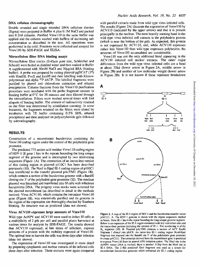
The predicted 775 amino acid residue Vmwl 10 coding regionof HSV-1 IE gene 1 lies in the repeats bounding the long uniquesegment of the genome and is interrupted by two interveningsequences (Figure 1A). The constuction of an intron-less versionof this coding region in plasmid pi 10C1 has been describedpreviously (10). The Ncol to Hpal IE-1 coding region of pi 10C1was transferred to the transfer plasmid pAcYMl (Figure IB),which contains a section of the baculovirus genome with a BamHIcloning site 3' of the polyhedrin gene promoter (32). The resultantplasmid was linearised and transfected into Sf cells with infectiousbaculovirus DNA. The progeny virus stocks were screened forthe desired recombinant (as described in detail in the methodssection). Virus ACV110, which contains the intronless Vmwl 10gene (Figure IB), was extensively purified and its genome inthe region of the expression site thoroughly checked by Southernblotting. Its structure was as predicted (data not shown).
Virus ACV110 expresses large amounts of VmwllOWild type AcNPV and ACV110 were used to infect Sf cells ata multiplicity of 2 pfu per cell and parallel plates harvested atvarious times for analysis by SDS-PAGE. The results showedthat ACV110 expressed, at late times of infection, copiousamounts of a protein with the mobility expected of VmwllO.The optimal time to harvest the cells was three days after infection(data not shown).
The expression of Vmwl 10 was investigated in more detailby preparing cytoplasmic and nuclear extracts of the infected cellsthree days after infection. These extracts were again compared
with parallel extracts made from wild type virus infected cells.The results (Figure 2A) illustrate the expression of Vmwl 10 byACV110 (indicated by the upper arrow) and that it is presentprincipally in the nucleus. The most heavily staining band in thewild type virus infected cell extracts is the polyhedrin protein(which is near the bottom of the gel). As expected, this proteinis not expressed by ACV110, and, while ACV110 expressesrather less Vmwl 10 than wild type expresses polyhedrin, theamounts of VmwllO accumulated are considerable.
VmwllO was not the only additional band appearing in theACV110 infected cell nuclear extracts. The other majordifferences from the wild type virus infected cells are a bandat about 35kd (lower arrow in Figure 2A, middle arrow inFigure 2B) and another of low molecular weight (lower arrowin Figure 2B). It is not known if these represent breakdown
-CD
IE-1 IE-1
tHpalS501
tSailSOS5
A
IXhol2940
\
Ncol1862
tSfllSOS
3' TAA5303 5097
ATG S'062 1734
B
nNcoIXhoI Sail Hpal
Figure 1. A map of the IE-1 region of HSV-1 and the baculovirus transfer vectorpAClll . A. The HSV-1 genome is shown with the repeat sequences markedas boxes. Gene IE-1 lies in the repeat bounding the long unique genome segment.Below is an expansion of the IE-1 region annotated with key restriction sites andthe transcribed portion (including the introns) of IE-1. The numbering is of theRL sequence (18). B. Plasmid pAcYMl contains a section of ACV EcoRIfragment I cloned into pUC8. An intron-less IE-1 coding region Ncol-Hpalfragment was inserted into the BamHI site 3' of the polyhedrin gene promoterto create pAC 111. The transcript derived from the recombinant gene is predictedto express Vmwl 10 from its natural ATG initiation codon. The Xhol site in theAcNPV vector DNA is marked; there is another 315bp from the Ncol site inIE-1 DNA. The 2.4kb predicted Xhol fragment was used as a screen forrecombinant baculovirus genomes which contained the IE-1 coding region.
at University of G
lasgow on D
ecember 19, 2012
http://nar.oxfordjournals.org/D
ownloaded from

6158 Nucleic Acids Research, Vol. 19, No. 22
ACV110WC C N P
ACVWTWC C N P MW
WT 110 MWHSV BHK Sf ACV110
IIy
ylt
Figure 2. Recombinant virus ACV110 expresses Vmwl 10. A. Whole cell (WC),cytoplasmic (C), nuclear (N) and insoluble (P) fractions of Sf cells which hadbeen infected for 72 hours with ACV 110 and AcNPV wild type virus separatedon a Coomassie blue stained 7.5% SDS polyacrylamide gel. The molecular weightmarkers (MW) are myosin (2O5kD), beta galactosidase (116kD), phosphorylaseb (97kD), bovine serum albumin (66kD), egg albumin (45kD) and carbonicanhydrase (29kD). The upper arrow points out Vmwl 10 and the lower indicatesanother prominent band not present in the wild type vims nuclear extract. Themajor stained band in the whole cell and pellet fractions of AcNPV extracts atabout 30kD is the polyhedrin protein. B. Nuclear extracts of AcNPV and A-CV110 separated on a 12.5% SDS polyacrylamide gel The molecular weightmarkers are bovine serum albumin (66kD), egg albumin (45kD), glyceraldehyde3 phosphate dehydrogenase (36kD), carbonic anhydrase (29kD), trypsinogen(24kD) and trypsin inhibitor (20kD). The uppermost of the three arrows indicatesVmwl 10 while the others indicate proteins not present in ACV WT extracts.C. A western blot of samples of HSV-1 infected (HSV) and uninfected (BHK)cells and ACV 110 infected and uninfected (Sf) cells using rabbit antiserum 14711.The arrow points out undegraded Vmwl 10.
B110 .01 .02 M
10 ISFraction number
Figure 4. Preparations of purified Vmwl 10 contain dimers. A. Glycerol gradientcentrifugation of Q-sepharose purified Vmwl 10. Vmwl 10 was detected by ELISAusing serum 14711 and the marker proteins by SDS-PAGE. The positions ofthe peaks fractions of the marker proteins, and their sedimentation co-efficients,are indicated. B. Glutaraldehyde cross linking of Vmwl 10. Q-sepharose purifiedVmwl 10 (110) was treated with 01 and .02% final concentrations ofglutaraldehyde and then analysed on a 5% SDS polyacrylamide gel. The lowerarrow indicates monomenc Vmwl 10, migrating slightly faster than the betagalactosidase marker, while the upper points out the dimer band, co-migratingwith the myosin marker. Note also the material of very low mobility in theglutaraldehyde treated tracks, which is not present in the untreated control.
N w « b 0-Z5M N»CI M OJM N«C1 <UM UIM M
I II
B NE PC QS M
1
Figure 3. Purification of Vmwl 10. A. The elution profile of an ACV 110 infectedcell nuclear extract (N) separated by successive steps of increasing ionic strength(as indicated) on a phosphocellulose column and analysed on 10% SDSpolyacrylamide gels. The molecular weight markers (M) are as in Figure 2A.Vmwl 10 is indicated by the arrow. B. A 10% SDS polyacrylamide gel stainedwith Coomassie blue showing ACV 110 infected cell nuclear extract (NE), pooledfractions after phosphocellulose column chromatography (PC) and a sample afterpurification on a Q-sepharose column (QS). Molecular weight markers (M) areas in Figure 2A.
products of Vmwl 10, cellular proteins that become preferentiallyeluted from the nucleus in the presence of Vmwl 10, or cellularproteins which are induced by the high level expression ofVmwl 10.
It was important to compare the Vmwl 10 proteins expressedby ACV110 and by HSV-1. Accordingly, BHK cells wereinfected with HSV-1 and harvested 16 hours later. Samples ofthe HSV-1 and ACV 110 infected cells were analysed by SDS-PA-GE with samples of mock infected BHK and Sf cells forcomparison. The proteins were transferred to nitrocellulose byWestern blotting and Vmwl 10 detected after incubation withrabbit antiserum 14711, which recognises a carboxy terminalpeptide of Vmwl 10 (10), followed by horseradish peroxidaseconjugated protein A. The results (Figure 2C) show that theVmwl 10 expressed by ACV110 is of the same mobility as thatexpressed by HSV-1, and that it possesses the same carboxyterminal epitope. These data suggest that the Vmwl 10 expressedby ACVI10 is very similar to that expressed during HSV-1infection. The bands of lower molecular weight in the ACV110track of Figure 2C are probably degradation products of Vmwl 10since they are not present in the Sf cell control.
Purification of VmwllO from ACV110 infected cellsVmwl 10 was extensively purified from nuclear extracts of A-CV110 infected cells by two steps of column chromatography.As described in the methods section, nuclear extracts were dilutedwith Buffer A without sodium chloride in order to reduce theionic strength to 0.2M. The solution was then loaded onto aphosphocellulose column equilibrated in the same buffer. Afterextensive washing, bound proteins were eluted with sequentialsteps of Buffer A containing 0.25, 0.3, 0.4 and 1.0M NaCl.
at University of G
lasgow on D
ecember 19, 2012
http://nar.oxfordjournals.org/D
ownloaded from

Nucleic Acids Research, Vol. 19, No. 22 6159
ACV110 MOCK0 -01.05 J 1 I 0 .01.05 J 1 2 oj polj dldC
load FT 0.1M0-2M0JM0JM1J1M1.0Mss-DNA cellulose
load FT 0JM<UM0JM0JM1.0M1.0Mds-DNA cellulose
Figure 5. Purified Vmwl 10 binds to single and double stranded DNA cellulosecolumns. Vmwl 10 was loaded onto the indicated columns and eluted by increasingionic strength as shown. The amount of Vmwl 10 in each fraction was determinedby ELISA.
Vmwl 10 was detected by SDS-PAGE and was found principallyin the 0.3M step elution (Figure 3A). For the second step,fractions from the 0.25M and 0.3M NaCl step elutions werepooled and loaded directly onto a Q-sepharose columnequilibrated with Buffer A containing 0.275M NaCl. The columnwas again washed and bound proteins eluted with steps of 0.3M,0.4M and 1.0M NaCl. Fractions containing Vmwl 10 with a highdegree of purity were eluted at 0.4M NaCl. A summary of theseresults, showing the initial extract, the pooled phosphocellulosefractions and the protein eluted from the Q-sepharose columnis shown in Figure 3B.
Preparations of purified Vmwl 10 contain ditneric moleculesThe purified Vmwl 10 preparation illustrated in Figure 3B wasanalysed by glycerol gradient centrifugation. Gradients wereloaded with a sample that contained Vmwl 10 and control proteinsbovine serum albumin, yeast alcohol dehydrogenase and bovinethyroglobulin. After centrifugation fractions were collected fromthe top of the tubes, and those containing the marker proteinswere identified by SDS-PAGE (data not shown). Suitable amountsof the fractions were used to coat wells for an ELISA estimationof the amount of Vmwl 10 using rabbit antiserum 14711. Theresults are shown in Figure 4A. Yeast ADH has a nativemolecular weight of 150kD and a sedimentation coefficient of6.7S. Vmwl 10 is clearly sedimenting with a peak position ofslightly higher S value (8.2S) than ADH. This is consistent withthe presence of dimeric Vmwl 10 molecules. To investigate thisfurther, the purified Vmwl 10 preparation was incubated in thepresence of 0.01% glutaraldehyde at room temperature for 1hour. Subsequent SDS-PAGE showed that glutaraldehydetreatment resulted in the appearance small amounts of a proteinof exactly twice the apparent molecular weight of Vmwl 10(Figure 4B). Thus it is clear that the preparation of Vmwl 10contains dimeric molecules. However, treatment withglutaraldehyde also converted some of the input protein intomaterial with very low gel mobility, seen as faint bands at thetop of the treated tracks, which is not present in the control track.Thus it is possible that higher order multimers of Vmwl 10 canalso be formed. Glutaraldehyde treatment of control proteins
I probe
Figure 6. Analysis of the DNA binding activity in nuclear extracts from ACV110infected and uninfected Sf cells. A gel retardation experiment using equal amountsof ACV 110 and uninfected cell nuclear extracts with the IE-3 EcoRI-BamHI probeand increasing amounts of polydl/polydC competitor.
known to be monomeric in solution did not produce novel bandsat the position expected of dimers (data not shown).
Analysis of Vmwl 10 by SDS-PAGE in the absence of reducingagent indicated that the dimers or multimers were not covalentlylinked by disulphide bridges (data not shown). Attempts tocharacterise further the nature of purified Vmwl 10 using nativepolyacrylamide gel electrophoresis were not successful; theprotein migrated as a high molecular weight smear (data notshown).
Purified Vmwl 10 binds to both single stranded and doublestranded DNA cellulose matricesOne of the crucial questions about the properties of Vmwl 10is whether it interacts directly with DNA, and, if so, whetherthis interaction is sequence specific. Earlier experiments hadshown that Vmwl 10 in crude nuclear extracts is retained on calfthymus DNA cellulose columns (17). However, this could beexplained by an interaction between Vmwl 10 and one or moreproteins which were binding to the column. The binding ofVmwl 10 to both single stranded and double stranded DNAcellulose was investigated using the purified preparation. Theresults (Figure 5) showed that the protein did indeed bind to DNAcellulose, eluting mainly at salt concentrations higher than 0.2M,and that its binding to double stranded DNA was essentiallyindistinguishable from its binding to single stranded DNA. Theseresults show that Vmwl 10 can interact directly with DNA.(Vmwl 10 prepared from HSV-1 infected BHK cells does notbind to cellulose slurries; unpublished data).
Purified Vmwl 10 does not appear to form a stable complexwith DNA in solutionThat Vmwl 10 binds to DNA cellulose columns does notnecessarily imply that it is a DNA binding protein, because itsretention by these columns could be explained by a non-specificelectrostatic interaction, in the same way that it binds tophosphocellulose. Therefore several attempts were made toinvestigate whether purified Vmwl 10 interacts with DNA insolution. Initial experiments adopted gel retardation technologyto compare the DNA binding activities of nuclear extracts of A-CV110 infected and uninfected cells. The probe contained theIE-3 promoter region from coordinates —108 to +27, and themethods were exactly as described (38). The results (Figure 6)
at University of G
lasgow on D
ecember 19, 2012
http://nar.oxfordjournals.org/D
ownloaded from

6160 Nucleic Acids Research, Vol. 19, No. 22
load FT 02 0.25 0.25 0 J OJ 0.4 1.0
Phosphocellulose
load FT 0.275 0 3 0.4Q-sepharost
Figure 7. The DNA binding activity in ACV110 infected cell nuclear extractsdoes not co-purify with Vmwl 10. A nuclear extract containing Vmwl 10 wasfractionated on phosphocellulose and Q-sepharose columns as indicated. The load,flowthrough, 0.2M, 0.25M (2 aliquots), 0.3M (2 aliquots), 0.4M and 1.0M NaCIsamples were used to detect the amount of Vmwl 10 by ELISA (open boxes)and the amount of DNA binding activity in filter binding experiments (filled boxes).The vertical scale on the display on the left refers to the DNA binding activity(cpmx 10~3), normalised for the amount of protein in each sample. That to theleft of the righthand display gives the amount of Vmwl 10 ELISA units.
showed that nuclear extracts containing Vmwl 10 displayed asimilar general or non-specific DNA binding potential touninfected nuclear extracts. Both contained sufficient DNAbinding activity to form complexes of low mobility (whichprobably contain many different proteins) in the absence ofcompetitor polydl/polydC. Increasing the amount of competitorresolved bands of higher mobility, but there was no indicationof any major differences between the two types of extract.
A gel retardation experiment of necessity can investigatebinding to only a limited set of DNA sequences. Therefore afilter binding assay, using of a mixture of probe fragments derivedfrom plasmid pgDCAT, was also used. Plasmid pgDCATresponds to activation by Vmwl 10 in transfection experiments(37). The probe fragments were incubated with the ACVI10nuclear extract and the mixture filtered through nitrocellulose.The DNA fragments bound to the filter by proteins in the extractwere eluted with SDS and analysed on polyacrylamide gels. Therewas no preferential retention of any of the fragments; all wereequally sensitive to competition by the addition of increasingamounts of polydl/polydC (results not shown). These resultssuggests that Vmwl 10 does not bind with high affinity to anyspecific DNA sequences in pgDCAT, but (taken with the resultsof the gel retardation experiment) they do not exclude thepossibility that Vmwl 10 has a non-specific DNA binding abilitywhich was masked by the total DNA binding capacity of the crudenuclear extract (Figure 6).
Therefore, the DNA binding activity in fractions throughoutthe Vmwl 10 purification procedure was measured by filterbinding experiments using the pgDCAT probe mixture. Theamount of Vmwl 10 in each fraction was quantitated by ELISA.The results (Figure 7) showed that fractions from thephosphocellulose column contained DNA binding activity, butbinding was not proportional to the amount of Vmwl 10 in thefractions. The 0.25M and 0.3M NaCI step fractions from thephosphocellulose column were pooled and passed down a Q-
seharose column. The binding activity clearly eluted with a peakat a much lower ionic strength than the Vmwl 10 elution peak.Indeed, DNA binding activity in the peak Vmwl 10 fractions wasessentially undetectable. These data can not be explained byinactivation of the DNA binding activity of VmwlO during Q-sepharose chromatography since the recoveries of both Vmwl 10and DNA binding activity were about 70%. The assays usingthe peak Vmwl 10 fractions contained a final salt concentration(90mM) which did not inhibit binding of Vmwl 10 to DNAcellulose columns (Figure 5). The fractions containing purifiedVmwl 10 were also completely inactive in a gel retardationexperiment in the absence of polydl/polydC competitor (resultsnot shown).
These results strongly suggest that purified Vmwl 10 is unableto form a stable complex with DNA in solution. It did not displaynon-specific DNA binding activity under a variety of conditions.If it were a sequence specific DNA binding protein, even if itsrecognition site is not present in a plasmid which responds toits activity, it would be expected to bind to other DNA sequencesin a plasmid the size of pgDCAT in the absence of competitor.
DISCUSSION
A fundamental understanding of the mechanism of action ofVmwl 10 has been impeded by its low level of expression duringa normal virus infection; it has been difficult to obtain the proteinin any great quantity or purity. The construction of a baculovirusrecombinant which expresses large quantities of Vmwl 10, andthe subsequent isolation of highly purified preparations of theprotein, is a first step towards a detailed understanding of itsbiochemistry and its interactions with other macromolecules. Thispaper describes the initial steps towards this goal: we have foundthat Vmwl 10 forms multimers, including dimers, in solution andthat it is apparently unable to form a stable interaction with DNAin solution.
The interaction of Vmwl 10 with DNA is of considerableinterest given its role in transcriptional activation. This studyconfirms a previous finding that Vmwl 10 binds to DNA cellulosecolumns (17) and extends it to show that highly purified proteinis still able to bind, both to double and single stranded DNA.However, the key question is whether this binding represents asimple ionic interaction with the phosphate backbone of DNA,a non-specific interaction with the nucleotide bases, or a specificinteraction with a defined sequence of bases. There is no evidenceof any sequence specific interaction, since the DNA bindingactivity of extracts containing Vmwl 10 could not be shown toselect any specific sequence from fragments comprising a plasmidwhich responds to activation by Vmwl 10. Perhaps this is notsurprising as no specific sequences have been defined which arerequired for activation by Vmwl 10. If Vmwl 10 interacts non-specifically with the nucleotide bases, this interaction must besufficiendy weak to be undetectable by a non-specific filterbinding assay conducted under conditions which would allowbinding of a wide range of DNA binding proteins (Figure 7).Therefore, since Vmwl 10 binds to negatively charged matricesduring purification, it seems likely that its retention on DNAcellulose columns is simply a reflection of this ionic interaction.It follows that its ability to activate gene expression is likely toresult from an interaction with other proteins or macromolecules.
As illustrated by a number of recent observations, activationof gene expression can be achieved without direct interaction withDNA by a variety of different mechanisms. These include:
at University of G
lasgow on D
ecember 19, 2012
http://nar.oxfordjournals.org/D
ownloaded from

Nucleic Acids Research, Vol. 19, No. 22 6161
activation of a bound cellular transcription factor (39,40); removalof repressors complexed to transcription factors (41—44);modification of the DNA binding specificity of a transcriptionfactor (45), and inhibition of the formation of a restrictivechromatin structure (46). The availability of large amounts ofpurified VmwllO should enable studies to determine which (ifany) of these stategies it employs.
A further area of interest with respect to Vmwl 10 is the recentdiscovery that a potential zinc finger domain within its secondexon is very highly conserved among a number of proteins ofdiverse evolutionary origin (23-29). Some of these proteins seemto be involved in a number of different aspects of DNAmetabolism and gene expression. The highly conserved natureof the potential zinc finger (which is distinguishable from thetwo other well described classes of zinc finger) suggests that itis involved in a process of general importance. It has beensuggested, as cited above, that the conserved motif might beinvolved in DNA sequence recognition since it is similar to otherzinc finger motifs which perform this function. However, theresults presented here imply that DNA binding is not the roleof at least this member of this particular class of zinc finger. Theavailability of purified preparations of one member of this familyof proteins will enable investigations relevent not only to herpesviruses and gene regulation, but also to a wide range of otheraspects of DNA function.
ACKNOWLEDGEMENTS
The encouragement of Professor J.H. Subak-Sharpe is muchappreciated. We are particularly grateful to Dr N.D. Stow formuch advice and material help during the setting up of the systemof baculovirus expression of VmwllO, and for constructivecriticism of the manuscript.
23. Freemont, P.S., Hanson, I.M. and Trowsdale, J. (1991). Cell 64, 483-484.24. Jones, J.S., Weber, S. and Prakash, L. (1988). Nucl. Acids Res. 16,
7119-7131.25. Patarca, R., Schwartz, J., Singh, R.P., Kong, Q.T., Murphy, E., Anderson,
Y., Sheng, F.W., Singh, P., Johnson, K.A., Guarnagia, S.M., Durfee, T.,Blattner, F., and Cantor, H. (1988). Proc. Natl. Acad. Sci. U.S.A. 85,2733-2737.
26. Takahashi, M., Inaguma, Y., Hiai, H. and Hirose, F. (1988). Mol. Cell.Biol. 8, 1853-1856.
27. Schatz, D.G., Oettinger, M.A. and Baltimore, D. (1989). Cell 59,1035-1048.
28. Haupt, Y., Alexander, W.S., Barri, G., Klinken, S.P. and Adams, J.M.(1991). Cell 65, 753-763.
29. van Lohuizen, M., Verbeek, S., Scheijen, B., Wientjens, E., van der Gulden,H. and Berns, A. (1991). Cell 65, 737-752.
30. Everett, R.D. (1991). J. Gen. Virol. 72, 651-659.31. Studier, F.W., Rosenberg, A.H., Dunn, J.J. and Dubendorff, J.W. (1990).
Methods in Enzymology 185, 60-89.32. Matsuura, Y., Possee, R.D., Overton, H.A. and Bishop, D.H.L. (1987).
J. Gen. Virol. 68, 1233-1250.33. Corsalo, CM. and Pearson, M.L. (1981). Somatic Cell Genetics 7, 603-616.34. Southern, E.M. (1975). J. Mol. Biol. 98, 503-517.35. Stow, N.D., McMonagle, E.C. and Davison, A.J. (1983). Nucl. Acids Res.
11, 8205-8220.36. Dignam, J.D., Lebovitz, R.M. and Roeder, R.G. (1983). Nucl. Acids Res.
11, 1475-1489.37. Everett, R.D. (1986). J. Gen. Virol. 67, 2507-2513.38. Paterson, T. and Everett, R.D. (1988). Nucl Acids Res. 16, 11005-11025.39. Preston, CM., Frame, M.C. and Campbell, M.E.M. (1988). Cell 52,
425-434.40. O'Hare, P. and Goding, C.R. (1988). Cell 52, 435-445.41. Baeuerle, P.A. and Baltimore, D. (1988). Cell 53, 211-217.42. Auwerx, J. and Sassone-corsi, P. (1991). Cell 64, 983-993.43. Bagchi, S., Weinmann, R. and Raychaudhuri, P. (1991). Cell 65,
1063-1072.44. Chellappan, S.P., Hiebert, S., Mudryj, M., Horowitz, J.M and Nevins, J.R.
(1991). Cell 65, 1053-1061.45. Maguire, H.F., Hoeffler, J.P. and Siddiqui, A. (1991). Science 252,
842-844.46. Croston, G.E., Kerrigan, L.A., Lira, L.M., Marshak, D.R. and Kadonaga,
J.T. (1991). Science 251, 643-649.
REFERENCES
1. McGeoch, D.J., Dalrymple, M.A., Davison, A.J., Dolan, A., Frame, M.C,McNab, D., Perry, L.J., Scott, J.E. and Taylor, P. (1988). J. Gen. Virol.69, 1531-1574.
2. Wagner, E.K. (1985). In: The Herpesviruses. Ed. B. Roizman. Plenum Press,New York/London. 3, 45-104.
3. Everett, R.D. (1987). Anticancer Research 7, 589-604.4. Everett, R.D. (1984). EMBO J. 3, 3135-3141.5. O'Hare, P. and Hayward, G.S. (1985). J. Virol. 53, 751-760.6. Gelman, I.H. and Silverstein, S. (1985). Proc. Natl. Acad. Sci. U.S.A. 82,
5265-5269.7. Quinlan, M.P. and Knipe, D.M. (1985). Mol. Cell. Biol. 5, 957-963.8. Stow, N.D. and Stow, E.C. (1986). J. Gen. Virol. 67, 2571-2585.9. Sacks, W.R. and Schaffer, P.A. (1987). J. Virol. 61, 829-839.
10. Everett, R.D. (1989). J. Gen. Virol. 70, 1185-1202.11. Cai, W. and Schaffer, P.A. (1989). J. Virol. 63, 4579-4589.12. Stow, E.C. and Stow, N.D. (1989). J. Gen. Virol. 70, 695-704.13. Russell, J., Stow, N.D., Stow, E.C. and Preston, CM. (1987). J. Gen.
Virol. 68, 3009-3018.14. Lieb, D.A., Coen, D.M., Bogard, C.L., Hicks, K.A., Yager, D.R., Knipe,
D.M., Tyler, K.L. and Schaffer, P.A. (1989). J. Virol. 63, 759-768.15. Clements, G.B. and Stow, N.D. (1989). J. Gen. Virol. 70, 2501-2506.16. Pereira, L., Wolff, M.H., Fenwick, M. and Roizman, B. (1977). Virology
77, 733-749.17. Hay, R.T. and Hay, J. (1980). Virology 104, 230-234.18. Perry, L.J., Rixon, F.J., Everett, R.D., Frame, M.C. and McGeoch, D.J.
(1986). J. Gen. Virol. 67, 2365-2380.19. Berg, J.M. (1986). Science 232, 485-487.20. Everett, R.D. (1987). EMBO J. 6, 2069-2076.21. Everett, R.D. (1988). J. Mol. Biol. 202, 87-96.22. Harris, R.A., Everett, R.D., Zhu, X., Silverstein, S. and Preston, C M .
(1989). J. Virol. 63, 3513-3515.
at University of G
lasgow on D
ecember 19, 2012
http://nar.oxfordjournals.org/D
ownloaded from