evans et al, 2009
DESCRIPTION
DAVID C. EVANS, 1 * RYAN RIDGELY, 2 AND LAWRENCE M. WITMER 2 1 Department of Natural History, Royal Ontario Museum, Toronto, Ontario, Canada 2 Department of Biomedical Sciences, College of Osteopathic Medicine, Ohio University, Athens, Ohio THE ANATOMICAL RECORD 292:1315–1337 (2009) V C 2009 WILEY-LISS, INC. V 1316 EVANS ET AL.TRANSCRIPT
THE ANATOMICAL RECORD 292:1315–1337 (2009)
Endocranial Anatomy of LambeosaurineHadrosaurids (Dinosauria:
Ornithischia): A SensorineuralPerspective on Cranial Crest Function
DAVID C. EVANS,1* RYAN RIDGELY,2 AND LAWRENCE M. WITMER2
1Department of Natural History, Royal Ontario Museum, Toronto, Ontario, Canada2Department of Biomedical Sciences, College of Osteopathic Medicine, Ohio University,
Athens, Ohio
ABSTRACTBrain and nasal cavity endocasts of four corythosaurian lambeosaur-
ines (Dinosauria: Ornithischia) were investigated to test hypotheses ofcranial crest function related to sensorineural systems. Endocasts weregenerated through computed tomography and three-dimensional render-ing and visualization software. The sample comprises a range of ontoge-netic stages from the taxa Lambeosaurus, Corythosaurus, andHypacrosaurus. Results show that the morphology of brain endocasts dif-fers little from that of hadrosaurines. The strikingly convoluted nasal ves-tibule of Hypacrosaurus altispinus, when interpreted in the context oflambeosaurine phylogeny, suggests selective pressure for nasal cavityfunction independent from changes in the external shape of the crest andassociated visual display function. The plesiomorphically small olfactorybulbs and apparently small olfactory region of the nasal cavity arguesagainst the hypothesis that increased olfactory acuity played a causalrole in crest evolution. The elongate cochlea of the inner ear reveals thathearing in lambeosaurines emphasized low frequencies consistent withthe hypothesized low-frequency calls made by the crests under the reso-nation model of crest function. The brain is relatively large in lambeo-saurines compared with many other large dinosaurs, and the cerebrum isrelatively larger than that of all non-hadrosaurian ornithischians andlarge theropods, but compares favorably with hadrosaurine hadrosauridsas well as some maniraptoran theropods. It is concluded that the largebrains of lambeosaurines are consistent with the range of social behaviorsinferred when the crest is interpreted as an intraspecific signalingstructure. Anat Rec, 292:1315–1337, 2009. VVC 2009 Wiley-Liss, Inc.
Keywords: archosaur; dinosaur; brain; Lambeosaurinae;Hadrosauridae; nasal cavity; inner ear; functionalmorphology
Grant sponsor: National Science Foundation; Contract grantnumbers: IBN-9601174, IBN-0343744; IOB-0517257; Grantsponsors: National Engineering Research Council of Canada(Discovery Grant), Ohio University College of OsteopathicMedicine, Ohio Supercomputing Center.
*Correspondence to: David C. Evans, Department of NaturalHistory, Royal Ontario Museum, 100 Queen’s Park, Toronto,ON, Canada. Fax: 416-586-5553. E-mail: [email protected]
Received 9 June 2009; Accepted 9 June 2009
DOI 10.1002/ar.20984Published online in Wiley InterScience (www.interscience.wiley.com).
VVC 2009 WILEY-LISS, INC.
Lambeosaurine dinosaurs have undergone appreciablestudy due to questions concerning the function of theirbizarre cranial crests and the associated evolutionaryhypertrophy of their nasal cavities (Ostrom, 1962;Hopson, 1975; Weishampel, 1981 a,b, 1997; Evans,2006). Despite this considerable attention and an abun-dance of fossil material, many aspects of lambeosaurineanatomy pertinent to cranial crest function, and lambeo-saurine paleobiology in general, remain incompletelyknown. Of particular consequence is the lack of data onthe morphology of the brain and the inner ear (Evans,2006). Cranial endocasts, the three-dimensional casts ofthe cavity that encapsulated the brain and associatedtissues, provide a wealth of information on the shape ofthe brain and the relative size of the brain parts (Jeri-son, 1973; Hopson, 1979; Witmer et al., 2008). Withregard to testing hypotheses of cranial crest functionin lambeosaurines, brain endocasts provides criticalnew information on brain structures associated witholfaction, and likewise casts of the endosseous laby-rinth of the inner ear provide key information on hear-ing sensitivity that can be related to vocal resonationof the crest.
Among hadrosaurids, brain cavity endocasts havebeen described for several hadrosaurine taxa includingEdmontosaurus regalis (Lambe, 1920; Ostrom, 1961),E. annectens (Lull and Wright, 1942), and Gryposaurusnotabilis (Ostrom, 1961; Hopson, 1979). Lambeosaurineendocasts are considerably more poorly documented.Lull and Wright (1942) erroneously listed Lambeosaurusas having a described endocast; Gilmore (1924) describedthe braincase foramina of the holotype of Lambeosauruslambei and commented on the shape of the cerebrum ina specimen now referred to Parasaurolophus sp. (Evanset al., in press), but a three-dimensional endocast wasnot figured or described. Young (1958) figured the endo-cast of Tsintaosaurus spinorhinus and on it labeled fivecranial nerves (V, IX, X, XI, and XII), the cerebellum, pi-tuitary, and optic lobe (Young, 1958). Tsintaosaurus isputatively a basal lambeosaurine (Buffetaut and Tong-Buffetaut, 1993; Godefroit et al., 2004a; Horner et al.,2004; Evans and Reisz, 2007), but this position is notfollowed by all workers (Wu, 2008, personal communica-tion; Weishampel, 1981b). Regardless of its status, Tsin-taosaurus does not seem to have the same degree ofnasal cavity hypertrophy seen in other lambeosaurines.Although Ostrom (1961, 1962) reconstructed the brainand associated vasculature in Corythosaurus and otherlambeosaurines based on braincase osteology, and Evans(2006) described a forebrain endocast for an indetermi-nate corythosaur, previous studies of lambeosaurinecrest function have not presented evidence from a com-plete brain cavity endocast.
Lambeosaurines tightly enclose most of the nasal cav-ity within the premaxilla and nasal bones, which becomeallometrically elaborated through ontogeny into a cra-nial crest with species-specific external and internalmorphologies (Dodson, 1975). The morphology and on-togeny of the crest cavities are still incompletely knownin virtually all lambeosaurine taxa. Published recon-structions of the crest cavities in most lambeosaurineshave been based on a few fortuitously broken specimensor even destructive ‘‘dissection’’ (Parasaurolophuswalkeri, P. cyrtocristatus) of the crest (Ostrom, 1963;
Weishampel, 1981b). Notable exceptions are the recon-struction of the nasal passages of Parasaurolophus tubi-cen by Sullivan and Williamson (1999) and unpublishedinvestigations of Hypacrosaurus stebingeri (Horner,1995; Horner et al., 2001), which were accomplishednondestructively using computed tomographic (CT) scan-ning and visualization software. These pioneering stud-ies demonstrated the considerable advantages of usingCT scanning to reveal the internal anatomy of the lam-beosaurine crest. This method can be used to visualizeendocasts of all intraosseous cavities, including the braincavity, nasal cavity, inner ear, vasculature, and airsinuses (Clark and Morrison, 1994; Ketcham and Carl-son, 2001; Witmer and Ridgely, 2008b; Witmer et al.,2008), and is ideally suited for illuminating complexinternal cranial anatomy of lambeosaurines. In this arti-cle, we present the first digital reconstructions of brainand nasal cavity endocasts for three species of fan-crested lambeosaurines: Hypacrosaurus altispinus, Cory-thosaurus sp., and Lambeosaurus sp. These endocasts ofthe brain cavity and endosseous labyrinth are the firstcomplete endocasts for lambeosaurine hadrosaurids. Thenasal cavity reconstructions provide new data on nasalcavity ontogeny and evolution and, together with thebrain cavity endocasts, permit the most complete sensor-ineural evaluation of crest functional hypotheses to date.
MATERIALS AND METHODSMaterials
The specimens included in this study were chosen asrepresentatives of the three most common genera ofNorth American lambeosaurines, and the sample con-sists of four specimens from three species of fan-crestedcorythosaurian lambeosaurines: Hypacrosaurus altispi-nus (Fig. 1, Table 1), Lambeosaurus sp. (Fig. 2), and Cor-ythosaurus sp. (Figs. 3, 4). The specimens differ withrespect to their overall size and corresponding degree ofcranial crest development, and were selected to covermost of the preserved ontogenetic stages of cranial crestdevelopment in the corythosaur clade. However, thereare gaps in the ontogenetic representation of each genusat this time. This study is part of a larger project oncomparative hadrosaurid cranial ontogeny, and weintend to scan more specimens to complete these ontoge-netic series that will be presented in future publications.
The taxonomic identification and provenance of thespecimens are as follows:
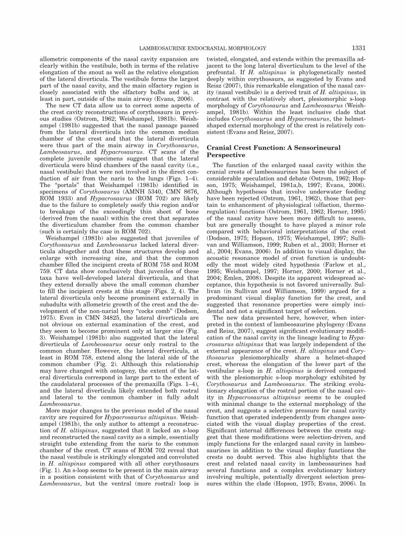
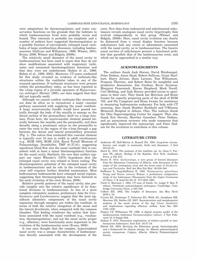
1. Hypacrosaurus altispinus (Royal Ontario Museum,Toronto, ROM 702): ROM 702 (Fig. 1) is a largely com-plete but disarticulated skull (skull length ¼ �700 mm)and associated partial tibia from a large individualwith a fully developed cranial crest. This specimen hasyet to be formally described, but can be referred toHypacrosaurus altispinus on the basis of a foreshort-ened caudolateral premaxillary process that contrib-utes to a helmet-shaped crest and a constrictedexternal naris (Russell and Chamney, 1967; Evans,in press). The specimen was collected from the lowerMaastrichtian part of the Horseshoe Canyon Forma-tion, Alberta, within the series of strata that haveyielded all of the known H. altispinus material (Russelland Chamney, 1967; Eberth, 2004; Evans, 2007).
1316 EVANS ET AL.
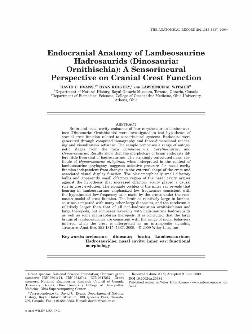
Fig. 1. Nasal cavity and other cephalic components of Hypacrosau-rus altispinus (ROM 702). Left premaxilla and nasal airway have beenmirrored from the right side. Labeled illustrations in (A) left lateralview; (B) left lateral view with transparent bone showing nasal airway;(C) left lateral view with transparent bone with nasal structures sagit-tally sectioned showing medial side of right nasal airway; (D) left ros-
trodorsolateral oblique view; (E) left rostrodorsolateral oblique viewwith transparent nasal airway to show the convoluted course ofinspired air; and (F) rostral view with transparent bone showing nasalairway. Anatomical abbreviations are provided in Table 1. Scale bar ¼20 cm.
LAMBEOSAURINE ENDOCRANIAL MORPHOLOGY 1317
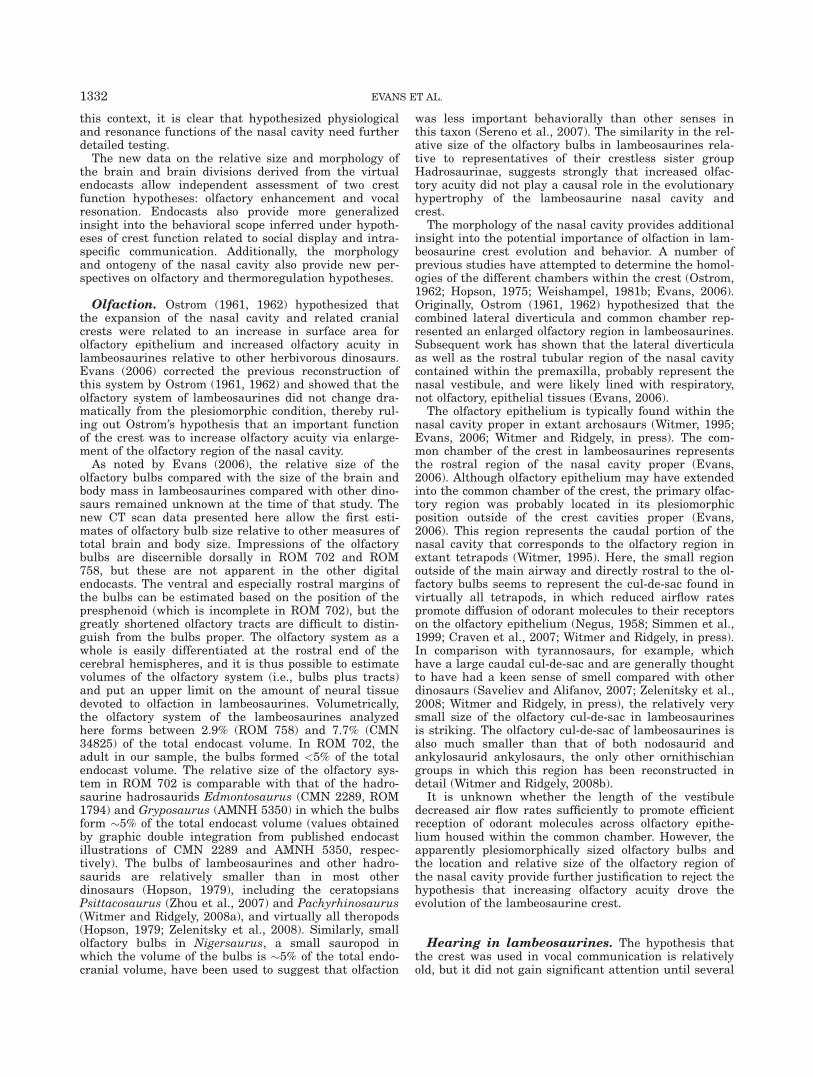
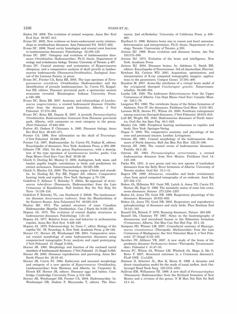
2. Lambeosaurus (ROM 758): ROM 758 (Fig. 2) is thesmallest skull in the sample (skull length ¼ 372 mm).It was originally designated the holotype of a distinct,small-bodied lambeosaurine species, Tetragonosauruspraeceps (Parks, 1931), but has more recently beeninterpreted as a juvenile Lambeosaurus by Dodson(1975) and Evans et al. (2005). This specimen was col-lected from the Campanian-aged strata of the Dino-saur Park Formation (76.5–74.8 Ma) at DinosaurProvincial Park, Alberta.
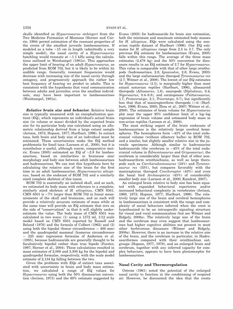
3. Corythosaurus (ROM 759, Canadian Museum of Na-ture, Ottawa, CMN 34825): ROM 759 (Fig. 4) wasoriginally designated the holotype of a small-bodiedlambeosaurine species, Tetragonosaurus erectofrons(Parks, 1931), but was reidentified as a juvenile Cory-
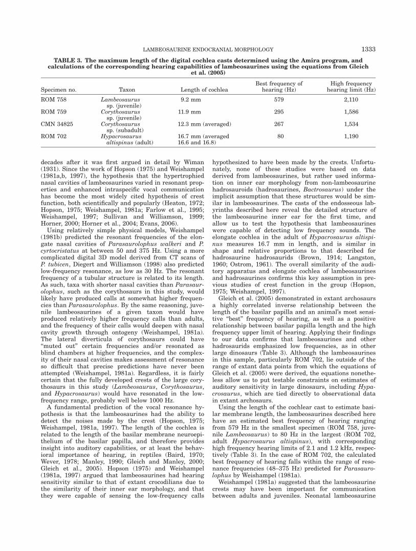
thosaurus by Dodson (1975) and Evans et al. (2005).This skull is only slightly larger than ROM 758, andit is incomplete in a number of areas, including therostral end of the premaxillae and most of the rightside of the face (Evans et al., 2005). Both of thesespecimens were collected from the Campanian-agedstrata of the Dinosaur Park Formation (76.5–74.8 Ma)at Dinosaur Provincial Park, Alberta. CMN 34825(Fig. 3) includes a complete skull (skull length ¼ 465mm) that is crushed on the left side. It is larger thanROM 759 and has a relatively larger cranial crest.
CT Scanning and 3D Visualization
All of the specimens were CT scanned at O’BlenessMemorial Hospital, Athens, Ohio, using a General Elec-tric (GE) LightSpeed Ultra Multislice CT scannerequipped with the Extended Hounsfield option (whichgreatly improves resolvability of detail from denseobjects such as fossils by extending the dynamic range ofimages as much as 16-fold) and a bow-tie filter (whichdecreases beam-hardening artifacts). All specimens werescanned helically at a slice thickness of 625 lm (exceptfor the crest of ROM 702, for which, given its large sizeand simpler anatomy, a coarser slice thickness of 1.25mm was used), 120–140 kV and 200–300 mA. The rawscan data were reconstructed using a bone algorithm.Data were output from the scanner in DICOM format,and then imported into Amira 4.1.2 and Amira 5.1 (Mer-cury-TGS, Chelmsford, MA) for viewing, analysis, andvisualization. All scan data, regardless of source, wereanalyzed on 32- and 64-bit PC workstations with 4 GBof RAM and nVidia Quadro FX 3000 or 4500 video cardsand running Microsoft Windows XP Professional, Win-dows XP Professional �64, or Linux 2.6.18 (Debian 4.0distribution). Anatomical features of interest (e.g., nasalcavity, cranial endocast, endosseous labyrinth, etc.) werehighlighted and digitally extracted using Amira’s seg-mentation tools for quantification and visualization.
The juvenile lambeosaurine specimens ROM 758 and759 were more difficult to scan because metal mountinghardware could not be removed before scanning withoutrisking damage to the specimens. Likewise, CMN 34825and its supporting jacket were too large to traverse thescanner’s gantry in a single pass. These specimensrequired multiple scans with different orientations andadditional data processing to minimize the adverseeffects of the scanning artifacts and, in the case of CMN34825, digital assembly of the different datasets inAmira. Because of missing portions and historic plasterreconstruction, the dataset generated for ROM 702 pre-sented some ambiguities that were difficult to interpret.Parts of our reconstruction of this specimen are there-fore tentative, but represent testable hypothesesgrounded in the context of the other corythosaurs andwill be refined in future studies. Because only one sideof the skull is preserved in ROM 702 and ROM 759, ourreconstructions include the necessary mirroring of ele-ments to provide a full picture of the skull.
Supplemental visualizations and native CT data forsome of the specimens are available on the websitewww.ohio.edu/witmerlab.
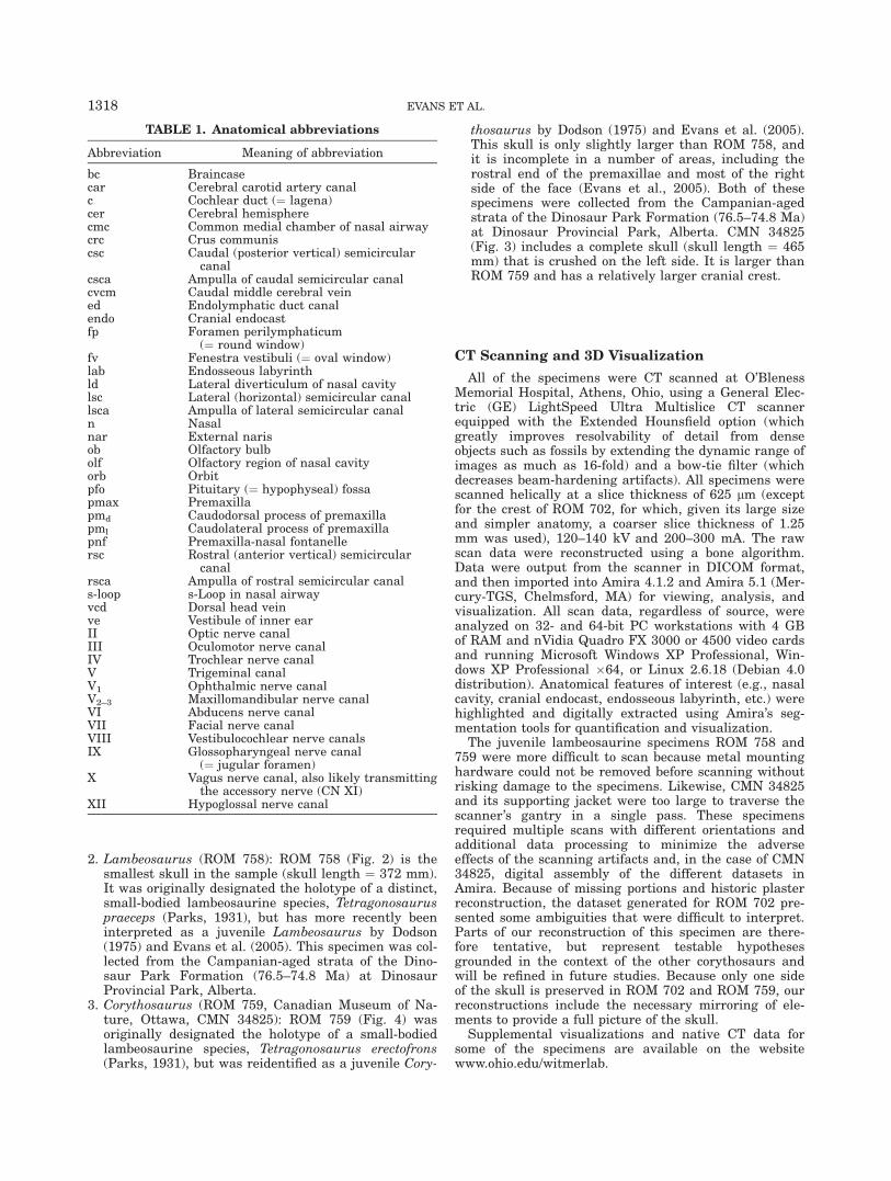
TABLE 1. Anatomical abbreviations
Abbreviation Meaning of abbreviation
bc Braincasecar Cerebral carotid artery canalc Cochlear duct (¼ lagena)cer Cerebral hemispherecmc Common medial chamber of nasal airwaycrc Crus communiscsc Caudal (posterior vertical) semicircular
canalcsca Ampulla of caudal semicircular canalcvcm Caudal middle cerebral veined Endolymphatic duct canalendo Cranial endocastfp Foramen perilymphaticum
(¼ round window)fv Fenestra vestibuli (¼ oval window)lab Endosseous labyrinthld Lateral diverticulum of nasal cavitylsc Lateral (horizontal) semicircular canallsca Ampulla of lateral semicircular canaln Nasalnar External narisob Olfactory bulbolf Olfactory region of nasal cavityorb Orbitpfo Pituitary (¼ hypophyseal) fossapmax Premaxillapmd Caudodorsal process of premaxillapml Caudolateral process of premaxillapnf Premaxilla-nasal fontanellersc Rostral (anterior vertical) semicircular
canalrsca Ampulla of rostral semicircular canals-loop s-Loop in nasal airwayvcd Dorsal head veinve Vestibule of inner earII Optic nerve canalIII Oculomotor nerve canalIV Trochlear nerve canalV Trigeminal canalV1 Ophthalmic nerve canalV2–3 Maxillomandibular nerve canalVI Abducens nerve canalVII Facial nerve canalVIII Vestibulocochlear nerve canalsIX Glossopharyngeal nerve canal
(¼ jugular foramen)X Vagus nerve canal, also likely transmitting
the accessory nerve (CN XI)XII Hypoglossal nerve canal
1318 EVANS ET AL.
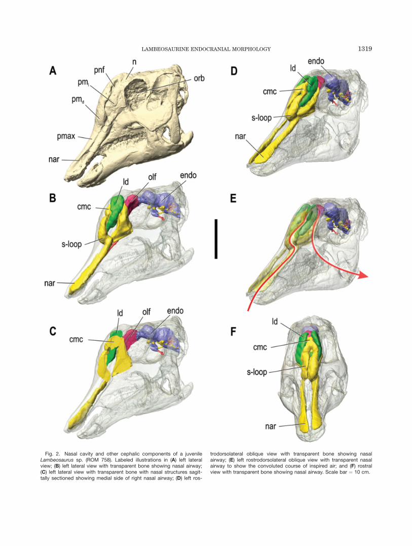
Fig. 2. Nasal cavity and other cephalic components of a juvenileLambeosaurus sp. (ROM 758). Labeled illustrations in (A) left lateralview; (B) left lateral view with transparent bone showing nasal airway;(C) left lateral view with transparent bone with nasal structures sagit-tally sectioned showing medial side of right nasal airway; (D) left ros-
trodorsolateral oblique view with transparent bone showing nasalairway; (E) left rostrodorsolateral oblique view with transparent nasalairway to show the convoluted course of inspired air; and (F) rostralview with transparent bone showing nasal airway. Scale bar ¼ 10 cm.
LAMBEOSAURINE ENDOCRANIAL MORPHOLOGY 1319
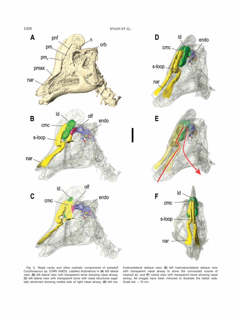
Fig. 3. Nasal cavity and other cephalic components of subadultCorythosaurus sp. (CMN 34825). Labeled illustrations in (A) left lateralview; (B) left lateral view with transparent bone showing nasal airway;(C) left lateral view with transparent bone with nasal structures sagit-tally sectioned showing medial side of right nasal airway; (D) left ros-
trodorsolateral oblique view; (E) left rostrodorsolateral oblique viewwith transparent nasal airway to show the convoluted course ofinspired air; and (F) rostral view with transparent bone showing nasalairway. All images have been mirrored to illustrate the better side.Scale bar ¼ 10 cm.
1320 EVANS ET AL.
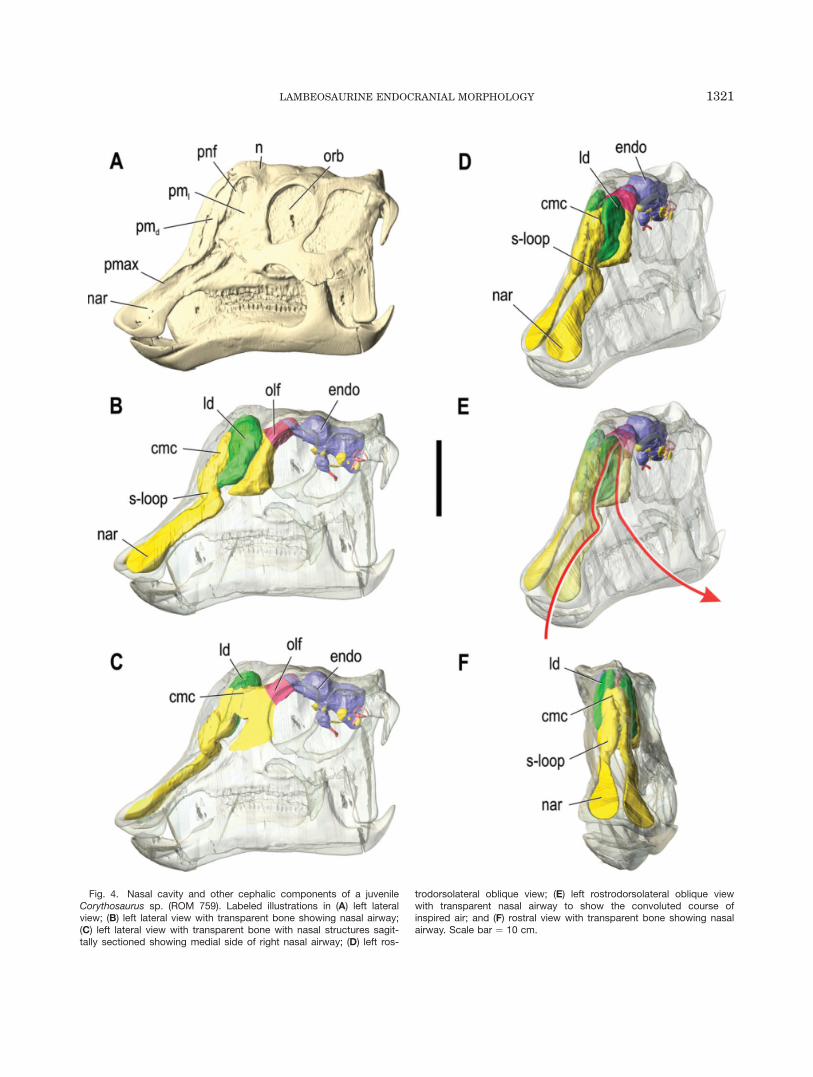
Fig. 4. Nasal cavity and other cephalic components of a juvenileCorythosaurus sp. (ROM 759). Labeled illustrations in (A) left lateralview; (B) left lateral view with transparent bone showing nasal airway;(C) left lateral view with transparent bone with nasal structures sagit-tally sectioned showing medial side of right nasal airway; (D) left ros-
trodorsolateral oblique view; (E) left rostrodorsolateral oblique viewwith transparent nasal airway to show the convoluted course ofinspired air; and (F) rostral view with transparent bone showing nasalairway. Scale bar ¼ 10 cm.
LAMBEOSAURINE ENDOCRANIAL MORPHOLOGY 1321
RESULTSNasal Cavity
Lambeosaurus and Corythosaurus. The nasalpassages of the smallest skulls, ROM 758 and ROM 759,are virtually identical (Figs. 2, 4). The nasal cavityascends caudodorsally within the premaxilla from thenaris at the rostrum to the region directly rostral to thedorsal process of the maxilla. Here, the nasal cavityturns rostrodorsally and then caudodorsally to form ans-shaped loop within the premaxilla. The nasal passagethen divides into the lateral diverticulum and a moremedial passage that coalesces with its complement toform the common undivided camber within the nasals,deep to the lateral diverticula. The lateral diverticulumis enclosed laterally by the caudolateral process of thepremaxilla and is exposed externally via the premaxilla-nasal fontanelle. These lateral chambers extend dorsalto the common chamber, are not inflated relative to therest of the crest, and do not seem to communicate withthe common median chamber or their counterparts cau-dal to it. The bony passage between the basal apertureof the lateral diverticulum and the rostral end of thecommon chamber occurs between the premaxilla andnasal bones and represents the homologue of the exter-nal naris in hadrosaurines and other non-lambeosaurineornithischians. The common chamber is defined predom-inantly by the nasals dorsally and laterally and by thepremaxillae rostroventrally, and occurs medial tothe large lateral diverticula. An aperture occurs betweenthe common chamber and each lateral diverticulum. Thecommon median chamber extends ventrally beyond thecrest proper into the antorbital cavity, deep to the pre-frontal. Ventral to the crest, a small but significant por-tion of the nasal cavity occurs caudal to the mainairway, between the main airway and the rostral end ofthe olfactory bulbs. This portion would house the olfac-tory region of the nasal cavity caudally, as well as thatportion of the respiratory region located below the crest.
This general pattern also occurs in CMN 34825, a sub-adult Corythosaurus (Fig. 3). The small crest of CMN34825, which extends dorsal to the orbits and overhangsthe skull roof, is relatively larger than in ROM 759, butremains less developed compared with large individualsof this genus (Dodson, 1975). The nasal passage remainspaired for a considerable distance caudal to the diver-gence of the lateral diverticula, and the lateral divertic-ula are notably large compared with the small commonchamber. There is an aperture between each lateral di-
verticulum and the common chamber, but this is lessclear between the diverticulum and the main airway ros-trally. Consistent with its ontogenetic stage, the commonchamber and lateral diverticula are more supraorbitallypositioned than in the smaller ROM 759, but not as welldeveloped as in large adults (Weishampel, 1981b).
Hypacrosaurus altispinus. The nasal cavity ofHypacrosaurus altispinus (Fig. 1) seems to be consider-ably more complex compared with the Corythosaurusand Lambeosaurus specimens described above, as wellas to previous reconstructions for this taxon (Weisham-pel, 1981b). From the constricted external narial aper-ture at the rostrum, the nasal cavity extendscaudodorsally as a narrow tube within the premaxilla tothe level of the prefrontal at the rostroventral region ofthe crest. This passage then loops laterally and rostrally,and extends rostroventrally to the level of dorsal processof the maxilla. In this region, the main airwayapproaches the lateral diverticulum, which extends cau-dodorsally in parallel to the main airway toward thecommon median chamber of the crest. The lateral diver-ticulum deepens dorsally to reach its maximum depth onthe lateral region of the crest such that the shape of thelateral diverticulum in lateral view mimics the generalshape of the caudodorsal process of the premaxilla thatencloses it laterally. The thin sheet of the nasal bonethat divides the common chamber from the lateral diver-ticulum is broken in ROM 702, and it is uncertainwhether or not there was a fenestra between thesechambers.
Brain Cavity Endocasts
The digital endocasts of the four specimens show con-siderable detail (Figs. 5–7). Traditionally, non-aviandinosaurs have been regarded as ‘‘reptilian’’ in that theirbrains were thought to have filled only a relatively smallportion of the endocranial cavity in contrast with theconditions in mammals and birds (Jerison, 1969, 1973;Hopson, 1977, 1979; Rogers, 1999; Larsson et al., 2000).In crocodilians, as in squamates, the brain typically fills<50% of the endocranium by volume. The remainder ofthe endocranial space consists of cerebrospinal fluidbetween the meninges and/or venous sinuses within thedura (Hopson, 1979). Lambeosaurines have recentlybeen shown to have dense vascular grooves that areessentially continuous across the lateral regions of the
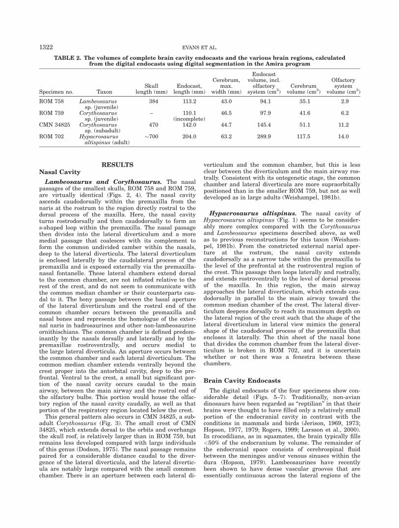
TABLE 2. The volumes of complete brain cavity endocasts and the various brain regions, calculatedfrom the digital endocasts using digital segmentation in the Amira program
Specimen no. TaxonSkull
length (mm)Endocast,
length (mm)
Cerebrum,max.
width (mm)
Endocastvolume, incl.olfactory
system (cm3)Cerebrum
volume (cm3)
Olfactorysystem
volume (cm3)
ROM 758 Lambeosaurussp. (juvenile)
384 113.2 43.0 94.1 35.1 2.9
ROM 759 Corythosaurussp. (juvenile)
– 110.1(incomplete)
46.5 97.9 41.6 6.2
CMN 34825 Corythosaurussp. (subadult)
470 142.0 44.7 145.4 51.1 11.2
ROM 702 Hypacrosaurusaltispinus (adult)
�700 204.0 63.2 289.9 117.5 14.0
1322 EVANS ET AL.
endocranium rostral to CN VII (Evans, 2005). This sug-gests that much of the rostral and ventral regions of thebrain were closely associated with the endocranial wall,and that the endocast generally reflects the shape of thebrain in this region (Evans, 2005). The largely undefineddorsal region of the endocast caudal to the cerebrumindicates the presence of a large longitudinal venoussinus, as in crocodilians, and a major ventrolateral divi-sion of the longitudinal sinus (the lateral head vein) isclearly present on the endocasts (Hopson, 1979). Theamorphous nature of the endocast in the postcerebral
region suggests that much of the hindbrain of hadro-saurids was not in close relationship to the endocranialwall (Evans, 2005).
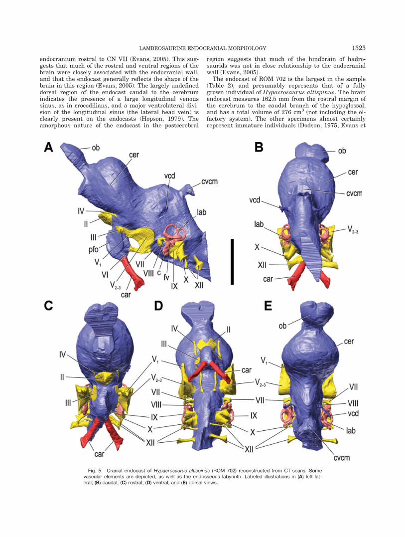
The endocast of ROM 702 is the largest in the sample(Table 2), and presumably represents that of a fullygrown individual of Hypacrosaurus altispinus. The brainendocast measures 162.5 mm from the rostral margin ofthe cerebrum to the caudal branch of the hypoglossal,and has a total volume of 276 cm3 (not including the ol-factory system). The other specimens almost certainlyrepresent immature individuals (Dodson, 1975; Evans et
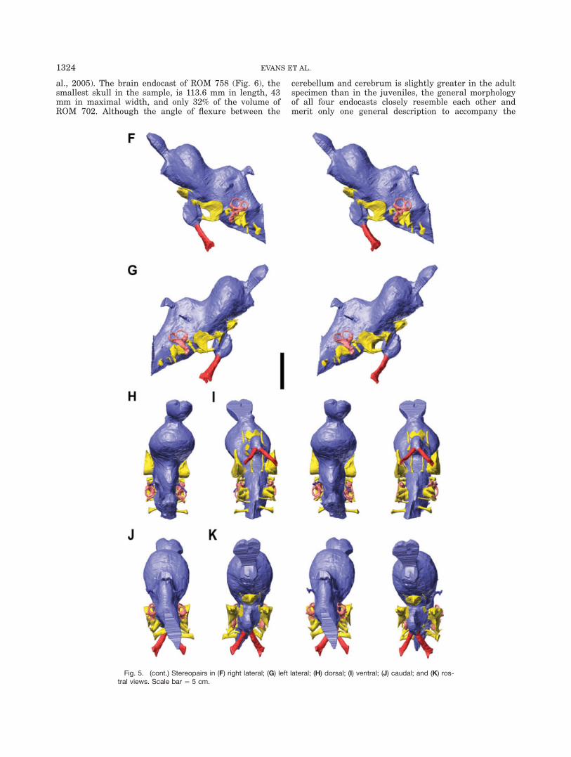
Fig. 5. Cranial endocast of Hypacrosaurus altispinus (ROM 702) reconstructed from CT scans. Somevascular elements are depicted, as well as the endosseous labyrinth. Labeled illustrations in (A) left lat-eral; (B) caudal; (C) rostral; (D) ventral; and (E) dorsal views.
LAMBEOSAURINE ENDOCRANIAL MORPHOLOGY 1323
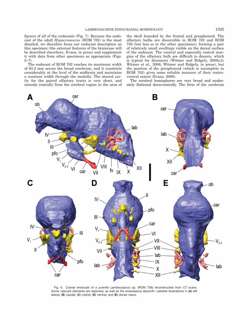
al., 2005). The brain endocast of ROM 758 (Fig. 6), thesmallest skull in the sample, is 113.6 mm in length, 43mm in maximal width, and only 32% of the volume ofROM 702. Although the angle of flexure between the
cerebellum and cerebrum is slightly greater in the adultspecimen than in the juveniles, the general morphologyof all four endocasts closely resemble each other andmerit only one general description to accompany the
Fig. 5. (cont.) Stereopairs in (F) right lateral; (G) left lateral; (H) dorsal; (I) ventral; (J) caudal; and (K) ros-tral views. Scale bar ¼ 5 cm.
1324 EVANS ET AL.
figures of all of the endocasts (Fig. 7). Because the endo-cast of the adult Hypacrosaurus (ROM 702) is the mostdetailed, we therefore focus our endocast description onthis specimen (the external features of the braincase willbe described elsewhere, Evans, in press) and supplementit with data from other specimens as appropriate (Figs.5–7).
The endocast of ROM 702 reaches its maximum widthof 63.2 mm across the broad cerebrum, and it constrictsconsiderably at the level of the midbrain and maintainsa constant width through the medulla. The shared cav-ity for the paired olfactory tracts is very short, andextends rostrally from the cerebral region in the area of
the skull bounded by the frontal and presphenoid. Theolfactory bulbs are discernible in ROM 702 and ROM758 (but less so in the other specimens), forming a pairof relatively small swellings visible on the dorsal surfaceof the endocast. The ventral and especially rostral mar-gins of the olfactory bulb are difficult to discern, whichis typical for dinosaurs (Witmer and Ridgely, 2008a,b;Witmer et al., 2008; Witmer and Ridgely, in press), butthe position of the presphenoid (which is incomplete inROM 702) gives some reliable measure of their rostro-ventral extent (Evans, 2006).
The cerebral hemispheres are very broad and moder-ately flattened dorsoventrally. The form of the cerebrum
Fig. 6. Cranial endocast of a juvenile Lambeosaurus sp. (ROM 758) reconstructed from CT scans.Some vascular elements are depicted, as well as the endosseous labyrinth. Labeled illustrations in (A) leftlateral; (B) caudal; (C) rostral; (D) ventral; and (E) dorsal views.
LAMBEOSAURINE ENDOCRANIAL MORPHOLOGY 1325
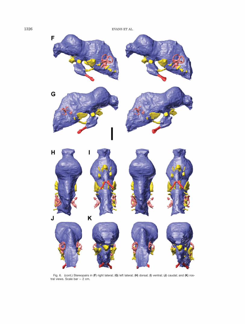
Fig. 6. (cont.) Stereopairs in (F) right lateral; (G) left lateral; (H) dorsal; (I) ventral; (J) caudal; and (K) ros-tral views. Scale bar ¼ 2 cm.
1326 EVANS ET AL.
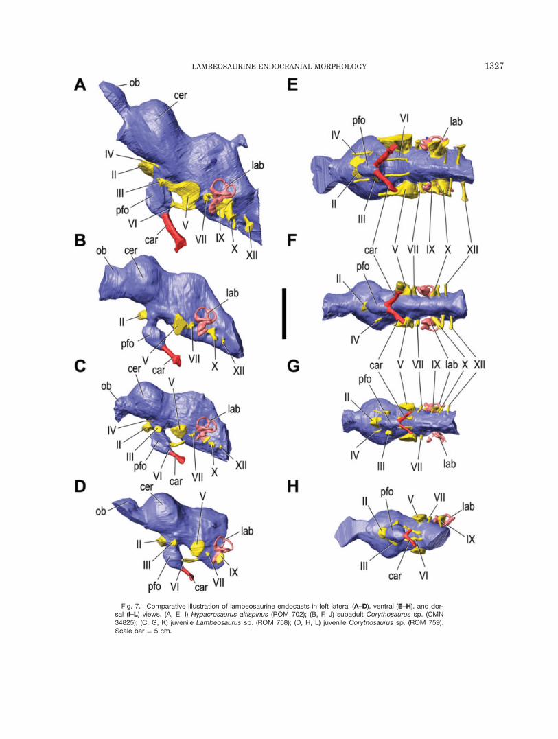
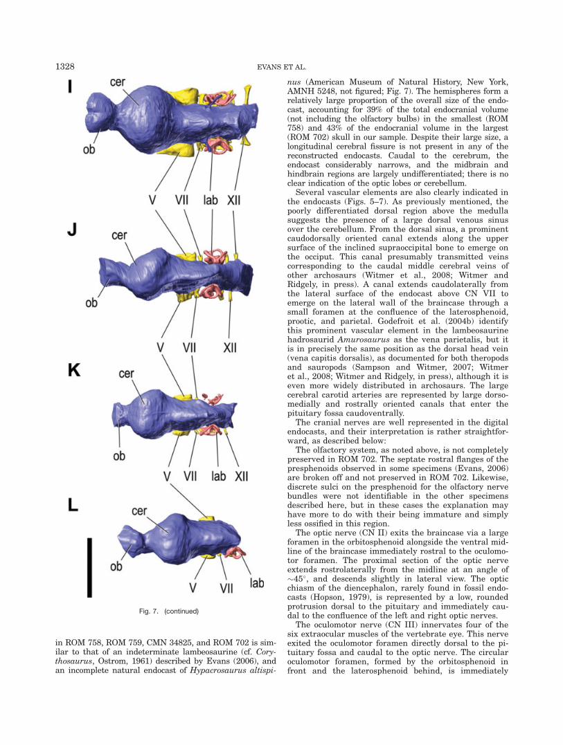
Fig. 7. Comparative illustration of lambeosaurine endocasts in left lateral (A–D), ventral (E–H), and dor-sal (I–L) views. (A, E, I) Hypacrosaurus altispinus (ROM 702); (B, F, J) subadult Corythosaurus sp. (CMN34825); (C, G, K) juvenile Lambeosaurus sp. (ROM 758); (D, H, L) juvenile Corythosaurus sp. (ROM 759).Scale bar ¼ 5 cm.
LAMBEOSAURINE ENDOCRANIAL MORPHOLOGY 1327
in ROM 758, ROM 759, CMN 34825, and ROM 702 is sim-ilar to that of an indeterminate lambeosaurine (cf. Cory-thosaurus, Ostrom, 1961) described by Evans (2006), andan incomplete natural endocast of Hypacrosaurus altispi-
nus (American Museum of Natural History, New York,AMNH 5248, not figured; Fig. 7). The hemispheres form arelatively large proportion of the overall size of the endo-cast, accounting for 39% of the total endocranial volume(not including the olfactory bulbs) in the smallest (ROM758) and 43% of the endocranial volume in the largest(ROM 702) skull in our sample. Despite their large size, alongitudinal cerebral fissure is not present in any of thereconstructed endocasts. Caudal to the cerebrum, theendocast considerably narrows, and the midbrain andhindbrain regions are largely undifferentiated; there is noclear indication of the optic lobes or cerebellum.
Several vascular elements are also clearly indicated inthe endocasts (Figs. 5–7). As previously mentioned, thepoorly differentiated dorsal region above the medullasuggests the presence of a large dorsal venous sinusover the cerebellum. From the dorsal sinus, a prominentcaudodorsally oriented canal extends along the uppersurface of the inclined supraoccipital bone to emerge onthe occiput. This canal presumably transmitted veinscorresponding to the caudal middle cerebral veins ofother archosaurs (Witmer et al., 2008; Witmer andRidgely, in press). A canal extends caudolaterally fromthe lateral surface of the endocast above CN VII toemerge on the lateral wall of the braincase through asmall foramen at the confluence of the laterosphenoid,prootic, and parietal. Godefroit et al. (2004b) identifythis prominent vascular element in the lambeosaurinehadrosaurid Amurosaurus as the vena parietalis, but itis in precisely the same position as the dorsal head vein(vena capitis dorsalis), as documented for both theropodsand sauropods (Sampson and Witmer, 2007; Witmeret al., 2008; Witmer and Ridgely, in press), although it iseven more widely distributed in archosaurs. The largecerebral carotid arteries are represented by large dorso-medially and rostrally oriented canals that enter thepituitary fossa caudoventrally.
The cranial nerves are well represented in the digitalendocasts, and their interpretation is rather straightfor-ward, as described below:
The olfactory system, as noted above, is not completelypreserved in ROM 702. The septate rostral flanges of thepresphenoids observed in some specimens (Evans, 2006)are broken off and not preserved in ROM 702. Likewise,discrete sulci on the presphenoid for the olfactory nervebundles were not identifiable in the other specimensdescribed here, but in these cases the explanation mayhave more to do with their being immature and simplyless ossified in this region.
The optic nerve (CN II) exits the braincase via a largeforamen in the orbitosphenoid alongside the ventral mid-line of the braincase immediately rostral to the oculomo-tor foramen. The proximal section of the optic nerveextends rostrolaterally from the midline at an angle of�45�, and descends slightly in lateral view. The opticchiasm of the diencephalon, rarely found in fossil endo-casts (Hopson, 1979), is represented by a low, roundedprotrusion dorsal to the pituitary and immediately cau-dal to the confluence of the left and right optic nerves.
The oculomotor nerve (CN III) innervates four of thesix extraocular muscles of the vertebrate eye. This nerveexited the oculomotor foramen directly dorsal to the pi-tuitary fossa and caudal to the optic nerve. The circularoculomotor foramen, formed by the orbitosphenoid infront and the laterosphenoid behind, is immediately
Fig. 7. (continued)
1328 EVANS ET AL.
dorsomedial to, and possibly continuous with, the fenes-tra on the lateral side of the pituitary fossa for the abdu-cens nerve (CN VI).
The small trochlear nerve (CN IV) passes rostrallyfrom the metencephalon via a long canal in the orbitos-phenoid bone on its path to the superior oblique extraoc-ular muscle. The trochlear nerve exits the braincase viaa dedicated foramen, as in extant Caiman and mostother dinosaurs (Hopson, 1979), that is located immedi-ately dorsal to the optic nerve foramen.
The trigeminal nerve (CN V) is located at the rostralend of the medulla, between CN III and CN VII. Itextends laterally via a characteristically large, laterallyexpanding, funnel shaped foramen, a distinctive land-mark on the lateral wall of the braincase. The large di-ameter of the external trigeminal foramen, 15 mm inROM 702, suggests that it housed the trigeminal gan-glion (Ostrom, 1961), from which the ophthalmic branch(CN V1) extends rostrally via a horizontal sulcus on thelaterosphenoid, and the combined maxillary and man-dibular branches (CN V2–3) extend ventrally at a rightangle to the ophthalmic branch. Distally, the maxillarybranch diverges from the mandibular branch rostral tothe alar process on the lateral surface of thebasisphenoid.
The abducens nerve (CN VI) passes rostroventrallyfrom the endocranial floor at the rostral end of the me-dulla, through the basisphenoid, to emerge in the pitui-tary fossa through a small foramen on the caudal wall ofthe dorsum sellae. The abducens then passes into theorbit via a large fenestra in the lateral side of the pitui-tary fossa.
The facial nerve (CN VII) exits the endocranial cavitythrough the prootic bone between the trigeminal fora-men and the fenestra vestibuli (¼ ovalis). The branchesof the facial nerve diverge near the lateral surface of theprootic. The hyomandibular branch passes caudodorsallyon its path above the fenestra vestibuli, and the palatinebranch extends ventrally within a sulcus on the lateralsurface of the prootic.
The morphology of the vestibulocochlear nerve (CNVIII) is particularly well preserved on the left side of theROM 702 endocast. It exits the endocranial cavity to-gether with the facial nerve, but before reaching the lat-eral wall of the prootic, curves caudally to enter thevestibular apparatus. Two distinct branches are present;one is directed dorsally to reach the otic vestibule, whileanother, the cochlear portion of the vestibulocochlearnerve, extends to the dorsal base of the cochlea at thelevel of the fenestra vestibuli.
The glossopharyngeal nerve (CN IX) exits the brain-case through the metotic foramen immediately caudal tothe fenestra vestibuli, and rostral to the vagus foramenopposite the metotic strut (Langston, 1960). The largeroot of the vagus nerve (CN X) extends caudolaterallyfrom the region of the metotic fissure on the lateral sideof the medulla oblongata to emerge via a large, oval fo-ramen in the exoccipital caudal to the metotic strut.Identification of the accessory nerve (CN XI) on endo-casts is difficult, as it is generally small in tetrapods.The accessory nerve most likely exited the braincasealong with the vagus (CN X), but may have exited withthe glossopharyngeal nerve (CN IX) through the metoticforamen. A small foramen ventral to the vagus foramenon the lateral wall of the exoccipital has recently been
suggested as the exit of the accessory nerve (Godefroit etal., 2004b). On the lateral wall of the braincase this fora-men is <2 mm in diameter in ROM 702. Like ROM 702,endocasts of hadrosaurines (e.g., Hopson, 1979) and dis-articulated bones of other lambeosaurines (ROM 1940)reveal that the canal originates from the ventral regionof the medulla, and not dorsolaterally on the medulla orin the region of the metotic fissure. Therefore, this canalmore likely represents a small rostral branch of thehypoglossal (Hopson, 1979) or possibly a vessel.
The hypoglossal nerve (CN XII) is represented by tworelatively large branches on the endocast of ROM 702.The more caudal branch passes caudolaterally throughthe base of the exoccipital near the occipital condyle,whereas the smaller branch extends rostrally from theventral surface of the medulla to a point immediatelycaudal to the vagus on the lateral wall of the braincase.In lambeosaurines, the most caudal foramen for thehypoglossal nerve is invariably the largest. The hadro-saurine Gryposaurus notabilis and the ceratopsids Tri-ceratops, Pachyrhinosaurus, and Anchiceratops havethree hypoglossal foramina (Hopson, 1979; Forster, 1996;Witmer and Ridgely, 2008a).
Endosseous labyrinth
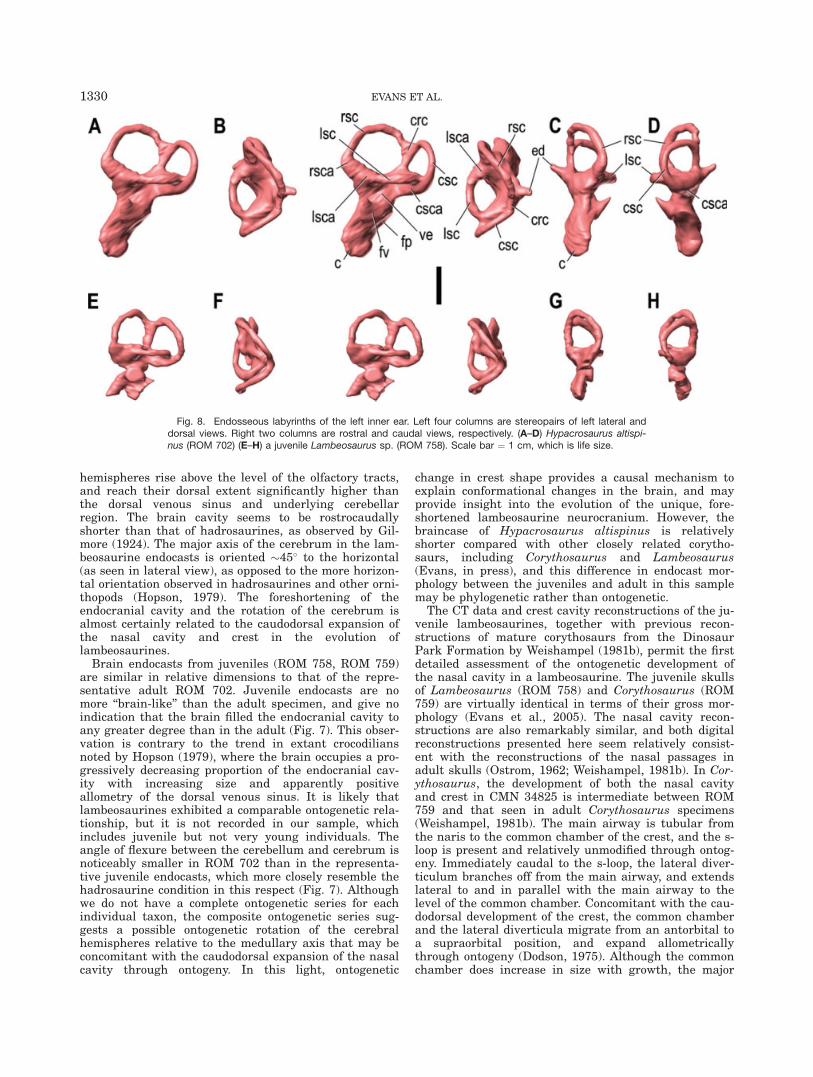
The digitally reconstructed vestibular apparatus iscomplete on at least one side of all four specimens in oursample, and is particularly well preserved on the leftside of ROM 702 and in ROM 758 (Fig. 8). It closelyresembles that of other hadrosaurids, and in generalform also bears a distinct resemblance to the conditionin extant crocodilians (Brown, 1914; Langston, 1960;Ostrom, 1961; Witmer et al., 2008). The three semicircu-lar canals are oriented in approximately the threeplanes of space, as in all tetrapods. The rostral semicir-cular canal is slightly longer than the caudal semicircu-lar canal, and the lateral (horizontal) semicircular canalis the shortest of the three. Of their correspondingampullae, the rostral ampulla is the largest, followed bythe lateral ampulla. The endolymphatic duct is repre-sented by a short, funnel-shaped process that extendsdorsomedially from the caudal wall of the vestibule atthe base of the common crus. The otic vestibule is diffi-cult to differentiate into the utriculus and sacculus. Thefenestra vestibuli faces laterally and is located at thebase of the cochlea, whereas the fenestra perilymphati-cum opens caudally into the metotic fissure. The promi-nent, finger-like cochlea extends rostroventrally fromthe vestibule.
DISCUSSION
These new data provide the first complete picture ofthe brain endocast in lambeosaurine hadrosaurids atthree different ontogenetic stages (Fig. 7). The overallshape of the endocast in corythosaurian lambeosaurinesis somewhat different from the well-described conditionin hadrosaurine hadrosaurids such as Gryposaurus andEdmontosaurus (Lambe, 1920; Lull and Wright, 1942;Ostrom, 1961; Hopson, 1979; Jerison, 2004). We confirmthat the olfactory tracts that connect the olfactory bulbsto the rest of the brain are very short such that thebulbs are located immediately rostral to the cerebralhemispheres (Evans, 2006). The broad cerebral
LAMBEOSAURINE ENDOCRANIAL MORPHOLOGY 1329
hemispheres rise above the level of the olfactory tracts,and reach their dorsal extent significantly higher thanthe dorsal venous sinus and underlying cerebellarregion. The brain cavity seems to be rostrocaudallyshorter than that of hadrosaurines, as observed by Gil-more (1924). The major axis of the cerebrum in the lam-beosaurine endocasts is oriented �45� to the horizontal(as seen in lateral view), as opposed to the more horizon-tal orientation observed in hadrosaurines and other orni-thopods (Hopson, 1979). The foreshortening of theendocranial cavity and the rotation of the cerebrum isalmost certainly related to the caudodorsal expansion ofthe nasal cavity and crest in the evolution oflambeosaurines.
Brain endocasts from juveniles (ROM 758, ROM 759)are similar in relative dimensions to that of the repre-sentative adult ROM 702. Juvenile endocasts are nomore ‘‘brain-like’’ than the adult specimen, and give noindication that the brain filled the endocranial cavity toany greater degree than in the adult (Fig. 7). This obser-vation is contrary to the trend in extant crocodiliansnoted by Hopson (1979), where the brain occupies a pro-gressively decreasing proportion of the endocranial cav-ity with increasing size and apparently positiveallometry of the dorsal venous sinus. It is likely thatlambeosaurines exhibited a comparable ontogenetic rela-tionship, but it is not recorded in our sample, whichincludes juvenile but not very young individuals. Theangle of flexure between the cerebellum and cerebrum isnoticeably smaller in ROM 702 than in the representa-tive juvenile endocasts, which more closely resemble thehadrosaurine condition in this respect (Fig. 7). Althoughwe do not have a complete ontogenetic series for eachindividual taxon, the composite ontogenetic series sug-gests a possible ontogenetic rotation of the cerebralhemispheres relative to the medullary axis that may beconcomitant with the caudodorsal expansion of the nasalcavity through ontogeny. In this light, ontogenetic
change in crest shape provides a causal mechanism toexplain conformational changes in the brain, and mayprovide insight into the evolution of the unique, fore-shortened lambeosaurine neurocranium. However, thebraincase of Hypacrosaurus altispinus is relativelyshorter compared with other closely related corytho-saurs, including Corythosaurus and Lambeosaurus(Evans, in press), and this difference in endocast mor-phology between the juveniles and adult in this samplemay be phylogenetic rather than ontogenetic.
The CT data and crest cavity reconstructions of the ju-venile lambeosaurines, together with previous recon-structions of mature corythosaurs from the DinosaurPark Formation by Weishampel (1981b), permit the firstdetailed assessment of the ontogenetic development ofthe nasal cavity in a lambeosaurine. The juvenile skullsof Lambeosaurus (ROM 758) and Corythosaurus (ROM759) are virtually identical in terms of their gross mor-phology (Evans et al., 2005). The nasal cavity recon-structions are also remarkably similar, and both digitalreconstructions presented here seem relatively consist-ent with the reconstructions of the nasal passages inadult skulls (Ostrom, 1962; Weishampel, 1981b). In Cor-ythosaurus, the development of both the nasal cavityand crest in CMN 34825 is intermediate between ROM759 and that seen in adult Corythosaurus specimens(Weishampel, 1981b). The main airway is tubular fromthe naris to the common chamber of the crest, and the s-loop is present and relatively unmodified through ontog-eny. Immediately caudal to the s-loop, the lateral diver-ticulum branches off from the main airway, and extendslateral to and in parallel with the main airway to thelevel of the common chamber. Concomitant with the cau-dodorsal development of the crest, the common chamberand the lateral diverticula migrate from an antorbital toa supraorbital position, and expand allometricallythrough ontogeny (Dodson, 1975). Although the commonchamber does increase in size with growth, the major
Fig. 8. Endosseous labyrinths of the left inner ear. Left four columns are stereopairs of left lateral anddorsal views. Right two columns are rostral and caudal views, respectively. (A–D) Hypacrosaurus altispi-nus (ROM 702) (E–H) a juvenile Lambeosaurus sp. (ROM 758). Scale bar ¼ 1 cm, which is life size.
1330 EVANS ET AL.
allometric components of the nasal cavity expansion areclearly within the vestibule, both in terms of the relativeelongation of the snout as well as the relative elongationof the lateral diverticula. The vestibule forms the largestpart of the nasal cavity, and the main olfactory region isclosely associated with the olfactory bulbs and is, atleast in part, outside of the main airway (Evans, 2006).
The new CT data allow us to correct some aspects ofthe crest cavity reconstructions of corythosaurs in previ-ous studies (Ostrom, 1962; Weishampel, 1981b). Weish-ampel (1981b) suggested that the nasal passage passedfrom the lateral diverticula into the common medianchamber of the crest and that the lateral diverticulawere thus part of the main airway in Corythosaurus,Lambeosaurus, and Hypacrosaurus. CT scans of thecomplete juvenile specimens suggest that the lateraldiverticula were blind chambers of the nasal cavity (i.e.,nasal vestibule) that were not involved in the direct con-duction of air from the naris to the lungs (Figs. 1–4).The ‘‘portals’’ that Weishampel (1981b) identified inspecimens of Corythosaurus (AMNH 5340, CMN 8676,ROM 1933) and Hypacrosaurus (ROM 702) are likelydue to the failure to completely ossify this region and/orto breakage of the exceedingly thin sheet of bone(derived from the nasal) within the crest that separatesthe diverticulum chamber from the common chamber(such is certainly the case in ROM 702).
Weishampel (1981b) also suggested that juveniles ofCorythosaurus and Lambeosaurus lacked lateral diver-ticula altogether and that these structures develop andenlarge with increasing size, and that the commonchamber filled the incipient crests of ROM 758 and ROM759. CT data show conclusively that juveniles of thesetaxa have well-developed lateral diverticula, and thatthey extend dorsally above the small common chamberto fill the incipient crests at this stage (Figs. 2, 4). Thelateral diverticula only become prominent externally insubadults with allometric growth of the crest and the de-velopment of the non-narial bony ‘‘cocks comb’’ (Dodson,1975). Even in CMN 34825, the lateral diverticula arenot obvious on external examination of the crest, andthey seem to become prominent only at larger size (Fig.3). Weishampel (1981b) also suggested that the lateraldiverticula of Lambeosaurus occur only rostral to thecommon chamber. However, the lateral diverticula, atleast in ROM 758, extend along the lateral side of thecommon chamber (Fig. 2). Although this relationshipmay have changed with ontogeny, the extent of the lat-eral diverticula correspond in large part to the extent ofthe caudolateral processes of the premaxilla (Figs. 1–4),and the lateral diverticula likely extended both rostraland lateral to the common chamber in fully adultLambeosaurus.
More major changes to the previous model of the nasalcavity are required for Hypacrosaurus altispinus. Weish-ampel (1981b), the only author to attempt a reconstruc-tion of H. altispinus, suggested that it lacked an s-loopand reconstructed the nasal cavity as a simple, essentiallystraight tube extending from the naris to the commonchamber of the crest. CT scans of ROM 702 reveal thatthe nasal vestibule is strikingly elongated and convolutedin H. altispinus compared with all other corythosaurs(Fig. 1). An s-loop seems to be present in the main airwayin a position consistent with that of Corythosaurus andLambeosaurus, but the ventral (more rostral) loop is
twisted, elongated, and extends within the premaxilla ad-jacent to the long lateral diverticulum to the level of theprefrontal. If H. altispinus is phylogenetically nesteddeeply within corythosaurs, as suggested by Evans andReisz (2007), this remarkable elongation of the nasal cav-ity (nasal vestibule) is a derived trait of H. altispinus, incontrast with the relatively short, plesiomorphic s-loopmorphology of Corythosaurus and Lambeosaurus (Weish-ampel, 1981b). Within the least inclusive clade thatincludes Corythosaurus and Hypacrosaurus, the helmet-shaped external morphology of the crest is relatively con-sistent (Evans and Reisz, 2007).
Cranial Crest Function: A SensorineuralPerspective
The function of the enlarged nasal cavity within thecranial crests of lambeosaurines has been the subject ofconsiderable speculation and debate (Ostrom, 1962; Hop-son, 1975; Weishampel, 1981a,b, 1997; Evans, 2006).Although hypotheses that involve underwater feedinghave been rejected (Ostrom, 1961, 1962), those that per-tain to enhancement of physiological (olfaction, thermo-regulation) functions (Ostrom, 1961, 1962; Horner, 1995)of the nasal cavity have been more difficult to assess,but are generally thought to have played a minor rolecompared with behavioral interpretations of the crest(Dodson, 1975; Hopson, 1975; Weishampel, 1997; Sulli-van and Williamson, 1999; Ruben et al., 2003; Horner etal., 2004; Evans, 2006). In addition to visual display, theacoustic resonance model of crest function is undoubt-edly the most widely cited hypothesis (Farlow et al.,1995; Weishampel, 1997; Horner, 2000; Horner et al.,2004; Emlen, 2008). Despite its apparent widespread ac-ceptance, this hypothesis is not favored universally. Sul-livan (in Sullivan and Williamson, 1999) argued for apredominant visual display function for the crest, andsuggested that resonance properties were simply inci-dental and not a significant target of selection.
The new data presented here, however, when inter-preted in the context of lambeosaurine phylogeny (Evansand Reisz, 2007), suggest significant evolutionary modifi-cation of the nasal cavity in the lineage leading to Hypa-crosaurus altispinus that was largely independent of theexternal appearance of the crest. H. altispinus and Cory-thosaurus plesiomorphically share a helmet-shapedcrest, whereas the elongation of the lower part of thevestibular s-loop in H. altispinus is derived comparedwith the plesiomorphic s-loop morphology exhibited byCorythosaurus and Lambeosaurus. The striking evolu-tionary elongation of the rostral portion of the nasal cav-ity in Hypacrosaurus altispinus seems to be coupledwith minimal change to the external morphology of thecrest, and suggests a selective pressure for nasal cavityfunction that operated independently from changes asso-ciated with the visual display properties of the crest.Significant internal differences between the crests sug-gest that these modifications were selection-driven, andimply functions for the enlarged nasal cavity in lambeo-saurines in addition to the visual display functions thecrests no doubt served. This also highlights that thecrest and related nasal cavity in lambeosaurines hadseveral functions and a complex evolutionary historyinvolving multiple, potentially divergent selection pres-sures within the clade (Hopson, 1975; Evans, 2006). In
LAMBEOSAURINE ENDOCRANIAL MORPHOLOGY 1331
this context, it is clear that hypothesized physiologicaland resonance functions of the nasal cavity need furtherdetailed testing.
The new data on the relative size and morphology ofthe brain and brain divisions derived from the virtualendocasts allow independent assessment of two crestfunction hypotheses: olfactory enhancement and vocalresonation. Endocasts also provide more generalizedinsight into the behavioral scope inferred under hypoth-eses of crest function related to social display and intra-specific communication. Additionally, the morphologyand ontogeny of the nasal cavity also provide new per-spectives on olfactory and thermoregulation hypotheses.
Olfaction. Ostrom (1961, 1962) hypothesized thatthe expansion of the nasal cavity and related cranialcrests were related to an increase in surface area forolfactory epithelium and increased olfactory acuity inlambeosaurines relative to other herbivorous dinosaurs.Evans (2006) corrected the previous reconstruction ofthis system by Ostrom (1961, 1962) and showed that theolfactory system of lambeosaurines did not change dra-matically from the plesiomorphic condition, thereby rul-ing out Ostrom’s hypothesis that an important functionof the crest was to increase olfactory acuity via enlarge-ment of the olfactory region of the nasal cavity.
As noted by Evans (2006), the relative size of theolfactory bulbs compared with the size of the brain andbody mass in lambeosaurines compared with other dino-saurs remained unknown at the time of that study. Thenew CT scan data presented here allow the first esti-mates of olfactory bulb size relative to other measures oftotal brain and body size. Impressions of the olfactorybulbs are discernible dorsally in ROM 702 and ROM758, but these are not apparent in the other digitalendocasts. The ventral and especially rostral margins ofthe bulbs can be estimated based on the position of thepresphenoid (which is incomplete in ROM 702), but thegreatly shortened olfactory tracts are difficult to distin-guish from the bulbs proper. The olfactory system as awhole is easily differentiated at the rostral end of thecerebral hemispheres, and it is thus possible to estimatevolumes of the olfactory system (i.e., bulbs plus tracts)and put an upper limit on the amount of neural tissuedevoted to olfaction in lambeosaurines. Volumetrically,the olfactory system of the lambeosaurines analyzedhere forms between 2.9% (ROM 758) and 7.7% (CMN34825) of the total endocast volume. In ROM 702, theadult in our sample, the bulbs formed <5% of the totalendocast volume. The relative size of the olfactory sys-tem in ROM 702 is comparable with that of the hadro-saurine hadrosaurids Edmontosaurus (CMN 2289, ROM1794) and Gryposaurus (AMNH 5350) in which the bulbsform �5% of the total endocast volume (values obtainedby graphic double integration from published endocastillustrations of CMN 2289 and AMNH 5350, respec-tively). The bulbs of lambeosaurines and other hadro-saurids are relatively smaller than in most otherdinosaurs (Hopson, 1979), including the ceratopsiansPsittacosaurus (Zhou et al., 2007) and Pachyrhinosaurus(Witmer and Ridgely, 2008a), and virtually all theropods(Hopson, 1979; Zelenitsky et al., 2008). Similarly, smallolfactory bulbs in Nigersaurus, a small sauropod inwhich the volume of the bulbs is �5% of the total endo-cranial volume, have been used to suggest that olfaction
was less important behaviorally than other senses inthis taxon (Sereno et al., 2007). The similarity in the rel-ative size of the olfactory bulbs in lambeosaurines rela-tive to representatives of their crestless sister groupHadrosaurinae, suggests strongly that increased olfac-tory acuity did not play a causal role in the evolutionaryhypertrophy of the lambeosaurine nasal cavity andcrest.
The morphology of the nasal cavity provides additionalinsight into the potential importance of olfaction in lam-beosaurine crest evolution and behavior. A number ofprevious studies have attempted to determine the homol-ogies of the different chambers within the crest (Ostrom,1962; Hopson, 1975; Weishampel, 1981b; Evans, 2006).Originally, Ostrom (1961, 1962) hypothesized that thecombined lateral diverticula and common chamber rep-resented an enlarged olfactory region in lambeosaurines.Subsequent work has shown that the lateral diverticulaas well as the rostral tubular region of the nasal cavitycontained within the premaxilla, probably represent thenasal vestibule, and were likely lined with respiratory,not olfactory, epithelial tissues (Evans, 2006).
The olfactory epithelium is typically found within thenasal cavity proper in extant archosaurs (Witmer, 1995;Evans, 2006; Witmer and Ridgely, in press). The com-mon chamber of the crest in lambeosaurines representsthe rostral region of the nasal cavity proper (Evans,2006). Although olfactory epithelium may have extendedinto the common chamber of the crest, the primary olfac-tory region was probably located in its plesiomorphicposition outside of the crest cavities proper (Evans,2006). This region represents the caudal portion of thenasal cavity that corresponds to the olfactory region inextant tetrapods (Witmer, 1995). Here, the small regionoutside of the main airway and directly rostral to the ol-factory bulbs seems to represent the cul-de-sac found invirtually all tetrapods, in which reduced airflow ratespromote diffusion of odorant molecules to their receptorson the olfactory epithelium (Negus, 1958; Simmen et al.,1999; Craven et al., 2007; Witmer and Ridgely, in press).In comparison with tyrannosaurs, for example, whichhave a large caudal cul-de-sac and are generally thoughtto have had a keen sense of smell compared with otherdinosaurs (Saveliev and Alifanov, 2007; Zelenitsky et al.,2008; Witmer and Ridgely, in press), the relatively verysmall size of the olfactory cul-de-sac in lambeosaurinesis striking. The olfactory cul-de-sac of lambeosaurines isalso much smaller than that of both nodosaurid andankylosaurid ankylosaurs, the only other ornithischiangroups in which this region has been reconstructed indetail (Witmer and Ridgely, 2008b).
It is unknown whether the length of the vestibuledecreased air flow rates sufficiently to promote efficientreception of odorant molecules across olfactory epithe-lium housed within the common chamber. However, theapparently plesiomorphically sized olfactory bulbs andthe location and relative size of the olfactory region ofthe nasal cavity provide further justification to reject thehypothesis that increasing olfactory acuity drove theevolution of the lambeosaurine crest.
Hearing in lambeosaurines. The hypothesis thatthe crest was used in vocal communication is relativelyold, but it did not gain significant attention until several
1332 EVANS ET AL.
decades after it was first argued in detail by Wiman(1931). Since the work of Hopson (1975) and Weishampel(1981a,b, 1997), the hypothesis that the hypertrophiednasal cavities of lambeosaurines varied in resonant prop-erties and enhanced intraspecific vocal communicationhas become the most widely cited hypothesis of crestfunction, both scientifically and popularly (Heaton, 1972;Hopson, 1975; Weishampel, 1981a; Farlow et al., 1995;Weishampel, 1997; Sullivan and Williamson, 1999;Horner, 2000; Horner et al., 2004; Evans, 2006).
Using relatively simple physical models, Weishampel(1981b) predicted the resonant frequencies of the elon-gate nasal cavities of Parasaurolophus walkeri and P.cyrtocristatus at between 50 and 375 Hz. Using a morecomplicated digital 3D model derived from CT scans ofP. tubicen, Diegert and Williamson (1998) also predictedlow-frequency resonance, as low as 30 Hz. The resonantfrequency of a tubular structure is related to its length.As such, taxa with shorter nasal cavities than Parasaur-olophus, such as the corythosaurs in this study, wouldlikely have produced calls at somewhat higher frequen-cies than Parasaurolophus. By the same reasoning, juve-nile lambeosaurines of a given taxon would haveproduced relatively higher frequency calls than adults,and the frequency of their calls would deepen with nasalcavity growth through ontogeny (Weishampel, 1981a).The lateral diverticula of corythosaurs could have‘‘muted out’’ certain frequencies and/or resonated asblind chambers at higher frequencies, and the complex-ity of their nasal cavities makes assessment of resonanceso difficult that precise predictions have never beenattempted (Weishampel, 1981a). Regardless, it is fairlycertain that the fully developed crests of the large cory-thosaurs in this study (Lambeosaurus, Corythosaurus,and Hypacrosaurus) would have resonated in the low-frequency range, probably well below 1000 Hz.
A fundamental prediction of the vocal resonance hy-pothesis is that the lambeosaurines had the ability todetect the noises made by the crest (Hopson, 1975;Weishampel, 1981a, 1997). The length of the cochlea isrelated to the length of the basilar membrane neuroepi-thelium of the basilar papilla, and therefore providesinsight into auditory capabilities, or at least the behav-ioral importance of hearing, in reptiles (Baird, 1970;Wever, 1978; Manley, 1990; Gleich and Manley, 2000;Gleich et al., 2005). Hopson (1975) and Weishampel(1981a, 1997) argued that lambeosaurines had hearingsensitivity similar to that of extant crocodilians due tothe similarity of their inner ear morphology, and thatthey were capable of sensing the low-frequency calls
hypothesized to have been made by the crests. Unfortu-nately, none of these studies were based on dataderived from lambeosaurines, but rather used informa-tion on inner ear morphology from non-lambeosaurinehadrosauroids (hadrosaurines, Bactrosaurus) under theimplicit assumption that these structures would be sim-ilar in lambeosaurines. The casts of the endosseous lab-yrinths described here reveal the detailed structure ofthe lambeosaurine inner ear for the first time, andallow us to test the hypothesis that lambeosaurineswere capable of detecting low frequency sounds. Theelongate cochlea in the adult of Hypacrosaurus altispi-nus measures 16.7 mm in length, and is similar inshape and relative proportions to that described forhadrosaurine hadrosaurids (Brown, 1914; Langston,1960; Ostrom, 1961). The overall similarity of the audi-tory apparatus and elongate cochlea of lambeosaurinesand hadrosaurines confirms this key assumption in pre-vious studies of crest function in the group (Hopson,1975; Weishampel, 1997).
Gleich et al. (2005) demonstrated in extant archosaursa highly correlated inverse relationship between thelength of the basilar papilla and an animal’s most sensi-tive ‘‘best’’ frequency of hearing, as well as a positiverelationship between basilar papilla length and the highfrequency upper limit of hearing. Applying their findingsto our data confirms that lambeosaurines and otherhadrosaurids emphasized low frequencies, as in otherlarge dinosaurs (Table 3). Although the lambeosaurinesin this sample, particularly ROM 702, lie outside of therange of extant data points from which the equations ofGleich et al. (2005) were derived, the equations nonethe-less allow us to put testable constraints on estimates ofauditory sensitivity in large dinosaurs, including Hypa-crosaurus, which are tied directly to observational datain extant archosaurs.
Using the length of the cochlear cast to estimate basi-lar membrane length, the lambeosaurines described herehave an estimated best frequency of hearing rangingfrom 579 Hz in the smallest specimen (ROM 758, juve-nile Lambeosaurus) to 80 Hz in the largest (ROM 702,adult Hypacrosaurus altispinus), with correspondinghigh frequency hearing limits of 2.1 and 1.2 kHz, respec-tively (Table 3). In the case of ROM 702, the calculatedbest frequency of hearing falls within the range of reso-nance frequencies (48–375 Hz) predicted for Parasauro-lophus by Weishampel (1981a).
Weishampel (1981a) suggested that the lambeosaurinecrests may have been important for communicationbetween adults and juveniles. Neonatal lambeosaurine
TABLE 3. The maximum length of the digital cochlea casts determined using the Amira program, andcalculations of the corresponding hearing capabilities of lambeosaurines using the equations from Gleich
et al. (2005)
Specimen no. Taxon Length of cochleaBest frequency of
hearing (Hz)High frequency
hearing limit (Hz)
ROM 758 Lambeosaurussp. (juvenile)
9.2 mm 579 2,110
ROM 759 Corythosaurussp. (juvenile)
11.9 mm 295 1,586
CMN 34825 Corythosaurussp. (subadult)
12.3 mm (averaged) 267 1,534
ROM 702 Hypacrosaurusaltispinus (adult)
16.7 mm (averaged16.6 and 16.8)
80 1,190
LAMBEOSAURINE ENDOCRANIAL MORPHOLOGY 1333
skulls identified as Hypacrosaurus stebingeri from theTwo Medicine Formation of Montana (Horner and Cur-rie, 1994) permit estimation of the resonant properties ofthe crests of the smallest juvenile lambeosaurines. Ifmodeled as a tube �15 cm in length (admittedly a verysimple model), the nasal cavity of a Hypacrosaurushatchling would resonate at �1.1 kHz using the calcula-tions outlined in Weishampel (1981a). This approachesthe upper limit of hearing of an adult Hypacrosaurus, aspredicted from ROM 702, but it is likely to be within itsaudible range. Generally, resonant frequencies woulddecrease with increasing size of the nasal cavity throughontogeny, and progressively approach the rather lowbest frequency of hearing we predict in adults. This isconsistent with the hypothesis that vocal communicationbetween adults and juveniles, even the smallest individ-uals, may have been important to lambeosaurines(Weishampel, 1981a).
Relative brain size and behavior. Relative brainsize is typically measured with an encephalization quo-tient (EQ), which represents an individual’s actual brainsize (in volume or mass) divided by the expected brainsize for its particular body size calculated using an allo-metric relationship derived from a large extant sample(Jerison, 1973; Hopson, 1977; Hurlburt, 1996). In extincttaxa, both brain and body size of an individual must beestimated. These uncertainties make the EQ methodproblematic for fossil taxa (Larsson et al., 2000), but it isnonetheless a useful, although coarse, comparative met-ric. Evans (2005) estimated an EQ of �2.8 for lambeo-saurines, based on similarities in braincase size/morphology and body size between adult lambeosaurinesand hadrosaurines. We can test this hypothesis here bycalculating the relative size of the brain for the firsttime in an adult lambeosaurine, Hypacrosaurus altispi-nus, based on the endocast of ROM 702 and a similarlysized complete skeleton of this taxon.
ROM 702 is essentially an isolated skull, and as such,we estimated its body mass with reference to a complete,similarly sized skeleton of H. altispinus, CMN 8501.CMN 8501 is <7% larger than ROM 702 in linear meas-urements of the skull and braincase, and as such willprovide a relatively accurate estimate of mass while atthe same time will provide an EQ estimate that errs onthe side of ‘‘conservatism’’ in that it will slightly under-estimate the value. The body mass of CMN 8501 wascalculated in two ways: (1) using a 1372 ml, 1/12 scalemodel based on CMN 8501 reported by Russell andBeland (1976) and the formula of Colbert (1962) and (2)using both the bipedal (femur circumference ¼ 400 mm)and the quadrupedal mammal (humerus circumference¼ 225 mm) regression formulae of Anderson et al.(1985), because hadrosaurids are generally thought to befacultatively bipedal rather than true bipeds (Forster,1997; Horner et al., 2004). These calculations resulted inmass estimates of 2,000 and 3,300 kg for the bipedal andquadrupedal formulae, respectively, with the scale modelestimate of 2,134 kg falling between the two.
Given the problems with EQs of extinct taxa associ-ated with uncertainty in brain and body mass estima-tion, we calculated a range of EQ values forHypacrosaurus using both the 50% dinosaurian conven-tion (Jerison, 1973) and the 60% estimate suggested by
Evans (2005) for hadrosaurids for brain size estimation,both the minimum and maximum estimated body massesfor H. altispinus. EQs were calculated using the non-avian reptile dataset of Hurlburt (1996). Our EQ esti-mates for H. altispinus range from 2.3 to 3.7. The onlyprevious EQ estimate for lambeosaurines (Evans, 2005)falls within this range. The average of the three massestimates (2,478 kg) and the 50% convention for dino-saurs results in an EQ estimate of 2.7 for Hypacrosaurus.This value is comparable with that of other large ornitho-pods (hadrosaurines, 2.8; Iguanodon, 2.6; Evans, 2005)and the large coelurosaurian theropod Tyrannosaurus rex(2.7; Witmer et al., 2008). The lowest of our EQ estimatesfor Hypacrosaurus (2.3), is marginally higher than mostextant nonavian reptiles (Hurlburt, 1996), allosauroidtheropods (Allosaurus, 1.6), sauropods (Diplodocus, 0.4;Nigersaurus, 0.4–0.8), and ceratopsians (Psittacosaurus,1.7; Protoceratops, 2.1; Triceratops, 0.7), but significantlyless that that of maniraptoriform theropods (>4) (Hurl-burt, 1996; Evans, 2005; Zhou et al., 2007; Witmer et al.,2008). The estimates of brain volume for Hypacrosaurusplot near the upper 95% confidence limit of a log–logregression of brain volume and estimated body mass innon-avian reptiles (Larsson et al., 2000).
The most striking aspect of the brain endocasts oflambeosaurines is the relatively large cerebral hemi-spheres. The hemispheres form �43% of the total endo-cranial volume (without olfactory bulbs) in ROM 702,and a similar, but slightly smaller relative size in the ju-venile specimens. Although similar to hadrosaurinehadrosaurids (the cerebrum is �45% of the total endo-cranial volume in Edmontosaurus and Gryposaurus), thecerebrum is considerably larger than that of other non-hadrosauriform ornithischians, as well as large thero-pods such as Carcharodontosaurus (24%) and Tyranno-saurus rex (33%), but compares favorably with themaniraptoran theropod Conchoraptor (43%) and eventhe basal bird Archaeopteryx (45%) of considerablysmaller body size (Larsson et al., 2000; Kundrat, 2007).
An enlarged brain relative to body size has been equa-ted with expanded behavioral repertoires and/orincreased behavioral complexity in vertebrates (Jerison,1969, 1973; Hopson, 1977; Hurlburt, 1996). The rela-tively large size of the brain and cerebral hemispheresin lambeosaurines is consistent with the range and com-plexity of social behaviors inferred when the crest ishypothesized to be an intraspecific signaling structurefor visual and vocal communication (but see Witmer andRidgely, 2008a). The relatively large size of the brainand the cerebrum may even suggest that lambeosaur-ines had higher cognitive abilities not present in mostother herbivorous dinosaurs (Witmer and Ridgely,2008a). However, there is an increase in the relative sizeof the brain, and the cerebrum in particular, in Hadro-sauriformes compared with their ornithischian out-groups (Hopson, 1977, 1979), and an enlarged brain andcerebrum, together with any inferred capacity for com-plex behaviors, appears to have been plesiomorphic forlambeosaurines.
Nasal Cavity and Thermoregulation
Ostrom (1961) noted the potential of the enlargednasal cavity to function in the conditioning of respiredair. However, he discounted the idea that the crests
1334 EVANS ET AL.
were adaptations for thermoregulatory and water con-servation functions on the grounds that the habitats inwhich lambeosaurines lived were probably warm andhumid. This rationale is probably too simplistic and anumber of authors have suggested thermoregulation asa possible function of convergently enlarged nasal vesti-bules of large ornithischian dinosaurs, including lambeo-saurines (Sullivan and Williamson, 1999; Witmer, 2001;Evans, 2006; Witmer and Ridgely, 2008b).
The relatively small diameter of the nasal cavity inlambeosaurines has been used to argue that they do notshow modifications associated with respiratory turbi-nates and associated thermoregulatory functions andthat they were not endothermic (Ruben et al., 1996;Ruben et al., 1998, 2003). Moreover, CT scans conductedfor this study revealed no evidence of turbinate-likestructures within the vestibular tubes in any of thescanned specimens. If turbinate structures were presentwithin the premaxillary tubes, as has been reported inthe s-loop region of a juvenile specimen of Hypacrosau-rus stebingeri (Horner, 1995), our data suggest that theydid not typically ossify.
Although we cannot confirm turbinate-like structures,our data do allow us to reconstruct a major vascularpathway associated with supplying the nasal vestibule.A large neurovascular bundle would have passed ros-trally through the body of the maxilla to emerge on thedorsal surface of the premaxillary shelf via a large fora-men. From here, the neurovascular element passed lat-erally between the maxilla and the premaxilla, and thendorsally along a prominent sulcus on the premaxilla toenter the crest in the region of the s-loop through a gapbetween the dorsal and lateral premaxillary processes(Fig. 1). The sulcus that conducted this vascular elementis typically over 10 mm in width in adult corythosaurs(e.g., ROM 1933, ROM 1218, Royal Tyrrell Museum ofPalaeontology, Drumheller, TMP 81.37.01), suggestingsignificant blood flow into the nasal vestibule that is con-sistent with at least a minor thermoregulatory functionfor the nasal cavity. Similarly, the new data neither sup-port nor reject Wheeler’s (1978) hypothesis that theenlarged nasal cavity was related to brain cooling. Thethermoregulatory potential of the enlarged nasal cavityin lambeosaurines and its role in the evolution of thecrest still requires further detailed examination. Mosthadrosaurine hadrosaurids have enlarged narial regions,suggesting that thermoregulation may have factored inthe early evolution of the crest (Evans, 2006).
Relative growth patterns of the nasal cavity also pro-vide insights into the relative significance of its func-tional divisions in lambeosaurines. In lieu of a morecomplete volumetric analysis, preliminary data for Cory-thosaurus and Lambeosaurus suggest that the most sig-nificant allometric components of the nasal cavityexpansion through ontogeny are within the vestibule, interms of both the relative elongation of the snout andthe lateral diverticula, when compared with the commonchamber. This finding reinforces the notion that func-tions associated with the nasal vestibule (e.g., vocaliza-tion, thermoregulation), and not the nasal cavity proper(e.g., olfaction), were functionally more important in theevolution of the lambeosaurine crest (Evans, 2006).
It was once thought that the complex, hypertrophiednasal cavity was a unique characteristic of lambeosaur-ines directly associated with the development of the
crest. New data from nodosaurid and ankylosaurid anky-losaurs reveals analogous nasal cavity hypertrophy thatevolved independently in this group (Witmer andRidgely, 2008b). Here, nasal cavity evolution can clearlybe distanced from a visual display function becauseankylosaurs lack any crests or adornments associatedwith the nasal cavity, as in lambeosaurines. The bizarrenasal cavities of ankylosaurs present a functional prob-lem that parallels that of the lambeosaurine crest, andwhich can be approached in a similar way.
ACKNOWLEDGMENTS
The authors thank Jack Horner, David Weishampel,Peter Dodson, Jason Head, Robert Sullivan, Grant Hurl-burt, Harry Jerison, Hans Larsson, Tom Williamson,Francois Therrien, and Robert Reisz for insightful andproductive discussions. Jim Gardner, Kevin Seymour,Margaret Feuerstack, Kieran Shepherd, Mark Norell,Carl Mehling, and Jack Horner provided access to speci-mens in their care. They thank Ian Morrison and BrainIwama for expertly preparing parts of the skull of ROM702, and Nic Campione and Brian Iwama for assistancein measuring hadrosaurine endocasts. For help with CTscanning, they thank Heather Rockhold and O’BlenessMemorial Hospital in Athens, Ohio. The authors thankthe Ohio Supercomputing Center for support. They alsothank Eric Snively, Merrilee Guenther, Peter Dodson,and an anonymous reviewer who made comments thatsignificantly improved the manuscript, and Peter Dod-son for the invitation to contribute to this volume.
LITERATURE CITED
Anderson JF, Hall-Martin A, Russell DA. 1985. Long-bone circum-ference and weight in mammals, birds and dinosaurs. J Zool207:53–61.
Baird IL. 1970. The anatomy of the reptilian ear. In: Gans C, Par-sons TS, editors. Biology of the Reptilia. New York: AcademicPress. p 193–275.
Brown B. 1914. Anchiceratops, a new genus of horned dinosaursfrom the Edmonton Cretaceous of Alberta, with discussion of theorigin of the ceratopsian crest and the brain casts of Anchicera-tops and Trachodon. Bull Am Mus Nat Hist 28:539–548.
Buffetaut E, Tong-Buffetaut H. 1993. Tsintaosaurus spinorhinusYoung and Tanius sinensis Wiman: a preliminary comparativestudy of two hadrosaurs (Dinosauria) from the Upper Cretaceousof China. C R Acad Sci Ser II 317:1255–1261.
Clark S, Morrison I. 1994. CT scan of fossils. In: Leiggi P, May P,editors. Vertebrate paleontological techniques. Cambridge: Cam-bridge University Press. p 323–329.
Colbert EH. 1962. The weights of dinosaurs. Am Mus Novit2076:1–16.
Craven BA, Neuberger T, Patterson EG, Webb AG, Josephson EM,Morrison EE, Settles GS. 2007. Reconstruction and morphometricanalysis of the nasal airway of the dog (Canis familiaris)and implications regarding olfactory airflow. Anat Rec 290:1325–1340.
Diegert CF, Williamson TE. 1998. A digital acoustic model of thelambeosaurine hadrosaur Parasaurolophus tubicen. J Vert Pale-ontol 18 (3 Suppl):38A.
Dodson P. 1975. Taxonomic implications of relative growth in lam-beosaurine hadrosaurs. Syst Zool 24:37–54.
Eberth DA. 2004. Revising the Edmonton group: biostratigraphyand a framework for climate change. In: Alberta palaeontologicalsociety symposium. Calgary, Alberta: Alberta PalaeontologicalSociety.
LAMBEOSAURINE ENDOCRANIAL MORPHOLOGY 1335
Emlen DJ. 2008. The evolution of animal weapons. Annu Rev EcolEvol Syst 39:387–413.
Evans DC. 2005. New evidence on brain-endocranial cavity relation-ships in ornithischian dinosaurs. Acta Palaeontol Pol 50:617–622.
Evans DC. 2006. Nasal cavity homologies and cranial crest functionin lambeosaurine dinosaurs. Paleobiology 32:109–125.
Evans DC. 2007. Ontogeny and evolution of lambeosaurine dino-saurs (Ornithischia: Hadrosauridae). Ph.D. thesis. Department ofecology and evolutionary biology. Toronto: University of Toronto. p 497.
Evans DC. Cranial anatomy and systematics of Hypacrosaurusaltispinus, and a comparative analysis of skull growth in lambeo-saurine hadrosaurids (Dinosauria:Ornithischia). Zoological Jour-nal of the Linnean Society, in press.
Evans DC, Forster CA, Reisz RR. 2005. The type specimen of Tetra-gonosaurus erectofrons (Ornithischia: Hadrosauridae) and theidentification of juvenile lambeosaurines. In: Currie PJ, Koppel-hus EB, editors. Dinosaur provincial park: a spectacular ancientecosystem revealed. Bloomington: Indiana University Press.p 349–366.
Evans DC, Reisz RR. 2007. Anatomy and relationships of Lambeo-saurus magnicristatus, a crested hadrosaurid dinosaur (Ornithi-schia) from the dinosaur park formation, Alberta. J VertPaleontol 27:373–393.
Evans DC, Reisz RR, Dupuis K. 2007. A juvenile Parasaurolophus(Ornithischia: Hadrosauridae) braincase from Dinosaur provincialpark, Alberta, with comments on crest ontogeny in the genus.JVert Paleontol 27:642–650.
Farlow JO, Dodson P, Chinsamy A. 1995. Dinosaur biology. AnnuRev Ecol Syst 26:445–471.
Forster CA. 1996. New information on the skull of Triceratops.J Vert Paleontol 16:246–258.
Forster CA. 1997. Hadrosauridae. In: Currie PJ, Padian K, editors.Encyclopedia of dinosaurs. New York: Academic Press. p 293–299.
Gilmore CW. 1924. On the genus Stephanosaurus, with a descrip-tion of the type specimen of Lambeosaurus lambei, parks. CanDept Mines (Geol Surv Can) Bull 38:29–48.
Gleich O, Dooling RJ, Manley G. 2005. Audiogram, body mass, andbasilar papilla length: correlations in birds and predictions forextinct archosaurs. Naturwissenschaften 92:595–598.
Gleich O, Manley GA. 2000. The hearing organ of birds and crocodi-lia. In: Dooling RJ, Fay RR, Popper AN, editors. Comparativehearing: birds and reptiles. New York: Springer. p 70–138.
Godefroit P, Alifanov V, Bolotsky Y. 2004a. Re-appraisal of Aralo-saurus tuberiferus (Dinosauria, Hadrosauridae) from the LateCretaceous of Kazakhstan. Bull Institut Roy Sci Nat Belg SciTerre 74:139–154.
Godefroit P, Bolotsky YL, van Itterbeeck J. 2004b. The lambeosaur-ine dinosaur Amurosaurus riabinini, from the Maastirchtian offar Eastern Russia. Acta Palaeontol Pol 49:585–618.
Heaton MJ. 1972. The palatal structure of some CanadianHadrosauridae (Reptilia: Ornithischia). Can J Earth Sci 9:185–205.
Hopson JA. 1975. The evolution of cranial display structures inhadrosaurian dinosaurs. Paleobiology 1:21–43.
Hopson JA. 1977. Relative brain size and behavior in archosaurianreptiles. Annu Rev Ecol Syst 8:429–448.
Hopson JA 1979. Paleoneurology. In: Gans C, editor. Biology of thereptilia Vol. IX: Neurology A. New York: Academic Press. p 39–146.
Horner CC, Horner JR, Weishampel DB. 2001. Comparative inter-nal cranial morphology of some hadrosaurian dinosaurs usingcomputerized tomographic X-ray analysis and rapid prototyping.J Vert Paleontol 21 (Suppl 3):64A.
Horner JR. 1995. Morphology and function of the enclosed narialchambers of lambeosaurid dinosaurs. J Vert Paleontol 15 (Suppl 3):36A.
Horner JR. 2000. Dinosaur reproduction and parenting. Annu RevEarth Planet Sci 28:19–45.
Horner JR, Currie PJ. 1994. Embryonic and neonatal morphologyand ontogeny of a new species of Hypacrosaurus (Ornithischia,Lambeosauridae) from Montana and Alberta. In: Carpenter K,Hirsch KF, Horner JR, editors. Dinosaur eggs and babies. Cam-bridge: Cambridge University Press. p 312–336.
Horner JR, Weishampel DB, Forster CA. 2004. Hadrosauridae. In:Weishampel DB, Dodson P, Maryanska T, editors. The Dino-
sauria. 2nd ed.Berkeley: University of California Press. p 438–463.
Hurlburt G. 1996. Relative brain size in recent and fossil amniotes:determination and interpretation. Ph.D. thesis. Department of zo-ology. Toronto: Unniveristy of Toronto. p 250.
Jerison HJ. 1969. Brain evolution and dinosaur brains. Am Nat103:575–588.
Jerison HJ. 1973. Evolution of the brain and intelligence. NewYork: Academic Press.
Jerison HJ. 2004. Dinosaur brains. In: Adelman G, Smith BH,editors. Encyclopedia of Neuroscience. 3rd ed.Amsterdam: Elsevier.
Ketcham RA, Carlson WD. 2001. Acquisition, optimization, andinterpretation of X-ray computed tomographic imagery: applica-tions to the geosciences. Comput Geosci 27:381–400.
Kundrat M. 2007. Avian-like attributes of a virtual brain model ofthe oviraptorid theropod Conchoraptor gracilis. Naturwissen-schaften 94:499–504.
Lambe LM. 1920. The hadrosaur Edmontosaurus from the UpperCretaceous of Alberta. Can Dept Mines (Geol Surv Canada) Mem-oir 120:1–79.
Langston WJ. 1960. The vertebrate fauna of the Selma formation ofAlabama. Part IV: the dinosaurs. Fieldiana Geol Mem 3:315–361.
Larsson HCE, Sereno PC, Wilson JA. 2000. Forebrain enlargementamong nonavian theropod dinosaurs. J Vert Paleontol 20:615–618.
Lull RS, Wright NE. 1942. Hadrosaurian dinosaurs of North Amer-ica. Geol Soc Am Spec Pap 40:1–242.
Manley GA. 1990. Peripheral hearing mechanisms in reptiles andbirds. New York: Springer-Verlag.
Negus V. 1958. The comparative anatomy and physiology of thenose and paranasal sinuses. London: Livingstone.
Ostrom JH. 1961. Cranial morphology of the hadrosaurian dino-saurs of North America. Bull Am Mus Nat Hist 122:33–186.
Ostrom JH. 1962. The cranial crests of hadrosaurian dinosaurs.Postilla 62:1–29.
Ostrom JH. 1963. Parasaurolophus cyrtocristatus, a crestedhadrosaurian dinosaur from New Mexico. Fieldiana Geol 14:143–168.
Parks WA. 1931. A new genus and two new species of trachodontdinosaurs from the Belly River formation of Alberta. University ofToronto studies. Geol Ser 31:1–11.
Rogers SW. 1999. Allosaurus, crocodiles and birds: evolutionaryclues from spiral computed tomography of an endocast. Anat Rec257:162–173.
Ruben JA, Hillenius WJ, Geist NR, Leitch A, Jones TD, Currie PJ,Horner JR, Espe G. 1996. The metabolic status of some late creta-ceous dinosaurs. Science 273:1204–1207.
Ruben JA, Jones TD, Geist NR. 1998. Respiratory physiology of thedinosaurs. Bioessays 20:852–856.
Ruben JA, Jones TD, Geist NR. 2003. Respiratory and reproductivepaleophysiology of dinosaurs and early birds. Phys Biochem Zool76:141–161.
Russell DA, Beland, P. 1976. Running dinosaurs. Nature 264:486.Russell DA, Chamney TP. 1967. Notes on the biostratigraphy ofdinosaurian and microfossil faunas in the Edmonton formation(Cretaceous), Alberta. Nat Mus Can Nat Hist Pap 35:1–22.
Sampson SD, Witmer LM. 2007. Craniofacial anatomy of Majunga-saurus crenatissimus (Theropoda: Abelisauridae) from the LateCretaceous of Madagascar. Soc Vert Paleontol Mem 8. J Vert Pale-ontol 27 (Suppl 2):32–102.
Saveliev SV, Alifanov VR. 2007. A new study of the brain of thepredatory dinosaur Tarbosaurus bataar (Theropoda, Tyrannosaur-idae). Paleontol J 41:47–55.
Sereno PC, Wilson JA, Witmer LM, Whitlock JA, Maga A, Ide O,Rowe T. 2007. Structural extremes in a Cretaceous dinosaur1.PLoS ONE 11:e1230.
Simmen D, Scherrer JL, Moe K, Heinz B. 1999. A dynamic anddirect visualization model for the study of nasal airflow. Arch Oto-laryngol Head Neck Surg 125:1015–1021.
Sullivan RM, Williamson TE. 1999. A new skull of Parasaurolophus(Dinosauria: Hadrosauridae) from the Kirtland formation of NewMexico and a revision of the genus. N M Mus Nat Hist Sci Bull15:1–41.
1336 EVANS ET AL.
Weishampel DB. 1981a. Acoustic analyses of potential vocalizationin lambeosaurine dinosaurs (Reptilia: Ornithischia). Paleobiology7:252–261.
Weishampel DB. 1981b. The nasal cavity of lambeosaurine hadro-saurids (Reptilia, Ornithischia): comparative anatomy and homol-ogies. J Paleontol 55:1046–1057.
Weishampel DB. 1997. Dinosaur cacophony: inferring function inextinct organisms. Bioscience 47:150–159.
Wever EG. 1978. The reptile ear. Princeton: Princeton UniverisityPress.
Wheeler PE. 1978. Elaborate CNS cooling structures in large dino-saurs. Nature 275:441–443.
Wiman C. 1931. Parasaurolophus tubicen n. sp. aus der Kreide inNew Mexico. Nova Acta Regiae Societatis Scientiarum Upsliensis,Ser. IV 7:1–11.
Witmer LM. 1995. Homology of facial structures in extant archo-saurs (birds and crocodilians), with special reference to paranasalpneumaticity and nasal conchae. J Morphol 225:269–327.
Witmer LM. 2001. Nostril position in dinosaurs and other vertebratesand its significance for nasal function. Science 293:850–853.
Witmer LM, Ridgely R. 2008a. Structure of the the brain and innerear of the centrosaurine dinosaur Pachyrhinosaurus based on CTscanning and 3D visualization. In: A new horned dinosaur from
the from and Upper Cretaceous bone bed in Alberta:NationalResearch Council of Canada. p 117–144.
Witmer LM, Ridgely R. 2008b. The paranasal air sinuses of preda-tory and armored dinosaurs (Archosauria: Theropoda and Ankylo-sauria) and their contribution to cephalic structure. Anat Rec291:1362–1388.
Witmer LM, Ridgely R. New insights into the brain, braincase, andear region of tyrannosaurs (Dinosauria, Theropoda), with implicationsfor sensory organization and behavior. Anat Rec, 292:1266–1296.
Witmer LM, Ridgely R, Dufeau DL, Semones MC. 2008. Using CTto peer into the past: 3D visualization of the brain and inner earregions of birds, crocodiles, and nonavain dinosaurs. In: Endo H,Frey R, editors. Anatomical imaging: towards a new morphology.Tokyo: Springer-Verlag. p 67–87.
Young CC. 1958. The dinosaurian remains of Liayang, Shantung.Palaeontol Sinica 142:1–139.
Zelenitsky D, Therrien F, Kobayashi Y. 2008. Olfactory acuity intheropods: palaeobiological and evolutionary implicaitons. Proc RSoc B doi:10.1098/rspb.2008.1075.
Zhou C-F, Gao K-Q, Fox RC, Du X-K. 2007. Endocranial morphologyof psittacosaurs (Dinosauria: Ceratopsia) based on CT scans ofnew fossils from the Lower Cretaceous, China. Palaeoworld16:285–293.
LAMBEOSAURINE ENDOCRANIAL MORPHOLOGY 1337